
Abstract
Background
Head and neck cutaneous squamous cell carcinoma (HNCSCC) is a non-melanoma skin cancer that is mostly caused by solar ultraviolet radiation exposure. While it usually has an excellent prognosis, a subset of patients (5%) develops nodal metastasis and has poor outcomes. The aim of this study was to systematically review the literature and evaluate the prognostic factors of HNCSCC in order to better understand which patients are the most likely to develop metastatic disease.
Methods
A comprehensive literature search was performed on PubMed and EMBASE to identify the studies that evaluated the prognostic factors of HNCSCC. Prognostic factors were deemed significant if they had a reported p-value of < 0.05. Proportions of studies that reported a given factor to be statistically significant were calculated for each prognostic factor.
Results
The search yielded a total of 958 citations. Forty studies, involving a total of 8535 patients, were included in the final analysis. The pre-operative/clinical prognostic factors with the highest proportion of significance were state of immunosuppression (73.3%) and age (53.3%); while post-operative/pathological prognostic factors of importance were number of lymph nodes involved with carcinoma (70.0%), margins involved with carcinoma (66.7%), and tumor depth (50.0%).
Conclusion
This systematic review is aimed to aid physicians in assessing the prognosis of HNCSCC and identifying the subsets of patients that are most susceptible to metastasis. It also suggests that immunosuppressed patients with a high-risk feature on biopsy, such as invasion beyond subcutaneous fat, could possibly benefit from a sentinel lymph node biopsy.
Graphical Abstract

Keywords
Background
Cutaneous squamous cell carcinoma (cSCC) is the second most common non-melanoma skin cancer (NMSC) [1]. As sun exposure is a major risk factor for cSCC, they arise commonly from the head and neck, commonly the ear, cheek, lip, and scalp [2, 3].
The recent literature reports a sharp increase in incidence of cSCC [4–6]. Over the last 30 years, longitudinal studies based in Canada and Australia have shown a 50–300% increase in the incidence of primary cSCC [1, 7, 8]. Despite the high incidence rates, it is reported that less than 5% of patients develop nodal metastases [9]. The majority of patients with cSCC undergoes electrodessication and curettage, cryosurgery, or Mohs’ surgery, and have an excellent prognosis. However, there is a subset of patients in which these therapies are unsuccessful and where cSCC appears to be far more aggressive, often resulting in metastasis and recurrence [1]. The poor survival rates for metastatic cSCC highlights the importance of identifying the patients who are susceptible to metastatic disease and poor outcome. Indeed, it has been shown that metastasis in cSCC patients is associated with a 50% decrease in five-year survival probability [10]. To date it remains however ambiguous to what defines this subset of patients. This may be accounted for by the inconsistency of the prognostic factors reported in the literature.
Moreover, in the last decade, sentinel lymph node biopsy (SLNB) has become a common practice in staging melanoma and has been utilized in other invasive cancers, such as oral squamous cell carcinoma [11, 12]. SLNB allow for pathological examination of the first echelon drainage node, helpful for determining accurate prognoses and therapy [11]. As 95% of cSCC patients have an excellent prognosis, it remains unclear as to which patients should be offered a SLNB.
Due to this therapeutic dilemma, and the fact that no study has evaluated in a thorough manner yet the prognostic factors associated with metastasis in head and neck cutaneous squamous cell carcinoma (HNCSCC), we felt appropriate to conduct a systematic review. The aim of the study was to determe the most significant negative prognostic factors in HNCSCC. This would provide an insight into which patients are more likely to develop metastasis and which could possibly benefit from a sentinel lymph node biopsy.
Materials and methods
Search strategy
We extracted information from all eligible publications using a standardised data extraction sheet and report the review according to equator network PRISMA guidelines. We searched for studies in the electronic databases PubMed and EMBASE for studies that analyzed prognostic factors in patients with HNCSCC. The following key words, as well as their synonyms, and abbreviations, were employed in the search strategy: Squamous cell carcinoma, Head and neck, Prognosis, and Cutaneous (Additional file 1). In order not to miss any appropriate study, we did not apply any time in our search. Only article published in English were considered. The reference lists of review articles were screened for potentially eligible studies.
The selection of studies involved an initial screening of the title and the abstract. In doubtful cases we obtained the full text. We entered articles in a data management software and eliminated the duplicates (Endnote 8®, Thompson Reuters Inc.)
Inclusion and exclusion criteria
Titles and abstracts were screened in order to identify the relevant studies. Articles were included based on the following criteria: (1) related to cutaneous squamous cell carcinoma; (2) related to cancers of the head and neck region; (3) Studies that were investigating prognostic factors. The full text of any papers missing abstracts or that had ambiguous titles and abstracts were screened in order to determine if they were relevant. After this elimination process, the following exclusion criteria were used to determine which articles were eligible for the data extraction: (1) No data extraction possible for head and neck only; (2) Studies that did not include prognostic factors as part of statistical analysis; (3) Studies that failed to perform multivariate analysis to assess significance of prognostic factors; (4) Literature reviews, case reports were also excluded.
Data extraction
Study demographics, sample size, and primary anatomic locations

Significance of prognostic factors in head and neck cutaneous squamous cell carcinoma

Data analysis
The prognostic factors evaluated in each study were classified as either significant or not significant. Significance was defined as having a p-value of < 0.05 and a 95% confidence interval. Prognostic factors’ significance calculated with univariate analyses were disregarded. The total amount of times that each prognostic factor was found significant and non-significant in the literature was recorded and proportions were calculated.
Results
Our search strategy yielded a total of 958 articles after excluding duplications. Following the review of titles and abstracts, 175 full-text articles were reviewed. In total, 40 [3, 5, 6, 9, 13–20, 22, 24–43, 45–48] papers were found to meet the complete list of inclusion and exclusion criteria as defined in the methods and were included in our review (Fig. 1).

Study selection flow chart
The total number of patients included in this systematic review was 8535 out of 40 studies, with the smallest study including 35 patients, while the biggest included 1434 patients. All patients had, according to the inclusion criteria, primary lesion located in the head or neck region. Out of the 40 studies included, 20 (50.0%) were conducted in Australia. The other populations included were the United States (17.5%), Germany (7.5%), Canada (5.0%), New Zealand (5.0%), Netherlands (5.0%), Israel (2.5%), Greece (2.5%), France (2.5%), and Chile (2.5%) (Table 1).
The analysis of patient factors and features found pre-operatively or on biopsy lead to the following results (Table 2): Out of the fifteen studies that assessed age, eight (53.3%) of them stated that age was a significant prognostic factor. Out of the eight that assessed sex, one deemed it as a significant factor. Only 2 studies out of six (33.3%) that assessed for the involved anatomical site found that it was significant. Similarly, three of six (50.0%) studies indicated that having a recurrence was significant. Out of the 15 studies that assessed immunosuppression, eleven (73.3%) suggested that it was significant. Tumor depth was found to be significant in three of six (50.0%) studies. Seven out of the 15 (46.5%) studies that assessed the histological differentiation grade claimed that it was a significant prognostic factor.
The analysis of the factors known post-operatively lead to the following results. Five of eleven (45.5%) studies found that tumor size was a significant prognostic factor. Nine of 18 (50.0%) studies indicated that extranodal extension was significant. Ten of 15 (66.7%) studies stated that margins involved with carcinoma was a significant prognostic factor. For the parotid and neck staging, six out of 13 (46.2%) studies suggested that parotid staging was a significant factor and thirteen out of the 24 studies (54.2%) found that neck staging was significant. Out of the ten studies that considered the number of lymph nodes (multiple nodes, ≥2) affected by carcinoma, 7 (70.0%) indicated that it was a significant prognostic factor. Seven of fourteen (50.0%) and three of eight (37.5%) studies suggested that perineural invasion and lymphovascular invasion, respectively, were significant prognostic factors. The ranking of the prognostic factors is shown in Fig. 2.

Percentage of significance of prognostic factors
The primary outcomes of the included studies varied with 39.5% (17/43) of studies reporting disease-specific survival, 32.6% (14/43) reporting overall survival, 18.6% (8/43) reporting disease-free survival, and 9.3% (4/43) reporting metastasis free survival.
Discussion
To our knowledge, we present the first systematic review looking at prognostic factors for HNCSCC. Given the low rate of metastasis from HNCSCC lesions, it can be challenging to identify the patients who are at high risk of having metastatic disease. We believe this review could help identify patients that would require a closer follow-up and those that could possibly profit from a SLNB.
State of immunosuppression
Out of the prognostic factors evaluated in the present study, immunosuppression was the most consistently reported significant prognostic factor (73.3%). It is postulated that a state of immunosuppression increases the risk of metastasis, as well as it decreases overall survival [13, 16, 17, 19, 20]. In their multivariate analysis, Brantsch et al. [5] reported that metastasis-free survival time was considerably affected in the immunosuppressed patients (HR 4.32; p = 0·0035). Oddone et al. [39] also deemed immunosuppression as a powerful prognostic indicator (HR 3.13; p = 0·006) and included it in their ITEM (immunosuppression, treatment, extranodal extension, and margin status) score, which suggests high-risk features for primary HNCSCC.
Solid-organ transplant recipients make up the majority of the immunosuppressed population. It was found that cSCC is the most common malignancy for patients who have received renal transplants, and these have an up to 82-fold increased likelihood of developing invasive cSCC compared to the general population [49]. As solid-organ transplant procedures are becoming more common [6], immunosuppression must be considered when evaluating risk stratification and prognosis of HNCSCC patients. Future studies shall allow better risk stratification according to the type and degree of immunosuppression,
Tumor factors
Tumor thickness (especially above 6 mm) and/or infiltration into the subcutaneous fat was found consistently to be a significant negative prognostic factor in HNCSCC. Depth of invasion has been a major prognostic factor for melanoma (Breslow thickness) and also recently been integrated in the stating system for oral squamous cell carcinoma. Further, perineural invasion (PNI) and lymphovascular invasion (LVI) were found to have a significant prognostic value in 50 and 37.5% of the included studies, respectively. Histological differentiation was also significant in 46.7% of studies, with all authors agreeing that a poorer grade of cellular differentiation lead to worse outcomes for patients.
Lymph node involvement and discrepancies in parotid and neck staging system
The number of lymph nodes affected by carcinoma showed to be a significant prognostic factor in 70.0% of studies. The higher number of lymph nodes (≥ two nodes) involved with HNCSCC, the worse the prognosis [3, 22, 30]. .Forest et al. demonstrated the significance of having multiple lymph nodes involved versus a single one (HR 3.8; p = 0·002) [22]. Another retrospective chart review showed that the five-year disease-free survival was significantly better in patients with a single node involved, compared to multiple nodes (75% vs. 64%; p = 0.04) [6]. Those findings suggest that patients with more than two lymph nodes should benefit from a closer follow-up.
Moreover, positive parotid and neck disease were found to be significant in 54.2 and 46.2% of the time, respectively. One important finding of our systematic review is the discrepancies in staging HNCSCC in the literature. In fact, there appears to be no standard staging system for this disease. Out of the 22 studies that assessed neck nodal staging, 9 studies used the O'Brien staging system (40.9%) [3], 9 studies used the AJCC staging system (40.9%), and 3 studies used the N1S3 staging system (13,6%) [22]. A study comparing the 7th edition of the AJCC staging system for HNCSCC and the N1S3 staging system stated the N1S3 staging system is superior to the AJCC staging system due to the fact that the N1S3 has statistically better distribution and stratification [9]. Discrepancies in the staging systems used in the literature can account for the inconsistency of the significance of parotid and neck staging. More studies are needed to compare those methods and to define a standard staging system for the field.
Margin status
The third most consistently significant prognostic factor was margin status. Out of the 15 studies that assessed margin status, 66.7% reported that margins involved with carcinoma lead to a poorer prognosis. A study by Hinerman et al. [27] concluded that positive surgical margins conferred a worse prognosis compared to negative and close [≤5 mm] margins when considering disease-free survival. Interestingly, O'Brien et al. [3] found that patients with advanced disease were more likely to have positive margins. Moreover, O'Brien's study showed that patients who underwent more radical surgeries were the ones with a higher chance of having positive margins. This can be explained by the fact that large facial primaries are more difficult to excise due to their size, depth and vital surrounding structures. Therefore, involved margins constitute an important prognosis factor in HNSCCS that should always be taken into consideration when evaluating prognosis in patients with HNCSCC.
Age
Age was found to be a significant prognostic factor in 53.3% of studies. Four studies23, 42–44 demonstrated that advanced age was significantly associated with a decreased overall survival. It is postulated that with age, the body accumulates somatic mutations and gets increasingly exposed to risk factors that favor the development of different cancers. Vasan et al. [46], in their retrospective analysis including 326 patients with a mean age of 73.3 years, showed that this factor negatively affected the disease-free survival (DFS) (HR 1.02; p = 0.02) and overall survival (HR 1.04; p < 0.001). Although statistically significant, the magnitude of the negative effect on DFS is low and unlikely to be of clinical significance. Interestingly, one study in the present review demonstrated a contrary observation. A retrospective study27 showed that older patients diagnosed with HNCSCC had a better prognosis than younger ones (HR 0.92; p = 0.002). Therefore, although age was shown to be a significant factor in approximately half the included studies, the extent of its effect on prognosis and use as a clinically significant marker is questionable. Given the discordance on its significance in the literature, more studies are needed to clarify its prognostic importance in HNCSCC.
Other findings
Gender was found to be significant in one of all the studies analyzed. Anatomic site and state of recurrence were each only studied in six studies. Extranodal extension (ENE) was significant in 50.0% of studies. Interestingly, ECS, which was included as one of the four prognostic factors on the Oddone's ITEM score, carries the heaviest prognostic significance amongst them [39].
Sentinel lymph node biopsy and comparison of results with current literature
Sentinel lymph node biopsy (SLNB) has been widely accepted as a minimally invasive and accurate technique for detecting occult nodal metastases in cutaneous melanoma and breast cancer [12]. However, its use in HNCSCC remains controversial, as it is unclear from the literature whether it has benefits with regards to survival. Ross and Schmults [50] showed in their systematic review that SLNB performed on cSCC in high-risk lesions were positive 21% of the time.
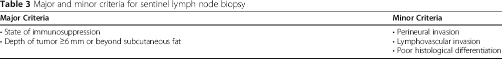
Major and minor criteria for sentinel lymph node biopsy
Limitations
There are several limitations to this systematic review. First, the majority of the included studies were based in Australia, involving 3921 out of the 8535 (45.9%) patients in this review, which may induce a selection bias and could limit the external validity of the study. Another important limitation is the fact that most studies on prognostic factors of cSCC were not specific to the head and neck region. Therefore, despite a large proportion of head and neck population included in most studies (see Table 1), single study results - allowing a meta-analysis - could not be extracted as they were not divided based on anatomical location. We chose to report how often a particular prognostic factor was found to be significant in each study, which gives an idea – although not quantitative – on how consistent a finding is. Lastly, some of our included studies had very small sample sizes, making the potential for beta-error high. In other words, some small study may give this impression that a given clinicopathological factor is not significantly associated with worse prognosis of HNSCC, although this may be due to limited sample size (false-negative). Another limitation of our study is the fact that most prognostic factors reported in the literature did not account for major treatment variables, such as the use of postoperative radiotherapy, for example. Future studies on cSCC should explore those factors, and differentiate results based on anatomical location, in order to expand the data on HNCSCC.
Conclusion
The prognostic factors for head and neck cutaneous squamous cell carcinoma that were most consistently reported as significant in the literature are a state of immunosuppression, tumor depth, margins involved, number of lymph nodes affected by carcinoma, parotideal disease, and age. This systematic review can aid physicians in assessing the prognosis of patients and possibly identifying the subsets of patients that are most susceptible to metastasis. We believe that immunosuppressed patients with high-risk features could possibly benefit from a sentinel lymph node biopsy at the time of the surgery. As some prognostic factors are poorly studied in the literature, more research is needed to assess their value in the prognosis of head and neck cutaneous squamous cell carcinoma.
Footnotes
Acknowledgements
Not applicable.
Author's information
JL, ML, and KS conducted the literature review, analyzed and interpreted the results. All authors read and approved the final manuscript.
Funding
Not applicable.
Availability of data and materials
All data generated and analyzed during this study are available in published articles available on the PudMed and EMBASE repositories: https://pubmed.ncbi.nlm.nih.gov, ![]() .
.
This material has never been published before and is not currently under consideration by another journal.
Declarations
Abbreviations
Springer Nature remains neutral with regard to jurisdictional claims in published maps and institutional affiliations.