
Abstract

Dear Editor,
I just wanted to reach out to you regarding the MSK article of July/August 2022 Full Thickness Achilles Tendon Tears: Two Case Studies.
I have been scanning MSK for about 10 years and work in a busy and dedicated sports medicine department. I am a registered MSK sonographer. I have been on the ARDMS MSK Task Force team for several years. That being said, I was thrilled to see Mr Carrigan’s MSK article!
My point of writing this letter was that the anatomy is labeled incorrectly in the images provided. See Figure 1 and Figure 4—G for the labeled structure as gastric is actually the soleus. The letter S is the flexor muscle. Figure 6 image was panoramic and may be gastric for G; however, the soleus does not look correctly labeled. In Figure 7, the scanning location is very distal in the lower leg, and you would not see the gastric muscle (corresponding image) and where the author marked KFP is where the soleus is connecting to the Achilles!
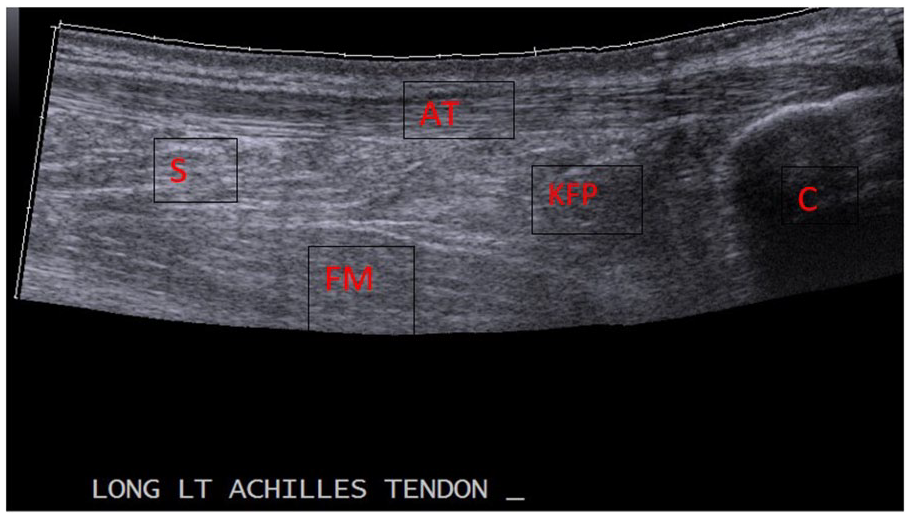
AT, Achilles tendon; S, soleus muscle; KFP, Kager’s fat pad; C, calcaneous; FM, one of the flexor muscles.
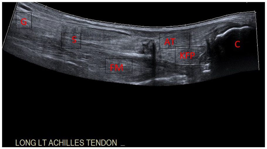
G, gastrocnemius muscle; S, soleus muscle; AT, Achilles tendon; KFP, Kager’s fat pad; C, calcaneous; FM, one of the flexor muscles.
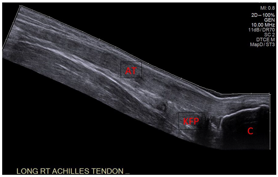
AT, Achilles tendon; KFP, Kager’s fat pad; C, calcaneous.
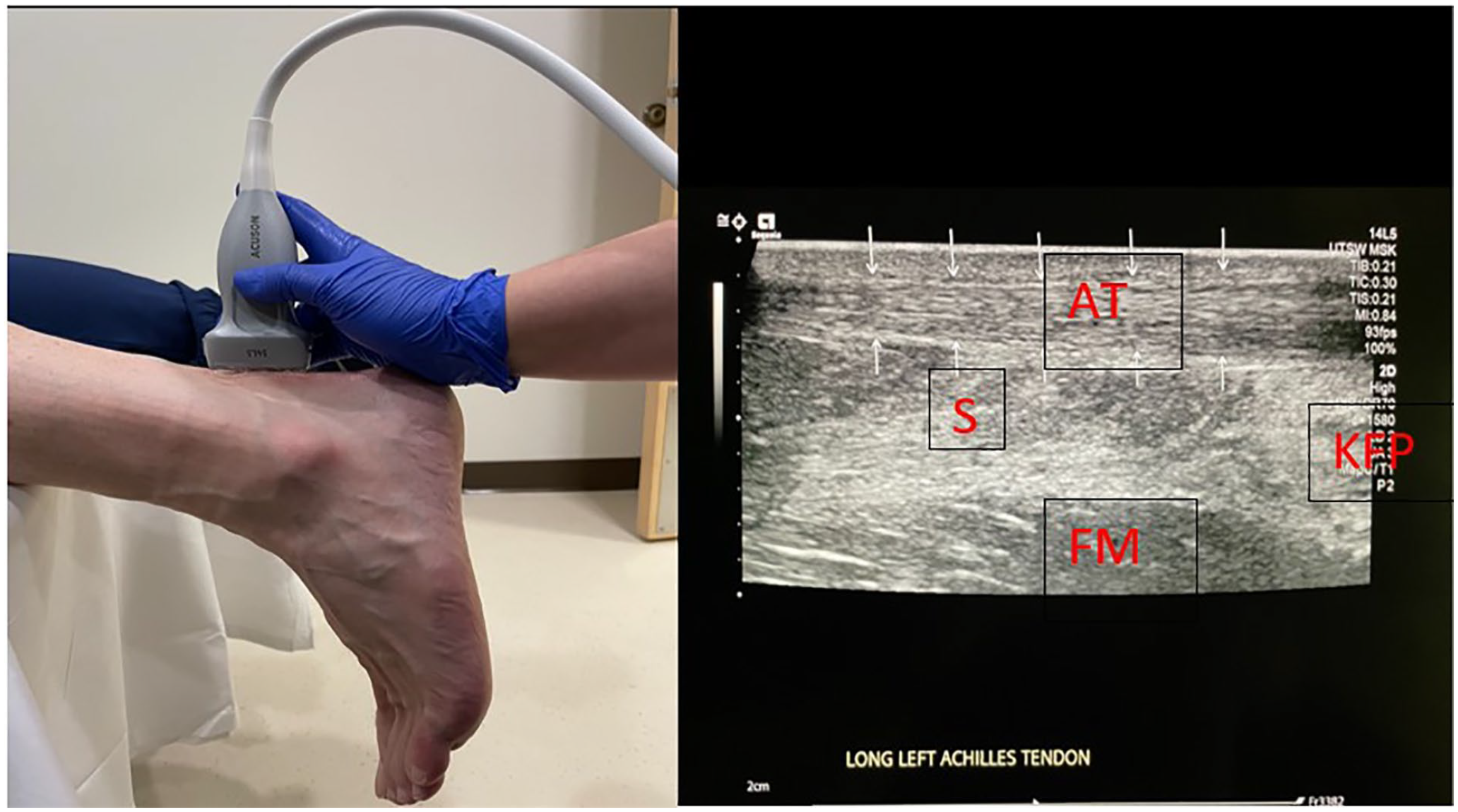
AT, Achilles tendon; S, soleus muscle; KFP, Kager’s fat pad; FM, one of the flexor muscles.
We are all continuing to learn more in our imaging modalities, and I would like the author to be successful and enjoy doing Achilles tendon scanning and MSK imaging.