




Abstract
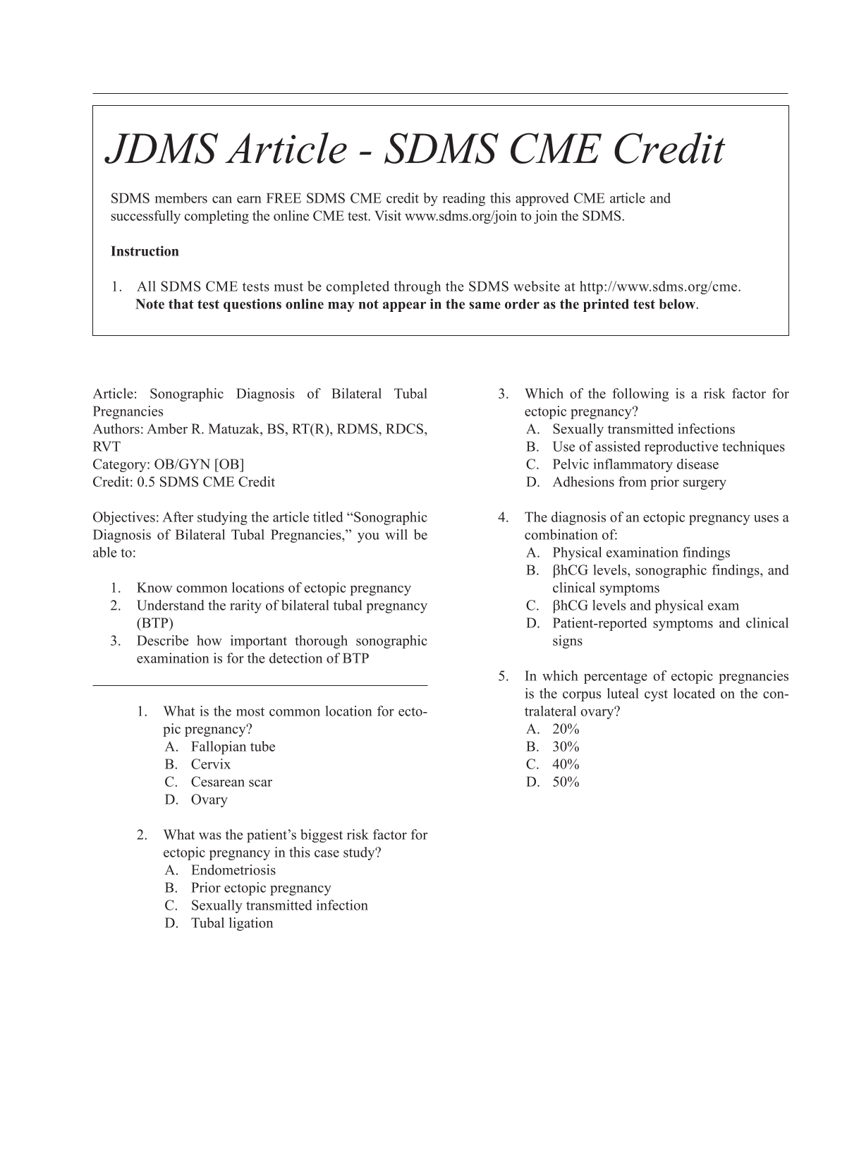
Article: Sonographic Diagnosis of Bilateral Tubal Pregnancies
Authors: Amber R. Matuzak, BS, RT(R), RDMS, RDCS, RVT
Category: OB/GYN [OB]
Credit: 0.5 SDMS CME Credit
Objectives: After studying the article titled “Sonographic Diagnosis of Bilateral Tubal Pregnancies,” you will be able to:
Know common locations of ectopic pregnancy
Understand the rarity of bilateral tubal pregnancy (BTP)
Describe how important thorough sonographic examination is for the detection of BTP
What is the most common location for ectopic pregnancy? Fallopian tube Cervix Cesarean scar Ovary
What was the patient’s biggest risk factor for ectopic pregnancy in this case study? Endometriosis Prior ectopic pregnancy Sexually transmitted infection Tubal ligation
Which of the following is a risk factor for ectopic pregnancy? Sexually transmitted infections Use of assisted reproductive techniques Pelvic inflammatory disease Adhesions from prior surgery
The diagnosis of an ectopic pregnancy uses a combination of: Physical examination findings βhCG levels, sonographic findings, and clinical symptoms βhCG levels and physical exam Patient-reported symptoms and clinical signs
In which percentage of ectopic pregnancies is the corpus luteal cyst located on the contralateral ovary? 20% 30% 40% 50%
Footnotes