
Abstract
This article is an overview of the field of small animal veterinary sonography, including its history, examples of utilization, and a brief review of the small animal abdominal sonographic examination that includes images of normal examples and typical pathology.
Sonography has become an essential imaging modality in the field of veterinary medicine and is increasing in popularity year by year. The use of sonography in small animal veterinary medicine has a history almost as long as diagnostic medical sonography utilization in the human medical field. Beginning in the 1960s, sonograms have been clinically indicated in animals for many of the same reasons as humans. Sonography is fast, is noninvasive, and can greatly further veterinarians diagnostic and therapeutic capabilities. Veterinary sonography uses similar equipment and protocols as in human medicine, with a very few minor differences.
Sonography plays a prominent role in the history of veterinary medicine. Gaining popularity in the mid 1980s, sonography-specific articles began appearing in the Journal of Veterinary Radiology. In 1983, an article on hepatic ultrasonography in dogs appeared, followed by an article covering the sonographic evaluation of pancreatitis in 1985.1,2 As a reflection of its increased popularity, the journal officially changed its name to the Journal of Veterinary Radiology and Ultrasound in 1992.
Veterinary sonography is utilized in many different instances. In general practice veterinary clinics, sonography is usually indicated for any chronic disease process in which the cause is unknown, as it serves as a comprehensive screening tool. The most common indication for ordering an ultrasound in general veterinary practices is chronically elevated liver enzymes. 3 Unfortunately, unless a mass is identified, the cause of the elevated liver enzymes is often inconclusive. Other common indications include urinary tract disease, gastrointestinal disease, endocrine disease, neoplasia, trauma, fever of unknown origin, and immune mediated diseases. Sonography can be used to diagnose many common diseases with a high sensitivity/specificity rate. Most often identified in dogs and cats is nonspecific inflammatory bowel disease and pancreatitis. Inflammatory bowel disease presents with a thickened bowel wall, more specifically, the muscularis layer. Pancreatitis presents sonographically as an enlarged hypoechoic gland. Another common use for sonography in animals, which is not a typical utilization in human medicine, is the identification of foreign bodies. Substances such as plastic, fabric, and wood cannot typically be visualized by radiographic tests such as plain film X-ray, which remains the first line of defense in imaging modalities in veterinary care. The University of Melbourne Veterinary Clinic and Hospital published a study in 2006 comparing the effectiveness of survey radiography and sonography at identifying gastrointestinal foreign bodies. 3 It was reported that for 16 small animals, sonography was able to identify the gastrointestinal foreign body in all 16 animals, whereas only 9 animals were noted to have foreign bodies radiographically. A similar study by Ober et al in 2008 showed that sonography had a 100% specificity rate in finding foreign bodies, specifically concerning a wooden splinter in a dog extremity. 4 Sonography is not primarily used for acute disease processes, as they are often treated symptomatically in veterinary medicine. If this method does not prove successful in resolving symptoms, sonography can be useful in providing differential diagnoses.
The other primary use of sonography in veterinary medicine is the identification of neoplasms, and more specifically for cancer staging.5,6 When there is a known mass, either visualized externally, on X-ray, or palpable, a sonogram can be performed to survey the extent of tumor invasion and the presence and extent of metastases as well as guide biopsies. The most common animal acquired cancers are lymphoma, mast cell tumors (or mastocytomas), transitional cell carcinoma, and hemangiosarcomas. In veterinary specialty clinics, sonography is utilized to follow the progress and success of animals undergoing radiation treatment, and to monitor specific disease processes. As in humans, sonography is also an exceptional tool for performing guided biopsies and is therefore commonly utilized. Ultrasound guidance has improved the overall percentage of positive diagnostic samples, as well as increasing the speed and safety of these procedures. 5 If a mass is identified in an animal, it is typically biopsied so as to determine the specific type of cancer. 6
The first line of defense in veterinary imaging remains the conventional plain film radiograph. Sonography and computed tomography (CT) are being used, but frequently on a limited basis due to a general lack of practicing knowledge of those imaging modalities and, particularly for CT, the relatively high cost of the tests. Not every animal owner is willing or able to spend several hundred dollars on a sonogram or several thousand on a CT. Many general veterinary practitioners do not train in the field of sonography specifically. Rather than investing the time in learning and mastering sonography, they will either get by without it or contract to outside specialty imaging agencies or mobile practices. This practice raises the questions of who can, and who should, perform animal sonograms. Similar to the evolution of the field of human sonography, veterinarians can either perform the sonograms themselves or a sonographer can perform them and send the images to a remote veterinary radiologist for interpretation. There are currently no veterinary sonography accreditation bodies or certifications (outside of possessing a doctor of veterinary medicine [DVM] degree), and no dedicated programs are currently being offered. There are some short-term training courses available for a substantial fee which do offer hands-on training, but the majority of veterinary sonographers learn the science and application through on the job training, just as diagnostic medical sonographers did so many years ago.
The field of veterinary sonography is just as operator dependent as in human medicine. Similarly, there are many different types of protocols currently being utilized. Unlike in human medicine, there are no established guidelines or standards of practice on what a specific examination must include for consideration for reimbursement; likewise, there is no accreditation by an outside governing body such as the American College of Radiology or the Intersocietal Accreditation Commission. In 2015, 11 different companies offer pet insurance, but these insurance companies do not have specific standards for sonogram requirements to merit reimbursement. For these reason, examinations obviously can vary greatly in extent and quality from one to the next.
Small animal sonography can often use the exact same equipment as used in human medicine. Some ultrasound equipment manufacturers have a line of models specifically geared to the veterinary community, with presets and patient data that are specific to animal use. One such model is the Esaote MyLab Gold (Esaote, Indianapolis, IN), a portable sonography machine tailored to animal use with various presets such as dog/cat abdomen and cardiovascular. The DVM and/or sonographer must choose the best transducer that is appropriate to the animal and the study to be done. For example, small dogs and cats can be effectively examined with 7.5- or 10.0-MHz linear array transducer. Medium-sized dogs do best with a combination of 7.5-MHz linear array for more superficial examinations to 5.0 MHz or less curvilinear arrays for deeper structures. Large breed dogs may require a transducer to go as low as 3.0 MHz in frequency for optimal visibility.7,8 Similar to performing a sonogram on people, the veterinary sonographer may need to switch transducers frequently during an examination, working for the best resolution possible while still maintaining the required penetration.
Preparation is an important factor as well. Animals that have a scheduled sonogram should ideally be fasted when possible. An animal that is receiving an abdominal sonogram will also need the abdomen completely shaved, from the xyphoid to the pubis, to eliminate the effects of air trapped within the fur and to increase sound wave conductivity. 9 An added challenge to veterinary medicine is that of dealing with a combative patient. The majority of the time, sonograms and biopsies are performed without the aid of sedatives. During the examination, dogs and cats should be placed dorsal side down in a padded v-trough or positioned on their side and restrained with assistance. When annotating veterinary images, the sonographer needs to remember that cranial and caudal are used in place of superior and inferior, and ventral and dorsal replace anterior and posterior. For example, the superior mesenteric artery should instead be referred to as the cranial mesenteric artery. The following is a basic example of what an abdominal canine sonogram could entail, including sample images of common pathology specific to that organ.
Urologic
The bladder is surveyed in sagittal and transverse planes, searching for any abnormalities. A common pathological finding is a growth in the bladder secondary to transitional cell carcinoma (Figure 1a). These masses tend to be located in the trigone of the urinary bladder, and can often be linked clinically to a dog/cat straining to urinate. Also noted during this survey is the prostate in the male dog. The size of the prostate in a nonneutered canine correlates with the dog’s age and weight, but should only be up to 2 cm in neutered dogs, and should display homogeneous echotexture (Figure 1b).10,11 In female dogs, the uterus can also be observed if the female has not been spayed.
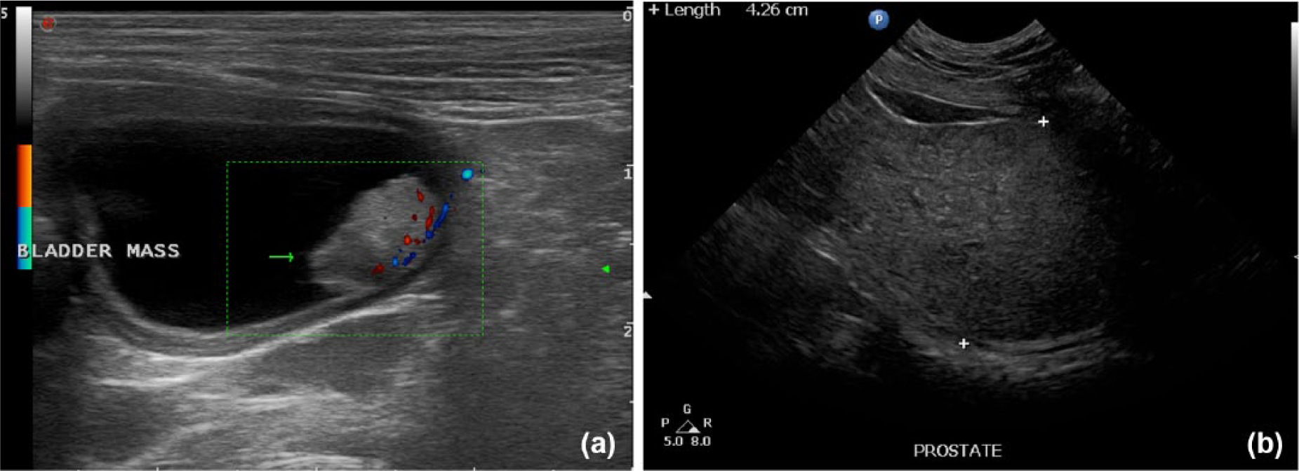
(a) Color Doppler image of the canine bladder showing a vascularized mass in the trigone that was shown to be a transitional cell carcinoma on biopsy. (b) Gray-scale image of an intact adult (nonneutered) canine prostate showing normal size (4.26 cm) and homogeneous echotexture.
Next, the medial iliac lymph nodes (MILNs) are surveyed in a longitudinal plane, and they are the most consistently visualized abdominal lymph nodes due to their size and relatively constant location. Located parallel to the distal aorta and caudal vena cava at the level of the fifth and sixth lumbar vertebrae, the MILNs are seen as long, thin structures with homogenous echotexture. They may not always be identifiable due to overlaying bowel filled with gas, but this is usually a sign of normalcy. The upper limits of normal for these lymph nodes are 4 cm in length, 2 cm in width, and 0.5 cm in thickness in adult dogs.9,10
When surveying the kidneys, it is typical to start on the left. Contrary to humans, the left kidney is the easiest to locate in canines. Animal kidneys also differ slightly in appearance compared to human anatomy. They have a homogenous cortex, which is moderately hypoechoic. Unlike in humans, the medullary region is often nearly anechoic; for those unfamiliar with animal kidneys, it can mimic the appearance of hydronephrosis. Kidneys should possess a well-defined cortico-medullay definition. Figure 2a demonstrates the appearance of actual hydronephrosis in a dog kidney. Animal kidneys can acquire many of the other same disease processes as humans such as chronic renal disease, obstructive renal disease, nephrolithiasis, cortical infarct, pyelonephritis, polycystic renal disease, renal adenocarcinoma, and renal lymphoma. 10 The adrenal glands should be surveyed and imaged after the same sided kidney is observed. Located medial and cranial to the kidney, the adrenal gland is homogenous in echotexture. It tends to be shaped similar to a peanut on the left side and a check mark on the right side in dogs. The maximum thickness should be taken, generally at the caudal pole, and should not exceed 7.4 mm (Figure 2b). 12 In the cat, the adrenal glands tend to be more oval in shape and more hypoechoic. Animals can present with various adrenal pathology, including hyperadrenocorticism (Cushing’s disease) and adrenal neoplasia (Figure 2c). 13
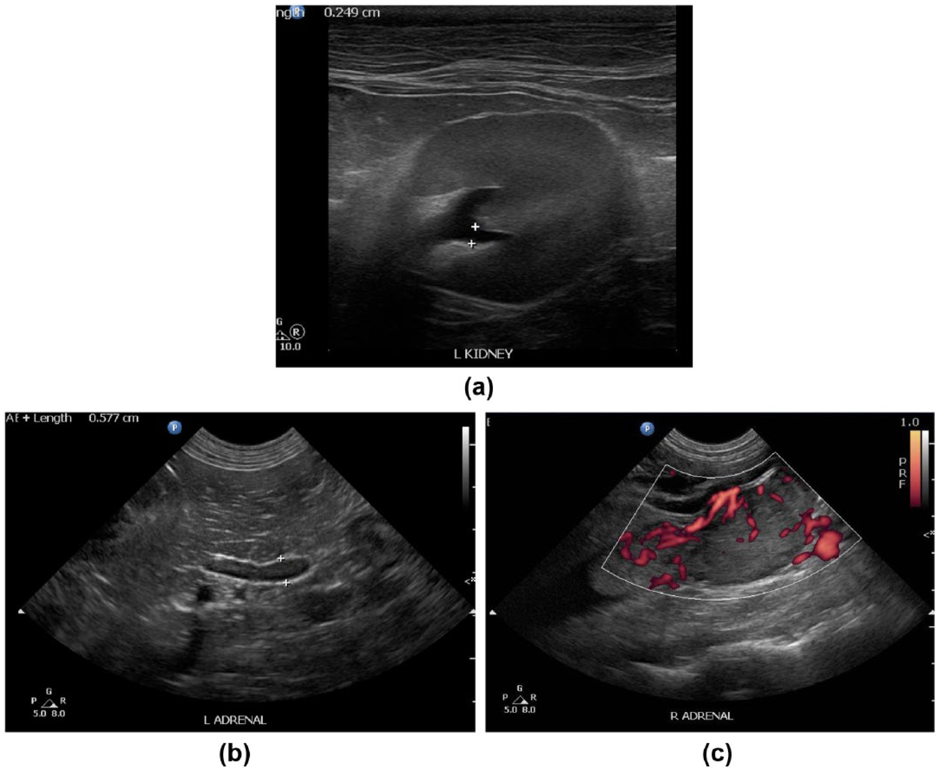
(a) Gray-scale image of an adult canine kidney with hydronephrosis. (b) Gray-scale image of the left adrenal gland in a canine showing normal homogeneous hypoechoic texture and normal size (5.8 mm). (c) Power Doppler image of a right adrenal gland in a canine with a neoplasia showing increased vascularity.
The liver in small animals serves the same physiological purpose as in humans, with very similar appearing anatomy.14-17 Typical portal vein velocity ranges from 10-25 cm/sec and the caudal vena cava velocity should be between 40-60 cm/sec.18,19 Scanning the liver of a dog and/or cat can be quite challenging depending on their body confirmation. A species such as a deep-chested boxer that has his liver tucked up high under his rib cage may need to be scanned intercostally, which provides a considerable challenge due to the small intercostal spaces (and the uncooperative nature of the dog in some cases). Small animals can have significant hepatic venous congestion, which is often secondary to such diseases as heartworms, pericardial effusion or Budd–Chiari Syndrome. They can also develop intra- and extrahepatic shunts, with a single congenital extrahepatic portosystemic shunt (PSS) being the most common variety; Yorkshire terriers are especially susceptible to this congenital abnormality (Figure 3a). 17 In the small animal, an abnormal liver appears sonographically the same as in humans: a coarse echotexture, irregular serosal margins, and nodules of various echogenicity. The differential diagnoses for an abnormal appearing liver are equally broad, from benign nodular hyperplasia, to lymphoma, to toxicity, just to mention a few more common reasons.15,16 An example of a canine liver with multiple hypoechoic nodules is shown in Figure 3b.
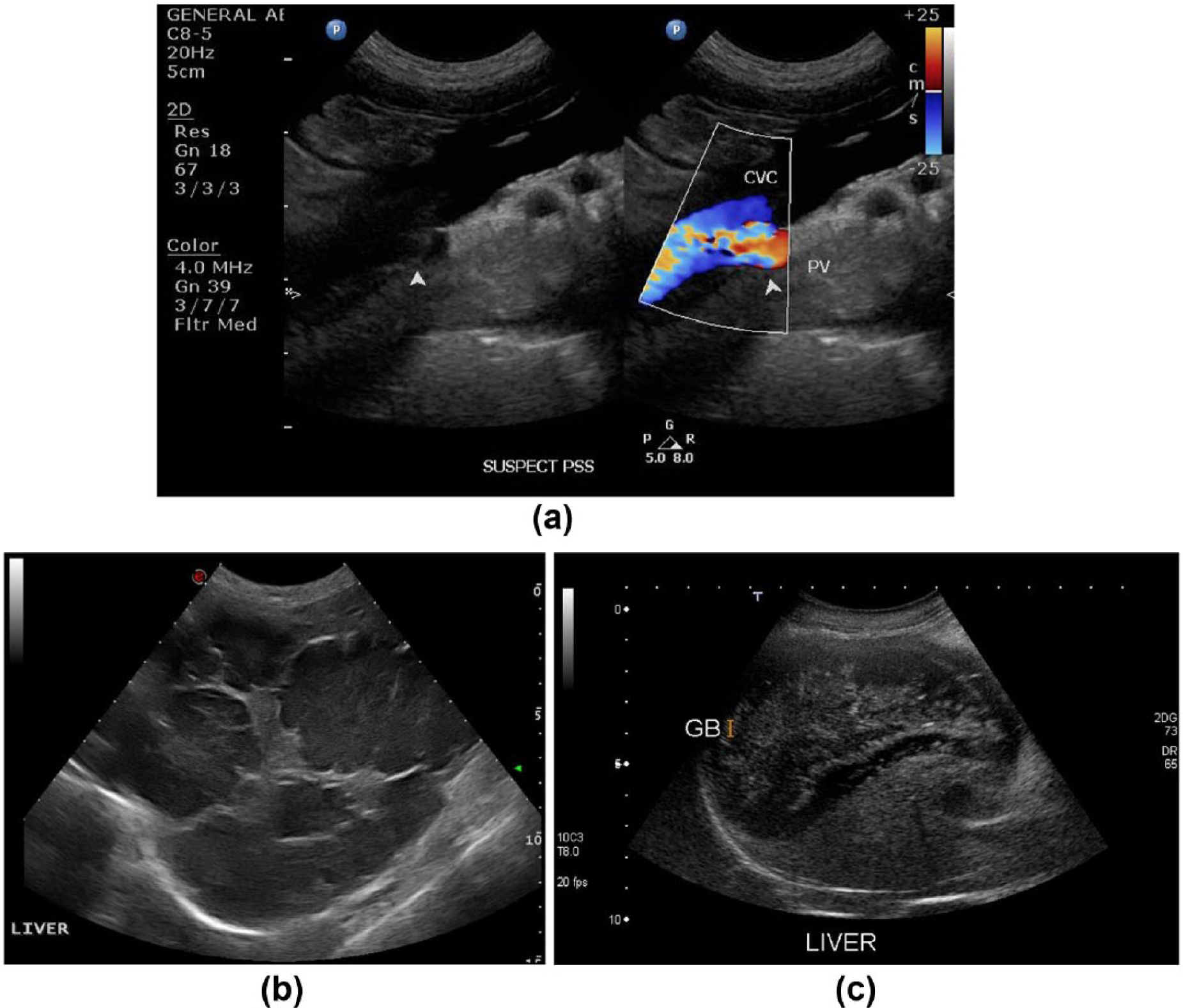
(a) Color Doppler image in an adult canine showing a single extrahepatic portosystemic shunt from the portal vein to the caudal vena cava. (b) Gray-scale image of an adult canine liver showing multiple hypoechoic nodules. (c) Gray-scale image of an adult canine liver and gallbladder showing the characteristic immobile stellate pattern of a biliary mucolcele.
The normal gallbladder in dogs appears similar to humans.15,19 It is round to oval in shape, filled with anechoic bile, and demonstrates a thin, smooth wall. It is consistently located in the right lateral liver. Normal cystic and bile ducts cannot usually be observed. Small animals can suffer from many gallbladder pathologies that also plague humans. These include, but are not limited to, cholelithiasis, “sludge,” and cholecystitis. Figure 3c shows a biliary mucocele, which is characterized by its “stellate” appearance. 20 Also indicative of a biliary mucocele is an “onion layered” or “honeybun” appearance. As opposed to sludge, the contents of a biliary mucocele occupy the entire lumen and do not move when agitated. Previously thought to be rare, this sonographic finding is becoming more common.
Unlike human pathology, the spleen in dogs and cats is susceptible to a large variety of pathology that can be identified sonographically.21-23 Splenic hematoma and nodular hyperplasia are the most common noncancerous lesions found in the spleen and account for 20-41% of all splenic lesions. Surgical removal is curative. Hemangiosarcoma is a common malignant tumor of the spleen usually seen in older dogs over years of age, with larger breeds at an increased risk, particularly German shepherds, golden retrievers, Labradors, and poodles. 23 A spleen with multifocal nodules that would be consistent with a type of round cell tumor, such as lymphoma or mast cell tumor, 24 is shown in Figure 4a. Figure 4b shows a spleen that has been infarcted, which in this case was caused by torsion. 25
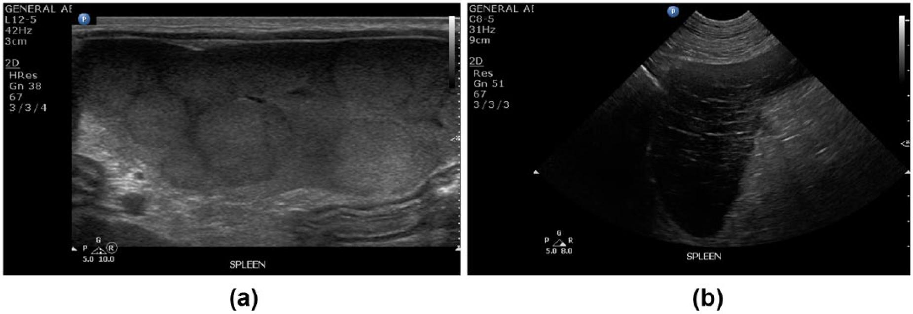
(a) Gray-scale image of an adult canine spleen showing multifocal nodular disease consistent with lymphoma or mast cell tumor. (b) Gray-scale image of a hypoechoic adult canine spleen that has an infarct secondary to torsion.
The pancreas in small animals lies in a slightly different orientation then that of a human. It is divided into right and left limbs, with the right located dorsomedial and adjacent to the descending duodenum and the left coursing caudal to the greater curvature of the stomach (Figures 5a and 5b). 26 Pancreatic ducts are not typically visualized sonographically, but the pancreaticoduodenal vein can usually be seen in the right limb in a dog. Small animals can be affected by pancreatitis just like humans, causing marked enlargement and a hypoechoic appearance in the acute stage.26-28
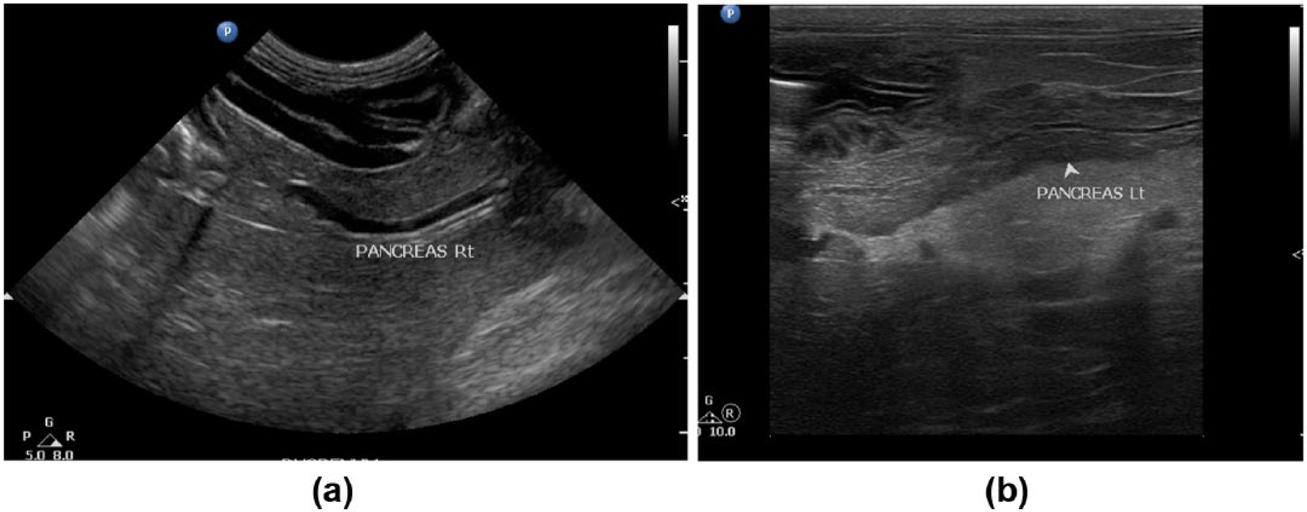
(a) Gray-scale image of the normal right pancreas in an adult canine. (b) Gray-scale image of the normal left pancreas in an adult canine.
Imaging the gastrointestinal tract is an important part of veterinary medicine that not all human sonographers are proficient. The gastrointestinal tract consists of the stomach, duodenum, jejunum, ileum, cecum, and colon. Imaging these areas can be difficult due to gas and artifact, but can be crucial for a correct diagnosis. Fasting of animals receiving an abdominal sonogram is ideal for better visualization. 29 Changes in the stomach wall and intestines can be caused by adenocarcinoma, lymphoma, polyps, chronic gastritis, uremic gastritis, and ulcers.30-36 The small intestines should display all 4 histologic layers: the serosa, muscularis, submucosa, and muscosa. 29 Thickening of the muscularis layer, as seen in Figure 6, can be indicative of inflammatory bowel disease or lymphoma, which can only be distinguished with a biopsy. The small intestine is also a common location for a simple obstruction due to a foreign object.3,34 The colon has much thinner layers and the lumen is typically not visible due to air artifact. Intussusception can also be diagnosed while imaging the gastrointestinal tract sonographically.
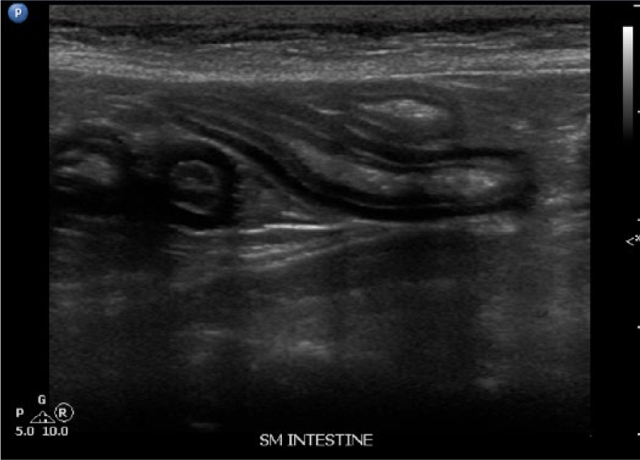
Gray-scale image of an adult canine small intestine showing diffuse muscularis layer thickening consistent with inflammatory bowel disease.
Conclusion
Small animal veterinary sonography has greatly impacted the entire veterinary field of medicine. Its importance simply cannot be understated since its introduction in 1966. 37 Just as human sonographers must learn the anatomy of the human body and the pathologies that are more common in men or women, and in different ethnicities, to produce high-quality diagnostic examinations, veterinary sonographers must know the anatomy and physiology of not only the species that they are examining, but all the breed-specific pathologies. Spanning back generations, this is a field that is as challenging as it is crucial to animal care. Significantly improving medical care in small animals, sonography is a growing imaging modality with many benefits and diagnostic abilities similar to that of human sonography.
Footnotes
Acknowledgements
The author would like to thank Clint Feagin, DVM, DACVR, for providing the images and for his help and guidance, without which this article would not have been possible. The author would also like to thank the staff at Veterinary Specialists of North Texas for sharing their extensive knowledge and allowing the opportunity to learn about small animal ultrasound.
Declaration of Conflicting Interests
The author declared no potential conflicts of interest with respect to the research, authorship, and/or publication of this article.
Funding
The author received no financial support for the research, authorship, and/or publication of this article.