
Abstract
Background:
Femoroacetabular impingement syndrome (FAIS) is a common condition in young adults that causes groin pain and predisposes patients to labral tears. Hip arthroscopy has become the gold standard in the treatment of FAIS and involves labral repair and osteoplasties to address any bony impingement. Traditionally, hip arthroscopy begins with access to the central compartment, followed by osteoplasties in the peripheral compartment (PC). However, initiating with the PC using percutaneous instrumentation has emerged as an effective alternative. This approach preserves capsular integrity and may simplify the procedure, especially when employing a periportal capsulotomy.
Indications:
Indications for the percutaneous approach include the treatment of FAIS and/or labral tears, especially in the context of tight/smaller joint spaces.
Technique Description:
This approach involves PC work first through the modified mid-anterior portal and anterolateral portal utilizing a 5.5-mm arthroscopic burr. Minimizing the degree of capsulotomy during this step allows for improved visualization due to decreased fluid loss. Following femoroplasty, a distal anterolateral portal can be created to address any labral tears without the need for further capsulotomy. Finally, capsular plication is performed to close any periportal incisions within the capsule.
Results:
The percutaneous approach with PC work first allows for a less invasive capsulotomy and femoroplasty in the setting of FAIS.
Discussion/Conclusion:
Percutaneous hip arthroscopy can be an effective capsular preserving technique that can lead to positive patient rehabilitation and functional outcomes. Performing the femoroplasty first with percutaneous instrumentation can mitigate the need for traditional capsulotomies.
Patient Consent Disclosure Statement:
The author(s) attests that consent has been obtained from any patient(s) appearing in this publication. If the individual may be identifiable, the author(s) has included a statement of release or other written form of approval from the patient(s) with this submission for publication.
This is a visual representation of the abstract.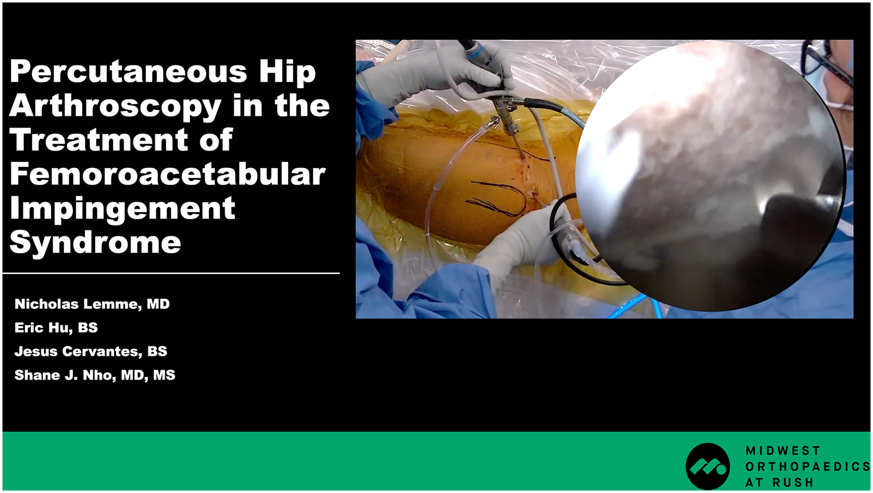
Video Transcript
In this video, we present the technique of a percutaneous hip arthroscopy utilizing a peripheral compartment (PC) first approach for the treatment of labral tears and femoroacetabular impingement syndrome (FAIS).
Here are our disclosures.
Background
Hip arthroscopy has become an effective treatment option for addressing FAIS and concomitant labral tears. It has been shown to have good patient outcomes and low reoperation rates up to 10 years after surgery. 1 Traditionally, this technique has been performed utilizing a central compartment (CC) first technique that creates 2 portals with an interportal or T-type capsulotomy. 4 However, extensive capsulotomy has been shown to be associated with hip instability, dislocation, and capsular insufficiency. To mitigate capsular disruption, the periportal capsulotomy technique has been adopted, although it presents challenges such as limited instrument mobility and suboptimal visualization. The PC first approach addresses these issues by minimizing capsulotomy during femoroplasty, enhancing visualization through a ballooning effect, and preserving capsular integrity. Furthermore, upon completion of PC work, the CC can be accessed under direct arthroscopic visualization, minimizing iatrogenic injury. 6 Furthermore, capsular preservation can protect against postoperative complications and facilitate the rehabilitation process.
Indications
Our patient is a 17-year-old active girl who has had left hip pain for 3 months. She denies any injury and experiences the pain in her groin that is sharp and an 8/10. The pain is worse when sitting and during activity, and while she recently participated in cross-country and track, she has been unable to run in the past 3 months. On physical examination, she has slightly limited external and internal rotation, as well as a positive subspine and flexion, adduction, and internal rotation (FADIR) test with a painful arc between 1- and 3-o’clock. Her strength is 5/5, and she is neurovascularly intact.
On preoperative radiographs, anteroposterior and Dunn lateral views of the pelvis were obtained. She had normal body alignment and was properly covered with a lateral center-edge angle of 33.8°. The α angle was measured to be 56.4°, suggesting a cam deformity. Preoperative magnetic resonance imaging arthrogram showed left anterosuperior acetabular labral tear with possible subcortical cystic change and femoroacetabular impingement.
Due to the patient's imaging findings, history, and failure with conservative treatment, she was recommended to undergo hip arthroscopy with labral repair and femoroplasty to address her labral tear and impingement.
Technique Description
In the operating room, the patient is placed supine on a hip positioning table under general anesthesia. A hip distracting system that allows for full range of motion of the operative hip is used. The patient is prepped and draped in typical sterile fashion, and appropriate landmarks are marked.
A spinal needle is used to first create the modified mid-anterior portal (MAP). With fluoroscopy guidance, the needle is passed through the lateral hip, pointing slightly upward toward the hip joint. A trocar is placed and advanced into the capsule with swift twisting motions. A 70° arthroscopic scope is inserted in the MAP to visualize the PC. Saline is run through the scope to insufflate the intact capsule surrounding the PC and produce good visualization via the ballooning effect. The PC first technique allows minimal loss of fluid from the capsule, which improves the visualization of the femoral head-neck junction and the surrounding synovial folds.
To address femoroacetabular impingement, an anterolateral portal is made, starting with a spinal needle and wire under direct view with the arthroscope. A switching stick is used to replace the initial dilator cannula with the sleeve of a 5.5-mm arthroscopic burr. The burr head is then inserted into its sleeve, and the rest of the arthroscopic burr handpiece is attached. This is done to minimize the size of portals and maintain the balloon effect when performing the femoroplasty. The burr is then used to address the cam lesion under fluoroscopy guidance. Flexion, internal rotation, and external rotation of the leg during this process allow for adequate resection of the lesion. Intermittently, a 4.0-mm arthroscopic shaver or radiofrequency is used to address any protruding tissue or synovium. After this initial resection, a dynamic examination is performed under fluoroscopy to ensure that the cam has been fully addressed. This is done with full flexion of the hip with approximately 30° of internal and external rotation, then 60° of hip flexion at 30° of internal and external rotation.
Attention is now turned to the CC. In this case, the patient has a labral tear likely secondary to her impingement and will be repaired. The camera is removed from the MAP and inserted into the anterolateral portal for better visualization. In addition, a distal anterolateral (DALA) portal is made to adequately introduce labral anchors. After identification of the tear, an arthroscopic shaver and burr are used to debride the labrum and decorticate the acetabular rim for anchor placement. A straight wire guide is placed at approximately the 12-o’clock position, and a 1.4-mm polyether ether ketone suture anchor is placed. The suture is passed through the labral tissue, and the resulting strands are tied. This is repeated for a total of 2 anchors for adequate repair of the labrum.
Capsule plication is performed for adequate closure of the capsule following work in the CC. Usage of the DALA portal for the arthroscope may be better for visualization when closing the capsule.
Pre- and postoperative radiographs in the Dunn lateral view are shown here. Adequate resection of the cam deformity can be appreciated on the postoperative radiograph, which was taken 2 weeks after surgery.
To improve surgical technique, several pearls are highlighted here. First, care should be taken when introducing instruments to minimize any unnecessary capsulotomy to maintain capsular preservation. This will also maximize the ballooning effect, which helps aid in visualization during PC viewing. In addition, when introducing cannulas or dilators, an 80%/20% rule can be used, where 80% of the motion should be twisting, and 20% should be pushing. 3 This minimizes the risk of iatrogenic cartilage or labral damage. Third, when switching between instrumentation during femoroplasty, utilizing the sleeve of the shaver or burr as a cannula may help with precision and feasibility of joint entry. Finally, proper establishment of the MAP is paramount in this technique. During needle placement, the needle should be perpendicular to the neck axis and pass through the anterolateral portion of the capsule. Care should be taken to not penetrate too medially, as this can limit lateral visualization of the head-neck junction, or proximally, as this may risk injuring the femoral head cartilage.
The percutaneous hip arthroscopy approach has several limitations. In larger and older patients, the freedom of movement with the burr may be restricted due to thicker, less pliable soft tissue. Additionally, larger and more complex cam deformities may necessitate further capsulotomy to adequately address a greater portion of the femoral neck. If PC visualization is inadequate, transition to contemporary hip arthroscopy can be seamless by initiating traction and using the same portals created to perform any necessary capsulotomy.
Results
After surgery, patients undergo a 4-phase rehabilitation protocol, each lasting 4 to 6 weeks. Phase 1 includes the gradual progression to weightbearing over the course of 6 weeks, with their abduction range of motion restricted. During phase 2, around week 6, range of motion and weightbearing restrictions are lifted, and patients initiate strength and gait training. By week 12, phase 3 begins, including initial return to prior activities, with a focus on functional training, including single-leg balance, hip rotation, or lunging exercises. By phase 4, patients are encouraged to slowly return to prior sports activity, with running, agility, cutting, and plyometric exercises being introduced. Patients may expect this process to be as long as 1 year for maximum recovery from a sports perspective.
Discussion/Conclusion
Prior studies have demonstrated improved patient outcomes following these PC first techniques. In a cohort study involving 704 hips, after 6 years of follow-up, 90.2% of patients reported satisfactory patient-reported outcomes, with only a 3.7% conversion to total hip arthroplasty (THA) and a 2.6% revision hip arthroscopy rate. 7 Another prospective case series study examined the outcomes of this technique at the 2-year follow-up, showing improvement in all functional patient-reported outcome scores measured by the 1-year follow-up. 5 Finally, another 2-year study from Dantas et al 2 reported similar findings, with significant improvements in patient-reported outcomes with significant impingement correction. This study reported a 3.7% rate of revision hip arthroscopy. Overall, while the literature on PC first hip arthroscopy is limited, the studies that have examined outcomes have been promising to date.
Here are our references.
Thank you.
Footnotes
Submitted November 19, 2024; accepted March 26, 2025.
One or more of the authors has declared a potential conflict of interest: S.J.N. receives research support from Allosource, Arthrex, Athletico, DJ Orthopaedics, Linvatec, Smith & Nephew, Miomed; is on the board or a committee member of the American Orthopaedic Society for Sports Medicine and Arthroscopy Association of North America; is on the editorial or governing board of the American Journal of Orthopedics; is a paid consultant and receives IP royalties from Ossur; receives publishing royalties (2019), financial or material support, and consulting fees (through 2019) from Springer; and is a paid consultant and receives research support, consulting fees, royalties and/or license, and education payments from Stryker. AOSSM checks author disclosures against the Open Payments Database (OPD). AOSSM has not conducted an independent investigation on the OPD and disclaims any liability or responsibility relating thereto.