
Abstract
Background:
Anterior cruciate ligament (ACL) injuries frequently present with lateral meniscal injuries, and when irreparable, this may lead to meniscectomy, increasing the risk for osteoarthritis. Lateral meniscal allograft transplant (LMAT) can restore knee function and proper contact pressures. When combined with osteochondral allograft (OCA) for chondral defects, results are highly positive.
Indications:
LMAT is indicated in relatively young patients (<50 years of age) with a symptomatic, meniscus-deficient knee that has failed conservative treatment. The knee must be stable, without articular cartilage damage that cannot be repaired, and patients should be able to adhere to postoperative rehabilitation and future care. Indications for OCA include young, active patients with posttraumatic osteochondral defects, osteonecrosis, osteochondritis dissecans, large focal defects, previous cartilage repair failure, or patellofemoral joint cartilage lesions.
Technique Description:
After a diagnostic arthroscopy is performed and concomitant injuries are ruled out, the lateral meniscal tissue is debrided to a 1- to 2-mm rim for the recipient site preparation. Tibial sockets for root fixation are created using tibial guides and passing sutures are placed. A capsulodesis is performed to reduce meniscal extrusion by securing the lateral capsule through a transtibial tunnel to the anteromedial tibial cortex with high-strength sutures. A meniscal allograft, prepared with bone plugs, is introduced through an enlarged anterolateral portal. After it is accurately positioned, it is stabilized using Fast-Fix Flex devices and circumferential sutures. The bone plug sutures are then fixed through the tibial tunnels to the anteromedial tibial cortex with a button. The large cartilage defect is addressed with an OCA transplant, involving defect measurement, careful reaming, and press-fit insertion of a donor plug, ensuring congruent articulation.
Results:
Patients can expect improved clinical outcomes and high patient satisfaction with LMAT and concomitant OCA. The use of bone plugs minimizes soft tissue dissection while achieving solid osseous fixation.
Discussion/Conclusions:
LMAT with OCA leads to restored contact pressures to near-physiological levels, a high patient satisfaction of over 85%, and mean allograft survival of 16 years.
Patient Consent Disclosure Statement:
The author(s) attests that consent has been obtained from any patient(s) appearing in this publication. If the individual may be identifiable, the author(s) has included a statement of release or other written form of approval from the patient(s) with this submission for publication.
This is a visual representation of the abstract.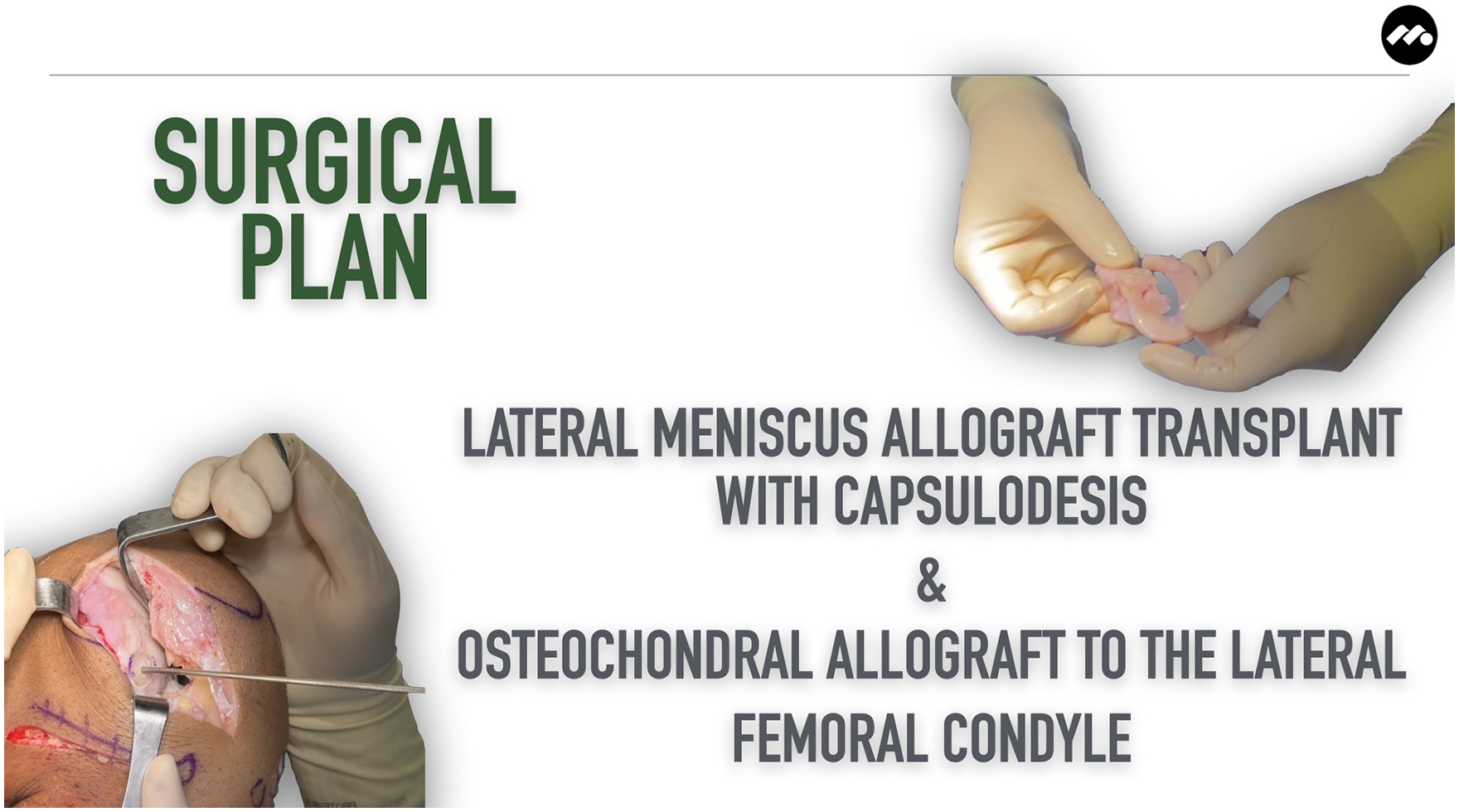
Video Transcript
The following is a video on lateral meniscus allograft transplant with capsulodesis and an osteochondral allograft (OCA) to the lateral femoral condyle.
The following are the authors’ disclosures.
Background
Anterior cruciate ligament (ACL) and meniscal injuries are a common challenge in orthopaedics. 6
Not all meniscal tears can be repaired, often requiring partial or complete meniscectomy. 6 Damage to the lateral meniscus, commonly associated with ACL injuries, can lead to elevated tibiofemoral contact pressures and an increased risk of early-onset osteoarthritis. 6 Lateral meniscal allograft transplant (LMAT) offers a promising solution to reinstate the integrity of a knee with a deficient lateral meniscus, as this can reinstate tibiofemoral contact pressures to near-physiological levels.8,9 Confronted with concurrent chondral defects, OCA transplant with meniscal allograft transplant can yield results comparable to those observed in knees without chondral defects. 4
A variety of LMAT techniques exist, including those using bone troughs, bone plugs, and soft tissue approaches. 1 Osseous fixation of the meniscal roots, in particular, has been shown to enhance the graft's stability, optimize patient outcomes, and extend graft longevity. 1 We advocate for a bone plug LMAT technique that capitalizes on standard arthroscopic portals. This approach minimizes soft tissue dissection while achieving solid osseous fixation, exemplifying an advancement in surgical technique that combines efficacy with minimally invasive principles.
We discuss the case of a 39-year-old man who presents with chronic left knee pain.
The patient has a history of an ACL reconstruction using a hamstring autograft and a subtotal lateral meniscectomy in India 15 years ago. Following the surgery, he experienced recurrent postactivity swelling. Within the past 2 years, he began to notice lateral knee pain, accompanied by swelling. He describes the pain as a persistent ache, occasionally experiencing sharp and intense exacerbations. His condition has progressed to the point where it not only prevents him from playing soccer but also affects his daily activities. Despite attempts at conservative management with physical therapy, nonsteroidal anti-inflammatory drugs, and intra-articular injections, his symptoms persisted.
On physical examination, his knee presented full range of motion, mild effusion, and tenderness along the lateral joint line, with no signs of instability or gait abnormalities.
Preoperative radiographs showed mild lateral osteoarthritis, with a Kellgren-Lawrence (KL) grade of 2 and normal limb alignment. Magnetic resonance imaging scans confirmed an intact ACL graft but revealed a deficiency of the lateral meniscus, cartilage damage on the lateral femoral condyle, and bone marrow edema within the lateral compartment.
Indications
Indications for meniscus allograft transplant include young patients with a symptomatic, meniscus-deficient knee that has failed conservative treatment. 2 Patients must have a stable knee with no concomitant articular cartilage damage that is not amenable to cartilage repair techniques. Patients also must be able to comply with postoperative rehabilitation and future care. 2 Contraindications for meniscus allograft transplant include patients over 50 years of age, body mass index (BMI) over 35 kg/m2, extensive cartilage lesions or multicompartmental osteoarthritis, uncorrected malalignment and ligamentous injury, inflammatory arthritis, synovial disease or prior joint infection, and noncompliant patients. 2
Indications for OCA transplant include young active patients, posttraumatic osteochondral defects, osteonecrosis, osteochondritis dissecans, large focal defects, previous cartilage repair failure, and patellofemoral joint cartilage lesions. 3 Relative contraindications to OCA transplant include a BMI over 35 kg/m2, concomitant ligament or meniscal injury, uncorrectable malalignment of the knee joint, inflammatory arthritis, and smoking or corticosteroid use. Severely advanced osteoarthritis is an absolute contraindication to OCA transplant. 3
Our surgical plan was for a left knee arthroscopy, LMAT with capsulodesis, and an OCA to the lateral femoral condyle.
Technique Description
The patient is positioned supine. A high thigh tourniquet is placed. A bilateral examination under anesthesia confirmed ligament stability and full range of motion. Standard prepping and draping of the operative side are done for knee arthroscopy.
The surgery begins with the arthroscopic portion of the procedure. We initiated with an anterolateral viewing portal, followed by the creation of an anteromedial portal.
Diagnostic arthroscopy reveals slight fraying of the medial meniscus that is not amenable to repair, in addition to a deficient lateral meniscus and grade 4 chondral injury of the lateral femoral condyle.
The recipient site is then prepared by debriding the remaining meniscal tissue to a thin peripheral rim of approximately 1 to 2 mm.
Anatomic landmarks are identified and used as guidance for the creation of the tibial sockets. First, a tibial guide is placed through the anterolateral portal, with the guide tip at the center of the posterior meniscal root footprint just off the articular surface.
After placing a guidewire, the posterior socket is retrodrilled 8 mm, and a passing suture is pulled through from the tibial socket into the joint and retrieved from the anterolateral portal.
A lateral incision is made to aid with performing the capsulodesis. A capsulodesis is performed to reduce meniscal extrusion. With a guide, a 2.4-mm tunnel is drilled from the anteromedial tibial cortex in an oblique direction to the edge of the lateral plateau where the capsule is most displaced. A passing suture is then passed through the tunnel. The capsule is captured with 2 spinal needles loaded with a polydioxanone suture, with an outside-in technique, which are then substituted with a high-strength suture. The 2 tails of the suture are then recovered through the tunnel, securing the articular capsule to the tibial plateau.
Two extra passing sutures are placed at the meniscocapsular junction in the meniscus body and retrieved through the anterolateral portal as well.
The anterior socket is created by 9-mm anterograde drilling with a 45° angle aiming posterodistally.
Another passing suture is pulled through the socket and retrieved from the anterolateral portal.
A fresh-frozen, size-matched, meniscal allograft is prepared on the back table.
Care is taken to preserve the integrity of the meniscal attachments during the creation of the 7-mm × 10-mm posterior bone plug and the 8-mm × 10-mm anterior bone plug at the meniscal roots.
A traction suture is securely attached to each bone plug, to be used as traction sutures for transtibial pull-through root fixation.
Last, 2 sutures are placed at the meniscus body for aiding in meniscal reduction and positioning as well.
The anterolateral portal is expanded to 2 to 3 cm in size to allow easy passage of the graft and instrumentation during implantation.
With sutures threaded through the tibial tunnels and meniscocapsular junction, the meniscal allograft is pulled into the joint. Sequential pulling, beginning with the posterior root, is performed to ensure adequate reduction of the posterior bone plug into the posterior tibia socket. This is then repeated for the anterior root bone block as well. The anterior horn is fixed to the capsule with a suture. This is done efficiently, and the meniscus is placed anatomically on the lateral tibial plateau.
Stability of the meniscus is then achieved using Fast-Fix Flex (Smith & Nephew) devices, applying multiple superior and inferior vertical mattress sutures along the entirety of the circumference of the meniscus. The number of Fast-Fix Flex devices used can vary depending on the specific needs of each case. It is crucial to apply as many superior and inferior Fast-Fix Flex devices as necessary to ensure optimal meniscal stability.
The anterior and posterior bone plug sutures, which are placed through the tibial tunnels, are finally fixed to the anteromedial tibial cortex with a button.
We then proceed with the lateral femoral condyle OCA.
Through a lateral parapatellar arthrotomy, the lateral femoral condyle osteochondral defect is identified after delicate dissection and flexion of the knee.
A cannulated cylindrical sizing guide is then used to determine the diameter of the defect. A guide pin is inserted to the center of the defect by passing through the cylindrical sizing guide.
The defect is scored with a cannulated circular reamer until healthy bleeding bone is exposed. The depth of the defect in this case was 7 mm, allowing for a secure press fit. The recipient site is dilated with a smooth cylinder to ensure the donor bone plug can be then inserted appropriately.
The depth of the reamed defect is then measured at the 3-, 6-, 9-, and 12-o’clock positions to determine appropriate sizing in preparation of the donor graft. The osteochondral donor plug is then harvested from the allograft with use of a coring reamer while using copious amounts of irrigation to prevent heat necrosis. The osteochondral dowel is removed, and the 12-o’clock position is marked with a marking pen. The dowel is then measured with a ruler and cut down to the appropriate depths based on the measurements previously taken from the recipient defect using a sagittal saw. Triple antibiotic solution is used to minimize the risk of an immune reaction. The bone plug is dipped in a sample of the patient's own blood or bone marrow aspirate concentrate.
The bone plug is then gently press-fitted into the recipient site after carefully aligning the 12-o’clock mark on the donor graft with that of the recipient site, to ensure a congruent articular surface.
Intraoperative testing demonstrates good graft stability throughout full knee range of motion.
Closure is achieved with layered sutures, ensuring a secure and clean operative field.
Results
At 2 weeks postoperatively, radiographs demonstrate well-preserved joint space, well-positioned OCA graft, and proper lateral meniscus transplant hardware placement.
Regarding outcomes, significant clinical improvements have been shown at the 5-year follow-up.5,8,10,11 Mean overall survival rate is 87% to 95% at 5 years, with a similar rate of 86.7% when performing meniscal allograft transplant with associated cartilage repair procedures.5,7,8,10,11 Patient satisfaction has been reported as 85% to 92%.5,8,10,11 Mean allograft survival has been shown to be 16.1 years.5,8,10,11 Increased age and BMI and noncompliant patients have been shown to negatively affect outcomes after meniscal allograft transplant.5,8,10,11
Complication rates are similar to those of standard meniscal repair surgery.5,8,10,11 The most frequent complication is tear of the graft. Other reported complications include graft failure, extrusion, infection, arthrofibrosis, aseptic synovitis, cellulitis, hematoma, joint effusion, and bone plug loosening.5,8,10,11 Reoperation rates have been reported ranging between 21% and 36%, most frequently for debridement of tears and less frequently for revision meniscal allograft transplant or total knee arthroplasty.5,8,10,11
Discussion
Regarding tips and tricks, bone plugs allow for adjustment of anterior and posterior horn positions. Bone plugs help to avoid interfering with tibial tunnel sites in ACL reconstruction. 1 Preservation of the anterior and posterior horn root attachments may serve as anatomic landmarks for allograft root insertion. 1
Regarding rehabilitation, from 0 to 6 weeks, strict nonweightbearing is employed with crutches. A knee immobilizer should be used until quad control is adequate, with limited range of motion for the first 2 weeks. From weeks 6 to 8, progress to full weightbearing, and by 8 weeks, discontinue the immobilizer and begin active range of motion and closed-chain exercises. For weeks 8 to 12, progress to full pain-free range of motion, closed-chain activities, proprioception, and balance exercises, and begin the stationary bike. At 12 weeks, progress to phase 3 and functional activities and advance core, glutes, and balance.
The following are our references.
Thank you for your time and attention.
Footnotes
One or more of the authors has declared the following potential conflict of interest or source of funding: J.C. was a board or committee member for AOSSM, Arthroscopy Association of North America, and International Society of Arthroscopy, Knee Surgery, and Orthopaedic Sports Medicine; was a paid consultant for Arthrex, Linvatec Corporation, Ossur, Smith & Nephew, DePuy Synthes Products, Vericel, and RTI Surgical; received a grant from Arthrex; received compensation for services other than consulting, including serving as faculty or as a speaker at a venue other than a continuing education program from Arthrex and Smith & Nephew; received compensation for serving as faculty or as a speaker for a nonaccredited and noncertified continuing education program from CONMED Corporation and Linvatec Corporation; received support for education from Arthrex and Medwest Associates; and received travel and lodging from Smith & Nephew and Medwest Associates. AOSSM checks author disclosures against the Open Payments Database (OPD). AOSSM has not conducted an independent investigation on the OPD and disclaims any liability or responsibility relating thereto.