
Abstract
Background:
Genu recurvatum is a challenging disorder and can negatively affect knee biomechanics, as reflected in its role as a risk factor for knee ligament injury and poor outcomes following ligament reconstruction or arthroplasty.
Indication:
Pathological recurvatum should be addressed in the presence of a causative correctible structural abnormality, whether due to decreased posterior tibial slope or due to multiligament injury. We described a posterior oblique ligament (POL) advancement technique for the treatment of knee recurvatum.
Technique Description:
After completion of all concomitant procedures such as ligament reconstructions, the procedure is initiated with a medial approach. The POL is isolated with an inverted L-shaped dissection and mobilized. Mattress sutures are passed through the resulting POL flap, and are re-tensioned and fixed anteriorly and distally, with the knee close to extension, in order for the imbrication to restrain hyperextension.
Results:
Anecdotal evidence points to satisfactory control of knee recurvatum postoperatively, which can potentially avoid its deleterious effects in surgical outcomes.
Discussion/Conclusion:
We present a promising option in the treatment of pathological recurvatum, with favorable cost-efficacy, low morbidity relative to slope-altering osteotomy or posterolateral soft tissue procedures, and focused on the biomechanically demonstrated main restrictors to knee hyperextension.
Patient Consent Disclosure Statement:
The author(s) attests that consent has been obtained from any patient(s) appearing in this publication. If the individual may be identifiable, the author(s) has included a statement of release or other written form of approval from the patient(s) with this submission for publication.
This is a visual representation of the abstract.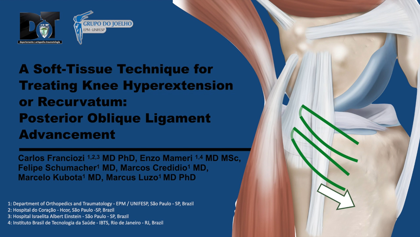
Video Transcript
Although there is abundant literature on coronal plane malalignment, sagittal imbalance of the knee has been less studied, largely due to its less common occurrence and the uncommon need for operative treatment. 13
There is still no consensus on the normality values for knee extension beyond 0°. Pathological knee recurvatum is typically characterized by extension beyond 15° or even smaller values when unilateral, asymmetrical, and/or symptomatic. 9 Of note, physiological hyperextension might also need specific intervention when there is added ligament injury, most commonly of the anterior cruciate ligament (ACL).1,7,9
Recurvatum can be caused by a variety of factors, such as physeal arrest, fracture malunion, collagen disorders, or neuromuscular weakness. The main culprits, however, are typically abnormal bone morphology in the form of decreased or inverted posterior tibial slope, or soft tissue related, due to a traumatic knee ligament injury or innate capsular laxity.4,5 A recent cadaveric investigation revealed incremental increases in heel height with injury patterns of ACL + fibular collateral ligament (FCL), ACL + posterolateral corner (PLC), and bicruciate tears + medial collateral ligament (MCL). 12
Clinical studies report that genu recurvatum is a risk factor for ACL tears, 2 increases failure rates following ACL reconstruction with a hamstring autograft, and results in worse patient outcomes following total knee arthroplasty.2,6,8,13 With that context in mind, pathological recurvatum should be treated operatively when there is causative correctible structural abnormality, whether due to decreased posterior tibial slope or due to multiligament injury.
We present here a soft tissue technique to address knee recurvatum, with a posterior oblique ligament (POL) advancement in a multiligament knee injury patient.
Our patient was a 24-year-old male recreational soccer player, with a history of traumatic left knee injury with varus moment during a match 14 months earlier. The patient complained of persistent diffuse pain and instability episodes upon pivoting. Physical examination revealed a neutral coronal alignment, with no varus thrust during gait; a 3+ Lachman, anterior drawer, and pivot-shift; a 2+ varus stress opening at 30° of flexion and in extension; as well as a positive posterolateral drawer and dial test at 30°. The patient presented with a notable and asymmetrical recurvatum on the injured side, as reflected in a positive recurvatum–external rotation test and heel-height test.
Preoperative radiographs revealed no significant degenerative joint disease on Rosenberg view and a neutral alignment on standing long-limb radiograph. On lateral view, however, the affected knee exhibited a posterior slope of 12°, in contrast with a 4° slope on the contralateral knee.
Magnetic resonance imaging (MRI) revealed a complete ACL tear and a suspected PLC injury, noted as thinning of the FCL and popliteal fibular ligament, which was later confirmed with a 4-mm side-to-side difference in varus gapping during manual stress radiographs.
In summary, the diagnosis was an ACL tear with an explosive pivot shift, a posterolateral corner injury, and pathological knee recurvatum most likely attributed to ligamentous laxity (with a 12° tibial slope). The planned procedure was an ACL reconstruction, PLC reconstruction, anterolateral ligament (ALL) reconstruction, and POL advancement to address the recurvatum.
The ACL reconstruction was performed with a bone-patellar tendon-bone autograft, using an anatomic anteromedial portal technique in standard fashion. The PLC reconstruction was performed with an anatomic technique using allografts, as described by LaPrade, reproducing the popliteus tendon, FCL, and popliteal fibular ligament.
Subsequently, we performed the ALL reconstruction with a modified technique with a single-strand gracilis autograft sharing the same femoral tunnel and fixation with the FCL. The graft is passed distally, deep to the superficial layer of the iliotibial band and the FCL. The tibial tunnel aperture is placed halfway between Gerdy's tubercle and the fibular head, and the tunnel is directed in a medial and distal orientation, where the suture limbs of the ALL graft are tied to those of the ACL in 30° of flexion.
Following completion of the ACL, ALL, and PLC reconstructions, there was still significant residual asymmetrical recurvatum, which we aimed to correct with the use of our proposed technique for POL advancement.
The advancement should take place following the complete fixation of all concomitant procedures. Through a 5-cm longitudinal medial approach, posterior to the medial epicondyle and extending distally to the posteromedial border of the medial tibial condyle, the POL is isolated by means of an L-shaped dissection, with the distal horizontal arm placed at the proximal end of the direct insertion of the semimembranosus insertion, and the proximal longitudinal arm placed at the posterior border of the superficial MCL. The resulting POL flap will be mobilized and re-tensioned in an anterior and distal direction, with 3 to 4 no. 2 nonabsorbable high-strength sutures, which are subsequently fixed with the knee at 30° to 45° of flexion, as some stretching out is expected. Following the completion of the POL advancement, we can observe the correction of the residual recurvatum.
Postoperative radiographs demonstrate the location of each tunnel for the ACL, ALL, and PLC reconstructions.
Another case example shows the improvement in knee recurvatum in a patient with Ehlers-Danlos disease and severe bilateral recurvatum who sustained an ACL tear and underwent ACL and ALL reconstructions with POL advancement.
Postoperatively, the patient should be kept in a hinged knee brace, with extension locked at 10° to 15° for 3 months. Gait retraining should emphasize avoidance of hyperextension during the stance phase, in order to avoid recurrence of the recurvatum.
Advantages of our proposed POL advancement technique include ease of use, a relatively low-risk medial approach, and no need for additional implants, making it cost-friendly.
Overall, operative management of genu recurvatum poses a significant challenge, and there are currently multiple options available, generally divided into osteotomies and soft tissue procedures. 4 Osteotomies are appropriate when the recurvatum is attributed to altered bone morphology, namely in the form of slope-increasing osteotomies, 10 as in this anterolateral biplanar proximal tibial opening-wedge osteotomy, for cases with combined valgus and recurvatum. 3 Osteotomies, however, are ill-suited when there is a normal or even increased tibial slope or in cases where increasing the tibial slope is detrimental to concomitant procedures such as an ACL reconstruction.
Furthermore, a slope-reducing osteotomy, which is indicated for cases of revision ACL reconstructions with an increased posterior slope, may lead to iatrogenic increase in knee recurvatum 14 and can potentially benefit from the addition of our proposed POL advancement technique as a means to prevent this unwanted effect.
In such cases, or in the setting of normal bone morphology where ligament laxity or traumatic injuries are the culprit, soft tissue procedures should be preferred. A recent cadaveric investigation by Frank Noyes demonstrated that the posteromedial capsule and POL are the main restraints against hyperextension, accounting for more than 20% of the mean resisting force, followed by the posterolateral capsule and fabello fibular ligament. 11 In addition to its greater contribution in resisting hyperextension, a posteromedial approach offers a technically easier and faster access, with less morbidity and iatrogenic neurovascular damage.
In summary, our proposed technique for POL advancement is a promising option in the treatment of pathological recurvatum—particularly in cases of soft tissue recurvatum or in addition to a slope-reducing high tibial osteotomy, as a means to prevent iatrogenic hyperextesion. POL advancement provides favorable cost-efficacy (given the obviated need for additional grafts and implants), with lower morbidity relative to slope-altering osteotomy or posterolateral soft tissue procedures, and which acts on the biomechanically demonstrated main restrictors to knee hyperextension.
Footnotes
Submitted June 9, 2023; accepted July 27, 2023.
One or more of the authors has declared the following potential conflict of interest or source of funding: C.E.F. has received educational support from Smith + Nephew. M.S.K. has received educational support from Johnson & Johnson. M.V.L. has received educational support from Johnson & Johnson and Zimmer Biomet. AOSSM checks author disclosures against the Open Payments Database (OPD). AOSSM has not conducted an independent investigation on the OPD and disclaims any liability or responsibility relating thereto.