
Abstract
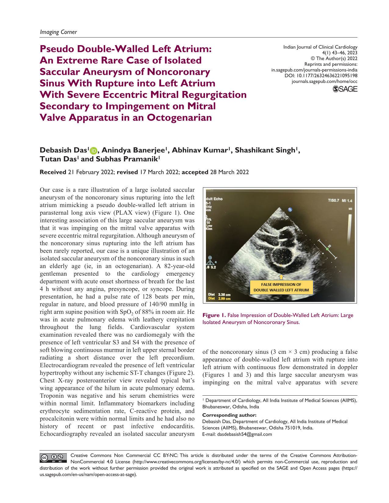
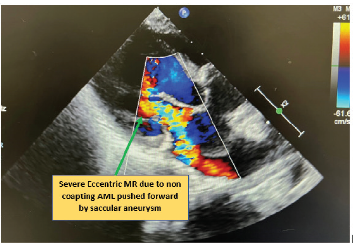
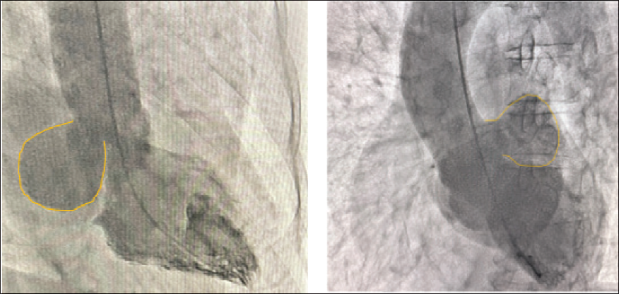
Our case is a rare illustration of a large isolated saccular aneurysm of the noncoronary sinus rupturing into the left atrium mimicking a pseudo double-walled left atrium in parasternal long axis view (PLAX view) (Figure 1). One interesting association of this large saccular aneurysm was that it was impinging on the mitral valve apparatus with severe eccentric mitral regurgitation. Although aneurysm of the noncoronary sinus rupturing into the left atrium has been rarely reported, our case is a unique illustration of an isolated saccular aneurysm of the noncoronary sinus in such an elderly age (ie, in an octogenarian). A 82-year-old gentleman presented to the cardiology emergency department with acute onset shortness of breath for the last 4 h without any angina, presyncope, or syncope. During presentation, he had a pulse rate of 128 beats per min, regular in nature, and blood pressure of 140/90 mmHg in right arm supine position with SpO2 of 88% in room air. He was in acute pulmonary edema with leathery crepitation throughout the lung fields. Cardiovascular system examination revealed there was no cardiomegaly with the presence of left ventricular S3 and S4 with the presence of soft blowing continuous murmur in left upper sternal border radiating a short distance over the left precordium. Electrocardiogram revealed the presence of left ventricular hypertrophy without any ischemic ST-T changes (Figure 2). Chest X-ray posteroanterior view revealed typical bat’s wing appearance of the hilum in acute pulmonary edema. Troponin was negative and his serum chemistries were within normal limit. Inflammatory biomarkers including erythrocyte sedimentation rate, C-reactive protein, and procalcitonin were within normal limits and he had also no history of recent or past infective endocarditis. Echocardiography revealed an isolated saccular aneurysm of the noncoronary sinus (3 cm × 3 cm) producing a false appearance of double-walled left atrium with rupture into left atrium with continuous flow demonstrated in doppler (Figures 1 and 3) and this large saccular aneurysm was impinging on the mitral valve apparatus with severe eccentric mitral regurgitation (Figure 4). In view of need for emergency repair of the ruptured sinus into left atrium, the patient was taken for coronary angiography and cardiac catheterization, which revealed normal coronaries and demonstrated the large saccular aneurysm of the posterior noncoronary sinus (Figure 5) with eccentric severe mitral regurgitation filling the whole left atrium. The patient was advised to undergo emergency double-patch closure of ruptured sinus of Valsalva after initial stabilization with oxygen inhalation, diuretic, and vasodilator therapy. Although aneurysm of noncoronary sinus with rupture into left atrium, 1 right atrium, 2 and right ventricle 3 have been described in paucity, our case is a rare description of isolated saccular aneurysm of the noncoronary sinus in such an elderly age producing a false sense of double-walled left atrium with impingement on the mitral valve apparatus and eccentric severe mitral regurgitation. Echocardiography with precision often adds to the clinical image.
False Impression of Double-Walled Left Atrium: Large Isolated Aneurysm of Noncoronary Sinus.
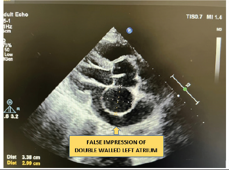
ECG Showing Left Ventricular Hypertrophy Without Ischemic ST-T Changes.
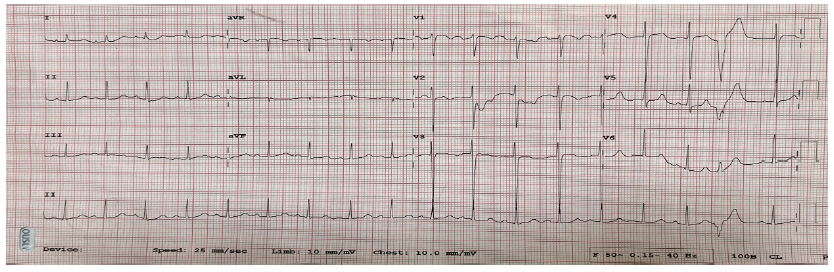
Bat’s Wing Appearance of Hilum Due to Acute Pulmonary Edema, Rupture of Sinus of Valsalva into Left Atrium With Continuous Doppler Flow.
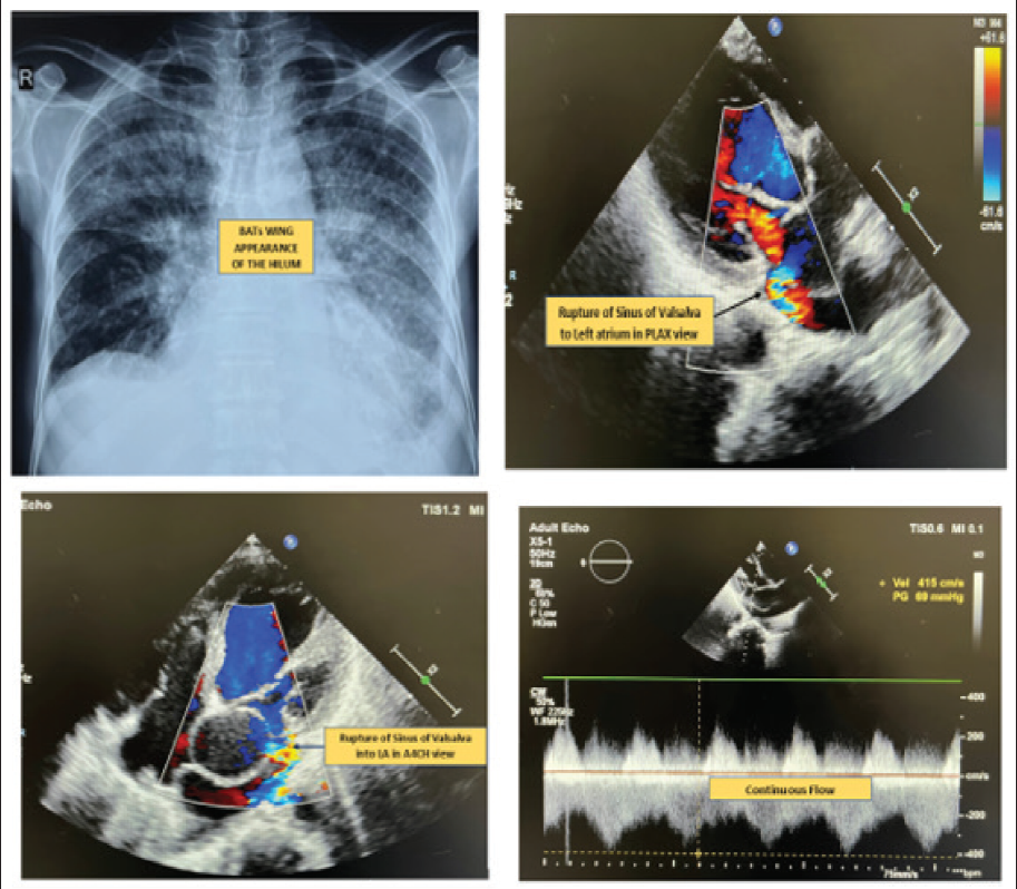
Severe Mitral Regurgitation (MR) due to Impingement of the Aneurysm on Mitral Valve Apparatus.
Large Saccular Aneurysm From Noncoronary Sinus in RAO 30 and LAO 50 20 View.
Footnotes
Declaration of Conflicting Interests
The authors declared no potential conflicts of interest with respect to the research, authorship, and/or publication of this article.
Funding
The authors received no financial support for the research, authorship, and/or publication of this article.