
Abstract

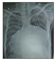
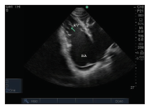
A 20-year-old boy presented with swelling over the body and breathlessness on exertion for last 4 years. His symptoms increased for last 1 month with increased ascites, pedal edema, and breathlessness, including orthopnea. Examination revealed signs of chronic heart failure. His electrocardiogram (ECG) showed atrial fibrillation with intraventricular conduction defect. Chest x-ray showed gross cardiomegaly, biatrial dilatation, pulmonary infundibular dilatation, and post-capillary pulmonary hypertension (Figure1) which are typical radiological features of biventricular endomyocardial fibrosis (EMF). Echocardiogram showed severe biatrial enlargement, moderate pericardial effusion, dilated inferior vena cava, severe mitral regurgitation and tricuspid regurgitation, moderately severe pulmonary arterial hypertension with patterns of involvement of severe biventricular EMF. Fibrosis involving the right ventricular inflow tract and endomyocardial soft echogenic infiltrates of left ventricular wall was seen suggestive of biventricular EMF (Figure 2). Echocardiogram is almost adequate for diagnosis of EMF.
There are 5 prime diagnostic echocardiographic features of EMF; apical fibrosis, ventricular wall fibrosis, huge atrium, atrioventricular valve regurgitation, and obliteration of ventricular cavity. The presence of pericardial effusion, endocardium fibrous shelf, and layering of the posterior wall lend further diagnostic support.
The diagnosis of EMF needs a high index of suspicion as it can be mislabeled as rheumatic heart disease or apical type of hypertrophic obstructive cardiomyopathy. Right ventricular EMF can also be confused with Ebstein’s disease of the tricuspid valve because of the similar dilatation of right atrium and right heart failure.
As with other forms of restrictive cardiomyopathy, EMF should be distinguished from constrictive pericarditis.
Other conditions to consider in patients with suspected EMF are:
Anthracycline toxicity
Carcinoid heart disease
Drug-induced cardiotoxicity (eg, serotonin, methysergide, ergotamine, mercurial agents)
Fabry disease
Gaucher disease
Glycogen storage disease
Metastatic cancers
Radiation
Cardiac MRI can provide additional value for the diagnosis of EMF. Cardiovascular magnetic resonance outlines the degree of chamber distortion and the extent of thrombosis. It may be an ideal tool for monitoring the response to treatment and for defining anatomic details before surgery.
Chest x-ray showing gross cardiomegaly, biatrial dilatation, pulmonary infundibular dilatation and PAH.
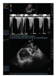
Echocardiogram showing moderately severe pulmonary arterial hypertension.
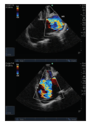
Echocardiogram showing severe biatrial dilatation, severe MR and TR.
Echocardiogram showing biatrial dilatation, moderate pericardial effusion and echogenic infiltrates of LV wall.
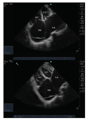
Echocardiogram showing fibrosis of RV inflow tract.
Footnotes
Declaration of Conflicting Interests
Funding
The authors received no financial support for the research, authorship and/or publication of this article.