
Abstract
Endomyocardial fibrosis is a rare cardiomyopathy. There has to be a high level of suspicion to make the diagnosis. The treatment is based on symptomatic relief and surgical management is based on the exact pathology found in the left ventricle apex. MRI is a robust investigation which can confirm diagnosis and provide management options and prognosis.
Case
A 63-year-old female presented with breathlessness NYHA CL II to III over 2 months. Clinical exam revealed pedal edema, elevated JVP, LVS3+, and bibasilar crepitation. A clinical diagnosis of heart failure was made. BNP was elevated at 55 pg/ml. Chest radiograph revealed mild cardiomegaly with pulmonary venous hypertension. ECG showed atrial fibrillation with fast ventricular rate with T wave inversion in V1 to V3. 2D echocardiogram showed obliteration of the LV apex with preserved biventricular systolic function. In order to make a definitive diagnosis and prognosticate the patient, a cardiac MRI was ordered.
MRI findings reveal:
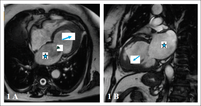
LV apical thickening with obliteration (Figure 1A-1B)
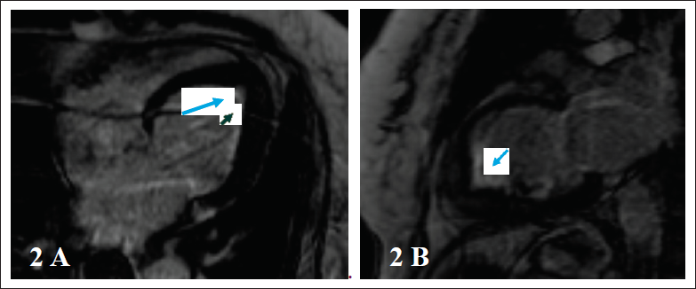
Subendocardial delayed enhancement of LV apex (Figure 2A-2B)
Preserved biventricular systolic function with LV diastolic dysfunction.
Normal biventricular volumes
Mild left atrial dilatation
Moderate MR
The patient was initiated on heart failure medication including diuretics, salt and water restriction, and lifestyle modification. Surgical opinion was taken for endocardiectomy which has been planned subsequently on follow-up. She has been placed on regular follow-up and registered on a transplant registry with a plan for future cardiac transplant if she does not respond to optimal medical therapy or endocardiectomy.
Discussion
EMF is the most common type of restrictive cardiomyopathy in tropical areas. It is characterized by fibrotic tissue deposition in the endocardium of the inflow tract and apex of one or both ventricles. Ventricular morphology is usually distorted with normal or reduced volumes, whereas atrial volumes are increased. The cause of EMF is unknown; however, early hypereosinophilia may play a role in its pathogenesis. Diastolic dysfunction is responsible for the severe heart failure.
Cardiac magnetic resonance (CMR) provides detailed information on ventricular morphology and function, including excellent visualization of the ventricular apex. Late gadolinium enhancement (LGE) by CMR allows the evaluation of the presence of myocardial inflammation and fibrosis.
MRI has definitive role in confirming and characterizing this pathology as compared to echocardiography.
Characteristic CMR features are as follows:
Major diagnostic criteria of EMF on CMR include ventricular apical obliteration with subendocardial delayed enhancement (typical “double V” sign).
It consists of a three-layered pattern of normal myocardium, thickened enhanced endomyocardium, and overlying thrombus at the ventricular apex with or without calcification. There is excellent correlation with histopathologic findings and it plays an important role in differentiating EMF from other cardiomyopathies.
Secondary atrial enlargement.
Atrioventricular valve regurgitation: Tricuspid and mitral valve regurgitation secondary to fibrosis of the papillary muscles.
Endomyocardial biopsy is not essential to diagnose EMF. Medical management usually includes treatment with diuretics and anticoagulants. Surgical treatment consists of resection of fibrotic tissue and valve repair or replacement. Transplantation is an option if the patient goes into refractory heart failure despite medical and surgical intervention.
Conclusion
EMF is a type of restrictive cardiomyopathy noted in tropics and commonly affecting females of middle age. The presentation is of heart failure with preserved ejection fraction. Advanced cases of EMF show calcification and valvular regurgitation of different grades. The pattern of post contrast delayed enhancement clearly differs from other types of cardiomyopathy (typical “double V” sign). Cardiac MRI is the modality of choice in EMF, to confirm the diagnosis and to assess the severity of involvement.