
Abstract
Dear Editor,
Approaching the vitreous body is of paramount importance when treating a considerable range of eye diseases. Reports of primordial vitrectomy attempts have been noted since the 17th century, 1 but only starting from the early 1970s has the procedure achieved its modern rationale and high standards. 2
The concept of vitrectomy involves a pars plana approach, where, as in abdominal video-laparoscopic surgery, the vitreous cavity is reached by means of three trocars, normally used for illumination, fluid infusion and surgical instruments. Substantial advances ensued with the development of smaller gauge instrumentation, higher cutting speeds, improved illumination, better microscopes and the use of perfluorocarbon liquids, which significantly improved outcome, making pars plana vitrectomy the standard approach for the majority of vitreoretinal diseases.
Current limitations
Vitrectomy is now a remarkably reproducible, fast and clean procedure. However, whenever we feel that everything is working smoothly, we run the risk of entering a comfort zone, and maybe ignoring the possibility of progress. We believe there are certain limiting factors to this standard procedure that require careful attention.
These limiting factors mainly involve the fact that various instruments need to be inserted and thereafter extracted, which might be necessary numerous times during surgery causing interruption and possible distraction, continuous changes of focus for the surgeon, as well as consuming precious time. This kind of procedural sequence can become tedious, as in the case of long interventions, or following complications that might require rapid countermeasures.
The interruptions can indeed be deleterious. As an expert surgeon generally plans each subsequent step in advance, any kind of distraction may block this stream of thought. Once the procedure has been paused, the surgeon needs to re-insert the instruments into the trocars. This involves abandoning the microscope eyepiece, selecting a new instrument, focusing on the operating field, inserting the desired probe and then returning to the microscope with its previous focus. This can be tiring, particularly for presbyopic operators, and moreover demanding in the presence of conjunctival chemosis, dim theatre lights or dazzling reflexes.
Finally, the abovementioned facts are time-consuming. Time is a priceless resource, and no one understands this better than the surgeon, who might have to deal with a leaking blood vessel or a ballooning choroidal detachment. Prompt reactions can be decisive in certain situations, hence precious time cannot be wasted.
Innovations
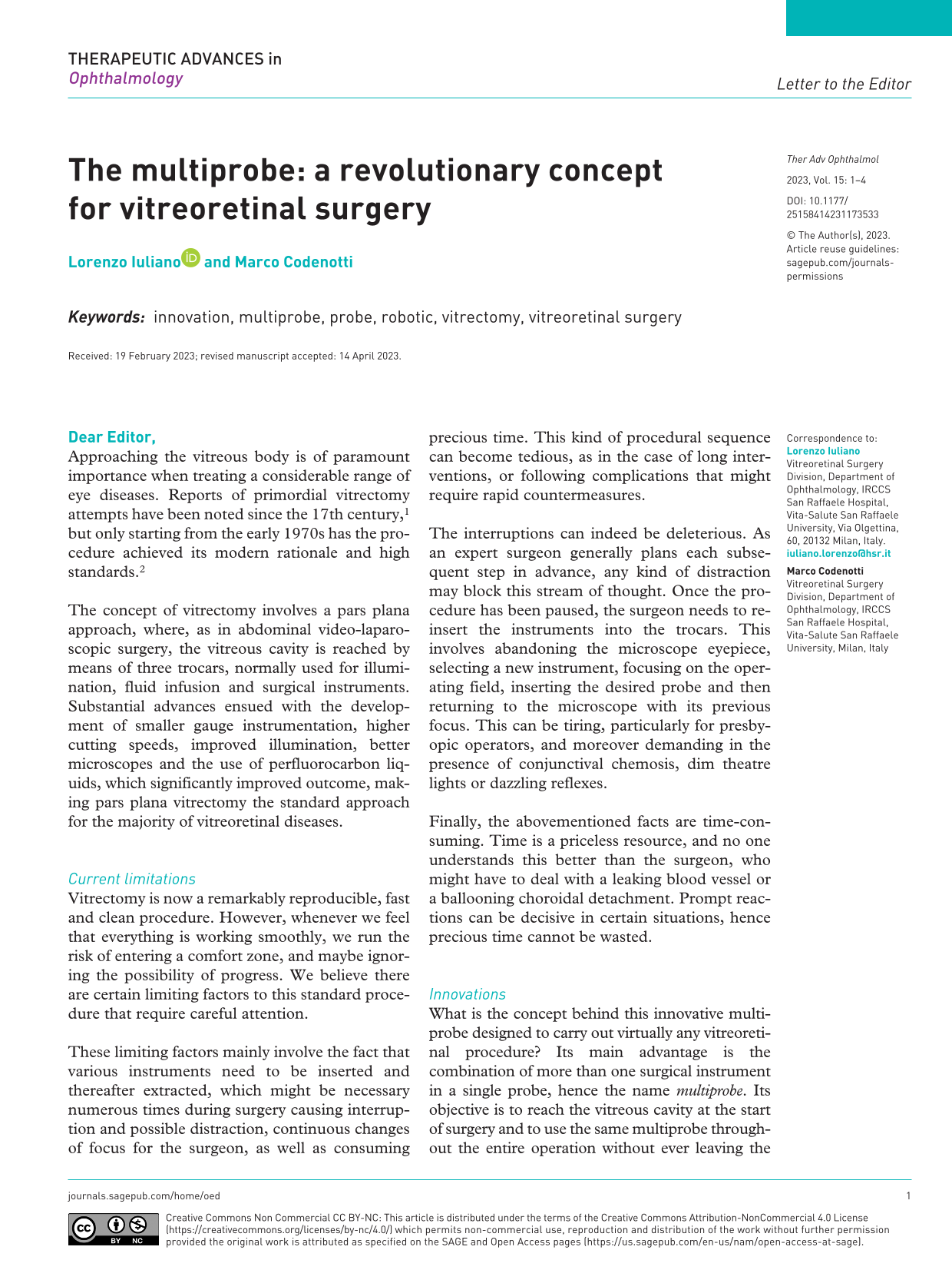
What is the concept behind this innovative multiprobe designed to carry out virtually any vitreoretinal procedure? Its main advantage is the combination of more than one surgical instrument in a single probe, hence the name multiprobe. Its objective is to reach the vitreous cavity at the start of surgery and to use the same multiprobe throughout the entire operation without ever leaving the eye. The multiprobe is equipped with a solid cannula, which needs to be inserted intraocularly using standard trocars. The surgical instruments are thereafter fitted into the probe’s handheld housing (Figure 1), from which a selector can extrude the desired device through the cannula to reach the vitreous cavity. When the surgeon needs a different instrument, extraction of the multiprobe is not required, but simply external selection (via a pedal or nurse-guided remote control) to carry out the instrument switch. The instrument being used (e.g. cutter) is thus retracted and is replaced by another (e.g. forceps).

Multiprobe rendering. Three-dimensional rendering of the multiprobe, with captions. (a) Front view. (b) Transparent skeletonized model. (c) Back view.
Changing instruments is totally automatic and mechanized, requiring a simple command.
Advantages
As often happens with innovations, our concept might appear intuitive or maybe even obvious. Naturally, we are not claiming to be pioneers of vitreoretinal surgery, although we strongly believe the idea of grouping together and automating instrument exchange to be both smart and innovative.
Surgical manoeuvres will logically become (1) considerably faster, reducing critical time-gaps for the precise control of eventual complications and accurate fluid exchange (fluid aspiration through retinotomies and instantaneous laser retinopexy); (2) significantly less exhausting, as the surgeon will avoid the cumbersome sequences involved in instrument switching; (3) much cleaner, as the risk of external contamination caused by continuous exchange is reduced.
Other indirect advantages need to be considered. We are now experiencing the thrill of the three-dimensional intervention, where surgery is progressively moving towards the heads-up approach. 3 In this eyepiece-free scenario, surgeons have two operating fields: the ultrawide high-definition monitor and the ‘naked’ patient’s eye. The exchange of instruments thus requires even more strenuous effort than before. We feel an automatic multiprobe is essential for this ergonomic change, as this revolution might otherwise seem incomplete.
Furthermore, we feel the future of vitreoretinal surgery will be unable to overlook robotic support.4,5 Although in its infancy, it is already a reality that rightfully represents the next generation. We wonder if our multiprobe might be also integrated with an optical coherence tomography-distance sensor to the tip of the probe, that has been already evaluated clinically in a robot-assisted setting. 6 How can we imagine robot-assisted surgery without mechanized automatic multiprobes?
Disadvantages
A mention to the possible disadvantages must be acknowledged. First, handling a heavier probe may decrease the dexterity and increase fatigue. About that, we disclose the true weight of the single instruments is remarkably low, hence their combination (moreover with a single casing for all) is expected to remain relatively handy. Furthermore, a slightly heavier probe may not necessarily affect precision as it can favour stability and steadiness.
Second, the augmented mechanization may raise the chance of malfunctions. This is unavoidably correct, but it should be disclosed that the innovative aspect of the automation relies on the insertion–extrusion mechanism, more than on the functioning of the single instruments, that actually remains unchanged. We hence believe this potential risk remains manageable, and, above all, harmless for the biological tissues.
Finally, it can be presumed the multiprobe may increase the costs. This might be reasonable, but this issue requires a more extended and comprehensive analysis of the benefit–cost ratio, including those abovementioned, more than a mere calculation of the single probe’s costs.
Conclusions
Our intention was not to invent a new surgical instrument but to propose an idea based on situations we regularly face in the operating room. Despite the engineering and design process are at present under development, its specifications go way beyond the scope of the present description. Although numerous studies are required to verify the efficacy of our proposal, we strongly believe that the use of a multiprobe would be more effective, precise and even safer than current standard procedures. We hope the multiprobe will help the mesmerizing world of vitreoretinal surgery take yet another revolutionary step forward.