
Abstract
The Phytexponent is used to treat pain and inflammation in complementary and alternative medicine practices; however, empirical data supporting its pharmacological efficacy and safety is scanty, hence the present study. We used the carrageenan-induced paw oedema and the acetic acid-induced writhing techniques to determine the anti-inflammatory and analgesic efficacies, respectively, of the Phytexponent in Swiss albino mice models. The 3-(4, 5-dimethylthiazol-2-yl)-2, 5-diphenyltetrazolium bromide (MTT) assay technique was used to investigate the in vitro cytotoxic effects of the Phytexponent in the Vero E6 cell line. The Phytexponent exerted significant (P < .05) anti-inflammatory effects in the carrageenan-induced paw oedema mouse model in a dose- and time-dependent manner, with significantly higher efficacy at 250 mg/Kg BW, than indomethacin (4 mg/Kg BW), in the first, second, and third hour (P < .05). Besides, the Phytexponent significantly reduced the acetic acid-induced writhing frequency in mice (P < .05), in a dose-dependent manner, depicting its analgesic efficacy. Notably, the Phytexponent (at doses: 125 mg/Kg BW and 250 mg/Kg BW) exhibited significantly higher analgesic efficacy than the Indomethacin (P<.05). Moreover, the Phytexponent was not cytotoxic to Vero E6 cells (CC50 >1000 µg/ml) compared to cyclophosphamide (CC50 = 2.48 µg/ml). Thus, the Phytexponent has significant in vivo anti-inflammatory and analgesic efficacy in mice models and is not cytotoxic to Vero E6 cell line, depicting its therapeutic potential upon further empirical investigation.
Introduction
Inflammation is a response of a tissue to a noxious stimuli, such as physical injury, irritant agents, and pathogens, which is characterised by increased vascular permeability, changes in blood flow, and migration of leucocytes to the affected sites. 1 Pain refers to an unpleasant emotional and sensory experience that results from tissue damage and acts as a signal to warn against further insults. 2 Pain, fever, and inflammation are associated with a myriad of pathological processes in the body. 3 There are various anti-inflammatory and analgesic drugs for the treatment of inflammation and pain;4,5 however, they are inaccessible and unaffordable especially in low income and remote settings, they are of low efficacy and cause adverse effects with life-threatening consequences.6,7 In this regard, focus has shifted to investigating natural products, especially medicinal plants, as one of the most promising therapeutic agents for inflammatory diseases. 8
Research data shows that plant-derived natural products are a bulwark of future drug discovery, especially for the treatment of inflammation and pain.9–11 This is encouraging, considering that more than 80% of the human population in third world countries, especially in Africa, depend on traditional medicine for healthcare needs. 12 In Kenya, various communities manage pain, fever, and inflammation using plant-derived remedies, as part of their traditional and complementary medicine practices13–19.
Even though medicinal plants have extensive and longstanding utilisation in alternative and complementary therapy, various concerns regarding their safety have been raised. 20 For instance, most countries lack legislative guidelines for the practice of traditional medicine, thus allowing unscrupulous practioners to thrive. 21 Additionally, there is scanty data on dosage regimens, herb-herb and herb-drug interactions, and associated effects to effectively guide prescriptions. 22 Furthermore, the lack empirical data on safety and toxicity profiles of many medicinal plants further cripples the confidence accorded to herbal medicine; hence, it is imperative to evaluate toxicity and safety of herbal preparations used to manage various diseases to avert the development of undesirable effects and fatalities.20,22
The Phytexponent is used in complementary and alternative medicine, in Kenya, and in many other countries, to manage inflammation and pain, and associated syndromes, with an appreciable level of efficacy. Some of the plants used to formulate the Phytexponent are used in traditional medicine since they possess various pharmacologic activities against a variety of disease conditions. For instance, extracts of Viola tricolor have been traditionally used to treat inflammatory lung disease and skin ailments, such as ulcers, itching, scabs, psoriasis, and eczema. 23 Besides, Echinacea purpurea is commonly used to alleviate common cold symptoms, and has been shown to posses anti-inflammatory and immunostimulatory properties. 23 Additionally, Allium sativum (Garlic) is widely used as a food ingredient, and its over 200 phytocemicals are used to treat various conditions that are associated with inflammation, including some types of cancer, and as an aphrodisiac. 23
Recently, Moriasi et al. 24 demonstrated significant in vitro anti-inflammatory and antioxidant activities, and the presence of bioactive phytochemicals with diverse pharmacologic effects, including anti-inflammation and antioxidation. However, there is a scarcity of documented studies on the in vivo efficacy of this polyherbal product, its mode(s) of action in various disease states, its toxicity, and safety.
Therefore, this study was designed to investigate the in vivo anti-inflammatory, analgesic, and cytotoxic effects of the Phytexponent formulation of Viola tricolor, Echinacea purpurea, Allium sativum, Matricaria chamomilla, and Triticum repens, as a potential alternative source of affordable, accessible, potent, and safe analgesic and anti-inflammatory lead compounds for drug discovery and development.
Materials and Methods
The Source of the Phytexponent Polyherbal Formulation
The Phytexponent formulation (Pharmapath 27, Belgium; LOT NO:17E19) was purchased from a local vendor and stored at room temperature according to the manufacturer's guidelines, and retrieved only when required.
Experimental Animals
Swiss-albino mice weighing 24 ± 1 g, and aged between four and five weeks old were used in this study. The experimental animals were housed under standard conditions (Temperature: 25 ± 2 °C; Relative humidity: 55%-61%; 12 hours of dark and 12 hrs of light cycle), in polypropylene rectangular cages measuring 30 cm × 20 cm × 13 cm, in which soft wood shavings were added as bedding material. The mice were fed on standard laboratory rodent food (pellets) and clean water ad-libitum, and maintained at natural day-night cycle. The experimental mice were acclimatised to the laboratory settings for 72 hours prior to experimentation.
Determination of
in Vivo
Anti-Inflammatory Activity of the Phytexponent Prepartion
The Carrageenan-induced paw oedema technique of Winter et al. 25 was adopted to investigate the antiinflammoty effects of the Phytexponent with minor modifications. Briefly, experimental mice were randomised into 8 groups comprising of 5 mice per group. The normal control group mice [A] were orally administered with 10 ml/Kg BW of normal saline. The negative control group mice [B] were given normal saline (10 ml/Kg BW) orally, and after 30 minutes, they were injected with 100 µl of 1% Carrageenan (Sigma-Aldrich, Germany) via the subplantar region of the right hind paw (s.p). The positive control group mice [C] received 10 mg/Kg BW of indomethacin orally and 100 µl of 1% carrageenan through the subplantar region of the right hind paw after 30 minutes. Mice in other experimental groups (D-H) were orally treated with the Phytexponent at dose levels of 15.625 mg/Kg BW, 31.25 mg/Kg BW, 62.5 mg/Kg BW, 125 mg/Kg BW, 250 mg/Kg BW and 500 mg/Kg BW, respectively, and injected with 100 µl of 1% Carrageenan via the subplantar route after 30 minutes. The changes in paw diameter sizes were measured before induction of inflammation, and after 1 hour, 2 hours, 3 hours, and 4 hours, respectively, following the induction of inflammation, using a plethysmographic technique, and the percentage inhibitions of inflammation were calculated and tabulated.
Determination of the Analgesic Effects of the Phytexponent
The analgesic activity of the Phytexponent was evaluated according to the method described by Koster et al. 26 with slight modifications. Briefly, the normal control group mice [A] received normal saline (10 mg/Kg BW; p.o) only. The negative control group [B] mice were orally admisntered with normal saline at a dose of 10 mg/Kg BW (p.o) and 100 µl of acetic acid (0.6% w/v; ip) (Lot#L148661503; Loba Chemie) after 30 minutes. On the other hand, the positive control group [C] mice received indomethacin (4 mg/Kg BW; p.o) and 100 µl of acetic acid (0.6% w/v; i.p) after 30 minutes. Besides, the mice in other experimental groups [D-H] were orally administered with the Phytexponent at dose levels of 15.625 mg/Kg BW, 31.25 mg/Kg BW, 62.5 mg/Kg BW, 125 mg/Kg BW, 250 mg/Kg BW, and 500 mg/Kg BW, respectively, 30 minutes before the intraperitoneal injection of 100 µl of 0.6% v/v of acetic acid. The total number of writhes was recorded for each experimental mouse after 5 minutes of writhing induction for 15 minutes, and expressed as the percentage inhibition of writhing.
In Vitro
Cytotoxicity Assay
Vero E6 cell line culture
The normal kidney epithelial cell line derived from the African green monkey (Vero E6) was obtained from the American Type Culture Collection (ATCC) (Rockville, USA). The Vero cell line (Vero E6) was cultured in T75 culture flasks containing Eagle's Minimum Essential Medium (EMEM)(ATCC® 30-2003™, Sigma-Aldrich, Chem, St. Louis, MO), in an aseptic environment to avoid contamination, and supplemented with penicillin (100 units/ml)-streptomycin(100 μg/ml) (Sigma-Aldrich, St. Louis, MO, USA) to reduce extraneous bacterial contamination, and 10% foetal bovine serum (10% FBS) (Bio Whittaker®, Verviers, Belgium). The culture was incubated at 37 °C in an incubator (SHEL LAB™, Sheldon Mfg., Inc., OR, USA) with 5% CO2 in air and 65% humidity. The growth of cells was controlled thrice a week, on Monday, Wednesday, and Friday, respectively, trypsinised, and passaged following the modified procedure of Bibi et al.. 27
Determination of the effects of the Phytexponent on cell viability
The standard 3-(4,5-dimethylthiazol-2-yl)-2, 5-diphenyltetrazolium bromide (MTT) assay technique27,28 was used to determine the viability of Vero E6 cells in the presence and absence of the Phytexponent. In this assay, 100 μl of the growth medium was transferred into each well of the 96-multiwell plate and then seeded with 20, 000 Vero E6 cells and allowed to attach overnight. Various serial concentrations of the Phytexponent and Cyclophosphamide (Sigma-Aldrich, St. Louis, MO, USA) (positive control) were added to respective wells in triplicate according to the assay protocol. After that, the multiwell plates were incubated for 48 hours at 37 oC, 5% CO2 and 95% relative humidity in an incubator.
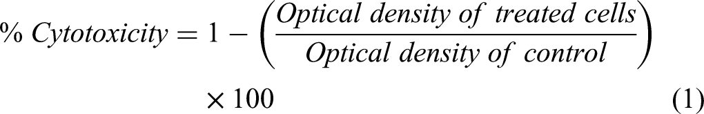
Following culturing, 10 μl of freshly prepared MTT reagent (Sigma-Aldrich, St. Louis, MO, USA) was added to each well and the plates were further incubated for 4 hours. With the help of a micropipette, there spective supernatants were aspirated followed by addition of 100 μl of dimethyl sulphoxide (DMSO) (Lot#A218101702, Loba Chemie) to solubilise the MTT crystals. The plates were then agitated and optical densities of each well were measured using an enzyme-linked immunosorbent assay (ELISA) scanning multiwell spectrophotometer (Multiskan Ex lab-systems) at 562 nm. The percentage inhibitions of cell proliferation (percentage cytotoxicity) was calculated using the following formula (Eq. 1) described by Fatemeh and Khosro.
29
Data Management and Statistical Analysis
The obtained data were first tabulated on Excel (Microsoft 365) spreadsheet and then exported to GraphPad prism version 9.2 for analysis. The data were subjected to descriptive statistics and the results were expressed as mean ± standard error of the mean (
Results
Anti-Inflammatory Activity of the Phytexponent in Swiss Albino Mice
The Phytexponent exerted significant (P < .05) anti-inflammatory effects in carrageenan-induced paw oedema mouse model in a time-dependent manner (Table 1). At a dose of 31.25 mg/Kg BW, the percentage inhibition of inflammation by the Phytexponent was 1.117 ± 0.193% at the first hour, while at the fourth hour it was 11.162 ± 0.091% (P < .05; Table 1). At a dose of 62.50 mg/Kg BW, the percentage inhibitions of inflammation ranged from 6.240 ± 0.242% at the first hour to 17.407 ± 0.186% at the fourth hour (P < .05; Table 1). The percentage inhibition of inflammation caused by the Phytexponent at a dose of 125 mg/Kg BW ranged from 9.645 ± 0.020% at the first hour to 31.795 ± 0.090% at the fourth hour (P < .05; Table 1). At a dose of 250 mg/Kg BW, the percentage inhibition of inflammation by the Phytexponent ranged from 14.000 ± 0.102% at the first hour to 37.931 ± 0.133% in the fourth hour (P < .05; Table 1). Notably, the Phytexponent formulation significantly inhibited inflammation in dose- and time-dependent manner (P < .05; Table 1).
Anti-Inflammatory Effects of the Phytexponent in Swiss Albino Mice.

Values are expressed as
Analgesic Activity of the Phytexponent
The findings revealed a positive dose-dependent significant increase in the percentage inhibition of acetic-induced writhing in mice (P < .05; Figure 1). Notably, at doses of 125 mg/Kg BW and 250 mg/Kg BW of the Phytexponent, the percentage inhibitions of acetic acid-induced writhing were significantly higher than the percentage inhibitions caused by indomethacin (reference drug) (P < .05; Figure 1). However, indomethacin exhibited a significantly higher inhibition of acetic acid-induced writhing in mice compared with the inhibitions caused by the Phytexponent at dose levels of 31.25 mg/Kg BW and 52.50 mg/Kg BW (P < .05; Figure 1).

Analgesic effects of the phytexponent in acetic acid-induced writhing in mice.
Bars with dissimilar letters are significantly different (One-Way ANOVA followed by Tukey's test; P < .05)
In Vitro Cytotoxic Effects of the Phytexponent
In this study, the results depicted a significantly positive dose-dependent increase percentage cytotoxicity of the Phytexponent on Vero cell line (normal cell line) (P < .05; Table 2). Similarly, the reference drug (cyclophosphamide) caused a dose-dependent increase in cytotoxicity to Vero cell in vitro (P < .05; Table 2). A comparison between the cytotoxic effects of the Phytexponent and cyclophosphamide revealed that, at all the tested concentrations, the cytotoxicity of cyclophosphamide was significantly higher than that of the Phytexponent in Vero cells (P < .05; Table 2). Furthermore, the median cytotoxic concentrations (CC50) were >1000 µg/ml (1137.83 µg/ml) for the Phytexponent and 2.48 µg/ml for cyclophosphamide (Table 2).
In Vitro Cytotoxic Effects of the Phytexponent on Vero Cell Line.

Values are expressed as
Discussion
Currently, the management of inflammation mostly utilises the non-steroidal anti-inflammatory drugs (NSAIDs) such as diclofenac, indomethacin, naproxen, ketoprofen, and ibuprofen, which inhibit the activity of- the cyclooxygenase-2 (COX-2) enzyme, thereby impeding the synthesis of prostaglandins likeprostaglandin E-2 (PGE2). 4 However, NSAID therapy causes dependence, is arguably unaffordable, inaccessible, and is often associated with adverse effects such as nephrotoxicity, cardiotoxicity, hepatotoxicity, intestinal bleeding, gastric ulcers, among other effects30–33. In light of this, we investigated the anti-inflammatory, analgesic, and cytotoxic effects of the Phytexponent of selected medicinal plants as a potential source of safer, efficacious, accessible, and affordable anti-inflammatory and analgesic molecules.
In this study, inflammation was induced in mice using carrageenan, a natural carbohydrate derived from edible red seaweed, widely used to screen plant extracts and molecules for anti-inflammatory efficacy. 25 Carrageenan induces a biphasic inflammatory response, whereby distinct modulators are produced. 34 In the early phase of carrageenan-inducedinflammation, cycloxygenase, histamine, and serotonin are produced, whereas in the late phase, which occurs after one hour, is characterised by PGE2 synthesis, mediated by bradykinin and leukotrienes.34,35 In this case, the early and late phases are characteristic of acute and chronic inflammation, respectively. 34 The upregulated synthesis of inflammatory mediators is due to the activation and enhanced activity of the inducible nitric oxide synthase (iNOS), COX-2 enzymes, pyrogenic cytokines, including the tumor necrosis factor alpha (TNF-α), interleukin-1 (IL-1), and interleukin (IL-6), among others.25,34,36 Therefore, for an agent to be considered as having anti-inflammatory activity, it ought to alter the consequences of carrageenan-induced inflammation, culminating in the amelioration of its typical features such as oedema, pyrexia, redness, algesia, and tissue dysfunction.36,37
Previous studies have established that a solution of 1% carrageenan (prepared in physiologic saline), when injected at a volume of 50-150 µl into the subplantar region, is sufficient to cause inflammation, which manifests in oedema.38,39 In this study, a subplantar injection of 100 µl of 1% carrageenan into the right hind paw of experimental mice effectively induced inflammation, as evidenced by well-pronounced swelling around the injected site. The negative inhibitions of oedema are indiciative of progressive increase in oedema size, due to the the inflammatory response to Carrageenan. The findings revealed a progressive increase in oedematous paw size of the negative control mice throughout the treatment period, which indicates a successful induction of inflammation. Conversely, the reference drug (indomethacin) and the Phytexponent effectively reduced oedema in a dose- and time-dependent manner in mice as depicted by the percentage inhibtions of paw oedema. Moreover, the Phytexponent successifully inhibited both the early and late phases of inflammation as depicted by the progresive increase in the percentage inhibitions of oedema. The time-dependent increase in percentage inhibition of oedema may be attributed to a higher bioavilability of the Phytexponent's active molecules, following metabolism and distribution to target sites. 40
Indomethacin is a non-steroidal anti-inflammatory drug that interferes with the synthesis of prostaglandins from arachidonic acid by inhibiting the cyclooxygenase (COX) enzyme. 41 The COX enzyme exists in two isoforms: COX-1 and COX-2, respectively. Scientific evidence shows that COX-1 mainly catalyses the synthesis of prostaglandins, which are essential for maintaining the health and proper functioning of the gastrointestinal tract, platelet activity, renal functioning, and other vital physiological functions in the body.41,42 On the other hand, COX-2 facilitates the synthesis of prostaglandins, which mediate pain, fever, and inflammation. 43 However, studies have demonstrated the existence, in some instances, of a crossover of the biological effects between COX-1 and COX-2 in the body. 44 Just like other NSAIDs, indomethacin nonselectively inhibits both COX-1 and COX-2 to confer anti-inflammatory activity.32,42,44 Even though the specific mode of action of the Phytexponent has not been established, the observations made herein partly suggest that its anti-inflammatory effects could be via the inhibition of the COX enzyme.
Pain is an unpleasant emotional and sensory experience resulting from tissue damage. 2 It acts as a warning signal to protect the body from actual or potential injury; however, it is associated with a disabling accompaniment of discomfort and adverse effects, characterising various medical conditions.45,46 As a result, pain forms a critical component of disease diagnosis, and its management is among the most important therapeutic priorities in medical practice.47,48 Various analgesic agents are used manage acute and chronic pain in patients.49,50 Currently, the most typical group of analgesic drugs used to manage pain comprises the NSAIDs, whose efficacy is based on the central and peripheral inhibition of prostaglandin synthesis. 51
Since NSAIDs interfere with prostaglandins’ normal synthesis and functioning, their side effects are predictable and include decreased homeostasis, renal dysfunction, hepatic dysfunction, peptic ulceration, intestinal bleeding, among others.30,31 Empirical evidence shows that over 20% of patients under long-term NSAID therapy develop duodenal and gastric ulcers with profound consequences.31,33 In light of these, the search for alternative, potent, safer, accessible, and affordable analgesics has attracted much attention in the realm of medical research.
The acetic acid-induced writhing is an experimental reflex model of visceral pain that has been extensively utilised to screen drugs and chemicals for analgesic efficacy in laboratory animals. 26 In the present study, 0.6% of acetic acid was intraperitoneally administered into experimental mice to induce pain by activating chemosensitive nociceptors, which manifests in writhing. 52 Writhing is described as the arching of the back, extension of limbs, and the abdominal musculature contraction. 53 In this experiment, the level of analgesia is indicated by the percentage reduction in abdominal writhing frequency.
This study showed a dose-dependent increase in the percentage inhibition of acetic acid-induced writhing by the Phytexponent in mice, indicating its potential analgesic efficacy. Similarly, indomethacin, the positive control drug, successfully inhibited the acetic acid-induced writhing in mice resulting in high percentage inhibitions. Moreover, the results showed that the Phytexponent at dose levels of 125 mg/Kg BW and 250 mg/Kg BW had significantly higher percentage inhibitions of writhing compared to indomethacin. These observations suggest that the Phytexpoent is more potent at these doses than indomethacin. Partly, this observation could be attributable to the various phytoactive principles present in the Phytexponent, 24 which may have acted at different sites in a multitarget fashion to thwart pain as opposed to a single target effect (inhibition of the COX enzyme) of indomethacin.
Preliminary studies have demonstrated that each medicinal plant, which comprises the Phytexponent, has anti-inflammatory and analgesic properties54–59. Additonaly, a recent study demonstrate significant in vitro anti-inflammatory and antioxidant efficacy of the Phytexponent. 24 Therefore, a combination of the analgesic- and anti-inflammatory-associated phytocompounds of individual plants in the Phytexponent may have synergistically conferred the bioactivities reported in the present study. 8 Moreover, studies have shown that chronic pain can be successfully managed by agents which modify the neurochemistry of the spinal cord dorsal horn, like anticonvulsants, local anaesthetic analogues, tricyclic antidepressants, γ-aminobutyric acid (GABA) agonists, and N-methyl-D-aspartate (NMDA) antagonists60–62. Opiates are useful in managing chronic pain; however, tolerance, dependence,and loss of efficacy limit their usefulness.63,64
To this end, only NMDA antagonists and epidural morphine have consistently demonstrated preemptive analgesic efficacies65–67. Therefore, to adequately manage pain and inflammation, a multifaceted approach using multitarget agents is the most viable strategy to alleviating pain in affected patients. 68 The results of this study, therefore, posit that the Phytexponent, by virtue of its analgesic and anti-inflammatory efficacy, could be a promising candidate for further development.
Even though the specific mode of action and the specific analgesic and anti-inflammatory bioactive molecules of the Phytexponent have not been elucidated, its is suggestive that it targets various pathways associated with immunity, inflammation, and pain. 69 Possibly, the phytocompounds present in this formulation could be maintaining the redox homeostasis, thereby preventing cell damage, and modulating immunity, modifying the inflammatory and pain transduction pathways, which together ensure the proper functioning of cellular molecules and avert cellular damage8,70–72.
Medicinal plants have longstanding usage in managing various diseases and play an integral role in meeting primary healthcare needs, especially in Sub-Saharan Africa. 73 Indeed the world health organisation estimates that over 80% of the global population depends on herbal medicine for their healthcare needs. 74 Despite the extensive utilisation of herbal products to manage various diseases, serious concerns regarding their efficacy and safety have been raised. 20
In the present study, the 3-(4,5-dimethylthiazol-2-yl)-2–5-diphenyltetrazolium bromide (MTT) colorimetric assay technique27,28 was employed to assess in vitro cytotoxicity and safety effects of the Phytexponent to assess its safety. This has was first described by Mosmann, 75 and has been extensively applied in the screening of anticancer potential of chemicals and plant extracts. The MTT assay measures the activity of mitochondrial enzymes, especially the succinate dehydrogenase (SDH), whose function is impaired by toxic agents leading to mitochondrial collapse and cell death. 28 During the assay, the mitochondrial nicotinamide adenine dinucleotide (NAD) reduced the MTT to a purple formazan product, which is determined calorimetrically at a specific wavelength (520 nm). The amount of formazan produced is directly proportional to the number of cells in a particular cell line. 28 This technique was selected due to its high reproducibility, safety, sensitivity, and robustness in determining cell viability and cytotoxicity. 76
According to the National Cancer Institute (NCI) criteria, plant extracts with CC50< 30 µg/ml are considered to be cytotoxic after 48-72-hour exposure to cells. 77 The observation from this study revealed that the reference drug (cyclophosphamide) was a potent cytotoxic agent by its low CC50 value (CC50 = 2.48 µg/ml) as consistently demonstrated in other studies.78,79 On the other hand, the Phytexponent demonstrated low cytotoxic effects, as witnessed by its high CC50 (CC50>1000 µg/ml). These results indicate that this polyherbal formulation might be safe and may be used to treat pain and inflammation without eliciting cytotoxic effects. However, extensive toxicity studies should be conducted to establish their safety profile.
In current medical practice, pharmaceutical drugs are designed to confer specific biological effects that are accompanied by specific side effects.41,80 However, medicinal plants demonstrate a broad spectrum of bioactivities; thus, there are no defined toxic profiles. 81 This is attributable to the enormous phytoconstituents that act synergistically to affect various physiological functions in a non-specific manner71,82–85. If a medicinal plant contains toxic compounds, the toxic effects elicited could be fatal; therefore, it is critical to validate medicinal plants’ safety to avert potential fatalities. This study's findings demonstrate that the Phytexponent is non-toxic to Vero cell-lines-normal cells and is a potential source of safe analgesic and anti-inflammatory agents.
Conclusions and Recommedations
Our study findings showed that the Phytexponent possess in vivo anti-inflammatory and remarkabale analgesic efficacy in experimental mice, and is non-toxic to Vero E6 cell line. Therefore, the Phytexponent is a promising source of efficacious anti-inflammatory and analgesic compounds, against various maladies, especially those it is used to manage in complementary and alternative medicine. Further studies aimed at establishing the specific mechanism(s) through which the Phytexponent confers the anti-inflammatory and analgesic effects should be done. Extensive toxicity and safety evaluation of this polyherbal product should be performed to give way for its further development.
Supplemental Material
sj-txt-1-jebim-10.1177_2515690X221082986 - Supplemental material for Anti-inflammatory, Analgesic, and Cytotoxic Effects of The Phytexponent: A Polyherbal Formulation
Supplemental material, sj-txt-1-jebim-10.1177_2515690X221082986 for Anti-inflammatory, Analgesic, and Cytotoxic Effects of The Phytexponent: A Polyherbal Formulation by Halvince O. Odira, Simon O. Mitema, Isaac M. Mapenay and Gervason A. Moriasi in Journal of Evidence-Based Integrative Medicine
Footnotes
Acknowledgments
The authors acknowledge the Laboratory staff of the Department of Public health, Pharmacology and Toxicology of the University of Nairobi, and the Kenya Medical Research Institute for their technical support. We also thank Prof. Epaphrodite Twahirwa of the Department of Pharmaceutics and Pharmaceutical Chemistry of Mount Kenya University for his mentorship.
Authors’ Contributions
Halvince Odira, performed the experiments, and drafted the manuscript under the supervision of Simon Mitema and Isaac Mapenay. Gervason Moriasi designed the experiments, donated research materials, performed data analysis, and interpreted the results. All authors reviewed the draft manuscript and approved the final version for publication.
Data Availability
All data is included in the manuscript, and any additional information is available from the authors upon request.
Declaration of Conflicting Interests
The author(s) declared no potential conflicts of interest with respect to the research, authorship, and/or publication of this article.
Ethical Approval
The experimental mice were used and disposed of as per the guidelines set out by the Faculty of veterinery Medicine (FVM) of the University of Nairobi biosafety, animal use, and ethics committee (BAUEC) (FVM BAUEC/2020/265). The cell line (Vero E6) was used and disposed of according to the protocols set out by the Scientific Ethical Review Unit (SERU) of the Kenya Medical Research Institute (KEMRI/RES/7/5/2). Permission to conduct this study was also obtained from the Kenya National commission for science, technology, and innovation (NACOSTI) (BAHAMAS ABS/P/20/7744).
Funding
The author(s) received no financial support for the research, authorship, and/or publication of this article.
Supplemental Material
Supplemental material for this article is available online.
References
Supplementary Material
Please find the following supplemental material available below.
For Open Access articles published under a Creative Commons License, all supplemental material carries the same license as the article it is associated with.
For non-Open Access articles published, all supplemental material carries a non-exclusive license, and permission requests for re-use of supplemental material or any part of supplemental material shall be sent directly to the copyright owner as specified in the copyright notice associated with the article.