
Abstract
Wound healing involves the interaction of blood cells, proteins, proteases, growth factors, and extracellular matrix components. Inflammation is one of the first events occurring during this process. Previously, we showed that the N-Methyl-(2S,4R)-trans-4-Hydroxy-L-Proline (NMP) from Sideroxylon obtusifolium leaves (a Brazilian medicinal species) presents an anti-inflammatory action. Considering inflammation as an important event in the wound healing process, the objectives were to investigate the topical effects of the NMP gel on a mice wound-induced model. Male Swiss mice were divided into 4 groups: Sham (surgical procedure only), Control (gel-base treated), and 3% or 10% NMP gel-treated groups. Measurements of wound areas and microscopic analyses (HE [hematoxylin-eosin] and PSR [picrosirius red] stainings) were carried out, at the 7th and 12th, days after the wound induction. Furthermore, immunohistochemical assays for iNOS (inducible nitric oxide synthase) and COX-2 (cyclooxygenase-2) and biochemical measurements for TBARS (thiobarbituric acid reactive substances), GSH (glutathione), and myeloperoxidase (MPO) were also performed, at the second day after the wound induction. The work showed that NMP decreases the wound areas, after topical application, relatively to the Sham and Control groups. In addition, microscopic alterations were reduced and collagen deposition was increased, at the 7th and 12th days, in the 10% NMP group. While iNOS and COX-2 immunostainings and GSH contents increased, in relation to the Sham and Control groups, TBARS and MPO decreased. Altogether, the results showed NMP to improve the wound healing process, by upregulating iNOS and COX-2 activities, reducing lipid peroxidation and MPO activity, and increasing GSH contents. In addition, NMP certainly contributes to the increased collagen deposition. These data may stimulate translational studies dealing with the possible use of NMP from Sideroxylon obtusifolium or from other sources for the management of wound healing.
Introduction
The process of wound healing consists of hemostasis, inflammation, proliferation, and remodeling. It involves several cell types, such as neutrophils, macrophages, lymphocytes, keratinocytes, fibroblasts, and endothelial cells. Multiple factors may cause impaired wound healing, by affecting one or more phases of the process. 1 Furthermore, wound healing is a clinical problem, requiring the development of new approaches for acute and chronic wound management. 2 In addition, its phases are highly organized and tightly regulated, depending on a balance between several cell types and mediators.
Although inflammation is a prerequisite to healing, chronic wounds exhibit a prolonged inflammatory response leading to bacterial infiltration and proliferation. 3 Studies in search for substances with cicatrizing activity are stimulated, due to the high costs for the chronic wounds treatment, so that the substances derived from natural sources are receiving great prominence. Many compounds obtained from natural products with antioxidant, anti-inflammatory, and antimicrobial properties have shown considerable importance in wound management. 4
The use of plants in the treatment of inflammatory skin diseases results from their influence on different stages of inflammation. Thus, they inhibit formation of cytokines and eicosanoids, preventing the inflammatory reaction cascade. 5 Several genus of medicinal plants, including the Sideroxylon genus and the species S inerme L, are used for this purpose in some South Africa tribes and elsewhere, for hyperpigmentation problems. 6
Sideroxylon obtusifolium (Roem. & Schult.), known in Brazil as “quixabeira,” is a native tree in Central and South America. The leaves and barks are generally used in the form of teas in popular therapy. 7 Chemical analyses performed with S obtusifolium identified several compounds, such as flavonoids, monoterpenes and sesquiterpenes, condensed proanthocyanidins and leucoanthocyanidins, triterpenes, steroids, and sugars. 8,9
Recently, we demonstrated the presence of an L-proline derivative, N-methyl-(2S,4R)-trans-4-hydroxy-l-proline (NMP), in the leaves of S. obtusifolium, as well as its potent anti-inflammatory activity. 10 Considering the inflammatory phase as an important event during the wound healing process, in the present work we investigate the effect of this bioactive compound on an experimental model of wound healing in rats, by means of histological, biochemical, and immunohistochemical analyses.
Materials and Methods
Plant Material and Isolation of N-Methyl-(2S,4R)-trans-4-Hydroxy-L-Proline
Leaves from the S obtusifolium species were collected at the municipality of Mauriti, Ceará State, in August 2014. A voucher specimen (Number 10 648) has been stored at the “Herbário Caririense Dárdano de Andrade Lima,” Regional University of Cariri (URCA), Crato, Ceará, Brazil. The bioactive compound, N-methyl-(2S,4R)-trans-4-hydroxy-L-proline (NMP), was obtained according to the procedure previously described. 10
Gel Preparation
The gel base was prepared at the Center for Cosmetics and Pharmaceutical Studies of the Federal University of Ceará. For that, the physicochemical characteristics of the bioactive compound (L-proline derivative [NMP]) and skin lesions (excisional wound) were taken into consideration. The compound was carried out with cold neutralization and under agitation in an aqueous solution, containing 1% carboxyvinyl polymer, 5% glycerin, and 1% polypropylene glycol. Methylparaben (0.15%) and propilparaben (0.05%) were used as preservants. Aminomethyl propanol (q.s.p. 100 g), pH 6.5, was used as the neutralizing agent.
Animals
Male Swiss mice (25-30 g) were provided by the Animal House of the Federal University of Ceará, Brazil. The animals were housed in plastic cages with sawdust, as beddings, and kept in a room with controlled temperature (25 ± 2°C), under a 12 hour light/12 hour dark cycle and given food and water ad libitum. The experiments were carried out according to the Guide for the Care and Use of Laboratory Animals (US Department of Health and Human Services). The project was previously approved by the Animal’s Ethics Committee of the Faculty of Medicine of the Federal University of Ceará, Approval Number 21/2017.
Wound-Induced Model
The mice were anesthetized by intraperitoneal injections of 10% ketamine hydrochloride (115 mg/kg) and 2% xylazine hydrochloride (10 mg/kg) before their surgical procedure. After shaving the dorsal surface skin, the region was prepared for aseptic surgery using 1% iodopovidone, followed by 70% ethanol. Two circular excisional wounds with 8 mm diameter were induced, with a surgical punch, on the dorsal surface of each animal. The excisional wounds were not sutured, so that the healing occurred by secondary intention. After the surgery, the animals were injected with dipyrone from Mendley, SP, Brazil (50 mg/kg, ip), for analgesic purposes. They were randomly divided into 4 groups, as follows: animals subjected only to surgical procedures (Sham), animals treated with gel base (Control), and animals treated with 3% and 10% NMP (prepared in gel base).
Macroscopic Analyses (Wound Area Measurements)
Macroscopic differences in neoformed tissue were monitored at the 7th and 12th days after surgery. 11 For that, areas of excisional wounds in mm2 were calculated with a pachymeter, as follows: A = Π × R × r, where A is the area and R and r are large and small rays, respectively. 12 Reepithelization was noted whenever a scab was not covering the wounds. 13 After the macroscopic study, 4 mm punch biopsies from neoformed tissue and excisional wounds with adjacent normal skin were removed (n = 5/group) for histological study. Then, the animals were sacrificed with an anesthetic overdose (ketamine, 350 mg/kg, ip).
Histopathological Analyses: Hematoxylin-Eosin (HE) and Picrosirius Red (PSR) Stainings
Neoformed tissues of incisional and excisional wounds were fixed in formaldehyde (10%, v/v) and resuspended in 0.01 M PBS (phosphate-buffered saline), pH 7.2, over 24 hours, for histological processing. Sections (5 μm) were stained with HE. The PSR staining was used for the examination of the collagen fiber deposition. PSR is one of the most important stains for studying collagen networks in tissues. 14 Photomicrographs of sections were obtained, 7 and 12 days after surgery, under a polarizing microscope. Images of connective tissue were captured and collagen fiber deposition was quantified in images obtained from 6 random fields. Collagen fibers were analyzed in nonpolarized images, according to their color, by the RGB (red, green, blue) system. The total percentage of collagen fibers was quantified by the ImageJ 1.46 software (National Institutes of Health, Bethesda, MD) in 3 different sections for each group. 11
Immunohistochemical Assays for Inducible Nitric Oxide Synthase (iNOS) and Cyclooxygenase-2 (COX-2)
For these assays, the streptavidin-biotin-peroxidase method was used. 15 The sections of excisional wounds were then deparaffinized, hydrated in xylol and ethanol, and immersed in 0.1 M citrate buffer (pH 6) under 18 minutes of microwave heating for antigen recovery. After cooling at room temperature for 20 minutes, the sections were washed with a phosphate-buffered solution, followed by a 15-minute blockade of endogenous peroxidase, with a 3% H2O2 solution. The 5 μm sections were incubated overnight (4°C) with the primary antibodies (anti-iNOS, anti-COX-2; Sigma-Aldrich, MO, and Santa Cruz Biotechnology, CA, respectively), and diluted in PBS, according the manufacturers’ instructions. On the next day, the sections were washed in PBS and incubated for 30 minutes with the secondary biotinylated rabbit antibody (anti-IgG), also diluted in PBS (1:200 dilution). After washing in PBS, the sections were incubated for 30 minutes with the conjugated streptavidin-peroxidase complex (ABC Vectastain complex, Vector Laboratories, Burlingame, CA). After another washing with PBS, the sections were stained with 3,3-diaminobenzidine-peroxide (DAB) cromophore, counterstained with Mayer hematoxylin, dehydrated and mounted in microscope slides for analyses, and the data were semiquantified (as relative optic densities) with the Image J software.
Determination of Lipid Peroxidation by Thiobarbituric Acid Reactive Substances (TBARS)
Lipid peroxidation expresses oxidative stress induced by ROS reactivity. A widely used method for measuring it is the determination of malondialdehyde (MDA) in biological samples. Although the lipid peroxidation products are MDA and 4-hydroxy-2-nonenal (4-HNE), MDA is a good biomarker of oxidative stress and an end product of lipid peroxidation. Wounds at the second day after surgery were used. For that, homogenates (10%) in 1.15% KCl were added (250 μL) to 1 mL 10% TCA, followed by the addition of 1 mL 0.6% thiobarbituric acid. After agitation, this mixture was maintained in a water bath (95°C to 100°C) for 15 minutes. Then, the mixture was cooled on ice and centrifuged (1500 × g/5 min). The TBARS content was determined in a plate reader, at 540 nm, with results expressed in μmol MDA/g tissue. A standard curve with several MDA concentrations was also performed. 16
Determination of the Concentration of Reduced Glutathione (GSH)
Determination of the GSH concentration was performed on the ELISA plate. 17 It is based on the reaction of the Ellman reagent, 5,5′-dithiobios (2-nitrobenzoic acid, DTNB), with the free thiol, giving a mixed disulfide plus 2-nitro-5-thiobenzoic acid. The sample preparation was done as follows: 40 μL of each sample (a 10% wound homogenate in phosphate buffer, at the second day) was added to an Eppendorf tube with 50 μL distilled water and 10 μL trichloroacetic acid (50%). The samples were centrifuged at 3000 rpm for 15 minutes at 4°C. The supernatants (60 μL) were added to the ELISA plate (kept cooled), as were the blanks. The mixture remained cooled throughout the test. Immediately before the readings at 412 nm, 102 μL of the Tris-HCl buffer and 0.65 mL of 0.01 M DTNB in methanol were added to each well. The concentration of GSH was expressed as μg/g tissue based on a standard curve of GSH.
Myeloperoxidase (MPO) Activity Measurement
The tissues collected from the excised wounds, at the second day after induction, were incubated in 0.5% HTAB (hexadecyltrimethylammonium bromide) solution, at the concentration of 50 mg tissue per mL, homogenized, and centrifuged (1500 × g/15 min, at 4°C). The supernatant obtained was transferred to Eppendorf tubes and subjected to heat shock in the cell pellet, in 3 freeze-thaw stages (−20°C, 10 minutes each). The supernatant was centrifuged again (1500 × g/15 min, at 4°C) for better removal of contaminants. The samples were then plated (duplicates of 7 μL in 96-well plates) and 200 μL of the reading solution (5 mg O-dianisidine, 15 μL H2O2 1%, 3 mL phosphate buffer, 27 mL H2O) was added. Readings were taken at 460 nm (t 0 = 0 minutes and t 1 = 1 minute) with an ELISA microplate reader. 18 The change in absorbance was plotted on a standard curve of neutrophils and the results were expressed as neutrophils/mg tissue and interpreted as MPO activity.
Statistical Analyses
All results are expressed as mean values of groups ± SEM. Statistical significance was assessed by ANOVA, followed by the Tukey test for multiple comparisons or Kruskal–Wallis test for medians. The level of significance was determined as P < .05, using the Graph Pad Prism, version 6.0.
Results
Macroscopic Analyses
The measurements of the wound area in mm2 were performed at the 7th and 12th days after surgery, in all 4 groups tested. The results of the 7th day showed similar values (around 45% decreases) in wound areas, after the treatments with 3% and 10% NMP, compared with the Sham group, F(3, 47) = 11.95, P < .0001. But no statistically significant difference was observed between the Sham and Control groups (Figure 1A). At the 12th day, reductions around 58% were demonstrated in wound areas, after treatments with 3% and 10% NMP, relatively to the Sham group, F(3, 46) = 8.701, P < .0001. Similarly to the 7th day data, no significant differences were observed between the Sham and Control groups (Figure 1B).

Wound areas measurements at the 7th and 12th days after induction. Seventh day: (a) versus Sham, q = 7.215, P < .001; (b) vs Control, q = 4.673, P < .01; (c) versus Sham, q = 6.443, P < .001; (d) versus Control, q = 3.909, P < .05. Twelfth day: (a) versus Sham, q = 5.791, P < .01; (b) versus Control, q = 3.929, P < .05; (c) versus Sham, q = 5.745, P < .01; (d) versus Control, q = 3.883, P < .05 (One-way ANOVA and Tukey as the post hoc test).
Histopathologic Analyses, HE Staining
The Sham group, at the 7th day, showed a proliferation of the extracellular matrix and also of the conjunctive, inflammatory, and adipose cells. At the 12th day, it presented necrosis, hemorrhage, edema (in the dermis and between inflammatory cells), as well as cellular proliferation in the extracellular matrix. The Control group, at the 7th day, presented epidermis disruption and extracellular matrix proliferation, as well as proliferation of conjunctive cells. In addition, it showed the presence of inflammatory cells and necrosis. At the 12th day, intense inflammatory cell infiltrates and deposits on the extracellular matrix were seen, as well as proliferation of extracellular matrix and fibroblasts, hemorrhage, and interstitial edema. The 10% NMP group showed an intense extracellular matrix and conjunctive cells proliferation at the 7th day. In addition, this group presented a normal epidermis and normal papillary dermis. At the 12th day, a proliferation of hair follicles on the reticular dermis and intense extracellular matrix proliferation, as well as proliferation of fibroblasts, collagen fibers, and conjunctive cells, were demonstrated. In addition, an intense proliferation of inflammatory cells, next to hair follicles and to extracellular matrix, and also a discontinuous epidermis were also observed (Figure 2).

Representative photomicrographs (HE staining, ×400) of the Sham, Control, and 10% NMP-treated groups, at the 7th and 12th days after the wound induction, showing the main macroscopic changes. The arrows indicate inflammatory infiltrate. Seventh day: Sham and Control groups show proliferation of the extracellular matrix and of conjunctive and inflammatory cells, while the 10% NMP-treated group showed intense proliferation of extracellular matrix and conjunctive cells, and also presented normal epidermis and normal papillary dermis. Twelfth day: The Sham group presented necrosis, hemorrhage, and edema in the dermis and between inflammatory cells, as well as extracellular matrix proliferation and cellular proliferation. The Control group showed intense inflammatory cells infiltrate and deposit on the extracellular matrix, as well as proliferation of extracellular matrix and fibroblasts, hemorrhage, and interstitial edema. The 10% NMP-treated group showed proliferation of hair follicles on the reticular dermis and intense extracellular matrix proliferation, as well as proliferation of fibroblasts, collagen fibers, and conjunctive cells. It also presented intense proliferation of inflammatory cells next to hair follicles and to extracellular matrix and presence of a discontinuous epidermis.
Histopathologic Analyses, PSR Staining
Figure 3A shows representative photomicrographs of the 4 groups tested, in nonpolarized (left column) and polarized lights (right columns), at the 7th and 12th days. At the 7th day, an almost 2-fold increase in the percentages of collagen fibers was observed, in the 3% and 10% NMP-treated groups, compared with the Sham group, F(3, 17) = 21.98, P < .0001). Lower values (ranging from 1.6- to 1.9-times increases) were attained, by comparisons with the Control group. At the 12th day, an average of 1.4-time increase was observed in both NMP-treated groups, in relation to either the Sham and Control groups, F(3, 17) = 7.968, P = .0016; Figure 3B).

Representative nonpolarized (left) and polarized (right) photomicrographs (PSR staining) of the Sham, Control, and 10% NMP-treated groups, at the 7th and 12th days after the wound induction, showing the percentage of increase in the number of collagen fibers after NMP treatments. The nonpolarized photomicrographs were used for measuring the collagen fibers by the RGB Measure Plus (Image J software). Seventh day: (a) versus Sham, q = 7.174, P < .001; (b) versus Control, q = 6.101, P < .001; (c) versus Sham, q = 9.638, P < .001; (d) versus Control, q = 8.518, P < .001. Twelfth day: (a) versus Sham, q = 4.134, P < .05; (b) versus Control, q = 3.908, P < .05; (c) versus Sham, q = 5.646, P < .01; (d) versus Control, q = 5.429, P < .01 (One-way ANOVA and Tukey as the post hoc test).
Immunohistochemistry for iNOS and COX-2
These assays were only performed with the Sham, Control, and 10% NMP groups, at the 2nd day after wound induction. The results showed a 53% increase in iNOS immunostaining, in the 10% NMP group, compared with the Sham group, while a 26% increase was detected in the 10% NMP group compared with the Control group, F(2, 11) = 19,26, P = .0003. No significant differences were detected between the Sham and Control groups (Figure 4). Similar increases (around 26%) were observed in COX-2 immunostaining of the 10% NMP group, related to the Sham and Control groups, F(2, 11) = 22.06, P < .0001. No significant differences were observed between the Sham and Control groups (Figure 5).
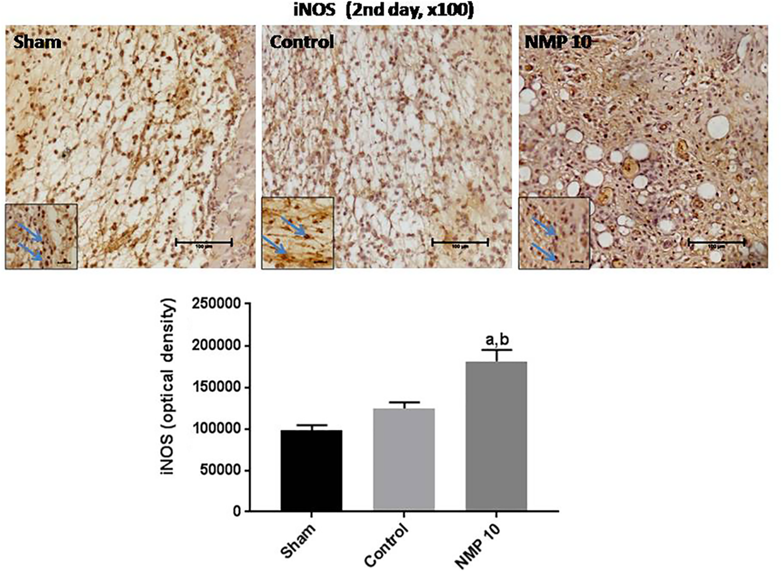
Representative photomicrographs (upper panels, ×100, scale = 100 μm), showing that the NMP-treated groups increased iNOS immunostainings, compared with the Sham and Control groups. The lower panels present the immunostaining quantification by the Image J software. Inserts present photomicrographs with higher magnification (×400). The blue arrows show PMN cells. (a) versus Sham, q = 8.736, P < .0001; (b) versus Control, q = 4.871, P < .0001 (One-way ANOVA and Tukey as the post hoc test).

Representative photomicrographs (upper panels, ×100, scale = 100 μm), showing that the NMP-treated groups increased both COX-2 immunostainings, compared to the Sham and Control groups. The lower panels present the immunostaining quantification by the Image J software. Inserts present photomicrographs with higher magnification (×400). The blue arrows show PMN cells. (a) versus Sham, q = 8.479, P < .001; (b) versus Control, q = 7.563, P < .01 (One-way ANOVA and Tukey as the post hoc test).
Lipid Peroxidation (TBARS) Determined in the Second Day of Wound Induction
The lipoperoxidation values, at the 2nd day of wound induction, were reduced in both 3% and 10% NMP-treated groups, in comparison with the Sham group, showing 32% and 44% decreases, respectively. A similar lipoperoxidation decrease (41%) was noticed in the 10% NMP-treated group, related with the Control group, F(3, 25) = 8.95, P < .0003 (Figure 6).

Lipid peroxidation measurements (as thiobarbituric acid reactive substances, TBARS), at the second day after the wound induction, in the Sham, Control, and NMP-treated groups. (a) versus Sham, q = 4.360, P < .05; (b) versus Sham, q = 6.212, P < .001; (c) versus Control, q = 5.577, P < .01 (One-way ANOVA and Tukey as the post hoc test).
Determination of Reduced Glutathione (GSH) in the Second Day After Wound Induction
The 10% NMP-treated group presented 2.8- and 2.3-times increases in GSH contents compared with the Sham and Control groups, respectively. No significant increase was seen in the 3% NMP-treated group, F(3, 40) = 7.017, P = .0007 (Figure 7).

The NMP-treated groups increased glutathione (GSH), at the second day after the wound induction, relatively to the Sham and Control groups. (a) versus Sham, q = 6.481, P < .01; (b) versus Control, q = 5.511, P < .01 (one-way ANOVA and Tukey as the post hoc test).
Inhibition of MPO Activity, Determined at the Second Day After Wound Induction
The 10% NMP-treated group showed 57% and 52% reductions in MPO activity, compared with the Sham and Control groups, respectively. No significant decreases were demonstrated in the 3% NMP group, in relation to either the Sham or Control groups, F(3, 21) = 12.07, P < .0001 (Figure 8).

The 10% NMP-treated groups decreased the myeloperoxidase (MPO) activity, at the second day after the wound induction, relatively to the Sham, Control, and 3% NMP-treated groups. (a) versus Sham, q = 8.219, P < .001; (b) versus Control, q = 6.114, P < .01; (c) versus 3% NMP, q = 4.400, P < .05 (One-way ANOVA and Tukey as the post hoc test).
Discussion
Wounds are physical injuries, leading to skin disruption whose repair requires a sequence of events, including inflammation, proliferation, and migration of different cell types. The inflammation occurs immediately after the injury, with vasoconstriction which favors homeostasis and releases inflammatory mediators. 19 In the present study and for the first time, we showed that the L-proline derivative (NMP) isolated from Sideroxylon obtusifolium leaves presents wound healing properties. This medicinal species has been object of study in our laboratory, and we recently 10 showed that NMP presents a potent anti-inflammatory action.
Our results demonstrated, after the topical application of a 10% NMP gel, an intense extracellular matrix proliferation, as well as proliferation of conjunctive cells, fibroblasts, and collagen fibers. These microscopic features point to an improvement in the wound healing process. Furthermore, the increased number of collagen fibers in the NMP group was confirmed by PSR staining, performed at both the 7th and 12th days after the wound induction. Collagen is the major insoluble fibrous protein in the extracellular matrix and in connective tissue of mammals. For a successful wound healing process, the synthesis of significant amounts of collagen is required. Furthermore, proline and hydroxyproline together with glycine are major precursors for collagen synthesis. 20,21 In addition, evidence indicates that the topical administration of L-proline accelerates wound healing. 22 These data demonstrate that the benefits of NMP in the wound healing process, as shown by us in the present study, are due to the content of the drug L-proline.
In addition, we also showed that the immunostaining for iNOS, the inducible isoform of nitric oxide synthase, was significantly increased in the second day after the wound induction in the NMP-treated group. iNOS is produced by inflammatory cells in the early phase of wound healing, and the NO released regulates collagen formation, among other steps during the wound healing process. 23 Furthermore, evidence indicated that iNOS deficiency causes significant impairment in wound healing–related properties of fibroblasts, suggesting that NO plays an important role in wound healing. 24 iNOS upregulation, following tissue injury, results in increased NO production, which is beneficial to normal healing 25 and may be attributed to the NO influence on angiogenesis, inflammation, cell proliferation, matrix deposition, and remodeling. 26 Interestingly, the amino acid arginine is of fundamental importance in wound healing, as its metabolites NO, proline, and polyamines that affect all phases of the wound healing process. 27
Similarly, significant increases were demonstrated in COX-2 immunostaining in the NMP-treated group, compared with the Sham and the Control groups. Nonsteroidal anti-inflammatory drugs inhibit the production of PGE2, an inflammatory mediating prostaglandin. These nonsteroidal anti-inflammatory drugs have an antiproliferative effect and delay the healing rate. Data from western blotting analyses demonstrated induction of COX-2 protein, peaking 3 days after skin injury. 28 These authors observed that the administration of a COX-2 inhibitor delayed reepithelization in the early phase of wound healing, making this enzyme an important factor in cutaneous wound healing.
Others, 29 however, showed that the administration of Celecoxib (a selective COX-2 inhibitor) improves the wound healing process, through decreased expression of iNOS and COX-2. Interestingly, inflammatory cells (among others) produce NO, partly in response to inflammatory cytokines released in the skin injury. NO mediates angiogenesis and inflammation, and thus, inhibition of iNOS impairs wound healing. 30 Furthermore, iNOS is highly active in the inflammatory phase, affecting COX-2 and the release of inflammatory mediators. 31
We also showed decreases in lipid peroxidation values in both NMP-treated groups. Evidences indicate that the alteration in the antioxidant profile and elevated levels of MDA, a marker of free radical damage or lipid peroxidation, may be attributed to impaired wound healing. 32 Furthermore, any compound that inhibits lipid peroxidation will increase the strength of collagen fibers and hasten the process of wound healing. 33 Evidence indicates that lipid peroxidation inhibition improves angiogenesis and may be an effective therapeutic approach in wound healing. 34 Interestingly, the amino acid proline was shown to be a potent scavenger of ROS 35 and to play major roles during stress. 36 However, in the present study NMP did show only a moderate antioxidant effect, as observed by the SOD assay (data not shown).
Furthermore, glutathione is considered to be the most important redox regulator that controls inflammatory processes. 37 Thus, depletion in glutathione levels has a role in delayed healing. We observed increased levels of GSH in the NMP-treated group, relatively to the Sham and Control groups, suggesting that this compound improves the wound healing process, at least in part, by increasing the antioxidant capacity. In addition, MPO release was significantly reduced after treatment with NMP. MPO is a heme protein released by leukocytes, mainly neutrophils, which play a crucial role in inflammation and oxidative stress in the cellular level. 38 Neutrophils are among the first immune cells recruited to a wound and produce cytokines and growth factors, among other mediators that activate several types of cells, such as inflammatory cells, keratinocytes, endothelial cells, and fibroblasts. 39
NMP, the drug studied in the present work, is a proline-derivative molecule. Not only proline but also hydroxyproline and mainly glycine are known to contribute to the majority of the total amino acids content present in collagen. 40 Thus, NMP could certainly play an important role in the wound healing property of S obtusifolium. More importantly, collagen is the most abundant insoluble fibrous protein in the animal kingdom, whose fibrils and networks comprise the majority of the extracellular matrix. 41 Collagen plays a crucial role in wound healing, and its biosynthesis includes unique biochemical processes, such as hydroxylation of proline to hydroxyproline. Furthermore, proline plays important roles in protein synthesis and structure, metabolism, and nutrition, as well as wound healing, antioxidative reactions, and immune responses. Proline and its metabolite (hydroxyproline) are unique amino acids and constitute one third of amino acids present in the collagen proteins, which comprise approximately 30% of body proteins. 42 –44
In conclusion, also present in the NMP molecule, proline is an important chemical component of the collagen structure and is also known to scavenger free radicals. 43 This antioxidant property of proline may explain why its concentration increases markedly, in response to the cellular oxidative stress. 45 In addition, exogenous application of proline resulted in a reduction of protein carbonylation, an increase in glutathione redox state, and the activity of glutathione peroxidase, glutathione-S-transferase, and glyoxalase. 46 All these data together point to the potential of proline and of proline-rich species, such as S obtusifolium, on wound healing processes, what stimulates translational studies focusing on this purpose.
Footnotes
Acknowledgments
The authors thank Prof M. O. L. Viana for the orthographic correction of the article.
Author Contributions
PEAA was responsible for the in vivo experiments and some of the biochemical determinations; TFGS helped with some of the in vivo experiments; FAS supervised some of the ex vivo experiments; AFSCV was responsible for the histological method (picrosirius red staining); BOL was responsible for the cream preparation used for the NMP formulation; LKAML supervised the cream preparation; TMR helped with the biochemical determination (ex vivo experiments); JSAME supervised and provided the final analyses of the histological experiments; NCA was responsible for the chemical characterization of the L-proline derivative (NMP); MNNA supervised some of the ex vivo experiments; ERS supervised the isolation and chemical characterization of the L-proline derivative (NMP); GSBV coordinated the work and was also responsible for the statistical analyses and for writhing the manuscript draft.
Declaration of Conflicting Interests
The authors declared no potential conflicts of interest with respect to the research, authorship, and/or publication of this article.
Funding
The authors disclosed receipt of the following financial support for the research, authorship, and/or publication of this article: The authors are grateful to the financial support from the Brazilian National Research Council (CNPq), Coordination for the Improvement of Higher Education Personnel (CAPES), and the Foundation for Research and Technological Development of the State of Ceará (FUNCAP).
Ethical Approval
The study follows international and institutional guidelines for the treatment of animals and complies with relevant legislation. The study did not involve unnecessary pain, distress, suffering, or lasting harm to animals. The study was submitted and approved by the Ethics Committee on Animal Research of the Federal University of Ceará (CEUA/UFC), Approval Number 59/17.