
Abstract
Saccharomyces cerevisiae Vac8 is a vacuolar membrane protein, which functions in vacuole inheritance and fusion, nucleus-vacuole junctions, autophagy and the cytoplasm-to-vacuole-targeting pathway. Here, we analyzed Vac8 of the yeast Hansenula polymorpha. We show that HpVac8 localizes to the vacuolar membrane and concentrates in patches at nucleus-vacuole junctions. Analysis of a VAC8 deletion strain indicated that HpVac8 is required for vacuole inheritance and the formation of nuclear-vacuole junctions, but not for vacuole fusion. Previously, organelle proteomics resulted in the identification of Vac8 in peroxisomal fractions isolated from H. polymorpha and S. cerevisiae. However, deletion of H. polymorpha VAC8 had no effect on peroxisome biogenesis or peroxisome-vacuole contact sites.
Saccharomyces cerevisiae Vac8 is a vacuolar membrane protein, which plays a role in vacuole inheritance (Tang et al., 2003) and fusion (Pan and Goldfarb, 1998), in the cytoplasm-to-vacuole pathway (Wang et al., 1998; Scott et al., 2000) and in autophagy by regulating the phagophore assembly site formation and fusion with the vacuole (Hollenstein et al., 2019; Gatica et al., 2020). In Pichia pastoris Vac8 was shown to be involved in pexophagy (Fry et al., 2006; Sakai et al., 2006). S. cerevisiae Vac8 is also important for the formation of nucleus-vacuole junctions (NVJs) (Pan et al., 2000), sites where piecemeal microautophagy of the nucleus (PMN) takes place (Roberts et al., 2003). In addition, NVJs may play a role in lipid transfer, because they contain several lipid binding proteins, such as Osh1, a member of the oxysterol-binding protein family (Loewen et al., 2003) and Mdm1, a protein that contains a PX domain, which binds phosphatidylinositol-3-phosphate (Henne et al., 2015).
Proteomic studies revealed the presence of Vac8 in peroxisomal fractions isolated from S. cerevisiae (Marelli et al., 2004) and Hansenula polymorpha (Singh, 2019). Because physical contacts exist between H. polymorpha peroxisomes and vacuoles, we reasoned that HpVac8 may play a role in these contacts as well (Wu et al., 2019). Moreover, a high-throughput fluorescence microscopy screen revealed localization of the NVJ protein Nvj2, an integral ER protein, at pre-peroxisomal vesicles in S. cerevisiae pex3, suggesting a role of Nvj2 in peroxisome biology as well (Wróblewska, 2020).
These observations prompted us to further analyze NVJ proteins in the yeast H. polymorpha. Our data show that H. polymorpha Vac8 localizes to the vacuolar membrane and is required for NVJ formation and vacuole inheritance. Unexpectedly, HpVac8 is not essential for vacuole fusion. We were unable to establish a role of HpVac8 and HpNvj2 in peroxisome formation.
Results
H. polymorpha Vac8 is an NVJ Protein
Analysis of the H. polymorpha genome resulted in the identification of homologs of key components of S. cerevisiae NVJs, namely Vac8, Nvj2, Nvj3, Osh1, Lam6, Sac1, Mdm1 and Vps13 (Malia and Ungermann, 2016). However, we were unable to identify Nvj1.
Fluorescence microscopy (FM) of cells producing Vac8 tagged with green fluorescent protein (GFP) confirmed that the protein localizes to the vacuolar membrane, where it forms patches of enhanced fluorescence (Figure 1A). These patches co-localize with the ER marker BiP-mCherry-HDEL at the nuclear envelope, indicating that these patches represent NVJs (Figure 1B). Electron microscopy (EM) confirmed the presence of NVJs in H. polymorpha (Figure 1C). In cells of an H. polymorpha VAC8 deletion strain (vac8) NVJs were not observed, indicating that Vac8 is required for NVJ formation (Figure 1D). This is confirmed by quantification of the distance between the membranes of the vacuoles and nuclear envelope in EM images (Figure 1E).

Vac8 Localizes to NVJs and Is Required for NVJ Formation. A and B: FM images of glucose-grown cells producing Vac8-mGFP and stained with the vacuolar dye CMAC (A) or co-producing the ER marker BIP-mCherry-HDEL (B). C and D: Electron micrographs of (C) glucose- or methanol-grown WT cells and (D) glucose-grown vac8 cells. E: Quantification of the distance between the nucleus and the vacuole in 25 random cell sections using EM. For both strains the distance is depicted as an interquartile box together with the distances of the individual cells. F: FM images of WT H. polymorpha cells producing Nvj2-mGFP and the ER marker BIP-mCherry-HDEL. The vacuole lumen was stained with CMAC. White arrows indicate NVJs. Scale bars represent 2 μm unless indicated otherwise.
FM of a strain producing a fusion protein of another NVJ protein, Nvj2, showed that Nvj2-mGFP predominantly co-localized with the ER marker BiP-mCherry-HDEL and only occasionally observed in patches at the NVJs (Figure 1F).
HpVac8 Is Required for Vacuole Inheritance, but Not for Vacuole Fusion
To analyze the requirement of HpVac8 for vacuole inheritance, vacuoles of WT and vac8 cells were labelled with FM4-64. Subsequently the cells were grown in the absence of FM4-64 for 3 hours. At these conditions yeast buds only contain fluorescently labelled vacuoles if these are inherited from the mother cell. As shown in Figure 2A, buds of vac8 cells were devoid of FM4-64 stained vacuoles, in contrast to the buds of WT control cells. Quantitative analysis of the number of vacuoles present in mother and daughter cells supported the conclusion that HpVac8 is required for vacuole inheritance (Figure 2B).

Deletion of VAC8 Disturbs Vacuole Inheritance. A: FM images of WT and vac8 cells stained with FM4-64 and subsequently grown for 3 h in medium without FM4-64. B: Quantification of mother and daughter cells containing vacuoles in WT and vac8 strains. 35 dividing cells were analyzed in two independent cultures. Error bars represent standard deviation (SD). The p value was calculated using a two-tailed student’s t test. C: FM images of WT and vac8 cells stained with FM4-64 before osmotic shock (T = 0), in the presence of 0.5 M NaCl and after 30 min recovery in fresh medium. D: Quantification of percentage of cells containing a vacuole larger than 1 µm in diameter. Data were obtained from two independent experiments. In each experiment vacuoles were counted in 200 cells. Scale bars represent 2 μm.
Next, we analyzed the role of HpVac8 in vacuole fusion. Upon exposure of cells stained with FM4-64 to medium containing 0.5 M NaCl, fragmented vacuoles were observed both in WT and vac8 cells (Figure 2C). After subsequent incubation of the cells in medium without NaCl, large round vacuoles reappeared in both WT and vac8 cells (Figure 2C). This was confirmed by quantification of the percentage of cells containing vacuoles larger than 1 µm in diameter, which revealed a similar behavior of vac8 and WT control cells (Figure 2D).
Taken together our observations demonstrate that HpVac8 is required for vacuole inheritance, but not for vacuolar fusion.
The Absence of Vac8 Does Not Affect Peroxisome-Vacuole Contact Sites or Peroxisome Biogenesis
In H. polymorpha peroxisomes can form tight physical contacts with vacuoles (Wu et al., 2019). EM analysis revealed that these contacts still occur in vac8 cells (Figure 3A). Localisation studies indicated that Vac8-mGFP patches localized close to the vacuole-peroxisome contact sites, but were not present at these contacts. Also, overexpression of VAC8 had no clear effect on peroxisome-vacuole contacts or peroxisome morphology (Figure 3B). Deletion of VAC8 did not alter peroxisome numbers (Figure 3C). Similar results were obtained upon deletion of NVJ2 (Figure 3C).

Deletion of VAC8 Does Not Affect Peroxisome-Vacuole Contact Sites or Peroxisome Biogenesis. A: EM images of vac8 cells grown for 4 h on methanol medium. B: FM images of cells producing Vac8-mGFP or overproducing Vac8-mGFP together with DsRed-SKL as peroxisomal marker. The FM images were captured with identical exposure settings, but processed differently for good visibility. The minimum and maximum pixel values used for the GFP channel are indicated in the images. This reveals that the Vac8-mGFP level of the overexpression strain is higher compared to the endogenous levels. Arrows point to Vac8-mGFP patches localizing close to the vacuole-peroxisome contacts. C: Examples of z-projected CLSM images of WT, vac8 and nvj2 cells producing Pmp47-mGFP grown on methanol used for quantification of peroxisome numbers (n = 2x200 cells). Error bars represent standard deviation. Scale bar reprents 2 µm unless indicated otherwise.
Discussion
Here we characterized H. polymorpha Vac8. ScVac8 plays an important role in vacuole inheritance and vacuole-vacuole fusion (Wang et al., 1998, 2001, Veit et al., 2001). While HpVac8 is also important for vacuole inheritance, it is not required for vacuole fusion.
The functions of S. cerevisiae Vac8 are related to its vacuole membrane localization and the presence of C-terminal armadillo repeats (Wang et al., 1998). Vac8 binding partners, such as Vac17 (Tang et al., 2003), Nvj1 (Pan et al., 2000) and Atg13 (Scott et al., 2000) bind to these repeats. In H. polymorpha vacuoles also can form tight contacts with peroxisomes (Wu et al., 2019). Here we show that Vac8 localizes close to these peroxisome-vacuole contacts, but is not present at the contacts. Together with the observations that neither deletion nor overexpression of VAC8 had an effect on peroxisome biogenesis or abundance, a role of Vac8 in peroxisome biology is unlikely.
In S. cerevisae, Nvj1 plays a crucial role in NVJ formation (Pan et al., 2000) and is important to restrict NVJs to the nuclear ER (Millen et al., 2008). We were unable to identify an H. polymorpha Nvj1 homologue. This is possibly due to very low homologies among Nvj1 proteins (Millen et al., 2008). Alternatively, an Nvj1 homolog is indeed absent and another protein fulfils the function of Nvj1 in H. polymorpha.
Materials and Methods
Strains and Growth Conditions
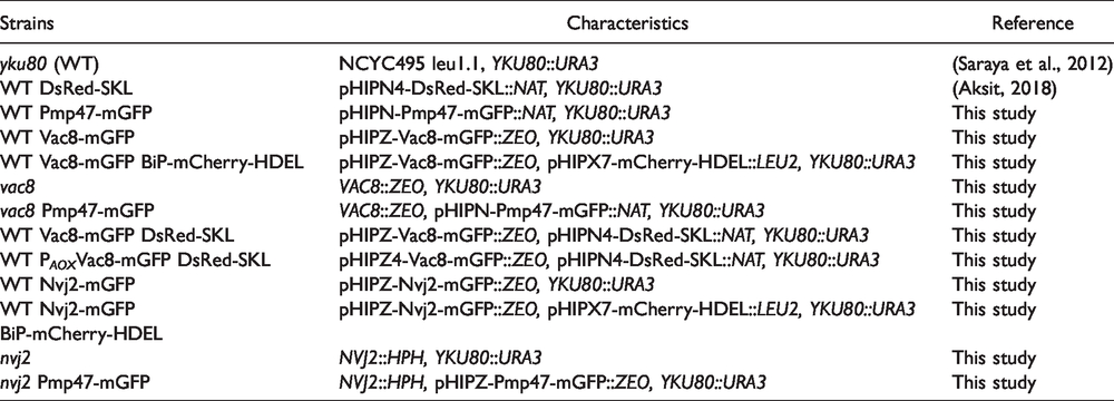
All H. polymorpha strains used in this study are listed in Table 1. Cells were grown in batch cultures on mineral media (van Dijken et al., 1976) supplemented with 0.5% glucose or 0.5% methanol, as carbon source and 0.25% ammonium sulfate as nitrogen source at 37 °C. When required leucine was added to a final concentration of 30 µg/ml. Transformants were selected using 100 µg/ml zeocin (Invitrogen), 100 µg/ml nourseothricin (Werner Bioagents) or 200 µg/ml hygromycin (Invitrogen). Escherichia coli DH5α was used for cloning. Cells were grown at 37 °C in Luria Bertani (LB) medium (1% bacto tryptone, 0.5% yeast extract and 0.5% NaCl) supplemented with ampicillin (100 µg/ml) or kanamycin (50 µg/ml). For growth on agar plates, 2% agar was added to the medium
Hansenula polymorpha Strains Used in This Study.
Vacuole Inheritance and Fusion Assay
To monitor vacuole inheritance, cells were grown on glucose media and incubated with 2 µM FM4-64 for 2 hours. Subsequently, cells were collected by centrifugation, washed and resuspended in fresh glucose-containing medium without FM4-64.
To analyse vacuole fragmentation, cells were grown to the mid-exponential growth phase on glucose medium in the presence of 2 µM FM4-64. Next, cells were incubated in medium containing 0.5 M NaCl for 2 minutes, collected by centrifugation and resuspended in fresh medium without NaCl.
Cloning and Strain Construction
The plasmids and primers used in this study are listed in Table 2 and Table 3. H. polymorpha was transformed as described before (Faber et al., 1994). All integrations were checked by colony PCR. Gene deletions were confirmed by southern blotting.
Plasmids Used in This Study.

Oligonucleotides Used in This Study.

Plasmid pHIPZ-Vac8-mGFP was constructed by amplification of the VAC8 gene, lacking the stop codon, using primers Vac8BglII R and Vac8 F and H. polymorpha genomic DNA as template. The resulting PCR product was digested with HindIII and BglII, and ligated between the HindIII and BglII sites of the pHIPZ-mGFP fusinator plasmid. The resulting plasmid was linearized with BclI to enable integration into the H. polymorpha genome of yku80 (reffered to as WT throughout this work) and WT producing DsRed-SKL peroxisomal marker. Subsequently, StuI linearized pHIPX7-mCherry-HDEL was transformed into the H. polymorpha strain producing Vac8-mGFP.
For the construction of P AOX -Vac8-mGFP, first, we amplified the GFP-2HA tag along with a STOP codon using primers GFP_NdeIF and GFP_2HA_SalIR and plasmid pHIPX4-VPS39-GFP-2HA as a template. The resulting fragment was digested with NdeI and SalI and cloned into the NdeI-SalI digested pHIPZ4-Nia plasmid. The resulting plasmid was digested with BamHi-NdeI enzyme combination. Next, we amplified the VAC8 gene, lacking the STOP codon using primers Vac8F_BamHI and Vac8R_NdeI and genomic DNA as a template. The resulting PCR fragment was digested with BamHI and NdeI and ligated with the previously digested plasmid containing the GFP_2HA tag. The final plasmid was linearized with StuI to enable integration into the WT H. polymorpha genome producing the peroxisomal marker DsRed-SKL.
The vac8 deletion strain was constructed by replacing the VAC8 region with the zeocin resistance gene. First, a PCR fragment containing the zeocin resistance gene and 50 bp of the VAC8 flanking regions was amplified using primers Vac8-Forward and Vac8-Reverse and the pMCE7 plasmid as a template. The resulting deletion cassette was transformed into WT cells. Zeocin resistant transformants were selected and checked by colony PCR with primers Vac8-cPCRF and Vac8-cPCRR. Correct deletion of VAC8 was confirmed by southern blotting. To construct a vac8 strain that produces Pmp47-mGFP, the MunI-linearized pHIPN-Pmp47-mGFP plasmid was transformed into vac8 cells.
A plasmid encoding Nvj2-mGFP was constructed by PCR amplification of a fragment encoding the C-terminus of Nvj2 using primers JWR_63 and JWR_64 and H. polymorpha genomic DNA as a template. The obtained PCR fragment was digested with HindIII and BglII, and inserted between the HindIII and BglII sites of pHIPZ-mGFP fusinator plasmid, resulting in a plasmid pHIPZ—Nvj2-mGFP (eJWR0016). MunI-linearized plasmid pJWR16 was transformed into WT cells. Next, the StuI-linearized pHIPX7-BiP-mCherry-HDEL plasmid was transformed into the WT strain producing Nvj2-mGFP.
An nvj2 deletion strain was constructed by replacing the NVJ2 region with the hygromycin resistance gene. First, a PCR fragment containing the hygromycin resistance gene and 50 bp of the NVJ2 flanking regions was amplified using primers JWR_74 and JWR_75 and the pHIPH4 plasmid as a template. The resulting deletion cassette was transformed into WT cells. Hygromycin resistant transformants were selected and checked by colony PCR with primers JWR_80 and JWR_81. To create a WT and nvj2 strains producing Pmp47-mGFP, the MunI-linearized pHIPZ-Pmp47-mGFP plasmid was transformed into WT and nvj2 cells.
Fluorescence Microscopy
Wide field images were captured using a 100x1.30 NA objective, a Zeiss Axioscope A1 fluorescence microscope (Carl Zeiss, Oberkochen, Germany), Micro-Manager 1.4 software and a CoolSNAP HQ2 camera. GFP fluorescence was visualized with a 470/40 nm bandpass excitation filter, a 495 nm dichromatic mirror, and a 525/50 nm bandpass emission filter. DsRed and FM4-64 fluorescence were visualized with a 546/12 nm bandpass excitation filter, a 560 nm dichromatic mirror, and a 575-640 nm bandpass emission filter. A 587/25 nm bandpass excitation filter, a 605 nm dichromatic mirror and a 647/70 nm bandpass emission filter were used to visualize mCherry fluorescence. The vacuolar lumen was stained with 100 µM CellTracker™ Blue CMAC Dye (7-amino-4-chloromethylcoumarin; Molecular Probes) by incubating cells at 37 °C. CMAC fluorescence was visualized with a 380/30 nm bandpass excitation filter, a 420 nm dichromatic mirror, and a 460/50 nm bandpass emission filter. Image analysis was performed using ImageJ.
Confocal Laser Scanning Microscopy
For quantification of peroxisomes, Z-stack images of cells were taken using a 100x1.40 NA objective using a confocal microscope (LSM800, Carl Zeiss) and Zen software. GFP signal was visualized by excitation with a 488 nm laser and the emission was detected from 490 – 650 nm using an GaAsp detector. Peroxisomes were detected and quantified automatically using a custom made plugin (Thomas et al., 2015).
Electron Microscopy
Cells were cryo-fixed using self-pressurized rapid freezing (Leunissen and Yi, 2009). The copper capillaries were sliced open longitudinally and placed on frozen freeze-substitution medium containing 1% osmium tetroxide, 0.5% uranyl acetate and 5% water in acetone. The cryo-fixed cells were dehydrated and fixed using the rapid freeze substitution method (McDonald and Webb, 2011). Samples were embedded in Epon and ultra-thin sections were collected on formvar coated and carbon evaporated copper grids and inspected using a CM12 (Philips) transmission electron microscope (TEM).
In Silico Analysis
Homologues of S. cerevisiae NVJ-related proteins in H. polymorpha were identified basically as described by (Saraya et al., 2010). In short, via Gapped Blast and Position Specific Iterated (PSI) Blast analyses, primary sequences of S. cerevisiae proteins were used as queries to search the budding yeast dataset (taxid: 4892) of the non-redundant protein database at the National Centre for Biotechnological Information (NCBI, version September 2017). In the PSI-Blast analyses a statistical significance value of 0,001 was used as a threshold for the inclusion of homologous sequences in each next iteration. Additionally, the H. polymorpha genome sequence (Hansenula polymorpha NCYC 495 leu1.1; ATCC MYA-335) was searched using TBlastN with identified protein sequences as queries for the presence of a particular NVJ-related protein. The Genbank accession numbers are MN958684 for HpVac8 and MN958685 for HpNvj2.
Footnotes
Declaration of Conflicting Interests
The author(s) declared no potential conflicts of interest with respect to the research, authorship, and/or publication of this article.
Funding
The authors disclosed receipt of the following financial supportfor the research, authorship, and/or publication of this article: This research was funded by the Marie Curie Initial Training Network PERFUME (Grant Agreement Number 316723).