Abstract
Research Type:
Level 3 - Retrospective cohort study, Case-control study, Meta-analysis of Level 3 studies
Introduction/Purpose:
The concept of peri-talar subluxation has been a recent focus in clinical studies of adult flatfoot deformity. It describes the incongruency in the talonavicular (TN), calcaneocuboid (CC) joints and the three facets of the subtalar joint. In adults, a fair amount of peri-talar subluxation has been found to be physiological rather than pathological but this has not been investigated in the pediatric flatfoot field (reference 1,2). This study used weightbearing CT scans to compare the peri-talar congruency status between symptomatic flexible pediatric flatfeet and a group of pediatric control feet, as well as between pediatric control feet and adult control feet.
Methods:
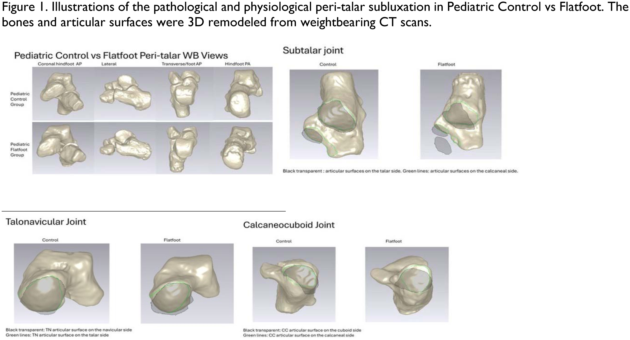
This was an IRB approved study. Weightbearing CT scans of 12 flexible pediatric flatfeet (average age 10.33y, range 7-12y) obtained for clinical investigation of the condition, 10 control feet (average 13.17y, range 12-15y) for evaluation of other etiologies were retrospectively reviewed. This study did not include flatfoot deformities with an accessory navicular, which may have a different presentation in terms of peri-talar subluxation. The peri-talar bones of each foot were segmented and remodeled using Mimics software, and then imported to Geomagic Studio 10 to map out the bony articular surfaces of the peri-talar joints, and to evaluate the extent of subluxation (reflected by percentage of uncoverage of each joint). Peri-talar subluxation was compared between the control and the flatfoot group. Data from a group of adult control feet was also used as a reference to compare the physiological peri-talar subluxation between normal controls in children and adults.
Results:
In children, in non-deformed feet, there was a range of physiological subluxation in the peri-talar joints during weightbearing (Subtalar posterior facet: 17.07±5.16% on the talar side, 10.03±3.36% on the calcaneal side; CC: 20.52±9.86% on the calcaneal side, 32.86±9.86% on the cuboid side; TN: 8.59±5.12% on the navicular side, and 29.65±8.17% on the talar side). The peri-talar subluxation was significantly greater in pediatric flexible flat feet than in controls in posterior facet evaluated on the calcaneal side, and talonavicular joint evaluated on the navicular side. The physiological peri-talar subluxation in children is in general greater than in adults with statistical significance in some joints (Table 1).
Conclusion:
As in adults, physiological peri-talar subluxation exists in normal feet in children. The pathological peri-talar subluxation status in pediatric flatfeet is significantly more severe in some peri-talar joints. In each specific joint, the subluxation is more significant when evaluated on one side than on the other side of the joint. This indicates that both the evaluation method and the concept of pathological peri-talar subluxation need to be further investigated.
References
1. Wallace, C., Zhu, M., Nocek, M., Huang, J., Nolte, C., Wittels, S., … & Li, S. (2024). Evaluating Morphology of the Peritalar Joints Using Surface Mapping Based 3D Remodeling Technique: A Large Sample Sized Cadaveric Study. Foot & Ankle Orthopaedics, 9(4), 2473011424S00274.
2. Zhu, M., Nocek, M., Wallace, A., Gu, W., Challapalli, A., Huang, J., Nolte, C., Orr, C., Myerson, M. S., & Li, S. (2025). Three-dimensional morphology and congruency of the cartilaginous articulation in peri-talar joints: A cadaveric study. Foot & Ankle International. (Manuscript under review).