Abstract
Research Type
Level 3 - Retrospective cohort study, Case-control study, Meta-analysis of Level 3 studies
Introduction/Purpose
Distal tibial deformities can significantly impact patients if left uncorrected, often leading to pain, alterations in gait, and the eventual development of post-traumatic arthritis. The criteria for surgical correction in these patients continue to be a subject of debate, while supramalleolar osteotomy (SMO) is an effective method for correcting distal tibial deformities. The purpose of this study was to evaluate and compare the clinical results of SMO using internal fixation or using computer-assisted hexapod external fixator in the treatment of distal tibial deformity.
Methods
A retrospective study was conducted on 290 patients who underwent SMO between June 2015 and January 2023. Forty-four patients met the inclusion and exclusion criteria. Among the participants, 19 underwent SMO combined with a computer-assisted hexapod external fixator, while 25 received SMO with plate and screw internal fixation. The tibial anterior surface (TAS) angle, tibial lateral surface (TLS) angle, the tibiotalar (TT) angle and the talocrural (TC) angle were assessed on weight-bearing X-ray films. Functional assessments were performed according to the American Orthopedic Foot and Ankle Society (AOFAS) ankle-hindfoot score.
Results
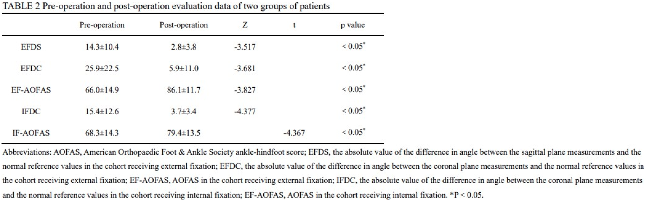
The study followed patients for an average of 31.7 ± 15.3 months (range: 12–67 months), achieving successful bone union in all cases. In the hexapod external fixator group, sagittal plane deformity improved from 14.3 ± 10.4° to 2.8 ± 3.8° (P < 0.05), and coronal plane deformity from 25.9 ± 22.5° to 5.9 ± 11.0° (P < 0.05). The AOFAS ankle-hindfoot score significantly improved. Internal fixation patients also showed improved coronal plane alignment and AOFAS scores. No significant differences were found between groups regarding gender, side, follow-up time, postoperative deformity deviation, or pre-/postoperative AOFAS scores.
Conclusion
In conclusion, comprehensive preoperative planning of SMO combined with either internal fixation or a hexapod external fixator for treating distal tibial deformities can achieve satisfactory outcomes. The utilization of a computer-assisted hexapod external fixator facilitates a gradual and precise correction process, which proved to be an effective and relatively safe method.