Abstract
Research Type:
Level 2: Prospective comparative study, Meta-analysis of Level 2 studies or Level 1 studies with inconsistent results
Introduction/Purpose:
Plantar plate rupture is a significant cause of metatarsophalangeal (MTP) joint instability, often leading to forefoot pain and deformity. Accurate diagnosis is crucial for timely and appropriate management. However, the radiographic findings that correlate with plantar plate integrity remain poorly defined. This study aims to identify radiographic markers that indicate MTP joint instability secondary to plantar plate rupture with neutral and dorsiflexion forces. Our assumption was that dorsiflexion will neutralize the tension from the collateral ligaments and will isolate the effect of plantar plate.
Methods:
Twenty-six MTP joints (including 2nd, 3rd, and 4th MTP) of nine fresh-frozen specimens were used to evaluate MTP joint stability under intact, partial tear, and complete tear plantar plate conditions. MTP Joint widening was assessed radiographically under four different axial traction forces (0N, 25N, 50N, and 75N), each traction force was applied at neutral and 40-degree dorsiflexion of the MTP joints. The joint space widening was compared under the plantar plate conditions using Welch’s ANOVA test with interactions, Tukey HSD as a post hoc. Receiver Operating Characteristic Curve (ROC) analysis was used to determine clinically relevant cutoff values for joint widening.
Statistical significance was set at 0.05.
Results:
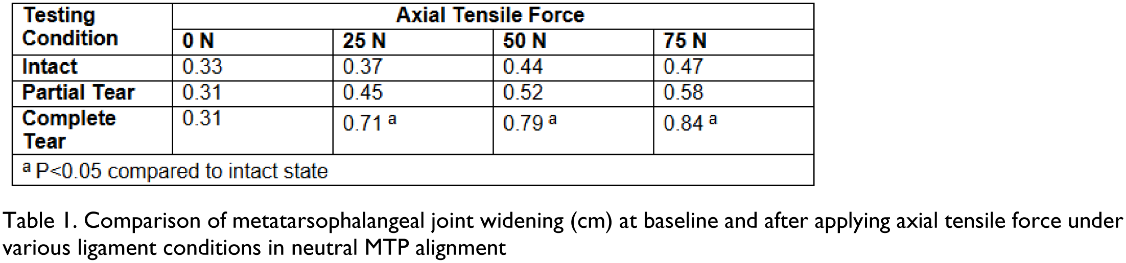
At neutral position there was joint widening at 25N in the complete tear condition as compared to the intact state (Mean Difference: 0.7 mm, p< 0.001) (Table 1). This effect was further amplified with increasing force; however, no significant difference was observed between 50N and 75N rendering 25N as the force of choice to determine instability. A joint widening cutoff value of 0.53 mm, with an area under the ROC of 0.96, showed a sensitivity of 92% and specificity of 86% for accurately detecting complete tears under 25N traction force. There was a significant difference between the MTP diastasis in intact and partially ruptured condition after applying 25N force at dorsiflexion (Mean difference: 0.7 mm; p=0.003) and at neutral position (Mean difference xx mm: p=0.03).
Conclusion:
This study highlights the potential of radiographic testing under traction force for diagnosing complete plantar tears. A traction force of 25N appears to be sufficient for accurately detecting complete tears if the patient can tolerate the pain. We should also admit that a less than 1 mm difference is hard to appreciate, especially in determining partial ruptures. Future clinical investigations are needed to validate these findings and establish definitive diagnostic criteria for plantar tears based on traction force measurements.