
Abstract
Research Type:
Level 3 - Retrospective cohort study, Case-control study, Meta-analysis of Level 3 studies
Introduction/Purpose:
The lesser TMT joints are recessed into a rigid configuration with the cuneiforms, which restricts motion at the second ray during the latter stages of the stance phase when load transfer occurs to the forefoot for toe-off. However, a detailed explanation of how arthritic changes in these stable lesser TMT joints affect gait has not yet been reported in the literature. The purpose of this study was to investigate the inter-segmental foot and ankle kinematics in patients with lesser tarsometatarsal (TMT) joint arthritis to elucidate the biomechanical implications of this condition.
Methods:
The study included 25 patients with lesser TMT arthritis and 50 healthy controls. Gait analysis was performed using the DuPont Foot Model with 15 reflective markers to evaluate the inter-segmental motions across the hallux, forefoot, and hindfoot. Spatiotemporal gait parameters, range of motion (ROM), and kinematic patterns during specific gait phases were analyzed. Statistical parametric mapping (SPM) was used to identify significant differences between the groups.
Results:
Patients with lesser TMT arthritis exhibited slower walking speeds, shorter stride lengths, and wider step widths compared to controls. Notable kinematic alterations included increased dorsiflexion in the hallux during mid-stance to terminal stance, valgus motion from heel contact to pre-swing, and elevated dorsiflexion of the forefoot during pre-swing. The hindfoot demonstrated increased dorsiflexion during initial contact, loading response, and swing phases. These changes suggest compensatory mechanisms to mitigate midfoot pain and instability while maintaining forward propulsion and load transfer. Reduced sagittal and transverse ROM in multiple segments were observed, reflecting functional limitations imposed by arthritic changes.
Conclusion:
Lesser TMT arthritis alters the inter-segmental foot and ankle kinematics during gait, emphasizing the critical role of the midfoot in stability and load transfer. The compensatory adaptations observed in the hallux, forefoot, and hindfoot highlight the biomechanical challenges posed by this condition.
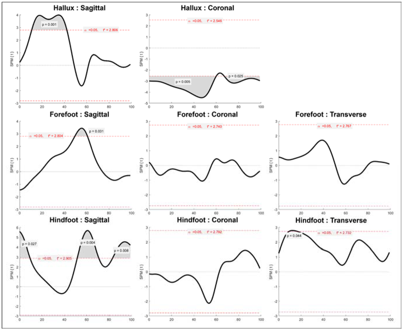
Figure 1. Differences between continuous curves of TMT and control groups, analyzed using statistical parametric mapping (SPM)
A p value of less than 0.05 was considered significant