
Abstract
Research Type:
Level 4 – Case series
Introduction/Purpose:
The dorsal bunion is a deformity consisting of elevation of the first metatarsal and often severe plantarflexion of the first metatarsophalangeal joint resulting from muscle imbalances across the foot and ankle. Most surgical procedures have utilized combinations of first metatarsal osteotomies or medial column arthrodeses, tendon transfers, and metatarsophalangeal capsular releases, to correct the deformity, but only Yong, et al have reported a series with radiographic or clinical outcomes. A new technique for arthrodesis of the naviculocuneiform-1 joint and the 1,2 intercuneiform joint using two bone-blocks to correct the primary deformity was previously published, but no clinical or radiographic results have been reported. The purpose of this study is to report the radiographic results of this new procedure in a retrospective case series.
Methods:
Nine feet in 7 patients were surgically treated for a symptomatic dorsal bunion by the author. All patients underwent a first and second naviculocuneiform and 1-2 intercuneiform joint arthrodesis with structural bone block grafts to reposition and stabilize the medial column of the forefoot utilizing a surgical technique as previously reported. Additional bone and soft tissue procedures were performed concurrently to address associated muscle imbalances, joint contractures or secondary deformities of the forefoot, hindfoot or ankle. The degree of deformity correction was assessed by comparing preoperative and postoperative radiographs. All radiographic measurements were made using a standardized technique on weightbearing radiographs, normalized for magnification and included the intermetatarsal (IM 1-2) angle, the MTP-1 angle, first metatarsal head height, the great toe flexion angle, first metatarsal declination angle and talar declination angle. A retrospective chart review was performed to assess clinical results and determine if any complications or reoperations had occurred.
Results:
Radiographic measures noted a mean decrease (improvement) in the first MT head height (MTHH) of 1.5 cm (0-3.5), decrease (improvement) of the great toe flexion angle (GTFA) of 33° (8-55), increase (improvement) in the first metatarsal declination angle (MTDA) of 13° (3-24) and increase (improvement) in the talar declination (Talar DA) angle of 5° (0-12). There were no non-unions or malunions. Final clinical follow-up at an average of 15 months postoperative (range 3-31 months) showed no complications. Two patients underwent painful hardware removal. All patients were significantly improved by the procedure and were wearing commercially available footwear without the need for foot orthoses. Four of the 6 patients returned to sports (soccer 2, basketball/golf 1 and cheerleading/softball 1).
Conclusion:
A dorsal bunion is a highly variable and challenging deformity requiring a combination of bone and soft-tissue procedures for correction. The double bone block naviculo-cuneiform arthrodesis technique achieved major improvements in the radiographic measures of the primary sagittal plane deformities exhibited in this group of patients with dorsal bunion deformity. This technique provided more correction than other techniques reported using metatarsal osteotomies and may provide improved deformity correction for patients with greater deformity where the medial column is less flexible.
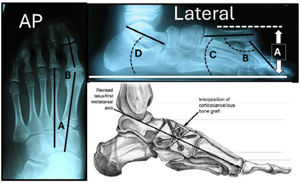
Dorsal Bone Block Fusion Technique and Radiographic Measures
AP view: A. IM1-2 angle B. MTP-1 angle ; Lateral view: A. MTH height B. Great toe flexion angle C. First MT declination angle D. Talar declination angle