
Abstract
Research Type:
Level 3 - Retrospective cohort study, Case-control study, Meta-analysis of Level 3 studies
Introduction/Purpose:
Flatfoot deformity is a complex three-dimensional condition characterized by abnormal talar positioning within the ankle mortise. Given that the deformity is centered around the talus, stabilizing the talus through isolated ankle arthrodesis may influence radiographic parameters associated with flatfoot. This study aimed to evaluate changes in radiographic parameters associated with flatfoot deformity following isolated ankle arthrodesis in end-stage ankle osteoarthritis.
Methods:
This retrospective case-control study included 80 patients with end-stage ankle osteoarthritis and concomitant flatfoot deformity who underwent isolated ankle arthrodesis. Patients were categorized into two groups based on preoperative lateral Meary’s angle: mild (4°–15°, Group A) and moderate (>15°, Group B) deformity.
Radiographic parameters, including AP and lateral Meary’s angle, talonavicular coverage angle, AP and lateral talocalcaneal angle, calcaneal pitch angle, and first tarsometatarsal (TMT) dorsoplantar gap, were assessed preoperatively and at final follow-up. Clinical outcomes were evaluated using the Foot and Ankle Outcome Score (FAOS), American Orthopedic Foot and Ankle Society (AOFAS) ankle-hindfoot scale, and visual analog scale (VAS). Pedobarographic analysis was conducted in a subgroup of 10 patients.
Results:
Significant improvements in radiographic parameters were observed postoperatively in both groups, with Group B demonstrating greater changes in AP and lateral Meary’s angle (p=0.014), talonavicular coverage angle (p=0.002), lateral talocalcaneal angle (p < 0.001), and first TMT dorsoplantar gap (p=0.008). Functional outcomes improved significantly in both groups (all p< 0.01), with no significant differences between them. Correlation analysis revealed associations between radiographic changes and functional improvements, though a direct causal relationship could not be established. Pedobarographic analysis suggested biomechanical adaptations postoperatively, with reduced midfoot load distribution.
Conclusion:
Isolated ankle arthrodesis led to significant changes in radiographic parameters related to flatfoot deformity, despite the absence of direct flatfoot corrective procedures. These findings suggest that talar stabilization within the ankle mortise may contribute to spontaneous correction of flatfoot alignment. Further prospective studies are needed to elucidate the long-term implications of these radiographic and biomechanical changes.
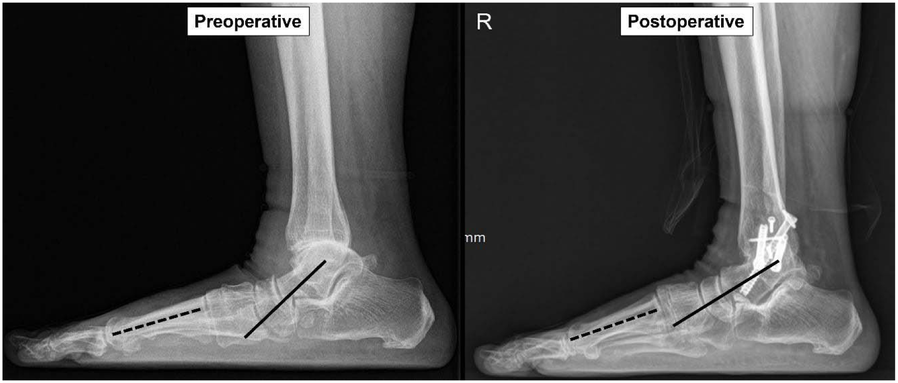
Figure 1. Changes in the lateral Meary’s angle before and after isolated ankle arthrodesis in patients with end-stage ankle osteoarthritis accompanied by moderate flatfoot.
Lateral Meary's angle = angle formed by the dashed and solid lines