
Abstract

Introduction
A primary challenge in studying medial longitudinal arch deformities lies in their definition. Clinically, pes cavus is characterized by a plantarflexion (equinus) deformity of the forefoot with an irreducible plantar flexion of the first ray, and pes planus is distinguished by the collapse of the medial arch when the patient is standing. 1 To objectively confirm its presence and assess its severity, radiologic evaluation and reproducible measurability is essential.
Méary’s eponymous angle in the lateral weight-bearing radiograph of the foot, presented in 1967 at the 39th Annual Meeting of the French Society of Orthopedics and Traumatology, was introduced as a measurement technique for pes cavus and pes planus. 1 Prior to 1967, various methods for measuring cavus had been proposed. These included the Rennotte angle, Hibbs angle, and the Lelièvre drop, which assesses the arch of the forefoot relative to the hindfoot (Figure 1). Méary’s innovation was to grade the severity of pes cavus and pes planus by measuring the angle between the axis of the first metatarsal and the longitudinal axis of the talus on a weightbearing radiograph (Figure 2). 1

Measurements of pes cavus by Rennotte, Hibbs, and Lelièvre, Symposium Le Pied Creux Essentiel, XXXIX Réunion Annuelle de la Société Française d’Orthopédie et de Traumatologie, Revue de Chirurgie Orthopédique et Répératrice de l’Appareil Moteur, Tome 53, July-August 1967, by R. Méary.

Measurement of pes cavus by Robert Méary, Symposium Le Pied Creux Essentiel, XXXIX Réunion Annuelle de la Société Française d’Orthopédie et de Traumatologie, Revue de Chirurgie Orthopédique et Répératrice de l’Appareil Moteur, Tome 53, July-August 1967.
Of note, Meary was also a pioneer regarding the radiographic assessment of hindfoot alignment. Specifically, the Meary “cerclage” view uses a weightbearing AP view of the ankle with a metal cerclage wire placed around the hindfoot to assess the relationship of the tibial axis to the axes of the hindfoot and talus.2 -4
Despite its routine use, many foot and ankle surgeons are unfamiliar with the origins of the Méary angle measurement. This manuscript introduces Robert Méary, MD, a highly innovative and respected Parisian orthopaedic surgeon. It also reviews the scientific research on Meary angle, and the evolution of measurement techniques for flatfoot and cavus deformities.
Historical Background
Robert Méary (Figure 3) was born in 1921 in Paris to Albert Méary and Marie-Louise Méary. He received a classical education at the Lycée Buffon and pursued medical studies at the Medical School of Paris. His education occurred amid the upheavals of World War II. He passed the externship examination in 1942 (French equivalent of the MCAT), and the internship examination in 1946 (French equivalent of the USMLE). During this period, the field of orthopaedic surgery began to take shape as a distinct specialty, with the establishment of the first orthopaedic surgery department at Foch Hospital in 1945 under Dr Robert Merle d’Aubigné.
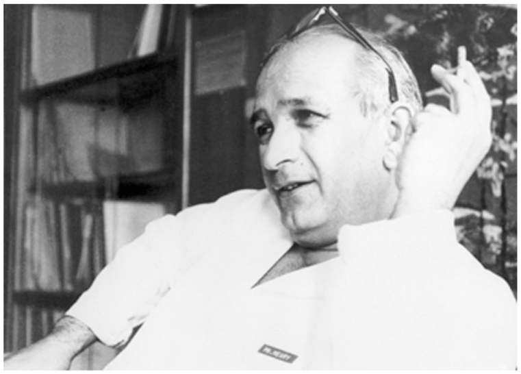
Photograph of Dr Méary in his office. (Courtesy of Drs B. Tomeno and R. Burgade.)
Orthopaedic surgery was officially recognized by the French Medical Board only in 1978. Méary initially began his residency in thoracic surgery at the children’s hospital, Enfants-Malades in Paris. However, he later moved his focus to pediatric orthopaedics. Méary’s exposure to posttraumatic reconstructive surgery during his time at Merle d’Aubigné’s department solidified his decision to specialize in orthopaedics. His thesis on foot drop marked the beginning of his interest in foot and ankle surgery. 5
From 1951 to 1955, Dr Méary served as an assistant professor in the orthopaedic department at Saint Vincent de Paul Hospital in Paris. He grew further intrigued by pediatric foot deformities, a relatively unexplored field at the time. In 1956, Méary was appointed assistant professor at Cochin Hospital, a leading orthopaedic institution in France, where he played a crucial role in organizing the newly created department (Figure 4).

Photograph of the orthopaedic surgeons at Cochin Hospital in 1957. (Courtesy of Drs B. Tomeno and R. Burgade.)
In 1959, Méary created the Documentation Center for the French Orthopedic Society, developing a numerical classification code for clinical diagnoses. 6 His system used 4-digit codes for diagnoses and treatments, providing precise classifications not available in conventional nosological systems. Typically, when a patient underwent surgery, their file would be labeled with 3 or 4 numbers to classify their diagnosis and treatments. Méary’s coding system was exceptionally precise. For instance, while the conventional and current International Classification of Diseases, Tenth Revision (ICD-10), coding assigns a single number to “malignant neoplasm of bone and articular cartilage,” Méary’s system provided distinct numbers for each type of primary bone sarcoma, such as osteosarcoma, juxtacortical sarcoma, chondrosarcoma, and Ewing sarcoma. In 1965, he became Chairman at Boucicaut Hospital, and later at the Ambroise-Paré Hospital in Boulogne in 1969. In 1970, following Merle d’Aubigné’s retirement, Méary took charge of one of the two sections of the orthopaedic department at Cochin Hospital.
Méary’s most notable contributions were in congenital dislocations of the hip, foot surgery, and orthopaedic oncology.6 -11 In 1970, Méary co-founded the Group for the Study of Bone Tumors (GETO). This multidisciplinary group met once a year in France to discuss orthopaedic oncology and is still active today.
Méary’s personal life was closely intertwined with his professional one. His wife, Odette, an anesthesiologist, was a constant presence. They had 3 children: Marion, Jean-Noël, and Eric. Eric was the only child who pursued a career in medicine, ironically specializing in radiology. Méary enjoyed family vacations involving mountain walks and various outdoor games. Robert Méary passed away from pancreatic cancer in January 1974, at the young age of 53 years. He was buried at the Vaugirard Cemetery in Paris together with his parents.
Meary Angle: Technique
When the clinical presentation of a medial arch deformity significantly impairs function, surgery can be considered as a corrective measure. An initial step is to accurately identify and quantify the deformity to properly plan the correction.
Historically, foot posture classification relied on visual observation, clinical measurements such as assessing navicular or midfoot arch height, and footprint analysis. However, these techniques can be challenging because of the soft tissues overlying the skeletal structures. As a result, radiographic methods are regarded as the gold standard for evaluating skeletal alignment of the foot.12 -17
The assessment of medial arch deformities requires weightbearing radiographs. Loading the joints within the foot may reveal or exaggerate deformities otherwise missed. In a cadaveric study, Kimura demonstrated that at least 25% of the total body weight must be applied to reveal the extent of forefoot arch collapse. 18 These results were confirmed by Shelton et al 19 in their clinical study. Figure 5 is an example of the difference between weightbearing and nonweightbearing radiographs of a mild flatfoot, where plantar flexion creates the impression of a pes cavus.

On the first radiograph, the foot is nonweightbearing and appears to be cavus; on the second radiograph on the same foot, the patient is weightbearing and the foot is flat. (Courtesy of Dr B. Tomeno.)
To ensure that the most inferior portion of the foot is not cut off at the edge of the image, the patient should stand with both feet side by side, leaving a 1-cm gap between the medial malleoli. The foot under evaluation is placed on a small wooden block 1 or 2 cm in height. The cassette is then inserted vertically between the 2 feet, allowing the patient to stand upright, and the image is made from lateral to medial on each foot independently.
Méary was the first to describe the use of the angle between the longitudinal axis of the talus and the axis of the first metatarsal in 1967. 1 This angle is also known as the “Méary-Tomeno Angle” because Bernard Tomeno, a student of Méary, went on to write extensively on its various applications.20 -22 The axis of the first metatarsal is relatively easy to draw, as it runs parallel to the dorsal cortex.
Weightbearing computed tomography (WBCT) is now increasingly available, offering complex angular measurements and 3-dimensional reconstructions23 -26 with low radiation doses.27,28 Measurement standards are evolving to adapt to these technological advancements.24,28 Carrara et al analyzed different measurement techniques with the WBCT. Similar to radiographic angles, the ground was defined as the transverse plane, with the orthogonal axis to the ground serving as the vertical axis. 26
In flatfoot or pes cavus, the axes of the talus and first metatarsal form an angle, A, quantifying the deformity (Figures 5 and 6). Méary originally described the normal Méary angle as 0 ± 4 degrees. 1 An angle greater than 4 degrees apex superior indicates pes cavus, whereas an angle greater than 4 degrees apex inferior indicates pes planus. The angle has also been used to classify the severity of the deformity: minimal if the angle is less than 15 degrees, moderate if between 15 and 30 degrees, and severe if greater than 30 degrees.29,30

Lateral radiograph of a pes planus foot. aa, the axis of the talus; mm, axis of the first metatarsal; A, Méary angle.
Current Concepts on the Méary Angle
Intraobserver and Interobserver Reliability
Lamm et al 31 demonstrated excellent interobserver reliability (intraclass correlation of 0.92) between their 2 authors (Table 1). In contrast, in Gibboney et al, 3 18 observers measured the Méary angle in 100 right feet and demonstrated a poor intraclass correlation (0.555) among all physicians. When broken down by training level, attendings and fourth-year podiatry students had moderate intraclass correlations, 0.711 and 0.726, respectively. However, podiatry residents had the lowest intraclass correlation (0.545) among the 3 levels of training.
Méary Angle Measurement in a Healthy Population.

Abbreviation: NA, not available.
Another study, conducted by Davids et al, 32 had a pediatric orthopaedic surgeon, a pediatric orthopaedic fellow, and a clinical research assistant analyze 60 radiographs of children’s feet and measured both the intra- and interobserver reliability of the Méary angle. Interobserver variability was determined by comparing the measurements for 10 cases between the pediatric orthopaedic surgeon and the clinical research assistant and calculated a Pearson correlation coefficient of 0.88, demonstrating very good interobserver reliability. Intraobserver variability was determined by repeated measurement of the 10 angles performed by the pediatric orthopaedic surgeon at 6 months apart and found excellent intraobserver reliability with a Pearson correlation coefficient of 0.98.
Additionally, Bryant 2 demonstrated the high intraobserver reliability of the Méary angle by analyzing 20 radiographs 2 weeks apart and demonstrated an excellent intraclass correlation coefficient, 0.913. This same study also demonstrated that feet in a straight-ahead position and feel in the natural position of gait did not demonstrate any statistically significant differences in the measurement of the Méary angle.
Mean Méary Angle Value
Although Méary first described the normal value of his angle as 0, more current research has reported “normal” values that range from 0 degrees to 13 degrees (Table 1).2,31-34
Researchers have studied variations on this measurement among different populations. Vanderwilde et al 35 noted an inconsistent decrease of the Méary angle with age in the pediatric population, from an average of 18 degrees to 6 degrees between the ages of 6 months and 10 years.
Meanwhile, Thomas et al reported an average adult Méary angle of 4.0 (±5.5) degrees (range −12.9 degrees to 19.5 degrees). In their male population (100 feet), the average Méary angle was 4.6 (±6.2) degrees (range 12.9-19.5 degrees). In their female population, the average Méary angle was 3.3 (±4.7) degrees (range −7.0 degrees to 17.6 degrees). 36 These authors also reported that in subjects <31 years of age (94 feet), the average Méary angle of 3.8 (±5.2) degrees (range −7.8 degrees to 19.1 degrees). In subjects >31 years (106 feet), the average Méary angle was 4.2 (±5.8) degrees (range −12.9 degrees to 19.5 degrees). Dong-Oh 13 also demonstrated a statistically significant difference between the mean Méary angle in 100 male feet (1.91 ± 1.44 degrees) and 100 female feet (−1.90 ± 1.47 degrees), P < .001. The mean Méary angle in the overall population found in this study was 0 (±7.7) degrees, similar to Méary’s original finding.
Pathologies such as hallux valgus were shown to have correlation with a larger Méary angle by Kim et al. 15 In their control group, the mean measurement of the Méary angle was 12.7 (±7.3) degrees, whereas in the hallux valgus groups, the Méary angle was significantly more, with a mean of 16.6 degrees (±8.5; P = .02). Cronin et al 37 demonstrated an association with a more apex plantar Méary angle as a strong predictor in second-ray hammer toe risk in patients with hallux valgus.
Measurement Techniques for the Talus Angle
In his publications, Méary never provided a specific method for drawing the talus axis, only stating that it should represent the longitudinal axis of the talus (Table 2). However, Tomeno precisely described the talus axis as the bisector of the lines passing through the 2 highest and 2 lowest points of the talus (Figure 6). 1
Drawing Methods for the Méary Angle.

Abbreviation: TC, Talo-calcaneal angle; CI, Calcaneal Inclination angle.
Other surgeons have chosen different methods of drawing the axis of the talus. Lamm et al 31 measured the Méary angle as the “angle formed between the bisection of the neck of the talus and the anatomic axis of the first metatarsal”. Therefore, their method of drawing the talus axis is based on the identification of the talar neck (which is very short, and subjective). Kim et al 15 seem to use the same definition as Lamm to determine the talus axis.
Thomas et al 36 reported that it was difficult to identify the bisection of the head and neck of the talus. To determine the talus axis, they used a line perpendicular to the bisector of the most dorsal and plantar extremes of the head. Davids et al 32 used the same technique: “long axis of the talus, created by connecting the midpoints of the margins of the articular surface of the talus and the margins of the neck of the talus.”
The Future of the Méary Angle
Although there seems that to be good interobserver and intraobserver reliability when determining the Meary angle, it is most important to precisely define the measurement technique of the talus axis. One area of future research that may increase reliability is use of an automatic program that identifies the widest axis, orthogonal to each other for each bone, corresponding to the 3-dimensional longitudinal, mediolateral, and dorsoplantar anatomical axes. Variations of up to 10 degrees can be found between the methods for angle calculation, with the 3D angle measurement proving more reliable than sagittal projections. Additionally, no gold standard measurement technique has yet been established for WBCT. Furthermore, as artificial intelligence (AI) comes to the forefront of technology worldwide, future research into the use of AI and measurement of radiologic angles may increase the reliability and aid surgeons in diagnosis and preoperative planning.
Conclusion
The Méary angle revolutionized how foot and ankle surgeons analyze and classify medial arch deformities. His contributions to our understanding of foot and ankle pathologies field cannot be understated and his lifelong dedication to the overall field of orthopaedics should be celebrated. Robert Méary laid the initial foundation into understanding the lateral radiographic of the foot and the relationship between the talus and the first metatarsal. That said, more research is needed for better characterization of what a “normal” value of this angle is in the general population and how best to enhance its reproducibility and inter- and intraobserver reliability. Research has shown that demographics, such as sex, and pathologies, such as hallux valgus, may affect a patient’s angle. Understanding not only the measurement, interpretation, and reliability of this widely acknowledged and highly taught radiographic angle, but also the variability in what is considered “normal” in current literature, is important for understanding how best to approach understanding medial arch deformities.
Supplemental Material
sj-pdf-1-fao-10.1177_24730114251386357 – Supplemental material for The Méary Angle: A Historical Perspective and Contemporary Review
Supplemental material, sj-pdf-1-fao-10.1177_24730114251386357 for The Méary Angle: A Historical Perspective and Contemporary Review by Alexandra M. Stein, Janice Havasy, Adam Bitterman and John E. Herzenberg in Foot & Ankle Orthopaedics
Footnotes
Acknowledgements
The authors thank and pay tribute to Bernard Tomeno, MD, PhD, a close colleague of Robert Méary, MD, PhD, for sharing his knowledge and providing iconographies. Sadly, he recently passed away. Mr Robert Farley assisted in sourcing original articles by Méary.
Ethical Considerations
Not applicable.
Informed consent
Not Required for this study
Funding
The authors received no financial support for the research, authorship, and/or publication of this article.
Declaration of Conflicting Interests
The authors declared no potential conflicts of interest with respect to the research, authorship, and/or publication of this article. Disclosure forms for all authors are available online.
References
Supplementary Material
Please find the following supplemental material available below.
For Open Access articles published under a Creative Commons License, all supplemental material carries the same license as the article it is associated with.
For non-Open Access articles published, all supplemental material carries a non-exclusive license, and permission requests for re-use of supplemental material or any part of supplemental material shall be sent directly to the copyright owner as specified in the copyright notice associated with the article.