
Abstract
Background:
Forefoot varus is a common component of flatfoot deformity that is often surgically addressed. Multiple options exist to plantarflex the medial column, with midfoot fusion and the Cotton osteotomy being the most common. This study analyzes radiographic outcomes and complications when a titanium wedge is used for structural support in a dorsal opening wedge Cotton osteotomy of the medial cuneiform.
Methods:
Between December 2016 and May 2018, 32 feet in 31 patients were treated with medial column titanium wedges for residual forefoot varus in association with flatfoot corrections. All participants had preoperative and weight-bearing postoperative radiographs examined for analysis of radiographic correction. The average age of the patients was 41.1 (range: 12-70). The average follow-up time for patients was 12.2 months (8-24).
Results:
All radiographic parameters were statistically significantly improved from preoperative to postoperative (P < .05). There were no instances of nonunion of the medial cuneiform osteotomy. There was 1 implant that loosened and was revised to a larger implant that healed uneventfully. No wedges were removed for continued pain at the osteotomy site.
Conclusion:
This study suggests that metal wedges are both safe and effective for use in medial column correction based on early follow-up data. Future studies comparing titanium wedges versus traditional bone grafting for Cotton osteotomies may provide further analysis of radiographic correction, operative time, procedure cost, and outcomes. There were no instances of pain over the titanium wedge site. Radiographic outcomes are similar to those reported for opening wedge Cotton osteotomies including bone grafting and wedge plates with screws. Future studies will help determine the long-term maintenance of correction and hardware survivorship.
Level of Evidence:
Level IV, case series.
Adult acquired flatfoot deformity is multifactorial, but commonly, posterior tibial tendon insufficiency is cited to be an initial pathologic inciting factor. In addition, other ligaments that help support the medial column and the arch of the foot begin to attenuate and cause further progression of the malalignment. This leads to anatomical derangement with multiple planes of hindfoot, midfoot, and forefoot malalignment. There are a variety of well-documented procedures for operative correction. Consequently, treatment algorithms differ based on the stage of the deformity described by Myerson and colleagues. 8,10
Stage II flatfoot deformity is characterized by flexible hindfoot valgus with varying amounts of forefoot abduction. Stage IIa is generally defined as less than 30% talar uncoverage on anteroposterior (AP) radiographs, whereas stage IIb has greater than 30% talar uncoverage. Stage III is characterized by rigid valgus of the hindfoot, and compensatory varus of the forefoot with subsequent elevation of the medial column and first ray. Medial column correction is aimed at increasing talonavicular joint coverage and plantarflexing the first ray to restore the lateral talus–first metatarsal angle. Historically, bony correction of the medial column has been accomplished by midfoot fusion or dorsal opening wedge osteotomies with bone grafting. 4,6,7 The use of porous metal wedges has recently been proposed as a bone graft substitute or augment for opening wedge osteotomies. Proponents suggest that metal augments avoid the need for bone grafting, shorten procedure time, expedite the recovery process, decrease complications, and more effectively maintain correction.
Titanium implants have been evaluated more extensively in the orthopedic arthroplasty literature, but currently little clinical data can be found on titanium metal augment wedge fixation in the foot. 2,12 –14 A small series evaluating radiographic outcomes and complications of metal augmentation for calcaneal lateral column lengthening in flatfoot correction has been reported. 3,5,9,11 Although this study provides an early follow-up period, we hypothesize that a titanium wedge implant will allow bone ingrowth and will provide acceptable structural correction with low complication rates when used for a medial cuneiform opening wedge osteotomy during flatfoot operative correction.
Methods
Between December 2016 and May 2018, a total of 32 feet in 31 patients underwent operative correction for flatfoot deformity with a porous titanium wedge used to correct the forefoot varus component of the multiplanar deformity. Surgeries were performed by 2 fellowship-trained foot and ankle orthopedic surgeons at independent academic institutions. The study was institutional review board approved and included a consecutive series of patients aged 0-100 years that had operative flatfoot correction with pre- and postoperative weight-bearing radiographs. Eligible patients were identified by searching the patient records database for CPT codes 27691, 2769, 28306, and 28307. Searches also used ICD-9 and ICD-10 codes M21.40, M21.41, M21.42, 734. Consecutive patients all had titanium wedge placement in the medial cuneiform at the time of dorsal opening wedge osteotomy. Excluded patients were those with less than 6 months of follow-up, those without postoperative weight-bearing radiographs, and those with medial column fusion rather than opening wedge osteotomy. The choice to use a titanium wedge versus an arthrodesis was at the discretion of the primary surgeon, mainly based on the absence of preoperative arthritic signs and symptoms in the midfoot. Patients in this study were also those whose midfoot sag was not centered around the naviculocuneiform joint as those patients were treated with other methods or corrections. One patient was excluded because of inadequate preoperative radiographic analysis. This left 31 feet with a minimum of 6 months’ clinical and radiographic follow-up.
Baseline demographics are as follows. The mean age was 41 years (range, 12-70 years), and gender stratification was 63% female and 37% male. The average time from initial appointment with the operative surgeon to surgery date was 211 days (range, 29-1296 days). Average follow-up was 12.2 months (range, 8-24 months). Radiographic images were analyzed using a calibrated ruler and angular measurements using Carestream Solutions, version 11.4.0.1253 (Carestream Health Inc, Rochester, NY). Measurements included AP talonavicular coverage angle, AP talocalcaneal angle, AP talus–first metatarsal angle, AP talar uncoverage angle, lateral talocalcaneal angle, lateral talus–first metatarsal angle, lateral medial cuneiform–fifth metatarsal height, and lateral calcaneal pitch. In an attempt to better isolate the effect of the Cotton osteotomy, we also evaluated the medial arch sag angle and the medial cuneiform articular angle pre- and postoperatively. The medial cuneiform articular angle was measured by a line parallel to proximal and distal articular surfaces of the medial cuneiform. As defined by Castaneda et al, 3 this value becomes negative when a line along the distal articular surface diverges from the proximal articular surface. Thus, a dorsiflexion osteotomy of the medial cuneiform likely produces a negative value compared to the preoperative state. As defined by Aiyer, 1 the medial arch sag angle was measured by a line paralleling the proximal articular surfaces of the navicular and the first metatarsal.
Initially, patients were seen at 2 weeks, 6 weeks, 3 months, and 6 months postoperatively with clinical examination and radiographs. During the study, we discontinued routine follow-up on patients at 6 months and beyond unless the patient had symptoms or concerns (Figures 1 and 2).

Preoperative. Solid line, medial cuneiform articular angle; dashed line, medial arch sag angle.

Postoperative. Solid line, medial cuneiform articular angle; dashed line, medial arch sag angle.
Operative Technique
All patients had multiple procedures performed as components of the flatfoot reconstruction, and the average number of simultaneous procedures was 6. The most common concomitant procedures are listed in Table 1. Twenty-seven of 31 patients first received a strayer gastrocnemius recession. Next, the hindfoot deformity was addressed with either subtalar fusion or medial slide calcaneal osteotomy at the surgeon’s discretion. An incision was then made in line with the insertion of the posterior tibial tendon and dissection was carried down to the medial column of the forefoot. This allowed for tenosynovectomy or excision of the posterior tibial tendon. In some patients, repair of the medial ankle deltoid and spring ligament complex was performed with no. 1 Vicryl suture and augmentation with fiber tape. Next, the flexor digitorum longus tendon was harvested from the plantar foot and transferred into the navicular. Lastly, the medial cuneiform was identified and the tibialis anterior tendon was retracted plantarly and proximally. Using fluoroscopic guidance, a dorsal opening wedge osteotomy was made in the medial cuneiform while attempting to leave plantar periosteal hinge intact. The appropriately sized titanium wedge was selected by trialing with lateral radiographic imaging. The mean sized final selection for the titanium wedge was 7 mm. Postoperatively, patients were placed into a plaster splint. A short leg cast was generally placed at the first follow-up visit. At 6 weeks, a walking boot was fitted and full weight bearing as well as physical therapy commenced. Patients were transitioned into standard footwear at a 3-month postoperative visit.
Concomitant Procedures.

Abbreviation: FDL, flexor digitorum longus.
Results
Each measured radiographic parameter had statistically significant improvement from preoperative to final postoperative radiographs, including the medial cuneiform articular angle which helps to isolate the effect of the titanium wedge implants (P < .05; Table 2). There were 14 patients included in our study that had greater than 12-month follow-up. Looking at the data on these 14 patients, with an average follow-up of 17.4 months, all radiographic parameters remained statistically significant with no complications in this group of patients. There was no evidence of osteotomy nonunion at final follow-up in any of the patients included in the study. Routine computed tomographic scanning was not used to evaluate union of the Cotton osteotomy as no patient had radiographic evidence of loosening or continued pain with weight bearing at the osteotomy site. One patient (3.2%) had evidence of plantar gapping with subsequent loosening of the wedge and required unanticipated revision surgery with removal of the original implant and conversion to a larger wedge. The patient went on to union without further complication or clinical dissatisfaction. At final follow-up, no patients had implant subsidence, migration, removal, or future planned medial column procedures. In 1 patient (3.2%), the metallic wedge had a nondisplaced fracture line through the metallic wedge that was first seen at 5 months postoperatively. The patient was allowed to continue to weight-bear as tolerated and was followed at regular intervals to assess stability of the implant. The patient was last seen at 17 months postoperatively. The patient reported no history of pain at the osteotomy site and had no tenderness to palpation over the site. Radiographs at 17 months postoperatively showed no propagation of the fracture line, no evidence of lucency around the implant, and no change in the alignment of her correction based on these serial radiographs. Two patients (6.4%) requested screw removal from the hindfoot after successful union. One patient (3.2%) underwent planned staged total ankle replacement 6 months after the flatfoot reconstruction (Figures 3 –6).
Radiographic Parameters.

Abbreviations: AP, anteroposterior; MT, metatarsal.
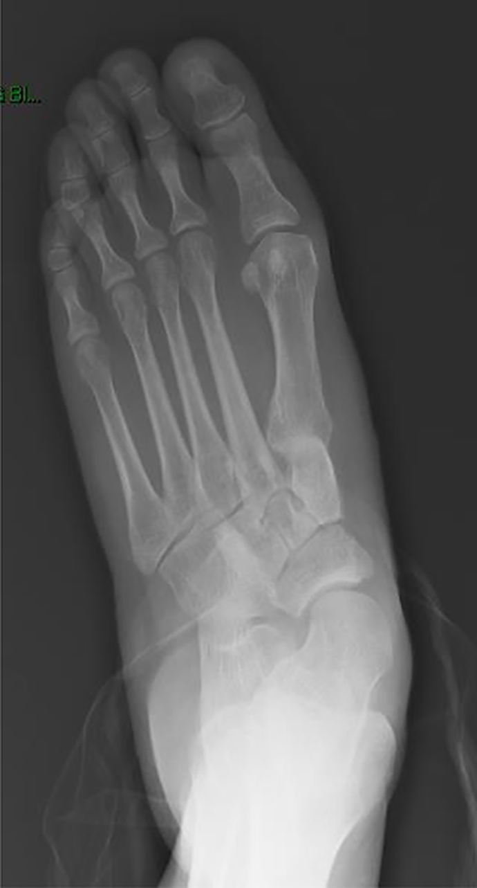
Preoperative anteroposterior foot radiograph.

Postoperative anteroposterior foot radiograph.
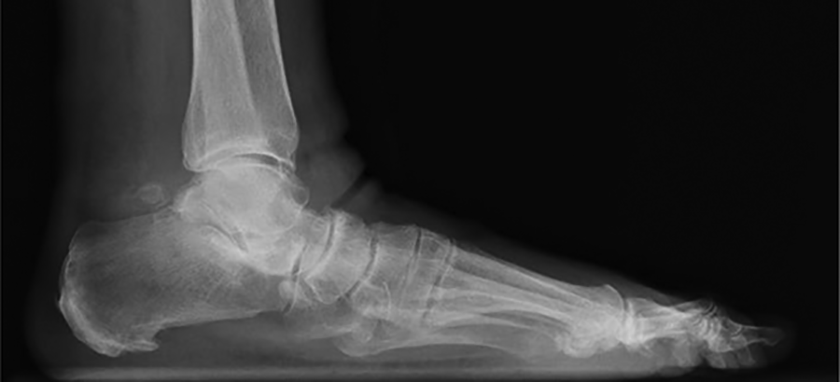
Preoperative lateral foot radiograph.

Postoperative lateral foot radiograph.
Discussion
This study evaluated the use of a structural titanium opening wedges for medial column correction in flatfoot corrective surgery. There are a few studies using a similar implant for lateral column lengthening as part of flatfoot reconstructions. 3,9 In one series, 28 feet were treated using a metal wedge, and all but 1 had bony incorporation. The authors noted significant radiographic deformity correction. The cost of the implant was similar to tricortical iliac crest allograft when accounting for operating room time. Another case series reported on the use of a titanium wedge in lateral column lengthening for flatfoot correction. 9 With 30 patients and 34 feet included, the results showed significant radiographic correction with zero cases of nonunion, and no wedges had to be removed.
The authors recognize some limitations of our study design. Although some of the earliest patients in the study had 24-month follow-up, our average follow-up was 12.2 months, with a range of 8 to 24 months. Initially all patients were seen at 1 year postoperatively, but after the initial learning curve and successful clinical outcomes were observed, we did not routinely bring patients back into the office after the 6-month postoperative visit if the patient was doing well without concerns. There were no additional surgeries for hardware removal of the Cotton wedge for any patient in the study. Future studies with longer follow-up would help to validate long-term maintenance of deformity correction and implant survivorship using metallic wedges. Moreover, this is a relatively small case series, and an increased number of patients would further validate the results; on the other hand, the group size in the current study is similar to that in prior reports on metallic wedge implant utilization in the foot. 3,9
Nearly every patient in our series underwent concomitant procedures as part of the flatfoot reconstruction, and therefore, it is difficult to elucidate the effect of a single procedure on radiographic correction. We did find significant correction of the medial arch sag angle and the medial cuneiform articular angle, which are most directly linked to the effects of the Cotton osteotomy. Our results suggest that metal wedges are both safe and effective for continued use in medial column correction. Future trials may provide further insight into the clinical implications of wedge augmentation, if the procedure continues to gain popularity among surgeons versus traditional grafting techniques. Although one patient required revision surgery, there were no instances of continued pain over the titanium wedge or need for implant removal.
Conclusion
Based on the early outcomes presented, the use of metal wedge augmentation in the medial column of the foot appear to provide successful radiographic correction with complication rates similar to other commonly accepted flatfoot corrective procedures.
Supplemental Material
Supplemental Material, FAO868971-ICMJE - Complications and Early Radiographic Outcomes of Flatfoot Deformity Correction With Metallic Midfoot Opening Wedge Implants
Supplemental Material, FAO868971-ICMJE for Complications and Early Radiographic Outcomes of Flatfoot Deformity Correction With Metallic Midfoot Opening Wedge Implants by Tyler W. Fraser, Anish R. Kadakia and Jesse F. Doty in Foot & Ankle Orthopaedics
Footnotes
Declaration of Conflicting Interests
The author(s) declared the following potential conflicts of interest with respect to the research, authorship, and/or publication of this article: Jesse F. Doty, MD, reports personal fees from Arthrex Inc, personal fees from Globus Medical Inc, and personal fees from Wright Medical Inc, outside the submitted work. Anish R. Kadakia, MD, reports grants and personal fees from Arthrex, during the conduct of the study; other from BME, other from Acumed, outside the submitted work. ICMJE forms for all authors are available online.
Funding
The author(s) received no financial support for the research, authorship, and/or publication of this article.
References
Supplementary Material
Please find the following supplemental material available below.
For Open Access articles published under a Creative Commons License, all supplemental material carries the same license as the article it is associated with.
For non-Open Access articles published, all supplemental material carries a non-exclusive license, and permission requests for re-use of supplemental material or any part of supplemental material shall be sent directly to the copyright owner as specified in the copyright notice associated with the article.