
Abstract
Background:
Soft tissue complications after Achilles tendon repair has led to increased interest in less invasive techniques. Various limited open techniques have gained popularity as an alternative to open operative repair. The purpose of this study was to biomechanically compare an open Krackow and limited open repair for Achilles tendon rupture. We hypothesized that there would be no statistical difference in load to failure, work to failure, and initial linear stiffness.
Methods:
A simulated Achilles tendon rupture was created 4 cm proximal to its insertion in 18 fresh-frozen cadaveric below-knee lower limbs. Specimens were randomized to open or limited open PARS Achilles Jig System repair. Repairs were loaded to failure at a rate of 25.4 mm/s to reflect loading during normal ankle range of motion. Load to failure, work to failure, and initial linear stiffness were compared between the 2 repair types.
Results:
The average load to failure (353.8 ± 88.8 N vs 313.3 ± 99.9 N; P = .38) and work to failure (6.4 ± 2.3 J vs 6.3 ± 3.5 J; P = .904) were not statistically different for Krackow and PARS repair, respectively. Mean initial linear stiffness of the Krackow repair (17.8 ± 5.4 N/mm) was significantly greater than PARS repair (11.8 ± 2.5 N/mm) (P = .011).
Conclusion:
No significant difference in repair strength was seen, but higher initial linear stiffness for Krackow repair suggests superior resistance to gap formation, which may occur during postoperative rehabilitation. With equal repair strength, but less soft tissue devitalization, the PARS may be a favorable option for patients with risk factors for soft tissue complications.
Introduction
Although nonoperative treatment of acute Achilles tendon ruptures is safe, inexpensive, and comparable to surgery with respect to rates of rerupture, recent randomized controlled trials suggest that operative repair is associated with earlier return to work and greater plantar flexion strength. 28,33 Operative repair can be performed using a percutaneous, limited open, or standard open technique. However, no one technique has been shown to be clearly superior. 2,6,11
Open repair is a familiar and readily available option for most surgeons. Although a standard posterior or posteromedial approach allows for direct visualization and optimal suture placement, open repair is associated with a substantial risk of soft tissue complications. Rates of non-rerupture complications, including superficial and deep infection, wound dehiscence, skin tethering, and hypertrophic scarring have been reported as high as 34% after open repair. 19,33 Given this risk, less invasive techniques, which use smaller incisions, have garnered increased interest. 3,8,12,16 For example, in the PARS Achilles Jig System (Arthrex, North Naples, FL), suture is passed percutaneously and placed deep to the crural fascia, while a small incision at the rupture site allows access to assess tendon apposition and quality of repair.
Clinical outcome comparisons between the 2 repair types yield mixed results. A recent retrospective comparison of the PARS repair to open augmented Krackow repair found no statistically significant differences in rerupture rate, sural neuritis, dehiscence, infection, or reoperation. 12 In one study, at 5 months’ follow-up, significantly more patients who underwent PARS repair returned to baseline activities (98% vs 82%) (P < .0001), whereas another study demonstrated earlier return to work in the open repair group. 18
Although other percutaneous repair constructs have been studied previously, there is a paucity of biomechanical studies specifically evaluating the PARS system. 10,15 A previous study demonstrated comparable strength among 3 percutaneous repair constructs, including PARS, subjected to a cyclic loading protocol intended to simulate aggressive rehabilitation. 4
In this study, we biomechanically compared an open, 2-strand Krackow repair with epitendinous weave to the limited open PARS Achilles Jig System for simulated midsubstance tears in human cadaver Achilles tendons. 12,33 Our primary objective was to compare the ultimate strength, measured by load to failure, between techniques.
Materials and Methods
Study Design
An a priori power analysis was performed using data from previous studies for maximum load to failure for a Krackow (276 N ± 87 N) and PARS repair (385 N ± 90 N). 7,13 Assuming paired specimens and an α-value of 0.05, a minimal sample size of n = 18 was calculated to satisfy a statistical power greater than 0.80.
Nine pairs of fresh-frozen human cadaver lower limbs (proximal tibia to toe) were included in this study (mean age, 66 years ± 8.16 [range 53-77]; 3 male, 6 female). Specimens were obtained from the United Tissue Network (Phoenix, AZ). Donors were screened for absence of systemic connective tissue disorder, inflammatory disease, ankle fracture, or prior definitive injury to the foot and ankle including triceps surae. One ankle from each cadaveric pair was randomly assigned to one of the 2 experimental groups. The contralateral ankle was then assigned to the other group, resulting in 9 specimens per group. Five left and 4 right-sided specimens were randomly allocated to the PARS group and 5 right and 4 left-sided specimens were randomly allocated to the open group. Specimens were stored at –20°C and were thawed for 12 hours at room temperature before dissection.
Specimen Preparation
The gastrocnemius-soleus unit was first carefully dissected free of all overlying skin and soft tissue to eliminate potential confounding due to eccentric suture placement during tendon repair. 10,13 Each tendon was transected proximally at its musculotendinous junction. The calcaneus was harvested using an oscillating saw. Care was taken to preserve the entirety of the tendo-osseus footprint. The width, thickness and circumference of each tendon were measured using a digital caliper at the planned site of tendon transection. Width was defined as the medial to lateral distance (mm) and thickness was defined as the anterior to posterior distance (mm). The Achilles tendon was then transected horizontally with a no. 10 blade 4 cm proximal to its calcaneal insertion, as ruptures tend to occur 2 to 7 cm proximal to the calcaneal insertion. 17
In the open group, a 2-strand Krackow repair with epitendinous weave was performed (Figure 1). No. 2 braided polyethylene/polyester multifilament (FiberWire; Arthrex) nonabsorbable suture was started at the cut edge of the tendon and passed in 4 sequential locking loops along its periphery. 20

Schematic diagram illustrating each repair and their suture configurations. The open repair consisted of (A) a 2-strand Krackow repair augmented with (B) epitendinous weave. After the Krackow stitch, the epitendinous weave was passed through the tendon 2.5 cm from its torn edge as described by Lee et al.22 (C) The PARS Achilles Jig System (Arthrex). The PARS repair consisted of 2 simple transverse and 1 locking suture.
This was then repeated distally along the adjacent side of the tendon before exiting at the stump end. Care was taken during suture passage to avoid severing or harpooning adjacent suture. This process was repeated in the remaining tendon stump, which resulted in 2 suture ends exiting from each cut tendon surface. The 2 suture pairs were then tightened to oppose the proximal and distal stumps and tied securely with 6 standard square knots based on existing biomechanical studies. 31 All knots were tied at the rupture site. Lastly, a cross-stitch epitendinous weave using no. 0 Vicryl (Ethilon; Ethicon, NJ) absorbable suture was performed at the rupture site as previously described. 22 This was tightened and tied securely with 6 standard square knots. At this time, complete apposition of tendon edges was once again confirmed.
In the PARS group, tendon repair using the PARS Achilles jig system was performed using no. 2 braided polyethylene/polyester multifilament (No. 2 FiberWire and TigerWire; Arthrex) nonabsorbable suture. All repairs were performed using the PARS jig under direct visualization to ensure that each suture passed through the mid-aspect of the tendon in the anteroposterior plane. 7,13,22 Because no biomechanical advantage has been shown for any particular suture configuration, all PARS repairs were performed as depicted in the manufacturer’s operative technique manual. 7 This resulted in a final configuration consisting of 2 simple transverse and 1 locked suture loop in each tendon stump (Figure 1). All suture ends were brought together and tightened until the tendon edges were approximated. Suture strands were tied in order from closest to farthest from the repair site per the operative technique manual. All knots were tied with 6 square standard operative knots at the site of the defect with no supplemental reinforcement stitches.
Specimen Testing and Biomechanical Outcomes
Specimens were tested in a servohydraulic material testing machine (858 Mini Bionix; MTS Systems, Eden Prairie, MN). Each calcaneal wedge was potted in a 5-cm-diameter PVC pipe using polymethylmethacrylate cement. Proximally, the tendon was tightly secured in a vise clamp with 2 opposing abrasive grit surfaces. The potted calcaneus was mounted onto the testing apparatus on the material testing systems machine’s base (Figure 2). The tendon length, defined as the distance between the inferior aspect of the clamp and insertion of the Achilles into the potted calcaneus, was standardized across specimens to allow for stiffness calculations.
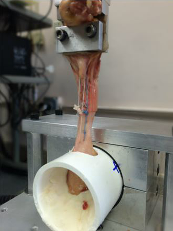
The repaired Achilles tendon secured onto the material testing machine (858 Mini Bionix, MTS Systems). The tendon is secured proximally in a tightened clamp. The calcaneal wedge is potted onto a 5-cm PVC pipe using polymethylmethacrylate cement.
A load to failure test was performed on each repaired Achilles tendon, with the load applied along its longitudinal physiological axis. An initial load of 0.5 N was applied to tension each specimen immediately prior to load to failure testing without a standardized duration. Load to failure was performed at a rate of 25.4 mm/s, to reflect the range at which loads are imposed on the tendon during normal ankle range of motion. 4,7,10,22 The maximal load prior to failure and location of failure were documented for each specimen. Failure was defined as a precipitous decline in measured load resulting from either complete failure at the suture-tendon interface or breakage of remaining suture. Possible mechanisms of failure included suture breakage, knot breakage, knot unraveling, and suture cutout. For each specimen, load displacement curves were created to calculate the initial linear stiffness, load to failure, and work to failure. Initial linear stiffness was calculated as the slope of the elastic deformation phase prior to failure from the start of testing. Although we did not collect data on displacement directly, prior studies have used initial linear stiffness as an acceptable surrogate for gapping resistance (Heitman et al). Work to failure was calculated by the area under the force-displacement curve from the start of the test to the point where the maximum load was achieved.
Sample Size and Statistical Analysis
Paired t tests were used to compare initial linear stiffness, load to failure, and work to failure between experimental groups. Pearson correlation coefficients were calculated to determine the relationship between tendon size and load to failure in both repair types. Statistical significance was set at P < .05. All statistical analysis was performed in SPSS version 22 (IBM Corp, Armonk, NY).
Results
We queried the laterality, age, gender, and proportion of failure location, by repair type (Table 1). We also calculated the average load to failure, work to failure, and initial linear stiffness by repair type (Table 2). The average load to failure for open repair was 353.8 ± 88.8 N (range, 266.7-521.4 N). The average load to failure for PARS repair was 313.3 ± 99.9 N (range, 174.8-498.2 N), which was not statistically different from that for open repair (P = .38). The average work to failure for open repair was 6.4 ± 2.3 J (range, 3.1-9.9 J). The average work to failure for PARS repair was 6.3 ± 3.5 J (range, 2.7-12.6 J), which was not statistically different from that for open repair (P = .904). Mean initial linear stiffness of the open repair (17.8 ± 5.4 N/mm; range 12.3-27.9 N/mm), was significantly greater than PARS repair (11.8 ± 2.5 N/mm; range, 8.7-16.6 N/mm) (P = .011) (Table 2).
Specimen Profiles by Repair Type.

Biomechanical Outcomes.a

aAll values expressed as Mean (Standard deviation).
For open repair, Krackow suture strands predominantly failed at the suture itself (7/9 open). This corresponded to a force-displacement curve that demonstrated the sequential failure of suture strands (Figure 3). The epitendinous weave suture predominantly failed at the suture itself (6/9 open), followed by the suture-tendon interface (3/9 open). In contrast, the PARS repair predominantly failed at the suture-tendon interface (7/9 PARS), with all 3 suture loops cutting out of the repaired tendon. This corresponded to a force-displacement curve demonstrating a broad all-at-once failure (Figure 4). In 2 specimens (2/9 PARS), 1 of 3 transverse suture loops failed at the suture itself. The other 2 loops in these 2 specimens failed at the suture-tendon interface.

Load displacement curve showing the predominant failure mechanism of augmented Krackow repair, which was sequential breakage of the running locking and epitendinous weave suture.

Load displacement curve showing the predominant failure mechanism of PARS repair, which was cut out of all 3 suture limbs.
No significant difference in mean tendon width (13.3 vs 13.3 mm; P = .976), thickness (5.69 vs 5.87 mm; P = .507), or circumference (36.3 vs 36.6 mm; P = .507) existed between the 2 experimental groups (Table 3). Pearson correlation coefficients between tendon size parameters and maximum load to failure revealed that increasing tendon width (R = 0.75; P = .029) and circumference (R = 0.72; P = .028) correlated with increasing load to failure for PARS repair (Table 4). No statistically significant correlation between measures of tendon size and load to failure were detected for open repair.
Average Values for Tendon Size (mm).a

aAll values are expressed as mean (standard deviation).
Tendon Size and Load to Failure Correlations.

Discussion
The principal finding of this study was that there was no significant difference in load to failure between PARS and an open 2-strand Krackow repair with epitendinous weave for simulated, midsubstance Achilles tendon ruptures in human cadavers.
The primary outcome of this study was ultimate strength, or load to failure, which has implications in the repaired tendon’s ability to withstand loading during early postoperative ankle motion and weight bearing. 5,30 The average load to failure for the PARS and open repairs were 313.3 and 353.8 N, respectively. These values exceed loads previously shown to occur across the Achilles tendon during passive ankle plantar flexion and protected weight bearing with a 1-inch heel lift, which are allowed as early as 2 weeks postoperatively in some protocols. 1,24,33 Our findings suggest that progressive, accelerated rehabilitation in a reliable and compliant patient would be practical after either Krackow or limited open PARS repair, as described in the present study.
Repair site gapping, which has been shown to affect plantarflexion strength and chance of rerupture, has come to the forefront in biomechanical studies of newer limited open techniques. 4,22 Prior studies have used initial linear stiffness as a surrogate measure of gapping. 10 The initial linear stiffness (N/mm) estimates the force (N) required to create a gap of 1 mm. Initial linear stiffness was significantly greater for Krackow repair, which suggests a greater resistance to gap formation compared to the PARS. Recent studies show that adding epitendinous suture reinforcement may improve tensile strength, gap resistance, and apposition of frayed tendon ends. 22,27 Lee et al showed in a cadaveric model that Krackow repairs with an epitendinous cross-stitch weave tolerated more cycles before gapping than nonaugmented Krackow repairs (2208 vs 502 cycles) (P = .024). 21 In the present study, it is possible that augmentation contributed to the greater initial linear stiffness seen after open repair.
This study also demonstrated that tendon size correlates with strength after limited open repair. In the PARS group, increasing tendon width and circumference strongly correlated with increasing load to failure. Because PARS repairs failed predominantly by suture cutout, a tendon with larger dimensions in the medial to lateral plane should better resist pullout of the transversely placed suture using the PARS jig. This finding suggests that limited open repair may exhibit greater ultimate strength in more robust Achilles tendons using the PARS. In the open repair group, no correlation between tendon size and ultimate strength was seen. Our findings suggest that the ultimate strength of Krackow repair is independent of tendon size, and this can be explained by its propensity to fail by suture breakage, which is similar to previous studies. 10,22 Further investigation is needed to better understand the impact of suture material, and knot quality and number, on ultimate repair strength. Also, future studies are needed to better understand the effect of accuracy of suture position in the PARS repair on biomechanical properties.
Since its introduction in 2010, only 2 studies have studied the biomechanical properties of the PARS Achilles Jig System. One study compared it to the Achillon device (Integra Life Sciences Corp, Plainsboro, NJ) in a 2-stage cyclic loading protocol ending in a single load to failure test. 7 Similar to the present study, all repairs were performed under direct visualization after removal of all overlying soft tissue. Overall, PARS repairs withstood a significantly greater average number of cycles prior to 2 and 9.5 mm of gapping. The PARS repair also demonstrated a significantly greater average load to failure at 385.0 N (range, 185.6-502.2 N), which is similar to the loads that are reported in the present study (313.3 ± 99.9 N; range, 174.8-498.2 N). Only 3 of the 21 PARS repair specimen failed at the suture tendon interface, which is a much smaller proportion than in our study. However, only 7 of these were repaired using the same suture configuration as in the present study and the authors did not specify failure location for each individual PARS subtype. Still this difference in predominant suture location may be accounted for by cyclic loading, which preceded their single load to failure test.
A more recent study compared open repair, consisting of a core of 3 Kessler sutures with epitendinous weave, to the PARS, Achillon, and SpeedBridge (Arthrex) in a progressive cyclic loading protocol. 4 Repair strength was quantified in terms of number of cycles to failure. Significantly less early elongation was seen for open repair compared to the 3 limited open techniques, but no difference in cycles to failure was seen. Similar to the present study, the predominant location of failure for PARS repair (5 of 9) was at the suture tendon interface.
The authors acknowledge that the present study is not without limitation. All repairs were performed in open fashion. 7,13,22 Although this successfully reduces the likelihood of eccentric suture placement, it fails to mimic in vivo operative conditions, which may have biased the results in favor of the PARS repair. However, the objective of this study was to biomechanically compare repair techniques under optimal conditions. Although the PARS jig has shown greater suture placement accuracy than other limited open techniques, the concern for superficial suture placement remains. 4 With this in mind, our methodology must be taken into consideration when comparing our findings with other studies.
A second limitation of our study is that biomechanical testing is a time zero representation of Achilles tendon rupture repair. It is well known that in vivo time, early motion, and progressive loading affect the strength of a healing tendon. 9,25,26 Therefore, the results of this study do not account for the impact of subsequent healing on repair strength.
Additionally, the tendon ruptures created in our study do not mimic the frayed tendon edges commonly seen clinically, a previously described drawback inherent to cadaveric biomechanical testing 10,32 Another limitation is that the Achilles tendons used in this study were predominantly from female cadavers with an average donor age of 66 years. Achilles tendon ruptures occur most frequently in men between the ages of 30 and 49. 14,29 Because matched specimens were used, it is unlikely that this had an effect on the difference in repair strength for either repair type.
Finally, our study used a single load to failure protocol. A cyclic loading protocol successfully simulates aggressive rehabilitation, 4 which may lead to lengthening of the operatively repaired tendon. Nevertheless, accidental falls or slips are commonly cited mechanisms of rerupture, suggesting that a single load to failure protocol remains clinically relevant. 23
Conclusion
Our results indicate that load to failure was not statistically different for the open augmented Krackow as compared to limited open PARS repairs. The initial linear stiffness of the open Krackow locking loop technique with epitendinous augmentation was significantly greater than the limited open PARS technique. In addition to ultimate strength, we believe that patient factors such as risk for infection, regard for cosmesis, and time to return to work or sport should be considered when deciding on a specific repair method for a given patient. 12 Surgeon familiarity and comfort, as well as cost and availability, should also be considered.
Footnotes
Declaration of Conflicting Interests
The author(s) declared no potential conflicts of interest with respect to the research, authorship, and/or publication of this article.
Funding
The author(s) disclosed receipt of the following financial support for the research, authorship, and/or publication of this article: Northwestern University Department of Orthopedic Surgery.