
Abstract

27th–30th April 2025 | Liverpool, UK
Book of abstracts
Table of Contents

Aging and Neurodegeneration
PM_001
Keywords: Glial cells, Neuroinflammation, Neuropathology, Alzheimer's disease, Tau hyperphosphorylation
Authors: Meg Watt, Fiona Houston, Danielle Gunn-Moore, Alexandra Malbon, Sophie Scrimgeour
1.Ageing is the main risk factor for most neurodegenerative diseases. The most common form of neurodegenerative dementia in humans is Alzheimer's disease (AD), which is characterised by amyloid plaques, neurofibrillary tangles (NFT) containing hyperphosphorylated tau and increased neuroinflammation. Cognitive dysfunction syndrome (CDS) is an age-related condition found in domestic cats, with clinical features similar to those seen in human dementia. Additionally, aged cats and sheep develop neuropathology similar to changes observed in the brains of humans diagnosed with AD, such as amyloid-β (Aβ) accumulation and NFT. However, there is limited knowledge of the neurodegenerative processes in these species, and how closely their age-related neuropathology resembles that seen in humans. As glial cells such as microglia and astrocytes have been implicated in the pathogenesis of neurodegenerative diseases, we aimed to assess age-related changes to glial cell morphology and density in both cats and sheep and compare the results with glial cell changes in the brains of cats diagnosed with CDS.
2.Transverse sections of brain regions were cut from formalin fixed, paraffin embedded blocks from cats and sheep of various ages, including cats diagnosed with CDS. Sections were labelled using antibodies for specific glial cell markers (Iba1 for microglia; GFAP for astrocytes) using standard immunohistochemical techniques. Images were taken from the grey matter, white matter, dentate gyrus and CA3 of labelled rostral and parietal sections from cat brains. ImageJ macros were used to determine cell size and density. Analysis of microglia process length and number was also performed.
3.Using GraphPad Prism v9.2, one-way ANOVA was performed assuming a two-sided 95% confidence interval and equal variance to compare the effect of age and cognitive decline on glial cell morphology and density. Two-sample t-tests compared the differences in glial cells between cats with and without Aβ and tau pathology. Non-parametric tests were used to evaluate non-normal data.
4.Microglia branch length decreased in the grey matter of the parietal and rostral cortex of brains from older cats diagnosed with CDS (16-19-years-old) compared to younger cats (2-6-years-old). Additionally, in some brain areas microglia branch length decreased and branch number increased in cats that were positive for intracellular hyperphosphorylated tau pathology. The study suggests that the glial cell morphological changes may be a response to neuropathology, which could be an age-related correlate of human AD. In sheep, we expect to see similar glial cell morphological changes with age and in response to age-related neuropathology.
Aging and Neurodegeneration
PM_005
Keywords: Alzheimer's disease, astrocytes, synapse, synaptic activity
Authors: Francesco Gobbo, Makis Tzioras, Declan King, Jane Tulloch, Colin Smith, Claire Durrant, Tara Spires-Jones
Synapse loss is an early phenomenon in Alzheimer’s disease (AD) and strongly correlates with cognitive decline. We recently showed that astrocytes contain ingested synapses around plaques in human post-mortem AD brains, and cultured astrocytes show increased ingestion of synapses isolated from AD tissue (Tzioras et al., Cell.Rep.Medicine,2023). It is unclear, however, whether synapse engulfment serves as a protective mechanism by removing dysfunctional synapses or as a detrimental process, either by targeting connections essential for normal function or by reducing the pool of synapses available to compensate for pathological synapse loss
Here, we address whether astrocytes ingest functionally active synapses in living brain slices using multiphoton fluorescence imaging. To model the response of healthy tissue to toxic Abeta species, we challenge organotypic mouse brain slices with Abeta-immunodepleted (17.3pM Abeta40, <0.9pM Abeta42) or mock-immunodepleted (52.5pM Abeta40, 31.5pM Abeta42) human AD brain homogenate (ADBH). Synaptic structure and activity are monitored with mScarlet/GCaMP7b expression in CA1 pyramidal neurons, while astrocytes express a blue fluorescent reporter (ECFP). Changes in synaptic activity from the baseline are monitored 2h after ADBH incubation, while structural synaptic loss is evaluated after 24h. Data are analysed with GLMM with animals included as a random effect to account for repeated measures. Calcium data is processed with ImageJ and is analysed blind with CaImAn/OASIS in Python (Giovannucci et al.,eLife,2019).
We demonstrate that Abeta+ ADBH induces a significant loss of synapses compared to ACSF or Abeta- ADBH treatment. We observe that Abeta+ ADBH induces a significant increase in the frequency of synaptic events evaluated with calcium imaging. We did not observe a significant difference between the rate of change in synaptic activity of surviving versus lost synapses at 24h. Furthermore, astrocyte contact had a minimal, non-significant effect on the rate of change in synaptic activity at 2h. Conversely, we found that synapses contacted by astrocytes were significantly more likely to survive at 24h after Abeta+ ADBH challenge (GLMM Survived fraction ~ Astrocyte + 1|(animal, slice) n=16 z=4.39 p<0.001).
Our findings suggest that our brain slice model effectively reproduces key features of early AD, including synapse loss and hyperexcitability. Furthermore, they indicate that astrocytes play a protective role in maintaining synapses, particularly under conditions of short-term exposure to low concentrations of toxic forms of Abeta. Further work will elucidate the role of synapse phagocytosis by astrocytes due to continued presence of Abeta species.
Aging and Neurodegeneration
PM_007
Keywords: Hyperexcitability, Mitochondria, Alpha synuclein
Authors: Lauren O'Neill, Bethany Dennis, Chun Chen, Gavin Clowry, Fiona LeBeau
Presymptomatic network hyperexcitability and changes in mitochondrial function contribute to the pathogenesis of various forms of dementia. However, the role of hippocampal hyperexcitability and mitochondria in the early stages of dementia with Lewy bodies (DLB) is unclear. Using multi-electrode array (MEA) recordings and quadruple immunohistochemistry, we sought to investigate how the overexpression of human mutant (A30P) alpha synuclein impacts hippocampal network activity and mitochondrial function. To investigate early pathology, we used young (2-4 months old) mice which harbour an alanine to proline point mutation (A30P) in the gene encoding alpha synuclein (SNCA), resulting in overexpression. For MEA experiments, 200 µm thick hippocampal slices from young male A30P (N=7 slices/5 mice) and wild-type (WT) (N=7 slices/4 mice) mice were mounted onto a 4000 electrode MEA chip (3Brain) followed by bath application of kainate (100 nM) and 4-aminopyridine (4-AP, 100 µM) to evoke spikes and burst activity. For immunohistochemical experiments, formalin-fixed paraffin embedded hippocampal tissue from young male A30P (N=10 sections/6 mice) and WT (N=12 sections/7 mice) mice were used. Sections were stained for mitochondrial complex I NADH dehydrogenase 1 beta subcomplex subunit 8 (NDUFB8), complex IV mitochondrial encoded cytochrome c oxidase I (MTCO1), mitochondrial mass (porin) in all hippocampal neurons (NeuN) and parvalbumin (PV) positive interneurons.
MEA recordings showed greater spikes and burst activity in the CA3 regions of A30P mice compared to control following 4-AP application (p <0.05, 2-way ANOVA) suggesting increased hippocampal excitability in A30P mice. Immunohistochemistry showed mitochondrial complex I subunit NDUFB8 expression was significantly increased in all hippocampal neurons (NeuN) and also within PV positive interneurons in both CA3 and CA1 of A30P mice compared to WT controls (p <0.05, Mann-Whitney). Correlation analysis revealed a significant positive relationship between the intensity of alpha synuclein expression and NDUFB8 expression per cell (Pearson R2=0.837, p <0.05), suggesting that as the cellular burden of alpha synuclein increases, there is a compensatory increase in mitochondrial complex I subunit.
In conclusion, our data suggest that young A30P mice exhibit greater hippocampal hyperexcitability compared to WT mice, in addition to an increase in complex I subunit NDUFB8 expression. The observed hyperexcitability may account for the increase in mitochondrial complex I subunit as the hippocampal network has an increased bioenergetic demand in A30P mice. Our data align with the incidence of hyperexcitability seen in patients with DLB and the observed changes to components of the mitochondrial respiratory chain in patients with Lewy body dementia.
Aging and Neurodegeneration
PM_008
Keywords: Alzheimer's Disease, Synapse Loss, Astrocytic Phagocytosis, APOE Alleles, Synaptoneurosomes
Authors: Sharon Meyers, Sowmya Sekizar, Rosemary Jackson, Tara Spires-Jones
Alzheimer’s disease (AD) is a progressive neurodegenerative disorder characterized by the pathological accumulation of amyloid-β in plaques and phosphorylated tau in tangles in the brain. Soluble forms of these pathological proteins are toxic to synapses, leading to synapse loss and subsequent cognitive deterioration. Recent findings from the Spires-Jones’ lab indicate that astrocytes in post-mortem human AD brain tissue contain synaptic proteins, and that astrocytes in vitro engulf synapses derived from AD brain more and faster than those from control tissue1. The factors influencing this process, particularly the role of different APOE alleles, remain poorly understood. APOE is a major genetic risk factor linked to late-onset AD, with APOE4 being well-known to increase the likelihood of developing the disease at an earlier age. In contrast, APOE2 substantially reduces the risk of AD, and the Christchurch variant in the neutral APOE3 allele (APOE3Ch) has been identified in case studies to protect people with familial Alzheimer’s disease mutations from developing clinical dementia.
This study aims to investigate the impact of APOE genotypes (APOE2, APOE3, APOE4, and APOE3Ch) on the potential of astrocytes to phagocytose synapses. Immortalized astrocyte cell lines from mice expressing humanized APOE alleles will be characterized to assess viability and expression of astrocyte markers; conditioned media and cell lysates will be collected for RNA and protein analysis. Astrocytes will be incubated with Phrodo-labeled synaptoneurosomes derived from AD (n=11) and control (n=11) donors, followed by live-cell imaging to analyze synaptic ingestion by astrocytes1. Phagocytic activity will be quantified using a linear mixed effects model, with blinded analysis conducted in ImageJ to ensure unbiased results. Observing astrocyte-mediated phagocytosis across APOE genotypes, which present with varied disease risk or protection, will offer unique and critical insight into the allele-specific processes behind synapse loss in AD. Exploring the molecular mechanisms underlying synaptic loss will support the identification of potential therapeutic targets to mitigate the cognitive devastation associated with Alzheimer’s disease.
1Tzioras M, Daniels MJD, Davies C, et al, Human Astrocytes and Microglia Show Augmented Ingestion of Synapses in Alzheimer’s Disease via MFG-E8, Cell Rep Med. 2023 Sep 19;4(9):101175. doi:10.1016/j.xcrm.2023.101175
Aging and Neurodegeneration
PM_011
Keywords: Synapse, Autophagy, LRRK2, Parkinson's, Lysosome
Authors: Shikha Kataria, Dayne Beccano-Kelly, Mattia Volta, Adrian Harwood
Mutations in the Leucine-Rich Repeat Kinase 2 (LRRK2) gene represent the most prevalent genetic cause of familial Parkinson's disease (PD). LRRK2 dysfunction has been implicated in the autolysosomal pathway (ALP) impairment seen in Parkinson’s. Furthermore, studies demonstrate robust neurotransmission and electrophysiological abnormalities in LRRK2 mutant PD models critical in the clinical phenotypes observed. Recent evidence alludes to a direct physiological relationship between these two pathways.
Specifically, heightened neuronal activity triggers lysosomal localisation to dendritic spines, thereby facilitating synaptic plasticity. Conversely, lysosomal calcium release may initiate neurotransmitter release. This process appears to be associated with glutamatergic activity, which exhibits abnormalities in LRRK2 mutant lines.
Whilst the mechanisms linking ALP and synaptic function is not fully elucidated, this connection presents a novel avenue for investigating PD pathogenesis.
Our research utilises isogenic induced pluripotent stem cells (iPSCs) carrying the most prevalent LRRK2 disease-associated mutations to derive cortical neuronal cultures. Through multielectrode array (MEA) analysis, we have documented alterations in neural network activity between 35 and 100 days in vitro, comparing control and isogenic LRRK2-PD lines. This project is currently in progress and due to the preliminary nature of this data, statistical analysis has currently not been performed. However, our preliminary MEA data reveals distinct electrophysiological signatures among different mutations. Additionally, we are employing DQ-BSA analysis to identify ALP disruptions, enabling investigation of temporal variations in the interplay between these critical cellular processes. Elucidating the relationship between these processes will establish a foundation for developing more dynamic and effective therapeutic interventions.
Aging and Neurodegeneration
PM_012
Keywords: Alzheimer’s disease, Tau toxicity, Neuronal vulnerability, Drosophila model, Genetic modifiers
Authors: Eva Fahey, Edmond Mouofo, Tara Spires-Jones, James Catterson, Claire Durrant
Alzheimer’s disease (AD) is a neurodegenerative disorder defined by the aggregation of pathological proteins, amyloid-β (Aβ) plaques and tau neurofibrillary tangles (NFTs), in a stereotyped pattern across brain regions. These protein aggregates disrupt cellular function, causing widespread neuronal loss and ultimately leading to cognitive decline and dementia. Tau tangles are closely linked to neurodegeneration and are the most reliable pathological indicator of clinical symptoms, emphasizing the central role of tau pathology in AD (Gibbons, Lee & Trojanowski, 2019).
Recent studies have revealed that certain neuronal subpopulations are more susceptible to tau-induced toxicity than others (Roussarie et al., 2020). Understanding this selective vulnerability is essential for detecting new potential therapeutic targets. The fruit fly, Drosophila melanogaster, will be utilised in this study, a powerful model system for molecular, cellular, and genetic approaches to understanding human tauopathies, including AD.
The first aim of this study focuses on identifying genetic modifiers of tau toxicity in specific neuronal populations previously recognized as highly susceptible to tau-induced degeneration. To achieve this, we will develop a high-throughput screening system utilizing luciferase-based bioluminescence as a sensitive and efficient method to quantify neuronal loss. This bioluminescent assay will systematically assess genetic interactions by crossing tau-expressing flies with the Bloomington Deficiency Kit, a library of chromosomal deletions in Drosophila. By analysing neuronal survival and degeneration through these crosses, we hope to pinpoint genetic regions that enhance or suppress tau-induced neurotoxicity.
The second aim is to conduct a genome-wide screen to identify genetic modifiers of tau toxicity that influence behaviour, particularly sleep-related phenotypes. Sleep disturbances are a common feature in tauopathies and are increasingly recognized as an early indicator of neurodegeneration. The Drosophila Activity Monitoring (DAM2) system will be utilised to analyse sleep/wake cycles in tau-expressing Drosophila. Behavioural phenotypes, such as changes in sleep patterns and locomotor activity, will be noted to show the functional consequences of tau overexpression. The Bloomington Deficiency Kit will again be used to identify genetic regions impacting tau-induced behavioural deficits. All analysis and statistics will be performed in RStudio using linear mixed effects models.
This study seeks to uncover novel genes implicated in tau-mediated neurotoxicity, providing insights into the molecular mechanisms underlying neuronal vulnerability. This work has the potential to make a significant contribution to AD research, deepening our understanding of the genetic factors contributing to tau toxicity. The results may lay the groundwork for the development of innovative therapeutic strategies to combat neurodegeneration.
Aging and Neurodegeneration
PM_013
Keywords: antibody, Huntington's, resilient, huntingtin, immune
Authors: Paulina Kolasinska-Zwierz, Donna Finch, James McCarthy, Sophie Sanford
With a lack of disease modifying therapeutics for neurodegenerative diseases such as Huntington’s disease, novel approaches are required to develop and identify treatments. Our approach begins by identifying resilient individuals, who lack disease manifestation despite a strong genetic or other predisposition. We want to understand how they overcome or resist disease, focusing on the B cell component of the adaptive immune response.
We used Next Generation Sequencing to profile the B cell receptor (BCR) repertoires of individuals with Huntington’s disease (HD). In collaboration with PREDICT-HD study consortium, we identified resilient individuals with Huntingtin (HTT) CAG repeat expansions >40 who did not manifest disease (UHDRS Q80 criteria), in the top quartile (semi-resilient) and top 5% (resilient) of CAG Repeat-Age Product (CAP) scores. We selected naturally occurring antibodies which were convergent across resilient patients, and identified a Huntingtin protein (HTT) binding antibody that was uniquely present in individuals with slow disease progression.
ATLX-1095 was identified in resilient and semi-resilient HD patients, as a HTT binding antibody with features indicative of chronic B cell activation, i.e. 20 somatic mutations from germline, class switched to IgG and part of a clonal expansion. In vitro, ATLX-1095 binds all tested forms of recombinant mutant HTT protein (48Q HTT), as well as aggregates from transgenic R6/2 mouse brains, expressing exon 1 of mutant human HTT with 150 CAG repeats. ATLX-1095 increases the phagocytosis of mutant HTT by iPSC derived microglia. In the transgenic R6/1 mouse model, which expresses human mutant HTT with 115 CAG repeats, ATLX-1095 decreases aggregates without affecting endogenous HTT. It has a favourable development profile, with no off-target effects.
ATLX-1095 is a novel therapeutic for HD treatment, identified in resilient individuals, and is in pre-clinical development. Our data suggests that naturally occurring auto-antibodies may confer protection against neurodegeneration. At Alchemab, we are applying this approach more broadly to find novel targets and treatments for hard-to-treat diseases such as neurodegenerative diseases and cancers.
Aging and Neurodegeneration
PM_014
Keywords: Alzheimer's Disease, Disrupted Sleep, Systemic Inflammation, Neurodegeneration, Objective Sleep Measurements
Authors: Sahar Uppal, Magdalena Kolanko, Lucia Li, Eyal Soreq, David Sharp
Sleep disturbances are common in Alzheimer's Disease (AD), and emerging evidence suggests a link to inflammation and neurodegeneration. However, this relationship in AD has not been extensively studied. This study investigated the relationships between objective sleep metrics, inflammatory markers and neurodegenerative markers in AD patients, using novel contactless sleep monitoring technology to contribute to our understanding of how sleep disruptions impact AD pathology. We hypothesised that disrupted sleep, measured as a 3-day average prior to blood sampling, would correlate with increased inflammatory markers (IL-6, TNF-α, CCL3, IL-10) and neurodegenerative markers (NfL, GFAP, pTau217).
Data from the MINDER clinical longitudinal study was obtained, including 40 participants diagnosed with AD (mean age: 81 ± 7.99 years, mean ADAS-Cog score: 39.6 ± 16.5, 40% female). Sleep metrics were derived using the Withings Sleep Analyser, an under-mattress pressure sensor device. Key sleep metrics included wakefulness during the night (WSA_AWAKE) and a composite dementia-related sleep disturbance index (SDI_RISK). Blood samples were collected to quantify inflammatory and neurodegenerative biomarkers using the OLINK® Target 48 Inflammation panel and Simoa®-HD1 platform. Sleep data was averaged over three nights prior to blood sampling to capture acute effects of sleep disruption.
Generalised Estimating Equations (GEE) models were employed to account for repeated measures within participants and test associations between sleep metrics, inflammatory markers and neurodegenerative markers. Covariates included age, sex and ADAS-Cog score, with confidence intervals calculated and FDR correction applied. Additional sensitivity analyses were conducted using 7-day sleep metrics.
A number of meaningful associations were identified between sleep metrics and biomarker levels. Increased wakefulness after sleep onset (WSA_AWAKE) was significantly associated with higher IL-6 (estimate = 1.47, SE = 0.52, 95% CI: 0.45–2.5, corrected p = 0.013), TNF-α (estimate = 0.9, SE = 0.3, 95% CI: 0.33–1.49, corrected p = 0.0079), and NfL (estimate = 1.39, SE = 0.54, 95% CI: 0.34–2.45, corrected p = 0.017). Additionally, sleep disturbance index (SDI_RISK) was positively associated with NfL levels (estimate = 0.15, SE = 0.041, 95% CI: 0.07−0.23, corrected p = 0.0029), highlighting a link between disrupted sleep with heightened inflammation and neuronal injury in AD patients. These findings illustrate that sleep quality may be a promising avenue for intervention. Future research should investigate establishing causality behind these relationships and the long-term impacts of sleep disruptions on inflammation and AD pathology.
Aging and Neurodegeneration
PM_015
Keywords: behaviour, olfaction, neurocircuitry, synaptic signalling, fibre photometry
Authors: Yasmina Bendriss, David Harrison, Thomas Akam, Mark Walton, Mariah Lelos, Dayne Beccano-Kelly
Parkinson’s disease (PD) has been primarily characterised as a neurodegenerative movement disorder with the symptomology attributed to dopaminergic cell loss and denervation of the substantia nigra. However, non-motor symptoms such as anosmia and cognitive impairment have been identified at earlier stages of the condition, frequently preceding motor symptom onset. Furthermore, synaptic signalling changes in PD are not exclusive to dopaminergic circuitry, but also affect glutamatergic and cholinergic systems. The most prevalent form of familial PD is the autosomal dominant leucine rich repeat kinase 2 (LRRK2) missense mutation, particularly the kinase domain mutation Gly2019Ser (G2019S). Here, we aim to identify early non-motor differences between mice carrying the G2019S knock-in (KI) mutation and wild-type (WT) littermates and to link these to underlying neurophysiological changes.
Associative learning and cognitive flexibility were assessed in 3-month-old mice using operant conditioning and olfaction was probed using a social odour discrimination paradigm. We identified significant differences between KI and WT mice across both tasks. An additional cohort of mice underwent classical conditioning alongside dopaminergic fibre photometry, probing dopamine release in the dorsolateral striatum. Moreover, an olfactory habituation dishabituation test was conducted, probing for non-social odour discrimination. This was paired with an immediate early gene analysis of relevant olfactory processing regions. Finally, additional neurophysiological differences will be explored ex vivo via immunohistochemistry and Western blot.
Fibre photometry data was processed and visualised in a custom-built python analysis pipeline, which employed a linear regression analysis to control for photobleaching and Pearson’s correlation coefficient to extract signal from control. One-tailed repeated measures ANOVAs with Tukey’s multiple comparisons tests were used to probe for inter-trial differences in the olfactory habituation dishabituation test.
As this work remains ongoing, fibre photometry and olfactory function results are preliminary at this stage. However, viral expression driving fibre photometry was confirmed by immunohistochemistry. While G2019S mice show a deficit in social odour discrimination in contrast to their WT littermates, preliminary habituation dishabituation olfaction data is suggesting a currently non-significant trend towards osmic hypersensitivity in G2019S mice. Our findings will link early olfactory and cognitive symptoms to changes in neurocircuitry.
Aging and Neurodegeneration
PM_016
Keywords: Healthy aging, Neuromuscular aging, Temporalis muscle,
Authors: Dace Apšvalka, Marius Mada
The temporalis muscle, essential for mastication, is emerging as a potential biomarker of skeletal muscle health and neuromuscular ageing. Beyond its role in chewing, mastication is increasingly recognised for its influence on brain function and cognitive ageing, with evidence linking chewing ability to hippocampal plasticity, spatial memory, and neuroprotection. Conversely, reduced masticatory function is associated with hippocampal atrophy, cognitive decline, and increased neuroinflammation in older adults. Despite these associations, the longitudinal trajectory of temporalis muscle decline and its relationship to brain ageing remain poorly understood.
We plan to investigate longitudinal changes in temporalis muscle cross-sectional area (CSA) in a healthy ageing population and explore its associations with cognition and brain structure and function. We hypothesise that:
* Temporalis muscle CSA decreases with age, consistent with established patterns of muscle atrophy.
* Declining temporalis muscle CSA is associated with concurrent brain structure and function changes.
* Muscle atrophy correlates with cognitive decline, indicating an interaction between neuromuscular and cognitive ageing processes.
By integrating structural, functional, and cognitive data, this study aims to determine whether changes in the temporalis muscle can serve as an early biomarker for broader neurobiological ageing processes, potentially informing early detection strategies for neurodegenerative conditions. This research will bridge the gap between neuromuscular health and cognitive ageing, providing novel insights into the interconnected nature of brain, muscle, and cognitive decline in healthy ageing.
We will leverage the Cambridge Centre for Ageing and Neuroscience (CamCAN) dataset, which includes MRI data from two time points approximately 10 years apart for 134 participants. Temporalis muscle CSA will be measured from T1-weighted structural MRI scans using an automated segmentation approach. Complementary structural, diffusion-weighted, and functional MRI scans will be used to examine associations between temporalis muscle atrophy and changes in sensorimotor and memory-related neural pathways.
* Linear mixed-effects models will assess longitudinal changes in temporalis CSA and its interactions with age, sex, and BMI.
* Multimodal brain imaging analyses will explore associations between muscle atrophy and brain structure and function alterations.
* Correlation analyses will determine relationships between temporalis CSA and cognitive performance, focusing on motor learning, fluid intelligence, and memory.
Aging and Neurodegeneration
PM_017
Keywords: Tau, Brain Clearance, Meninges, Cells, Animal Modelling
Authors: Sophie Llewellyn, Jack Wells, Jason Rihel, Mark Lythgoe, Ian Harrison
In neurodegenerative disease endogenous proteins spontaneously misfold, and aggregate into oligomeric and fibrillar states in the brain, such as tau neurofibrillary tangles and β-amyloid plaques in Alzheimer’s disease (AD). ‘Seeds’ from these fibrils can propagate protein misfolding in neighboring neurons and can be found in the cerebrospinal fluid (CSF) of AD patients following their clearance from the brain parenchyma. Leptomeningeal Lymphatic Endothelial Cells (LLECs) are a recently discovered cell type for homeostatic surveillance and waste clearance from circulating CSF in the meninges. These single cells do not lumenise and are distinct from other meningeal cell types (e.g. macrophages and Mato cells). They have been found to internalize β-amyloid from the CSF and degrade it using lysosomal vesicles. The aim of this study is to use the Thy1-hTau.P301S mouse model of tau propagation, to investigate the ability of LLECs to clear tau protein from the brain; the other hallmark protein associated with AD.
Brain extract from wildtype or aged-P301S mice will be injected into the hippocampus and overlying cortex of 2-month-old P301S mice. Over 10 weeks, phenotypic testing will be used to confirm cognitive decline (novel object recognition), hippocampal/cortical atrophy (structural MRI) and tau aggregation (post-mortem AT8 immunofluorescence) of tau-injected mice. Immunofluorescence staining for LLECs (MRC1, LYVE1, VEGFR3) and tau (AT8) will then be used to determine the ability of LLECs to uptake brain-derived tau.
Time spent interacting with each object and the discrimination index will be calculated for novel object tasks. MR images will be automatically mapped to a brain atlas and parcellated with hippocampal/cortical volumes extracted. Immunofluorescence (percentage area covered) will be calculated and co-localisation of tau (AT8) and LLEC markers will be measured. Statistical analysis between group means will be made using two-way ANOVA with Bonferroni Multiple Comparison Test, n=10-12 for all comparisons.
Aging and Neurodegeneration
PM_018
Keywords: Germinal matrix-intraventricular hemorrhage, Preterm newborn, VP3.15
Authors: Isabel Atienza-Navarro, Shelei Pan, Isabel Benavente-Fernandez, Carmen Gil, Ana Martinez, Simon Lubian-Lopez, Jennifer M Strahle, Monica Garcia-Alloza
Germinal matrix-intraventricular hemorrhage (GM-IVH) results in the worst neurocognitive outcomes of all infants born preterm. There are no successful treatments for GM-IVH. VP3.15 is a glycogen synthase kinase 3β and phosphodiesterase 7 dual inhibitor with neuroprotective activity in neurodegenerative diseases. Specifically, VP3.15 is effective in limiting neurodegeneration, promoting remyelination and maintaining axonal integrity, among others. Therefore, we have assessed the effects of VP3.15 on neuronal and myelination alterations as well as on vascular damage in a murine model of GM-IVH.
GM-IVH was induced to P7 CD1 mice by intraventricular infusion of collagenase. Animals were treated with VP3.15 (10 mg/kg/day) i.p., or vehicle for 7 consecutive days. In the short term (P14), ventricle size was measured as well as the presence of periventricular hemorrhages and hemosiderin deposits using a Bruker 9.7T MRI small-animal scanner with T2-weighted fast spin echo sequences. Images were quantified using ITK-SNAP software. Postmortem studies included analysis of neuronal density by NeuN-DAPI staining and the presence of hemorrhages with Prussian blue staining in the periventricular region. Myelin basic protein levels were measured by ELISA. One-way ANOVA was performed for independent samples, followed by Tukey’s b or Tamhane tests as required.
Acute VP3.15 treatment reduced ventricular enlargement and periventricular hemorrhage. Likewise, neuronal compromise in the area surrounding the ventricles where the GM-IVH was induced was ameliorated. In addition, significantly lower white matter volumes and myelin basic protein levels that were found after GM-IVH were restored after treatment with VP3.15. Finally, VP3.15 also decreased the volume of hemosiderin deposits, as well as the presence of hemorrhages in the periventricular zone after GM-IVH.
Our data show that glycogen synthase kinase 3β and phosphodiesterase 7 dual inhibition by VP3.15 reduced not only grey and withe matter alterations, but also decreased GM-IVH induced vascular damage. In summary, Our results support the neuroprotective role of VP3.15 in GM-IVH-related pathology.

Aging and Neurodegeneration
PM_019
Keywords: Alzheimer's disease, APOE4, TDP43, IPSCs, Neurodegeneration
Authors: Antonio Fusciardi, Jonathan Mill, Akshay Bhinge
Alzheimer’s disease has several known genetic risk factors, among which APOE4 is the strongest. Interestingly a post mortem study of severe cases of Alzheimer’s disease found that up to 80% of patients possessed with TDP43 inclusions in the cortex. This suggests TDP43 inclusions may play a role in the progression of Alzheimer's disease. This project aims to investigate if a dual-hit model of both APOE4 expression and TDP43 mislocalisation can result in a synergistic increase in neurodegeneration in IPSC-derived cortical neurones.
We optimised a technique for generating neurones expressing cortical layer 2-3 markers from human IPSCs via over-expression of various transcription factors. This is in addition to incubation with various small molecules to prevent proliferation. Immunostaining and RTQPCR techniques were used to identify these population of neurones. We intend to create our dual hit APOE4-TDP43 mislocalisation model via overexpression of APOE4. Through the use of TDP43-GFP tagged IPSCs, and the expression of GFP-specific nanobodies tagged with a nuclear export signal we will achieve TDP43 mislocalisation. Our lab has previously generated GFP-specific nanobodies that can be expressed via lentiviruses, in addition to a GFP tagged TDP43 IPSC line. To confirm the generation of an Alzheimer’s like phenotype we will perform quantification of Alzheimer's disease biomarkers such as amyloid beta and phospho-tau. Neurodegeneration will be quantified via a multiplex cell viability assay. RNA sequencing will identify differences between our dual hit model and controls.
We will include control groups such as inducing an APOE2/3 dual hit model and compare to our APOE4 model using ANOVA statistical analysis to confirm a significant difference in the degree of neurodegeneration between our models, as well as identifying significant differences in Alzheimer's disease biomarkers such as Amyloid beta and Phospho-tau.
Currently we have successfully improved upon the current methods for cortical neuronal generation, from a 50% population yield to 70%. We have had success in generating neurones expressing a biomarker profile reminiscent of a deeper cortical layer. This is with the caveat that a minority of our population of neurones express motor neuronal markers.
Overall Our results show great progress as we are successfully generating cortical neurones and as several previous studies provided sufficient justification that demonstrates a role for TDP43 mislocalisation in Alzheimer's disease, thus the establishment of a synergistic effect on the progression of neurodegeneration could open new pathways for research and clinical studies.
Aging and Neurodegeneration
PM_020
Keywords: Plasmalogens, Zebrafish, Lipidomics, Mass Spectrometry, Alzheimer's Disease
Authors: Angelo Robles, Iwan Gane, Emma Kenyon, Roberto Angelini
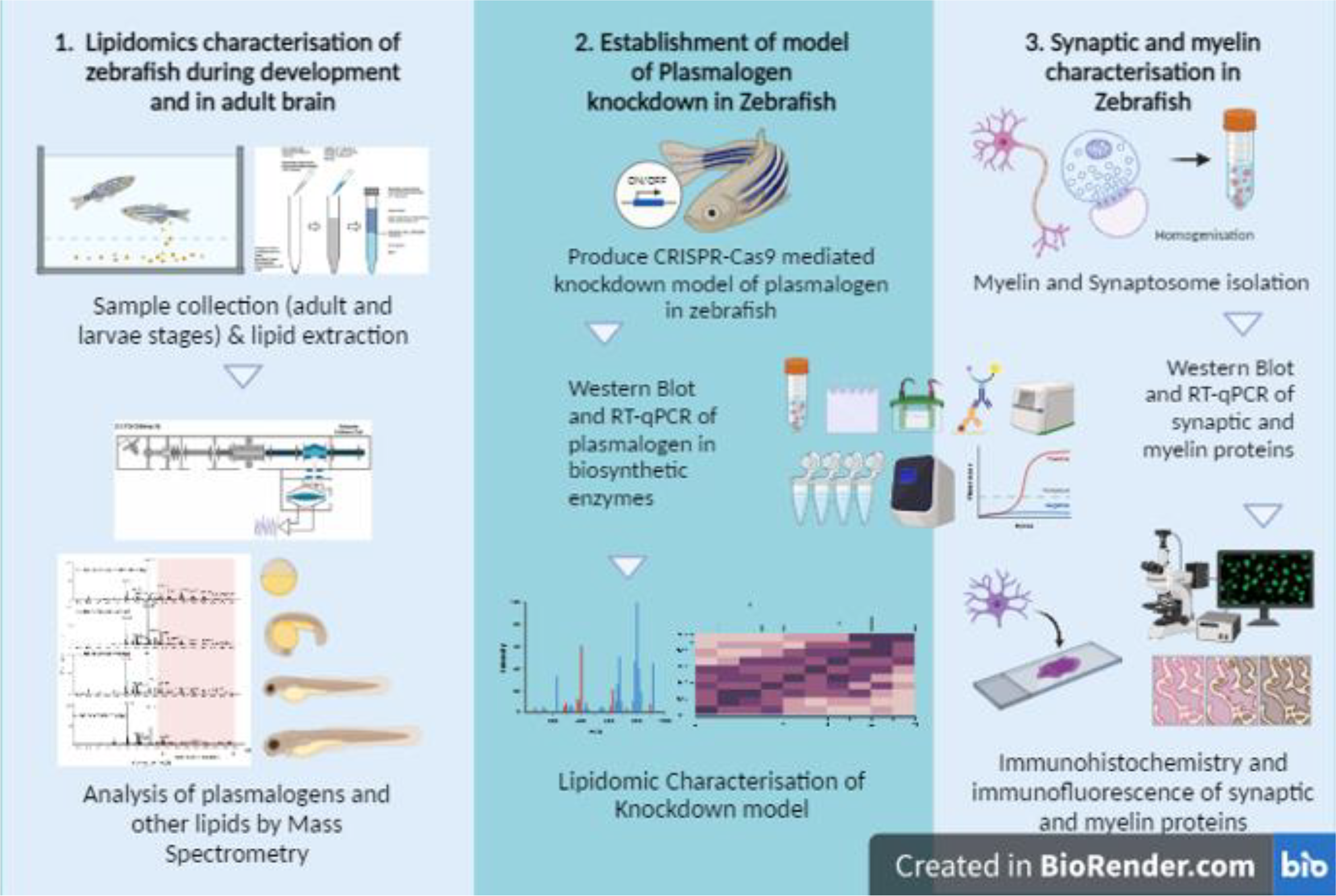
Neurolipidomics (Han, 2007) is an emerging field at the interface of lipid research and neuroscience, aiming to illuminate how brain lipids function and how their dysregulation contributes to neurological disorders. Lipids—such as fatty acids, cholesterol, and phospholipids—play fundamental roles in glial and neuronal health. When lipid metabolism goes awry, it can lead to or exacerbate neurodegenerative conditions like Alzheimer’s disease (AD) (William et al., 2010; Kunkle, 2019).
Among phospholipids, ethanolamine plasmalogen (PE-p) has garnered particular attention for its apparent link to AD: lower PE-p levels correlate with cognitive decline and disease severity (Kling et al., 2020). Despite this importance, PE-p’s presence and function in the brain of the zebrafish (Danio rerio) remain unclear. Zebrafish are a popular model in neuroscience because of their human-like nervous system, high fecundity, and relatively short life cycle (Stewart et al., 2014). Their fully sequenced genome and transparent embryos support diverse experimental approaches throughout development (Xi et al., 2011; Kalueff et al., 2014). Still, the lipid composition of zebrafish remains insufficiently characterized, particularly regarding PE-p. Conflicting reports suggest uncertainty about its presence in this species (Van Amerongen, 2014; Fraher, 2016), although plasmalogens have been detected in other fish (Chen, 2022).
Addressing this gap, our research set out to definitively confirm the presence of PE-p and characterize its potential roles in the zebrafish brain. We employed shotgun lipidomics to analyze zebrafish at various developmental stages (0, 24, 48, 72 hours, and 5 days post-fertilization) and in adults (8 months, both sexes). Lipids were extracted using the Bligh and Dyer method, followed by targeted MDSL-MS (multi-dimensional shotgun lipidomics mass spectrometry) on an LTQ-Orbitrap XL (ThermoFisher Scientific) with a TriVersa NanoMate (Advion Biosciences), in accordance with established protocols (Nielsen et al., 2020). Plasmalogen species were validated through MSn analyses (Hsu, 2018), and quantitative assessment utilized internal standards (PE 12:0, PC 12:0).
Our preliminary data confirm that PE-p is present throughout zebrafish development and in the adult brain, with seven candidate plasmalogen species identified. Future studies will extend this work to aging zebrafish, using gene silencing to deplete plasmalogens. This approach may clarify how variations in plasmalogen levels influence brain function and resilience to neurodegeneration, potentially translating into novel insights for conditions such as Alzheimer’s disease.
Aging and Neurodegeneration
PM_021
Keywords: APP, Synapse, Alzheimer's Disease, Integrin, Epilepsy
Authors: Ben Goult
Misprocessing of amyloid precursor protein (APP) is one of the major causes of Alzheimer’s disease. APP comprises a large extracellular region, a single transmembrane helix and a short cytoplasmic tail containing an NPxY motif (normally referred to as the YENPTY motif). Talins are synaptic scaffold proteins that connect the cytoskeletal machinery to the plasma membrane via binding NPxY motifs in the cytoplasmic tail of integrins. Here, we report the crystal structure of an APP/talin1 complex identifying a new way to couple the cytoskeletal machinery to synaptic sites through APP. Proximity ligation assay (PLA) confirmed the close proximity of talin1 and APP in primary neurons, and talin1 depletion had a dramatic effect on APP processing in cells. Structural modelling reveals APP might form an extracellular meshwork that mechanically couples the cytoskeletons of the pre- and post-synaptic compartments. We propose APP processing represents a mechanical signalling pathway whereby under tension, the cleavage sites in APP have varying accessibility to cleavage by secretases. During synaptogenesis in healthy neurons, the APP/talin linkage would provide an exquisite mechanical coupling between synapses, with tightly controlled APP processing providing instructions to maintain this synchrony. Furthermore, APP directly coupling to the binary switches in talin indicates a role for APP in mechanical memory storage as postulated by the MeshCODE theory of a mechanical basis of memory.
This leads us to propose a new hypothesis for Alzheimer’s, where misregulated APP dynamics result in loss of the mechanical integrity of the synapse, corruption and loss of mechanical binary data, and excessive generation of toxic plaque-forming Aβ42 peptide.
Aging and Neurodegeneration
PM_022
Keywords: alzheimer's disease, gamma oscillation, microglia
Authors: Kai (Jerry) Lo & Michael M Kohl
Alzheimer's disease has become a leading cause of debilitating illness and death. Current treatments like Lecanemab (Leqembi) can cause side effects such as cerebral oedema and haemorrhage. In this study, I aim to use a non-invasive method with minimal side effects to clear amyloid-beta plaques. I hypothesised that after twelve days of sensory stimulation, microglia would change in number and size, and amyloid plaques will reduce either in number or volume. I will also use the fear conditioning test to assess potential cognitive improvements in animals.
I used 12-month-old wildtype or APP NL-G-F/NL-G-F mice and subjected them to one hour of light (white LED) and sound stimulation at gamma frequency (40 Hz) for twelve consecutive days. A one-hour light-off period served as control. This was followed by contextual fear conditioning (cFC) and post-mortem histological analysis of microglia and amyloid beta plaques.
I observed a significant difference in the freezing rate during the test in wild-type mice with sensory stimulation (40Hz light and sound) but no difference in the wild-type dark group. As for the APP mice, I observe difference in the freezing rate on the test day between the 40Hz group and the dark group. I also found a slight increase in the number of inactive microglia with smaller sizes (10-1000μm) in the RSC brain region after sensory stimulation in both APP and wild-type mice. However, there was no change in the CA1 region. For active microglia with larger sizes (over 1000μm), which are in a phagocytic state, there was a decrease in both their number and percentage area in the CA1 region, but I did not observe the same change in the RSC. Multimodal sensory stimulation resulted in a significant reduction in the area covered by amyloid plaques in the CA1 region but had no effect in the RSC. This result may indicate a potential decrease in amyloid-β plaques, reducing the need for active microglial intervention. However, further clarification and investigation are required.
Sensory stimulation alters microglial morphology and amyloid plaques with region-specific effects in the RSC and CA1 of mice, suggesting its potential as a therapeutic approach for Alzheimer’s disease.
Aging and Neurodegeneration
PM_023
Keywords: Glymphatics, alpha-Synuclein, Synucleinopathy, Aquaporin-4
Authors: Annabell Rickert, Douglas M Lopes, Sophie K Llewellyn, Jack A Wells, Guglielmo Verona, Mark F Lythgoe, Ian F Harrison
The glymphatic system mediates the clearance of extracellular solutes from the cerebral interstitial space. Misfolded proteins forming aggregates and causing neurodegeneration, e.g. alpha-Synuclein (αSyn), can be removed from the brain via this system, highlighting its therapeutic potential. As a key component of the glymphatic system, the water channel Aquaporin-4 (AQP4) has gained scientific interest regarding its role in the spread of pathology and disease progression. Recent work showed that inhibition of AQP4 leads to exacerbated aggregation of αSyn; enhancing AQP4 function may therefore increase the clearance of extracellular αSyn, and slow its neuron-to-neuron propagation. This study will evaluate the therapeutic efficacy of pharmacological AQP4-facilitation in synucleinopathy using the novel compound TGN-073. Moreover, we will investigate the potential mechanisms-of-action via which TGN-073 mediates AQP4-facilitation, with AQP4 and the dystrophin-associated complex (DAC) as potential targets.
hSNCA-A53T mutant mice were inoculated with αSyn pre-formed fibrils. Mice received TGN-073 (or vehicle) intraperitoneally 3 times per week for 6 weeks. Open field data was acquired prior, during and after the treatment course. Structural MRI scans were acquired before brains were collected for biochemical analysis.
Image analysis and biochemical investigation will be employed to determine the efficacy of pharmacological AQP4-facilitation as a therapeutic approach against αSyn pathogenesis. Animal tracking parameters from open field videos will be acquired to determine alterations in locomotion and thigmotaxis. Structural MRI data will be manually segmented for volumetric analysis of nigro-striatal pathways to determine degrees of brain atrophy. Extraction of RNA and protein will be conducted to quantify αSyn deposition and dopaminergic neurodegeneration, using Western blot. Dot blot quantification of αSyn in CSF samples will serve to identify potential corresponding changes in CSF αSyn load. Further Western blotting will be utilised to investigate the drug’s potential mechanism-of-action, by studying AQP4 polarisation, and RNA from brain samples will be used for RT-qPCR to determine any potential expression changes in AQP4 and the DAC.
Statistical analysis will be conducted using either one-way or two-way ANOVA to determine any significant between-group differences (p < 0.05). Pearson’s correlation coefficient will be calculated to detect/quantify any correlations across obtained parameters. For all experiments, n = 12 per group will be used.
These data will represent the first investigation of the potential of an AQP4 targeting agent as a therapeutic intervention in synucleinopathy. If successful, it will provide rationale for further investigation of this target for disease modification in neurodegenerative disease.
Aging and Neurodegeneration
PM_024
Keywords: Mitochondria, Tau, Two-Photon, Barasertib, GCaMP
Authors: Marie Sabec, Michael Ashby
Mitochondria are actively transported along axons to presynaptic sites, where they support neural health and function as local sites for both calcium regulation and energy production. The importance of mitochondrial trafficking is highlighted by the range of disorders associated with disrupted mitochondrial motility, including neurodegenerative tauopathy. We therefore used PS19 mice, which carry a P301S mutation associated with frontotemporal dementia, to investigate the impact of pathological tau on mitochondrial trafficking in vivo and to examine the subsequent consequences.
We employed a combination of in vivo and ex vivo optical strategies to assess mitochondrial dynamics, calcium activity, and histopathology. Mice were injected with viral vectors into the cortex to dual label axons and mitochondria with distinct fluorescent constructs within the same neuron. Dynamic mitochondrial trafficking (Mito-STagRFP) and axonal calcium transients (axon-GCaMP8m) were then recorded through implanted cranial windows using two-photon microscopy. In parallel to the dynamic imaging, immunofluorescent staining and quantitative confocal analysis was conducted to evaluate concurrent changes in markers of synaptic, axonal, and neuronal health in post-mortem brains. Finally, the influence of pharmacological intervention using an aurora kinase B inhibitor, Barasertib, to up-regulate mitochondrial trafficking in PS19 and control mice was tested. Data were statistically analysed using general linear models, t-tests, or chi-square tests of proportions, as appropriate.
We show that the P301S tau mutation induced a significant reduction to motile mitochondria in an age-dependent manner. These trafficking deficits emerged before detectable structural degeneration. However, they were associated with a significant de-correlation of presynaptic axonal activity in the tau-burdened brains. Together, this study demonstrates the pathological cascade triggered by tau-mediated transport disruption and highlights the therapeutic potential of targeting early mitochondrial trafficking deficits.
Aging and Neurodegeneration
PM_025
Keywords: Normal ageing, Diffusion tensor imaging, Glymphatic function, Perivascular space, Cognition
Authors: Zeyan LI, Liwei GUO, Hanna LU
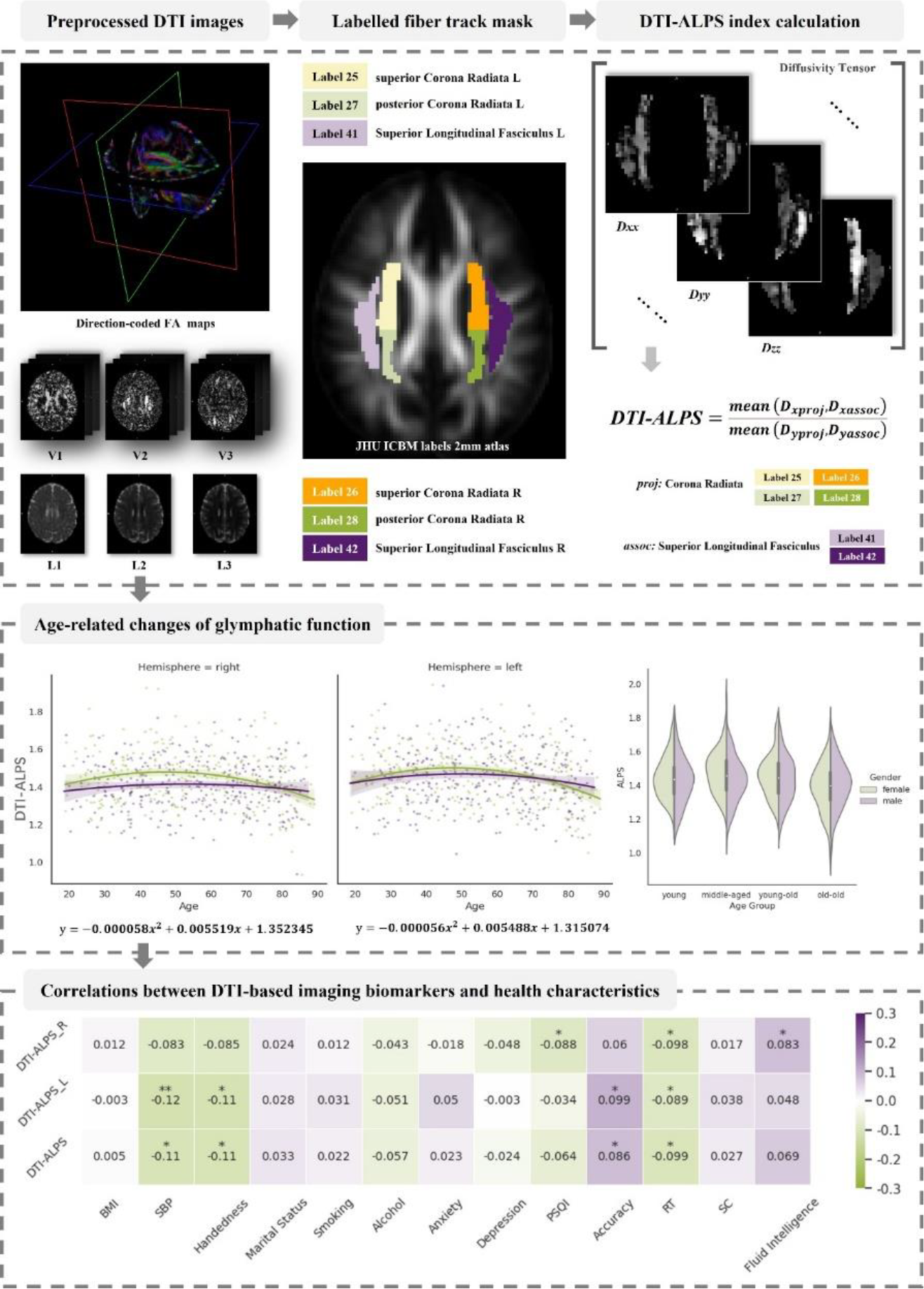
The study investigated temporal changes within the cerebral environment during normal ageing, using the non-invasive Diffusion Tensor Imaging (DTI) data. We quantitatively evaluate the diffusivity along the perivascular space (ALPS) to explore the age-related changes in glymphatic function, with a novel marker on the DTI-ALPS index. We examined the relations between imaging biomarkers, health-related characteristics, considering age as a risk factor influencing glymphatic system functionality.
Dataset from the Cam-CAN project was used in this study, comprising a total of 582 neurologically healthy adults aged 18 to 88 years. To facilitate our analyses, a standard pipeline was developed to calculate the DTI-ALPS index, thereby enabling the assessment of glymphatic function. Statistical Analysis Regression and correlation analyses were conducted to determine the trajectory of the DTI-ALPS index decline and investigate the relationship between the glymphatic function and health-related characteristics.
Aging and Neurodegeneration
PM_026
Keywords: Brain Slice Cultures, Lipopolysaccharide, Neuroinflammation, Synapse Loss, Electrophysiology
Authors: Lewis Taylor, Soraya Meftah, Calum Bonthron, Robert McGeachan, Imran Liaquat, Sam Booker, Paul Brennan, Claire Durrant
Neuroinflammation likely contributes to the progression of neurodegenerative diseases including Alzheimer’s disease, Parkinson’s disease and Multiple Sclerosis. However, the effects of neuroinflammatory pathways on synaptic and neuronal structure and function, the mechanisms by which these effects occur and whether they can be pharmacologically reversed remain to be fully investigated. Here, we treated murine organotypic hippocampal slice cultures (mOHSCs) with lipopolysaccharide (LPS), a toll-like 4 receptor agonist, to investigate the effects of a pro-inflammatory state on synaptic and neuronal integrity.
A single LPS concentration, 1 μg/ml, was used to induce neuroinflammation in mOHSCs from wild-type P6-9 mouse pups. To confirm the pro-inflammatory response following shorter and longer LPS exposure durations, mOHSCs were immunostained for GFAP, for reactive astrocytes, and IBA-1, for activated microglia, following both 24 hours and 7 days of LPS treatment. TNF-α protein concentration in slice culture medium from separate mOHSCs was quantified using enzyme-linked immunosorbent assay (ELISA). Excitatory and inhibitory synaptic protein expression in 7-day-treated mOHSCs was assessed using western blot. In separate 7-day-treated mOHSCs, CA1 pyramidal neuron synaptic activity was assayed in voltage-clamp mode, and neuronal excitability was assayed in current-clamp mode, using whole-cell patch-clamp electrophysiology. For western blot and electrophysiology experiments, paired t-tests were performed using a linear mixed effects model: Dependent Variable ~ Treatment + Sex + (1|Litter/Animal). For the ELISA experiment, a three-way ANOVA was performed using a linear mixed effects model: Dependent Variable ~ Treatment*Timepoint + Sex + (1|Litter/Animal).
In mOHSCs, 24 hours of LPS treatment increased the TNF-α protein concentration in slice culture medium (P < 0.001, N = 9 animals). Following both 24 hours and 7 days LPS treatment, a qualitative increase in astrocytic reactivity (GFAP signal) and microglial activation (IBA-1 signal) was observed. LPS lowered both excitatory (VGlut1: P = 0.003, PSD-95: P = 0.0005) and inhibitory (VGAT: P = 0.002, Gephyrin: P = 0.011) synaptic proteins to a similar magnitude (N = 11 animals) and decreased CA1 pyramidal neuron excitability (P = 0.012, N = 6 animals), but had a variable effect on synaptic activity, in mOHSCs.
This preliminary work provides a novel characterisation of the effects of lipopolysaccharide on excitatory and inhibitory synaptic structural and functional integrity in mOHSCs. Future experiments will optimise an effective LPS concentration in live human brain slice cultures (HBSCs), and characterise the impact of LPS on synaptic and neuronal physiology in HBSCs.
Aging and Neurodegeneration
PM_027
Keywords: Aging, Alzheimer's Disease, fNIRS, EEG, Phase Coherence
Authors: Juliane Bjerkan, Gemma Lancaster, Bernard Meglič, Jan Kobal, Trevor Crawford, Peter McClintock, Aneta Stefanovska
Vascular hypothesis of Alzheimer’s disease (AD) argues that vascular changes, such as impairment to the blood-brain-barrier and the neurovascular unit are critical events in causing neuropathology in AD [1]. Our aim was to investigate the phase interactions between cardiovascular oscillations and brain waves, and to establish how they change with AD by using novel non-linear methods of analysis.
As shown in figure B, the wavelet power of fNIRS signals, fNIRS phase coherence and fNIRS-EEG phase coherence were significantly reduced with age and further with AD [3,4]. results for oscillations with periods of 20-50 seconds are shown (0.052-0.145Hz, corresponding to what is known as myogenic oscillations [5]).
Our results confirm that AD is associated with altered cardiovascular and neurovascular dynamics. The observation of altered dynamics paves the way for a simple method to monitor the progression of Alzheimer’s disease non-invasively and evaluate the efficacy of treatments.
[1] de la Torre, J. C., & Mussivand, T. (1993). Can disturbed brain microcirculation cause Alzheimer's disease? Neurol. Res.,15(3), 146–153.
[2] Iatsenko, D., McClintock, P. V. E., & Stefanovska, A. (2016). Extraction of instantaneous frequencies from ridges in time-frequency representations of signals. Signal Process., 125, 290–303.
[3] Bjerkan, J., Lancaster, G., Meglič, B., Kobal, J., Crawford, TJ., McClintock, PVE. & Stefanovska, A. (2023) Aging affects the phase coherence between spontaneous oscillations in brain oxygenation and neural activity. Brain Res. Bull., 201, 110704.
[4] Bjerkan, J., Meglič, B., Lancaster, G., Kobal, J., Crawford, TJ., McClintock, PVE. & Stefanovska, A. (2025) Neurovascular dynamics is altered in Alzheimer’s disease. Brain Commun., in press.
[5] Stefanovska A. (2007). Coupled oscillators. Complex but not complicated cardiovascular and brain interactions. IEEE Eng. Med. Biol. Mag., 26(6), 25–29.

Aging and Neurodegeneration
PM_028
Keywords: white matter, cognition, ageing, cardiovascular, diffusion
Authors: Richard Henson, Petar Raykov
Magnetic Resonance Imaging (MRI) offers many ways to non-invasively estimate the properties of white matter (WM) in the brain. In addition to the various metrics derived from diffusion-weighted MRI, one can estimate total WM volume from T1-weighted MRI, WM hyper-intensities from T2-weighted MRI, myelination from the T1:T2 ratio, or from the magnetisation-transfer ratio (MTR).
Here we utilise the presence of all of these MR contrasts in a population based life-span cohort of 650 healthy adults [CamCAN cohort] to identify the latent factors underlying the covariance of 11 commonly-used WM metrics.
Four factors were needed to explain 89% of the variance, which we interpreted in terms of 1) fibre density / myelination, 2) free-water / tissue damage, 3) fibre-crossing complexity and 4) microstructural complexity. These factors showed distinct effects of age and sex. To test the validity of these factors, we related them to measures of cardiovascular health and cognitive performance. Specifically, we ran path analyses 1) linking cardio-vascular measures to the WM factors, given the idea that WM health is related to cardiovascular health, and 2) linking the WM factors to cognitive measure, given the idea that WM health is important for cognition.
Even after adjusting for age, we found that a vascular factor related to pulse pressure predicted the WM factor capturing free-water / tissue damage, and that several WM factors made unique predictions for fluid intelligence and processing speed. Our results show that there is both complementary and redundant information across common MR measures of WM, and their underlying latent factors may be useful for pinpointing the differential causes and contributions of white matter health in healthy aging.
Aging and Neurodegeneration
PM_031
Keywords: PET imaging, Huntington's disease, Protein aggregation, Disease monitoring, Imaging biomarker
Authors: Paul Sharp, Michael Fairclough, Rosemary Shoop, Tracy Hall, Dhifaf Jasim, Juliana Maynard
Huntington’s disease (HD) is a fatal neurodegenerative disorder caused by an expansion of CAG repeats in exon 1 of the human huntingtin gene (HTT). The expanded HTT sequence encodes unstable mutant huntingtin (mHTT) protein that aggregates and plays a key pathophysiological role in selective neuronal loss. Several therapeutic strategies aimed at lowering mHTT are under development and as such sensitive non-invasive PET-based methods that directly measure mHTT would be of great benefit in assessing treatment response in preclinical and human trials. The CHDI foundation developed the first-generation PET ligands based on the 11C-radioisotope, which advanced to clinical evaluation. Further optimization generated an 18F radiolabel with improved metabolic stability, brain exposure and specificity, as defined by PET imaging in wild-type mice and autoradiography in HD brain samples. Here, we characterised the PET radioligand [18F]CHDI-650 in the R6/1 mouse model of HD to determine its sensitivity for monitoring treatment response of mHTT lowering therapies.
[18F]CHDI-650 was produced using an automated TRACERlab FX-FE radiochemistry system. Radiochemical purity and molar activity of the final [18F]CHDI-650 product was calculated following reverse-phase HPLC analysis. Dynamic PET/CT images were acquired on Molecubes (Bruker) scanners. In vivo imaging was conducted using hemizygous R6/1 mice and wild-type littermates (both sexes) at various disease stages (7-16 weeks), including the early presymptomatic period. Image processing and kinetic modelling (2TCM) analyses were performed using PMOD software. Ex vivo brain radioactivity was measured using the Wizard2 automated gamma counter (Perkin Elmer).
Statistical analyses were performed with GraphPad Prism (Version 9). Quantitative data are expressed as mean ± SEM. For two group comparison, unpaired two-tailed t-tests were used, and statistical significance was set at p < 0.05.
[18F]CHDI-650 was produced (1.3 ± 0.2 GBq) with an average radiochemical purity of 99.9 ± 0.1 % and a molar activity of 32.4 ± 5.2 GBq/µmol (n = 9). The radioligand was able to discriminate R6/1 HET from WT mice at an early presymptomatic age for both dynamic PET imaging (striatal volumes) and ex vivo whole brain gamma. A well defined age associated increase was also evident using both measures. Therefore, [18F]CHDI-650 PET shows great potential as a non-invasive tool for visualising the development of mHTT pathology and as a biomarker for preclinical efficacy studies.
Aging and Neurodegeneration
PM_035
Keywords: ataxia, cerebellum, neurotrophins, cerebellar granule cells, TrkB signalling
Authors: Elena Eliseeva, Mohd Yaseen Malik, Liliana Minichiello
Ataxia disorders, such as spinocerebellar ataxia 6 (SCA6), are characterised by motor incoordination often caused by cerebellar dysfunction. Cerebellar Purkinje cell (PC) degeneration is commonly linked to ataxia, and emerging evidence suggests that disrupted BDNF-TrkB signalling contributes to PC dysfunction and motor incoordination in SCA6. However, the impact of disrupted TrkB signalling in cerebellar granule cells (GCs) on PC function and motor coordination is unclear. Since TrkB receptors are more abundant in GCs, which provide extensive input to PC, dysfunctional TrkB signalling in GCs may impair PCs and contribute to ataxia. Using TrkbPenk-KO mice, in which TrkB was ablated from a specific subset of GCs, we investigated whether this dysfunction is sufficient to induce ataxia.
Motor coordination and gait were assessed with ledge and CatWalk tests, while molecular and cellular cerebellar changes were explored using Western blotting and immunohistochemistry. Since TrkbPenk-KO mice also lack TrkB in a subset of striatal neurons, we are employing a targeted rescue of striatal dysfunction to isolate cerebellar contributions to the ataxia phenotype.
Statistical Analysis Assumptions of normality of residuals and equality of variances were tested. Two-tailed unpaired t-tests were used for parametric data, and Mann-Whitey tests for nonparametric data. Bonferroni-Dunn or Holm-Bonferroni corrections were applied for multiple comparisons.
At 3 months, TrkbPenk-KO mice exhibited motor incoordination with a higher (worse) ledge score (p = 0.005; controls, n = 13, median score = 0; mutants, n = 12, median score = 2). CatWalk detected gait deficits in TrkbPenk-KO mice (Fig.1). Despite these motor deficits, cerebellar morphology and levels of selected synaptic markers were unaffected. Ongoing striatal rescue experiments will delineate striatal versus cerebellar contributions to ataxia.
Figure 1. Altered gait in TrkbPenk-KO mice at 3-5 months. (A) CatWalk footprints of TrkbPenk-KO and control mice. (B) Print positions of left paws (red frame in A) were elevated in TrkbPenk-KO mice (p = 0.015, n = 5 controls, 9 mutants). (C) CatWalk gait diagrams. (D) Left hind swing duration (red arrows in C) was reduced in TrkbPenk-KO mice (p = 0.029, n = 5 controls, 9 mutants). Paw colours: right front (blue); right hind (pink); left front (yellow); left hind (green).
These findings provide insight into how GC-specific TrkB signalling contributes to motor incoordination and suggest that GC dysfunction should be considered in cerebellar ataxias. Ongoing research will clarify cerebellar-striatal circuit interplay in motor coordination and gait performance.

Aging and Neurodegeneration
PM_039
Keywords: ALZHEIMER’S DISEASE, TYPE TWO DIABETES, BLOOD BRAIN BARRIER
Authors: Inés González-Reyes, Ángel Del Marco, Alan W. Stitt, Rafael Simó, Mónica García-Alloza
Age is the main risk factor for developing Alzheimer's disease (AD), the leading cause of dementia. However, previous studies show that type 2 diabetes (T2D) also increases the risk to suffer AD. In addition, both diseases share some pathological features, such as alterations of the blood brain barrier (BBB). Therefore, we have assessed the brain vasculature in a mixed murine model of AD and T2D, the APP/PS1xdb/db mouse as diseases progress.
Structural BBB alterations were analyzed in control, AD (APP/PS1), T2D (db/db) and AD-T2D (APP/PS1xdb/db) mice at 4 (before the onset of AD), 14 (when T2D has commenced), and 26 weeks of age (when both diseases are fully established). Cortical acellular capillaries were analyzed by immunostaining using anti-collagen IV antibody and isolectin B4 (IB4). Neuron-glia antigen 2 (NG2), glial fibrillary acidic protein (GFAP) and aquaporin 4 (AQP4) immunostainings were performed to analyze pericytes, astrocytes and their water channels respectively. Cerebral amyloid angiopathy (CAA) was analyzed by immunostaining with 4G8 antibody. The statistical analysis was performed by one-way ANOVA, followed by Tukey B or Tamhane tests, as required. Student’s t-test for independent samples was used when 2 populations were compared.
We observed an overall increase of cortical acellular capillaries at late stages of the diseases (26 weeks of age) in AD, T2D and AD-T2D mice. In this line, a reduction of isolectin B4 and AQP4 levels was also detected when individual vessels were analyzed at 26 weeks of age. On the other hand, CAA is increased in AD-T2D animals when compared with AD mice. Interestingly, other vascular markers including pericyte and astrocyte end-feet densities were altered in AD and T2D mice at early ages (4 weeks of age), and a synergistic effect was observed in AD-T2D mice as diseases progressed, supporting the accelerator role of T2D on AD .
Our data show the disruption of the BBB structure as AD and T2D progress and support a synergistic cerebrovascular damage in APP/PS1xdb/db mice, when AD and T2D coexists in the long term.

Aging and Neurodegeneration
PM_059
Keywords: Vasomotion, Neurovascular Coupling, Optical Imaging Spectros, Alzheimer's Diease
Authors: Runchong Wang, Shannon O’Connor, Paul Sharp, Osman Shabir, Chris Martin, Clare Howarth, Michael Okun, Jason Berwick
Vasomotion refers to low-frequency vascular oscillations (~0.1 Hz) occurring across tissues, potentially serving as a biomarker and therapeutic target for neurodegenerative diseases. However, its origins, vascular structure in the brain, independence from neural activity, and alterations in Alzheimer’s disease (AD) remain unclear.
This study analysed data from anaesthetised rats (Hooded Lister, female, 12–18 months) and mice (wildtype: C57bl/6; AD: hAPP-J20; male, 9–12 months). Neuronal and haemodynamic activity were recorded using a 16-channel electrode and 2D optical imaging spectroscopy, respectively. Rats also had tissue oxygen measured via an oxygen probe (OxyLite Pro, Oxford Optronix UK). Vasomotion was manipulated using blood pressure changes in rats, and inhaled oxygen levels (100% oxygen vs. medical air, to alter tissue oxygen level) in mice. The power of low-frequency total haemoglobin concentration (HbT) oscillations (~0.1 Hz) was calculated. Kernel regression analysis assessed the linear relationship between HbT time series and neuronal activity, with predicted HbT calculated by convolving neural activity and the kernel. Independence of vasomotion from neural activity was evaluated based on the correlation between measured and predicted HbT across conditions.
A mixed ANOVA assessed HbT power, neural activity, and vasomotion independence across conditions (and groups for mice).
Rats: HbT oscillations (~0.1 Hz) were driven by the arterial tree, causing pronounced saturation changes in downstream veins. These oscillations were associated with low tissue oxygen and were largely independent of neuronal activity. Thus, vasomotion in rats (~0.1 Hz) is not driven by spontaneous neurovascular coupling. Its correlation with tissue oxygen suggests that vasomotion may be modulated by oxygen levels, a hypothesis tested in mice.
Mice: Experimental manipulations altered HbT and neural activity power (~0.1 Hz), but the proportion of HbT predicted by neural activity remained unchanged, suggesting neural activity-derived HbT power changes. Group differences between wildtype and AD mice in 0.1 Hz HbT power across manipulations were only observed when electrodes were implanted.
In mice, oxygen level manipulations influenced vascular oscillation power, largely explained by spontaneous neuronal activity. Group differences were only observed with electrode implantation, suggesting cortical spreading depression as a contributing factor. Future studies should explore non-invasive methods (e.g., widefield calcium imaging) to compare AD and wildtype mice.
In conclusion, the results reveal species-specific drivers of low-frequency HbT oscillations, highlighting the need for further investigation into these mechanisms and improved methods for manipulating vasomotion (independent of spontaneous neurovascular coupling) in mice.
Aging and Neurodegeneration
PM_066
Keywords: Parkinson's disease, white matter, diffusion imaging
Authors: Bethany Facer, Corey Ratcliffe, Antonella Macerollo, Simon Keller
Parkinson’s disease (PD) is the second most prevalent neurodegenerative disorder, primarily affecting the substantia nigra and other areas of the basal ganglia. The disease presents with clinical heterogeneity and symptoms beyond motor impairment, and likely impact multiple brain areas. Fixel-based analysis (FBA), measures intra-axonal volume specific to fibre populations (fibre density (FD)), the macroscopic cross-sectional size of fibre bundles (FC), and combined metrics (FDC). Previous studies have identified increased FC in the corticospinal tract in tremor-dominant PD (TD-PD). However, the dentato-rubro-thalamic tract (DRTT)—a key pathway connecting the cerebellum to the thalamus—remains underexplored. This study aims to investigate whether white matter microstructural abnormalities of the DRTT exist in patients with de-novo PD using FBA.
Data was obtained from the Parkinson’s Progression Markers Initiative, including DTI and clinical data from individuals with de novo PD. Patients were stratified into tremor-dominant (TD) and postural instability gait difficulty (PIGD) groups to examine motor phenotype-specific white matter alterations. Data preprocessing and FBA were performed using MRtrix3. A DRTT mask was delineated on the white matter population template, defined by manual inclusion and exclusion ROIs. GLMs were conducted using fixelcfestats to assess between-group differences in fixels across the whole brain followed by the DRTT. Connectivity-clustered family-wise error corrected fixel metric outputs and standardised effect sizes (ES) were overlaid on the template and visually inspected for anatomical consistency, and for local maxima.
A total of 65 controls (24 females; mean age 60.3 ± 10.9), 103 tremor-dominant PD (TD-PD) patients (39 females; mean age 61.5 ± 8.9), and 31 PIGD-PD patients (9 females; mean age 64.1 ± 9.1) were included. In an exploratory whole brain analysis, patients with TD-PD showed significantly larger FC (p= .015, ES=0.931) and trending increases in FDC (p=.078, ES=0.795) in the cerebellum compared to controls. Controls exhibited trending higher FD values compared to patients with PIGD-PD (p= .149, ES=1.062) in the occipital lobe and TD-PD (p= .172; ES=0.844) in the parietal lobe. In the DRTT specific analysis, trending higher FC values were observed in PIGD-PD relative to controls (p= .124, ES=0.711).
Consistent with previous studies, we observed whole-brain differences in FC and FDC between TD-PD and controls. This study extends prior findings, as the increased DRTT FC observed in PIGD-PD compared to controls could suggest a potential compensatory mechanism, with a larger fibre bundle cross section in this tract potentially mitigating tremor symptoms.
Aging and Neurodegeneration
PT_003
Keywords: Huntington's disease, Neurotrophin Signalling, BDNF-TrkB, Dopamine, Energy metabolism
Authors: Mohd Yaseen Malik, Fei Guo, Liliana Minichiello
Disrupted brain-derived neurotrophic factor (BDNF) signalling through its receptor tropomyosin receptor kinase B (TrkB) plays a crucial role in Huntington's disease (HD), but its precise contribution has been difficult to determine. To better understand its role, particularly in presymptomatic HD, we investigated BDNF-TrkB signalling in indirect spiny projection neurons (iSPNs), particularly susceptible to HD pathology.
We used an in-house (C57BL/6J;129), Trkb conditional knock-out (TrkbPenk-KO) mouse model and wildtype littermates at 3 and 8 months (M) of age. We employed immunohistochemistry (IF), immunoblotting (IB), and HPLC to study the dopamine (DA) dynamics. RNA-seq analysis was used to study the transcriptional profile of relevant neurons in the presence and absence of TrkB signalling at 3 and 8M. High-resolution respirometry (HRR) was used to analyse the various components of oxidative phosphorylation and electron transport system. Behavioural analysis was performed using different tasks, including open-field recordings.
Two-way ANOVA was used to analyse the behavioural data and HRR data. Other data were analysed using two-tailed unpaired Student’s t-tests and/or one-way ANOVAs. For detailed methods, see [1].
TrkbPenk-KO mice at early ages (3M) showed a significant increase in TH protein expression (IF, p = 0.003, n=3), TH protein levels (IB, p = 0.02, n=4) and DA concentrations (HPLC, p = 0.030, n=4). No such differences were seen at 8M. Transcriptomic analysis showed substantially altered striatal glutathione metabolism-related genes (Gsto2 upregulation, adj p = 4.2 × 10⁻⁷, n=6) and energy metabolism deficits at 3M. Gsto2 upregulation preceded the onset of hyperdopaminergia. HRR study validated the impaired energy metabolism results, showing a substantial decrease in all major respiratory states in the striatum in mutant mice at 3M, exacerbating with age (8M). Mutant mice reared significantly less (p = 0.023) from an early age and eventually developed a hyperlocomotor phenotype at 8M (p = 0.022). Selective Gsto2 knockdown prevented the development of hyperdopaminergia, energy metabolism deficits, and the onset of motor symptoms in TrkbPenk-KO mice [1].
Our findings underscore the critical role of BDNF-TrkB signalling in striatal protection, dopamine circuitry, energy metabolism, and the regulation of cellular metabolic pathways, such as glutathione–ascorbate homeostasis. Dysfunction in these pathways can trigger neurodegeneration and motor dysfunction.
1. Malik, M.Y., Guo, F., Asif-Malik, A., Eftychidis, V., Barkas, N., Eliseeva, E., Timm, K.N., Wolska, A., Bergin, D., Zonta, B. and Ratz-Wirsching, V., 2024. Impaired striatal glutathione–ascorbate metabolism induces transient dopamine increase and motor dysfunction. Nature Metabolism, pp.1-18.
Aging and Neurodegeneration
PT_004
Keywords: Neurodegeneration, Motor neuron disease, Gene therapy
Authors: Puja Mehta, Tomas Solomon, Peter Harley, Simone Barattucci, Adrian Isaacs, Matthew Keuss, Pietro Fratta, Marc-David Ruepp
Amyotrophic lateral sclerosis (ALS), or motor neuron disease, is a rapidly progressive paralysing illness resulting from the neurodegeneration of upper and motor neurons. The lifetime risk is 1 in 300, with a median survival of 3 years, and there is only one globally licensed disease-modifying treatment that prolongs life by 2-3 months. There is a pressing need for novel therapies. ~97% of people with ALS exhibit the pathological hallmark of 'TDP-43 proteinopathy', characterised by the nuclear-to-cytoplasmic mislocalisation of the RNA-binding protein, TDP-43. The nuclear depletion of TDP-43 is a key driver of ALS pathophysiology, through the resultant mis-splicing of pre-mRNAs. This leads to erroneous inclusion of usually intronic sequences, called "cryptic exons (CEs)", in mature mRNA transcripts, and resultant loss of proteins crucial for neuronal function. CEs in the STMN2 gene (encoding a protein crucial for neurite growth) and UNC13A gene (encoding a protein crucial for synaptic transmission) have been shown to have an important mechanistic role in ALS. Thus, novel splicing therapies targeting these functionally relevant CEs are needed.
A modified U7 small nuclear RNA (snRNA) gene therapy approach has been developed to correct cryptic splicing seen in ALS. Using human iPSC-derived neurons, their ability to rescue RNA and protein levels, as well as relevant functional phenotypes were investigated in vitro.
At least three biological replicates were performed. Data were determined to be parametric or non-parametric before applying the appropriate statistical analyses.
Our U7s successfully correct STMN2 and UNC13A mis-splicing and rescue of levels of both proteins in TDP-43-depleted iPSC-derived neurons, which serve as a model for TDP-43 nuclear loss seen in people with ALS. Crucially, our STMN2 U7 therapy rescues neurite outgrowth, and our UNC13A U7 therapy rescues synaptic activity. U7s are currently in a Phase I/IIa trial for Duchenne muscular dystrophy, but have not been used in ALS. Therapeutic U7s have the benefit of being stably expressed, negating the need for repeated, invasive intrathecal administration in patients, unlike with antisense oligonucleotides. Another advantage is the possibility of correcting multiple CEs, using one gene therapy construct, which is useful given the discovery of multiple disease-relevant CEs. As such, we have also developed a combined U7 that rescues STMN2 and UNC13A mis-splicing together. Building on the exciting discovery that CEs underpin ALS disease mechanisms, our findings have huge translational potential for people with ALS and other TDP-43 proteinopathies.
Aging and Neurodegeneration
PT_010
Keywords: cerebellum, hippocampus, ageing, functional connectivity, memory
Authors: Kavishini Apasamy, Samuel Berry, Marie-Lucie Read, Narender Ramnani, Carl Hodgetts
Evidence from nonhuman animals show that the hippocampus and cerebellum share close anatomical and physiological relationships (Watson et al., 2019). Electrophysiological evidence shows that cerebellar disruption influences the firing of hippocampal place cells and affects spatial navigation (Rochefort et al., 2011). Both of these structures are also thought to undergo similar age-related changes. Nonetheless, the hippocampal-cerebellar connectivity in the human brain is not understood well.
Seed-based connectivity analyses were performed on CamCAN resting-state fMRI data (N= 479, age range = 18-88) using CONN Toolbox. Hippocampal regions of interest were created using Harvard-Oxford and Julich Histological atlases. Time series for four hippocampal seeds (left, right, anterior and posterior) were entered as co-variates in separate General Linear Models (GLMs) to obtain correlations between these seeds and every other voxel in the brain. To understand ageing effects, a second set of GLMs included age as an additional subject-level effect. Connectivity with the cerebellar cortex was visualised using SUIT.
We found widespread functional connectivity between the hippocampus and the cerebellar cortex at the border of lobule HV and HVI, lobules HIX and HVIIA (Crus I and II). Left and right hippocampi connected with a contralateral region of lobule HVIIA. Anterior hippocampus connected bilaterally with areas in lobule HVIIA (Crus II) whilst the posterior hippocampus connected with lobule V. We also found age-related reductions in functional connectivity between hippocampal areas (left, right and anterior) around the primary fissure.
These results give novel insights into the organisation of the hippocampal-cerebellar connectivity in the human brain and aligns with previous nonhuman animal evidence (e.g., connectivity with lobules HVI and HVIIA). Additionally, we observed connectivity with lobules HIX and HX. Anterior and posterior hippocampal areas are known to have distinct functional connectivity profiles and in our study they also connect with distinct cerebellar areas. Decreased functional connectivity during ageing, in lobules HV and HVI, resonate with findings that show age-related atrophy in lobule HVI accompanied by hippocampal-dependent navigation deficits (Ramanoël et al., 2013). Our future work will investigate the possible role of cerebellar forward models in the automation of hippocampal processes.
Rochefort C et al. (2011) Cerebellum shapes hippocampal spatial code. Science 334: 385-389.
Watson TC (2019) Anatomical and physiological foundations of cerebello-hippocampal interaction. ELife 8
Ramanoël S et al. (2023) An appraisal of the role of the neocerebellum for spatial navigation in healthy aging. The Cerebellum 22: 235-239.
Aging and Neurodegeneration
PT_032
Keywords: Alzheimer's disease, Dendritic spines, Compensation
Authors: Ya Yin Chang, Jessica Willshaw, Zoran Boskovic, Tim Castello-Waldow, Annalisa Paolino, Patricio Opazo
The loss of excitatory synapses is known to underlie the cognitive deficits in Alzheimer’s disease (AD). Although much is known about the mechanisms underlying synaptic loss in AD, how neurons compensate for this loss and whether this provides cognitive benefits remains almost completely unexplored. Here, we provide evidence for two potential compensatory mechanisms implemented following synaptic loss: the enlargement of the surviving neighbouring synapses and the regeneration of synapses.
Second, we developed opto- and chemogenetic tools for the artificial elimination of dendritic spines with high spatiotemporal control. Similar to AD, we found that the elimination of dendritic spines using chromophore-assisted light inactivation (CALI) in WT rat hippocampal organotypic slices led to the enlargement of the remaining synapses (p<.01). More remarkably, we discovered that synapses were regenerated one week after their loss both in-vitro (p<.01) and in-vivo (p<.01) preparations.
Data obtained from in-vitro organotypic slice culture imaging was carried out either using a general linear mixed-effects model (treatment with oAβ) or t-test (optogenetic photoactivation of Drebrin-KillerRed). The general linear mixed-effects model explores differences between control and treated slices while accounting for fixed and random effects simultaneously. In our analysis, individual animals (from which multiple slices could be obtained) were included as a random variable to avoid statistical pseudoreplication. Data obtained from in-vivo imaging was carried out using t-tests.
We believe that understanding the molecular mechanisms behind these forms of synaptic compensation and regeneration will be critical for the development of therapeutics aiming at delaying the onset of cognitive deficits in AD.
Aging and Neurodegeneration
PT_033
Keywords: Human Brain Slice Culture, Biomarkers, Pathology, Amyloid, Tau
Authors: Soraya Meftah, Lewis Taylor, Robert McGeachan, Calum Bonthron, Danilo Negro, Declan King, Jane Tulloch, Imran Liaquat, Tara Spires-Jones, Sam Booker, Paul Brennan, Claire Durrant
The absence of translatable models for dementia is a significant barrier to the development of effective treatments and a cure. To address this, we have developed a live human brain slice culture model derived from adult human brain tissue, providing a valuable platform to study dementia-related processes in an ex vivo environment. We sought to characterise how patient characteristics such as age, sex, AOPE genotype and brain region relates to dementia-associated proteins released by the slice cultures into the medium.
Access human brain tissue, obtained from tumour debulking surgery, was cultured for up to 7 days in vitro (DIV). The culture medium collected from slices at DIV 7 was analysed using ELISA and Luminex technology to measure levels of several dementia-associated proteins, including Amyloid-β1-40, Amyloid-β1-42, Tau, KLK-6, NCAM-1, Neurogranin, and TDP-43, in the culture medium derived from different brain regions (cortical). Additionally, samples were screened histologically for endogenous amyloid-β and tau pathology. Linear mixed models were used to assess statistical differences, using age as a random factor. Different patient characteristics such as brain region or sex were selected as fixed factors to investigate the specific impact of these factors. Each analyte was individually tested for statistical differences.
Samples from 20 individual patients were used for biomarker analysis within this study. Within the cortex, increased NCAM-1 (p=0.02) and TDP-43 (p=0.03) release into the culture medium was seen in male patient samples compared to females. KLK-6 and tau showed differential release with APOE genotype and numerous markers (tau, KLK-6, NCAM-1, Neurogranin) showed differential release by brain region. The presence of tau, but not amyloid pathology, led to alterations in the release of KLK-6 and the ratio of amyloid-β1-42:1-40 ratio.
This novel human brain slice culture model offers a unique approach to studying the molecular and pathological features of dementia. Our results demonstrate that cultured human brain slices exhibit protein expression patterns that recapitulate some aspects observed in patients with dementia. This model provides a translatable platform to further understand dementia pathogenesis and screen for potential therapeutic targets.
Aging and Neurodegeneration
PT_034
Keywords: Alzheimer's Disease, Microglia, ABI3,
Authors: Shania Ibarra, Katerina Gospodinova, Emma Mead
Alzheimer’s disease (AD) is the most common form of dementia, affecting millions worldwide, with limited disease-modifying treatments currently available. Microglial dysfunction plays a key role in disease progression, and ABI3, an adaptor protein involved in actin cytoskeleton dynamics, has been genetically linked to AD. The AD risk ABI3 S209F variant is associated with reduced phosphorylation, potentially affecting ABI3 function and thus modulating microglial phagocytosis and migration to sites of damage. The kinases responsible for ABI3 phosphorylation remain unknown. This project aims to characterise ABI3’s role in human induced pluripotent stem cell (hiPSC) derived macrophages and identify kinases that regulate ABI3 phosphorylation for potential therapeutic intervention.
Isogenic hiPSC lines with ABI3 knockout (KO) and S209F knock-in (KI) mutations were generated using CRISPR-Cas9 and differentiated into macrophages following protocols from van Wilgenburg et al. (2013) and Haenseler et al. (2017). Macrophages were characterised by western blotting and flow cytometry. Microglial functions, including phagocytosis and migration, were assessed using live imaging on the Incucyte.
XY plots and bar charts were generated using GraphPad Prism v10.3.1. Data were collected from a minimum of three independent experiments. All values are presented as the mean ± standard deviation (SD). Statistical analysis was performed using one-way ANOVA followed by Dunnett's multiple comparison test. Statistical significance was determined at p < 0.05.
Flow cytometry confirmed similar expression of key macrophage cell surface markers in both parent and edited clones, indicating that the genetic modifications did not affect differentiation. Western blotting verified ABI3 deletion in KO clones and the presence of both phosphorylated and unphosphorylated ABI3 in parent macrophages, while S209F KI clones showed reduced phosphorylation.
Further analysis revealed that ABI3 KO and S209F KI significantly decreased the expression of the Wave Regulatory Complex (WRC) components CYFIP1 and NCKAP1L, potentially disrupting WRC formation. ABI3 KO macrophages exhibited impaired phagocytosis of apoptotic SH-SY5Y cells, whereas phagocytosis of zymosan bioparticles remained unaffected in both ABI3 KO and S209F KI macrophages. Additionally, preliminary data suggest that the loss of TREM2 signalling components may reduce total and phosphorylated ABI3 levels, pointing to a potential link between TREM2 activation and ABI3 phosphorylation.
These findings provide insights into ABI3’s role in microglial function and lay the groundwork for a kinase screening strategy to identify therapeutic targets.
Aging and Neurodegeneration
PT_036
Keywords: retrosplenial cortex, wandering, spatial disorientation, 5-Ht2 receptors, memory
Authors: Omar Ahmed
Most people living with Alzheimer’s disease (AD) suffer from pronounced spatial disorientation, contributing to the wandering symptoms associated with AD. Similar symptoms are seen in patients with specific lesions of the retrosplenial cortex (RSC), a brain region critical for spatial orientation and memory. The present study focused on understanding and repairing RSC cellular dysfunction in preclinical AD mouse models.
Using physiological, synaptic, and morphological analyses across 379 neurons (67 mice) we investigated cell-type-specific RSC impairments at two age points in 5xFAD mice and attempted to repair them using a single systemic dose of psychedelic drug 25CN-NBOH.
Statistical analyses of synaptic physiology and spine density were performed in GraphPad Prism (version 10.0.1). Data were tested to determine normal or lognormal distributions before being tested with parametric (unpaired t-test) or nonparametric (Mann-Whitney) tests, respectively.
The defining cell type of the RSC shows specific impairments in synaptic activity in 5xFAD mice at 12 months of age. A single psychedelic dose reverses this deficit, boosting synapses and spines onto these neurons. These results highlight cell-type-specific RSC dysfunction in AD and motivate further preclinical and translational evaluation of psychedelic medicine for alleviating spatial disorientation symptoms in AD.
Aging and Neurodegeneration
PT_037
Keywords: SV2A, Biomarker, PET, Array Tomography, Alzheimer's Disease
Authors: Lauren Young, Declan King, Ya Yin Chang, Kristjan Holt, Scott Gladstein, Sjoerd Finnema
Human in-vivo Positron Emission Tomography (PET) shows Synaptic Vesicle Glycoprotein 2A (SV2A) decreases in Alzheimer's disease (AD) patient brains compared to controls. Whether this decreasing signal reflects synapse loss or a loss of SV2A protein from remaining synapses is unclear. We use array tomography, a high-resolution imaging technique, to validate whether SV2A accurately detects synapse loss in AD. The group’s previous work shows a loss of SV2A positive synapses in the entorhinal cortex and slightly increased density of SV2A positive synapses in the cerebellum from AD brains with likely maintenance of SV2A protein levels in remaining synapses in both regions. The current project will investigate five more brain regions to test the hypotheses that (1) there will be regional variability in SV2A positive synapse loss; and (2) SV2A density correlates with synaptophysin density across all brain regions.
We will compare SV2A puncta density, localisation and intensity with synaptophysin in human post-mortem tissue samples from the inferior temporal gyrus, dorsolateral prefrontal cortex, anterior cingulate gyrus, primary visual cortex, parietal cortex, and putamen from end-stage AD and age-matched control subjects (n=11-19 cases/group/region). Fixed, resin-embedded brain samples cut into ribbons of 70nm serial sections are immunostained and imaged along the ribbon on a confocal microscope. In-house image analysis macros (https://github.com/Spires-Jones-Lab) are used to process and analyse images.
Statistical Analysis Cohort and regional differences will be assessed with generalised linear mixed effects models with disease, brain region, and sex as fixed effects and brain donor as a random effect to account for multiple measurements per donor. Other fixed and random effects will be explored using systematic analyses. ANOVAs will be run on the final models. Spearman’s correlations on average densities per case/region of SV2A and synaptophysin will be performed.
The data will contribute to other ongoing work from the Foundations for the National Institutes of Health SV2A project team, with the aim of determining whether SV2A PET imaging should be proposed as a reliable identifier of AD-associated synapse loss in living people.
Aging and Neurodegeneration
PT_038
Keywords: Alzheimer's disease, FRET, Array tomography
Authors: Elizabeth Simzer, Claire Durrant, Blanca Díaz-Castro, Declan King, Colin Smith, Tara Spires-Jones
In Alzheimer’s disease (AD), tau pathology spreads through the brain in a stereotypical pattern through neural circuits. Our previous research demonstrated that oligomeric tau propagates from pre- to post-synapses in mouse models and is present in pre- and post-synaptic pairs in human post-mortem AD brain tissue. This compelling evidence strongly supports trans-synaptic spread as a mechanism of tau propagation in the brain. Additionally, preliminary data suggests the accumulation of synaptic proteins co-localised with pathological tau within astrocytes and microglia in AD brain, implicating glial cells in trans-synaptic tau propagation. Several candidate tau interactors, including BIN1, have been identified in model systems as potential mediators of tau spread, but their roles have yet to be confirmed in the human brain.
In this study, we will use tissue embedded for array tomography combined with two high-resolution microscopy techniques: array tomography and Förster resonance energy transfer (FRET), to examine BIN1-tau interactions within pre-and post-synapses and the processes of astrocytes in human AD (n=10) and age and sex-matched control (n=10) brain tissue.
Data will be analysed using linear mixed-effects models, including fixed and random variables. Denominator degrees of freedom will be calculated using Type III analysis of variance (ANOVA) with Satterthwaite's method on linear mixed-effect models. Post hoc testing will be performed for pairwise comparisons and estimated marginal means with 95% confidence intervals calculated using the “emmeans” R package, with Tukey’s adjustment for multiple comparisons.
These data will further characterise specific proteins that pathological tau binds to in the human brain, prompting further research on whether these interactions mediate tau spread.
Aging and Neurodegeneration
PT_040
Keywords: White Matter Hyperintensities, Stroke, Neuroimaging
Authors: Debora Mucida Alvim, Susana Muñoz Maniega, Maria Valdés Hernández, Carmen Arteaga-Reyes, Dany Jaime Garcia, Mark Bastin, Joanna Wardlaw
White matter hyperintensities (WMH) are a hallmark of cerebral small vessel disease (SVD), associated with microstructural damage, cognitive decline, and functional impairments (Wardlaw et al., 2015). Diffusion MRI metrics, such as fractional anisotropy (FA) and mean diffusivity (MD), quantify WMH-related microstructural disruptions, but the relationship between WMH volume, its spatial distribution within white matter tracts, and stroke populations' structural and functional impacts is not fully understood (Wardlaw et al., 2015; Tuladhar et al., 2015). We study the contribution of total WMH volume and the proportion of WMH within white matter tracts to gradients of disruption in diffusion metrics, with a particular focus on areas within and surrounding the WMH. We aim to determine how these disruptions relate to cognitive and functional outcomes.
This study utilises data from the Mild Stroke Study 3 (MSS3), a prospective cohort of over 200 adults who experienced a mild-to-moderate stroke (Clancy et al., 2021), with clinical, cognitive, lifestyle, physiological, retinal, and brain MRIs at baseline and one year later. WMH volume and its proportion within specific white matter tracts were quantified using automated segmentation. Diffusion MRI metrics, including FA and MD, were extracted for tracts crossed by WMH and nearby tracts. These metrics were sampled at increasing distances from the lesion site (2mm to 10mm along the tract).
Linear mixed models will be used to assess FA and MD gradients, along the affected tracts, accounting for age, stroke severity, and vascular risk factors. Multivariable regression will identify predictors of cognition and functional scores, while mediation analyses will evaluate vascular risk factors as moderators between WMH volume, tract integrity, and clinical outcomes. Model assumptions will be validated, and multiple comparisons corrected using false discovery rate.
Understanding how WMH-related disruptions in tract integrity contribute to cognitive and functional impairments could reveal mechanisms underlying stroke-related deficits and guide targeted rehabilitation strategies to preserve white matter integrity and improve patient outcomes.
Clancy U, et al. (2021) Rationale and design of a longitudinal study of cerebral small vessel diseases, clinical and imaging outcomes in patients presenting with mild ischaemic stroke: Mild Stroke Study 3. European Stroke Journal 6(1): 81–88.
Tuladhar AM, et al. (2015) White matter integrity in small vessel disease is related to cognition. NeuroImage: Clinical 7: 518–524.
Wardlaw JM, et al. (2015) What are white matter hyperintensities made of? Relevance to vascular cognitive impairment. Journal of the American Heart Association 4(6): e001140.

Aging and Neurodegeneration
PT_041
Keywords: Alzheimer's disease, Hippocampus, Learning, Experience, Optogenetics
Authors: Elizabeth Williams, Szu-Han Wang
Learning experiences impact future events and subsequent memories. These experiences can be modelled by training animals in different tasks and environments. It has been shown that the brain networks and receptor mechanisms required for learning change with experience. The hippocampus (HPC) is involved in learning and memory and the hippocampal regions required for these processes change between a first and second learning.
Male and female 3-4 month-old C56BL6/J (Exp1) or APPswe/PS1de9 (Exp2) mice were used. A dual contextual fear conditioning (CFC) was carried out using two contexts, altered in shape, sound, smell, and light, were used for conditioning and long-term memory tests. 2 x 0.3mA footshocks were delivered at 180 and 240s.
Exp. 1: Mice were infused with 150nl of AAV9-CAMKII-ArchT-gfp into the dorsal HPC or 330nl of AAV5-CAMKII-ArchT-gfp and optic fibres implanted into the ventral HPC. 550nm light was shone during the entire conditioning.
Exp. 2: 10mg/kg of NMDAr antagonist 3-(2-Carboxypiperazin-4-yl)propyl-1-phosphonic acid (CPP) or saline (SAL) injections (i.p.) were given 30 min prior to conditioning session. 300nl (5:1:4 ratio) of PBS:AAV9-cFos-tTA:AAV9-TRE-ChR2-eYFP was infused into the dorsal DG and optic fibres implanted above at the same site for the optogenetic reactivation experiment.
Statistical Analysis
For comparisons of a single factor, normality in experimental groups was examined using a Shapiro-Wilk test and where met, an independent samples t-test was carried out. Where normality was not met, a Mann-Whitney U test was carried out. Comparisons of two factors, such as effect of drug or genotype, were carried out using a 2-way ANOVA, followed by post-hoc simple effects analysis using Sidak’s correction for multiple comparisons. To investigate correlations of two measures, a Pearson’s correlation was carried out.
Using AAV9-CAMKII-ArchT-GFP optogenetic inhibition, the dorsal CA1 was shown to be required for the first learning (n=3). The dorsal-ventral HPC projections were found to be required for only the first learning (n=4-7) and not the second (n=6-10), suggesting the requirement of these intra-HPC connections change with experience.
APPswe/PS1de9 mice were shown to still require NMDA receptors for both experiences, suggesting they do not undergo experience-dependent changes (n=9-12). This effect was restored only by reducing the time between learning events to zero (n=5-7). When using optogenetic reactivation of cells expressing cFos in the first learning, restoration of the NMDA-receptor ‘shift’ was observed (n=5-9).
The intra-HPC connections required for learning change between experiences, and experience-dependent changes are impacted in APP/PS1 mice.
Aging and Neurodegeneration
PT_042
Keywords: blood brain barrier, Nrf2, neuroinflammation, endothelial cell
Authors: Haoyu Zou
The blood-brain barrier (BBB), composed of endothelial cells, pericytes, and astrocytes, plays a critical role in maintaining brain homeostasis by supplying nutrients and blocking toxic substances. BBB dysfunction during systemic inflammation result in neuroinflammation, contributing to acute delirium and chronic neurodegeneration. The nuclear factor erythroid 2-related factor 2 (Nrf2), a master antioxidant regulator in endothelial cells, has emerged as a key regulator in maintaining endothelial barrier function. In the brain, Nrf2 is highly expressed in endothelial cells, microglia and astrocytes, with low expression in neurons. However, cell-type specific roles of Nrf2 are not well understood. In this study, we hypothesised that EC Nrf2 specifically plays a role in regulating trans-BBB inflammatory signalling
We developed a human polarized BBB model to investigate Nrf2 activation effects on barrier integrity through Transepithelial/Transendothelial Electrical Resistance (TEER) measurements and secretome profiles. We also generated endothelial-specific Nrf2 knockout mice and administrated mice with RTA 404 for 4 consecutive days with 24 hours apart, following a dose of LPS by intraperitoneal injection.
one-way ANOVA, two-way ANOVA, unpaired t-test etc.
Our results show that Nrf2 activation significantly enhanced the barrier functionality of human endothelial cells. Systemic inflammation induces macrophage infiltration, microglial activation, and astrogliosis, which were alleviated by the Nrf2 activator RTA-404. However, these protective effects were completely abolished in endothelial cell-specific Nrf2 knockout mice, highlighting the critical role of endothelial Nrf2 in modulating neuroinflammation.
These findings revealed that endothelial cell Nrf2 represents a promising, peripherally accessible therapeutic target for reducing the adverse effects of systemic inflammation on the central nervous system.
Aging and Neurodegeneration
PT_043
Keywords: Alzheimer's Disease, Tau, Metabolism, Neurodegeneration
Authors: Mohammed Al-Onaizi
Insulin resistance due to diabetes has been associated with dementia, characterized by Alzheimer’s disease like pathology. Diabetes related dementia is characterized by tau hyperphosphorylation and aggregation. Tau is a microtubule associated protein, essential for microtubule stabilization and axonal transport in neurons. The role and mechanism of tau pathology in diabetes related dementia remains unclear. The objective of our study is to delineate the effect of metabolic dysfunction on Tau pathology using a genetic model of diabetes and a diet induced model of insulin resistance.
The diabetes mouse model db/db was compared with age matched controls and grouped as young (5 months) and aged (12 months). For diet induced model, C57BL/6J mice were randomly assigned to be fed normal chow diet or commercial high fat diet, composed of 10.48% and 60% calories from fat respectively. OGTT and ITT were performed, body weights were recorded and mice were sacrificed after a short-term diet (12 weeks) and a long-term diet (27 weeks). Cerebral cortex and hippocampal tissue were used to perform western blot analysis against Tau, PhosphoTau (T231, S202/T205, S396), PP2Ac, PP2B, GSK-3b, Akt, b-Actin and GAPDH. Band intensities were quantified and statistical analysis was performed using the unpaired Student’s t-test. Only male mice were used in the study and n³3 for all groups.
HFDc-fed mice exhibited significantly higher body weights compared to controls, alongside marked hyperglycemia and insulin resistance, confirming metabolic dysfunction. In the db/db model, total Tau levels were significantly reduced in both the cortex and hippocampus. Tau hyperphosphorylation at S396 was prominent in the young db/db cortex and hippocampus, whereas hyperphosphorylation at T231 and S202/T205 was observed in the cortex of aged db/db and HFDc-fed mice. Additionally, a notable reduction in the levels of Tau phosphatases, PP2B and PP2Ac, was identified, with a significant decrease in PP2Ac activity in db/db mice.
Our findings highlight the impact of metabolic dysfunction on Tau pathology, showing region- and age-specific Tau hyperphosphorylation. Decreased Tau phosphatase levels and activity suggest impaired dephosphorylation as a key mechanism. These findings identify potential therapeutic targets for diabetes-related dementia.
We thank the Animal Resources Centre and Research Core facility (SRUL02/13; GM01/15).
Funding Agency-grant RM01/19 and RM01/23 to MAO through KU
Aging and Neurodegeneration
PT_044
Keywords: cognitive reserve, neuroimaging biomarkers, Cognition, Alzheimer's disease
Authors: Lizhi Guo, Yajing Zhou, Hanna Lu, Helene Hoi-Lam Fung
Objective: This systematic review aims to investigate the moderating effects of cognitive reserve (CR) on the relationships between cognitive function and multimodal Alzheimer’s disease (AD)-signatured brain changes, including positron emission tomography (PET), structural magnetic resonance imaging (sMRI) and functional MRI (fMRI).
Through a comprehensive search of PubMed, Scopus, and Web of Science, we identified 52 eligible studies examining the moderating effect of CR on the relationship between neuroimaging biomarkers and cognitive outcomes. CR measurements include sociobehavioral proxies (e.g., education, leisure activities), residual measures, and functional imaging measures.
Findings were mixed. Of the fifty-two studies, 42 studies reported a protective effect of CR, suggesting its buffering effect against cognitive decline or AD progression during aging. However, eight studies reported no interaction between CR and cognitive decline. The remaining three studies suggested a detrimental effect of CR in the middle-to-late stages of the disease (e.g., mild cognitive impairment and AD). The discrepancies may reflect that the influence of CR on cognitive function depends on disease stage. In early stages, it exhibits protective effects, but as pathology accumulates, it may accelerate cognitive decline.
This review revealed CR has heterogeneous effects on AD progression. We preliminary identified a “critical point” for CR within the continuum between cognitively unimpaired (CU) and MCI, although its precise pathological determinants require further clarification. Establishing standardized CR metrics and conducting longitudinal biomarker-integrated studies are critical to pinpointing optimal intervention windows for maximizing CR’s protective effects.
Aging and Neurodegeneration
PT_045
Keywords: Ageing, Spatial Navigation, Memory, Prior knowledge, schemas
Authors: Cristina Varela Chacon, Xiana Fowles, Jessica Talbot, Dorothy Tse
Memories are stronger when connected to existing knowledge or schemas. Research on schema formation in animal models has provided valuable insights into the neurobiology underlying memory consolidation (Tse et al., 2007; 2011; Wang et al., 2012). However, a critical gap remains due to the lack of a translational paradigm for human studies. This project aims to address this gap by adapting a novel schema memory paradigm from animals to humans.
In addition, spatial navigation has also been identified as a potential cognitive marker for preclinical Alzheimer's disease (AD). This research seeks to develop a translatable behavioural model from animals to humans to detect AD-related changes effectively.
We have designed a computer-based task simulating a virtual shopping mall where participants navigate and learn the locations of various items through a paired-associate task. The task aims to measure spatial navigation abilities in older adults and explore schema formation and the timing of memory consolidation. Participants in the experimental group will navigate a consistent map where items and locations remain the same, while those in the control group will navigate an inconsistent map with varying items and locations.
The study will involve two groups: young adults (18–30; n=30) and healthy elderly participants (+60; n=30). We hypothesize that the experimental group will perform better and learn new paired associates faster than the control group, based on the schema hypothesis that a consistent schema aids in learning. We also expect fewer errors in the experimental group when navigating the inconsistent map condition. R software will be used to carry out ANOVA tests for the different measurable variables (error, latency and distance) on both experimental and control conditions in the young and old group separately.
Tse, D., Langston, R. F., Kakeyama, M., Bethus, I., Spooner, P. A., Wood, E. R., Witter, M. P., & Morris, R. G. (2007). Schemas and memory consolidation. Science (New York, N.Y.), 316(5821), 76–82.
Tse, D., Takeuchi, T., Kakeyama, M., Kajii, Y., Okuno, H., Tohyama, C., Bito, H., & Morris, R. G. (2011). Schema-dependent gene activation and memory encoding in neocortex. Science (New York, N.Y.), 333(6044), 891–895.
Wang, S. H., Tse, D., & Morris, R. G. (2012). Anterior cingulate cortex in schema assimilation and expression. Learning & memory (Cold Spring Harbor, N.Y.), 19(8), 315–318.
Aging and Neurodegeneration
PT_046
Keywords: amyotrophic lateral sclerosis, spatial transcriptomic, inhibitory synapses, interneurons,
Authors: Santiago Mora, Roser Montanana-Rosell, Alexander Rodon, Sarah Newell, Marco Fernandes, Thomas Als, Kasper Thorsen, Ilary Allodi
Amyotrophic lateral sclerosis (ALS) is a fatal disorder characterized by degeneration of somatic motor neurons (MNs). As the final output of the brain, MNs directly connect to the muscles, but they are activated by a complex network of excitatory and inhibitory spinal interneurons (INs). Previous research in our lab (1) showed that spinal V1 spinal inhibitory INs, positive for Engrailed 1 (En1) transcription factor (2) are affected in the disease prior to muscle denervation and MN degeneration (1): specifically, they lose their connections to MNs. Inhibitory IN dysfunctions can trigger maladaptive MN hyperexcitability, resulting in increased intracellular Ca2+ levels, oxidative stress, and endoplasmic reticulum stress, extensively reported in ALS (3). However, this loss of synaptic inputs can be caused either by MN- or V1 IN-degenerative mechanisms. To understand which cell type initiates degeneration, spatial transcriptomics was performed on V1 INs and MNs at three different timepoints: PD45, 63 and 83, in the SOD1G93A ALS mouse model (4), with Wild-type littermates used as controls. The novel GeoMX Digital Spatial Profiling technique (Nanostring) (5), coupled with RNAscope, enabled us to isolate V1 INs and MNs, and the whole transcriptome was obtained from En1+ (V1) and Chat+ (MN) neurons. Bioinformatic analysis was performed while maintaining spatial resolution of neurons at the different timepoints. Differential gene expression (DEGs) and enrichment analysis performed in control V1 INs and MNs showed a differential signature in the overall synaptic machinery of the two populations: specifically, transcripts involved in neurosecretion, synaptic vesicle priming, pre and post synaptic element regulation were preferentially expressed in V1 INs. Moreover, Unc13a, Unc13c, Snap25 and Stxbp1 were found among the DEGs during disease progression in ALS mice. Overall, these results report differences in the synaptic signature of V1 INs and MNs and highlight the contribution of early changes in synaptic maintenance of V1 INs in ALS. Finally, changes in transcript expression were validated in SOD1 and TDP43-DNS mice (6) using RNAscope.
1) Allodi I et al 2021 Nature Communications 12, 3251 2) Gosgnach S et al 2006 Nature 440(7081):215-9 3) Jensen DB et al 2020 Journal of Physiology 598 (19), 4385-4403 4) Gurney ME et al 1994 N Engl J Med 331:1721-2 5) Beechem JM (2020) Methods in Molecular Biology vol 2055 6) Walker AK et al 2015 Acta Neuropathol 130 (5), 643-60.
Funding: IA: School of Psychology and Neuroscience, University of St Andrews and MRC UKRI (UK), Louis-Hansen Foundation (Denmark).
Aging and Neurodegeneration
PT_047
Keywords: Mitochondria, Alzheimer's disease, Mitochondrial transfer, Imaging, Neurodegeneration
Authors: Yijun Dong, Susan Chalmers
Neurons rely on functional mitochondria for metabolism and synaptic transmission, and mitochondrial dysfunction is linked to neurodegenerative diseases like Alzheimer’s disease (AD). Recent studies suggest that exogenous mitochondria can improve cognitive performance, and astrocytic mitochondria may transfer to neurons to ameliorate damage following stroke. However, the frequency and mechanisms underlying glia-to-neuron mitochondrial transfer in neurodegeneration are less known. This project aims to explore the role and mechanisms of intercellular mitochondrial transfer in neurodegenerative conditions, specifically in AD.
Primary neurons and astrocytes will be grown in vitro separately, with mitochondria in each cell type labelled using different fluorophores. Following co-culture, live cell imaging using a wide-field or con-focal microscope will track mitochondrial movements and potential intercellular transfer. Immunocytochemistry (ICC) will confirm mitochondrial localisation relative to cell membranes and F-actin structures. KymoAnalyzer, an ImageJ plug-in, will be used to quantify various parameters of mitochondrial motility from live-cell time-lapse imaging. Kymographs will be used to analyse the motion paths of differential fluorophore-tagged mitochondria
Following the establishment of an imaging quantification system, in vitro AD models will be generated by exposing cultures to oligomeric Aβ1-42 or by using primary brain cells from 5xFAD transgenic mice. Mitochondrial transfer rates between glia and neurons will be compared between control and amyloid-exposed conditions to explore whether neurodegeneration alters intercellular mitochondrial transfer, and, if so, whether this transfer supports neuronal survival. Additionally, oxygen-glucose deprivation will be applied to mimic small vessel disease in amyloid-susceptible patients. To explore whether mitochondrial transfer occurs via extracellular vesicles (EVs), astrocyte-conditioned media (ACM) and mitochondria-depleted ACM will be collected and applied to control or amyloid-exposed neurons, followed by neuronal viability assessments.
Quantitative data on mitochondrial transfer events will be analysed using appropriate statistical tests following tests for data normality. Comparisons between experimental groups (e.g., control Aβ42-1 reverse peptide versus. Aβ1-42 exposure) will be assessed using independent-sample t-tests or one-way ANOVA if multiple conditions are involved. statistical analyses will be performed using GraphPad Prism or Minitab, with significance set at p < 0.05. As this is exploratory pilot work, an initial n=7 per group will be used, with effect sizes informing future larger studies.
Aging and Neurodegeneration
PT_049
Keywords: Parkinson's Disease, Psychosis, Cognitive decline, APOE ɛ4
Authors: Sara Pisani, Sajini Kuruppu, Latha Velayudhan, Sagnik Bhattacharyya
Psychosis in Parkinson’s Disease (PDP) is associated with poor quality of life and onset of dementia. PDP patients also experience worse cognitive decline compared to those without such symptoms. However, the presence of APOE ɛ4 can also lead to more cognitive deficits, and the relationship between the APOE ɛ4 and declining cognitive performance in PDP patients remains unclear. Here, we examined differences in cognitive changes over several years in PDP patients and its relationship with APOE ɛ4.
Data were drawn from the Parkinson’s Progression Markers Initiative (PPMI), an international observational study with PD and health controls (HC) followed up for 14 years. These data were accessed and downloaded in October 2024 and are openly available from the PPMI database (www.ppmi-info.org/access-dataspecimens/download-data), RRID:SCR 006431. For up-to-date information, visit www.ppmi-info.org. The PPMI study was approved by the institutional review boards at each site, and the participants provided written informed consent. We conducted linear mixed-effect model on R with a three-way interaction to compare the cognitive trajectory between PD patients who developed psychosis (PDP) and those who do not (PDnP), and HC, study years (up to year 11 due to small sample at years 12-14), and presence of APOE ɛ4. We combined participants with >1 APOE ɛ 4 alleles due to the small sample size. We controlled for socio-demographic, depression, REM sleep behaviour disorder, sleepiness, and motor symptom severity.
At baseline, there were 869 PDnP patients (age, mean±SD=63.278±9.518, 65.36% male, PD duration=8.978±7.103, 10.4% APOE ɛ4, genotyping data missing for 522 patients), 162 PDP patients (age, mean±SD=63.558±8.745, 64.20% male, PD duration= 7.463±7.397, 20.1% APOE ɛ4, genotyping data missing for 18 patients) and 297 healthy controls (age, mean±SD= 62.032±11.501, 62.29% male, 19.2% APOE ɛ4, genotyping data missing for 73 HC).
Adjusted analysis showed PDP had a worse trajectory on immediate recall (b=-0.22, p=0.001), delayed recall (b=-0.10, p=0.001), processing speed (b=-0.39, p<0.001), visuo-spatial (b=-0.05, p=0.048), MoCA (b=-0.10, p<0.001), and TMT A test (b=1.36, p=0.049) compared to PDnP patients, independently of covariates and APOE e4. Psychosis and presence of APOE was associated with poor trajectory on working memory (b=-1.43, p=0.007), visuo-spatial (b=-0.88, p=0.044), and MoCA (b=-1.17, p=0.026).
Greater cognitive decline occurs in PDP patients regardless of APOE ɛ4 status, as compared to PDnP patients, in immediate and delayed recall, processing speed, visuo-spatial abilities, and TMT A.
Aging and Neurodegeneration
PT_050
Keywords: Deep brain stimulation, brain stimulation, Parkinson's, Impulsivity, Psychiatry
Authors: Arteen Ahmed, Edward Newman, Anjum misbahuddin, Antonella Macerollo, Monty Silverdale, Nagaraja Sarangmat, Michael Samuel, David Okai, Paul Shotbolt
Clinical Response of Impulsive Behaviours to Deep Brain Stimulation in Parkinson’s Disease (CRISP) study
The CRISP study is a multicenter observational study involving Parkinson’s patients undergoing DBS at seven UK centers. Participants complete self-rated and clinician-rated questionnaires, including QUIP-RS and Parkinson’s Impulse Control Scale (PICs), before DBS activation and at 3, 6, and 12 months post-operation.
Objective: 1) Does DBS worsen, improve, or lead to new cases of impulsive behaviors (QUIP-RS)? 2) Does DBS affect other psychiatric symptoms, including mood, apathy, personality traits, and carer’s burden?
Seventy-three patients (50 male) were recruited, with 61 completing the 6-month follow-up. At baseline, 26% showed impulsivity, with 32% hobbyism-punding and 17.8% binge eating. Clinical symptoms included 53% depression, 16% anxiety, and 96% carer’s burden. By 6 months, impulsivity, hypersexuality, and hobbyism-punding decreased, anxiety improved, while apathy worsened. Personality traits remained stable, and carer’s burden decreased. Improvement in impulse control correlated with baseline anhedonia and higher depression and anxiety in new cases.
Discussion: STN-DBS generally positively affects impulsive behaviors but varies among sub-types. More data is needed for new cases. At 6 months, anxiety improved, no effect on depression was observed, and apathy worsened, highlighting the need for careful medication management and electrode programming.
At the 6-month follow-up, STN-DBS positively affects Parkinson’s patients' psychiatric profiles, except for apathy. Its impact at 12 months is still unknown.
Aging and Neurodegeneration
PT_051
Keywords:
Authors: Aleksandra Pokrovskaya, Barry Bradford, Neil Mabbott
Transmissible spongiform encephalopathies, or prion diseases, are a group of rare neurodegenerative disorders affecting the brain and nervous system of animals and humans. Prion diseases occur following structural modification and accumulation of misfolded isoforms of the cellular prion protein, PrP, causing spongiosis of the grey matter, reactive gliosis and neurodegeneration. Despite ongoing research into the mechanisms underlying prion disease neuropathology no cure is currently available for treatment of the disease, making it a uniformly fatal condition. Microglia are innate immune cells residing within CNS parenchyma that are highly sensitive regulators of brain homeostasis and dysfunction. Microglia can undergo morphological and functional changes, adopting either pro-inflammatory and anti-inflammatory states. During the early stages of prion disease, microglia predominantly display an anti-inflammatory, neuroprotective profile, characterised by increased phagocytosis. As prion pathology progresses, the microglia population expands and shifts towards a disease-associated, pro-inflammatory state.
Brains were analysed from mice infected with a range of distinct prion agent strains. Different combinations of host mouse genotypes and prion agent strain were included in this study to allow comparison. Histopathological assessment of mouse brain tissue was performed using anti-AIF1, anti-TMEM119, and anti-LGALS3 antibodies to detect microglia, alongside anti-PrP to detect the abnormal accumulations of prion disease-specific PrP.
Statistical analyses were performed on percentage area coverage of each microglial marker across different brain regions. Two-way ANOVA and appropriate post-hoc comparisons were performed at p<0.05 level of significance.
AIF1 (Iba-1) was used as a pan microglia marker, TMEM119 was used to detect homeostatic microglia, and LGALS3 (Galectin-3) was used to detect disease-associated microglia. The distribution of these microglial subsets was compared throughout the brains of mice infected with a range of distinct prion agent strains. The differences in distribution of microglial phenotypes were dependent on both the infective prion strain agent and the genetic background of the host mouse alone and in combination. Inverse correlation was observed between TMEM119 and LGALS3 microglia subsets, highlighting the diversity within microglia population.
This study revealed the regional specificity of microglia subsets in the brains of mice with terminal prion disease. Understanding the interplay between different microglia phenotypes and their regional distributions in neurodegeneration would allow the development of novel therapeutic targets.
Microglia in the mouse brain express AIF1 (Iba-1). The images below show the distinct distribution of homeostatic (TMEM119+) and disease-associated (LGALS3+) microglia populations in the corpus collosum region of a prion-infected mouse brain.

Aging and Neurodegeneration
PT_052
Keywords: motor neuron disease, immune system, therapy development, neurodegeneration
Authors: Bernát Nógrádi, Kinga Molnár, Rebeka Kristóf, Yu-Ting Huang, Zara Ridgway, Amaia Elicegui, Sonia Alonso-Martin, Gábor J. Szebeni, Hannah L Smith, László Siklós, Péter Klivényi, Tom Gillingwater, Helena Chaytow
Amyotrophic lateral sclerosis (ALS) is the most common form of adult-onset motor neuron disease (MND), characterised by progressive motor neuron cell death which is eventually fatal within 2 years of diagnosis. Systemic immune changes have been implicated in the pathogenesis ALS, but the precise mechanisms and cellular targets remain unknown. Here we report a marked leukocyte infiltration in skeletal muscle of ALS patients. We show that robust leukocyte and macrophage infiltration was recapitulated in the gastrocnemius muscle of the Thy1-hTDP-43 (hTDP-43Tg/Tg) mouse model. The tibialis anterior (TA) and extensor digitorum longus (EDL) muscles, which only display moderate NMJ denervation, were less affected by these changes. Proteomic analysis revealed elevated levels of pro-inflammatory chemokines CCL2, 3, 4 and 5 in the gastrocnemius muscle, but not in the less affected TA and EDL muscles. CCL2, which showed the highest fold change, was expressed in cells of close proximity to the NMJ and the ratio of these perisynaptic CCL2-expressing cells was also increased, along with the number of CCR2-positive immune cells in the gastrocnemius muscle. Interestingly, some of these changes were already present at the pre- and early symptomatic disease stage, suggesting a potential pathogenic role of these immune processes. Increased numbers of CCR2+ cells were also observed in skeletal muscle from ALS patients. Local immunomodulatory treatment with CCL2-neutralizing or high-dose IgG antibody reduced the leukocyte infiltration and increased the ratio of innervated NMJs in the gastrocnemius muscle. These results demonstrate that the CCL2-CCR2 axis drives immune cell infiltration targeting NMJs in ALS and suggest that these immune pathways can be therapeutically modulated to protect NMJs from denervation.
Aging and Neurodegeneration
PT_053
Keywords: Parkinson's Disease, Retinoids, Neuroprotection, Therapy, 6-OHDA
Authors: Joseph Allison, Katarzyna Dyjach, Andy Whiting, Susan Duty
Current treatments for Parkinson’s disease (PD) provide symptomatic relief but fail to address the underlying degeneration of dopaminergic neurons in the substantia nigra pars compacta (SNpc). The pathogenesis of PD is complex and multifactorial combining, for example, α-synuclein aggregation, neuroinflammation, mitochondrial impairment, proteasomal deficits, and loss of neurotrophic support. A novel class of retinoic acid receptor modulator (RAR-M) developed by Nevrargenics Ltd, has been shown to, reduce inflammation, oxidative stress and excitotoxicity; to drive growth factor production; to increase neurite outgrowth; and to protect against proteasome inhibitor-induced cell death in cultured cells (Khatib et al., 2019; Kouchmenshky et al., 2024). Given this, we hypothesise that these RAR-Ms will protect against nigrostriatal tract degeneration in the 6-hydroxydopamine-lesioned rat model of PD.
Male and female Wistar rats received a single i.p. injection of increasing doses of two RAR-Ms, ellorarxine (n=7) or GZ25 (n=,12). The brains were removed at 2- and 4-hrs post-dose, respectively, for qPCR analysis to confirm target engagement. Following this, male Wistar rats (n=8-11 per group) were intra-striatally injected with 6-OHDA to induce a unilateral partial lesion model of PD. Rats underwent twice-weekly dosing with either ellorarxine (0.03mg/kg and 0.06mg/kg) or GZ25 (0.3mg/kg and 0.8mg/kg), starting on the day of lesioning. Behaviours were measured using the cylinder, beam-walk or ladder tests at baseline (pre-lesion) then at 11-14 days post-lesion. Amphetamine (3.5mg/kg)-induced rotations were performed on day 14, 24h prior to culling. Brains were dissected and the SNpc was taken for histological analysis of TH-positive cell counts. One-way and Two-way ANOVA were used for statistical testing with Dunnett’s post-hoc testing when the ANOVA reached significance threshold (p<0.05).
We found that both ellorarxine and GZ25 increased striatal mRNA expression of the retinoid-related genes CYP26b1 and RARβ by 5-7 fold, and 1.5-2 fold, respectively at the highest doses tested. In the unilateral 6-OHDA lesioned rat, ellorarxine produced a 20% increase in dopaminergic cell survival . No significant improvements were seen in behavioural tests. The results of GZ25-dependent neuroprotection are pending analysis. In conclusion, these studies confirm striatal target engagement of these RAR-Ms and support a neuroprotective action, at least of the lead compound, ellorarxine. Future studies using alternative PD models and examining neurorepair are warranted.
Khatib et al. (2019). Cell Commun Signal 17(1):40.
Kouchmenshky et al. (2024). Front Neurosci 18:1422294.
Aging and Neurodegeneration
PT_055
Keywords: Synapse, Dementia, RNA-binding proteins, ALS/FTD
Authors: Margarita Toneva, Fanbo Kong, Scott Mitchell, Marc-David Ruepp, Jernej Ule, Kei Cho
Fused in sarcoma (FUS) is an RNA-binding protein involved in mRNA transport, stabilisation, and translation. FUS aggregates have been observed in amyotrophic lateral sclerosis and frontotemporal dementia (FTD). Interestingly, in sporadic FTD, pathological FUS aggregates are hypomethylated. This post-translational modification has been shown to drive aberrant liquid-liquid phase separation (LLPS) and the formation of FUS condensates. Persistent condensates could disrupt RNA metabolism and impair synaptic function. Indeed, we have previously shown that mimicking FUS hypomethylation induces synaptic weakening (Kim et al., 2023). However, the exact mechanisms underlying FUS-mediated synapse degeneration remain unclear. Here, we use the FUS16R hypomethylation mimic to uncover the progression of FUS pathophysiology at a receptor subtype level.
Rat organotypic hippocampal slice cultures were biolistically transfected with either: human wild-type FUS (overexpression control), FUS16R (hypomethylation mimic), FUS16R-LLPS (hypomethylation mimic incapable of undergoing LLPS), or FUS16R-NLS (hypomethylation mimic with nuclear expression); and structural/functional markers described below. The effect of FUS on circuit connectivity was assessed via calcium imaging (Fluo8) and glutamate uncaging at FUS+ neurons to trigger an evoked network response. Apical dendrites of CA1 neurons were assessed for changes in spine density (TdTomato) or single-spine calcium activity (XCaMP-R). In addition, XCaMP-R was utilised to monitor neuronal responses to ionotropic and metabotropic receptor agonists. Group differences were tested by ANOVA with post-hoc Tukey tests.
FUS16R condensation induced presynaptic deficits, as the ability of FUS16R+ neurons to activate their postsynaptic partners was impaired. FUS16R also induced postsynaptic structural and functional weakening, as spine density and spontaneous spine calcium activity was reduced in presence of FUS16R aggregates. Notably, all synaptic deficits were abolished by inhibiting LLPS of FUS or restricting FUS to the nucleus. Critically, FUS16R selectively impaired ionotropic glutamate receptors (AMPAR and NMDAR) and spared G-protein coupled receptors (mGluR1/5 and mAChR). Interestingly, RNA-CLIP data suggest WT-FUS interacts with transcripts encoding ionotropic but not metabotropic receptor subunits.
Our findings suggest FUS hypomethylation and subsequent aberrant LLPS drive synapse dysfunction. Critically, our data indicate FUS hypomethylation only affects postsynaptic receptors whose transcripts are bound by FUS (i.e. ionotropic receptors), revealing a previously unknown selectivity of pathophysiological progression.
Kim, S.C., Mitchell, S.J., Qamar, S., Whitcomb, D.J., Ruepp, M.D., St George-Hyslop, P. and Cho, K., 2023. Mimicking hypomethylation of FUS requires liquid–liquid phase separation to induce synaptic dysfunctions. Acta Neuropathologica Communications, 11(1), p.199.
Aging and Neurodegeneration
PT_056
Keywords: oligodendrocytes, myelin, Alzheimer's disease, APOE, human
Authors: Christina Brown, Irene Molina-Gonzalez, Alison Munro, Felicia New, Jamie Rose, Krystal Laing, Harpreet Saini, Brent Graham, Heather Weir, Elena Di Daniel, Carla Bento, Axel Montagne, Giles Hardingham, Veronique Miron
White matter health is critical for learning and memory. Accordingly, white matter abnormalities are associated with cognitive decline in ageing and neurodegenerative disease, and are detected in the brain in preclinical Alzheimer’s disease (AD) prior to overt axonal degeneration. This highlights white matter as a critical therapeutic target for early intervention in AD, however a comprehensive assessment of white matter neuropathology in AD is lacking.
We assessed myelin, oligodendrocyte and glial markers in APOE3/4 vs APOE3 harbouring individuals, comparing young non-symptomatic controls (NS) to aged AD cases, to gauge temporal progression and regional susceptibility within white matter tracts (N=5/group). Histological staining was investigated alongside spatial transcriptomics (GeoMx, Bruker) in adjacent tissue to map associated changes in oligodendrocyte lineage cells.
When comparing 3 or more genotyped human tissue groups, one-way ANOVAs were applied. When comparing AD against NS cases, student t-tests were applied. When analysing within areas of tissue, and comparing between groups, two-way repeated ANOVAs were applied. Tukey's multiple comparison post-hoc tests were applied after ANOVAs. To analyse differential gene expression from our GeoMx results, a linear mixed effect model was applied to account for subsampling per tissue.
We observed heterogeneous white matter integrity particularly within the posterior cingulate gyrus (BA23; PCG) in all APOE 3/4 individuals, with worse myelin integrity in AD vs NS, and compared to APOE3/3 aged and young controls (One-way ANOVA, p<0.001). This revealed that white matter pathology is a common feature of APOE3/4 harbouring individuals as early as middle age regardless of symptomatic status. Surprisingly, we found relatively increased oligodendrocyte lineage cells in demyelinated vs myelinated areas of both groups, with greater densities compared to APOE3/3 controls (One-way ANOVA, p<0.05). Spatial transcriptomics of oligodendrocytes (N=5/group) uncovered dysregulated translation pathways related to mTOR signalling in demyelinated areas of AD compared to NS. This was validated by immunohistochemistry, showing downregulation of MTORC1 proteins Phosho s6 and Raptor, but upregulation of an MTORC2 downstream target, Rictor, in AD oligodendrocytes (N=5/group; student t-test, p<0.05).
We reveal that white matter pathology is a common feature of APOE3/4 harbouring individuals as early as middle-age which worsened with AD, revealing dysregulated translation in oligodendrocytes as a potential contributor. Our discovery of temporal and regional susceptibility to white matter pathology in AD highlights oligodendrocytes as critical therapeutic targets for AD.
Aging and Neurodegeneration
PW_006
Keywords: Alzheimers disease, iPSC-derived neurons, amyloid beta, PSEN1, APP
Authors: Rebecca Northeast, Aurora Veteleanu, Tony Oosterveen, Prachi Bhagwatar, George Shipley, Hannah Garnett, Chris Aruthan, Sejla Salic-Hainzl, Tilmann Buerckstuemmer, Amanda Turner, Farah Patell-Socha, Oliver Dovey, Will Bernard, Mark Kotter
ALZHEIMER’S DISEASE MODELS IN IPSC-DERIVED GLUTAMATERGIC NEURONS SHOW INCREASED SECRETION OF PATHOGENIC AMYLOID BETA PEPTIDES
Alzheimer’s disease (AD), a complex, multifactorial neurodegenerative disease, is challenging to study in vitro due to a lack of physiologically relevant models. ioGlutamatergic Neurons are deterministically programmed human iPSC-derived excitatory neurons that provide a consistent and scalable model to study such diseases. A panel of AD models in ioGlutamatergic Neurons was developed and characterised to determine the effects of mutations in PSEN1 and APP on amyloid beta (A-beta) production.
CRISPR-cas9 was used to engineer heterozygous and homozygous PSEN1 M146L, APP KM670/671NL or APP V717I mutations in the parental iPSC-line of the ioGlutamatergic Neurons, which were subsequently programmed using opti-ox technology to generate the disease model cells. The disease models were cultured for 30 days alongside their genetically matched wild type control. Supernatant was collected on days 10, 20 and 30, and concentrations of A-beta38, A-beta40 and A-beta42 were determined using the V-PLEX A-beta Peptide Panel ELISA kit.
ioGlutamatergic Neurons carrying the PSEN1 M146L and APP V717I mutations secreted significantly more A-beta42 compared to their wild type control, showing higher A-beta42:40 ratios at days 20 and 30. Importantly, a clear correlation between genotype and A-beta42:40 ratios was observed, as wild type, heterozygous and homozygous mutants showed a stepwise increase in A-beta42 production relative to A-beta40. ioGlutamatergic Neurons APP KM670/671NL secreted significantly more A-beta38, A-beta40, and A-beta42 than their wild type control but the A-beta42:40 ratio did not increase, as expected. ioGlutamatergic Neurons with mutations in PSEN1 or APP recapitulate the increase in A-beta42 secretion observed in Alzheimer’s patients. This demonstrates their validity as an in vitro model to study AD and for the discovery of drugs targeting the pathogenic A-beta pathway.
Aging and Neurodegeneration
PW_009
Keywords: Type-2 Diabetes, Neurogenesis, Diet, SH-SY5Y
Authors: Tim Craig, Imogen Targett, Lucy Crompton
Type 2 diabetes mellitus (T2DM) is one of the most significant risk factors for late-onset Alzheimer’s Disease (LOAD), associated with a nearly 2-fold increase in risk, although the reasons for this are not fully understood. Several recent studies have reported dysregulated hippocampal neurogenesis in AD patients and mouse models, and there is evidence that this also occurs in T2DM, suggesting this could explain the connection between T2DM and AD. This study aims to investigate how chronic exposure to fatty acids, which are raised in T2DM and obesity, affect neuronal differentiation, serving here as a model of neurogenesis.
Human SH-SY5Y neuroblastoma cells were subjected to a 10-day differentiation protocol involving retinoic acid and brain-derived neurotrophic factor, which resulted in robust differentiation with abundant neuronal processes in control cells. To mimic conditions in Type-2 Diabetes, cells were exposed to a physiologically relevant dose of oleic or palmitic acid (OA or PA; monosaturated and saturated fatty acids respectively which are raised T2DM and AD) throughout differentiation. Morphology, differentiation, and signalling markers were evaluated by Western blotting and confocal microscopy at different timepoints during this process.
All data is presented as mean +/- SEM. Statistical tests used were either 1- or 2-way ANOVA with Bonferroni’s post-hoc tests for comparing pairs of mean. * indicates p<0.05, ** indicates p<0.01, *** indicates p<0.001.
Chronic PA treatment caused a profound dysregulation of differentiation, including decreased expression of pre- and postsynaptic markers (synaptophysin and PSD-95). PA also disrupted key signalling pathways, including Akt, CDK5/p35 and GSK3β. Importantly, none of these effects occurred if the cells were exposed to the same concentration of PA post-differentiation. All of these effects were absent or less severe in OA-exposed cells. Furthermore, PA-exposed cells displayed severe insulin resistance and a dysregulation of CREB phosphorylation, although there was no increase in either Aβ production or Tau phosphorylation. Overall, our data suggest that differentiating neurones are uniquely vulnerable to levels of PA observed in T2DM and obese patients, and that this exposure triggers a dysfunction in neurogenesis. We conclude that this dysregulation may represent an early event in the link between T2DM and AD, preceding both amyloid and tau pathology. We therefore hypothesise that chronic saturated fatty acid exposure is one of the mechanistic links between T2DM and AD.
Aging and Neurodegeneration
PW_057
Keywords: Parkinson's, microglia, human pluripotent stem cells, transplantation, disease modeling
Authors: Marine Krzisch, Bingbing Yuan, Wenyu Chen, Tatsuya Osaki, Dongdong Fu, Carrie Garrett-Engele, Devon Svoboda, Kristin Andrykovich
The authors are, in this order: M. Krzisch, B. Yuan, W. Chen, T. Osaki, D. Fu, C.M. Garrett-Engele, D.S.S. Svoboda, K.R. Andrykovich, M.D. Gallagher, M. Sur, R. Jaenisch.
Parkinson’s disease (PD) is the second most common neurodegenerative disorder, characterized by the aggregation of α-synuclein into Lewy bodies and neurites. Microglia-driven neuroinflammation may contribute to neuronal death in PD, yet their precise role remains poorly understood. The A53T mutation in α-synuclein is associated with early-onset PD, and exposure to A53T mutant human α-synuclein increases inflammatory potential in murine microglia. However, its effects on human microglia have not been explored. While mouse models provide insights, they do not fully replicate PD pathogenesis, as human microglia express unique genes relevant to neurodegenerative diseases. Therefore, there is a need to study human-relevant pathways using human microglia. Here, we hypothesized that A53T mutant human microglia exhibit cell-autonomous disease-relevant phenotypes, including heightened inflammatory responses that may impact neuronal health.
Conventional culture methods fall short of replicating human microglia physiology. In contrast, myeloid precursors (MPs) derived from human pluripotent stem cells (hPSCs) can be transplanted into the brains of immune-deficient mice, where they differentiate into microglia that retain their human characteristics.
Here, we used both the transplantation of A53T mutant (PD) hPSC-derived MPs and isogenic controls into mice and their differentiation into microglia in vitro to characterize the effects of the A53T mutation on human microglia. We used Rag2/IL2rg double knockout mice expressing the human allele of CSF1 (C;129S4- Rag2tm1.1Flv Csf1tm1(CSF1) Flv Il2rgtm1.1Flv/J, Jackson labs).
Statistical analyses were performed using GraphPad Prism (v.9). Microglial morphology was assessed using two-way repeated measures ANOVA or mixed-effects models, with appropriate corrections for multiple comparisons. For smaller datasets, unpaired t-tests were employed, and normality was assessed to determine the appropriate statistical tests.
Under non-inflammatory conditions, A53T mutant human microglia exhibited decreased catalase expression and increased oxidative stress. In proinflammatory conditions, these microglia demonstrated significantly heightened proinflammatory activation compared to controls (Figure).
These findings indicate that A53T mutant microglia possess an intrinsic propensity for proinflammatory activation, even in a healthy young mouse brain environment. Additionally, these microglia showed reduced catalase expression and increased oxidative stress. This suggests that A53T mutations lead to cell-autonomous phenotypes that may contribute to neuronal damage in early-onset PD.

Aging and Neurodegeneration
PW_058
Keywords: lymphatic vessel, foramina, skull base, immunohistochemistry, human
Authors: Damlasu Altınöz, Ilke Ali Gürses, Safiye Çavdar
The brain has long been considered an immune-privileged organ, isolated from immune surveillance. However, recent studies show lymphatic vessels transport macromolecules and immune cells from cerebrospinal fluid along dural sinuses to deep cervical lymph nodes. Impaired lymphatic circulation may lead to neurological diseases such as Alzheimer’s, Parkinson’s, and Multiple Sclerosis diseases. This study investigates whether Meckel’s cave, internal acoustic meatus, foramen lacerum, internal jugular canal, and hypoglossal canal at the skull base contribute to the extracranial transport of brain lymphatics. We use podoplanin, LYVE-1 (lymphatic endothelial markers), and CD31 (vascular endothelial marker) to visualize lymphatic vessels in postmortem human samples.
Tissue samples from these regions were obtained from seven human cadavers. Immunohistochemistry using podoplanin, LYVE-1, and CD31 was performed on 5 μm thick transverse paraffin sections, counterstained with hematoxylin, and observed under a light microscope.
Immunohistochemical staining revealed podoplanin and LYVE-1 positive lymphatic vessels in all foramina of varying diameters: Meckel’s cave, 13.0-586.3 µm; internal acoustic meatus, 12.8-255.6 µm; foramen lacerum, 22.2-149.62 µm; internal jugular canal, 16.3-244.3 µm; hypoglossal canal, 13.1-414.9 µm. Fluid channels were present in all specimens (diameter range: 13.1-67 µm), except for the internal acoustic meatus.
This study highlights the presence of lymphatic vessels in skull base foramina, suggesting a potential pathway for brain waste drainage with clinical implications for neurosurgery, especially in procedures involving foramina such as the Meckel’s cave, internal acoustic meatus, foramen lacerum, jugular foramen, and hypoglossal canal. Awareness of these vessels is crucial during skull base surgeries, including vestibular schwannoma removal, microvascular decompression, and resection of meningiomas and paragangliomas. These findings also have implications for understanding the potential side effects of neurosurgeries that disturb lymphatic circulation in these regions.
Louveau, A, Smirnov, I, Keyes, TJ, Eccles, J.D., Rouhani, SJ, Peske, JD, Derecki, NC, Castle, D, Mandell, JW, Lee, KS and Harris, TH, (2015) Structural and functional features of central nervous system lymphatic vessels. Nature 24(523): 337–341.
Mezey, É, Szalayova, I, Hogden, CT, Brady, A, Dósa, Á, Sótonyi, P and Palkovits, M, (2021). An immunohistochemical study of lymphatic elements in the human brain. Proceedings of the National Academy of Sciences 118(3): e2002574118.

Aging and Neurodegeneration
PW_060
Keywords: Brain Predicted Age Difference, Chronic Pain, Convolutional Neural Networks, Psychological Variables
Authors: Calum Kinch, Georg Meyer, Christopher Brown
Chronic pain has been associated with structural, volumetric and density changes in both white and grey matter. MRI and machine learning models enable the prediction of brain age. Subtracting this discrete measure from chronological age results in the brain predicted age difference (Brain-PAD) biomarker, which has been shown as a good predictor of disease related brain changes. The current literature indicates chronic pain patients with an increased brain-PAD experience more psychological distress, specifically a partial correlation between pain catastrophising (PCS) and brain-PAD (Johnson et al., 2022). Therefore, the current study aimed to test whether psychological and clinical variables are associated with brain-PAD across two discrete chronic pain conditions (fibromyalgia & inflammatory arthritis). It was hypothesised that PCS, pain score, disability, depression and pain duration would be positively associated with brain-PAD, suggesting accelerated brain age in those with higher psychological suffering.
Participants were grouped into healthy controls (n=22), inflammatory arthritis (n=34) & Fibromyalgia (n=34). Brain-PAD was generated for all participants (n=90) from T1 images pre-processed using the Pyment preprocessing pipeline and fed into the Pyment convolutional neural network (CNN) model called SFCN-reg (Leonardsen et al., 2022). An ANCOVA for brain-PAD and group, controlling for chronological age was run, as well as individual spearman correlation analysis for Brain-PAD vs each psychological variable for each group.
The CNN model accuracy was good, with a mean absolute error of 4.65 years and a correlation between chronological age and brain predicted age of (r = 0.800, p = <.001, N = 90). A rank-based ANCOVA, controlling for age, showed no significant differences in Brain-PAD between the three groups (F(2,86) = 0.243, p = 0.785). Spearman’s correlation analysis revealed no significant association between Brain-PAD and the variables (r_s = 0.021 to 0.221, p = 0.209 to 0.908, N = 34) for the fibromyalgia group. For the inflammatory arthritis group, no significant associations were found between Brain-PAD and the variables (r_s = 0.009 to 0.193, p = 0.274 to 0.962, N = 33 to 34).
The current study is the second study to find no significant difference in Brain-PAD between chronic pain sufferers and healthy controls. However, previously only shown in non-cancer pain sufferers (Sörös and Bantel, 2020). Our results are potentially due to several confounding factors, mainly the lack of data around potential engagement in treatment, which has been demonstrated to reduce Brain-PAD in chronic pain patients, and the small sample size within each pain group.
Aging and Neurodegeneration
PW_061
Keywords: Alzheimer's Disease, Plasmalogen, Lipids, Synapse, Neurodegeneration
Authors: Iwan Gane, Ilke Guntan, Anna Sadilova, Ann Hunter, Roberto Angelini
Nearly 1-million people in the UK currently live with dementia, most prominently caused by Alzheimer’s Disease (AD). Synaptic dysfunction and neuronal cell death drive the impairment of memory and cognition characteristic of AD progression. Plasmalogen phospholipids are enriched in the brain, and their loss correlates with dementia severity in AD (Su et al., 2019). Plasmalogen lipids possess unique physicochemical properties, with their conical geometry making them uniquely capable of stabilising negative membrane curvature and fusion (Koivuniemi, 2017), though their exact role in neurons has yet to be elucidated. Plasmalogens represent a potential therapeutic direction for AD, though clinical trials have yielded limited success due to gaps in our understanding of plasmalogen metabolism and transport (Bozelli and Epand, 2021). Our goal is to shed light on the neuronal function of plasmalogen lipids and contribute to research on plasmalogen therapies.
Plasmalogen levels are modulated in a neuronal model of SH-SY5Y containing an inducible lentiviral vector encoding shRNA targeting FAR1, the rate-limiting enzyme of plasmalogen biosynthesis. Cells were differentiated over 15 days, with depletion of plasmalogen levels via shRNA-induced FAR1 knockdown, followed by supplementation with astrocytes-conditioned media (ACM) and plasmalogen precursors. Global lipid levels were quantified by shotgun lipidomics. Protein expression was monitored by Western Blot and rt-qPCR. Cell morphology and synaptic assembly were determined by ICC/IF.
Statistics: Normality tested by Kolmogorov-Smirnov test. Comparisons between groups made by one-way ANOVA with Tukey HSD. Minimum n=3.
Our results show FAR1 knockdown significantly reduces plasmalogen levels with a compensatory increase in other conical phospholipids. Plasmalogen knockdown disrupts neuronal cell morphology, impedes synaptic assembly, and alters endocytic recycling. ACM supplementation restores lipid levels and ameliorates the impacts of plasmalogen knockdown. Similarly, plasmalogen precursors replenish plasmalogen levels, improving synaptic assembly and endocytic activity critical for neurotransmitter release and recycling.
Our findings suggest that plasmalogens play a vital role in neuronal membrane homeostasis by mediating vesicle fusion and supporting synaptic assembly, and that plasmalogen loss contributes to synaptic dysfunction in AD. Our study also demonstrates that precursor supplementation effectively restores plasmalogen levels and synaptic assembly, informing novel therapies.
Bozelli JC Jr and Epand RM (2021) Plasmalogen Replacement Therapy. Membranes 11(11): 838.
Koivuniemi A. (2017) The biophysical properties of plasmalogens originating from their unique molecular architecture. FEBS letters 591(18): 2700–2713.
Su XQ, Wang J and Sinclair AJ (2019) Plasmalogens and Alzheimer's disease: a review. Lipids Health Disease 18(1):100.
Aging and Neurodegeneration
PW_062
Keywords: Tau, Dementia, Brain region, MEA, ELISA
Authors: Millie Bryce, Claire Durrant, Soraya Meftah
In Alzheimer’s disease (AD) abnormal hyperphosphorylation and aggregation of the microtubule-associated protein tau is associated with neuronal dysfunction, loss of synapses and neurodegeneration. Throughout disease progression, pathology typically initiates in the entorhinal cortex and progressively “spreads” throughout the brain, whilst sparing neighbouring regions and other areas like the cerebellum. This suggests regional-specific variations in vulnerability to abnormal tau. Therefore, we will use mouse organotypic slice cultures to investigate regional vulnerability in response to tau related insults.
Using organotypic mouse slice cultures from C57Bl6/j mice from the hippocampus, cerebellum and three cortical subregions, this work aims to uncover regional differences in susceptibility to aberrant tau. An enzyme-linked immunosorbent assay (ELISA) will be used to quantify differences in mouse tau levels in the slice media between brain regions, at baseline and following increased activity induced by potassium-chloride (KCl) application. Using multi-electrode arrays (MEA) to take electrophysiological recordings, we will establish baseline activity levels per brain region. Once baseline data has been established, these techniques will then aid in identifying regional differences to different tau-related insults (e.g. tau knock-out mice, tau antisense oligonucleotide application, tau oligomers/homogenate application, etc). Generalised linear mixed effect models will be used to analyse the datasets generated. Data will be assessed for normality, linearity and homogeneity of variance prior to modelling. For brain region comparison, the model will factor in brain region, with animal number taken as a random factor. Other factors such as sex will also be included. For perturbations, the model will likely include brain region and perturbation to identify differential responses per animal per brain region.
We predict a differential response in both function and tau release in response to tau-related insults within cerebellar cultures, when compared with hippocampal and cortical cultures.
Aging and Neurodegeneration
PW_063
Keywords: Alzheimer's disease, Prefrontal cortex, Electrophysiology, Neuronal excitability, 3xTg mouse model
Authors: Grace Cunliffe, Li Yang Tan, Sarah Luo, Jonathan Turner, Jung Sangyong, John Gigg
Executive dysfunction encompasses altered decision-making, excessive risk-taking behaviours, and inefficient planning. Alongside memory loss, these cognitive deficits are amongst the most frequently reported symptoms of Alzheimer’s disease (AD). Normal executive function depends on connectivity between the ventral hippocampus (vHIP) and medial prefrontal cortex (mPFC), yet how abnormalities in this pathway lead to cognitive dysfunction in AD has yet to be elucidated. As the Iowa Gambling task effectively measures executive function in the clinic, the rodent 4-Choice Gambling task (4CGT) touchscreen version was used to assess decision-making in 6-month-old male control and 3xTgAD mice. All groups successfully trained to criterion; however, during testing 3xTg mice were unable to choose significantly more advantageous (lower risk) over disadvantageous (higher risk) options, indicative of abnormal executive function. In a separate cohort of male 3xTgAD and control mice, underlying modifications to hippocampal-mPFC connectivity at this age were explored using extracellular or patch-clamp electrophysiology on infralimbic cortex layer II/III and V neurons receiving hippocampal input. 3xTg neurons displayed a smaller action potential amplitude and shorter AHP latency in layer V and II/III, respectively. Local field potentials exhibited paired pulse depression in layer V, and reduced input-output connectivity in layer II/III. Bulk RNA sequencing of mPFC is ongoing to identify gene expression changes associated with these network alterations. Overall, these results support a platform for testing executive function in AD models and provide insight into how early-stage, sex-dependent excitability changes in the hippocampal-prefrontal pathway may underlie executive dysfunction in AD.
Aging and Neurodegeneration
PW_064
Keywords: Astrocytes, Neuroinflammation, Neurodegeneration, Transcriptomics, AI
Authors: Tamara Modebadze, Mark Gurney, Megan Paterson, Hollie Scott, Sarah McCafferty, Kinga Keska-Izworska, Lucas Fillinger, Matthias Ley, Paul Perco, Klaus Kratochwill, Elise Malavasi
Astrocytes, a predominant glial cell type in the human central nervous system, normally play critical roles in neuroprotection, immunity, and homeostasis. However, upon exposure to specific neuroinflammatory cues, astrocytes assume a pro-inflammatory, neurotoxic phenotype. Neurotoxic astrocytes promote chronic neuroinflammation, a common feature of many neurodegenerative diseases that exacerbates neurodegeneration. Thus, identifying and targeting the signalling pathways that lead to the formation of neurotoxic astrocytes can provide novel therapeutic approaches to halt or reverse neurodegeneration.
The aim of this study is to identify novel therapeutic targets in human astrocytes, then predict and test candidate compounds that have the potential to inhibit or reverse the pro-inflammatory neurotoxic phenotype.
We have generated bulk RNA-sequencing (RNA-seq) data from human iPSC-derived astrocytes exposed to either Vehicle or the proinflammatory “TIC” cytokine cocktail (30 ng/mL TNFα, 3 ng/mL IL-1α, and 400 ng/mL C1q) for 24 hours. RNA-seq libraries were generated using the Roche KAPPA mRNA HyperPrep kit and data was analysed using DESeq2. Adjusted p-values were calculated with the Benjamini-Hochberg procedure.
Downstream target prioritization will make use of the Hyper-C computational software platform (Delta 4, Vienna, Austria) to (i) deduce a molecular signature of astrocyte differentiation from the transcriptomic perturbation data and (ii) use its computational target prioritization pipeline to rank recently discovered and novel drug targets for reversing the pro-inflammatory neurotoxic phenotype in astrocytes. Mode-of-action hypotheses for candidate molecules will be (iii) projected onto a big-data derived knowledge graph to allow interpretation of affected hubs and pathways in relation to existing evidence regarding astrocyte differentiation.
Promising candidates will be evaluated in vitro to assess their ability to inhibit the inflammatory polarization of human iPSC-derived astrocytes exposed to the TIC cocktail of cytokines, without inducing cell toxicity. Astrocyte phenotype will be assessed by quantifying gene expression levels of key polarization markers, such as Complement C3 (C3), TIMP metallopeptidase inhibitor 1 (TIMP1) and Guanylate-Binding Protein 2 (GBP2), and by measuring secreted levels of selected pro-inflammatory cytokines and chemokines, with concomitant assessment of cell viability.
Aging and Neurodegeneration
PW_065
Keywords: Aging, Neurodegenerative diseases, Autophagy, Oxidative stress, Microtubules
Authors: Haifa Alhadyian, Pilar Okenve-Ramos, Rory Gosling, Samuel Shields, Kriti Gupta, Monika Chojnowska-Monga, Ceryce Collie, Emilia Gregory, Natalia Sanchez-Soriano
Nerve cells have an extreme morphology, demanding high levels of metabolism and must survive for our entire lifetime. Unsurprisingly, ageing neurons display gradual axonal and synaptic decay, are vulnerable to environmental or genetic stressors and are at risk of developing pathologies reminiscent of neurodegenerative diseases. However, the cellular mechanisms driving neuronal ageing are not well-understood.
Here, we introduce a novel cellular model within the invertebrate Drosophila brain to study neuronal ageing. This model allows fast study of the cell biology of ageing and tissue-specific genetic manipulations to investigate the pathways driving neuronal deterioration during ageing. Most importantly, Drosophila’s genome shares nearly 75% of human disease-causing genes, rendering this model ideal to understand neuronal decay at an advanced age.
Using this model, we report classical ageing hallmarks previously observed in the primate brain, including the presence of axonal swellings, microtubules cytoskeletal decay, a reduction in axonal calibre and morphological changes arising at synaptic terminals. We used these ageing hallmarks to determine the degree of neuronal atrophy with time (Okenve et al., 2024).
results Our investigations in this model revealed a cascade of pathological events during ageing: aberrant autophagy causes oxidative stress, both of which negatively impact the integrity and functionality of the microtubule cytoskeleton. Microtubule decay drives axonal atrophy and the appearance of ageing hallmarks. At the molecular level, we identified essential microtubules regulators, the activities of which decrease with age, playing a pivotal role in driving the decay of ageing neurons. In agreement with our findings, we demonstrate that interventions aimed at protecting the microtubule cytoskeleton not only halt axonal decay during physiological ageing but also rescue axonal and synaptic atrophy when challenged with oxidative stress or defective autophagy (Okenve et al., 2024, Shields et al., 2025).
Taken together, we demonstrate a mechanistic pathway linking defective autophagy, oxidative stress, the microtubule cytoskeleton, and axonal deterioration during ageing (Okenve et al., 2024, Shields et al., 2025). This work highlights the therapeutic potential of the cytoskeleton to provide resilience and improve neuronal health.
Okenve-Ramos P, Gosling R, Chojnowska-Monga M, Gupta K, Shields S, et al. (2024) Neuronal ageing is promoted by the decay of the microtubule cytoskeleton. PLOS Biology 22(3): e3002504.
Shields, S., Gregory, E., Wilkes, O., Gozes, I., Sanchez-Soriano, N. (2025) Oxidative stress promotes axonal atropgy through alterations in microtubules and EB11 function. Aging and Disease. Epub ahead of print 16 January 2025.16(6).
Aging and Neurodegeneration
PW_067
Keywords: Tactile Acuity, Central Mechanisms, Aging, Healthy Aging, Somatosensory Function
Authors: Joshua Underwood, lsobel Kevan, Nick Fallon, Car; Roberts, Jessica Henderson
Tactile acuity, defined as the perceived accuracy of touch, serves as a reliable measure of somatosensory system function (Grant et al., 2006). It is assessed through behavioural perceptual testing, with existing literature consistently demonstrating that tactile acuity thresholds increase with age, indicating a decline in tactile sensitivity (Kalisch et al., 2009). Recent reviews have highlighted the role of the peripheral nervous system in age-related declines in tactile acuity, attributing these changes to reductions in mechanosensitive neuron density, degeneration of receptor endings, and alterations in ion channel activity (Garcia-Piqueras et al., 2019). However, a substantial body of research also implicates central mechanisms in these declines. For instance, elevated tactile thresholds have been significantly correlated with structural changes in the brain, including both increases and decreases in brain volume, as well as functional alterations, such as heightened or diminished neural activation in specific neural regions (Johnson et al., 2021; Brodoehl et al., 2013). Therefore, this systematic review seeks to synthesise existing evidence on the central mechanisms underlying age-related declines in tactile acuity, exploring both structural and functional dimensions and examining potential compensatory factors that may influence these changes during the healthy aging process. This review was registered on PROSPERO (22/01/2025, CRD42025636780) and is at the preliminary stages of data exploration.
Brodoehl, S., Klingner, C., Stieglitz, K., & Witte, O. W. (2013). Age-related changes in the somatosensory processing of tactile stimulation—An fMRI study. Behavioural Brain Research, 238, 259-264. https://doi.org/10.1016/j.bbr.2012.10.038
García-Piqueras, J., García-Mesa, Y., Cárcaba, L., Feito, J., Torres-Parejo, I., Martín-Biedma, B., Cobo, J., García-Suárez, O., & Vega, J. A. (2019). Ageing of the somatosensory system at the periphery: Age-related changes in cutaneous mechanoreceptors. Journal of Anatomy, 234(6), 839-852. https://doi.org/10.1111/joa.12983
Grant, A. C., Fernandez, R., Shilian, P., Yanni, E., & Hill, M. A. (2006). Tactile spatial acuity differs between fingers: A study comparing two testing paradigms. Perception & Psychophysics, 68(8), 1359-1362. https://doi.org/10.3758/bf03193734
Johnson, A. J., Wilson, A. T., Laffitte Nodarse, C., Montesino-Goicolea, S., Valdes-Hernandez, P. A., Somerville, J., Peraza, J. A., Fillingim, R. B., Bialosky, J., & Cruz-Almeida, Y. (2021). Age differences in multimodal quantitative sensory testing and associations with brain volume. Innovation in Aging, 5(3). https://doi.org/10.1093/geroni/igab033
Kalisch, T., Ragert, P., Schwenkreis, P., Dinse, H. R., & Tegenthoff, M. (2008). Impaired tactile acuity in old age is accompanied by enlarged hand representations in Somatosensory cortex. Cerebral Cortex, 19(7), 1530-1538. https://doi.org/10.1093/cercor/bhn190
Aging and Neurodegeneration
PW_068
Keywords: RNA Processing, Computational Biology, Huntington's Disease, Transcriptomics
Authors: Sarah Langley, Swizel Colaco
Huntington’s disease (HD) is a neurodegenerative disease that is caused by a CAG trinucleotide repeat expansion mutation in the Huntingtin (HTT) gene and is characterised by the loss of neurons in the striatum region of the brain. Widespread dysregulation in alternative splicing has been observed in HD, but there has been little work in assessing differential transcript abundance because of altered RNA processing. Here we will investigate dysregulated transcript abundance associated with cell type and the length of the CAG repeat mutation in HD.
With deep RNA-sequencing data, we previously performed alternative splicing in an established human isogenic system with embryonic stem cells (ESCs), neural progenitor cells (NPCs) and neurons (NEU) across a range of CAG mutation lengths corresponding to control (n=3), early onset (81Q, n=3) and late onset (45Q, n=3) (Ooi et al., 2019; Tano et al., 2023). The genes that are alternatively spliced with respect to CAG length are significantly enriched (FDR<10%) for functional terms including RNA processing, neuronal function, and epigenetic modifications.
We will extend this splicing analysis by performing differential transcript abundance analyses (EdgeR, FDR<10%, Baldoni et al., 2024) in the same RNA sequencing data: ESCs, NPCs and NEU across control (n=3 per cell type), early onset (81Q, n=3 per cell type) and late onset (45Q, n=3 per cell type). We will compare with the previously derived alternative splicing results as well perform corresponding functional enrichment analyses (Yu et al., 2012) to identify convergent and divergent implicated pathways.
We aim to identify widespread transcript abundance dysregulation in HD in an early cell model of neuronal development and further inform on the functional implications of impaired RNA processing in HD.
Baldoni PL, Chen L and Smyth GK (2024) Faster and more accurate assessment of differential transcript expression with Gibbs sampling and edgeR v4. NAR Genomics and Bioinformatics 6(4): lqae151.
Ooi J, Langley SR, Xu X, et al. (2019) Unbiased Profiling of Isogenic Huntington Disease hPSC-Derived CNS and Peripheral Cells Reveals Strong Cell-Type Specificity of CAG Length Effects. Cell Reports 26(9): 2494-2508.e7.
Tano V, Utami KH, Yusof NABM, et al. (2023) Widespread dysregulation of mRNA splicing implicates RNA processing in the development and progression of Huntington’s disease. eBioMedicine 94. Elsevier.
Yu G, Wang L-G, Han Y, et al. (2012) clusterProfiler: an R Package for Comparing Biological Themes Among Gene Clusters. OMICS : a Journal of Integrative Biology 16(5): 284–287.
Aging and Neurodegeneration
PW_069
Keywords: Genetics, Neurodegeneration, Gene Regulation
Authors: Megan Allen, Ben Middlehurst, John Quinn, Vivien J Bubb
The accumulation of Tau in the Central Nervous System is a known contributor of neurodegenerative disease progression like Parkinsons and Alzheimer Disease. This is driven through a combination of protein expression and phosphorylation state. To understand the pathological mechanisms of Tau, we need to understand the regulation of the gene encoding Tau, MAPT. The MAPT gene is found on chromosome 17q21.31, a complicated region containing various known neurodegenerative disease associated genes, such as KANSL1 and LRRC37A. This project focuses on the role of non-coding regulatory elements, specifically the retrotransposon family of transposable elements which contribute to the regulation of these genes. Previous research identified a SINE-VNTR-Alu (SVA) element termed SVA_67 within this locus which regulates the expression of multiple key genes within the locus.
This project aims to understand the mechanisms through which SVA_67 can elicit effects on gene expression of MAPT, KANSL1 and LRRC37A amongst others. We will focus on ZNF91, a zinc finger known to bind to SVA’s and recruit epigenetic modulators which supress activity. We will assess the binding capacity of ZNF91 to SVA_67 and other SVA constructs which consist of various domains that can be found the composite SVA structure . This will provide insights into how SVA activity can be affected by ZNF91 binding based on structural variation.
Additionally, another aspect of this project will look at the effect of SVA_67 on gene expression which can help provide insights to SVA_67 as a potential risk factor for neurodegeneration. We have genotyped healthy brain tissue DNA from the North American Brain Expression Consortium (NABEC), for the presence and absence of SVA_67 and intend to perform differential gene expression analysis using RNA-seq datasets available from NABEC. This will help to understand the effects of SVA_67 on individual gene expression for all the genes within the wider MAPT locus. This data will be compared to data from published analysis of SVA_67 in neurodegenerative disease cohorts including motor neurone disease and Parkinson’s disease.
In summary, this project hypothesises that SVA_67 is a contributing factor for neurodegeneration and this project aims to develop an understanding of the mechanisms of its action.
Aging and Neurodegeneration
PW_070
Keywords: primary neurons, Ara-C, FdUrd, neuronal enrichment, neurodegeneration
Authors: Victoria Gorberg, Paul Fowler, Peter McCaffery
Neurodegenerative disorders such as motor neurone disease (MND) and frontotemporal dementia (FTD) lack effective treatments and require new therapeutics. In vitro models are essential for research and development of new therapeutic approaches. Primary cultured neurons provide an effective tool for this. Cytarabine (Ara-C) and 5-fluoro-2'-deoxyuridine (FdUrd) are effective in preparing murine neuronal enriched cultures by inhibiting glial proliferation. However, the effectiveness of Ara-C and FdUrd for human primary cortical neurons is still to be investigated.
Mouse pup (postnatal-day 0) and human fetal (gestational age 80-120 days) primary cortical neurons were grown on a poly-D-lysine coated coverslip or 96-well plates in a supplemented neurobasal-A or neurobasal medium. Human primary cortical neuronal mixed cultures were treated with 10 µM of Ara-C or FdUrd. After 3 weeks in vitro, cells were immunostained for glial and neuronal markers. Cells in 96-well plates were treated with hydrogen peroxide (H2O2) for two hours to establish the MTT viability assay conditions for potential drug screening. Human fetuses were available through the SAFeR Study (NHS ethics approval: REC 15/NS/012).
Treatment of human primary cortical neurons with both Ara-C and FdUrd at 10 µM, 48 hours after seeding, showed a reduction in glial cells. Two-hour treatment with H2O2 of mixed mouse primary cortical neurons under the tested conditions produced an IC50 of 2.3 µM.
Ara-C and FdUrd at 10 µM can be used to enrich for neurons in human primary cortical cultures. Establishing working protocols for neuronal enrichment and conditions for viability assays in primary cultures can be an effective tool for screening potential new treatments.
Aging and Neurodegeneration
PW_071
Keywords: Spatial and liquid Multi-omics, Neurodegenerative Diseases, Precision Medicine, Biomarker Discovery, Molecular Signatures
Authors: Kerry Shea, Lucy Frost, Eleanor Platt, Irma O'Meara, Ana-Maria Nastase, Tara Bowen, Maike Langini, Regine Anderson, Caitlin Shaw, Paul Sharp, James Mccarthy, Donna Finch, Gayle Marshall
The aim of this study was to utilise various multi-omics technology platforms to explore biomarkers in neurodegenerative disease across multiple sample types. This includes both human clinical samples and pre-clinical samples from in vivo models of disease. Using both targeted and untargeted omics approaches and advanced proteomic, lipidomic and metabolomic techniques, we wanted to identify molecular signatures that may aid in the understanding of the diseases and their progression.
The future of treatment for patients with neurodegenerative disease has the potential to be revolutionised using cutting-edge technologies, enabling us to discover new targets and potential treatments that can alter the course of these illnesses and significantly improve patient care outcomes. In this study we utilised multi-omics platforms to identify biomarkers from a diverse range of human clinical samples in addition to pre-clinical samples taken from in vivo models of the disease. By applying both focused and comprehensive omics techniques using advanced proteomics, lipidomics and metabolomics profiling, we sought to identify molecular signatures to improve the understanding of the diseases and their progression and to inform the development of more effective, patient-specific treatments.
Human plasma and cerebrospinal fluid samples were collected from clinical cohorts, and comparative profiling was conducted on preclinical samples from a relevant animal model. A comprehensive omics-based strategy using both bulk and spatial analysis was employed, examining targeted proteomic profiles using both Luminex (including a customized multiplex assay build) and Olink platforms. Untargeted proteomics, lipidomics, and metabolomics profiling was achieved using a mass spectrometry platform. For spatial transcriptomic and lipidomic analysis of in vivo brain tissue, GeoMx Digital Spatial Profiling and Mass Spectrometry Imaging was used.
Preliminary results from the transcriptomic, proteomic, lipidomic and metabolomic analyses revealed a set of candidate molecules showing differential expression across human plasma and CSF, with some overlap observed in the animal model.
This exploratory study demonstrates the potential of multi-omics approaches for biomarker discovery in human and animal samples. While initial findings are promising, further investigation and validation are required to elucidate the clinical relevance of the identified molecules. The integration of targeted and untargeted techniques shows potential for advancing biomarker research.
Aging and Neurodegeneration
PW_072
Keywords: Multiple Sclerosis, Cytokines, Neurodegenerations, serum, immune system
Authors: Nimah Alsomali
Multiple Sclerosis (MS) is a neurological disorder that disables young adults, primarily due to inflammation causing brain damage.
This cross-sectional study aimed to characterize the serum cytokine profile and its correlations with the expanded disability status scale (EDSS) in Saudi MS patients.
Serum levels of inflammatory cytokines (IFN-γ, sCD40L, IL-8, TNF-α, IL-6, IL-12P70, IL-1β, IL-5) and anti-inflammatory cytokines (IL-10, IL-4, IL-13, IL-1Ra, IL-2) in 80 MS Saudi patients and 80 matched healthy controls were measured. .
The study found significant dysregulation of cytokine levels in MS patients, with elevated levels of INF-γ, sCD40L, IL-8, TNF-α, IL-6, IL-12P70, IL-1β, IL-5, IL-13, IL-4, IL-1Ra, and IL-2, and a significant decrease in IL-10 compared to controls.
Male MS patients had higher serum concentrations of INF-γ, IL-8, IL-6, IL-13, and IL-1β, and lower IL-10 levels compared to females. Treated MS patients had higher sCD40L levels and lower levels of IL-8, IL-12P70, IL-13, IL-1Ra, and IL-5 compared to untreated patients. No significant correlation was found between cytokines and EDSS.
MS patients exhibit considerable dysregulation in cytokine levels, aiding in understanding the clinical course of MS. Higher cytokine levels were observed in males versus females.
Behavioural neuroscience
PM_073
Keywords: Traumatic brain injury (TBI), cognitive impairment., Oman.
Authors: Mai Helmy, Amal Saki Malehi, Aziz Al-Naamani, Samir Al-Adawi
Traumatic brain injury (TBI) is one of the main causes of disability and dependency worldwide. There is a shortage of studies on the effect of TBI on neuropsychological functioning in the Arabian Gulf population, where TBIs are increasingly becoming rife.
AIMS: This study compares cognitive performance between patients with TBI and a healthy control group. By comparing TBI with healthy controls, the related aim is to identify specific cognitive deficits associated with TBI. Therefore, both groups underwent identical cognitive tests.
Consecutive patients admitted or attending inpatient and outpatient clinics were evaluated for their sociodemographic and clinical status and their ability to undergo a prolonged neuropsychological evaluation. Therefore, only participants with mild cognitive impairment were included. Cognitive batteries included those that tapped into attention and concentration (Digit Span), learning and remembering (Buschke Selective Reminding Test), and executive functioning (Controlled Oral Word Association Test, Wisconsin Card Sorting Test and Tower of London).
The study had patients with TBI (n = 57) and healthy controls (n=42). Both groups were predominantly male (80.6% for TBI, 74.7% for controls) and had similar mean ages (± SD) (27.08 ± 11.25 years for TBI, 25.79 ± 10.24years for controls, p=0.36). The educational levels were comparable and most patients with TBI had completed secondary education, while the control group was more evenly distributed between the educational levels (p=0.511). In cognitive assessment, patients with TBI demonstrated significantly lower performance than controls in batteries assessing cognitive domains such as attention, concentration, learning, memory, and executive functioning (P<0.05).
This study underscores the significant impact of TBI on cognitive functioning, with notable deficits in attention, memory, and executive function among patients with TBI compared to healthy controls. These results are consistent with the existing literature on TBI-related cognitive impairments. Cognitive status should be routinely evaluated in both acute and chronic phases of TBI care, as cognitive deficits can profoundly affect daily functioning and quality of life. Future research should build on these findings by exploring long-term cognitive outcomes in Omani patients with TBI and developing culturally tailored rehabilitation strategies that improve recovery and improve quality of life in this population.
Behavioural neuroscience
PM_075
Keywords: Acetylcholine, Uncertainty, Retrosplenial Cortex, Neuromodulation, Virtual reality
Authors: Daniel Goodwin, Jack Mellor, Jon Brown, Jon Witton
Novel changes in our environment introduce uncertainty in spatial representations to which neural systems must adapt for optimal behaviour. Acetylcholine (ACh) is a key neuromodulator hypothesised to facilitate this adaptation by modulating attention and promoting plasticity[1]. The retrosplenial cortex (RSC), a critical hub for spatial and novelty processing[2], may also play a role in dealing with this uncertainty through integrating and relaying environmental information across brain regions. However, the characteristics of ACh release in the RSC during different forms of uncertainty remains unclear.
We developed a virtual reality paradigm in which mice (n = 12, 6 males and 6 females, 10-18 weeks old) were trained to run along a linear track and receive a liquid sucrose reward at a fixed location. Different forms of uncertainty (unexpected and expected) were introduced through changes in environment or changes in reward location (Fig 1). ACh dynamics in the RSC were recorded using the GRAB-ACh3.5[3] biosensor with two-photon microscopy (ScanImage), alongside pupillometry data (IC Capture 2.5), which was captured as a measure of attention in different states of uncertainty[4]. All analyses were performed using custom MATLAB scripts. Statistical comparisons were conducted using repeated measures ANOVA.
Unexpected uncertainty induced the largest changes in ACh in the RSC, with first exposure to an environment switch produced the largest and most sustained increase in ACh. Subsequent environment switching resulted in a smaller and less sustained response, suggesting a habituation to this form of uncertainty. Shifting the reward location produced a smaller ACh signal than environment switching. Expected uncertainty, introduced by broadening the potential reward zone, led to the smallest increase in ACh, but this effect remained sustained over several laps. Changes in pupil diameter closely matched the fluctuations in ACh levels across different forms of uncertainty, suggesting that cholinergic signalling in the RSC may contribute to shifts in cognitive state. These findings highlight the sensitivity of RSC ACh release to different forms of uncertainty, suggesting distinct neuromodulatory processes involved in adapting to changes in spatial context.
[1] Hasselmo, M.E. et al. (2011). Neuropsychopharmacology 36(1), pp.52-73. https://doi.org/10.1038/npp.2010.104
[2] Vann, S.D. (2009). Nature Reviews Neuroscience, 10(11), pp.792-802. https://doi.org/10.1038/nrn2733
[3] Jing, M. (2018). Nature Biotechnology, 36(8), pp.726-737. https://doi.org/10.1038/nbt.4184
[4] Keene, P.A. (2022). Attention, Perception, & Psychophysics, 84(8), pp.2472-2482. https://doi.org/10.3758/s13414-022-02557-5

Behavioural neuroscience
PM_076
Keywords: Psilocybin, Fear Conditioning, Threat Processing, Learning and memory, Psychedelics
Authors: Michael Hogan, Abigail Hogan, Ben Grayson, Caroline Lea-Carnall, Riccardo Storchi, John Gigg
Psilocybin has been shown to diminish fear behaviour in traditional fear conditioning paradigms (Woodburn et al., 2024). However, the precise nature of these changes and whether they persist in ecologically relevant threat scenarios remains unclear. This study investigates how psilocybin alters freezing behaviour in response to a shock-conditioned stimulus with innate threat valence (looming), both acutely and one-month post-administration. We hypothesised psilocybin-treated mice would exhibit reduced freezing, relative to controls, due to enhanced fear extinction learning over repeated exposure to conditioned stimuli, with persistent effects at long-term follow-up.
Male C57BL/6J mice (CharlesRiver) (n=30) were randomly assigned to psilocybin (1 mg/kg) or vehicle (saline) treatment groups. Test days were structured as follows: (1) Acquisition (looming disc stimulus paired with footshock, 3 repeats, 2-min ISI, (2) Extinction (24 hours post-acquisition: loom stimulus presented 20× without shock, 1–3 min ISI; animals were dosed here IP 30mins prior to testing), and (3) Renewal (28 days post-acquisition: loom stimulus presented 20× without shock, no dosing). Behaviour was recorded for 20s post-stimulus using an overhead camera, and body landmark data were obtained using DeepLabCut. Freezing was defined as movement of a central landmark <0.8 cm/s for ≥1s. Two-way ANOVA was used to assess interactions between mean freezing times, group and session with Fisher’s LSD post-hoc test. A logistic regression model was employed to estimate freezing probability as a function of time (within-trials), trial, and treatment group.
General locomotion (recorded in 5-min period before extinction) did not differ between treatment groups (P =0.548). Psilocybin-treated mice froze significantly less than controls post-stimulus during extinction (P = 0.003). A significant group×time interaction (P = 0.0058) indicated that freezing probability decayed faster within trials for psilocybin-treated mice. However, group×trial interaction was not significant (P = 0.174), suggesting extinction rates across trials were similar between groups. At 1-month renewal test, previously psilocybin-treated mice continued to show significantly less mean freezing responses post-stimulus (P = 0.041).
These results suggest psilocybin alters the acute expression of fear responses rather than enhancing extinction learning. Contrary to our hypothesis, freezing probability decayed faster within individual trials but extinction learning across trials remained unchanged. This indicates psilocybin may act directly on circuits underlying sensory processing and/or fear expression, rather than facilitating extinction memory formation. Freezing remained lower in the psilocybin group for at least one month after a single dose, indicating sustained impact on conditioned fear.

Behavioural neuroscience
PM_077
Keywords: Hallucination proneness, Functional MRI (fMRI), Signal detection theory, Cognitive control
Authors: Haya Althuwaini, Georg Meyer, Abdullah Alotaibi
Background
Hallucination proneness (HP) in the general population has been linked to altered perceptual and cognitive processing, yet its neural underpinnings remain unclear. This study examined behavioural and neural responses to auditory and audiovisual stimuli in individuals with varying levels of hallucination proneness, using fMRI and behavioural performance measures.
A two-phase cross-sectional study was conducted. In Phase 1, 491 participants completed the Launay-Slade Hallucinations Scale–Extended version (LSHS-E), from which a subsample (N = 66) was recruited for Phase 2, involving fMRI scanning and associated cognitive tasks. Two experimental tasks were implemented: (1) an auditory target detection task, where participants selectively attended to speech stimuli (forward vs. reversed speech) while detecting a target number, and (2) an audiovisual target detection task, where participants responded to visual stimuli while ignoring auditory distractors. Behavioural performance was assessed using signal detection theory (d′prime, response bias) and reaction time measures.
fMRI data were analysed using SPM12, with whole-brain and ROI analyses. A 2×2 factorial design with age as a covariant was used for the auditory task. Two-sample t-tests, controlling for age, were conducted to compare group differences in both tasks. Behavioural data were analysed using 2x2 ANCOVA with post-hoc comparisons in SPSS.
• Behavioural performance: in the audio task individuals with high HP exhibited reduced perceptual sensitivity (d′) (p < .001), indicating poorer speech discrimination, but no differences in response bias. In the audiovisual task, the high hallucination group showed significantly faster reaction times across all conditions (p < .024), particularly when auditory stimuli preceded visual stimuli.
• fMRI findings: between-group comparisons showed trends towards higher activation in the right central operculum for the high hallucination group (p_uncor < .06). However, no significant differences were observed in the audiovisual task. ROI-based correlation analyses revealed a significant negative correlation between LSHS-E scores and activation in the left opercular inferior frontal gyrus (p < .035, r = -0.39) for the hallucination-prone group, suggesting that higher HP was associated with altered activation in regions related to language and executive functioning.
These findings suggest that hallucination-prone individuals exhibit altered speech discrimination, increased auditory-driven responses, and reduced engagement of language and executive control regions. The faster response times observed in the audiovisual task, particularly in auditory-first conditions, suggest increased sensitivity to auditory cues, potentially contributing to hallucinatory experiences. The results support the notion that HP is associated with increased bottom-up auditory processing and reduced top-down regulation of perceptual inputs.
Behavioural neuroscience
PM_078
Keywords: Decision Making, Cognitive flexibility, Functional Connectivity, Optogenetics, Calcium Imaging
Authors: Matthew Harvey, Angus Chadwick, Laura Andreae, Adil Khan
Rapid modulation of brain wide effective connectivity enables flexible behaviour
Matthew Harvey, Angus Chadwick, Laura Andreae, Adil Khan
Cognitive flexibility involves adapting behaviour to changing rules or contexts, or modifying input-output mappings of brain-wide networks to achieve context-appropriate behaviour. This process relies on distinct neural representations of different rules or contexts in multiple brain regions, as well as potentially changes in the interactions between brain regions. While context-dependent changes in neural responses have been largely studied locally in single brain regions, it is not clear to what extent different cognitive contexts relate to changes in the effective connectivity between brain regions. To date investigations into the interactions between brain regions have relied primarily on correlational approaches, from which it is challenging to conclude causal relationships.
We imaged widefield calcium activity from the entire dorsal cortex of mice as they switched between two distinct cognitive contexts in an attention-switching task. In one context visual stimuli were relevant to decision making and had to be attended to, in another context visual stimuli were irrelevant to the task and could be ignored. Following this we performed simultaneous optogenetic inhibition and widefield calcium imaging to causally probe cortical effective connectivity and investigate how this was modulated by the current contextual rule. Finally, we performed in-vivo two-photon calcium imaging to examine the extent to which changes in mesoscale effective connectivity interact with changes in local population dynamics. We used generalised linear models to estimate the contribution of stimuli, overt movements and contextual rules on cortical activity, as well as the effects of optogenetic perturbation.
When visual stimuli were relevant to decision making, they evoked greater activity in secondary motor cortices, compared to when the same stimuli were irrelevant to decision making, even when the overt behavioural movements were matched. Optogenetic mapping of effective connectivity also revealed greater connectivity between posterior parietal and secondary motor cortices when visual stimuli were relevant to decision making. At the neural population level, when visual stimuli were relevant to decision making, they evoked population activity in secondary motor cortex along coding dimensions which were associated with lick initiation. In contrast when visual stimuli were irrelevant to decision making, they evoked less activity along lick coding dimensions.
These results demonstrate that changes in mesoscale effective connectivity provide a substrate for flexibly rerouting information across brain-wide networks with changing cognitive demands.
Behavioural neuroscience
PM_079
Keywords: neurodevelopment disorders, ecologically relevant, behaviour, transcriptome, environment
Authors: Raven Hickson, Anjanette Harris, Andrew Sutherland, Mosi Li, Zrinko Kozic, Behaviour Team, Vanesa Salazar-Sanchez, Giles Hardingham, Owen Dando, Oliver Hardt, Peter Kind
The richness and flexibility of the rat behavioural repertoire make them well suited as models of the cognitive and social aspects of neurodevelopmental disorders. The Habitat provides a spatially and socially complex environment, affording the expression of natural behaviours. Observing rats in this environment generates testable hypotheses. We are thoroughly characterising behaviour in rat models of NDD under different developmental and environmental conditions to reveal robust behavioural phenotypes and their dependence on genotype x phenotype interactions.
The Habitat is a modular housing structure consisting of 16 modules interconnected by tunnels and ladders. Three cohorts of 10-25 Long-Evans rat models of SYNGAP1 haploinsufficiency (Syngap+/-) were housed in the Habitat or in standard laboratory housing from ~5wks old and then underwent behavioural testing (prey capture, marble interaction, social interaction) or were humanely culled for tissue at ~16wks. Bulk and single nucleus RNA-seq was performed on pre-frontal cortex and hippocampus samples (n=4 Habitat vs n=4 standard housed WT females) as described in O’Keeffe et al. (2025). ANOVA or GLMM has been applied where appropriate.
Behaviour: There was a main effect of genotype (ANOVA, F(1,69)=25.97, p<0.0001) on time spent interacting with marbles. WT rats spent significantly more time interacting with marbles than Syngap+/- rats, regardless of housing condition (WT N=38, mean=196.7sec, SD=138.4; Syngap+/- N= 35, mean=57.11sec, SD=70.00sec). There were main effects of genotype and housing on time spent socially investigating a novel rat (ANOVA, housing F(1,89)=8.502, p=0.0045; genotype F(2,89)=11.84, p<0.0001). Syngap+/- rats spent significantly less time engaged in social exploration than did WT rats (Syngap+/- N=30, mean=116.5sec, SD=61.92sec; WT N=35, mean=159.00sec, SD=51.38sec), and Habitat housed rats spent significantly more time in social exploration than did standard housed rats (Habitat mean=158.8sec, SD=47.48; standard mean=122.8sec, SD=65.03).
RNA-seq: 295 genes were significantly differentially expressed in pre-frontal cortex (116 up-regulated in Habitat, 179 down-regulated), 43 genes were differentially expressed in hippocampus (20 up-regulated, 23 down-regulated in Habitat). Preliminary investigation of single-nucleus sequencing of pre-frontal cortex samples from the same rats indicates excitatory neurons are likely to be most affected.
Experience living in the Habitat appears to alter transcriptome profiles and behavioural phenotypes in some empirical behavioural tasks. More work is needed to elucidate the experiential factors that may contribute to the observed changes in physiology and behaviour associated with living in a complex environment such as the Habitat.
Behavioural neuroscience
PM_080
Keywords: Reversal learning, Hippocampus, Cognitive flexibility, GABA, Behaviour
Authors: Rachel Grasmeder Allen, Jacob Juty, Jacco Renstrom, Charlotte Taylor, Joanna Loayza, Luke O'Hara, Patricia Radu, Silvia Maggi, Tobias Bast
Reversal learning, a form of cognitive flexibility disrupted in many neuropsychiatric disorders, involves switching from one response to another when the reward contingencies of the responses are reversed. The hippocampus, especially ventral hippocampus (VH), projects to fronto-striatal circuits regulating reversal learning. In previous animal studies, hippocampal lesions impaired, whereas chemogenetic activation of VH facilitated, the acquisition of reversal learning. However, there is limited evidence on how VH activity affects distinct stages of reversal learning, including reversal acquisition and during established reversal learning.
We examined how VH functional inhibition or disinhibition, via infusion of a GABAA receptor agonist (muscimol) and antagonist (picrotoxin), affects repeated reversal learning and reversal acquisition, in young adult male Lister hooded rats performing a two-lever discrimination task. To study repeated reversals, rats were trained to acquire a spatial discrimination (press left or right lever) followed by four reversals, to achieve stable performance levels. Then, the impact of VH muscimol, picrotoxin and saline infusions on repeated reversals was compared within-subjects (n=13). To study reversal acquisition, we examined the impact of VH muscimol, picrotoxin or saline on spatial discrimination and three reversals, using a between-subjects sequential design (currently n=5–7 per group, study ongoing). Alongside classical performance measures, we used Bayesian trial-by-trial analysis of behavioural strategies. Data were analysed by ANOVAs, using infusion as within-subjects factor (repeated reversal), or infusion as between-subjects and task stage as within-subjects factor (reversal acquisition).
VH inhibition, but not disinhibition, impaired repeated reversal learning, as reflected by increased responses to criterion and increased regressive errors. This suggests that repeated reversal learning requires VH activity, but not balanced levels of VH activity. In contrast, VH disinhibition impaired expression of the previous response during reminder trials preceding reversal trials, increasing response latencies and omissions. This was supported by strategy analysis, which revealed VH inhibition tended to increase, and VH disinhibition tended to decrease, the use of the previous rule around reversal. VH manipulations did not affect win-stay or lose-shift strategies around reversal, suggesting that VH activity changes do not affect exploration or exploitation, and contrasting with our findings for the medial prefrontal cortex. Our reversal acquisition study is ongoing: VH disinhibition tended to improve reversal acquisition, but this may reflect impaired expression of the previous response during reminder trials. Our findings suggest the VH is required for repeated reversals, whereas VH disinhibition disrupts expression of the old rule during reversals, which may facilitate reversal learning acquisition.
Behavioural neuroscience
PM_081
Keywords: uncertainty, stress, computational modelling, propranolol, citalopram
Authors: Matthew Hilton, Clara Velazquez-Sanchez, Nace Mikus, Lucia Marti-Prats, Christoph Mathys, Rebecca P. Lawson, Jeffrey W. Dalley
Both early life stress (ELS) and adulthood stressors are risk factors for the development of depressive and anxiety-related disorders. These disorders have also been associated with altered processing of uncertainty within dynamically changing environments. In addition, it has been suggested that the targets of common pharmacological treatments for these disorders, such as selective serotonin reuptake inhibitors (SSRIs) and beta-blockers, may be involved in processing and representing uncertainty. However, it is unclear if either ELS or acute stress alone can cause the altered uncertainty processing seen in depressive and anxiety-related disorders. Furthermore, it is unclear how SSRIs and beta-blockers may affect representations of uncertainty in dynamically changing environments. In order to answer these questions, we will carry out repeated maternal separation in Lister-hooded rats, as a model of ELS. Propranolol, a beta-blocker, and citalopram, an SSRI, will be administered to each subject at three different doses (1 mg/kg, 3 mg/kg and 10 mg/kg). Twenty minutes later animals will be tested on a novel adaptation of the probabilistic reversal learning task (PRL). This task has been inspired by an adaptation used with humans and assesses learning across volatile and stable environments (Cole et al., 2020). The effects of each compound will be compared against a vehicle injection (saline). Subjects’ trial-by-trial representations of environmental volatility will be modelled using the hierarchical Gaussian filter, giving us an insight into how they process uncertainty within the environment. Group comparisons of the parameter values extracted from the model will also be performed. The subjects will then undergo footshock stress, as a model of uncontrollable stress in adulthood. Following the acute stressors, propranolol and citalopram will be administered as described above, and performance on the novel PRL will be assessed and behaviour modelled. Mixed-effects linear models will be used for all statistical analyses, with post-hoc comparisons using Tukey’s HSD. We hypothesise that early life stress will be associated with decreased overall performance on the novel PRL task and increased volatility beliefs, and that adulthood stress will exacerbate these impairments. Furthermore, we hypothesise that citalopram and propranolol will recover the impairments on the probabilistic reversal learning task following both early life and adulthood stress.
Cole DM, Diaconescu AO, Pfeiffer UJ et al. (2020) Atypical processing of uncertainty in individuals at risk for psychosis. NeuroImage: Clinical 26(102239): 1-14.
Behavioural neuroscience
PM_082
Keywords: vocal learning, neural tract tracing, viral tracers, bats, vocal motor control
Authors: Alexa Clarke, Stephen Hoerpel, Sonja Vernes
Vocal production learning (VPL) describes producing novel vocalizations based on other’s vocalizations. While key to human speech learning, VPL is relatively rare in other species and is primarily studied in songbirds, which have limited anatomical homology with humans. Mammalian vocal learners, such as tractable Phyllostomus discolor bats which modify social vocalizations through VPL, are well suited to comparative neuroscience (Lattenkamp et al., 2020). The current leading hypothesis to explain VPL abilities posits that direct innervation of vocal organs (e.g., mammalian larynx) is required and therefore present in all vocal learners (Jarvis, 2019). Such connectivity exists in humans and songbirds. We seek to test this hypothesis by tracing neural connectivity between laryngeal muscles and motor cortex for the first time in a mammalian VPL model, P. discolor. We employ a novel system of two viral tracers: glycoprotein-deleted (ΔG) green fluorescent protein (GFP) tagged N2c-rabies and adeno-associated virus (AAV) containing N2c-rabies glycoprotein. The addition of the helper AAV enables the ΔG-N2c-rabies to cross one synapse (Lee et al., 2023). Simultaneous injection into laryngeal muscles allows ΔG-N2c-rabies to traverse the laryngeal motor nerve into the brain. We will use immunofluorescent staining on brain slices to visualise traced neurons. To analyse this tracing data, we will compare the number of neurons infected with only ΔG-N2c-rabies that receive input from uninfected, only AAV9 infected, and AAV9 and ΔG-N2c-rabies infected neurons using an ANOVA. The path of the ΔG-N2c-rabies will express GFP and will only be found in the motor cortex by crossing one synapse from motor nerve to motor cortex neuron. If tracing identifies this direct connection, then P. discolor laryngeal-motor connectivity supports the direct connection hypothesis. The absence of a direct connection would suggest this is not a requirement for VPL. Understanding the required neural connectivity for VPL in a mammalian model will help further our understanding of the neural underpinnings of speech and language in humans and their relevance to language acquisition.
Jarvis ED (2019) Evolution of vocal learning and spoken language. Science 366(6461): 50-54.
Lattenkamp EZ, Vernes SC and Wiegrebe L (2020) Vocal production learning in the pale spear-nosed bat, Phyllostomus discolor. Biology Letters 16(4): 20190928.
Lee H, Ciabatti E, Gonzalez-Rueda A, et al. (2023) Combining long-term circuit mapping and network transcriptomics with SiR-N2c. Nat Methods 20(4): 580-589.
Behavioural neuroscience
PM_083
Keywords: autism, anxiety, immediate early genes, Setd5, interoception
Authors: Simone Sartori, Nino Kobakhidze, Sarah Gorkiewicz, Francesca Silvagni, Arnau Ramos-Prats, Claudia Schmuckermair, Pawel M Matulewicz, Gaia Novarino, Francesco Ferraguti, Nicolas Singewald
Anxiety is a frequently co-occurring condition in patients with autism spectrum disorder (ASD), exacerbating ASD symptoms and causing functional impairments. In contrast to anxiety in normotypic populations, our knowledge about the neuronal correlates of anxiety in ASD is very limited, in particular concerning anxiety related to the subjective experience of internal sensation processing (interoception). To address this, we investigated behavioral responses to carbon dioxide (CO2) inhalation in Setd5 haploinsufficient (Setd5+/-) mice, a line exhibiting signs of ASD and altered anxiety-related behavior, compared with their wildtype littermates. Inducing hypercapnia by CO2 inhalation is a highly translational paradigm known to activate interoception and to reliably trigger anxiety-related behavior across species from mice to humans.
Setd5+/- and Setd5+/+ mice of both sexes were exposed to either CO2-enriched (10%) air, synthetic atmospheric air, or chamber-only for 20 minutes. Locomotor activity and anxiety-related behavioral parameters were assessed during the test period. To investigate the neuronal substrates involved in processing CO2-induced anxiety, immediate early gene expression was analyzed using c-Fos and Zif268 as markers of neuronal activation in key brain anxiety/stress-related brain regions. Data were analysed by multi-factor ANOVA and Pearson’s coefficient analysis.
Exposure to 10% CO2 decreased locomotion and exploratory behaviors and increased behavioral measures indicative of anxiety-related behavior in both lines compared to control conditions (n=10-12). Relative to synthetic air, CO2-induced behavioral inhibition was more pronounced in Setd5+/- than in wildtype controls, with notable sex-specific differences. Setd5+/- mice exhibited reduced activation of the paraventricular hypothalamic nucleus (PVN) and anterior insula compared to wildtype controls, with some sex-specific variations (n=5-10). Notably, a distinct increase in activation was observed in the superficial layer of the posterior agranular insula. Overall, neuronal activity among insular and amygdaloid subregions were more synchronised in the Setd5+/- model than in wildtype controls (n=5-10).
The heightened anxiety-related behavioral inhibition observed during CO2 exposure in Setd5+/- mice may indicate a deranged detection of aversive interoceptive stimuli, a characteristic also associated with ASD in humans. This behavioural alteration may be associated with atypical activation of the anterior agranular insula, alongside other stress-responsive brain regions. Thus, the CO2 challenge paradigm, with its strong translational relevance, proves to be a valuable tool for investigating the neurobiological mechanisms underlying the altered processing of aversive interoceptive stimuli in ASD models.
Supported by the Austrian Science Fund FWF FG 18-B.
Behavioural neuroscience
PM_084
Keywords: Engrams, Infralimbic Cortex, Bed nucleus of the stria terminalis, FosTRAP, Memory extinction
Authors: Adriana Casado Rodriguez, Paul Banks, Gabriella Margetts-Smith, Emma N Cahill
Genetic tools allow the targeted manipulation of specific neuronal populations, so-called ‘engrams’. The FosTRAP technique uses Cre-recombinase expression driven by the gene cFos to label neurones. This approach has been widely used in mice to study brain circuits involved in aversive stimuli responses. The main objectives of this study are to i) translate the FosTRAP technique into a rat model ii) map connections between the infralimbic prefrontal cortex (IL) and the bed nucleus of the stria terminalis (BNST) using a retrograde virus iii) examine the activation of putative IL and BNST neurons and FosTRAP labelled IL neurons after aversive stimuli extinction.
Extinction involves repeatedly presenting the conditioned stimulus (CS, the tone) without the aversive unconditioned stimulus (US, footshock). The rat learns the tone no longer predicts the US, and the conditioned response, freezing, gradually diminishes.
To trace potential connections from the BNST and IL, a retrograde virus AAVrg-hSyn1-mCherry was injected in the IL of Lister Hooded rats (6 females, 6 males). Rats will undergo aversive conditioning, followed by either a single CS memory test or extinction training and an extinction persistence test 24 hours later. Immunofluorescence analysis of ‘tagged’ cFos-positive cells will be carried out on the brain tissue.
FosTRAP labelling will be performed using a tagging virus (AAV5-cFos-CreERT2 -WPRE-SV40p(A)), which produces Fos-driven, tamoxifen-inducible, Cre-mediated recombination that when combined with a reporter virus (AAV-Syn-DIO-GFP) produces activity-dependent GFP labelling. Pilot injections in tdTomato Cre-reporter mice (Ai14) confirmed a lack of tagging in the absence of tamoxifen and a lack of GFP expression in the absence of Cre.
A second cohort (8 female, 8 male) will be stereotaxically injected with a mixture of tagging and reporter viruses in the right IL hemisphere . In the left IL only the reporter virus will be injected, as a control. After 24 days, an intraperitoneal injection of 50mg/kg tamoxifen will be administered prior to behavioural testing. The rats will undergo the same behavioural procedures to test for memory retrieval and extinction.
Both behavioural (freezing) and image analyses will be conducted to explore activation patterns in IL and BNST circuits during extinction and memory retrieval. Two-way ANOVA will be performed to assess differences in freezing behaviour and the number of tagged cFos-positive cells, with factors including ‘extinction group’ (extinction vs retrieval) and ‘sex’ (male vs female).
Behavioural neuroscience
PT_085
Keywords: epilepsy, seizure, physical activty, sports, exercise
Authors: RICARDO ARIDA, Amanda Mosini, Sarah Collard, Maria Alice Susemihl, Heitor Franco Santos, Lavínia Teixeira-Machado
Epilepsy is a common chronic neurological disorder with a great impact on the general health and quality of life of patients, their relatives, and caregivers. Clinical and animal studies have demonstrated that exercise programs are essential in this picture [1]. Despite the demonstrated benefits, people with epilepsy (PWE) are more sedentary and do not engage in physical/sports activities at the recommended intensity or frequency compared to individuals with other chronic diseases or the general population [2]. In 2003, our group carried out a survey to evaluate the physical activity habits of PWE in Brazil [3]. Analyzing this scenario two decades later would involve examining whether there have been advancements in understanding the barriers and facilitators of physical activity participation among PWE.
The data were analyzed using both descriptive and inferential statistics. Categorical variables were available and compared using the Chi-square test or Fisher's test (p-value < 0.05).
In this cross-sectional study, 432 PWE assessed the questionnaire. Of these, 387 (age: 33 ± 10.58, 81.4% women) completed the questionnaire. Among them, 60.5% were involved in regular physical activities. Among those who exercise regularly, the majority (96.5%) exercised between 2 and 5 times weekly and had been physically active for at least two years. Only 23.3% were prohibited by their doctors from participating in physical activities. Only 10% reported that seizures occur during exercise, and 55.8% believed that exercise improves or has no effect on seizure frequency. Most (79.06%) did not or rarely believe exercise can trigger seizures, and 57.3% were not afraid to have seizures during exercise. Most believed exercise improves cognition (80.9%), depression, and anxiety (93.8%). The most frequent sports were walking/running (104) and weight training (95). We visited this scenario two decades early. In our previous study, only 15% exercised regularly, while about 60% routinely exercised in this survey. Our findings also align with our initial survey, indicating a low seizure frequency during regular exercise, a belief that physical activity reduces seizures, and a reduction in the fear that exercise might trigger seizures.
Our results show that after two decades, there have been several improvements in physical and sports activity participation among PWE. Strategies to reduce barriers to exercise for PWE and educate them on the benefits of physical activity can help improve this scenario.
Arida RM et al. Epilepsy Behav. 2013;26:273-8
Capovilla G et al. Epilepsia 2016;57:6-12
Arida RM et al. Epilepsy Behav 2003;4:507-10

Behavioural neuroscience
PT_086
Keywords: Autonomic Nervous System, Autism Spectrum Disorder, Fragile X Syndrome, Heart Rate Variability, Blood Pressure
Authors: Harry Bradford-Dunk, Kevin Stewart, William Mungall, Philip Coan, Peter Kind, Andrew Stanfield, Sally Till
Neurodevelopmental disorders (NDDs) including autism spectrum disorder (ASD), intellectual disability (ID) and attention-deficit/hyperactivity disorder typically emerge during childhood. ASD affects approximately 1.18% of children globally, with monogenic forms of ASD accounting for nearly 15% of all cases. Both diagnosis of and treatment for ASD are hindered by a lack objective biomarkers and translatable preclinical outcomes. Dysregulation of the autonomic nervous system (ANS) and cardiovascular system commonly co-occur in ASD. Investigating changes in these systems in affected individuals could contribute towards the development of a biomarker or diagnostic tool for ASD. This project examines ANS dysfunction in a monogenic NDD related to ASD, Fragile X Syndrome, with the hypothesis that ANS dysregulation is a key feature of this condition.
14 Fmr1-/y and 8 wild-type rats at age 2 months were implanted with a radio-telemetry blood pressure device enabling wireless recordings of freely moving animals. Implanted animals were put through a behavioural pipeline of varying emotional stress/intensity; tasks included a social interaction, marble interaction, ultrasonic vocalisation playback, the hind-paw tape assay, fear conditioning and baseline recordings. Blood pressure was continually sampled throughout these paradigms allowing us to derive heart rate (HR), blood pressure (BP) and heart rate variability (HRV). BP was sampled at 500Hz and analysed using Ponemah® acquisition and review software respectively. statistical analyses were performed using GraphPad Prism 10. Comparison of genotypes were made using Student’s t-test and two-way repeated measures ANOVA. All data are expressed as mean±SEM. P<0.05 was considered statistically significant.
We found no behavioural differences between genotypes in relation to time spent exploring a social conspecific, interacting with marbles, removing tape, or freezing during recall in response to an aversive conditioned stimulus. However, cardiovascular recordings suggest potential variations in autonomic regulation between genotypes, particularly during baseline recordings and fear conditioning. Furthermore, HRV analysis indicates possible ANS dysfunction in Fmr1-/y rats.
Our findings suggest that while behavioural responses remain largely unchanged, Fmr1-/y rats exhibit changes in regulation of cardiovascular parameters and significant ANS dysfunction, contributing towards our understanding of ANS involvement in monogenic NDDs related to ASD.
Behavioural neuroscience
PT_087
Keywords: Social touch, Sex differences, Corticosterone, Stress, Oxytocin
Authors: Luc van der Voet, Chrysanthi Fergani
Social touch is reported to reduce behavioural and physiological markers of stress in humans and rodents. However, there is little understanding of how neurobiological sex-differences may influence such stress-buffering effects of social touch. The present study aimed to investigate sex differences in anxiety-like and social behaviours, and activation of the hormonal stress response following stroking touch administered to rats.
20 male and 20 female Wistar rats (10-12 weeks old) were used. On every second day for 3 weeks, half the males (n=10) and half the females (n=10) received 5 minutes of stroking touch, while the others were handled without stroking. Following this, a 5-minute open-field test assessed anxiety-like behaviour (more time spent in the central area indicated reduced anxiety). A social preference-avoidance paradigm assessed interaction times with a non-social, novel-social and familiar-social stimulus, each in a separate 5-minute trial. Additionally, a social-stress experiment assessed changes in ELISA-determined corticosterone in urine samples taken immediately before, after, and the following day of exposure to an acute social stressor. Brain tissue was collected and stored for future neuroanatomical analysis.
Most data were analysed using 2x2 independent ANOVAs, whereby sex (male and female) and touch condition (control and stroked) were used as independent variables. Independent t-tests were used to assess differences in urinary corticosterone between control and stroked males and females at each time point.
Stroked males spent more time in the central area of the open-field arena than control males (87.3 sec +/- 9.4 vs. 69 sec +/- 11.1; p = .008). There was no difference in females. Stroked males spent more time interacting with social stimuli compared to control males (novel social stimulus: 195 sec +/- 20 vs. 115.1 sec +/- 19.9, p = .01; familiar social stimulus: 182 sec +/- 10.9 vs. 128.5 sec +/- 16.7, p = .04). There was no difference in females. Stroked and control males exhibited no differences in corticosterone during the social stress experiment, whereas control females displayed higher corticosterone than stroked females on the following day of stress exposure (7003.2 pg/ml +/- 1598 vs. 2473.6 pg/ml +/- 411.2; p = .03). To conclude, behavioural effects of stroking were observed in males but not females, while hormonal effects were observed in females but not males. We aim to delineate the mechanisms underlying these sex-specific effects in our future neuroanatomical analysis of the brain tissue.
Behavioural neuroscience
PT_088
Keywords: light, clock, Rhabdomys pumilio, diurnal rodent, home-cage monitoring
Authors: Rebecca Hughes, Beatriz Bano-Otalora, Martha Davey, Franck Martial, Aadhithyan Babu, Timothy Brown, Robert Lucas
In sensory biology there is a fundamental distinction between stimuli generated externally or those produced by one’s own actions (reafferent). This distinction is particularly important in circadian photoentrainment as, in modern society, we have free access to ‘self-selected light’ (SSL) at any time of day. Light entrains the suprachiasmatic nucleus (circadian clock) to a 24h light dark (LD) cycle by altering circadian phase. But how does SSL impact the circadian clock? In humans SSL coincides with activities which may themselves alter the phase of the clock, such as exercising and socialising. To isolate the fundamental reciprocal relationship between SSL and the clock we measured behavioural activity:rest rhythms in Rhabdomys pumilio, a diurnal rodent.
We first confirmed that light impacts the Rhabdomys clock using established methods, with timed 30min light pulses having phase-dependent effects on the clock. Thus, we saw large phase delays in the afternoon/early evening (CT 9, 13, 17 (where activity onset = CT0); 92% of animals phase delayed by 4±3h Mean±SD, n=4-5 per timepoint) and smaller phase advances in late night/early morning (CT 21,1; 90% of animals phase advanced by 2±2h Mean±SD, n=5 per timepoint). Light at night also induced activity in Rhabdomys, with a 30min light pulse 4 hrs after lights off producing an acute increase in locomotor activity (Wilcoxon matched pairs test, p<0.05, n=10).
To understand the relationship between SSL and the clock we allowed Rhabdomys to control their home-cage light exposure. We first demonstrated how activity can influence light choice. We allowed animals to adjust the intensity of their light exposure by visiting one platform to turn the lights up and another to turn the lights down in a series of light steps (0, 10, 300, 1200 lux). During the animals’ active phase we found a significant relationship between light intensity and activity, the higher the light intensity the higher the activity (Repeated Measures ANOVA, p<0.05, n=4). Furthermore, all animals showed a circadian rhythm in light preference in this paradigm, choosing bright light when active and dim light or darkness for rest.
We are continuing to establish how SSL impacts circadian clock properties, by allowing animals access to bright or dim SSL, under constant dark conditions or entrainment to an LD cycle. Output measures of circadian clock properties will include period, phase and the fraction of each circadian cycle spent active (alpha), with and without access to SSL.
Behavioural neuroscience
PT_089
Keywords: psilocybin, rat, reversal learning, head twitch, wet dog shake
Authors: Patricia Radu, Michael Harte, Ben Grayson, John Gigg, Tobias Bast
Recent studies suggest that acute psilocybin treatment facilitates psychotherapy of mental health disorders, including depression, anxiety and PTSD. This effect may be linked to increased cognitive flexibility- the ability to adapt mental processes in response to environmental change. Behavioural assays, including reversal learning and set-shifting, can be used across species to measure distinct aspects of cognitive flexibility. Emerging evidence suggests that psilocybin and related serotonergic psychedelics may enhance performance on these tasks.
Our overarching aim is to investigate how psilocybin affects reversal-learning performance in male and female Lister hooded rats on a lever-press task. In preparation for these studies, we first aimed to establish an effective psilocybin dose and half-life for the induction of stereotypic behaviours (including head twitches and wet dog shakes). Such stereotypic behaviours are widely used as rodent behavioural markers of neuropharmacological mechanisms underlying the psychedelic effects of 5HT2A agonists. Additionally, we aimed to measure the locomotor impact of these doses. Based on previous rat studies, we hypothesised that 1 mg/kg psilocybin will produce marked stereotypical behaviours, whereas 0.1 mg/kg will not. For locomotor effects, we expected an inverted-U-shaped dose-response relation, with increased activity at lower doses and sedation at higher doses.
and preliminary observations: We used 8 male and 8 female Lister Hooded rats (9–10 weeks old). Rats were injected intraperitoneally with different psilocybin doses or saline, and their behaviour was recorded for 2 h following injection, in a within-subjects design. All injections were separated by at least 7 days. In sessions 1 and 2, rats received 0.1 or 1 mg/kg in a cross-over design. Visual inspection during the first two sessions did not indicate a substantial psilocybin-induced increase in wet dog shakes and head twitches. Therefore, we ran two additional sessions, where rats received either 1 mg/kg or a higher dose of psilocybin of 3 mg/kg in a cross-over design. While visual inspection did not indicate a substantial increase in stereotypical behaviours, the 3 mg/kg dose seemed to have slightly sedative effects.
Ongoing measurements and planned Statistical Analysis Measurement of stereotypical behaviours and of locomotor activity based on the recordings is ongoing with reversal learning in due course. We will analyse data using ANOVA with different doses of psilocybin (0.1 mg/kg; 1 mg/kg; 3 mg/kg) and time points after injection as repeated-measures factor and sex as between-subjects factor.
Behavioural neuroscience
PT_090
Keywords: Perineuronal, Hippocampus, Prefrontal, Parvalbumin, Extracellular matrix
Authors: Jacob Juty, Jennifer Fletcher, Ayesha Mohamed Sherief, John Gigg, Michael Harte, Tobias Bast
Perineuronal nets (PNNs) in many brain regions surround, and may support, parvalbumin-expressing interneurons (PVIs). However, in rat dorsal hippocampal CA2/3, most PNN-like structures do not surround PVIs (Lensjo et al, 2017, eNeuro). PNN-PVI co-localisation in ventral/intermediate hippocampus has not previously been quantified.
Sub-chronic phencyclidine (scPCP) treatment in rats is used to model schizophrenia-relevant NMDA-receptor hypofunction. In previous studies, scPCP reduced PNN and PVI density in medial prefrontal cortex (mPFC) (Fletcher et al., unpublished) and PVI density in dorsal CA2/3 and dentate gyrus (DG) (Abdul-Monim et al, 2007, J. Psychopharmacology). However, the effect of scPCP on hippocampal PNN density has not been investigated.
Young adult female Lister hooded rats (n=10 per group) underwent vehicle or scPCP treatment (2mg/kg, bidaily, 7 days, brains removed around 3 weeks later). After perfusion-fixation, immunofluorescence was used to visualise PNN and PVI densities, and PNN-PVI co-localisation in mPFC, as well as CA1-3 and DG, throughout the dorso-ventral axis. PNNs and PVIs were labelled with Wisteria floribunda agglutinin (WFA) and PV antibodies, respectively. WFA-stained structures in CA2, CA3 and DG did not meet the morphological definition of PNNs (as we could not ascertain they enwrapped proximal dendrites, due to high density), so we refer to these structures as PNN-like. In mPFC and CA1, PNNs, PVIs and their co-localisation were manually counted. In CA2, CA3 and DG, where PNN-like structures were too dense to count manually, semi-automated detection was used. Group and regional differences in histological measures were assessed using 2-way mixed ANOVA.
Figure 1 shows typical staining of PNNs/PNN-like structures and PVIs in mPFC and hippocampus. In mPFC and CA1, most PNNs (70% and 86%, respectively) surrounded PVIs, and around 50% of PVIs were surrounded by PNNs. In CA2 and CA3, PNN-like structures were difficult to quantify, but they mostly did not surround PVIs; however, most PVIs (>80%) were surrounded by PNN-like structures. In CA1 and CA3, PNN/PNN-like structure density was higher in ventral/intermediate than dorsal hippocampus. In DG, we could not identify PNNs or PNN-like structures. scPCP did not significantly affect PNN and PVI densities, nor PNN-PVI co-localisation, in any region.
Overall, PNNs in rat mPFC and CA1 (along its dorso-ventral axis) mainly surrounded PVIs. However, this was not found for PNN-like structures in CA2 and CA3. In CA1 and CA3, PNN/PNN-like structure density declined towards the dorsal third. scPCP did not affect densities of PNNs/PNN-like structures or PVIs.

Behavioural neuroscience
PT_091
Keywords: hippocampus, cerebellum, spatial navigation, memory, fMRI
Authors: Kavishini Apasamy, Aleksandra Rydzkowska, Szonya Durant, Narender Ramnani, Carl Hodgetts
Experience with spatial environments gives rise to different forms of spatial knowledge, ranging from knowledge of individual route sequences to flexible map-based knowledge. While the hippocampus supports map-based spatial knowledge, rodent studies indicate that hippocampal-cerebellar circuitry may underlie sequence-based navigation, suggesting a bidirectional relationship between these forms of knowledge (Bellmund et al., 2020; Hilton & Wiener, 2023). Given recent evidence of hippocampal-cerebellar functional connectivity in humans (Apasamy et al., 2024), we will investigate their role in navigational learning. Specifically, we predict greater hippocampal and cerebellar engagement during a novel virtual reality route-learning task, as well as evidence of hippocampal-cerebellar interactions during trials requiring the integration of different routes (forward models) into a common metric space.
Participants will learn several non-overlapping routes through a large-scale virtual city. Behavioural analyses will assess participants’ proficiency in automating individual routes through learning. We will also assess their ability to integrate these into a cognitive map when they experience detours (measured with angular errors). We will use event-related fMRI to test the hypotheses above. Behavioural data will be analysed using t-tests and ANOVA to compare cognitive map accuracy across training conditions. fMRI data will be preprocessed (motion correction, normalisation, smoothing). General Linear Models (GLMs) will include separate regressors for trained, novel, and detour routes. Key contrasts will test for hippocampal and cerebellar activations during route integration (e.g., detour vs. non-detour). ROI and whole-brain analyses will be complemented by PPI to examine task-modulated connectivity.
Apasamy K et al. (2024) Mapping hippocampal-cerebellar functional connectivity across the adult lifespan. bioRxiv
Bellmund JLS (2020) Piecing Together Cognitive Maps One Dimension at a Time. Neuron 170: 996-999
Hilton C and Wiener J (2023) Route sequence knowledge supports the formation of cognitive maps. Hippocampus 33: 1161-1170
Behavioural neuroscience
PT_092
Keywords: schizophrenia, functional MRI, smoking, psychiatric symptoms, psychological flexibility
Authors: Yim Wah Mak, Xuelin Zhang, Tin Yan Ng
Patients with schizophrenia show higher smoking rates and difficulty in quitting smoking, and abnormal neural connections may be the basis of psychiatric symptoms and smoking behavior. Although Acceptance and Commitment Therapy (ACT) shows promise in addiction treatment by enhancing psychological flexibility, its neural mechanisms remain unclear (Krotter et al., 2024). Psychological flexibility works through improved cognitive assessment of situations and adaptive attention to cues. We hypothesized that psychological flexibility would modulate abnormal connectivity patterns associated with smoking behavior and psychiatric symptoms severity, particularly in networks supporting cognitive control and attention.
48 participants (schizophrenic smokers and non-smokers) underwent functional magnetic resonance imaging (fMRI) scans during rest and with 30 smoking-cue images and 30 neural images (randomized blocks: 10 smoking/neutral images, 2s/image, 1s inter-trial interval, 12-18s inter-block delays) at baseline in ACT intervention study. Neural connectivity patterns were analyzed using ROI-level multivariate pattern analysis (MVPA, omnibus p < 0.01) and connection-level thresholding (p < 0.01) using CONN toolbox in MATLAB. Between-subject random-effects analyses examined how psychological flexibility (PF-VQ) interacted with smoking status, psychiatric symptoms (BPRS Brief Psychiatric Rating Scale), and nicotine dependence (ND) in modulating neural connectivity.
Two mechanisms were identified:
1. Resting-state: Higher nicotine dependence and psychiatric symptom severity are associated with weaker frontostriatal connectivity and reduced coupling of salience networks that are critical for cognitive assessment. Psychological flexibility was negatively associated with ND and symptom severity, suggesting that nicotine dependence is reduced and psychiatric symptoms may occur even when there are weaker connectivity patterns are maintained.
2. Task-based: Compared with smokers, non-smokers showed weaker attentional visual connections during smoking cues (BA6-BA19). High psychological flexibility is associated with greater connectivity in non-smokers, suggesting that smokers can remain non-smoking status despite generally having stronger connectivity patterns.
These baseline findings reveal a dual mechanism of psychological flexibility: at rest, it improves nicotine dependence and psychiatric symptoms even while maintaining adapted connectivity patterns, and during tasks, despite exhibiting more symptoms typically associated with smoking, it still allows maintenance of non-smoking behavior. This suggests ACT intervention may simultaneously address the underlying neural pathways of psychiatric symptoms and smoking behavior through different mechanisms.
Krotter, A., Aonso-Diego, G., González-Menéndez, A., González-Roz, A., Secades-Villa, R., & García-Pérez, Á. (2024). Effectiveness of Acceptance and Commitment Therapy for Addictive Behaviors: A Systematic Review and Meta-analysis. Journal of Contextual Behavioral Science, 100773.
(389 words)
Behavioural neuroscience
PT_093
Keywords: Acetylcholine, Attention, Signal Detection, Behavioural Pharmacology, Nicotinic Receptors
Authors: Harry J. Robson, Livia Wilod Versprille, Fynn S. Wesemann, Peter Schorn, Johann F. du Hoffmann, Jeffrey W. Dalley
Cholinergic dysfunction contributes to attentional deficits in various neuropsychiatric disorders. Cortical cholinergic inputs are fundamentally linked to attention and phasic acetylcholine (ACh) release causally mediates performance in tasks involving cue-directed responding. We investigated the role of nicotinic ACh receptors (nAChRs) in attentional performance by applying systemic manipulations in adult male Sprague-Dawley rats during the Signal Detection Task (SDT), and a novel Cross-Modal Divided Attention Task (CMDA). In the SDT, subjects (n = 15) detect and respond to the absence or presence of visual light to obtain food reward. In the CMDA, subjects (n = 16) detect and respond to four cue-types across two sensory modalities to obtain food reward. Outcome measures included accuracy (%), trial initiation latency (s), and response latencies (s). Dose-dependency was established using a Latin square design with a 48-hour washout between each compound. Each compound was tested against its respective vehicle. Data were analysed using two-way repeated measures ANOVAs or linear mixed-effects regressions, where appropriate.
In the CMDA, MK-801 (0.45 mg/kg), an NMDA receptor antagonist, significantly impaired subjects’ accuracy, trial initiation and response latencies (all p < .001), and subsequently was chosen as a pharmacological challenge. Mecamylamine (6 mg/kg), a non-selective nAChR antagonist, also impaired subjects across outcome measures (all p < .05), but was deemed an unsuitable intervention due to corresponding reductions in trial completion rate. Various nAChR-targeting compounds were then assessed on both tasks. An α7 nAChR partial agonist (0.3 mg/kg, 1.0 mg/kg), failed to significantly affect SDT performance across outcome measures. Varenicline (0.1mg/kg, 1.0 mg/kg), an α7 nAChR full agonist, and partial α4β2 agonist, also failed to significantly affect SDT performance across outcome measures. Both compounds (Each 1.0 mg/kg) were unable to rescue MK-801-induced deficits on the CMDA. Lastly, high-dose AT-1001 (10 mg/kg), an α3β4 nAChR functional antagonist, significantly impaired accuracy, trial initiation and response latencies (all p < .05) in the SDT. AT-1001 was not assessed in the CMDA.
Combining results from two operant tasks suggests that direct agonism of nAChRs does not sufficiently enhance fast, accurate performance in attentional tasks involving signal detection. This experiment also demonstrates the utility of our novel CMDA, which can be trained more rapidly (~20 sessions) than previous versions of this task. Our findings inform future development of cholinergic interventions to mimic attentional impairment, by providing novel operant tools and guiding research towards more subtle manipulations, such as allosteric modulators.
Behavioural neuroscience
PT_094
Keywords: Neurodevelopmental condition, Motor function, Attention deficit hyperactivity disorder, Neurofibromatosis type 1, Prospective longitudinal design
Authors: Sadali Wanniarachchi
Neurofibromatosis type 1 (NF1) is a monogenic neurodevelopmental condition that is associated with an increased risk of attention deficit hyperactivity disorder (ADHD). Despite its high prevalence and significant impact, the mechanisms underlying this co-occurrence remain poorly understood. Emerging evidence suggests that ADHD in individuals with NF1 may differ from idiopathic ADHD. Clarifying differences between these two forms in early childhood may illuminate shared and distinct neurodevelopmental pathways and inform personalized treatment approaches.
This study investigates early motor activity as a potential marker of NF1- and idiopathic-associated ADHD phenotypes. The primary aim is to evaluate whether early differences in motor activity are observable between infants with NF1, those at elevated likelihood of idiopathic ADHD (EL-ADHD), and typically developing (TD) infants. It also aims to assess the relationship between early activity and later ADHD behavioural dimensions. It is hypothesized that group differences will emerge between the first to second year of life, a developmental period when prior research suggests activity-related disparities become observable. These differences are expected to correlate with ADHD traits at 2 years of age.
Data was collected as part of the Early Development in Neurofibromatosis Type 1 (EDEN) project, a longitudinal study examining functional development in NF1 during early childhood. Infants at 10 and 14 months of age across three cohorts (NF1, EL-ADHD, and TD) were assessed using a multi-modal approach to measure motor activity. Data was collected using accelerometery, behavioural video coding, and standardized assessments. ADHD traits were evaluated using the Child Behaviour Checklist (CBCL) at 2 years of age.
Generalised linear models will be performed to assess the effects of group and timepoint on motor activity. To examine the relationship between early motor activity and ADHD traits at 2 years, linear regression analyses will be conducted using motor measures as predictors and CBCL scores as outcomes. Interaction terms will be tested to examine whether these relationships vary by group. 95% confidence intervals will be used for all tests and models. statistical analyses will be performed in R (version 4.3.2, http://www.r-project.org).
Data analysis is ongoing, and findings will be discussed in the poster.
Behavioural neuroscience
PT_095
Keywords: Visual Processing, 3D tracking, Balance, Visual Thalamus, Neural coding
Authors: Rucha Joshi, Aghileh Ebrahimi, Fengxi Jin, Riccardo Storchi
The effect of movement on visual processing has primarily been studied during self-initiated behaviours, where the brain can predict and proactively control visual input. However, in natural environments, movements are also required to respond to external perturbations to maintain balance. While motor reflexes such as the vestibulo-ocular reflex (VOR) are well characterized, how postural challenges influence visual information transmission remains largely unknown.
To investigate this, we developed a freely moving assay to examine how postural challenges affect visual responses in the mouse dorsal lateral geniculate nucleus (dLGN) and Lateral Posterior (LP) thalamus. Mice explored a motorised arena where visual stimuli were presented under both stationary and tilted conditions. Using machine learning, we reconstructed 3D body postures and eye movements from key points on the mouse body and eye.
Tilting of the arena shifted visual responses in ~30% of dLGN cells, reducing light-induced decrements and amplifying light-induced increments in neuronal activity. Visual modulation in dLGN was not driven by changes in visual input, as responses persisted in darkness and remained stable across arousal states measured via pupil imaging. Instead, neural activity was closely linked to the animal’s active movements. Ongoing experiments aim to compare these effects in LP, a region implicated in multisensory integration and visuomotor processing, to better understand how postural challenges shape visual perception.
Behavioural neuroscience
PT_096
Keywords: Pharmacology, Behaviour, GABA, Inhibition, Perception
Authors: Matthew Bailey, Fynn-Sebastian Wesemann, Peter Schorn, Zoe Kourtzi, Johann Faustus Du Hoffmann, Jeffrey Dalley
Gamma-aminobutyric acid (GABA) plays a key role in learning and brain plasticity by regulating functional connectivity and oscillatory activity in local circuits and global brain networks. Altered cortical inhibition is associated with perceptual and learning impairments in psychiatric disorders such as schizophrenia. Despite the importance of GABAergic inhibition for brain function, we lack a mechanistic understanding of the inhibitory network dynamics supporting adaptive behaviour from training. To unravel these mechanisms, we previously developed a progressive 3-stimuli touchscreen orientation discrimination task for rodents derived from the human perceptual paradigm, which demonstrated effective perceptual challenge and sensitivity to GABAergic pharmacology.
To further investigate the inhibitory contributions in perception we adapted our orientation discrimination paradigm into a temporal discrimination paradigm, performing systemic GABAergic pharmacological interventions to ascertain if spatial and temporal perceptual decisions are governed by analogous inhibitory microcircuitry.
Here, trials proceeded with magazine initiation, 3 LED stimuli were then randomly presented in the central 3 response ports of a 5-choice serial reaction time panel on the opposite side of the chamber. 1 target flashing at 8Hz, 1 solid distractor and a second distractor flashing at block-wise dependent frequencies (1Hz, 3Hz, 6Hz). Responses at the target received 10% sucrose reward, responses at either distractor were punished with 5s timeout. Sessions consisted of 3 blocks of 50 trials where the flashing distractor progressively converged on the target. All effects were assessed using linear mixed-effects regression to vehicle.
Baseline validation with 18 male Lister-hooded rats demonstrated effective perceptual challenge, with significant and orthogonal accuracy and distractor error rates between progressive distractor frequency blocks (p<0.001 all cases). Full-task systemic GABAergic pharmacology revealed a negative impact of high-dose GABA-A α5 positive allosteric modulator (Alogabat) on accuracy (30mg/kg, p=0.0378), whilst metabolic potentiation (tiagabine, main-effect p=0.9812), GABA-B receptor agonism (R-baclofen, main-effect p=0.5558) and negative allosteric modulation of the GABA-A α5 subunit (A5-NAM, main-effect p=0.9227) proved ineffective in modulating performance. When target and flashing distractor were far apart (windows 1–3), high-dose Alogabat showed a tendency to reduce performance (30mg/kg, p=0.076), whilst there was also a main-effect interaction between A5-NAM dose and distractor frequency (p=0.0351). These effects were not seen in other GABAergic compounds, or when target and flashing distractor were adjacent to one another.
Together this suggests that GABA-A α5 subunit-containing receptors play a role in rodent temporal discrimination that appears to depend on attentional load and distractor interference.
Project funded by Wellcome Trust (223131/Z/21/Z) and Boehringer Ingelheim Pharma.
Behavioural neuroscience
PT_361
Keywords: Exploit-explore, Decision making, Reward, Reinforcement learning
Authors: Emma Scholey, Matthew Apps, Mark Humphries
Choosing to exploit remaining resources or to seek better options elsewhere is a fundamental decision across species. While optimal patch foraging theories propose deterministic rules for when to leave a depleting resource, real-world foraging behaviour shows considerable variability of leaving within individuals. This variability could stem from deliberate stochastic choices that encourage random exploration of alternative options.
To test this hypothesis, we developed a model of stochastic choice for stay-or-leave decisions, inspired by random exploration models in reinforcement learning. Our model gives the probability of leaving at any time, driven by both reward-independent and reward-dependent exploration biases. We derived predictions from this model for how the expected leaving times and, crucially, their variability depended on the agent’s propensity to explore, and tested those predictions in three, cross-species datasets of patch foraging tasks, two in humans and one in rodents.
The model predicted that variability of patch leaving depended on how resources deplete in a patch, is driven by both reward-dependent and independent biases to explore, and that the degree of random exploration is directly linked to the environment’s average reward rate. Surprisingly, leaving variability could also be independent of the rewards available in the environment. These predictions were confirmed in all three foraging experiments, across three tasks, two species, and two different models of how resources deplete in a patch.
Our findings show that random exploration can explain variability in leaving behaviour across tasks and species. This suggests that humans and rats share a common model of stochastic stay-or-leave choice. While optimal foraging theories predict forager’s behaviour is always sensitive to the reward structure in the environment, our model explains how patch-leaving variability can be independent of that environment. Deliberate stochastic choice is thus an underappreciated but powerful mechanism of foraging decisions.
Behavioural neuroscience
PW_074
Keywords: place cells, ATN, subiculum, chemogenetics
Authors: Monika H Bielska, Michael T Craig, Kate Jeffery
Spatial navigation, a critical skill to animal’s survival, is a product of neuronal activity within multiple brain regions, including midline thalamus. One of its underlying features are spatially-modulated neurons which represent animal’s position in the environment. Place cells — neurons that fire when animal is in a specific location — have been recorded from hippocampus, subiculum, and, more recently, from anterior thalamic nuclei (ATN) (Lomi et al., 2023). This discovery suggests a hierarchical organisation of place signal across multiple structures, though it remains unclear which regions dictate place coding. Here, we hypothesise that thalamic place cell activity is driven by subiculum – a major hippocampal output structure with direct projections to the ATN. This project aims to investigate the effects of disrupting subicular inputs to ATN on thalamic place fields.
This will be tested in mice (N=6) by using chemogenetic inactivation of subicular projections to ATN, combined with in vivo electrophysiology to record neuronal activity from the ATN. We will use a Cre-loxP approach, where an AAV vector expressing a Cre-dependent inhibitory chemogenetic tool will be delivered to subiculum, and a retrograde AAV vector driving expression of Cre recombinase will be injected in the ATN, allowing delivery of the tool specifically to ATN-projecting neurons. The control group (N=6) will receive a fluorophore-only vector. All animals will receive agonist which will act on the chemogenetic tool, allowing selective and reversible inhibition of subicular ATN-projecting neurons. We will then collect single-unit recordings from the ATN of freely-moving mice during open field exploration. Neurons will be recorded before and after chemogenetic agonist administration to enable within-subjects comparison of thalamic place fields before and after subicular input inactivation. We will analyse Our results using a mixed-effects model, which will account for interaction between group (experimental and control) and condition (pre- and post-inhibition), while treating each animal as a random effect. If significant (p<0.05) effects of inhibition are detected, Tukey’s post hoc test will be conducted to assess how inactivation of subicular projections affects single-cell firing characteristics in ATN. We predict that inactivation of subicular projections will cause a disruption of place signalling, demonstrating that subiculum drives thalamic place representation. This work will provide deeper insight into the roles of thalamus and subiculum in place representation, enhancing our understanding of the mechanisms underlying spatial navigation.
Lomi E, Jeffery KJ and Mitchell AS (2023) Convergence of location, direction, and theta in the rat anteroventral thalamic nucleus. iScience 26: 106993.
Behavioural neuroscience
PW_097
Keywords: panic, anxiety, early life stress, resource scarcity, undernutrition
Authors: Arthur Rocha-Gomes, Jefferson Manoel do Nascimento Silva, Beatriz Cassimiro Cabral, Tania Regina Riul, Alceu Afonso Jordão, Valentina Mosienko, Helio Zangrossi Jr
Panic disorder (PD) is a debilitating mental health condition affecting about 1 million people in the UK and characterized by unexpected and recurrent panic attacks. In humans, exposure to early life adversities can enhance vulnerability to psychiatric conditions in later life, such as PD. The limited bedding and nesting (LBN) is a model that simulates early life adverse conditions, representing a neonatal environmental impoverishment. We previously observed that early exposure to LBN increases the vulnerability of male mice offspring to a panicogenic challenge in adulthood (Vilela-Costa et al., 2024). Similarly, protein restriction (PR) is a model of intense food insecurity that, during developmental stages, is capable of altering offspring behavior in adulthood, such as impaired risk assessment and impulsivity (Riul and Almeida, 2020). Together, LBN and PR may create an environment of increased adversity and impoverishment early in life. The present study aimed to evaluate the behavioral effects induced by the LBN in association with a PR diet (8% protein) in adult male and female offspring.
We subjected C57BL/6J mice to a PR diet during the phases of premating, mating, gestation, and lactation. Additionally, LBN was implemented during lactation, between postnatal days 2-9. We then assessed the effects of this association on offspring anxiety-like behaviors as measured in the elevated plus maze, open field, and light-dark box and responses to a panicogenic stimulus (20% CO2) in adulthood (n = 16-19/group/sex). Data were analyzed using two-way ANOVA and Duncan’s post-hoc test.
Neonatal exposure to LBN or LBN+PR increased panic-like jumping during the CO2 challenge in male offspring (p < 0.01), but not female mice. In contrast, PR and LBN+PR increased the time spent in the open arms (p < 0.05) of the elevated plus maze and the distance traveled (p < 0.01) in the open field for both males and females. Additionally, it increased the number of transitions (p < 0.01) for females in the light-dark box.
These findings indicate that each neonatal stressor can promote different alterations in behavior, increasing susceptibility to panic- and alterations in the anxiety-like behaviors in adulthood.
Riul TR and Almeida SS (2020). Feed restriction since lactation has reduced anxiety in adult wistar rats. Journal of Nutrition. 33:e190143
Vilela-Costa HH, Hernandes PM, Nascimento-Silva JM, et al. (2024) Neonatal limited bedding and nesting experience may lead to a sex-dependent increase in panic-like defensive behaviours in adult mice. European Journal of Neuroscience. 60(8):5900-5911
Behavioural neuroscience
PW_098
Keywords: Neuropeptide Y, feeding state, hunger, obesity, NPY neurons distribution
Authors: Yuliia Martynova, Freya Main, Karen Hislop, Antonio Gonzalez
Neuropeptide Y (NPY) a hunger stimulating neuropeptide widely expressed throughout the brain. The most studied NPY population in feeding behaviour and metabolism is the one in the arcuate nucleus of the hypothalamus (ARC).
Our aim was to characterise the distribution of NPY populations in regions involved in feeding behaviour and metabolism regulation - ARC, the nucleus accumbens (ACB), lateral hypothalamus (LH), central amygdala (CEA), and intergeniculate nucleus of the thalamus (IGL), and determine their response to feeding states.
The study was performed on young NPY-hrGFP mice (JAX stock #006417), n=18 (n=3 per sex and feeding state – ad libitum, overnight fasted or refed for 70 min). The brain was sampled by perfusion-fixation. The intrinsic green signal was used to analyse the distribution of NPY neurons from Bregma (Paxinos and Franklin, 2019). Immunohistochemistry for a marker of the neuronal activity cFos was used subsequently. One section/ level from Bregma was analysed for the co-expression percentage of reactive NPY cells (cFos+NPY+). Mixed-effect model (REML) was used for the NPY distribution analysis (sex and level from Bregma as independent variables); two-way ANOVA (sex and feeding state as independent variables) was used for cFos+NPY+, and Tukey post hoc was used for both. Person r test (one-tailed) was used for the correlation analysis of food intake and cFos+NPY+ in the regions showed differences in cFos+NPY+ at refeeding (males and females combined).
First, we described the distribution of the NPY populations in ARC, ACB, LH, CEA, IGL (Fig. 1).
Next, we found that only ARC (Feeding state F(2, 10)=27.59, P<0.0001), ACB (Feeding state F(2, 12)=23.43, P<0.0001) and CEA (Feeding State F(2, 12)=6.302, P=0.0135) showed significant differences in NPY+cFos+ in response to the feeding states. Conversely to ARC where the highest cFos+NPY+ was noted at fasting, the highest cFos+NPY+ in ACB was found at refeeding. Moreover, a strong negative correlation between food intake normalised to BW and cFos+NPY+ in ACB (r=-0.911, p=0.006, n=6) was revealed.
We established that although NPY is known as a hunger stimulating neuropeptide, activation of NPY neurones in relation to feeding state is not uniform across the different brain regions underpinning feeding behaviour and metabolism. This difference can be behind challenges in targeting NPY in obesity.
Paxinos G and Franklin KBL (2019) The Mouse Brain in Stereotaxic Coordinates. Elsevier

Behavioural neuroscience
PW_099
Keywords: affective touch, parental nurturing touch, CT-afferents, psychophysiology, infant mental health
Authors: Louise Staring, Martine Van Puyvelde
Adverse early experiences, often associated with a lack of nurturing parental touch, can leave lasting effects on psychosocial development. Affective touch, mediated by C-Tactile afferents, plays a vital role in healthy development. However, prior research is largely retrospective, cross-sectional, and focused on either the infant, mother, or mother-infant dyad, leaving gaps in understanding infant affective touch sensitivity (CT-sensitivity) and the broader parental context involving both parents. The Sensitouch project takes a novel, prospective approach to study CT-sensitivity development in infants during their first six months, considering both parents' roles.
This ongoing prospective study involves 74 non-clinical parent-infant dyads. Infant CT-sensitivity is assessed at 1, 3, 8, 12, and 24 weeks of age, and parental CT-sensitivity is assessed before birth and at 12 and 24 weeks using an experiential stroking touch paradigm. ECG and respiration data are collected to calculate RR-interval (RRI), respiration rate (fR), and respiratory sinus arrhythmia (RSA). Parental CT-sensitivity is also assessed vicariously, through ratings of perceived pleasantness of touch interaction videos. Broader parental context is examined through pre-birth trait questionnaires covering personality, attachment, emotion regulation, touch attitudes, and loneliness, alongside state questionnaires administered pre- and post-birth assessments measuring couple satisfaction, caregiving touch, postnatal depression, sleep quality, bonding, and touch longing.
Exploratory analyses, including correlation and ANOVA, will be conducted to examine associations and group differences in parent-infant CT-sensitivity and contextual factors. These findings will inform a Structural Equation Model (SEM) to integrate key variables and assess direct and indirect influences on early CT-sensitivity development.
Preliminary findings will be presented, offering a first contextual understanding of CT-sensitivity development in early life. The goal of this study is to identify risk and protective factors associated with early parental touch deprivation, to ultimately enhance prevention and intervention efforts.
Behavioural neuroscience
PW_100
Keywords: Artificial blue light, Mobile phones, Anxiety, Novel object recognition memory, Hippocampus
Authors: Iffath Ahmed, Roshan Atif Bashir Eltayeb, Hamdan Iftikhar Siddiqui, Yadukrishnan Moothedath Balan, Baby Chakrapani P. S., Sareesh Narayanan Naduvil
The artificial blue light exposure from electronic gadgets at night raises concerns about its impact on mood and brain functions. This study is designed to evaluate the effect of chronic exposure to artificial blue light from a light-emitting diode (LED) on emotionality, locomotion, novel object recognition memory and hippocampal cytoarchitecture in adolescent rats.
Male albino Wistar rats (2-month-old, n=20) were randomly divided into control and test groups. The test group was exposed to artificial blue light (450–495 nm, 100 Lux) for 12 hours/day in the night phase for 14 days from a LED-light source. Later, their emotionality, general locomotor behaviours and novel object recognition memory were tested by a computerised behavioural assessment system. Following this, animals were euthanized to study hippocampal cytoarchitecture by Nissl and Golgi-Cox staining.
Student ‘t’ test was used to analyse the data. A ‘p’ value ≤ 0.05 was considered as statistically significant.
In the open field test, latency to enter the centre zone was significantly increased in light-exposed animals compared to controls. Total entries to the centre zone and percentage of time spent in the centre zone were slightly reduced in light-exposed animals. Mean speed of animals in the centre, total zone transitions, and total distance travelled, were not different between the two groups. In the elevated plus maze test, entries to the open arm were significantly reduced in light-exposed animals. Time spent and distance travelled on the open arm were slightly reduced in light-exposed animals. Entries to the closed arm and time spent in the closed arm were slightly increased but, the distance travelled in the closed arm was significantly reduced in the light-exposed group. The mean speed of light-exposed animals on the open arm, closed arm and centre were comparable in two groups. Rearing duration and total zone transitions were slightly reduced but the total distance travelled was significantly reduced in the light-exposed group. Novel object recognition was altered in the light-exposed group as indicated by their negative discrimination index. Hippocampal CA1 and CA3 regions demonstrated pyknotic cells and CA1 apical dendritic spine density was reduced in light-exposed rats. Artificial blue light exposure induced anxiety-like behaviours, and significantly altered novel object recognition memory, but only mildly affected the general locomotor behaviours in adolescent rats. Moreover, it induced hippocampal cellular pyknosis and reduced cornu ammonis-1 (CA1) apical dendritic spine density in blue light-exposed adolescent rats.
Behavioural neuroscience
PW_102
Keywords: olfaction, emotions, EEG, pleasantness, landscapes
Authors: Lucrezia Pacinotti, Hasan Ali, Nicholas Fallon, Carl Roberts, Timo Giesbrecht
In a prior behavioural study, we investigated how four pleasant odours (vanilla, rose, lavender, lemon) affected the hedonic perception of various image types (faces, objects, landscapes) with different valences (neutral vs. positive), compared to a control condition (clean air). We also used the Liverpool Emotion and Odour Scale (LEOS; Ferdenzi et al., 2013) to assess how seven emotional domains (happiness, nostalgia, disgust, thirst, sensuality, relaxation, and energy) influenced the relationship between odours and images. Our results showed an increase in pleasantness for all types of images during exposure to pleasant odours, especially for positive images. Furthermore, different emotional domains were found to modulate the relationship between odours and each image type. Building on these findings, we are now conducting an EEG study to further investigate the role of emotions in the relationship between pleasant odours and images, and how this interaction is reflected at the cortical level. We hypothesise that lavender will increase the hedonic ratings compared to the control (clean air) for both positive and neutral landscapes. Moreover, we will explore which neural correlates are involved in this influence of a pleasant odour and landscapes as visual cues, as well as examine which affective dimensions are covarying with this influence using LEOS.
We plan to test approximately 60 healthy, mixed-gender participants between 18 and 35 years old. Participants will first rate their emotions using the LEOS from the behavioural pilot, followed by ratings of neutral and positive landscape images on a scale from 0 (neutral) to 10 (very pleasant) while smelling either clean air (control) or lavender. This within-subject block design will be complemented by EEG data recorded with a 128-channel sponge-based Geodesic Sensory Net using the EGI system. Odours will be delivered via an olfactometer. Behavioural data will then be analysed using a series of ANOVAs and ANCOVAs (with emotions as covariates). ERP analysis, and potentially time-frequency analysis, will be conducted on the EEG data using MATLAB with the HAPPE pipeline for pre-processing stages, and ERPLab and EEGLab packages.
Ferdenzi C, Roberts SC, Schirmer A, Delplanque S, Cekic S, Porcherot C, Cayeux I, Sander D and Grandjean D (2013) Variability of affective responses to odors: culture, gender, and olfactory knowledge. Chemical senses, 38(2): 175-186.
Behavioural neuroscience
PW_103
Keywords: Tourette Syndrome, GABA, Magnetic resonance spectroscopy, Exteroception,
Authors: Caitlin Mairi Smith, Adam Berrington, Susan T. Francis, Stephen R. Jackson
Tourette Syndrome (TS) is a neurodevelopmental disorder characterised by involuntary movements and vocalisations. Sensory processing abnormalities are a commonly reported source of discomfort in TS (Isaacs and Riordan, 2020), and while underexplored, are proposed to stem from inhibitory dysfunction in TS. However, the precise relationship between any disruptions in sensorimotor cortical inhibition and sensory processing is unclear.
To investigate this, we measured quantitative sensory thresholds for amplitude discrimination and temporal order judgement and self-reported external sensitivity using the Sensory Processing Sensitivity Questionnaire (Malinakova et al., 2021) in 19 typically developing (TD) controls (13F, mean age ±SD = 27.6 ±6.4) and 16 participants with TS (13F, mean age ±SD = 25.9 ±9.6). In vivo 1H 7T sLASER MRS was also collected to quantify concentrations of the inhibitory neurometabolite, GABA, within a 2x2x2 cm3 voxel over the left sensorimotor cortex. Five TD control participants and three participants with TS were absent from MRS analysis due to data quality concerns or data unavailability. Statistical comparison of group means were conducted using t-tests and Bayesian inference. In each group, individual quantitative sensory thresholds and SPSQ scores were correlated with individual MRS-GABA concentrations.
Results are shown in Figure 1. No significant differences were evident in SPSQ scores between the TS group and TD control group (Figure 1a); p = 0.17, BF10 = 0.31. However, there was Bayesian anecdotal evidence to suggest that quantitative sensory thresholds are poorer for a sequential amplitude discrimination task in the TS group, compared to the TD control group; p = 0.11, BF10 = 1.08. A t-test also revealed a trend of lower MRS-GABA concentrations in the TS group, compared to the TD control group (Figure 1b; p = 0.07, BF10 = 1.47). Moreover, while sensory thresholds and MRS-GABA were not correlated in either group (p > 0.05), self-reported external sensitivity positively correlated with MRS-GABA in TS (Figure 1c; r = 0.749, p = 0.01).
These findings provide preliminary support that while some disruptions in sensory processing and inhibition are evident in TS, there may only be a link between perceived external sensitivity and inhibitory functioning.
Isaacs D and Riordan H (2020) Sensory hypersensitivity in Tourette syndrome: A review. Brain and Development 42(9): 627–638.
Malinakova K, Novak L, Trnka R and Tavel P (2021) Sensory Processing Sensitivity Questionnaire: A Psychometric Evaluation and Associations with Experiencing the COVID-19 Pandemic. International Journal of Environmental Research and Public Health 18(24): 12962.

Behavioural neuroscience
PW_104
Keywords: Olfaction, Electroencephalography, Face perception
Authors: Nick Fallon, Carl Roberts, Mark Randle, Margaret Scott, Hasan Ali, Sen Soumitra, Simone Sethna, Timo Giesbrecht
Previous research from our lab has investigated cross-modal influence of olfactory stimuli on perception and evaluation of self and other face images, demonstrating how pleasant odours can ‘boost’ positive evaluations and influence electrophysiological processing. However, the potential mechanisms by which different fragrances impact processing of images of self and others is still not fully understood.
EEG data were pre-processed with the Harvard Automated Processing Pipeline for Electroencephalography (HAPPE), and analysed using EEGlab and FieldTrip software. Event-related potentials (ERPs) associated with onset of face stimuli were analysed to consider interactions between fragrance and self-other image-type. Significant effects were further explored using pairwise comparisons with Bonferroni correction and hypothesis-independent permutation analysis to control for multiple comparisons across timepoints and electrodes. statistical analyses involved 2x2 within-subjects ANOVAs to examine differences in behavioural and electrophysiological measures across two fragrance conditions (pleasant fragrance vs. clean air) and two face types (self vs. other).
In the pleasant fragrance condition, subjective ratings of attractiveness, confidence, femininity and glamorousness were greater relative to the clean air condition for self and other face images. ERP components of the EEG involved in face processing revealed a cross-modal fragrance-face interaction. The presence of the pleasant fragrance particularly affected P3 and Late Positive Potential components of ERPs for self-images, which are known for their importance in determining stimuli salience and affective aspects of face processing. The findings further contribute to our understanding of the impact of seemingly unrelated sensory stimuli in the olfactory modality and how these modulate evaluations of oneself and others in the visual domain via rapid cross-modal sensory integration. Our study highlights that fragrances can play an important role beyond olfaction by impacting one’s perception of one’s self.
Behavioural neuroscience
PW_105
Keywords: Genetic taster status, Facial Electromyography, Hedonic response, Implicit, Liking
Authors: Rachel Hagan, Ralph Pawling, Susannah Walker
Food choice and food intake are guided by both multisensory and metabolic processes. Genetic taster status (GTS) is an inherited relative sensitivity to taste stimulation, assessed via density of the fungiform papillae and/or perceptual sensitivity to the bitter compound 6-n-Propylthiouracil (PROP). However, the effects of GTS on food preferences goes beyond just bitter gustatory tastants. For example, significant relationships have been reported between GTS and responses to somatosensory (trigeminal), chemesthetic and astringent stimuli. To capture these varied sensory experiences, hedonic responses to oral stimuli are often assessed using subjective rating scales. However, these ratings do not always accurately reflect an individual's true affective state. As such, this study aimed to investigate whether facial Electromyography (EMG), a well-established implicit measure of affect, can predict individual differences in the liking or disliking of bitter, chemesthetic, and astringent compounds. Participants were pre-screened for GTS. Then, trial by trial, facial EMG responses to threshold and super-threshold concentrations of caffeine (bitter), menthol (chemesthetic) and alum (astringent) were measured. EMG responses were analysed using repeated measures ANOVA to assess within and between subject effects of GTS across stimulus conditions. It was found that super-tasters perceived all three stimuli to be more intense than non-tasters, with EMG reliably differentiating between the two groups. These findings demonstrate that facial EMG is an effective measure for differentiating Super-Tasters from Non-Tasters in their hedonic response to not only bitter but also chemesthetic and astringent sensations.

Behavioural neuroscience
PW_107
Keywords: working memory training, behavioural neuroscience, transfer, machine learning, computational cognitive neuroscience
Authors: Dorota Styk, Eddy J. Davelaar
An impact of working memory training (WMT) on cognitive performance is being investigated. There has been substantial research undertaken on the effects of WMT on individuals with learning disabilities, ADHD, neurological disorders, as well as typically developing children and healthy adults. The issue of WMT has received a considerable critical attention as results of multiple studies are still inconsistent, studies based on designs that differ in sample composition, training duration, outcome measures, and training paradigm. However, despite a few recommendations, no ‘golden standard’ has been proposed yet answering still outstanding questions about particular factors that play a role in successful transfer to everyday life abilities (far transfer). Till this day, no known empirical research has focused on exploring machine learning (ML) models that would create more fitted training environment to test whether individual differences play a role in training outcome. A web application of gamified working memory tasks is being developed using Hidden Markov Model to predict a level of difficulty based on individual’s performance. Prediction is that the results of studies of this research will reveal what factors are responsible for successful transfer and also will shed a light on the importance to upgrade current testing methods, as it seems like field of psychology is long overdue with creating new innovations for behavioural testing.
Behavioural neuroscience
PW_108
Keywords: Pets, Cognitive Health, Aging
Authors: Xi Ni, Hanna Lu
Cognitive decline in older adults represents a critical public health challenge, with modifiable lifestyle factors gaining attention for dementia prevention. Current research suggests that pet ownership may offer significant benefits to cognitive function among older adults, however its longitudinal relationship with cognitive aging remains underexplored. Furthermore, as prior studies aggregate all pet types, ignoring potential mechanistic differences: dogs may promote structured exercise and social engagement, whereas cats or ornamental pets (e.g., fish) likely provide passive companionship.
Using 10 years’ data of around1200 community-dwelling older adults(aged 60+) surveyed by the US Health and Retirement Study(HRS), this study tries to address two research gaps: (1) Whether pet type differentially impacts 10-year cognitive trajectories and (2) whether different types of pets also have differentiated effects on the various cognitive domains.
In HRS, participants were assessed cognitive function through the Mini-Mental State Examination biennially. We further categorize participants into different groups according to their pet ownership status as well as their pet types. Moreover, considering the unobservable differences between these groups, Propensity Score Matching is used to balance the baseline sociodemographic, health status, and cognition between different populations, with Inverse Probability Weighting address the health-related selection bias in pet ownership. Additional measures included social engagement, physical activity levels, and psychological well-being, cardiovascular health, and depressive symptoms.
Preliminary results indicate that pet owners demonstrate a slower rate of cognitive decline compared to non-pet owners, with significant effects observed across various cognitive domains. We also notice that there exist great differences in performance across owners of different kinds of pets on different cognitive domains.
These findings suggest that pet ownership may serve as a protective factor against cognitive decline in older adults, highlighting the potential role of companion animals in aging research and public health initiatives. Further research is needed to explore the underlying mechanisms and to understand how pet ownership can be effectively integrated into interventions aimed at promoting cognitive health in the aging population.
Behavioural neuroscience
PW_109
Keywords: Schizophrenia, Genetic mouse models, Home cage analysis, Early life experiences, Out-of-cage adult behavioural testing
Authors: Fiachra McEnaney, Ewan Selkirk, Rasneer Bains, Claire Witham, Michał Milczarek, Mark Platt, Michael Ashby, Cathy Fernandes, Laura Andreae, Peter Oliver, Neil Dawson, Steven Clapcote, Anthony Isles
Schizophrenia is a leading global health burden with a worldwide prevalence of ~1%. Despite this high prevalence, little progress has been made in advancing therapeutics in the past few decades, with current medications having limited efficacy and significant side effects. Identifying the impact of schizophrenia risk gene mutations provides a significant stepping stone in advancing our understanding of disease aetiology and the validation of novel therapeutics. A major gap in our knowledge exists around the impact of these mutations on neurodevelopment. To better understand the effects of such mutations on developmental trajectories, and to identify novel clinically relevant intervention points in schizophrenia, the MURIDAE consortium is establishing a standardised, integrated platform for early-life phenotyping of mouse models harbouring schizophrenia-associated mutations (Grin2a-Q655*, Grin2a-R695Q, Nrxn1 heterozygosity and Sp4-Y136*) on a C57BL/6N background. This includes developing a non-invasive dual-camera home cage video behavioural monitoring system coupled with machine learning to detect changes in early-life behaviour, including elucidating risk gene impacts on maternal behaviour and mother-pup interactions. In the first stage of the project, changes in mother-pup interactions and maternal behaviour between mixed and single genotype litters will be characterised. Mixed effect models will then be used to determine if early-life experiences interact with genetic risk to affect adult out-of-cage behavioural phenotyping. These behavioural assays will measure schizophrenia relevant behaviours, including anxiety-like behaviour (open field), social behaviour (3-chambered test), sensorimotor gating (pre-pulse inhibition) and cognition (touchscreen testing). Performance across the different adulthood behavioural assays will be analysed using univariate analysis of variance (ANOVAs). These data will provide new insight into the early life impact of schizophrenia risk genes and their interaction with maternal behaviours on offspring behavioural outcomes.
Brain repair and regeneration
PM_111
Keywords: Epilepsy, Sleep, ER stress, Antisense oligos, mouse model
Authors: Jose Prius Mengual, Peter Oliver, Vladyslav Vyazovskiy
Hyperexcitability in the brain can be caused by many different molecular mechanisms but the pathogenesis of epilepsy is not yet fully understood. Epilepsy and sleep are interrelated. Seizures during sleep have better prognosis and sleep deprivation can trigger seizures1. In addition, the role of endoplasmic reticulum (ER) stress mechanisms has been associated with both sleep, which expression levels correlate with sleep-wake history of the subject, and epilepsy, in which seizures trigger the expression of ER stress genes. Treatments targeting components of ER stress pathway have demonstrated antiepileptic and neuroprotective effects. The aim of the study is to expand our understanding of the potential key functional and mechanistic link between sleep and epilepsy, using the novel Tbc1d24 mouse model of epilepsy.
Variants in the gene encoding TBC1D24 occur in a range neurodevelopmental disorders characterised by seizures. Functionally, the protein is implicated in vesicle trafficking at the synapse. Using a tamoxifen-driven system in adult animals, the expression of Tbc1d24 gradually reduces in the brain over a period of 4 weeks in mice carrying both a cre-recombinase transgene (Cre+) and the floxed Tbc1d24 allele (Tbc1d24fl/fl). Using chronically implanted EEG electrodes, vigilance states and brain signals are recorded continuously in combination with local drug infusion of antisense oligos (ASOs) targeting ER stress pathways, testing the effects on signatures of local sleep as well as local paroxysmal activity and cell survival.
The data processing for in vivo analysis will focus on cortical electrophysiological recordings2. The expected effect size in the EEGs will be approximately 20% difference between groups. With a significance level of 0.05 and a power of 0.9 with a two-sided unpaired t-test, the N per group will be at least 7. Mixed model ANOVA with several factors such as genotype, treatment, dose, sex, time of the day or behavioural state will be chosen as a preferred option. Different treatments will be randomly allocated using a balanced design when possible. The dosage and concentration of the oligos will be optimized using pilot experiments in vitro and in vivo to achieve a sufficient concentration to generate quantifiable and repeatable effects.
1. Karoly, P.J., Rao, V.R., Gregg, N.M. et al. Cycles in epilepsy. Nat Rev Neurol 17, 267–284 (2021). https://doi.org/10.1038/s41582-021-00464-1
2. McKillop LE, Fisher SP, Milinski L, Krone LB, Vyazovskiy VV. Diazepam effects on local cortical neural activity during sleep in mice. Biochem Pharmacol. 2021 Sep;191:114515. doi: 10.1016/j.bcp.2021.114515.
Brain repair and regeneration
PM_112
Keywords: neonatal neurodegeneration, Curcumin, cell death, tissue loss, glial activation
Authors: Dipa Begum
Neonatal hypoxia-ischemia (HI), caused by restricted oxygen and cerebral blood flow, is a major contributor to neonatal morbidity and mortality, often leading to epilepsy and neurodevelopmental impairments. While therapeutic hypothermia (TH) is the only approved treatment, 40% of TH-treated neonates still develop disabilities, highlighting the need for alternative therapies. Curcumin shows strong antioxidant and neuroprotective potential in HI but suffers from poor bioavailability due to its hydrophobicity. Curcumin derivatives (J147, CNB-001) offer improved potency and solubility, while nanoparticle (NP)-based drug delivery enhances bioavailability, stability, and targeted release.
This experiment aimed to (i) compare the therapeutic efficacy of curcumin derivatives (dissolved in dimethyl sulfoxide) in vivo using a neonatal HI mouse model and (ii) evaluate the biophysical and toxicity properties of CNB-001, J147, and their nanoparticle-encapsulated forms. We hypothesized that curcumin derivatives would significantly reduce cell death and glial activation compared to HI-only groups, with minimal differences in biophysical properties. In vivo treatment (C57BL/6, P9 pups, LCCA occlusion) was assessed via a negative geotaxis assay (motor coordination) and histological analyses of cell death (TUNEL), tissue loss (Nissl), and glial activation (CD11b, GFAP). In vitro, nanoformulations were evaluated for pharmaceutical properties using a DPPH antioxidant assay, laser diffraction, and N2A cell viability. Statistical analysis involved Kruskal-Wallis with Dunn’s posthoc (in vivo) and one-way ANOVA with Tukey’s posthoc (in vitro).
The results showed that the biophysical properties of CNB-001 and J147 nanoformulations have significant differences in zeta potential, with J147 being 14-fold more negatively charged than CNB-001. Interestingly, CNB-001 NPs exhibited superior antioxidant effects in vitro compared to J147 NP, with CNB-001 demonstrating a significantly lower mean IC50 (14.3µg/mL) compared to J147 (25.1µg/mL; p=0.004, n=3). Both derivatives maintained >65% cell viability at 9.4µg/mL. The negative geotaxis results showed that CNB-001 (mean=10.5s) compared to J147 (mean=16.75s) showed shorter completion times compared to the HI and DMSO groups (mean=20.75s; mean = 23.16s, respectively). However, CNB-001 demonstrates a superior effect over J147 by significantly reducing the mean time compared to the HI group, whereas the reduction from J147 did not achieve significance (p=0.0418; p=0.3206, respectively, n=16). Lastly, for the total TUNEL+ cells, CNB-001 showed a significant reduction in the total mean TUNEL+ cells (mean=8.1; p=0.0436) compared to the HI group (mean=27.4), whereas J147 did not reach significance (mean=13.4; p>0.9999).
In conclusion, nanoencapsulated CNB-001 demonstrates strong neuroprotective potential and warrants further investigation as an HI therapy, particularly its effects on neurotrophic markers like BDNF.

Brain repair and regeneration
PT_113
Keywords: Neural tissue engineering, Scaffolds, Polymers, Biomaterials, Regenerative medicine
Authors: Ruxandra Birea, Stuart Jenkins
Neuromimetic in vitro models, simulating in vivo architecture/organization, are urgently needed to reduce experimental reliance on live animals for neuroregenerative research. Our group recently reported a novel brain tissue derivation protocol, simultaneously deriving all major cortical cell types in a facile protocol, generating a network of neurons in a single growth medium, which was interfaced with nanomaterial scaffolds. This represents a significant advance, as tissue engineers overwhelmingly use diverse methods to derive/combine individual brain cells for testing with biomaterials. Previous models also lacked cellular directionality/structural organization (unlike the highly organized cortex in vivo).
Nanofiber constructs are of high value in tissue engineering, providing directional cues for cells. Most neuro-nanofiber studies employ simple monocultures of either astrocytes or neurons, and commonly use peripheral neurons, rather than central nervous system populations. Here, we test whether this cortical co-culture system can be combined with nanofibres.
We hypothesised that highly aligned nanofibres would guide process extension for co-cultured neurons and astrocytes, resulting in parallel elongated cells, mimicking cortical tissue.
Primary mouse cortex (CD-1; postnatal days 0-2) was dissociated and cultured on either coverslips or PCL nanofibres (randomly-oriented or highly-aligned; coverslips and fibres pre-coated with polyornithine and laminin). Culture medium supported survival of all cell types: Neurobasal-B27. At 5 and 9 days in vitro, cultures were fixed and immunostained (Tuj1, neuronal marker; GFAP, astrocyte). Phase contrast and scanning electron micrographs were analysed to quantify fibre diameter and alignment. Fluorescence microscopy and ImageJ software were used to assess cellular morphologies, and compare cellular orientation with fibre orientation. Substrates were compared at each timepoint: Kruskal-Wallis test, and post-tests.
This complex brain model (neurons/astrocytes derived simultaneously) was successfully interfaced with randomly-oriented and aligned PCL fibre meshes. Both cell types showed targeted extension along aligned fibers, versus coverslips or random fibers: increased process lengths, and increased aspect ratio.
A new analysis method developed in-house demonstrated that peak orientations for astrocytes and neurons correlated with aligned nano-fibers (n=3-4; Nutt et al. 2025, in peer review).
Induced by highly-aligned PCL nanofibres, neurons and astrocytes exhibited elongated parallel processes, supporting the concept that nanofiber scaffolds can achieve organized growth of mixed cortical neural cell populations, mimicking neural architecture. This model could be adapted to mimic neuropathology, and thus test therapeutic interventions.
Nutt et al. (2025) Electrospun polycaprolactone (PCL) nanofibers induce elongation and alignment of co-cultured primary cortical astrocytes and neurons (Micromachines, in peer review)

Brain repair and regeneration
PT_114
Keywords: Vitamin A, Retinoids, Regenerative medicine, Alzheimer's disease, Drug discovery
Authors: Lorenzo Croce, Peter McCaffery
Retinoids, collectively referred to as Vitamin A, are a class of micronutrients essential to human life: they include retinol, retinal and retinoic acid, the active metabolite.
Besides its many vital functions in vision and immunity, retinoic acid has been demonstrated as neuroprotective and necessary for adult neuroplasticity.
The present synthetic ligands were designed, in partnership with Nevrargenics ltd., to bind and modulate the nuclear retinoic acid receptor (RAR). This collaboration aims to provide a novel drug discovery pipeline that studies a class of new small molecules that can enhance neuroprotection, regeneration and repair.
The present study tested the preclinical toxicology of four synthetic RAR modulator (RAR-M) compounds: DC645, DC528, DC716 and MH21.
All these RAR-Ms were tested for safety and side effects by intraperitoneal injection in C57BL/6 adult wild-type mice for four weeks at different doses until the appearance of the first mild side effects. Tissue was collected post-mortem for brain areas of interest (hippocampus and cortex) and retinoid-regulated gene expression was measured via qPCR.
A further timecourse study of DC645 was also performed on WT mice at 0.05mg/kg, to probe the temporal dimension of the gene expression response to treatment.
A further correlation was finally tested between these preclinical results and previous in vitro evidence regarding the same compounds.
A one-way ANOVA, with Bonferroni post-hoc test, was applied to all qPCR results.
A linear regression was used to calculate the statistics and significance of the final correlation.
All maximum safe doses were confirmed for each of the four compounds.
Gene expression quantification confirmed that DC528 and DC645 can activate retinoid gene expression in cortex at the doses tested, while DC716 and MH21 did not, likely due to poor blood-brain barrier penetration. Hippocampus also responded to the same treatments but with a smaller effect size.
The timecourse study of DC645 showed a peak retinoid-reporter gene expression response at 6h in both cortex and hippocampus and also downregulation of Alzheimer's-associated gene expression at the peak timepoint (6h).
The final correlations found a negative trend between the safety of the compounds (maximum dose) presently reported and previously tested in vitro non-canonical retinoid function.
Altogether, these results suggest a potential role of synthetic retinoid compounds as safe candidates against neurodegeneration and point to non-canonical retinoid functions as a clue to the mechanisms behind the reported safe doses.
Brain repair and regeneration
PW_115
Keywords: Intermittent Hypoxia, fMRS, GABA, Lactate, Glutamate
Authors: Ferrida Ponce, Paul Mullins, Jamie Macdonald, Sam Oliver, Richard Edden
Harnessing intermittent hypoxia for its potential therapeutic outcomes has gained traction in the past decade as it has shown to facilitate neural plasticity and, improve cardiovascular and cognitive function. Although the acute cerebrovascular and cardiovascular responses to IH have been extensively studied, the regional effects of IH on the underlying concentrations of excitatory and inhibitory neurometabolites remains unknown. To further understand the therapeutic potential of IH, we aimed to assess the brains neurometabolic responses to IH in the posterior cingulate cortex (PCC) and occipital cortex (OC), brain regions known to have differential responses to chronic hypoxia. As this is a novel data acquisition and stimuli combination, hypotheses stemmed from neurometabolic responses to chronic hypoxia exposures. (1) Lactate will increase during the hypoxic bouts and return to baseline during normoxic bouts as the body switches to anaerobic means of energy production. (2) GABA will increase in the PCC during hypoxia, reflecting increased inhibitory processes. (3) Glutamate will decrease in hypoxic bouts as glutamine and α-ketoglutarate, from which synthesis of neuronal glutamate is derived, are limited in availability in lower oxygen levels.
In a counterbalanced, crossover design, participants completed two experimental trials separated by at least 6 days. Trials involved 40 minutes of IH [4 cycles, 5 minutes of normoxia followed by 5 minutes of hypoxia (target PETO2 = 50mmHg)]. A functional magnetic resonance spectroscopy (fMRS) experiment utilising an edited MRS sequence (HERCULES) to measure glutamate, γ-amino butyric acid (GABA), and lactate data in 4 minute 24 second blocks is employed. An MRI-compatible end-tidal targeting system (RespirAct, Thornhill Medical) allowed for the simultaneous supply of the IH stimulus and collection of breath-by-breath end-tidal gases.
Statistical Analysis Neurometabolic concentration estimations will be calculated with Osprey and be presented as ratio quantities referenced to Tissue Corrected Water (TCwater). A repeated measures two-way analysis of variance will be performed to assess differences between neurometabolites across Brain Area (OC/PCC) vs Time (IH bout). A post hoc analysis using Bonferroni multiple comparison correction will used to establish if there are differences between initial and final bouts of hypoxia and normoxia. A sample size estimation for the effects of hypoxia on GABA indicated that 6 participants would be needed for an effect size of d = .79 and a desired power of 80%, however, due to the intricate nature of fMRS work and the incorporation hypoxic stimulus, we aim to recruit 20 participants minimum.

Brain repair and regeneration
PW_116
Keywords: Microglia, Neuroinflamation, Isoform, Long-read RNA sequencing, Alternative Splicing
Authors: Yan Liang, Yilai Han, Xiyue Zhang, Jiamei Lan, Bingxue Jin, Mingyang Wang, Ming Lin, Junwei Hao, Guoliang Chai
Microglia exhibit dynamic morphological and functional changes in response to neuroinflammation or injury, playing a pivotal role in immune responses and neural repair. While transcriptional alterations following inflammation have been extensively characterized, the role of post-transcriptional regulation, particularly isoform-specific functions, remains underexplored. This study investigates whether RNA isoforms of inflammation-associated genes exert distinct functional roles, facilitating the potential for isoform-targeted therapeutic strategies. Using full-length transcriptome sequencing of human iPSC-derived microglia, we reveal inflammation-specific isoform dysregulation, shedding light on previously unrecognized regulatory mechanisms.
Human iPSC-derived microglia (n=12) were collected at baseline, 4 hours, and 24 hours post-LPS treatment. Each sample underwent full-length transcriptome sequencing using individual Oxford Nanopore PromethION flow cells. Reads were aligned to the GRCh38 genome using minimap2, and isoform identification and quantification were performed with IsoQuant. SQANTI3 was employed to filter out artificial isoforms, ensuring high-quality annotation. Functional enrichment analysis was conducted using ClusterProfiler, and results were visualized with ggplot2.
Statistical Analysis
Differentially expressed genes (DEGs) and transcripts (DETs) were identified using DESeq2. DRIMSeq was employed to analyze differential isoform usage (DIU) within genes. Major isoform switching was assessed using maSigPro, while tappAS was used to identify alternative polyadenylation (APA) site utilization.
This study presents a comprehensive full-length transcriptomic analysis of human microglia, identifying 31,964 transcripts, including 9,658 novel isoforms absent from GENCODE v47. These findings expand the human proteome by 24,042 putative proteoforms. Among the identified transcripts, 532 exhibited significant DIU, while 8,811 were DETs. Notably, 1,398 isoforms displayed distinct regulatory patterns compared to their parent genes, with some showing opposing expression trends or heightened expression variation at the transcript level, despite minimal gene-level differences. Such isoform-specific regulation, often undetectable via short-read sequencing, underscores the complexity of gene expression regulation. Functionally, differentially expressed isoforms were enriched in key inflammatory pathways, including the MAPK cascade, critical for cellular signaling and immune responses. These findings highlight the significance of isoform-level regulation in microglial function and neuroinflammation, providing a foundation for future therapeutic strategies targeting isoform-specific mechanisms in neuroinflammatory diseases.
Brain repair and regeneration
PW_117
Keywords: Spinal Cord Injury, Hydrogels, Decellularisation
Authors: Zaw Wunna
Spinal cord injury (SCI) results in severe physical and financial burdens due to the limited efficacy of current treatments, which primarily focus on preventing complications rather than restoring function. Injectable decellularized extracellular matrix (DECM) hydrogels show promise in axon regeneration, offering angiogenic, neurotropic, and immunomodulatory properties. However, conventional DECM hydrogels degrade too rapidly to support long-term recovery (1).
Genipin-crosslinking has been shown to improve degradation resistance while preserving bioactivity (2). However, prior studies have used high detergent concentrations, which may alter extracellular matrix (ECM) structure and bioactive factors (3). Additionally, while macrophage polarization by DECM hydrogels is well-documented, their immunomodulatory effects on microglia—the resident immune cells of the central nervous system—remain underexplored.
This study evaluates the structural integrity, degradation resistance, and immunomodulatory properties of low-detergent-concentration genipin-crosslinked porcine sciatic DECM hydrogels (L/G) compared to non-crosslinked DECM hydrogels (L/N) and alginate control hydrogels.
Structural Integrity & Composition Analysis
* Quantify DNA, collagen, glycosaminoglycans, and laminin using biochemical assays.
* Assess rheological and turbidimetric properties for biomechanical stability.
* Verify crosslinking efficiency of L/G hydrogels.
Degradation Resistance
* Conduct collagenase degradation assays to simulate ECM remodeling in SCI.
* Hypothesis: L/G hydrogels will demonstrate superior resistance.
Immunomodulatory & Angiogenic Properties
* Culture LPS-activated BV2 microglia on hydrogels.
* Measure TNF-α, IL-10, FGFs, and Angiopoietin-1 using multiplex cytokine assays.
* Expectation: L/G hydrogels will promote M2 microglia polarization and angiogenesis.
Cytotoxicity & Neurotropic Potential
* Assess 3T3 fibroblast cytotoxicity using live/dead assays.
* Measure neurite outgrowth of SH-SY5Y cells at 24h, 3-day, and 7-day intervals.
* Expectation: L/G hydrogels will enhance neuronal differentiation and neurite extension.
Statistical Analysis
* Data will be analyzed using one-way ANOVA with Tukey’s post hoc test (p < 0.05).
1. Jiang W, Zhang X, Yu S, et al. (2023) Decellularized extracellular matrix in the treatment of spinal cord injury. Experimental neurology 368(114506): 114506.
2. Výborný K, Vallová J, Kočí Z, et al. (2019) Genipin and EDC crosslinking of extracellular matrix hydrogel derived from human umbilical cord for neural tissue repair. Scientific reports 9(1): 1–15.
3. Cornelison RC, Gonzalez-Rothi EJ, Porvasnik SL, et al. (2018) Injectable hydrogels of optimized acellular nerve for injection in the injured spinal cord. Biomedical materials (Bristol, England) 13(3): 034110.
Brain/Body interactions
PM_118
Keywords: Cerebrovascular Health, White Matter
Authors: Katie Moran, Daniela Montaldi, Nils Muhlert
Poor cerebrovascular health is proposed to impact cognition through the disruption of white matter networks. Developing predictive models to assess white matter pathology, particularly during the early stages of poor cerebrovascular health and forecast future outcomes is critical for identifying high-risk individuals. Given the comorbidity of vascular risk factors, understanding their collective impact can provide insight into disease mechanisms and inform targeted interventions. Here, we used predictive modelling techniques to estimate white matter pathology based on the presence of vascular risk factors.
A sample of 4,123 adults (46.45% male, 53.55% female) aged 46–81 with MRI data at two time points (~3 years apart) was analysed. Vascular risk factors recorded at baseline included systolic and diastolic blood pressure, cholesterol (medication), smoking status, waist-to-hip ratio, BMI, diabetes status, and duration-related metrics (smoking pack-years, years since hypertension and diabetes diagnoses).
Partial least squares regression (PLSR) models were used to determine how vascular risk factors and demographic variables cluster to predict white matter pathology - specifically, white matter hyperintensity volume (WMH), and anterior white matter integrity (DTI metrics: fractional anisotropy (FA) & mean diffusivity (MD)). The PLSR models were applied to predict pathology at baseline, time point 2 and rate of change between the two time points. Cross-validation and train & test methods were used to assess model stability.
Here, we show that high levels of cardiometabolic risk factors cluster together to form the strongest latent predictor, loaded with variables such as systolic blood pressure, hypertension duration and cholesterol. PLSR showed anthropometric measures formed 2 additional principal components, loaded with diastolic blood pressure, BMI & waist to hip ratio - reflecting both high and low levels of these risk factors separately. PLSR models performed reasonably well in predicting white matter pathology at time point 1 (R²: FA: 17%, MD: 20%, WMH: 17%) and time point 2 (R²: FA: 20%, MD: 25%, WMH: 18%), but poorly predicted the rate of change in white matter between time points (R²: FA: 2.4%, MD: 2.6%, WMH: <1%). Future work will aim to further enhance predictive accuracy of models and explore nonlinear interactions to better explain the rate of white matter change and understand how these effects contribute to cognitive dysfunction. Additionally, future research will also focus on establishing the optimal parameters for intervention of cardiometabolic and anthropometric risk factors across the lifespan in order to optimise white matter health and prevent cognitive dysfunction.
Brain/Body interactions
PM_119
Keywords: C-LTMR, Affective touch, Stress regulation, Gut microbiome diversity, Anxiety related behaviour
Authors: Francis McGlone, Rebecca Woods, Jen Fletcher, Michael Harte, Chris Murgatroyd
Chronic stress causes many adverse changes in the brain and the gut such as increased release of glucocorticoids and widespread inflammation. Imbalance in the gut microbiome leads to gut microbiota contributing to intestinal permeability allowing bacteria to cross the intestinal microbiota, accessing both immune cells and neuronal cells of the enteric nervous system. Stress decreases gut bacteria diversity. However, affiliative tactile interactions, mediated by a population of slowly conducting low threshold mechanoreceptive afferents, the preferred stimuls for which is a gentle caressing touch, are present in human skin (C-tactile afferents, CTs) and non-human species, (C-low threshold mechanoreceptors, C-LTMRs) have been shown to buffer social mammals against neurobiological and behavioural effects of stress. Here we ask 'does C-LTMR directed touch impact on diversity of the gut microbiome'?
Female Lister Hooded rats were stroked for 1-minute at the C-LTMR targeted velocity or non-C-LTMR velocity 18 times over 7 days. Anxiety scored were measured; extraction for microbial DNA from faecal pellets; nanopore sequencing performed using 10ng pf extracted DNA from each sample.
Slow stroking was associated with a significant reduction in anxiety related behaviour; a significant increase in diversity and richness in the microbiome; significant increases and decreases in several specific genera of bacteria, many of which are known to have important roles in regulating inflammation and gut health.
Discussion: Anxiety and stress are linked to altered gut microbiome composition leading to a reduced diversity and richness, measures associated with poorer physical and mental health, obesity and inflammatory diseases. results presented here are the first to suggest the existence of a Skin-Gut-Brain axis, the input arm of which is C-LTMRs. Our findings expose a complex reciprocal interplay between social behaviours mediated by affective touch, the gut microbiome, stress regulatory systems, the immune system, and the brain, all of which play a critical role in maintaining allostasis and wellbeing.

Brain/Body interactions
PM_120
Keywords: Transcranial magnetic stimulation, Tourette Syndrome, longitudinal study, tics, motor inhibition
Authors: Joanna Loayza, Therese-Marie Sartoris, Vridhi Rohira, Cyril Atkinson-Clement, Andreas Hartmann, Martina Bracco, Cecile Gallea, Jean-Charles Lamy, Yulia Worbe
Tourette Syndrome (TS) is characterised by chronic vocal and/or motor tics. Tics are thought to arise from loss of inhibitory function (i.e. GABA) in the striatum, leading to disinhibition of the thalamus and hyperexcitability of the motor cortex. Transcranial magnetic stimulation (TMS) is widely used to assess GABAergic function, with short-interval intracortical inhibition (SICI) and cortical silent period (CSP). Whether cortical inhibition, as assessed with SICI and CSP, is different in patients and how inhibition and tic severity (as measured by the Yale Global Tic Severity Scale (YGTSS)) change after 1.5 years, remain to be investigated.
This study included 68 healthy controls (HC), and 64 TS patients aged 13–18 years at the first visit (V1). Patients returned for a second visit (V2) approximately 1.5 years later. Tic severity YGTSS scores (scale 50 and 100) were compared between visits (paired t-tests). TMS with a figure-of-eight coil stimulated the motor cortex representation of the first dorsal interosseous (FDI) muscle. Motor evoked potentials (MEPs) were recorded from the dominant FDI and abductor digiti minimi (ADM) muscles. Resting motor thresholds (RMTs) in FDI were compared between HC and TS using unpaired t-tests. SICI and CSP were compared between HC and patients, using ANCOVA with age and height as covariates, and between visits for patients, using ANCOVA with tic severity as a covariate.
There was no significant difference in RMTs between patients and HC. SICI in ADM was significantly higher in patients at V1 than in HC (p=0.006), but not different at V2. SICI decreased from V1 to V2 in ADM, with YGTSS score (scale 50 and 100) as a covariate (p=0.022, p=0.011) alongside CSP (p=0.005, p=0.002). HC and patients (V1 or V2) had similar SICI in FDI, CSP in ADM and CSP in FDI. In FDI, SICI decreased from V1 to V2, significant only at scale 100 (p =0.063, p=0.049). CSP in FDI was similar between visits. Overall, tic severity decreased over time in both scales (p=0.004, p=0.001).
HC and patients had similar RMTs, suggesting that the difference in SICI is due to inhibition rather than overall corticospinal excitability. Increased inhibition in patients (V1), may reflect compensatory mechanisms, aimed at suppressing unwanted tics. This inhibition reduced in patients, while tics improved. Improvement in tics is consistent with the typical TS developmental time-course. Differences in findings for the ADM and FDI may indicate that these muscles are differently affected in TS.
Brain/Body interactions
PM_121
Keywords: itch, affective touch, pleasant touch, pruritus, CTs
Authors: Hasan Ali, Nicholas Fallon, Timo Giesbrecht, Carl Roberts
Itch is a commonly experienced symptom of skin diseases such as eczema. Topical corticosteroid medications are widely used in chronic itch conditions but can lead to skin thinning, and in certain cases, topical corticosteroid withdrawal. As such, non-pharmaceutical alternatives are being researched. The present research explored affective touch (slow stroking, gentle touch signalled by C-tactile afferents) as a strategy to reduce histamine induced itch.
Whilst experiencing histamine induced itch on the volar side of the forearms/wrist, participants (n=60) were subjected to 3 experimental conditions of modulatory somatosensation applied to the volar aspect of the same forearm relative to the site of itch induction (18 trials of each); 1) affective touch ( stroking the forearm with a soft brush at 3 cm/s), 2) non-affective touch (stroking the forearm with a soft brush at 18 cm/s) and 3) active control (static brush tapping on the forearm at 1Hz). Participants were asked to rate the severity of itch, and pleasantness of touch, after each trial. We also investigated whether changes in itch severity scores during the affective touch condition were moderated by individual differences in somatosensory experiences and attitudes as measured on the Touch Experiences and Attitudes Questionnaire (TEAQ), and the Pain Vigilance and Awareness Questionnaire (PVAQ).
A linear mixed effects model indicated a main effect of condition on itch severity, whereby affective touch significantly reduced itch severity compared to non-affective touch (p <.001) and active control (p <.001). The TEAQ and PVAQ scores did not correlate significantly with itch scores in the affective touch condition.
These results suggest that affective touch has a relieving effect on histamine-induced itch. Our findings lend further credibility to the idea that affective touch might be able to serve as an effective non-pharmaceutical treatment of itch conditions complementing established approaches.
Brain/Body interactions
PT_122
Keywords: Functional Neurological Disorder, Chronic Pain, Pain Management Programme,
Authors: Catriona Drew, Jenny Morris, Matthew Liptrot, Nathan Goss, Jacqueline Mather, Katie Herron
The prevalence of chronic pain in people with Functional Neurological Disorder (FND) is 55% (Steinruecke et al. 2024). Eight percent of patients referred to our Pain Management Programme (PMP) also have FND. Both conditions impact quality of life and present complexities for pain treatment where medical approaches are limited. Dissociative seizures, mobility problems, cognitive changes and fatigue associated with FND affects pain management. PMPs are recommended rehabilitative treatment based on the biospsychosocial model of chronic pain to enhance coping with symptoms. There are limited studies reporting outcomes of those with chronic pain and FND attending PMP. We conducted a service evaluation to: 1) understand the treatment pathway for FND patients referred for PMP, 2) highlight support required and 3) assess PMP treatment effects.
Demographics, pain intensity, pain catastrophising, mood, physical functioning and goal progress data was extracted from FND patients between 2014-2024. Effect size for clinical outcomes was calculated (Cohen’s d) and benchmarked against the minimum effect expected in a RCT PMP as outlined by Fenton and Morley’s (2013) for all domains listed apart from goal progress.
Sixty-three patients with FND were assessed by the multidisciplinary team and 28 offered PMP. Of these, 24 required preparation sessions with at least one discipline and 15 completed a PMP to date (2 dropped out) with the remaining in active PMP treatment. Thirty-two patients were not PMP suitable due to impactful FND symptoms including extensive deconditioning, fatigue, lack of diagnosis understanding or unreadiness for self-management. Three patients required a seizure plan and none experienced a seizure during a PMP. Medium (>0.56) to large (>0.1.1) treatment effects that surpassed the expected benchmarks were found for all outcomes.
This preliminary service evaluation suggests PMP treatment is effective in those who have FND alongside chronic pain to manage pain symptoms. Whilst specific rehabilitation programmes exist for FND, such patients who also have chronic pain may require PMP input however the pathway is dependent on the prominent symptom and ability to engage on PMP.
Fenton, G. and Morley, S. (2013) ‘A tale of two RCTs: using randomized controlled trials to benchmark routine clinical (psychological) treatments for chronic pain’, Pain, 154(10), pp. 2108–2119. Available at: https://doi.org/10.1016/j.pain.2013.06.033.
Steinruecke, M. et al. (2024) ‘Pain and functional neurological disorder: a systematic review and meta-analysis’, Journal of Neurology, Neurosurgery, and Psychiatry, 95(9), pp. 874–885. Available at: https://doi.org/10.1136/jnnp-2023-332810.
Brain/Body interactions
PT_123
Keywords: Human Gut Microbiome, Cortisol Awakening Response (CAR), Stress, Whole Shotgun Sequencing, Functional Metagenomics
Authors: Connor Bryan O'Reilly, John Tyson-Carr, Timo Giesbrecht, Nick Fallon, Carl Roberts
The human gut contains approximately 100 trillion microorganisms, which are vital in various physiological processes. The Microbiota-Gut-Brain Axis (MGBA) refers to the bidirectional communication between the Central Nervous System (CNS) and intestinal microbiota (Cryan et al., 2019). One prominent factor that has been implicated in influencing the composition of the gut microbiome is the hypothalamic-pituitary-adrenal (HPA) Axis (Mayer, 2000). The HPA is crucial for responding to stress, primarily via the release of the glucocorticoid cortisol. The HPA functions on a diurnal rhythm, demonstrating an acute spike in cortisol of approximately 38-75% within the first hour upon awakening (Powell and Schlotz, 2012), known as the Cortisol Awakening Response (CAR). It has been posited that the HPA axis may influence the composition and diversity of the gut microbiome by regulating intestinal permeability and nutrient availability (Mayer, 2000).
The present study aims to examine the extent to which subjective and physiological markers of stress determine the function and composition of the human gut microbiome between distinct periods of stress (i.e. academic examinations) and non-stress. It is hypothesised that alterations in the reactivity of the CAR will be associated with a corresponding change in gut microbiota function and composition.
40 healthy undergraduate students were recruited for a counter-balanced, repeated measures quasi-experimental study. Participants were required to provide samples of stool and three measures of waking salivary cortisol (0, 30 and 45 minutes post-awakening) during two conditions. The Stress condition was characterised by a period of academic examinations, whilst the non-stress period was approximately six weeks before or after. To examine associations between subjective and physiological measures of stress between conditions, Wilcoxon signed rank tests and paired samples t-tests were utilised. To examine the Gut Microbiome, Whole Shotgun metagenomic sequencing will be used to generate taxonomic/ functional profiles and diversity matrices, namely Bray-Curtis dissimilarity. Subsequently, MaAsLin (Microbiome Multivariable Association with Linear Models) will be utilised to identify significant associations between subjective and physiological measures of stress and taxonomic/functional features across conditions.

Brain/Body interactions
PT_124
Keywords: Pupil dynamics, Eye and body movements, Brain-body interactions, Visual and vestibular processing, Freely moving mice
Authors: Fengxi Jin, Zahra Montazeri, Aghileh Ebrahimi, Qian Huang, Michael Hogan, Franck Martial, Riccardo Storchi
Most current knowledge of visual processing comes from studies using immobilized animals. However, real-world vision is inherently linked to movements of the eyes, head, and body. To gain a more comprehensive understanding of the relation between pupil size, eye and body movements in freely moving animals we ran two sets of experiments aimed at eliciting (i) visually guided defensive behaviours and (ii) postural reactions induced by tilting of the arena floor.
· Initial Setup: We built a lightweight eye-tracking system allowing free head movements. A multi-camera array placed around the behavioural arenas was used to track head and body movements in 3D.
· Visual Stimulation: Visual stimuli were presented with an overhead projector while mice were freely exploring a behavioural arena. We delivered visually threatening stimuli, threatening sound stimulation (white noise or pure tones) and non threatening changes in global illumination
· Postural challenges: Mice were freely exploring the arena. The arena was motorised to deliver controlled left or right tilts of 12 degrees around its main axis.
Body and eye movements were labelled via DeepLabCut [1]. We then used a three-dimensional unsupervised probabilistic pose reconstruction (3D-UPPER [2]) to transform and denoise 2D data into 3D. Tracking data (Eye movement, pupil size, head and body 3D poses) were analysed using linear regression, cross-correlation, and change-point detection. Segmented regression analysis was used to examine the relationship between pupil response, behavioural response, stimulation type, and animal ID. This approach allows for the identification of distinct response patterns and potential breakpoints, capturing variability in individual sensitivity.
· Visual Stimulation: Pupil responses to non threatening changes in global illumination were robust and reproducible across animals. Instead, pupil responses to visual threats (looming and sweeping stimuli) and auditory threats (white noise or loud tones) were variable across trials and individuals. Eye movements were strongly coupled to head movements and locomotion and largely independent from visual and auditory stimulation.
· Postural challenge: Arena tilts elicited more reliable increases in pupil size and eye movements compared with visual and auditory threats. Eye movements we strongly coupled to increase in body movements and always preceded increases in pupil size.
[1] Mathis, Alexander, et al. "DeepLabCut: markerless pose estimation of user-defined body parts with deep learning." Nature neuroscience 21.9 (2018): 1281-1289.
[2] Ebrahimi, Aghileh S., et al. "Three-dimensional unsupervised probabilistic pose reconstruction (3D-UPPER) for freely moving animals." Scientific Reports 13.1 (2023): 155.
Brain/Body interactions
PW_125
Keywords: Fibromyalgia Syndrome, Chronic pain, Empathy, Healthcare communication, Clinical practices
Authors: Maria Planes Alias, Charlotte Krahé, David Moore, Nicholas Fallon, Katie Herron
Empathy is a multifaceted construct with dissociable affective, cognitive, and behavioural components, each supported by distinct yet interconnected neural networks (Weisz & Cikara, 2021). These components are critical in modulating social interactions, including the healthcare professional (HCP)-patient dynamic, where empathy can influence subjective experiences, such as pain, and neural responses. Fibromyalgia Syndrome (FMS), a chronic pain condition characterised by widespread pain, fatigue, and cognitive difficulties among others, is particularly sensitive to these dynamics. FMS patients often face stigma and social isolation, exacerbating symptoms and diminishing quality of life (Arnold et al., 2008; Choy et al., 2009). Empathic therapeutic relationships are associated with high-quality patient care, resulting in greater satisfaction and reduced pain perception in FMS patients (Canovas et al., 2017, Lobo et al. 2014). However, how empathy is communicated in healthcare consultations and its impact on patient outcomes remains unclear. This research comprises two studies investigating clinical empathy in FMS, focusing on HCPs’ and patients’ perspectives and interpretations.
Study 1 employs Q-
Given FMS’s impact on affective and cognitive processing, Study 2 examines how people with FMS interpret socio-emotional cues using an Interpretation Bias Task (IBT) tailored to perspective-taking in clinical contexts. Participants – FMS patients and a non-pain comparison group – evaluate ambiguous vignettes by rating the likelihood of positive, neutral, and negative interpretations. We expect that FMS patients may show biases towards negative interpretations compared to the non-pain group.
Findings aim to deepen our understanding of empathy-related challenges faced by FMS patients in clinical settings. These insights may help us identify key factors influencing satisfaction and distress during consultations. This research is part of a broader PhD project aiming to improve clinical practices for FMS patients, including an electroencephalography study to explore the neural underpinnings of empathy and pain during HCP-patient interactions.
Brain/Body interactions
PW_126
Keywords: Sleep Apnoea, OSA, Respiration, HRV, Correlation
Authors: Mohamed Abou-Sherif, Cem Ozkoc, Gregoris A. Orphanides, Alberto Capurro
The prevalence of sleep apnoea in the UK is estimated to be between 8 and 10 million people, with more men affected than women (1). This study evaluates physiological variables in sleep apnoea patients and control subjects along sleep stages. The autonomic system activity was estimated using the heart rate's power spectrum. All variables were correlated across sleep stages during the whole night sleep.
This study used open-source data from the ISRUC-sleep dataset (2). Data from 10 sleep apnoea patients and 10 age-matched controls were analysed using MATLAB. Comparisons between both groups were performed using the Man-Whitney U test. The Wilcoxon signed rank test was used for paired comparisons against non-effect and the correlation between variables was evaluated using Spearman test.
The breathing rate showed a significant negative correlation with sleeping time in the control group (rho = -0.35 p = 0.001). This trend was not observed for the apnoea patients. The linear regression slopes were significantly different in both groups. The variability of the parasympathetic activity (high-frequency band (0.15Hz – 0.40Hz)) was decreased across all sleep stages in the apnoea group. In agreement with previous reports (3), there is a decrease of sympathetic activity (low-frequency band (0.05Hz – 0.15Hz)) as sleep progresses from NREM1 to NREM3, with a sudden increase during REM.
Apnoea patients showed a more rigid parasympathetic tone throughout sleep, which was also reflected in a decreased heart rate variability during REM. The autonomic balance evolution described above appears to be blurred in apnoea patients. These differences reveal a disbalance between both divisions of the autonomic system in individuals suffering from sleep apnoea, likely due to the disruption of the sleep architecture.
This work was sponsored by the BSc Neuroscience program of QMUL and produced by third year students of the career.
1. Bonsignore MR., Baiamonte P., Mazzuca E., Castrogiovanni A. and Marrone O. Obstructive sleep apnea and comorbidities: a dangerous liaison. Multidisciplinary Respiratory Medicine (2019) 14:8.
2. Khalighi S, Sousa T, Santos JM, Nunes U. ISRUC-Sleep: A comprehensive public dataset for sleep researchers. Comput methods Programs Biomed. 2016 Feb;124:180–92.
3. Orphanides GA, Karittevlis C, Alsadder L, Ioannides AA. Using spectral continuity to extract breathing rate from heart rate and its applications in sleep physiology. Front Physiol [Internet]. 2024 Aug 2 [cited 2024 Aug 22];15. Available from: /pmc/articles/PMC11327063/
Brain/Body interactions
PW_127
Keywords: Interoception, Heartbeat evoked potential, Arrhythmia, Neuropharmacology
Authors: Samuel Wray, Hugo Critchley, Charlotte Rae
The heartbeat-evoked potential (HEP), generated by averaging EEG data time-locked to the R-peak of the ECG, is an increasingly utilised measure of interoceptive processing. However, the sources and function of the HEP remain unclear. Furthermore, relatively little research has investigated how the brain processes signals from aberrant heartbeats.
A participant taking part in a selective attention EEG study was noted to have premature ventricular contractions (PVCs). We identified the PVCs from their ECG and epoched the EEG data around the R-peak of the PVC (-200 to 600ms), followed by averaging. We also epoched and averaged EEG data around the R-peaks pre- and post-PVC. This process was done for PVCs identified during a resting-state, heart-focus and exteroceptive focus condition.
Pre-processing steps included a band-pass filter (0.4-40Hz), resampling to 250Hz, referencing to the average of the mastoids, and independent component analysis (ICA) to attenuate the effect of the cardiac-field artifact.
A cluster-based permutation test across the 32 frontal-central EEG channels and 51 time points (+400ms to +600ms post-R-peak) was performed. A paired-sample t-test was conducted at each electrode and time point, with an initial significance threshold of p < 0.05. Adjacent significant points in time and space were grouped into clusters, and cluster-level statistics were computed as the sum of t-values. To assess significance, we performed 1000 permutations, randomly shuffling condition labels within participants. Observed cluster statistics were compared against this null distribution, and clusters were considered significant at p < 0.05.
Differences were observed in the HEPs generated from PVCs and healthy heartbeats. The potential causes of these differences are discussed, including the reduction in cardiac output (and a subsequent fall in baroreceptor firing) associated with a PVC, the fact that the PVC is an unpredictable cardiac event. Methodological limitations and considerations are also discussed, including the baseline correction time-range used.
Brain/Body interactions
PW_128
Keywords: CO2 sensitive hemichannels connexin 26, dopaminergic neurons, hypercapnia, arousal
Authors: Lumei Huang
Neurons in the ventral tegmental area (VTA) contribute to the control of the sleep wake states. Cx26, a CO2-sensitive connexin, is expressed in some neuronal subtypes in the VTA. We proposed that these neurons could play a role in hypercapnic arousal, a vital reflex that promotes sleep to wake transitions when inspired CO2 is increased. The CO2-dependent opening of Cx26 hemichannels reduces the excitability of sleep-promoting GABAergic neurons in the VTA. However, Cx26 hemichannels are also expressed in nerve terminals that surround wake-promoting VTA dopaminergic neurons. We hypothesized CO2-dependent opening of the hemichannels in these terminals could alter the excitability of dopaminergic neurons by permitting direct neurotransmitter release. We now report that Cx26 specifically localizes to serotonergic terminals juxtaposed to dopaminergic neurons in the VTA. By expressing Cx26 in HeLa cells, we have shown CO2-dependent release of serotonin via this hemichannel. In acute brain slices, we found that brief elevation of PCO2 (1 minute) modulated dopaminergic neuron excitability in two ways: excitation and inhibition mediated via 5HT3 and 5HT2A/C receptors respectively. Longer lasting increases in PCO2 (5 minutes) mainly evoked the inhibitory effect mediated via 5HT2A/C receptors. We propose that direct release of serotonin via CO2 sensitive Cx26 hemichannels regulates the excitability of dopaminergic neurons in the VTA and thus may contribute to hypercapnic arousal.
Computational and theoretical neuroscience
PM_129
Keywords: decision-making, controllability, addiction, IP2, effective connectivity
Authors: Imogen Kruse
The Controllability Hypothesis posits the involvement of a parietal mechanism in decision-making and motivation. The mechanism operates via the computation of ‘Interpreted Controllability’ (IC), which refers to the extent to which the brain interprets outcomes as controllable or ‘within reach’. A core concept is that actions on controllable constructs have a higher probability of success, and are therefore associated with higher expected value, action values and motivation. IC may be elevated by negative emotional states to elicit motivation to avoid aversive outcomes. Under circumstances where increased effort cannot improve outcomes, the resulting distortion of action values can lead to behaviour being represented by the brain as being more valuable than it really is, which may lead to compulsive behaviours.
The aim was to model the theory using the OBMS and to identify human brain area(s) for the computation of IC.
Effective connectivity data using the HCP-MMP atlas were used to investigate connectivity between parietal areas, and orbitofrontal and anterior cingulate areas, as these are implicated in the representation of expected value and action values respectively, as well as in emotional responses and affective experience. Tasks eliciting activation in relevant parietal areas were identified using Neurosynth. Connectivity and imaging data informed the development of the model.
Bi-directional connectivity was found between newly-emerged intraparietal area IP2 and orbitofrontal areas and anterior cingulate areas. This connectivity was unusual and largely absent for other parietal areas. IP2 activity was modulated by events predicted to elicit IC, including: viewing reachable objects; decision-making under risk; near-miss outcomes; cue reactivity; endowment effect; experience of pain or negative emotion; and frustrative non-reward.
These results implicate IP2 in the computation of IC. The OBMS enabled development of a framework providing conceptual clarity, and facilitating theory integration and intervention development. Possible future interventions/theory tests include ultrasonic deactivation of IP2.
Hale, J., et al., (2020). An ontology-based modelling system (OBMS) for representing behaviour change theories applied to 76 theories. Wellcome Open Res. 5:177.
Kruse, I. (2019).The Controllability Hypothesis: Near-miss effect points to common neurological machinery in posterior parietal cortex for controllable objects and concepts. Eur J Neurosci. Dec;50(11):3786-3803
Rolls, E.T., et al., (2023). The human posterior parietal cortex: effective connectome, and its relation to function. Cereb Cortex. Mar 10;33(6):3142-3170.

Computational and theoretical neuroscience
PM_130
Keywords: dopamine, parvalbumin interneurons, prefrontal cortex, sustained attention, spiking neural networks
Authors: Tsvetoslav Ivanov, Mary Kate Joyce, Fenna Krienen, Anirvan Nandy, Jude Mitchell, Guillermo Gonzalez-Burgos, Seán Froudist-Walsh, Amy Arnsten
Marmosets are an emerging primate model of cognition because, unlike macaques, they are easy to handle and amenable to gene editing. However, marmosets perform worse and appear more distractible in cognitive tasks than macaques. Macaque research suggests that under optimal D1R stimulation pyramidal neurons and inhibitory PV neurons in layer III dlPFC generate and, respectively, stabilise persistent activity for sustained attention. We propose that higher D1R expression in layer III dlPFC PV neurons contributes to higher marmoset distractibility by increasing PV-mediated inhibition, which enables distractors to disrupt persistent activity at the D1R stimulation level that optimizes cognitive performance in macaques.
We compared the ability of eight marmosets and two macaques to sustain attention in a visual fixation task with distractors, and the in situ D1R expression in layer III dlPFC PV neurons between species using immunohistochemistry. We also built a spiking model of a dlPFC microcircuit where Cue cells, encoding visual distractors, drove D1R-expressing PV cells, which mediated feedforward inhibition onto Fixation cells that sustained overt attention (Figure 1A). Then, we modulated PV spiking thresholds in marmoset-D1R and macaque-D1R networks based on the cross-species difference in D1R expression in layer III dlPFC PV neurons (Figure 1B). Thus, PV cells in the marmoset-D1R (vs. macaque-D1R) network were more excitable across all D1R stimulation levels. Finally, we simulated our behavioural task.
Marmosets could ignore distractors in the behavioural task for a significantly shorter duration (Figure 1C; marmosets: 1.05 secs, macaques: 2.57 secs; two-tailed Ranksum test, p=0.044). Our neurochemical analyses revealed higher normalized D1R expression in PV neurons in marmoset layer III dlPFC (Figure 1D; marmosets: 0.53+/-0.03, macaques: 0.39+/-0.01; two-tailed t-test, t(2)=7.28, p=0.018). Finally, the modelling reproduced the significantly shorter fixation duration in marmosets (Figure 1E; marmoset-D1R network: 1.31+/-1.04 secs; macaque-D1R network: 2.62+/-0.75 secs; permutation test, p<0.001).
We confirm marmosets are more distractible than macaques—a crucial step in understanding the translational validity of marmosets in cognitive research. Our findings indicate that higher D1R expression in layer III dlPFC PV neurons may contribute to the higher distractibility in marmosets by making dlPFC microcircuits more vulnerable to disruptions of their task-related persistent activity, especially when dopamine is released in dlPFC in response to salient environmental stimuli. Finally, our research suggests a testable hypothesis that the D1R overexpression observed in dlPFC in schizophrenia may be partly localized to layer III PV neurons, which could contribute to the cognitive deficits of the disorder.
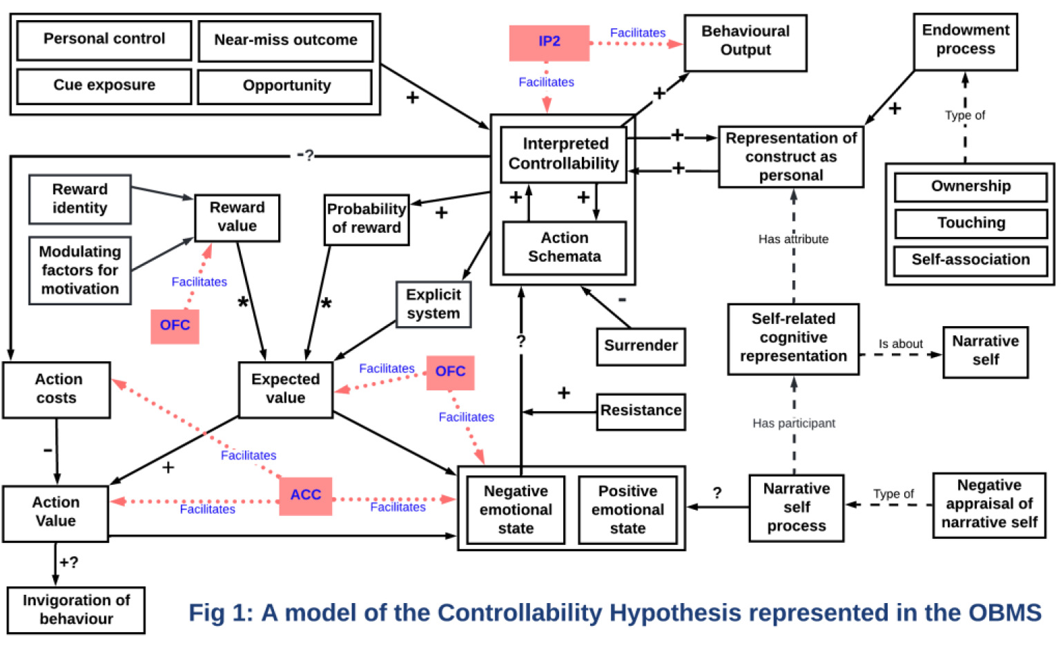
Computational and theoretical neuroscience
PM_132
Keywords: Protein-membrane interactions, AlphaFold and CHARMM-GUI modeling, ER stress disorders, Bioinformatics,
Authors: Filippo Dall'Armellina, Dan Rigden, Sylvie Urbe
Traditional biochemical methods like protein expression, construct testing, and pull-down assays are invaluable for studying protein function but face limitations when dealing with membrane-embedded proteins. Computational bioinformatics offers an efficient alternative, enabling systematic mutation screening, structural predictions, and interaction analysis to address these challenges.
Here, we investigate the interaction between ARL6IP1, an ER membrane protein, and INPP5K, a cytosolic phospholipid phosphatase that targets signalling molecules like IP3 and PIP3. Disruption of this interaction can result in disorders with motor and cognitive impairments, driven by ER stress and imbalanced lipid compositions. Importantly, ER proteins like ARL6IP1 can form dimers or adopt diverse stoichiometries, influencing their functional behaviour. We applied bioinformatics tools to predict molecular mechanisms, functional hotspots, and the impact of mutations on protein stability. Protein structures were modelled using AlphaFold3, while single-point mutations were analysed for folding and binding stability using FoldX, MAESTRO, mCSM, and MuPro. Pathogenicity was assessed with AlphaMissense, contextualized with evolutionary conservation and catalytic site proximity. Membrane bilayer interactions were modelled using CHARMM-GUI to evaluate which regions of INPP5K interact with the ER membrane. Our results identified critical hotspots in ARL6IP1 and INPP5K that govern binding and catalytic function. Specific mutations, particularly near conserved catalytic sites and membrane-embedded regions, caused significant destabilisation of the complex, with predictions validated across tools. This study highlights how bioinformatics can overcome biochemical limitations, such as membrane complexity and stoichiometric variability, to predict mutation-driven structural and functional disruptions. By identifying key molecular changes, we streamline experimental focus, uncover disease mechanisms, and provide a foundation for targeted therapeutic strategies and advanced research tools, including custom antibody development. Poster abstract figure. A. AlphaFold 3-generated INPP5K-ARL6IP1 protein complex and score metrics. B. INPP5K-ARL6IP1 model interaction energies calculated with FoldX Suite AnalyseComplex. C. Determining interface hotspots with AlphaMissense and machine learning methods for folding and binding stability. D. Protein abundance in HeLa cells (data from Bekker-Jensen et al 2017).
Computational and theoretical neuroscience
PM_133
Keywords: antidepressants, delayed therapeutic effects, reinforcement learning, computational modelling, alternative hypothesis
Authors: Phoenix Pavlik, Toby Wise
The mechanism by which antidepressant drugs induce delayed therapeutic effects still largely remains unknown. Those prescribed antidepressants, including selective serotonin reuptake inhibitors (SSRIs) and serotonin-noradrenaline reuptake inhibitors (SNRIs), typically don’t see improvements for 2-4 weeks. One hypothesis suggests that antidepressants act immediately upon cognitive biases (e.g., attention to negative stimuli), but take repeated interaction with the external environment for such biases to update and form positive associations (Harmer et al., 2009). Here, we postulate an alternative hypothesis. This hypothesis proposes that antidepressants encourage a form of internal offline learning, as a way of escaping beliefs responsible for reinforcing depressive/anxious states. That is, antidepressants nudge this offline learning process away from aversive prior beliefs, therefore improving mood. This may explain delayed therapeutic effects, as such updating takes time.
We aim to test this hypothesis using simulations of the offline learning process, while applying a reinforcement learning approach (specifically, the SARSA algorithm) combined with simple artificial neural networks. The model will learn a) a predictive model of the environment, and b) a policy within this environment based on previously experienced state transitions and rewards. This generative model will then be used to generate trajectories through the environment, reflecting the agent’s interactions and experiences over time. This trajectory generation process will be manipulated (simulating the effect of administering an antidepressant) to make it a) more exploratory, and b) more positive. By retraining the reinforcement learning model on this simulated data, we expect to see that it gradually forms more positive expectations, revealing parallels with the proposed offline learning process during the delay period between antidepressant drug administration and therapeutic onset. Changes in expected reward value will be analysed using t-tests to compare the effects of different interventions on agent behaviour (e.g., manipulating action selection exploration by adjusting the softmax temperature). Such results would support our alternative hypothesis for delay antidepressant effects and advance our understanding of the mechanisms underlying their efficacy.
Harmer CJ, Goodwin GM and Cowen PJ (2009) Why do antidepressants take so long to work? A cognitive neuropsychological model of antidepressant drug action. The British Journal of Psychiatry 195(2):102-108.
Computational and theoretical neuroscience
PM_134
Keywords: Oxytocin, Autism, Magnocellular neurons, Single Cell RNA Sequencing, Cross-Species
Authors: Victor Duque, João Victor Nani, André Mecawi
The magnocellular neurons (MCNs) are highly conserved across species and are essential for regulating fluid balance, reproductive function, and behavioral responses. Among the subpopulations of MCNs, they can be divided between those secreting vasopressin (AVP) or oxytocin (OXT). OXT is pivotal for social interactions and bonding, and its dysregulation has been implicated in autism spectrum disorder (ASD). Despite this important role, little is known about the characterization of these neurons and how their transcriptional profiles are conserved in different species. Elucidating these conserved features may offer new insights to the understanding of MCNs-OXT functionality and their association with psychiatric disorders.
Objective: To characterize MCNs-OXT at single-cell level and identify evolutionarily conserved mechanisms shared across species.
Single-nucleus data from the hypothalamic tissue from humans (10.1126/science.add7046), mice (10.1038/s42255-022-00657-y), marmoset (10.1126/sciadv.adk3986), and rats (Duque et al., 2025/Under Review) were clustered using the Seurat package (v.5.0). MCNs were identified using established markers from the literature. The classification of MCN subpopulations was based on the proportion of OXT/AVP, considering the normalized counts. Clusters from different species were integrated using the BENGAL pipeline, converting genes to the human code based on the highest homology according to the Ensembl. The transcription factors (TFs) were identified based on established literature (10.1016/j.cell.2018.01.029). The differential gene expression (DGEs) between MCNs clusters was analyzed using the Wilcoxon test in Seurat (v.5). Spearman correlations were performed to detect genes co-expressed with OXT using the Hmisc package (v5.1).
A conserved MCNs-OXT cluster was identified across all species. Each species showed over 300 DGEs (padj < 0.05) distinguishing MCNs-OXT from MCNs-AVP, with 13 consistently enriched in all species. Among these, the TFs Teashirt Zinc Finger Homeobox 3 (TSHZ3) and Forkhead Box Protein P1 (FOXP1), both being strongly associated with ASD, emerged as key conserved regulators. Besides its association with ASD, the dysregulation of FOXP1 also leads to the development of FOXP1 syndrome, typically characterized by developmental delay, severe speech and language deficits, and intellectual disability. Further, the correlation analysis of OXT expression revealed 115 conserved genes (r > 0.1, p < 0.05) across species, including the TF DR1 Associated Protein 1 (JUND) and ASD-related genes, such as L1 Cell Adhesion Molecule (L1CAM) and Seizure Related 6 Homolog Like 2 (SEZ6L2). These findings underscore the evolutionary conservation of MCNs-OXT among species, highlighting a core essential transcriptional network for their functionality, including genes associated with ASD.
Computational and theoretical neuroscience
PM_142
Keywords: Speech Segregration, Neural Mass Model, Computational Modelling
Authors: Andrew Shannon, David Barton, Conor Houghton, Martin Homer
We employ the Next Generation neural mass model (NGNMM) (Byrne et al. (2017)) to investigate the origin of oscillatory behaviour in syllable segregation by modelling the entrainment of neural activity to periodic linguistic stimuli. Multiple underlying mechanisms are proposed for this entrainment, with some debate existing on whether it results from activity evoked by acoustic edge features or through phase resetting of endogenous oscillations. Cucu et al. (2022) investigated this by analysing the entrainment of EEG activity to periodic sequences of consonant-vowel (CV) phonemes. A correlation between the sharpness of the phoneme sound envelope and the entrainment strength was identified (-0.84, p<0.001). These phenomena are characterisable by macro-scale oscillatory neural dynamics. As the NGNMM was developed with this kind of dynamics in mind, it was selected for investigating this aspect of neural language processing.
The experimental audio stimuli, ∼4Hz sequences of 20 phonemes, were transformed into envelopes. Each sequence comprised a consonant paired with 20 randomly selected vowels, e.g. “be, ba, bo, bo, bu,...”. The sound envelopes were used to drive the neural mass model, with additive noise. The Inter Trial Phase Coherence was used to evaluate the entrainment to the periodic stimuli. We characterised the sharpness of the phoneme sound envelopes using their time-to-maximum-derivative. Correlations were computed using the sample correlation coefficient.
The NGNMM demonstrated entrainment to the phoneme sequences across a range of conditions in agreement with Cucu et al. (2022). Under specific conditions, the model reproduced the correlation between phoneme sound envelope sharpness and entrainment strength, exhibiting a correlation coefficient of -0.66 (p<0.05, two-tailed t-test, 13 degrees of freedom).
The NGNMM can reproduce, at least roughly, the relationship between syllable sharpness and the neural response. A correlation between phoneme sharpness and entrainment strength was obtained, however the agreement with the experimental result (Cucu et al. (2022)) varied. The readiness with which a correlation was produced is promising for future investigations using the NGNMM to probe the origin of these neural dynamics. This result supports recent attempts to include detailed biophysical dynamics within neural mass models.
Byrne A, Brookes MJ and Coombes S (2017) A mean field model for movement induced changes in the beta rhythm Journal of computational neuroscience 43: 143–158.
Cucu MO, Kazanina N and Houghton C (2022) Syllable-initial phonemes affect neural entrainment to consonant-vowel syllables. Frontiers in Neuroscience 16: 826105
Computational and theoretical neuroscience
PT_135
Keywords: Cerebellum, Boolean algebra, Motor control, Neocerebellar syndrome, Intention tremor
Authors: Gregoris A. Orphanides, Christoforos Demosthenous, Ariadni Georgiannakis, Vasilis Stylianides, Petros Kyriakou, Konstantinos Antoniou, Alberto Capurro
Despite a plethora of cerebellum studies the role of the cerebellar circuit (CC) in motor coordination remains debated. This study aimed to model the function of CCs in motor control, assuming that the climbing fibers (CF) convey information about desired joint positions, whereas mossy fibers (MF) relay proprioceptive information about their actual position.
Each neuron of the CC was modelled in MATLAB using Boolean algebra. When a neuron’s threshold was reached, it was considered active, feeding input to the next neuron(s) in the CC. The connectivity of each CC was based on current histological descriptions, accounting for excitatory and inhibitory synaptic contacts (Takahashi and Shinoda, 2021). Cortex firing caused muscle flexion and Deep Cerebellar Nucleus (DCN) firing caused muscle extension. In each time sample of the model realisation, the model simulated muscle position and the state of every neuron in the circuit across parallel CCs.
The firing patterns of modelled DCN and Purkinje neurons during flexion and extension were similar to human recordings (Purves et al., 2012). By decreasing the number of CCs, a phenomenon resembling the cerebellar intention tremor emerged (Figure 1). Our simulations revealed that the CF frequency determines the DCN frequency, which is symmetrically modulated by the difference between the MF and CF frequencies. This study termed the aforementioned phenomenon “Shifting Central Frequency Hypothesis” (SCFH).
The SCFH intends to explain the widely accepted comparator functionality of the cerebellum. Thus, synchronising DCN and motor cortex firings allows for accurate control of intended movements. The Boolean algorithm proposed in this study introduces a novel approach to understanding the CC. Although we are aware that the model includes huge simplifications of complex biological processes, insights gained with this
Purves, D., Augustine, G. J., Fitzpatrick, D., Hall, W. C., LaManita, A.-S., & White, L. E. Neuroscience (5th edition). Sinauer Associates.
Takahashi M and Shinoda Y (2021) Neural Circuits of Inputs and Outputs of the Cerebellar Cortex and Nuclei. Neuroscience 462. Pergamon: 70–88.
Figure 1: Change in muscle power, muscle position and intentional tremor while increasing the number of simulated Cerebellar Circuits (columns). The black line indicates the desired muscle position whereas the blue line represents the achieved position.

Computational and theoretical neuroscience
PT_136
Keywords: Information theory, Complexity, Lempel-Ziv, Entropy, Neural dynamics
Authors: Claudia Pascovich, Julieta Ruiz, Alejo Rodriguez, Mayda Rivas, Andrea Devera, Francisco Flores, Patricia Lagos, Pablo Torterolo, Diego Mateos
A great number of studies support the notion of a lower electrocortical complexity during slow wave (SW) states like deep sleep and anaesthesia compared to wakefulness state or rapid eye movement (REM) sleep. In this work we investigated the neural complexity dynamics in different levels, from the mesoscale in the cortex to the microscale at the neuronal firing in the raphe nuclei, and how they are modulated during sub-states of urethane anaesthesia: SW and active state (AS). The central hypothesis posits a reduction in neuronal firing complexity during SW states.
A total of 64 male Wistar rats were used in this study. Recordings included 167 single neurons from the median and dorsal raphe nuclei under urethane anaesthesia. Simultaneously, local field potentials (LFPs) were recorded from the same region, as well as from the frontoparietal, motor, and visual cortices. To test our hypothesis, we first studied the neural complexity at the cortical level and LFP of the raphe nuclei. Two different measures of complexity were computed: Lempel-Ziv complexity (LZC) and Permutation Lempel–Ziv complexity (PLZC). At the neuronal level, three distinct measures were employed: Lempel–Ziv Complexity of Spike Trains (LZC–ST), LZC of Inter-spike Intervals (ISI) binarized (LZC–ISI bin), and PLZC of ISI (PLZC–ISI).
In this work, group comparisons were performed using Multilevel Linear models, and the null hypothesis was rejected at p‹0.05. The rat was included in the model as a random effect, and the measure of complexity as the fixed effect.
According to Our results, in the cortex LZC shows a significant and robust decrease during SW compared to AS (0.33±0.01 to 0.19±0.01, p‹0.001), and PLZC has a significant but smaller decrease (PLZC: 0.57±0.01 to 0.54±0.01, p‹0.05). The LZC and PLZC of the raphe LFP also shows a significant decrease during SW state (0.39±0.01 to 0.20±9.3x10⁻⁴, p‹0.001; 0.67±0.04 to 0.61±2.4x10⁻⁴; respectively). At the neuronal level of the raphe, none of the complexity measures studied (LZC–ST, LZC–ISI bin, or PLZC–ISI) show significant differences between AS and SW sub–states.
In conclusion, the state–dependent decrease in complexity occurs both in the cortex and raphe but disappears when calculated at the single-neuron level. This suggests that the information underlying changes in complexity may not be contained within the firing of a single neuron but is possibly distributed across a multi–neuronal circuit.
Computational and theoretical neuroscience
PT_137
Keywords: Olfaction, Odour frequency, Piriform Cortex, Network Model
Authors: Debanjan Dasgupta, Ankit Bishnoi
The mammalian olfactory system possesses the remarkable ability to encode rapid temporal features present in natural odor stimuli (Ackels et al., 2021; Dasgupta et al., 2022), which are generated by the natural turbulence in air flow. Furthermore, in mice the olfactory bulb (OB) projection neuronal activity can couple to specific odor stimulation frequency (Dasgupta et al., 2022). However, little is known how this information can get encoded in higher cortical areas like the Piriform Cortex (PCx). Here, we test this hypothesis using a biophysically relevant network model of PCx.
We used a conductance based biophysically relevant network model of PCx using the NEURON simulation environment (Fig. 1A). Next, we simulated the OB input onto this network model using in-vivo recordings from mitral and tufted cells (Dasgupta et al., 2022) in response to combinations of two odor frequencies (2Hz and 20Hz) and three odor mixtures (A, B and Mix(A+B)). We performed 336 trials using random shuffling of the OB inputs to the PCx network. The spiking data obtained from 500 excitatory pyramidal cells (Pyr) were used for two tailed T-tests to perform statistical comparisons between activities obtained for different odorant conditions pairwise (eg: A2Hz vs A 20Hz). We used Convolutional Neural Network (CNN) algorithms to train models on 70% and test the remaining 30% of the Pyr’s activity data. Furthermore, specific virtual knockouts were performed to test the effect of specific circuit connections on the ability to discriminate odor frequency.
We observed that 4.4-11.8% of the Pyr population could discriminate 2Hz from 20Hz odor frequency (Fig 1B). The virtual knockout of Pyr-Pyr showed similar proportion of frequency encoding cells (4.8-11.4%). However, the virtual knockout of feedback interneurons to Pyr increased the proportion of frequency discriminating cells (5-22.6%). When classified based on odor identity we observed that 8-22.6% (A), 7.4-18.4% (B), and 4.4%-7% (Mix) were the overall proportion of frequency discriminating cells. CNN’s accuracy on Odor A data from first 500ms after stimulus onset was 93±1.71%, while shuffle accuracy was 49.85±4.28%.
The PCx network model shows that a substantial proportion of the network can discriminate odor frequency 2Hz from 20Hz as validated by the performance of the CCN model. Furthermore, the frequency discrimination is strongly affected by the inhibitory synaptic connections while is largely unaffected by the excitatory recurrent connections. This suggests a strong circuit basis underlying the phenomena.
Computational and theoretical neuroscience
PT_138
Keywords: molecular dynamics
Authors: Love Onwuzuruike, Robin Corey
Serotonin receptors(5-HTR) regulate neurotransmission and impact various neuropsychiatric disorders. Despite their importance in several neurological processes, much remains unknown about their mechanisms and downstream activity.
There are multiple families/subtypes of 5-HTR. Using computational modelling, and molecular dynamic (MD) simulations-based methods, we aim to assess various functional features of three of these subtypes: 5-HT2a, 5-HT2c and 5-HT7.
Our project has several specific aims. Firstly, we will use MD to assess the structural basis of previously characterised residue mutations that decrease the ability of 5-HT2a to be activated by psychedelics such as LSD; this includes both mutations in the human system as well as the native inability of rat 5-HT2a to be activated by LSD. Further, we will use modelling and coarse-grained (CG) MD to assess the dynamics of 5-HT2c in different membrane environments whilst quantifying key lipid interactions for activation. Finally, we will run a comparative structural analysis of 5-HT7r to understand the role of ligand binding on receptor activation, with a particular focus on screening prospective antagonists against the inactive state of the receptor. Using Isambard 3 HPC, we will employ Well-Tempered Metadynamics (WTMetaD) on the 5-HTR to map activation/inactivation pathways and assess the potential allosteric role of lipid binding.
All findings will be validated using wet lab experimental methods including mutagenesis with functional cell-based BRET assays and imaging using FRET-FILM and FRAP.
Our data will reveal structural details about the underlying mechanism of several 5HTRs. We hope this will provide insights into 5HTR signalling, informing future therapeutic strategies.
Computational and theoretical neuroscience
PT_139
Keywords: Drosophila melanogaster, Computational neuroscience, Kenyon cells, Mushroom body
Authors: Nada Abdelrahman, Katie Greenin-Whitehead, Julian S Jung, Melissa Wei Yi Tan, Jiamu Jiang, Daichi Yamada, Toshihide Hige, Mark C W van Rossum, Andrew C Lin
Why are brains the way they are? Are their circuit architectures and synaptic plasticity rules in some sense ‘optimal’? If so, in what sense, or in what contexts? We address these questions using olfactory associative memory in the fruit fly Drosophila. Flies can learn to associate a particular odour with a reward (e.g., food) or punishment (e.g., shock) and thereafter approach or avoid the trained odour. These associative memories are stored in Kenyon cells in the mushroom body, by weakening synapses from odour-responsive Kenyon cells onto mushroom body output neurons (MBONs) that lead to incorrect actions (e.g., odour+punishment weakens KC->Approach synapses). Why weaken incorrect actions rather than strengthening correct actions? Notably, synaptic depression is also used for learning in the vertebrate cerebellum, which has a remarkably similar architecture to the insect mushroom body, suggesting that using depression may be functionally advantageous.
We show both analytically and using simulations that depression outperforms potentiation for discriminating odours with overlapping KC representations, under a particular condition: if behaviour depends on the relative, not the absolute, difference between Avoid vs. Approach MBON activities (i.e. divisive rather than subtractive normalisation). To test whether behaviour depends on the relative difference, we measured aversive learning for a range of odour concentrations and punishment intensities, in an individual-fly T-maze (n=95-442 flies per condition). We automatically tracked the flies’ decisions to enter or leave the side with the punished odour, and from the statistical distributions of these stochastic decisions, we inferred the mean and variance of the flies’ underlying preference for/against the odour. We fitted these data to alternative mechanistic models for learned decision-making that used subtractive or divisive normalisation, and the fit was better with divisive normalisation. These results suggest that flies learn by synaptic depression because, in the mushroom body, it is computationally superior to synaptic potentiation. These results illustrate how quantitative analysis of natural behaviour illuminates neural mechanisms underlying learned decision-making.
Computational and theoretical neuroscience
PT_140
Keywords: Machine Learning, neuropsychiatric and neurodegenerative disorders, neurotransmission, Alzheimer's disease, Parkinson's disease
Authors: Mariya Ivanova, Nicola Russo, Konstantin Nikolic
This work presents a novel, cost-saving and time-efficient
To demonstrate the approach, four bioassays provided by PubChem (NIH, 2025), the world's largest public chemical information repository, were explored.
(i) PubChem AID 652054 on the antagonists of the human dopamine D3 receptor, which receptor is a target for the treatment of several neuropsychiatric disorders;
(ii) PubChem AID 485297 on the Rab9 promoter activators related to the Niemann Pick Type C (NPC) neurodegenerative lipidosis, and mitochondrial dysfunction and cognitive deficits In Alzheimer's disease;
(iii) PubChem AID 2732 on small molecule inhibitors of DNA damage-inducible transcript 3, also known as C/EBP homologous protein (CHOP) to regulate the unfolded protein response to endoplasmic reticulum (ER) stress that have the potential of therapeutic application to diverse diseases, including Alzheimer's and Parkinson's diseases;
(iv) PubChem AID 588852 identifies antagonists of the human M1 muscarinic receptor, which receptor modulates the dynamics of cholinergic and dopaminergic neurotransmission.
Datasets, derived from the compound IDs (CIDs), structure IDs (SIDs), and targets of the bioassays described above, were used to develop ML models. These models were built using algorithms from the scikit-learn Python library, as detailed in Ivanova, Russo, and Nikolic (2025).
D3. Accuracy: 85.8%; ROC 85.8 % CHOP. Accuracy: 90.1 %; ROC 89.2 %
Rab9. Accuracy: 75.4%; ROC 75.4 % M1. Accuracy: 83.2 %; ROC 83.2 %
The approach demonstrates strong generalizability with an average accuracy of 83.63% (with 5.35 standard deviation) and ROC of 83.4% (with 5.09 standard deviation) across the considered cases. This implies the probability of its applicability to other objects of interest when the dataset generation requirements are fulfilled.
Ivanova ML, Russo N and Nikolic K (2025) Predicting novel pharmacological activities of compounds using PubChem IDs and machine learning (CID-SID ML model). ArXiv. doi.org/10.48550/arXiv.2501.02154
NIH (2025) PubChem. Available at: https://pubchem.ncbi.nlm.nih.gov/ (accessed 14 Jan 2025)
Computational and theoretical neuroscience
PT_373
Keywords: perceptual learning, visual cortex, network dynamics, balance of excitation and inhibition, deep learning
Authors: Monika Jozsa, Clara Pecci-Terroba, Ke Jia, Mengxin Wang, Zoe Kourtzi, Yashar Ahmadian
Training in perceptual tasks can enhance the brain’s internal representations of task-relevant features for improved decision making and perceptual acuity. Recent ultra-high-field neuroimaging studies have found that training in fine discrimination tasks increase the signal-to-noise ratio of activity patterns in the superficial layers of primary visual cortex (V1), which is accompanied by an increase in GABAergic inhibition. However, the causal circuit mechanisms underlying the layer-specific representational changes, and the layer-specificity of changes in cortical excitation and inhibition, remain unknown. Here, we theoretically study these questions by training a biologically-constrained mechanistic model of V1 in a fine orientation discrimination task near a fixed orientation. We found that training led to (1) strengthening (weakening) of cortical inhibition (excitation), which was larger and more robust in the model's superficial layer, and (2) sharpening of the superficial-layer tuning curves at the trained orientation, as previously reported in neurophysiology experiments. Further, these changes correlated with improvement in the network's task performance. Finally, the mechanistic nature of our model allows making testable predictions about the causal pathways linking layer-specific changes in excitation and inhibition with representational or behavioral improvements. In particular, using causal ablations we show that the existence of long-range horizontal connections in and the emergence of feedforward pathways from the superficial layers play a role in biasing functional and structural changes to those layers.
Computational and theoretical neuroscience
PW_131
Keywords: Multiple sclerosis, Cognitive impairment, Thalamus, Network controllability, Resting-state fMRI
Authors: Yuping Yang, Anna Woollams, Ilona Lipp, Zhizheng Zhuo, Marta Czime Litwińczuk, Valentina Tomassini, Yaou Liu, Nelson J Trujillo-Barreto, Nils Muhlert
Recent research suggests that individuals with multiple sclerosis (MS) and cognitive impairment exhibit more effortful transitions in brain network activity.1 Previous studies further highlight the increased vulnerability of core brain regions, particularly the thalamus, to disease-related damage in MS.2 This study investigates whether MS affects the controllability of core brain regions in driving network activity transitions across the brain, and examines the relationship between these changes and cognitive impairment in patients.
Resting-state fMRI and neuropsychological data were collected from 102 MS and 27 healthy controls. Functional network controllability analysis was performed to quantify how specific regions influence transitions between brain activity patterns. Disease alterations in controllability metrics were evaluated in the main dataset, then replicated in an independent dataset of 95 MS patients and 45 healthy controls. Controllability metrics were then used to distinguish MS from healthy controls and predict cognitive status in MS.
Group differences in controllability metrics were evaluated using permutation f-tests and post-hoc t-tests with 95% confidence intervals. Linear SVM classifiers were trained and tested using 10-fold cross-validation, with controllability and/or volumetric metrics as predictive features. A set of performance metrics, including accuracy, precision, sensitivity, specificity, and area under the curve (AUC) were computed to access the classification performance.
MS-specific controllability changes were predominantly observed in the thalamus, which were further confirmed in the replication dataset. Cognitively impaired patients showed greater difficulty in the thalamus driving brain transitions towards difficult-to-reach states, which are typically associated with high-energy-cost cognitive functions. Thalamic controllability proved more effective than thalamic volume in distinguishing MS from healthy controls (AUC = 88.3%) and in predicting cognitive status in MS (AUC = 80.7%). Building on previous research highlighting early thalamic damage in MS, this study examines how such damage disrupts activity transitions across the brain and may predict cognitive deficits, suggesting a potential pathological mechanism linking thalamic functional changes to cognitive impairment in MS.
1. Broeders TAA, van Dam M, Pontillo G, et al. Energy Associated With Dynamic Network Changes in Patients With Multiple Sclerosis and Cognitive Impairment. Neurology. 2024;103(9):e209952. doi:10.1212/WNL.0000000000209952
2. Mahajan KR, Nakamura K, Cohen JA, Trapp BD, Ontaneda D. Intrinsic and Extrinsic Mechanisms of Thalamic Pathology in Multiple Sclerosis. Ann Neurol. 2020;88(1):81-92. doi:10.1002/ana.25743

Computational and theoretical neuroscience
PW_141
Keywords: Histamine, Allen Brain Cell Atlas, Cell types, Dimensionality reduction,
Authors: Amrita Benoy, Srikanth Ramaswamy
Advances in spatial transcriptomics have revolutionized the ability to classify mammalian brain cell types based on diverse marker gene expression. In this study, we investigate the diverse landscape of histamine receptor expression in the adult mouse brain, leveraging data from the Allen Brain Cell Atlas (https://portal.brain-map.org/atlases-and-data/bkp/abc-atlas). While histamine is a crucial neurotransmitter, heterogeneity of histamine receptor expression in different cell types of the brain is underexplored (Benoy and Ramaswamy, 2024). We aim to bridge this gap in knowledge by performing a comprehensive cell type-specific computational analyses of histamine receptor expression in the adult mouse brain based on pre-existing open-source data available in the Allen Brain Cell Atlas (Zhang et al. 2023).
Using data from the Allen Brain Cell Atlas, we systematically analyze histamine receptor expression across neurotransmitter-releasing cells, including glutamatergic, GABAergic, and various modulatory neuron populations. We also extend this investigation to diverse molecularly defined cell classes and subclasses in different brain regions. Through principal component analysis-based dimensionality reduction and k-means clustering of cell type-specific expression profiles, the analyses reveal distinct patterns of histamine receptor expression, shedding light on cell type-specific signalling and functional diversity.
results
We find diverse expression patterns of the three histamine receptors Hrh1, Hrh2, and Hrh3 across multiple transcriptomically and anatomically defined cell classes and subclasses in the adult mouse brain, with a particular dominance of Hrh3 receptor expression in several cell types.
The
Allen Brain Cell Atlas. Available at: https://portal.brain-map.org/atlases-and-data/bkp/abc-atlas (Accessed: 31 January 2025).
Benoy, A., & Ramaswamy, S. (2024). Histamine in the neocortex: Towards integrating multiscale effectors. European Journal of Neuroscience, 60(4): 4597-4623.
Zhang, M., Pan, X., Jung, W., Halpern, A. R., Eichhorn, S. W., Lei, Z., ... & Zhuang, X. (2023). Molecularly defined and spatially resolved cell atlas of the whole mouse brain. Nature, 624(7991): 343-354.
Computational and theoretical neuroscience
PW_143
Keywords: Machine Learning, autism spectrum disorder, dementia, CNS tumors, Alzheimer's disease
Authors: Mariya Ivanova, Nicola Russo, Konstantin Nikolic
The G9a enzyme, Histone Lysine Methyltransferase, has been reported for having potential as a therapeutic target for neurodegenerative conditions, such as dementia, Alzheimer's disease, autism spectrum disorder, neuropsychiatric disorder and CNS tumors. To support research in this direction a machine learning (ML) model was developed that predicts the magnitude of G9a inhibitors efficacy, and in this way decrease the time and cost in early stage of drug discovery.
The labels and compound identifiers were extracted from the PubChem AID 504332 bioassay (NIH, 2011). The features for ML were generated using the approach provided by study Ivanova, Russo and Nikolic (2025). Additionally, nine new features were added, increasing the number of the features to seventy-two. However, the final model contained only three of them. ML was performed using algorithms from the scikit-learn Python library for ML. The ML models were evaluated with mean absolute error (MAE), root mean square error (RNSE), R-square (R2) and mean relative error (MRE). The code is available on GitHub (2025).
The "CID_SID ML model" (Ivanova, Russo and Nikolic, 2025) was developed to complement the main study. It predicts G9a inhibitors solely based on PubChem identifiers, providing researchers with a convenient tool for evaluating the potential of their compounds for such antagonism (GitHub, 2025).
The ML model (Gradient Boosting Regressor) regarding the G9a inhibitor efficacy achieved:
MAE: 21.48%; RMSE: 27.4%; R2: 0.05%; MRE: 17.81%;
Regarding the “CID_SID ML” model, the eXtreme Gradient Boosting classifier achieved:
Accuracy: 78.7%; Precision: 84.2%; Recall: 70.7%;
F1: 76.9%; ROC: 78.7%; Cross-validation score: 0.7995;
Although the ML model behaves more like statistical, than ML, it still provides insights predicting the magnitude of the G9a inhibitor efficacy. However, further research is needed to reduce prediction errors. The
GitHub (2025) Efficacy of the G9a Inhibitors. Available at https://github.com/articlesmli/G9a_Inhibitors_efficacy.git (accessed 14 Jan 2025)
Ivanova ML, Russo N, Djaid N and Nikolic K (2024) Application of machine learning for predicting G9a inhibitors. Digital Discovery 3(10):2010-2018. doi.10.1039/d4dd00101j
Ivanova ML, Russo N and Nikolic K (2025) Predicting novel pharmacological activities of compounds using PubChem IDs and machine learning (CID-SID ML model). ArXiv. doi.org/10.48550/arXiv.2501.02154
NIH (2011) QHTS ASSAY FOR INHIBITORS OF HISTONE LYSINE METHYLTRANSFERASE G9A. AVAILABLE AT: HTTPS://PUBCHEM.NCBI.NLM.NIH.GOV/BIOASSAY/504332 (ACCESSED 14 JAN 2025)
Computational and theoretical neuroscience
PW_144
Keywords: Machine Learning, neuropathic pain regulator, schizophrenia, depression, drug addiction
Authors: Mariya Ivanova, Nicola Russo, Konstantin Nikolic
G protein-coupled receptor 151 (GPR151) in nociceptors has been reported to regulate the neuropathic pain induced by nerve injury. It is also thought to regulate the habenula complex, which in turn plays a significant role in the pathophysiology of neuropsychiatric disorders such as schizophrenia, depression and drug addiction.
Data used in the study derived from a PubChem AID 1508602 bioassay on GPR151 protein activators (NIH, 2020). The IUPAC names of the compounds were parsed and the resulting segments used to create 4323 dataset features. The algorithms used to rank these features originated from scikit learn Python based library for machine learning (ML). A feature reduction technique was applied, which helped generate three datasets that were subsequently used for ML. The performances of the ML models were compared and the dataset whose ML model obtained the best results was selected as the final list of functional groups ordered in descending order of their influence.
The “CID_SID ML model” (Ivanova, Russo and Nikolic, 2025) was developed to complement the main study. It predicts the GPR151 activators solely based on PubChem identifiers, providing researchers with a convenient tool to evaluate the potential of their compounds for the considered functionality.
Rank of the functional groups as activators of the GPR151 protein
Functional group Score Functional group Score Functional group Score
methylsulfonylanilino 16.85 dimethoxyphenyl 10.04 dimethylphenyl 8.63
cyano 14.4 tetrahydro 9.07 methylpyrimidin 8.44
carboxylate 13.26 diamine 8.9 ylpyridazin 8.42
cyclopenta 12.11 imino 8.79 piperazine 8.22
oxoethyl 10.75 methylbenzoate 8.78 thiophene 7.81
pyrimidine 10.37 quinoline 8.76 methylphenyl 7.47
carbonitrile 10.3 azaspiro 8.74 benzene 7.44
ethanol 10.06 iodide 8.73 butylacetamide 7.37
Regarding the CID_SID ML model, the GradientBoostingClassifier achieved:
Accuracy: 61.4%; Precision: 61.2% Recall: 62.5%
F1: 61.8%; ROC: 61.4%; Cross-validation score: 0.6193
The method applies to any research case where a dataset with a significant number of records is available. Although it does not guarantee perfect accuracy, reducing the functional groups from over 4,000 to a convenient set could streamline the initial phase of drug discovery research.
GitHub (2025) Selecting functional Groups GPR151. Available at: https://github.com/articlesmli/selecting_functional_groups_UIPAC_GPR151/blob/main/README.md (accessed 20 January 2025)
Ivanova ML, Russo N and Nikolic K (2025) Predicting novel pharmacological activities of compounds using PubChem IDs and machine learning (CID-SID ML model). ArXiv. doi.org/10.48550/arXiv.2501.02154
NIH (2020) PubChem bioassay AID 1508602: Cell-based high throughput primary assay to identify activators of GPR151. Available at: https://pubchem.ncbi.nlm.nih.gov/bioassay/1508602 (accessed 14 Jan 2025)
Computational and theoretical neuroscience
PW_145
Keywords: Machine Learning, neuropsychiatric disorders, endocrine disorders, receptor-based antiparkinsonian drug, antipsychotic agents
Authors: Mariya Ivanova, Nicola Russo, Konstantin Nikolic
This study contributes to ongoing research that aims to overcome challenges in predicting the bioapplicability of nanoformulations (Ivanova, Russo and Nikolic, 2025a). It incorporates machine learning and 13C NMR spectroscopy data, which data was obtained by converting the canonical Simplified Molecular Input Line Entry System (SMILES) notations to 13C NMR spectroscopic signals by the NMRDB software.
The unique magnetic property of the Carbon-13 (¹³C) isomer is used in 13C NMR spectroscopy technology for exploration of organic and organometallic compounds, providing information for the connectivity and relative position of the carbon atoms in the molecule`s chemical environment. To test the hypothesis that such data could support the development of a robust ML model, the PubChem AID 504652 bioassay (NIH, 2011) was employed to predict human D1 dopamine receptor antagonists.
To generate the required dataset, the PubChem compound identifiers, their relevant labels, and SMILES notations were extracted from the bioassay. This dataset was then pre-processed and used to develop an ML model, applying scikit-learn algorithms as described in Ivanova, Russo and Nikolic (2025a).
The "CID_SID ML model" (Ivanova, Russo and Nikolic, 2025b) was developed to complement the main study. It predicts human D1 dopamine receptor antagonism solely based on PubChem identifiers, providing researchers with a convenient tool for evaluating the potential of their compounds for such antagonism.
Regarding the 13C NMR ML model, the Support Vector Classifier obtained:
Accuracy: 71.5%; Precision: 77.4 % Recall: 60.6 %
F1: 68.0 % ROC: 71.5 % Cross-validation score: 0.7487
Regarding the CID_SID ML model, the XGBoost classifier achieved:
Accuracy: 80.2%; Precision: 86.3 % Recall: 70.4 %
F1: 77.6 % ROC :79.9 % Cross-validation score: 0.8071
Given the 13C NMR ML model results it can be concluded that there is a potential for further investigations in this direction. The Methodology can be applied to any other bioassay with a significant number of records.
Ivanova ML, Russo N and Nikolic K (2025a) Predicting the Influence of Nanoformulations on Biomolecule Functionalities Using Machine Learning and 13C NMR Spectroscopy Data Derived from SMILES: A Case Study on Human Dopamine D1 Receptor Antagonists. Arxiv. doi.org/10.48550/arXiv.2501.14044
Ivanova ML, Russo N and Nikolic K (2025b) Predicting novel pharmacological activities of compounds using PubChem IDs and machine learning (CID-SID ML model). ArXiv. doi.org/10.48550/arXiv.2501.02154
NIH (2011) Antagonist of Human D 1 Dopamine Receptor: qHTS. Available at: https://pubchem.ncbi.nlm.nih.gov/bioassay/504652 (accessed 14 Jan 2025)
Human cognitive neuroscience
PM_146
Keywords: fNIRS, Virtual Reality, Minecraft, Human Social Cognitive Neuroscience, Human Spatial Navigation
Authors: Emre Yavuz, Cian Xu, Weitao Liu, Anna Mitchell, Jhumana Ali, Nicole Bloom, Naadirah Khatun, Peter Kirk
Many species, including rodents, bats, monkeys, and humans, can neurally encode the distance to a stationary goal. In humans, this extends to moving goals, with the entorhinal cortex and its cortical connections able to track the location of a moving human avatar. However, the role of the prefrontal cortex (PFC) in tracking moving goals, especially in dynamic, group-based behaviors, remains underexplored. A classic example of goal tracking that requires coordination among individuals is group hunting. This study investigated the PFC’s role in group hunting using a virtual environment (Minecraft). Two participants acted as predators hunting a human prey (a confederate) in Hunt trials, while in Follow trials, they followed the path of a human experimenter. Functional near-infrared spectroscopy (fNIRS) recorded predators' brain activity. Behaviorally, successful hunting was associated with maintaining a closer proximity to the prey, exhibiting adaptability in adjusting this distance, and achieving greater coordination with one’s co-hunter. Neurally, greater variation in the euclidean distance between the self and the prey during the hunt was associated with greater anterior prefrontal (frontopolar) cortex activity. This preliminary finding suggests a possible role for the PFC in coordinating goal-tracking behavior, warranting further investigation with larger samples to understand the underlying neurobiological mechanisms.
Full author list below:
Anterior Prefrontal Cortex Tracks Variation in Distance to Prey During Group Hunting
Emre Yavuz1, Cian Xu1, Weitao Liu1, Callum Slinn1, Anna Mitchell1, Jhumana Ali1, Nicole Bloom1, Naadirah Khatun1, Peter Kirk2, Ilias Tachtsidis3, Paola Pinti3, Flaminia Ronca4, Paul Burgess5*, Antonia Hamilton5*, Hugo Spiers1*
1. Institute of Behavioural Neuroscience, Department of Experimental Psychology, Division of Psychology and Language Sciences, University College London, London, UK
2. Emotion and Development Branch, National Institute of Mental Health, Bethesda, Maryland, USA
3. Department of Medical Physics and Biomedical Engineering, University College London, UK
4. Institute of Sport Exercise and Health, University College London, London, UK
1. Institute of Cognitive Neuroscience, University College London, UK
Human cognitive neuroscience
PM_148
Keywords: tDCS, speech motor adaptation, altered auditory feedback
Authors: Qiming Yuan, Daniel R. Lametti, Izara Williams, Hui Zhu, Kate E. Watkins
Speech production involves careful monitoring of sensory feedback to detect and correct for discrepancies between intended and actual sensory consequences. This form of sensorimotor learning is critical in the development and maintenance of speech production. Speech adaptation is studied using altered auditory feedback. During the task, speech is produced and fed back in near real-time with a shift in the frequency of a vowel formant. Participants adapt by changing the frequency of their vowel production to offset the perceived error. Previously, we showed that anodal transcranial direct current stimulation (tDCS) over left speech motor cortex and right cerebellum enhanced speech adaptation (Lametti et al., 2018). Here, we aimed to replicate these effects.
Participants read the words “head”, “bed” and “dead” out loud. Speech was recorded and fed back to them either normally (baseline) or with a 110-Mel increase in the frequency of the first vowel formant (adaptation). Retention of the adapted state was measured when feedback was masked with a speech-shaped noise (noise) and was returned to normal (after-effect). Thirty participants received 2-mA anodal tDCS over left speech motor cortex, 30 received 2-mA anodal tDCS over the right cerebellum and 30 received sham stimulation. The allocation of participants to receive active or sham stimulation was concealed from them and from experimenters. We tested differences in speech adaption among the three groups using one-way ANOVAs.
All groups showed significant adaptation while receiving altered auditory feedback (see Figure). Contrary to the previous study, we found no impact of anodal tDCS on speech adaptation (F(2,87) = 0.149, p = 0.862, Cohen’s d = 0.109). The positive results of the previous study are explained by unusual task performance in many participants in the original sham group rather than by the effects of tDCS.
Data averaged in 15 trials and error bars show standard error.
Speech adaptation is not affected by anodal tDCS over speech motor cortex or cerebellum. This study shows tDCS’s inconsistent effects on behavioural tasks. We conclude that larger samples are needed to avoid spurious results arising from individual differences in task performance.
Lametti, D. R., Smith, H. J., Freidin, P. F., & Watkins, K. E. (2018). Cortico-cerebellar Networks Drive Sensorimotor Learning in Speech. J Cogn Neurosci, 30(4), 540-551.

Human cognitive neuroscience
PM_151
Keywords: Cerebellum, Connectivity, Resting-state fMRI, Modularity
Authors: Kieran Allen, Iqra Arshad, Gabriele Bellucci, Narender Ramnani
It has been argued that connectivity in the cortico-cerebellar system is highly modular. ‘Motor’ and ‘prefrontal’ loops that connect the cerebellar cortex and parts of the cerebral cortex are considered to be anatomically segregated (Kelly and Strick, 2003). We tested this possibility systematically using resting-state functional MRI to map the connectivity of 411 cerebellar locations with grey matter target locations in the rest of the brain. For each target, we report the locations of its functionally connected counterparts in the cerebellar cortex, and the proportions of the cerebellar cortex that each target is functionally connected with.
We applied standard preprocessing to resting-state fMRI data from 150 human participants (Cambridge Centre for Ageing and Neuroscience dataset; Shafto et al., 2014; SPM12 normalisation to MNI standard space; smoothing, 4mm). We placed 411 evenly spaced seed voxels in the left cerebellar cortex, 6mm apart. First and second level GLMs were estimated for each seed voxel (SPM12/CONN Toolbox; FWE, p<0.05). Outputs reported their connectivity with ~160,000 extra-cerebellar grey matter target voxels. We binarised the resulting seed-to-target matrix and used it to identify seeds that were functionally connected with each target. We also calculated the proportion of the 411 seeds that each target was functionally connected with.
Our preliminary findings show that although most targets connected with some cerebellar seeds, 64 target clusters connected with at least 20% of cerebellar seeds. These included the angular gyrus (PGa/PGp) which connected with 45% of seeds. The peak voxel in this cluster showed connectivity with all cerebellar lobules from HV to HIX. Other examples included medial frontal cortex (Fp2, 29%; lobules HVI to HVIIIA, and HIX), middle frontal gyrus (posterior; all lobules from HV to HIX), and posterior parts of the superior frontal sulcus (SMA/6mc, 39%; from lobule HVIIA HVIIIA to X). The clusters with the 15 largest proportions typically showed connectivity with both classically defined ‘motor’ and ‘prefrontal’ lobules.
While most areas in the cerebral cortex are functionally connected with a few cerebellar cortical locations, there are also a few areas in the cerebral cortex that are connected with large proportions of the cerebellar cortex. This may allow some cortical areas to exert a global, rather than a modular, influence over cerebellar circuitry. Also, the ability of ‘motor’ and ‘prefrontal’ cortico-cerebellar loops to interact with common brain areas suggests that they may not be as independent as previously thought.
Human cognitive neuroscience
PM_152
Keywords: Neurofibromatosis 1, Working memory, (f)MRS, Excitation/inhibition balance, Non invasive brain stimulation
Authors: Anna Wild
Neurofibromatosis type 1 (NF1) is a rare genetic condition, caused by mutations in neurofibromin, leading to RAS/MAPK overactivity, which regulates cell growth and differentiation. Given its widespread effects on brain development, learning difficulties are common and may stem from excitation/inhibition (E/I) imbalances. While animal models suggest that excess GABAergic activity contributes to learning difficulties, translation to human models using magnetic resonance spectroscopy (MRS) suggests a more complex relationship, as MRS is limited to baseline measurements of E/I. Previously, Garg et al. (2022) investigated the effect of transcranial direct current stimulation (tDCS) on MRS GABA and working memory in NF1, finding that while tDCS reduced GABA, it did not improve task performance. We build on Garg et al. (2022) by exploring the effect of transcranial alternating current stimulation (tACS), in addition to tDCS, on E/I balance and working memory in NF1. We also use functional MRS (fMRS) to obtain dynamic measures of the E/I balance.
Our primary aim is to determine how tDCS and tACS modulate glutamate, GABA and working memory. Furthermore, our secondary aim is to investigate whether changes in the E/I balance due to tDCS/tACS correlate with improved working memory. Together, our study will provide insight into how the E/I balance is affected in NF1 and whether its modulation could improve learning difficulties; thereby informing therapeutic interventions.
Thirty adolescents with NF1, aged 11-17 years, are undergoing three visits, receiving sham, tACS or tDCS stimulation during an N-back working memory task, with concurrent glutamate fMRS measurements. We also use MRS to measure GABA and glutamate at rest, pre and post stimulation. In addition, working memory, attention and inhibition tasks are administered pre and post scan to investigate any lasting effects of stimulation.
We will use mixed-effects models to address our primary aim, comparing glutamate during stimulation, GABA changes and cognitive task performance changes due to tDCS, tACS and sham stimulation. Additionally, regression and correlation analyses will examine the relationship between E/I modulation and performance changes due to tDCS/tACS/sham conditions, as well as how baseline GABA and glutamate are associated with baseline task performance.
Garg S, Williams S, Jung J, Pobric G, Nandi T, Lim B, Vassallo G, Green J, Evans DG, Stagg CJ, Parkes LM, and Stivaros S (2022) Non-invasive brain stimulation modulates GABAergic activity in neurofibromatosis 1. Sci Rep 12(1): 18297.
Human cognitive neuroscience
PM_153
Keywords: Covid-19, Long-term memory, Cognitive Function, Structural MRI, Diffusion MRI
Authors: Josefina Weinerova, Sabine Yeung, Denis Schluppeck, Andrew Reid, Roni Tibon, Lucy Cheke
SARS-CoV-2, the virus responsible for the Covid-19 pandemic, has been shown to have an impact on cognitive function and the brain, but the specific areas that are affected remain unclear, increasing the need for replication studies. We present findings from two studies looking at this topic. In Study 1, we analysed cognitive data collected online from 296 participants (209 who had experienced Covid-19 infection and 87 who did not). Using Frequentist and Bayesian analyses, we have replicated a previously found effect of Covid status on accuracy in 2 long-term memory tasks (verbal item memory task and nonverbal associative memory task), but did not replicate previously reported effect on reaction times. Further, across 4 long-term memory tasks, we found consistent effect of Covid status on memory accuracy but not reaction times. Contrary to our predictions, we did not find an interaction with memory type (associative vs. item) or stimulus type (verbal vs. nonverbal). Moreover, we compared cognitive functioning amongst vaccinated and unvaccinated individuals to explore the role of vaccination status in cognitive symptoms associated with Covid-19, and did not find conclusive evidence for either the null or alternative hypothesis. In Study 2 (currently ongoing), we are looking at the Covid-19 case-control dataset from the UK Biobank (N=2092), a case control matched sample of Britain dwelling adults (51-83 years) to assess the impact of SARS-CoV-2 on cognition and brain structure. Cognitive scores and imaging derived phenotypes (IDPs) from MRI and dMRI scans of participants who recovered from Covid-19 are being compared to pre-pandemic scores, to those of matched controls also tested pre- and post-pandemic, and to those of a second control group of participants (N= 2360, 49-82 years) who underwent both testing sessions before the pandemic. Using linear mixed-effects model, we predict a significant deterioration of cognitive functions and reduced brain volume in the second testing instance of the Covid group versus all controls (i.e., first instant of Covid group, and two control groups). Overall, these studies provide valuable insights into the effect of (or, in some cases, lack of) SARS-CoV-2 infection on cognition and the brain, and can inform policy makers and healthcare services.
Human cognitive neuroscience
PM_156
Keywords: neuromodulation, freezing, neural activation, parkinson, eeg
Authors: Sena Demirtaş, Lütfü Hanoğlu
Freezing of Gait (FoG) is an involuntary walking interruption in Parkinson's patients, affecting 25% of early-stage and 63% of idiopathic Parkinson’s Disease (PD) cases (1). FoG is related to cortical-subcortical dysfunctions. Particularly in the frontoparietal and parieto-occipital regions. EEG studies associate changes in theta and beta frequencies with impaired motor control and increased cognitive load. The aim of this study is to propose a neuromodulation approach to alleviate FoG episodes. Given its relatively limited study compared to other methods, Transcranial Alternating Current Stimulation (tACS) has been selected for this study.
The study includes four groups and three measurements. All patients will receive clinical evaluations (MoCA, UPDRS, TUG) and EEG recordings. Based on EEG results, patients with high spectral power in fast frequencies will be classified into one group, while those with high spectral power in slow frequencies will be placed in another (2). Each group will be subdivided into two subgroups receiving either tDCS (DLPFC) or tACS. Sessions will last 20 minutes over 10 sessions.
The frequency and placement of tACS electrodes will be determined as follows:
• Theta-tACS for high fast-frequency power at F3, P4, or CZ (3).
• Beta-tACS for high slow-frequency power at C3 - C4.
Assessments will occur before treatment, after 10 sessions, and two weeks post-stimulation.
Groups will be analyzed separately, comparing within-group pre/post/follow-up phases and between-group effectiveness of tDCS vs. tACS. A "repeated measures, within-between interaction ANOVA" will be used. Power analysis (effect size: 0.8, α: 0.05, power: 0.95) determined a sample size of 8 per group, with 16 patients in total (four per group).
1. Rahimpour, S., Gaztanaga, W., Yadav, A. P., Chang, S. J., Krucoff, M. O., Cajigas, I., Turner, D. A., & Wang, D. D. (2021). Freezing of Gait in Parkinson’s Disease: Invasive and Noninvasive Neuromodulation. Neuromodulation: Technology at the Neural Interface, 24(5), 829-842. https://doi.org/10.1111/ner.13347
2. Del Felice, A., Castiglia, L., Formaggio, E., Cattelan, M., Scarpa, B., Manganotti, P., Tenconi, E., & Masiero, S. (2019). Personalized transcranial alternating current stimulation (tACS) and physical therapy to treat motor and cognitive symptoms in Parkinson’s disease: A randomized cross-over trial. NeuroImage: Clinical, 22, 101768. https://doi.org/10.1016/j.nicl.2019.101768
3. Marquez, J. S., Hasan, S. M. S., Siddiquee, M. R., Luca, C. C., Mishra, V. R., Mari, Z., & Bai, O. (2020). Neural Correlates of Freezing of Gait in Parkinson’s Disease: An Electrophysiology Mini-Review. Frontiers in Neurology, 11, 571086. https://doi.org/10.3389/fneur.2020.571086

Human cognitive neuroscience
PM_159
Keywords: auditory cognition, magnetoencephalography, functional connectivity, MVPA, tonal pitch
Authors: Pradeep Dheerendra, Timothy Griffiths
We aim to understand the dynamics underlying auditory working memory for maintaining 'simple' tones. We recorded magnetoencephalography (MEG) in 17 subjects while they maintained one of the two presented tones (or ignore both in the control condition). After 12s, subjects compared the pitch of a test tone with the maintained tone.
Analysis of evoked responses showed persistent activity throughout maintenance compared to the pre-stimulus silent baseline but only at the start of maintenance when compared to the control condition. The evoked response during maintenance was source localised against baseline to bilateral auditory cortex. Analysis of induced responses showed suppressed alpha in the left auditory cortex, enhanced theta in medial prefrontal cortex, and enhanced beta in cerebellum.
In a second experiment, 19 new subjects were presented with a tone and a Gabor patch and a retro-cue indicating whether to maintain auditory or visual information for 12s. Analysis of the induced responses in auditory condition yielded results similar to those observed in the first experiment.
Connectivity analysis showed that the theta activity in medial prefrontal was phase-locked to activity in the left hippocampus and left auditory cortex. The beta activity in cerebellum was phase-locked to left Inferior Frontal Gyrus (IFG) activity and correlated to subject’s task accuracy.
Using MVPA, a LDA classifier was trained to decode the contents of AWM (discriminate between low vs high pitched tone) using beta band Phase Locking Value with right cerebellum as its features. A channel searchlight analysis showed that decoder performance at right Anterior Cingulate Gyrus (56.17% acc.) was above chance. Further, the decoder performance at Right STG & MTG was correlated (rho=0.725, p<0.01) to subject’s task accuracy, showing a correspondence between encoding distance and behavioural performance.
Our data clearly shows a network of brain areas involving pre-frontal and hippocampus, IFG and cerebellum for maintaining sounds in the auditory cortex, consistent with previous fMRI [1] and ECoG experiments [2].
[1] Kumar, S., Joseph, S., Gander, P.E., Barascud, N., Halpern, A.R. and Griffiths, T.D., 2016. A brain system for auditory working memory. Journal of Neuroscience, 36(16), pp.4492-4505.
[2] Kumar, S., Gander, P.E., Berger, J.I., Billig, A.J., Nourski, K.V., Oya, H., Kawasaki, H., Howard III, M.A. and Griffiths, T.D., 2021. Oscillatory correlates of auditory working memory examined with human electrocorticography. Neuropsychologia, 150, p.107691.
Human cognitive neuroscience
PM_163
Keywords: mood, light, everyday-life, naturalistic, longitudinal
Authors: Chloe Roddis, Altug Didikoglu, Amy Gillespie, Catherine Harmer, Beatriz Bano-Otalora, Nina Milosavljevic
Light is crucial not only for vision but also for regulating mood, cognition, and performance. As a non-invasive therapeutic tool, light has the potential to enhance sleep, mood, and well-being. However, to fully harness its benefits, further research is required to understand the relationship between light exposure and mood in everyday life.
This study investigates the relationship between naturalistic light exposure and emotional bias. Fifty-one healthy volunteers participated in a one-week study, using a light monitor (Condor Actlumus) to measure melanopic Equivalent Daylight Illuminance (Melanopic-EDI) and a Fitbit to track sleep and physiological metrics. Participants also completed online surveys, sleep diaries, and mood assessments, including subjective questionnaires and validated objective tasks (Emotional Categorisation Test (ECAT), Emotional Recall Task (EREC), and Emotional Recognition Memory Task (EMEM)). This study marks the first time these objective tasks are adapted for measuring emotional bias across multiple days.
Preliminary results from five participants confirmed the feasibility of the protocol, with an average of 7.5 days of continuous melanopic-EDI data collected. ECAT results suggest participants categorised adjectives as positive or negative with around 98% accuracy. In addition, EREC results showed a trend toward greater recall of positive words compared to negative words, though this difference was not statistically significant (χ²(2) = 2, p = 0.3896). Furthermore, EMEM results also showed a trend towards greater positive word recall, with a significant effect in the Kruskal-Wallis test looking for any differences between variables (χ²(3) = 14.75, p = 0.0020). However, posthoc Dunn’s pairwise comparisons revealed no significant differences between positive and negative bias, possibly due to the small sample size. Lastly, multiple linear regressions found no significant practice effects when comparing daily objective mood scores for any task (ECAT: R² = 0.001, p = 0.8599; EREC: R² = 0.04, p = 0.2617; EMEM: R² = 0.038, p = 0.3004).
These preliminary findings suggest that light exposure monitoring is both feasible and can detect variability but may require a larger sample to observe affective bias-related differences. Future research will analyse the impact of acute light exposure and light history on mood and affective bias.
Human cognitive neuroscience
PM_167
Keywords: Mild Traumatic Brain Injury, Sleep-Wake Disturbances, Pediatrics, EEG, Cognition
Authors: Kalia Lofitou, Haris Achilleos, Evangelos Paraskevopoulos, Fofi Constantinidou
Sleep-wake disturbances (SWD) commonly occur following mild traumatic brain injury (mTBI) in children, often leading to cognitive difficulties, particularly in attention and processing speed. These disturbances may reflect underlying neurophysiological alterations, which can be objectively assessed through electroencephalographic (EEG) analysis. This study aims to investigate EEG patterns in children with mTBI to identify potential neurophysiological biomarkers associated with sleep-related disruptions.
A total of 24 pediatric participants (aged 6–17 years) were included, comprising age-matched children with mTBI (mean age = 10.86 years, SD = 3.08) and typically developing children (mean age = 9.93 years, SD = 2.40). Time since injury ranged from 6 to 15 months. EEG electrodes were placed on participants' scalps according to the 10–20 system to record brain activity during the Psychomotor Vigilance Task (PVT), a 20-minute task measuring attention and processing speed. EEG data were continuously recorded during the cognitive task to capture neurophysiological activity associated with cognitive processing. Preprocessing was conducted using BESA software. Group comparisons were performed using unpaired t-tests, with statistical significance set at p < 0.05.
Preliminary EEG findings suggest distinct neurophysiological patterns indicative of SWD in pediatric mTBI patients. Significant group differences were observed in EEG power modulation between children with mTBI and typically developing children. Statistical analysis indicated a significant group difference (p = 0.05), suggesting that neurophysiological activity during cognitive engagement is altered in pediatric mTBI. These findings reveal a potential link between SWD and cognitive performance, particularly in attention and processing speed.
The study suggests that pediatric mTBI patients may exhibit distinct EEG patterns, potentially reflecting SWD-related neurophysiological alterations. The observed differences in brain activity during cognitive engagement provide further evidence of the impact of mTBI on sleep and cognition. Identifying EEG biomarkers may facilitate early diagnosis and inform targeted interventions to improve outcomes for children with mTBI.
Human cognitive neuroscience
PT_150
Keywords: Human Neuroscience, Electroencephalography, Face Processing, Emotion Recognition, Cognitive Neuroscience
Authors: Constantin-Iulian Chiță, Simon Paul Liversedge, Philipp Ruhnau
Successful facial emotion processing relies on a wide range of neuroanatomical structures that integrate information received from visual stimuli. Although the current literature describes the existence of two individual neural pathways involved in face recognition (dorsal and ventral - Freiwald et al., 2016), their level of independence is not currently well understood.
To test the extent of their independence, we conducted an electrophysiological investigation of the effects of antithetic emotions (Happy, Angry, and Neutral) on processes of encoding and consolidation of morphed facial stimuli (Low, Medium, and High morphing levels). A mixed behavioural (accuracy-reaction time) and electrophysiological (event-related potentials - ERPs) methodological approach was used to assess the time course and spatial scalp distribution of short-term face familiarity effects, and to observe the modulatory influence of emotional expressions on such processing. From a behavioural perspective, we hypothesised that participants would be faster and more accurate while responding to low-morph emotional faces, with increased amplitudes and lower latencies of the face recognition ERPs (N170, N250, P300) for happy and angry stimuli.
A series of ANOVAs and Linear Mixed-Effects Models revealed that participants were significantly faster and more accurate during the recognition of stimuli displaying lower levels of morphing across all three emotions, but contrary to our hypotheses, the responses to Neutral faces were faster and more accurate than Happy or Angry ones. Analyses of our ERPs data are currently underway, and Our results will be discussed in accordance with Duchaine and Yovel’s (2015) and Grill-Spector et al.’s (2017) theoretical dual-pathway accounts of face processing.
The findings of this study suggest that short-term familiarity with previously unknown facial identities appears to be strongly modulated by the presence of neutral personal identity features, rather than emotional ones. Further exploration is still required to understand the neural correlates of these results.
Duchaine, B. and Yovel, G., 2015. A revised neural framework for face processing. Annual review of vision science, 1(1), pp.393-416
Freiwald, W., Duchaine, B. and Yovel, G., 2016. Face processing systems: from neurons to real-world social perception. Annual review of neuroscience, 39(1), pp.325-346.
Grill-Spector, K., Weiner, K.S., Kay, K. and Gomez, J., 2017. The functional neuroanatomy of human face perception. Annual review of vision science, 3(1), pp.167-196.

Dot plots showcasing the Emotion-Morph interactions for Similarity Ratings (Left) and Reaction Times (Right).
Human cognitive neuroscience
PT_154
Keywords: Memory, Pupillometry, Neurotransmission, Neurodevelopment, Magnetic Resonance Spectroscopy
Authors: Matthew McCowen
GABA and glutamate are integral neurotransmitters regulating the excitation/inhibition (E/I) balance, essential for synaptic plasticity, cognition, memory and learning. This includes memory and learning. Animal models have implicated an E/I imbalance to underpin memory and learning difficulties in various conditions including Neurofibromatosis type 1 (NF1) and Noonan syndrome (NS). Additionally, pupillary dynamics have provided insight into the neurocognitive mechanisms of learning and memory, yet their relationship with neurotransmission during memory remains only supported by theoretical evidence based on animal models and indirect functional magnetic resonance imaging evidence. A multimodal approach integrating magnetic resonance spectroscopy (MRS) and pupillometry can clarify this relationship in humans. NF1 and NS, linked to atypical neurotransmission, offer unique models to explore how neurotransmitter imbalances affect pupillary responses and memory processes. Establishing these connections may position NF1 and NS as genetic models for studying the interplay between neurotransmission, pupil responses and memory.
The aim is to investigate the relationship between GABA/glutamate ratios and the pupillary response during memory processes and performance in NF1, NS and controls. Individuals with NF1 and NS will show altered GABA/glutamate ratios in the dorsolateral prefrontal cortex (DLPFC) compared to controls, which will be associated with differences in pupillary responses during memory processes.
Participants will be recruited into one of three groups, an NF1, NS or control group and they will be between the ages of 11 to 17. Two memory tasks will be employed before the MRS scan. One assesses recognition memory through an Old/New paradigm and the other assesses working memory through an n-back task. Pupillometry will also be conducted simultaneously with the memory tasks. Brain scans collected will include structural imaging (T1/T2) and MRS to measure GABA and glutamate concentrations in the DLPFC. Parametric and non-parametric tests (one-way ANOVAs and Kruskal-Wallis) will be run to assess differences in GABA/glutamate ratios, pupil responses during memory processes and memory performance between groups. To evaluate the relationship between memory performance and predictor variables (GABA/glutamate ratios, pupil responses during memory processes) a multiple linear regression analysis will be conducted.
Human cognitive neuroscience
PT_155
Keywords: fMRI, Brain function, Employment, Working hours, UK Biobank
Authors: Nicholas E. Souter, Rhiannon M. Armitage, Martina Sladekova, Theodoros Karapanagiotidis, Chris Racey, Charlotte L. Rae
Prior research has demonstrated that elements of our working lives – for example, occupational stress – interact with brain structure and function. To date, the effect of length of working week on brain function has not been directly researched. We will investigate the effect of weekly working hours on the brain using task-based and resting-state functional magnetic resonance imaging (fMRI) using data from the UK Biobank. It is predicted that longer working hours will be associated with increased activation in the amygdala and decreased activation in the insula during an emotional face matching task, with no effect in the fusiform face area (FFA). Working more hours is also hypothesised to be associated with altered connectivity in associative networks, including the default mode network, but not in sensory and motor networks.
Data will be accessed from the UK Biobank, which includes fMRI data for approximately 60,000 participants. Detailed employment data, including average length of working week, is also available. Weekly working hours will be used as an independent variable in several forms: (1) as a categorical variable with participants placed into five groups of longer and shorter hours, (2) as a binary categorical variable comparing participants working the equivalent hours of a 4-day and 5-day week, and (3) as a continuous variable. We will assess the effect of working hours on mean percent signal change in BOLD activation in the bilateral amygdala, insula, and FFA during task performance. Using resting-state fMRI data, we will also observe the effect of working hours on functional connectivity between and within seven brain networks, including the default mode network.
Linear models in R will be used to assess the effect of weekly working hours on each task-based and resting-state fMRI outcome measure. Covariates will be introduced to analysis in a sequential manner, covering covariate families (each containing multiple covariates) of ‘neuroimaging’, ‘demographics’, ‘work life’, ‘household/socioeconomic status’, and ‘health’. Prior to statistical analysis, random forest machine learning will be used to narrow down these covariates to those relevant to weekly working hours in this dataset. Together, these analyses will allow us to infer how brain function is predicted by length of working week as defined categorically and continuously, and whether this effect holds when introducing particular sets of covariates. This work will contribute to ongoing conversations concerning the benefits of working time reduction for the brain, and therefore for health and wellbeing.

Human cognitive neuroscience
PT_157
Keywords: neurodevelopmental conditions, developmental prosopagnosia, autism spectrum condition, face perception, face recognition
Authors: Nadia Nawi, Nicola van Rijsbergen, Edwin Burns
Developmental prosopagnosia (DP) is a neurodevelopmental condition characterised by lifelong difficulties in social functioning, specifically impaired face recognition. Intriguingly, while females are overrepresented in prosopagnosia studies, they are often neglected when seeking a diagnosis of autistic spectrum disorder (ASD), which is also characterised life lifelong difficulties in social functioning. Given the high rate of occurrence of DP within ASD, it is possible that many females self-reporting Prosopagnosia are high-functioning individuals with autism who traditional approaches to diagnosing the condition have missed. This study aims to investigate underdiagnosed ASD females in reporting prosopagnosia. We hypothesised that individuals with ASD will exhibit elevated levels of prosopagnosia and that females reporting prosopagnosia will show higher levels of autistic traits than males.
Data collection began in September 2024, and to date, 138 participants (50 males, 88 females) who self-reported a diagnosis of ASD, DP (or lifelong difficulties recognising faces), or as neurotypical controls have been recruited from online platforms and the local community. The study aims to recruit 250 participants, with an equal number of males and females, by the end of February 2025. This 60-minute online study consisted of i) standardised questionnaires assessing neurodevelopmental traits (e.g., autism with the CATI, prosopagnosia with PI20, alexithymia with TAS-20, dyspraxia with ADC, and aphantasia ( with VVIQ) and ii) validated behavioural tasks measuring face recognition (e.g., face memory, perception, and emotion recognition), with object memory tests serving as controls.
Preliminary linear regression analyses demonstrated a significant interaction between ASD diagnosis and PI20 score (β = -0.39, p = .012), suggesting that the effect of PI20 score on CATI score differed based on ASD diagnosis and the predictive effect was significantly stronger in the undiagnosed group (figure 1). Interestingly, there is no interaction effect between PI20 and sex at birth (β = -0.14, p = .386). In terms of objective measures of prosopagnosia, face perception but not face memory is predictive of autistic traits (β = -0.25, p = .008).
Preliminary findings suggest PI20 is predictive of autistic traits, particularly in those without an ASD diagnosis. Furthermore, face perception, rather than face memory, was predictive of autistic traits, suggesting that perceptual processing difficulties may underlie face recognition impairments in ASD. However, no significant sex differences were found, in the preliminary analyses. The implications of our findings suggest that reporting lifelong difficulties with face recognition should lead to screening for ASD.

Human cognitive neuroscience
PT_158
Keywords: Non-perceptual Consciousness, Attention, Expectations, N170/VPP, Precision-weighting
Authors: Becky Tyler, Jason Clarke, Jon Silas
Psychophysical studies have found that up to 70% of participants report seeing an absent yet expected stimulus during inattention (Erol et al., 2018), an effect coined expectation awareness (Clarke, 2024). One explanation for this effect is that participants had a non-perceptual conscious experience (e.g. hallucination, visual imagery) of the absent stimulus due to strong expected precision and low sensory precision. Alternatively, participants may have been cognitively inferring that they had seen the stimulus based on cognitive reasoning. While there is consensus that attention is necessary for conscious perception, the present pre-registered experiment will be the first to use EEG to investigate whether attention is necessary for non-perceptual conscious experiences, thus testing the expectation awareness theory. Event-related potentials (ERPs) associated with content-specific neural correlates of face imagery and hallucination (i.e., the N170 and vertex positive potential [VPP] components) will be measured using a modified version of the ERP-adapted inattentional blindness no-report paradigm (Shafto & Pitts, 2015). If participants are hallucinating or imagining the face in its absence and without attention, support for expectation awareness would be marked by the presence of the N170 and VPP components on face absent trials during inattention. This would provide neural evidence that attention may not be necessary for all conscious experiences, with implications for our understanding of theoretical models of consciousness and perceptual reality monitoring.
The experiment consisted of three attention phases (see Figure 1). Participants will be assigned to the Expectation Awareness or Noticers group based on their response during the first ‘surprise’ awareness assessment, where they are asked to select the images they saw during each phase.
Figure 1: Adapted from Shafto & Pitts (2015).
Mean left/right N170 and VPP amplitudes in each attention phase will be compared using three mixed ANOVAs with one between-subjects IV: Awareness Group (Expectation Awareness, Noticers) and two within-subjects IV: Attention (Inattention, Divided Attention, Full Attention) and Face Condition (Face Present Noncritical, Face Present Critical, Face Absent Noncritical and Face Absent Critical). ANOVA’s will be unpacked using pairwise comparisons using a Bonferroni correction.
Clarke J (2024) Constructing experience: Expectation and attention in perception. Cambridge University Press.
Erol M, Mack A and Clarke J (2018) Expectation blindness: Seeing a face when there is none. Journal of Vision 18: 1115.
Shafto J and Pitts M (2015) Neural signatures of conscious face perception in an inattentional blindness paradigm. The Journal of Neuroscience 35(31): 10940-10948.

Human cognitive neuroscience
PT_160
Keywords: facial expression recognition, hemispheric lateralisation, handedness, TMS, face network
Authors: Ellie Williamson, James Duggan, Magdalena Sliwinska
Faces are a fundamental source of information in human communication. Recognizing facial expressions is vital for social interactions, helping us interpret emotions, intentions, and attitudes of others to guide our own responses. Classical models of face processing suggest a bilaterally distributed brain network involved in processing facial expressions, but emphasise right-hemispheric dominance. Indeed, this hemispheric dominance is evident in neuroimaging studies which show significantly greater activation in the right hemisphere during facial expression matching tasks (Pitcher et al., 2023) while non-invasive brain stimulation of right-hemisphere face regions affects expression recognition significantly stronger than left-hemisphere stimulation (Sliwinska and Pitcher, 2018). However, most studies exclusively test right-handed participants, limiting our understanding of facial expression recognition in left-handers who make up approximately 10% of the world’s population. Addressing this gap in knowledge is important not only for our theoretical insights into the brain mechanisms but also for practical applications in clinical brain function mappings. The current study will use TMS to explore differences in lateralisation of facial emotion recognition between right- and left-handers at both group and individual levels. Furthermore, it will examine whether task complexity influences lateralisation patterns.
Forty adult participants (20 left-handed) will be recruited for the study. Each participant will complete four sessions on separate days. In the first two sessions, participants will perform a behavioural computer-based facial expression matching task while TMS is applied to one of two experimental face regions: 1) occipital face area (OFA); posterior superior temporal sulcus (pSTS); or a control Vertex site. One session will involve stimulation of the experimental regions in the right hemisphere while the other will target the left hemisphere. The order of hemisphere and stimulated brain region will be counterbalanced across participants. These sessions will use an easier, slow-paced, version of the task. The final two sessions will follow identical procedure but a more challenging, fast-paced, version of the task will be used. Accuracy data from each experimental region will be analysed using repeated measures ANOVA, followed by appropriate post-hoc comparison tests.
Pitcher, D., Ianni, G.R., Holiday, K. and Ungerleider, L.G. (2023), ‘Identifying the cortical face network with dynamic face stimuli: A large group fMRI study’ bioRxiv, pp.2023-09.
Sliwinska, M.W. and Pitcher, D. (2018). ‘TMS demonstrates that both right and left superior temporal sulci are important for facial expression recognition’, NeuroImage, 183, pp.394-400.
Human cognitive neuroscience
PT_161
Keywords: EEG, Infant, Toddler, Visual habituation, Learning and memory
Authors: Ryan Stanyard, Claire Monroy
Early exposure to a multisensory landscape is thought to guide infant learning and cognition, including motor and language development. Conversely, the absence or impaired (degraded) perception of multisensory cues is thought to impede sensory learning. Hearing loss represents one such case, and is one of the most common birth defects, with an estimated prevalence between 0.10-0.30% (Al-Ani, 2023). Infants born deaf (DI) have been shown to spend longer looking at objects than their age-matched typically developing (TD) hearing peers, with a slower decline in attention across visual trials (Monroy et al., 2019). This “visual habituation” process provides an insight into the infants’ information processing and efficiency, yet it is unknown whether longer habituation reflects a compensatory depth of processing as opposed to reduced encoding efficiency. Recent work indicates that these learning processes impact cognition in deaf children (Conway et al., 2009), including at school ages (Jones et al., 2020). We seek to answer two key questions: firstly, whether early neural markers of visual habituation (to colourful images) are related to learning and information processing, and if so, whether their properties differ between DI and age-matched TD infants. Second, whether these neural correlates predict cognitive (language) outcomes in these infants 12 months later.
Infants aged 8-12 months (nTD=40, nDI=40) will undergo a visual habituation paradigm whilst wearing an EEG cap and sitting on their caregiver’s lap. They will be habituated to a colourful image before seeing a (novel) image interspersed amongst a block containing the familiar image. Following a break, a repeating movie clip (Fantasia) will be introduced with a checkerboard flashing at variable intervals. Language outcomes will subsequently be measured using the preschool language scales (PLS-5) one year later. Higher post-habituation marker amplitudes (negative central [Nc], P300) in DI vs TD infants in response to novel stimuli may suggest enhanced attentional/cognitive effort is required. Similarly, larger peri-habituation aperiodic exponents (AE) may suggest reduced information processing in DI. Moreover, slower habituation to novel stimuli may be revealed by larger post-habituation late slow wave (LSW) amplitude differences between novel and familiar stimuli, highlighting deeper memory encoding and greater attentional allocation. Finally, lower DI LSW amplitudes during the habituation phase may suggest more complete encoding. Mixed effects models will be employed to characterise trial-by-trial differences in these neural markers between groups (a) between novel versus familiar stimuli, (b) during the habituation phase, and (c) in predicting language outcomes.
Human cognitive neuroscience
PT_334
Keywords: white matter asymmetries, language lateralisation, tractography, hemispheric asymmetries, diffusion MRI
Authors: Ieva Andrulyte, Eszter Demirkan, Francesca Branzi, Laura Bonnett, Simon Keller
The relationship between functional language lateralisation and diffusion MRI-based white matter metrics remains a subject of considerable interest and complexity. This systematic review aims to synthesise existing diffusion MRI studies examining white matter correlates of functional language dominance. Twenty-five studies were identified through searches of Web of Science, Scopus, and Ovid MEDLINE (search period: inception to 16th March 2023) involving adults with epilepsy, tumours, or healthy controls. The results suggest that while the arcuate fasciculus and corpus callosum are commonly associated with language lateralisation in clinical populations, the findings in healthy individuals are more variable, often influenced by factors such as handedness. Other white matter tracts, including the inferior fronto-occipital fasciculus and uncinate fasciculus, showed less consistent associations with language dominance across studies. Interestingly, temporal lobe regions, especially those involved in semantic processing, exhibited stronger correlations with diffusion measures compared to frontal areas associated with phonological tasks. Methodological inconsistencies, such as variability in sample selection, task design, and analytical techniques, were identified as significant challenges in comparing findings across studies. Future research should aim for larger, more diverse sample sizes, whole-brain approaches, and a wider range of fMRI tasks to better elucidate the role of white matter in language lateralisation. If regions of interest (ROI)-based studies are utilised, a more standardised approach to tract segmentation should be adopted to ensure consistency and improve comparability across studies.
Human cognitive neuroscience
PW_147
Keywords: Thalamus, MRI, Functional connectivity, Hearing
Authors: Vassilis Pelekanos, Martin Craig, Ali-Reza Mohammadi-Nejad, Stamatios Sotiropoulos, Michael Akeroyd
Apart from their role in transmitting the auditory signal into perception, subcortical auditory brain structures also modulate auditory functions and are implicated in hearing disorders like tinnitus1,2. However, systematic measurement of the auditory subcortical structures, non-invasively, in the human brain is particularly challenging due to the structures’ deep location, small size, and variable anatomy across individuals. Here, we used magnetic resonance imaging (MRI) data from a sample of participants in the Human Connectome Project, to precisely localise the thalamic medial geniculate body (MGB), an important subcortical auditory nucleus that conveys information to the primary auditory cortex (PAC).
We performed connectivity-based localisation using each participant’s diffusion-weighted MRI data, where we applied tractography techniques to estimate the connectivity probability between each thalamic voxel and different, exclusive, cortical target masks3. This allowed us to localise the MGB, defined as the thalamic voxels connected to the PAC with higher proportion than to the other cortical targets, in each individual. Then, we used the participants’ resting-state functional MRI data to estimate functional connectivity between the PAC and the MGB, expecting that, compared to the MGB derived from group-level brain atlasese.g.,4, which, by definition, represent a population average and are not subject-specific, the PAC’s functional connectivity to the individual-specific MGB delineation should be stronger for that delineation to be functionally-relevant. We calculated linear correlation coefficients across our regions of interest timeseries data, and transformed the coefficients to Fisher Z values to allow calculating confidence intervals. Our preliminary findings suggest stronger functional connectivity between the PAC and our individual-specific MGB, compared to the Juelich histological atlas-derived MGB.
To explore generalisability of the MGB connectivity profiles, we are also investigating data in the UK Biobank (UKB), an independent dataset of different MRI quality and resolution that is closer to what can be achieved in the clinic. Since the UKB also contains hearing-related data, we are considering participants with tinnitus and healthy controls to disentangle the role of tinnitus in MGB localisation and connectivity. Previous studies5 report tinnitus-related alterations in functional connectivity between the MGB and the PAC, and we aim to capture MGB’s individual variability and investigate its potential in predicting hearing disorders in thousands of people included in the dataset.
[1] Auerbach et al, Front Neurol 5:206, 2014. [2] Sedley, Neuroscience 407:213-28, 2019. [3] Behrens et al, Nat Neurosci, 6:750-57, 2003. [4] Sitek et al, Elife 8:e48932, 2019. [5] Almasabi et al, Brain Res, 1779:1-8, 2022.
Human cognitive neuroscience
PW_149
Keywords: EEG, Dementia Risk, attention networks, inflammation, delirium
Authors: Aline Nixon, Helen McGettrick, Yessica Martinez Serrato, Thomas Jackson, Ali Mazaheri
Elective surgeries are common among older adults due to age-related health conditions, yet these procedures often result in cognitive decline and diminished quality of life. Disruptions in attention networks, particularly the alerting network, have been associated with post-surgical cognitive decline, including delirium and memory issues. This study explores how task-related changes in EEG, specifically alpha modulation during the Attention Network Task (ANT), can predict post-operative cognitive outcomes. Additionally, we investigate the inflammatory profiles of patients, as inflammation is thought to play a role in cognitive vulnerability after surgery.
Pre-surgical EEG recordings will be collected during the ANT, which measures the three core attention networks: alerting, orienting, and executive control. The focus will be on alpha modulation within the alerting network, with the hypothesis that changes in this EEG signature will predict post-operative cognitive resilience or vulnerability. Inflammatory markers will be assessed to examine their relationship with attention network function and cognitive outcomes. These task-related EEG changes and inflammatory profiles will be analyzed alongside clinical data to identify predictors of cognitive outcomes following surgery. The study is conducted in collaboration with multidisciplinary teams at the University of Birmingham and NHS Trusts.
We hypothesize that efficient alpha modulation in the alerting network will be associated with better cognitive resilience post-operatively, while disrupted or diminished alpha modulation may signal a higher risk for cognitive decline, including delirium and memory deficits. Furthermore, we expect that elevated inflammatory markers may correlate with disrupted alpha modulation in the alerting network, suggesting a potential interaction between inflammation and attention network function in predicting post-surgical cognitive outcomes. These findings highlight the potential of EEG-based task assessments and inflammatory profiles as tools for predicting cognitive resilience or decline after surgery.
This research emphasizes the role of alpha modulation in the alerting network, as assessed by the Attention Network Task, in predicting post-surgical cognitive outcomes. By incorporating inflammatory profiles into pre-surgical evaluations, we aim to develop a more comprehensive approach to identifying patients at risk for cognitive impairment, facilitating targeted interventions that improve recovery outcomes for elderly patients.
Human cognitive neuroscience
PW_162
Keywords: Toddler, Preschooler, Resting-state, EEG, Naturalistic
Authors: Ryan Stanyard, Claire Monroy
An understanding of how neural mechanisms driving brain organisation and information processing unfolds in toddlers and preschoolers is currently lacking (Stanyard et al., 2024). Specifically, mechanisms guiding brain maturation are not well understood for the “resting state”. Previous studies have largely focused on understanding toddler social dynamics. However, the toddlers’ sensory world is saturated with rich visual content, increasingly delivered via screen-based media. It is unknown whether naturalistic versus screen-based learning involves different neural mechanisms for information processing. Some studies suggest increased screen-based media use is associated with increased sensation-seeking (Heffler et al., 2024), but this has not been characterised during early typical development (TD). Neural measures such as the aperiodic exponent (AE) may provide insights into toddler/preschooler information processing, with recent work suggesting AE indexes neural excitation-inhibition balance (Gao et al., 2017) and is associated with cognitive (language) development. Moreover, understanding how toddlers/preschoolers process information in naturalistic and screen-based contexts can be achieved by using age-relevant naturalistic and screen-based video stimuli. Age-appropriate and engaging stimuli are essential for prolonged recording lengths as children at these ages become irritable/fussy. Furthermore, we show in single-subject pilot AE data that naturalistic and screened-based EEG paradigms are practical, as in a naturalistic bubble-blowing context. methods We seek to investigate neural differences in information processing in TD toddlers/preschoolers (2-5yrs, n=65) between naturalistic and screen-based contexts using resting-state electroencephalography (EEG). We aim to answer four questions: (1) are there differences in neural measures of information processing in toddlers between naturalistic versus screen-based contexts? Next, (2) are there differences between screen-based and naturalistic information processing mechanisms across toddlerhood, and (3) if so, are these differences associated with age-related changes in visual sensation seeking? Finally, (4) can these neural measures predict language outcomes in preschoolers? Toddlers will be invited to return every 3-months from 3-5yrs for EEG assessment and measurement of sensation-seeking behaviour (using the infant-toddler sensory profile, ITSP). In addition, language outcomes will be measured using the preschool learning scale (PLS-5) at the final timepoint. We hypothesise that (1) differences in neural measures will be observed between naturalistic versus screen-based contexts, (2) that this will vary based on toddler sensation seeking and (3) toddler neural measures will predict preschooler language outcomes. Longitudinal analyses (mixed effect models) will isolate individual-level and group effects for information process maturation in both naturalistic and screen-based contexts, as well as the impact of sensation seeking.
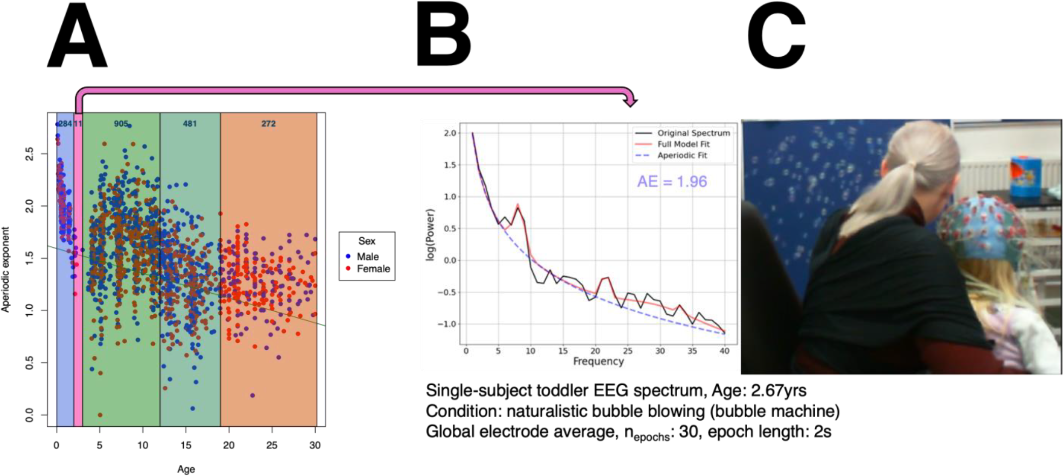
Human cognitive neuroscience
PW_164
Keywords: Decision Making, EEG, Optimal Stopping Problems, Modelling, Information Sampling
Authors: Christina Dimitriadou, Nicholas Furl
Optimal stopping problems, such as the beads task, provide a framework for studying decision-making under uncertainty, balancing the trade-off between evidence-gathering and decision commitment (Furl and Averbeck, 2011; Moutoussis et all, 2011). We investigated deviations from normative strategies in human decision-making and examined the neural mechanisms underlying these processes using EEG.
Forty participants completed a beads task under two probability conditions (0.8 and 0.6) while their EEG activity was recorded. A Bayesian ideal observer model and parametrised models were used to predict participant behaviour, revealing significant effects of agent type (humans, ideal observer, and parametrised model), probability condition (0.8 and 0.6), and their interaction on number of draws and accuracy. Participants systematically deviated from Bayesian ideal observer predictions, demonstrating undersampling behaviour, particularly in the more ambiguous 0.6 probability condition, where they sampled more than in the 0.8 condition but still less than the optimal strategy. Model comparisons indicated that the parametrised model, which incorporated a cost-to-sample parameter and an inverse temperature parameter, more closely approximated human behaviour than the ideal observer.
Neurally, P300 amplitudes were modulated by both task difficulty (probability condition) and response type (draw choice vs. urn choice), with larger amplitudes observed for urn choices, reflecting increased cognitive demands when committing to a decision. Unexpectedly, P300 amplitudes were larger in the more difficult 0.6 probability condition, suggesting greater neural engagement under higher uncertainty. Beta oscillatory activity also distinguished between probability conditions and choice types, with fast beta (20–30 Hz) showing greater engagement during urn choices, supporting its role in decision commitment. Additionally, beta power in left parietal regions was significantly associated with trial-wise model-derived action values, highlighting its relevance in evidence accumulation and uncertainty resolution.
Our findings confirm and extend prior work, demonstrating that deviations from normative strategies are systematic across task manipulations. By integrating behavioural, computational, and neurophysiological data, this study advances our understanding of how humans process and act upon uncertainty in probabilistic environments, reinforcing the role of beta oscillations and the P300 component in balancing exploration and exploitation in decision-making.
Furl, N. and Averbeck, B.B., 2011. Parietal cortex and insula relate to evidence seeking relevant to reward-related decisions. Journal of Neuroscience, 31(48), pp.17572-17582.
Moutoussis, M., Bentall, R.P., El-Deredy, W. and Dayan, P., 2011. Bayesian modelling of Jumping-to-Conclusions
bias in delusional patients. Cognitive neuropsychiatry, 16(5), pp.422-447.
Human cognitive neuroscience
PW_165
Keywords: Mild Cognitive Impairment, Cognitive dysfunction, Type 2 Diabetes, Metabolic Disease
Authors: Dana AlTarrah, Wafaa AlDuhaimi, Zahraa Abdulrasoul, Eman AlEnezi, Modhy AlHasawi, Fawaz AlZaid, Mohammed AlOnaizi
Cognitive impairment is a significant complication of type 2 diabetes mellitus (T2DM), adversely affecting quality of life and glycemic control. Mild cognitive impairment (MCI) in T2DM is particularly concerning, impacting memory and executive functions, and complicating diabetes management. Despite an increasing volume of literature on MCI prevalence in T2DM, findings remain inconsistent. This study systematically synthesizes current evidence to estimate MCI prevalence and identify factors contributing to cognitive decline in T2DM patients.
Following PRISMA guidelines, observational studies published between June 2020 and May 2024 were systematically reviewed from PubMed, Web of Science, and Embase. Observational studies reporting MCI prevalence in T2DM patients, assessed using validated neuropsychological tools, were included. Exclusions were made for studies focusing on dementia or those with insufficient data. Key data on prevalence rates, diagnostic tools, and study characteristics were extracted, and the STROBE checklist was used to assess bias. Study protocol registered at PROSPERO ID CRD42024562218.
Forty studies encompassing 14,974 T2DM patients were analyzed. MCI prevalence ranged from 9.94% to 74.9%, with a pooled prevalence of 35.7% (95% CI=0.305−0.409). Subgroup analyses revealed the highest prevalence in Europe (62%, 95% CI=0.38−0.86, I²=97%) compared to Asia (35%, 95% CI=0.30−0.40, I²=97%). Patients under 60 years showed slightly higher prevalence (38%, 95% CI=0.31−0.44, I²=98%) than those aged 60 and above (32%, 95% CI=0.23−0.41, I²=98%). The Montreal Cognitive Assessment (MoCA) reported higher prevalence rates (41%, 95% CI=0.34−0.48, I²=96%) compared to the Mini-Mental State Examination (MMSE) (34%, 95% CI=0.23−0.45, I²=97%).
MCI prevalence in T2DM patients varies widely due to methodological differences across studies. Standardizing cognitive and diabetes diagnostic criteria and expanding research in underrepresented regions are crucial. Early detection and intervention are essential for managing cognitive impairment in T2DM.
Human cognitive neuroscience
PW_166
Keywords: grey matter volume, visual spatial attention gradient, tunnel vision, trait anxiety, cerebellum
Authors: Mrinmoy Chakrabarty, Suhail Rafiq Mir, Varsha Singh
Flexible allocation of visual attentional resources to different spatial loci is crucial for visual cognition, the inability of which may lead to compromised function, e.g., tunnel vision. This is relevant in trait anxious individuals, known to have biases in visual processing. The relationship between brain structure and trait anxiety towards predicting the visual attention gradient is unclear, which we explored in this study.
We used a visual-spatial attention task with structural magnetic resonance imaging (sMRI; 3Tesla) in healthy young adults with varying degrees of trait anxiety (n = 60; 23 females; [mean ± s.d.] age = 22.8 ± 3.8 years; trait anxiety = 46.52 ± 11.04). Using objective measures from the behavioural task (attention gradient) and sMRI (gray matter volume - GMV), we explored if an association existed between the whole-brain GMV and trait anxiety towards predicting inter-individual differences in attention gradient. We calculated the attention gradient (metric for tunnel vision) using measures of attentional efficiency near the central fixation (1.5 degrees) minus far from it (6 degrees). Greater positive gradient values reflected better attentional efficiency at the near relative to farther spatial loci or higher degrees of tunnel vision.
Multiple linear regression was performed with the GMV, trait anxiety, GMV x trait anxiety interaction as the independent variables of interest and attention gradient as the dependent variable after controlling for total intracranial volume and age. Analyses were done using the Computational Anatomy Toolbox v12.8.1 (r2043) hosted on MATLAB 2022a. The brain regions were visualized after height and cluster-level thresholds with corrected p-values < 0.05.
The attention gradient correlated negatively with the GMV of the right cerebellum lobule VI (height threshold p < 0.001, cluster extent threshold = 1250 voxels, cluster-level p corrected for Family-Wise Error = 0.003). The strength of this negative correlation within bilateral cerebellar lobule VI was moderated by inter-individual differences of anxiety (height threshold p < 0.001, cluster extent threshold = 871 voxels, both cluster-level p corrected for Family-Wise Error ≤ 0.025), that varied between females and males. A machine-learning analysis with balanced cross-validation reliably predicted inter-individual differences of attention gradient using bilateral cerebellar lobule VI GMV.
Overall, individuals with lesser GMV of cerebellar lobule VI manifested greater individual severities of tunnel vision, which was more pronounced in females than males. Cerebellar lobule VI may interact with trait anxiety in tuning fine-grained spatial attention which may be predictive of tunnel vision.

Human cognitive neuroscience
PW_168
Keywords: Laparoscopy, EEG, fNIRS, Skill acquisition, Longitudinal training
Authors: Esra Zeynep Dudukcu, Alexander Luke Sumich, Philip Breedon, Zohreh Zakeri, Ahmet Omurtag
Laparoscopy training is a fundamental component of surgical education. In the past ten years, studies have sought to find objective ways to evaluate the skills of trainees by neuroimaging methods. Currently, there is no standardized evaluation protocol in this field, which makes it difficult to objectively assess skills. Moreover, research has indicated that cognitive skills influence the learning and retention of laparoscopic techniques, an aspect that is often neglected. Obtained data will be analysed by machine learning (ML) to pursue three main objectives: (i) quantifying progress in longitudinal training and its relationship with cognitive abilities, (ii) discovering the multi-neural correlates of proficiency and automation in the laparoscopy and (iii) comparative understanding of the expert surgeon’s brain to novice’s.
We will collect and analyse data from behavioural, electro-neurophysiological and hemodynamic activities. Behavioural data based on the motor performance of the trainee will be evaluated by speed, precision and task difficulty in addition to the fundamental laparoscopy skills score. In addition to that, brain-based metrics will be used as quantitative data.
Forty participants (20 novices and 20 experts) will be recruited in total. In this study, novices will receive laparoscopy training for ten consecutive days, while experts will participate for two days, alongside EEG-fNIRS recordings. Laparoscopy tasks will cover pattern cutting and ligating a loop with extra-corporeal Roeder knotting. Cognitive assessments will be held on by, the Wisconsin Card Sorting Task, the Attentional Network Task, the Mental Rotation Task and the Peg Board task for bimanual dexterity. On the fifth to the tenth days, participants will encounter a complex appendectomy model. All laparoscopy training will be performed using a training box equipped with instruments that emulate real, tissue-like models and offer realistic tactile feedback (LapAR, Inovus Medical). Following each laparoscopy session, participants will complete the NASA Task Load Index (NASA-TLX) questionnaire.
Since the study involves multiple aspects such as two groups of participants (novices and experts), and longitudinal training, the collected data on cognitive scores and laparoscopy training will be analysed in a multifaceted manner. Overall, descriptive statistics will be complemented by machine learning methods (ML), such as linear regression, support vector machines, linear discriminant analysis, and generalized estimating equations for learning curves. These are common ML methods applied to EEG-fNIRS data in laparoscopy studies. The results of the study are expected to significantly improve the laparoscopy learning process of medical students and objectively assess the acquired skills.
Human cognitive neuroscience
PW_169
Keywords: Event boundary, Temporal memory, Cognition
Authors: Qiying Liu, Tobias Sommer, Deborah Talmi
Although time appears continuous, our memories are not. Instead, we remember experiences as distinct events, marked by event boundaries that define their beginnings and endings. These boundaries influence how we recall event sequences. Research shows that people have better temporal order memory for sequences occurring within a single event (within-event items) than for those crossing event boundaries. However, this contradicts real-world experiences, where cross-boundary actions—such as setting the table after cooking—are often more vividly recalled than within-event details, like whether salt was added before or after stirring a sauce.
Wen and Egner (2022) suggested that this discrepancy arises because laboratory studies lack ecological validity. In real life, contexts are meaningful and distinct, whereas lab studies typically use repetitive, abstract stimuli to define event boundaries. Wen and Egner replicated two well-established effects: (1) people remembered within-event sequences more accurately than cross-boundary sequences, and (2) items separated by an event boundary were perceived as further apart in time. However, they also found that when events were embedded in meaningful, unique contexts—and those contexts were reinstated at retrieval—memory for cross-boundary sequences improved, reversing the classic TOM effect. This finding highlights the importance of distinct, real-world contexts in memory formation and retrieval.
The present study aims to replicate Wen and Egner’s findings while further enhancing ecological validity. In their study, event boundaries were defined by a change in a color frame or a shift in goal state and stimulus set, with some repetitions across trials. Our study structures event boundaries around real-world contexts, such as visiting a museum or staying at a hotel. Each boundary represents a transition to a unique location and activity, ensuring no repetition throughout the session. Participants will be instructed to actively associate target stimuli with their respective event contexts and engage in deep, elaborative processing.
In our experiment, participants will be assigned to either a context-present or context-absent group. Those in the context-present group will receive a contextual reminder during the memory test, while those in the context-absent group will not. To analyze whether accuracy rates and perceived temporal distance vary across conditions, we will fit a mixed-effects model using the lme4 package in R. The model will include context, boundary, and their interaction as fixed effects, while accounting for individual differences with random intercepts for participants. ANOVA will be performed on each model to assess the significance of the fixed effects and their interactions.
Neurodevelopment, stem cells and associated disorders
PM_171
Keywords: Autism Spectrum Disorder, Exercise Movement Techniques, Neuronal Plasticity
Authors: Lavínia Teixeira-Machado
Autism is a common neurodevelopmental condition defined by functioning difficulties that interfere with social, academic, or occupational demands and expectancies. It is known that physical exercise influences synaptogenesis and changes in structural and functional brain areas responsible for cognition, communication, and social reciprocity. Some gaps remain unknown in both animal models and human intervention about exercise neuroplasticity and autism core sets. We explored the mechanisms and structural and functional brain relations of applying physical exercises of autism core sets. methods The methodology was conducted in accordance with Joanna Briggs Institute's approach. We conducted electronic database searches, and 899 studies were recovered. results Of the 899, twelve studies met the inclusion criteria. Two hundred sixty-eight autistic participants, 186 boys (69.4%), age range 3-18 years old. Physical activity programs included arts (music, instruments, theatre, dance), martial arts (karate, Nei Yang Gong), sports (mini-basketball, cycling, aquatic exercises), equine-assisted therapy, virtual reality-based training, and physical exercise training. The included studies reported changes in mTOR signaling, TrkB, and BDNF after physical activity interventions. This review highlights changes in resting-state neural activity in regions important for social cognition and communication, as well as increased functional connectivity in brain areas responsible for inhibitory control, action outcomes monitoring, and self-regulation.
The results demonstrate a wide-ranging application of physical activity and movement interventions that explain neural effects mechanisms using different methods across various contexts. The effect of long- and short-term interventions on autism core sets is mixed. Future research should focus on undertaking randomized controlled trials with large sample sizes and participants with all levels of support to suggest the generalizability of the study findings. The lack of population-specific, reliable, and validated molecular mechanisms and neural effects outcome measures should be addressed.
American Psychiatric Association. (2013). Diagnostic and statistical manual of mental disorders (5th ed.).
Ranieri A, et al. (2023). Positive effects of physical activity in autism spectrum disorder: how influences behavior, metabolic disorder, and gut microbiota. Front Psychiatry.
Yu, H. et al. (2021). Alteration of Effective Connectivity in the Default Mode Network of Autism After an Intervention. Front Neuroscience.

Neurodevelopment, stem cells and associated disorders
PM_172
Keywords: Autism spectrum condition, Migraine disorder, Neurodevelopment, Sensory Processing, Cross-Condition Networks
Authors: Amparo Gimenez Rios, Fiona Kerr
Autism spectrum condition and migraine disorder (MD) are polygenic conditions shaping neurological function and sensory processing. Both share atypical sensory experiences and structural and functional brain changes, such as increased somatosensory cortex thickness and thalamic connectivity. Previous studies showed higher rates of migraines among autistic individuals (Pan et al., 2021, Underwood et al., 2019), suggesting a link between both. We explored shared genetic mechanisms and protein-protein interactions (PPI) network between autism and MD to identify biological processes linked to their co-occurrence and improve clinical approaches for autistic migraineurs.
Genome-wide association studies (GWAS) data for autism (1,032,825 autistic and 1,316,616 control) and MD (165,300,119 MD and 326,260,027 control) were retrieved from the GWAS Catalog. Overlapping genes were identified using the Bioconductor GeneOverlap package for R. Protein-coding genes were analysed to explore PPI networks and functional relationships using STRING. High-confidence interactions were filtered to obtain enriched biological processes from Gene Ontology (Figure 1). Chi-square and Bonferroni tests were applied to compare enriched biological processes in autism-MD, autism-only, and MD-only PPI networks.
63 significant overlapping genes were identified between autism and MD (p-value = 6e-12, OR = 2.9). PPI analysis revealed a highly enriched cross-condition network (p-value = 3.19e-12), highlighting overrepresented processes like "basal dendrite arborization" and "neuron-glial cell signaling." Biological process comparisons highlighted significant differences between autism-only, migraine-only, and combined networks, with unique enrichments appearing in the combined network.
Our findings enhance understanding of the shared genetics between autism and migraine, suggesting a complex interplay when these conditions coexist. A unique migraine profile may manifest in autistic migraineurs, and our data support further research into the causal roles of identified genes and processes linking these conditions. This could identify new precision medicine targets to tailor migraine assessment and treatment in autistic individuals.
Pan, P.Y., Bölte, S., Kaur, P., Jamil, S. and Jonsson, U., 2021. Neurological disorders in autism: A systematic review and meta-analysis. Autism, 25(3), pp.812-830. DOI: https://doi.org/10.1177/1362361320951370
Underwood, J.F., Kendall, K.M., Berrett, J., Lewis, C., Anney, R., Van den Bree, M.B. and Hall, J., 2019. Autism spectrum disorder diagnosis in adults: Phenotype and genotype findings from a clinically derived cohort. The British Journal of Psychiatry, 215(5), pp.647-653. DOI: https://doi.org/10.1192/bjp.2019.30
Lee, T.Y., Tsai, S.J., Chen, T.J. and Chen, M.H., 2021. Risk of migraine development among children and adolescents with autism spectrum disorder: A nationwide longitudinal study. Research in Autism Spectrum Disorders, 89, p.101880. DOI: https://doi.org/10.1016/j.rasd.2021.101880
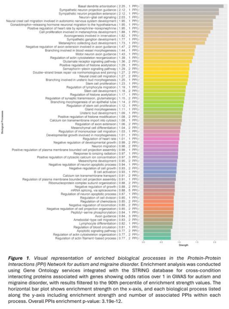
Neurodevelopment, stem cells and associated disorders
PM_173
Keywords: Dentate gyrus granule cells, Neurodevelopment, scRNA sequencing, snATAC sequencing, TF-gene regulons
Authors: Jacek Szymanski, Sanskar Ranglani, Claus Nerlov, Liliana Minichiello
The developing dentate gyrus undergoes extensive neurogenesis and provides critical input to the hippocampal circuitry. Defects in this process are associated with neurodevelopmental disorders. While the dentate gyrus has been extensively studied, comprehending the transcriptional and epigenetic events occurring in various developmental transitions remains elusive. To this end, we performed single-nucleus RNA and ATAC sequencing to uncover the developmental transitions of neurons at a single-cell resolution. The overall objective was to unbiasedly identify cell-type-specific genes and associated transcription factors (TFs) regulating dentate granule cell development.
We dissected the mouse dentate gyri from both sexes, 3-week-old mice (N = 3), and performed library preparation using the Chromium Next GEM Single Cell Multiome ATAC + Gene Expression kit followed by Illumina sequencing. The data were pre-processed using Cell Ranger. R Studio was utilised to filter out low-quality cells, perform normalisation, carry out dimensionality reduction, and integrate bimodal data. Clustering and cell type annotation were performed based on canonical gene marker expression, yielding a total of 17,005 cells included in the final dataset. TF motif accessibility was calculated using chromVAR, whereas FigR package allowed the construction of cell-type specific gene regulatory networks (GRN).
Bioinformatic analyses revealed seven different cell types along a developmental continuum – radial glia-like cells, neural progenitors, neuroblasts, immature granule cells stage 1, immature granule cells stage 2, young granule cells, and mature granule cells. Differential gene expression and motif enrichment analysis revealed novel genes and TFs important for each developmental stage of the dentate granule cells (Figure 1). Gene string enrichment analysis revealed cell-type specific pathways, whereas construction of GRN revealed previously unidentified regulons vital in maintaining cell-type identity.
This study uncovers a previously unappreciated developmental trajectory of the dentate granule cells. These findings provide a comprehensive framework for understanding dentate granule cell development and pave the way for further functional validation and unbiased exploration of its regulatory mechanisms.
Graphical abstract
Figure made with www.biorender.com
Neurodevelopment, stem cells and associated disorders
PM_177
Keywords: Autism Spectrum Disorder, Copy number variation, Social Responsiveness, Autism Genetic Resource Exchange
Authors: Hanna Ganderton, Zoe Mia, Asami Oguro-Ando, Rebecca Smith
Autism Spectrum Disorder (ASD) is a neurodevelopmental condition affecting 1-1.5% of the population, characterised by deficits in social communication as well as restricted and repetitive behaviours. ASD exhibits significant genetic and phenotypic heterogeneity, making it a challenging condition to fully comprehend. Specific risk genes (e.g., SHANK3, CNTNAP2, etc.) are strongly associated with the disorder, and these genes contribute to our understanding of the underlying biological mechanisms. Genetic studies have identified single nucleotide polymorphisms (SNPs) and copy number variants (CNVs) as key markers of ASD risk loci. However, genome-wide association studies (GWAS) often face challenges due to the interplay of de novo and inherited mutations, necessitating refined strategies to elucidate the genetic basis of ASD.
Investigating sub-phenotypes of ASD, such as those derived from the Social Responsiveness Scale (SRS), offers a promising strategy for identifying genetic loci associated with specific behavioural traits. The SRS measures various aspects of social responsiveness, which are highly relevant in distinguishing between behavioural profiles within the ASD population. Sub-phenotype stratification offers a promising approach for linking genetic loci to specific behavioural traits, as shown by Constantino and Todd (2003), who demonstrated SRS's utility in identifying social communication deficits in ASD populations.
This study will analyse phenotypic and genetic data from the Autism Genetic Resource Exchange (AGRE) database, comprising approximately 2500 individuals with ASD. Participants will be stratified into subgroups based on SRS domain scores (social communication, awareness, motivation, cognition, and mannerisms) or total SRS scores, depending on inter-domain correlations assessed via Pearson's correlation coefficient. Linear regression models will assess the effects of age, sex, and potential covariates on SRS scores. GWAS will be conducted using PLINK software, with genomic loci identified at a genome-wide significance threshold of p < 5×10⁻⁸. Multiple testing correction will be applied using the Bonferroni method. Key results will be visualised through Manhattan and QQ plots.
This study is expected to identify specific genetic loci associated with social responsiveness sub-phenotypes, thereby contributing to a deeper understanding of ASD's genetic architecture. These findings could inform personalised interventions and advance precision medicine approaches for ASD.
Constantino JN and Todd RD (2003) Autistic Traits in the General Population: A Twin Study. Archives of General Psychiatry 60(5). American Medical Association: 524–530.
Neurodevelopment, stem cells and associated disorders
PM_178
Keywords: Epilepsy, Sleep, Absence Seizures, SYNGAP1, Cognition
Authors: Lucy Pritchard, Sally Till, Alfredo Gonzalez-Sulser
Sleep disturbances and epilepsy often co-occur in neurodevelopmental disorders (NDDs), but the relationships between these phenotypes and cognition remains elusive. de novo pathogenic mutations in SYNGAP1 are a common underlying factor in NDDs and are associated with a range of phenotypes including autism, intellectual disability (ID), and absence epilepsy (Hamdan et al.,2009, doi:10.1056/NEJMoa0805392). Sleep disturbances are also highly prevalent in this population (Vlaskamp et al.,2019, doi:10.1212/WNL.0000000000006729).
A heterozygous rat model in which the SYNGAP C2 and GAP domains are deleted (Syngap+/Δ-GAP) recapitulates several features associated with SYNGAP1-related ID, including absence seizure-like spike-wave discharges (SWDs), REM sleep disturbances (Buller-Peralta et al.,2022, doi:10.1093/braincomms/fcac263), and impaired fear extinction (Katsanevaki et al.,2024, doi:10.1016/j.celrep.2024.114733). We previously showed that anti-seizure drug (ASD) ethosuximide blocked SWDs (Katsanevaki et al.,2024) and preliminary findings suggest it also rescues fear extinction impairments and REM sleep deficits. The proposed work aims to further characterise the effect of seizure suppression by ASDs on sleep and determine whether REM sleep modulation affects seizure suppression by ASDs.
Given the reciprocal interactions between sleep and epilepsy, and the high prevalence of sleep disturbances in individuals with SYNGAP1 mutations, understanding potential relationships between seizures and sleep may provide insight into novel therapeutics.
Male Syngap+/Δ-GAP rats and wild type (WT) littermates (3-4 months old), will be implanted with skull screw electrodes to record electrophysiology over the somatosensory cortex and reticular thalamus. Using the TAINITEC acquisition system, freely moving animals will be recorded 24-hours a day consecutively for two weeks. The recording protocol will begin with 48 hours of baseline electrophysiology recording. SWDs will be pharmacologically modulated on days 3, 5, 7, 9 and 11 via intraperitoneal injection of anti-seizure drugs ethosuximide (100mg/kg), levetiracetam (100mg/kg), valproic acid (100mg/kg), GABA agonist 4,5,6,7-tetrahydroisoxazolopyridin-3-ol (10mg/kg), or saline.
The number and duration of SWDs will be quantified using a MATLAB-based automated scoring algorithm. Time spent in each physiological state and sleep bout number and duration will also be quantified. Syngap+/Δ-GAP and WT rats will be compared across baseline, saline, and drug conditions using 2-way mixed ANOVA to characterise changes in seizure-activity and sleep following administration of ASDs.
Neurodevelopment, stem cells and associated disorders
PM_179
Keywords: 22q11.2DS, Bioinformatics, Neurodevelopment, microRNAs
Authors: Sabrina Burton, Gemma Wilkinson, Jamie Wood, Peter Maycox, Adrian Harwood, Lawrence Wilkinson
MicroRNAs (miRNAs) are small molecules that fine tune gene expression and are essential to regulating cell differentiation. 22q11.2 Deletion Syndrome (22q11.2DS) is a genetic syndrome which increases the likelihood of individuals developing schizophrenia by 25 times. This disorder is caused by a large deletion on chromosome 22 that spans many genes(Khan et al., 2020). The deletion includes the gene DiGeorge Syndrome Critical Region 8 (DGCR8) which is a master controller of miRNA production (Koscianska E et al., 2011). We are investigating the contribution that low gene dosage of DGCR8 and consequent effects on miRNA metabolism makes to the significantly elevated risk for psychiatric symptoms observed in 22q11.2DS patients.
Informatic approaches can been used to generate knowledge guided prediction models used in silico that predict mRNA targeted by miRNAs and hence draw insights into the cellular process that are dysregulated in disease states (Lin et al., 2018). We are building such a model for 22q11.2DS and will test our predictions by transcription profiling (RT-qPCR) at multiple time points representing stages from human pluripotent stem cells to mature neurons. We have shown that the DGCR8 protein changes during neuronal differentiation, indicating that there may be specific time points where key regulatory steps that control neurodevelopment occur. RT-qPCR data will be analysed using two-way ANOVA to look at differences between controls and 22q11.2DS samples across the time course. In the next step, we will correlate changes of miRNA abundance with the biological processes that they regulate and establish cell phenotypes observed in 22q11.2DS during neurodevelopment that are likely to be due to reduced DGCR8, with the ultimate objective of their suppression via direct miRNA manipulation.
Khan, TA, Revah, O, Gordon, A et al. (2020) ‘Neuronal defects in a human cellular model of 22q11.2 deletion syndrome’, Nature Medicine, 26: 1888–1898. Available at: https://doi.org/10.1038/s41591-020-1043-9
Koscianska E, Starega-Roslan J, Sznajder LJ et al. ‘Northern blotting analysis of microRNAs, their precursors and RNA interference triggers’ BMC Mol Biol. 2011 Apr 11;12:14. Available at: doi: 10.1186/1471-2199-12-14
Lin, Y, Chen, F, Shen, L et al. (2018) ‘Biomarker microRNAs for prostate cancer metastasis: screened with a network vulnerability analysis model’, Journal of Translational Medicine, 16(1): 134. Available at: https://doi.org/10.1186/s12967-018-1506-7
Neurodevelopment, stem cells and associated disorders
PM_180
Keywords: Trapp complex, postnatal microcephaly, lipid droplet, hippocampus, neural stem cells
Authors: Sultan Aljuraysi, Mark Platt, Michela Pulix, Harish Poptani, Antonius Plagge
Mutations of the human TRAFFICKING PROTEIN PARTICLE COMPLEX SUBUNIT 9 (TRAPPC9) cause a neurodevelopmental disorder characterised by postnatal microcephaly and intellectual disability. Trappc9 is a subunit specific to the intracellular membrane-associated TrappII complex, which activates Rab11 and Rab18. Rab18 is located on lipid droplets (LDs) and also interacts with the LD coat protein Perilipin-2 (Plin2). Trappc9 co-localises with Rab18 and Plin2 on LDs. We analysed the phenotype of Trappc9 knock-out mice and LD characteristics in neurons.
Brain volumes of Trappc9 knock-outs were measured by magnetic resonance imaging (MRI) and white matter was characterised by diffusion tensor imaging (DTI). Behavioural analysis comprised open field, rotarod, plug puzzle and novel object recognition tests. Gene expression was analysed by RNAscope and hippocampal neural stem and progenitor cells (NSPCs) were quantified by Sox2 immunohistochemistry. Primary hippocampal neurons from newborn mice were cultured with Oleic Acid (OA) to stimulate LD formation. LDs were stained with LipidTOX combined with immunohistochemistry for LD proteins. Statistics were undertaken via Student’s t-test, two-way ANOVA or Mann-Whitney U-test, depending on data distribution.
Trappc9 knock-outs developed microcephaly postnatally with grey and white matter regions being affected. MRI identified a disproportionately stronger volume reduction of the hippocampus, which was associated with a significant loss of Sox2-positive NSPCs. DTI indicated a reduced organisation or integrity of white matter. Trappc9 is expressed in NSPCs and neurons. Trappc9 knock-outs displayed behavioural abnormalities in all the above-mentioned tests, indicating deficits in exploration, learning and memory. Trappc9-deficient primary hippocampal neurons accumulated a larger total LD volume per cell after 12 hrs of OA exposure. Individual LDs were larger in knock-out neurons at 6 hrs. Although the majority of LDs was Plin2-positive in wild-type and knock-out neurons, the percentage of Plin2-coated LDs was significantly lower in Trappc9-deficient neurons after 12 hrs of OA. At both timepoints, the LD surface area that overlapped with Plin2 staining was reduced in Trappc9-deficient neurons. Beyond these brain-related phenotypes, Trappc9 knock-out mice developed obesity, which was significantly more severe in females than in males.
Trappc9 knock-out mice model the symptoms of the human disorder. Dysfunctions in LD homeostasis contribute to the neurodevelopmental symptoms of Trappc9 deficiency. LDs and lipid metabolism play an important role in neurodevelopment, NSPC proliferation and differentiation.
Aljuraysi S, Platt M, et al. (2024) Microcephaly with a disproportionate hippocampal reduction, stem cell loss and neuronal lipid droplet symptoms in Trappc9 KO mice. Neurobiol. Dis. 192:106431
Neurodevelopment, stem cells and associated disorders
PM_181
Keywords: Cortical neurodevelopment, GABAergic interneurons, Serotonin, Cortex, SSRI
Authors: Abhishek Saha, Veronica Munday, Gabriel Ocana-Santero, Simon J.B. Butt
Serotonin (5-HT) is a neuromodulator important for neurodevelopment and function, regulating early developmental processes that shape sensory representation in the neocortex. Diverse GABAergic interneuron populations are critical for initial formation of these sensory maps, and mediating information transfer in both the developing and adult brain. Deficits in this cell type are linked to a range of neurodevelopmental psychiatric disorders including ASD and ADHD, as is altered 5-HT signalling, either via in utero selective serotonin reuptake inhibitor (SSRI) exposure or serotonin reuptake transporter (SERT) genetic polymorphisms. To further investigate this connection, we examined how disrupted 5-HT signalling during early critical periods in the neocortex impacts GABAergic interneurons. Specifically, we assessed the effects of chronic genetic versus acute pharmacological SERT perturbations on their development in mouse somatosensory barrel cortex (S1BF).
Our two models were (1) wild-type/heterozygous/homozygous SERT knockout (SERTKO) mice and (2) wild-type mice dosed postnatally with the SSRI fluoxetine (versus sucrose controls). All mice expressed a red fluorophore (tdTomato) in specific interneuron subtypes (VIP+ or Nkx2-1+) to enable visualisation of these populations in ex vivo preparations. Brains were sectioned and stained for a further delineator of interneuron diversity, the calcium-binding protein parvalbumin (PV), followed by confocal imaging of S1BF. Automated QuPath analysis pipelines quantified distinct interneuron populations. statistical analyses were performed using Python 3.9. Non-parametric tests were utilised as appropriate, including Mann-Whitney U test, Kruskal-Wallis test, or two-way permutation-based ANOVA with Dunn’s post-hoc tests. Significance was defined as *p<0.05.
Our analysis revealed decreased expression of PV across S1BF in SERTKO (p=0.004) but not in SSRI-treated mice, despite the latter showing a decrease in the broader Nkx2-1+ population (p=0.032) which includes PV+ subtypes. VIP+ interneuron numbers were unaffected in SERTKO mice but increased in our SSRI model (p=0.032), further demonstrating distinct outcomes following acute versus chronic 5-HT alterations. Despite these changes, laminar organisation in S1BF was preserved in both models, suggesting that perinatal 5-HT alterations primarily impact interneuron survival rather than migration.
These findings reveal a subtype-specific role for 5-HT in interneuron development, with PV+ interneuron survival impacted by chronic 5-HT perturbation whereas VIP+ interneuron survival increased following acute postnatal 5-HT elevation. By demonstrating distinct impacts of SERTKO and SSRI treatments, this study advances our understanding of how 5-HT regulates GABAergic circuit development and provides novel insights into early cortical mechanisms essential for normal function.
Neurodevelopment, stem cells and associated disorders
PM_182
Keywords: Prefrontal cortex, Neurodevelopment, Neurocircuitry, Neurophysiology, Adolescence
Authors: Gabriella Margetts-Smith, Claire Montmasson, Paul Anastasiades
Normal development of the medial prefrontal cortex (mPFC) is critical for a range of cognitive processes, and dysfunction of this region is associated with various neurological and psychiatric disorders. Whilst the micro- and macro-circuitry of this area has been well documented, there is a limited amount of research examining the development of mPFC neurons across the adolescent sensitive period – particularly at the cellular level.
To address this, we will perform whole-cell patch-clamp recordings in layer 3 pyramidal and parvalbumin-expressing (PV) neurons in the prelimbic area at three time points: before (P25), during (P40) and immediately after (P55) the murine adolescent period. We will examine the development of both the intrinsic physiological properties, and the spontaneous postsynaptic activity of these two cell types. ANOVA, or appropriate non-parametric alternatives, will be used to compare between the three age points for various measures of membrane and firing properties, as well as excitatory and inhibitory spontaneous miniature release events.
Whilst the mPFC undergoes critical development at a later stage than lower-order sensory cortices, we nevertheless anticipate we will see similar changes in the physiological properties of pyramidal and PV cells as those described in early postnatal development in areas such as the visual and somatosensory cortices: for example, increased action potential thresholds and decreased firing rates in L3 pyramidal cells. Additionally, previous work within our group describes a peak in synaptic maturation during the adolescence period, therefore we predict we will find significant changes in the amplitudes and kinetics of both excitatory and inhibitory postsynaptic currents in these cells – as well as a shift in E/I balance – during this time period. Understanding the typical development of these cells and changes to their local circuitry during adolescence will provide us with a strong foundation from which we can better investigate how the underlying mechanisms of this development, and how it can be perturbed.
Neurodevelopment, stem cells and associated disorders
PM_183
Keywords: neurodevelopmental disorders, touch, pain, rat models
Authors: Katarzyna Mazur, Ying Sze, Sarfaraz Nawaz, Lewis Scott, Sally Till, Peter Kind, Carole Torsney
De novo mutations in SYNGAP1 are one of the most common genetic causes of neurodevelopmental disorders linked to intellectual disability, epilepsy, and autism spectrum disorder (ASD) (Satterstrom et al., 2020). SYNGAP1 haploinsufficiency has also been associated with somatosensory processing alterations in humans and in mice (Michaelson et al., 2018). Many ASD-associated genes are key components of activity-dependent processes, suggesting potential involvement in postnatal maturation of spinal somatosensory circuits (Ebert and Greenberg, 2013; Beggs et al., 2002). Our aim is to investigate the impact of heterozygous loss of SYNGAP1 on touch and pain behavioural phenotypes and underlying spinal processing in a SYNGAP1 haploinsufficiency rat model.
Adult (6-8-week-old) male and female Syngap+/- (n=20; 9M/11F) and wild-type (n=19; 10M/9F) rats underwent tactile, noxious mechanical, and noxious thermal behavioural testing. Spinal cord homogenates were immunoblotted for the SynGAP protein (Syngap+/- n=6/wild-type n=4). Spinal cord (n=7/genotype) was immunostained for markers of tactile (VGLUT1) and nociceptive (IB4) fibres. Glabrous skin (n=7/genotype) was immunostained for markers of tactile end organs (S100/NF200/ColIV). Isolated dorsal roots were stimulated at 0.2Hz/1-500µA in compound action potential (CAP) recordings (n=8/genotype). Aβ-fibre refractory period was measured at 1Hz/20µA in 2 ms steps between 20ms-2ms inter-pulse intervals. Dorsal root-ventral root potential (DR-VRP) recordings (n=7/genotype) were performed by stimulating dorsal roots at 0.2Hz/2.5-300µA.
Statistical analysis was carried out using GraphPad Prism 9 software. Two-way ANOVA was used to compare genotypes across male and female behavioural datasets. Unpaired t-test or Mann-Whitney test was used to compare between genotypes depending on normality of the data, as assessed using D'Agostino-Pearson and Shapiro-Wilk tests. Chi-square test was used to compare tape removal. p<0.05 was considered statistically significant.
Syngap+/- rats show reduced tactile reactivity but unaltered reactivity to noxious stimuli. SynGAP protein expression in the spinal cord dorsal horn is reduced by 50%. The gross spinal termination pattern of tactile and nociceptive afferents is unaltered. Glabrous hindpaw skin structure and the density of tactile corpuscles does not differ between genotypes. There are no differences in electrical properties of the Aβ, Aδ, and C-fibre primary afferent components in CAP recordings, including no difference in the Aβ-fibre refractory period. Spinal reflex networks have an increased response threshold in DR-VRP recordings, but unaltered response magnitude at maximal response intensity. In conclusion, tactile reactivity is reduced but acute pain reactivity is unaltered in Syngap+/- rats. Electrophysiological evidence indicates a spinal functional deficit contributes to the tactile hypo-reactivity phenotype we have observed in Syngap+/- rats.
Neurodevelopment, stem cells and associated disorders
PM_184
Keywords: Prefrontal, MRS, metabolites, depression, development
Authors: Spatika Jayaram, Arek Stasiak, Stacey Gould, Gemma Cockcroft, Taylor Lynn-Jones, Angela C. Roberts, Stephen J. Sawiak
Mood disorders are commonly diagnosed in adolescence, a period of significant prefrontal cortical development. Two regions implicated in depression are the subgenual anterior cingulate cortex (sgACC-25) and the dorsolateral prefrontal cortex (dlPFC-46). Both are targets for neuromodulation, with success related to the extent of their anti-correlated activity. Earlier studies in marmoset monkeys outlined distinct trajectories of morphological development for these subregions (Sawiak et al.,2018, Cerebral Cortex: 28:4440-4453). In this study, we examined the potential metabolic mechanisms underlying these differences.
Magnetic resonance spectroscopy data were analyzed from 77 marmoset monkeys, imaged 1-5 times from infancy to adulthood (3-30 months, 202 scans). Linear mixed-effects models assessed developmental changes, with age, region, and sex as fixed effects. Nine metabolites were examined, and those showing significant developmental changes were further analyzed. Non-linear spline modeling mapped metabolic trajectories for comparison with morphological trajectories.
Four metabolites (inositol, creatine, n-Acetylaspartate (NAA) and choline) showed significant differences in absolute levels and trajectories between sgACC-25 and dlPFC-46.
Inositol and creatine (involved in glial proliferation, membrane turnover and energy metabolism) were significantly higher in sgACC-25 and increased across development, reflective of an actively developing region. NAA, a marker of mature neurons, was lower in sgACC-25 but increased with age, reflecting sgACC-25’s protracted development.
Development in dlPFC-46 was marked by a decrease in choline, likely reflective of earlier myelination here since decreasing choline levels are linked to its incorporation into the myelin sheath. As myelination is one of the last developmental processes, this highlights the earlier metabolic maturation of dlPFC-46. Elevated NAA in this region further supports this, indicating a greater density of mature neurons than in sgACC-25.
Comparison of metabolic and structural trajectories showed greater synchrony between metabolic and volumetric change in dlPFC-46. Metabolic changes in dlPFC-46 were most pronounced from mid to late adolescence, overlapping considerably with the window of structural maturation. In contrast, the extended profile of metabolic change in sgACC-25 broadly mirrored its structural profile but appeared displaced later in development. Metabolic changes began after structural decline had begun, and continued into adulthood, with steady-state values occurring only after structural maturation was complete.
We hypothesise that the prolonged and elevated metabolic demands in sgACC-25 may be driven by its extended structural development, potentially increasing its susceptibility to dysregulation across adolescence and into adulthood. This may, in turn, disrupt circuit development, including its interaction with dlPFC-46, contributing to heightened vulnerability to mood disorders.

Neurodevelopment, stem cells and associated disorders
PM_186
Keywords: human pluripotent stem cells (hPSCs), neuropsychiatric disorders, neurogenesis, oxysterol, liver X receptor
Authors: Maria Cruz-Santos, Ethan Kidd, Zongzi Li, Marija Fjodorova, Meng Li
GABAergic inhibitory neurons derived from the medial ganglionic eminence (MGE) regulate pyramidal neuron activity in the neocortex. Dysfunctions of these neurons, arising during development, are associated with psychiatric and neurodevelopmental disorders (NDD). Oxysterols, oxidized forms of cholesterol or its precursors, interact with signalling molecules and play critical roles in brain development and function. 24S,25-epoxycholesterol (EC), the most abundant oxysterol in the fetal brain, has been shown to underlie distorted cortical neurogenesis in a stem cell model of NDD (De La Fuente et al., 2024). This study investigates the impact of EC on pluripotent stem cell (PSC)-derived MGE-like neurons.
Human PSCs were directed towards MGE fate by dual-SMAD inhibition and Sonic Hedgehog-induced ventralization (Noakes et al., 2019). EC was added at 0.5 µM and 1.0 µM from days 15 to 25 with vehicle alone as negative control. Effects of EC on neuronal differentiation were assessed by EdU incorporation, immunocytochemistry and RT-PCR. GABAergic neuron heterogeneity was investigated by single cell RNA sequencing (scRNA-seq) using the 10X Genomics platform. statistical analyses included ANOVA and pairwise t-tests, while scRNA-seq data were interpreted using the Seurat package in R.
EC treatment accelerated LHX6+ interneuron production during iPSC differentiation. EdU and Ki67 staining showed EC-treated cultures had fewer mitotic cells and a higher cell cycle exit index compared to controls, indicating increased GABAergic neurogenesis. Accordingly, scRNA-seq revealed a reduced neural progenitor pool due to EC treatment and an intriguing selective increase in two of the seven neuronal clusters. Transcriptomic profiling showed differential expression of liver X receptor (LXR) target genes in EC-treated samples, a finding independently confirmed by RT-PCR. Moreover, EC-dependent increase in GABAergic neurogenesis was abolished in LXRKO lines and blocked by LXR antagonists in wildtype iPSC neural cultures, while LXR agonists mimicked EC’s effect.
This study demonstrates that EC modulates MGE-like interneuron development by altering progenitor fate and promoting differentiation through LXR signalling. These findings highlight EC's role in interneuron development and its implications for disorders associated with interneuron dysfunction.
De La Fuente DC, Tamburini C, Stonelake E, et al. (2024) Impaired oxysterol-liver X receptor signaling underlies aberrant cortical neurogenesis in a stem cell model of neurodevelopmental disorder. Cell Reports 43(3). Elsevier B.V.
Noakes Z, Keefe F, Tamburini C, et al. (2019) Human Pluripotent Stem Cell-Derived Striatal Interneurons: Differentiation and Maturation In Vitro and in the Rat Brain. Stem Cell Reports 12(2). Cell Press: 191–200.
Neurodevelopment, stem cells and associated disorders
PM_203
Keywords: Microbiome, motor development, zebrafish, synaptic transmission
Authors: Shughoofa Karimi
The gut microbiome is a key modulator of neurodevelopment, yet its role in motor system maturation remains unclear. While behavioural changes following microbiome depletion have been explored, cellular and synaptic-level effects remain unexamined. Given the bidirectional gut-brain connection, we hypothesised that microbiome depletion would alter synaptic function, motor circuit organisation, and behaviour, with partial reversal upon microbiota reintroduction (conventionalisation).
Zebrafish larvae were raised under germ-free (gf), control, and conventionalised conditions. Whole-cell patch-clamp recordings assessed glutamatergic and glycinergic miniature postsynaptic currents in the spinal cord. Immunostaining was carried out to explore changes in synaptic organisation, primary and secondary motor neuron axons, and motor neuron differentiation. Behavioural assays assessed coiling (17–26 hpf), touch-evoked startle (2 dpf), burst swimming (3 dpf), free swimming (4 dpf), acoustic startle (4 dpf), thigmotaxis (4 dpf), and light/dark preference (4 dpf). Cortisol ELISA corroborated anxiety-related findings. 16S rRNA amplicon sequencing compared microbial communities at 4 dpf and RNA sequencing assessed gene expression changes from 1–4 dpf.
Data normality was assessed using the Shapiro-Wilk test. Parametric data were analysed using ANOVA, while non-parametric data were analysed using Kruskal-Wallis with Dunn’s post-hoc test in GraphPad Prism 10. p < 0.05 was considered significant.
Gf larvae exhibited reduced glutamatergic and increased glycinergic synaptic event frequency at 4 dpf, both restored in conventionalised larvae. Primary motor neuron branching was significantly reduced in gf larvae, however, secondary motor neuron complexity was unchanged. Motor neuron expression was also markedly downregulated in gf larvae at 3 and 4 dpf. Behaviourally, gf larvae displayed reduced coiling at 18–24 hpf, but no differences at 26 hpf. Free-swimming velocity and distance were reduced at 4 dpf, with increased thigmotaxis, both restored in conventionalised larvae. Gf larvae spent more time in the dark, suggesting heightened anxiety-like behaviour. Acoustic/touch-evoked startle and burst swimming showed no differences. From the 16S sequencing results, Proteobacteria and Fusobacteria dominance was restored in conventionalised larvae at 4 dpf.
Microbiome depletion disrupts spinal synaptic transmission, motor neuron development, and locomotion in zebrafish larvae, with restoration following conventionalisation. The observed increase in inhibitory neurotransmission alongside reduced excitation suggests a microbiome-mediated role in balancing excitatory and inhibitory circuits during motor development. These findings underscore the microbiome’s role in early motor circuit formation and behaviour, offering insights into gut-brain interactions during neurodevelopment.
Neurodevelopment, stem cells and associated disorders
PM_209
Keywords: Schizophrenia, Organoids, Neural precursor cells, DLG2, Neurodevelopment
Authors: Oliver Rowley, Eunju Jenny Shin
Schizophrenia (SZ) is a neurodevelopmental, neuropsychiatric disorder. Both environmental and genetic factors are involved in the disease aetiology. Population studies have identified a large number of genetic risk factors including de novo deletions in the postsynaptic density protein DLG2 which dramatically increases an individual’s risk of developing SZ. Our previous study highlighted its expression and function in human cortical neurons during the prenatal neurogenesis period. Although neuronal phenotypes were investigated in that study, evidence points to the earlier disease process in neural precursor cells (NPCs).
NPCs are a diverse population, and their various types and modes of division are important for generating the right quantity and types of cortical neurons. 3D brain organoids offer a great platform to study NPC organisation and function. We aim to study the effect of DLG2 deficiency in human cortical NPC types, their behaviour, interaction with neurons and find its interacting partners to reveal their signalling pathway during early neurogenesis.
Using the cerebral organoids kit (STEMCELL technologies) based on Lancaster et al., 2013, cerebral organoids were generated with H7 hESC line-derived DLG2-/- and sister DLG2+/+ lines. Due to the non-desired morphology of H7-derived cerebral organoids, we also generated organoids using the widely used hESC line H9. In addition, we are testing the culture condition of undifferentiated H7 to make the line respond to the protocol.
To determine DLG2-interacting partners using proximity labelling, we are generating DLG2-TurboID knock-in hESCs. TurboID will be inserted into the genome via homology-independent targeted insertion.
Presented data will be analysed where appropriate using either unpaired T-tests or ANOVA if the data is normally distributed, or if the data is not normally distributed, non-parametric tests such as Mann-Whitney test will be performed.
H7 hESCs didn’t produce the morphology nor the protein expression seen in typical cerebral organoids. Instead, they expressed markers of neural crest cells and the choroid plexus. In contrast, H9 showed complex human foetal brain-like morphology and cytoarchitecture as well as different types of NPCs such as ventral radial glia (RG), outer RG, truncated RG, and intermediate progenitor cells. It also showed the generation of deep- and upper-layer neurons. On-going work on H7-derived organoids and generation of the DLG2 TurboID knock-in lines will be presented.
Studying the role of DLG2 during early cortical neurogenesis may enable a better understanding of the cellular pathways contributing to SZ.
Neurodevelopment, stem cells and associated disorders
PT_175
Keywords: BDNF, TrkB, Multiome, Dentate gyrus, GABA shift
Authors: Jacek Szymanski, Sanskar Ranglani, Roy Drissen, Claus Nerlov, Liliana Minichiello
Early neuronal signalling disruptions can hinder neural circuit formation, contributing to neurodevelopmental disorders. The early GABA shift from excitatory to inhibitory activity is a critical, time-sensitive event regulated by the cation-chloride cotransporters NKCC1 and KCC2. BDNF-TrkB signalling controls KCC2 expression during neuronal development, influencing the GABA shift. Using a murine model, we demonstrated that conditional deletion of Ntrk2/Trkb in immature hippocampal dentate granule cells (DGCs) disrupts their maturation and integration within the dentate gyrus, leading to a premature shift from depolarising to hyperpolarising GABAergic activity. This is characterised by reduced expression of NKCC1 in the CA3 principal neurons, the target cells of the DGCs. The premature GABA shift disrupts synchronised neuronal activity, hinders CA3 pyramidal neuron maturation, and undermines hippocampal synaptic plasticity and cognitive function in adulthood. We took a holistic approach to explore how BDNF-TrkB signalling contributes to this complex biological process, such as hippocampal circuit formation, using multiomics integration of a paired single-nucleus transcriptomic and epigenetic sequencing approach (Figure 1).
and Statistical Analysis We performed multiomic single-nucleus sequencing of control and Trkb-deficient DGCs from mice of both sexes at postnatal day 21 (P21) (n = 6) and 2 months of age (n = 4). Paired snRNA-seq and snATAC-seq libraries were prepared using the Chromium Next GEM Single Cell Multiome ATAC + Gene Expression kit, followed by Illumina sequencing. Preprocessing was done with Cell Ranger, followed by R Studio for quality control, normalisation, integration of bimodal data, dimensionality reduction (UMAP), motif accessibility analysis (ChromVAR), and gene regulatory network (GRN) reconstruction (FigR). Canonical gene markers were used for cell annotation, and the MAST test assessed differential gene expression (DEG) and motif accessibility (DAM) between cell types.
We identified seven distinct cell populations in developing DGCs: radial glia-like cells, neural progenitors, neuroblasts, immature DGCs stage 1, immature DGCs stage 2, young DGCs, and mature DGCs. At P21, we identified 1294 DEGs and 311 DAMs in Trkb-deficient mice, and at 2 months, 723 DEGs and 216 DAMs. Key pathways essential for cell development were disrupted in the Trkb-deficient mice, as revealed by gene string enrichment.
Our findings highlight the molecular mechanisms and pathways involved in hippocampal circuit formation and demonstrate the critical role of BDNF-TrkB signalling in their proper development. This study also presents a multiome approach for assessing DGC development, offering a foundation for further functional exploration of the identified molecular mechanisms and pathways.
Neurodevelopment, stem cells and associated disorders
PT_185
Keywords: Neurodevelopment, Preclinical Models, Schizophrenia, Neuroimaging, Behaviour
Authors: Mark Platt, Michael Ashby, Steven Clapcote, Neil Dawson, Peter Oliver, Anthony Isles, Laura Andreae, Cathy Fernandes, Jason Lerch
There has been little progress in treating neurodevelopmental disorders such as schizophrenia, ADHD and autism in recent decades, despite these carrying a high disease burden and significant economic impact globally (Ferrari et al., 2022). The aetiolo25gy of these conditions is still poorly understood which has impeded the development of effective treatments.
In vivo mouse models present an opportunity to study the complex relationship between genes, environment, brain structure and behaviour, offering insight into how these factors interact to contribute to the development of neurological disorders. However, to be effective, these preclinical models must be appropriately translatable to the human disorders they aim to recapitulate and key neurodevelopmental windows must be investigated.
The MURIDAE Cluster aims to establish a standardised platform for early-life phenotyping which elucidates the developmental trajectories that give rise to neuropsychiatric disorders and to identify novel intervention points that are therapeutically relevant. In line with this aim, four genetic models of schizophrenia–associated polymorphisms have been produced (Nrxn1, Grin2a-Q655*, Grin2a-R695Q, sp4-Y136*) that will undergo neuroimaging alongside behavioural assays.
Magnetic Resonance Imaging (MRI) can be a powerful tool to study neurodevelopment, especially when applied longitudinally across the lifespan (Qiu et al. 2018). MRI will be performed at key developmental time points p10, p28, 8w, and 6m, starting in neonates, progressing through adolescence and into adulthood. A combination of high resolution MRI, diffusion MRI and functional MRI will provide insight into the gross anatomy, structural connectivity and functional activation of the brain during these important developmental periods. Brains will be analysed both at ROI and voxel level. Linear mixed effects models will independently model every voxel/ROI against a spline fit against age, sex, and genotype with a random factor for mouse ID. Multiple comparisons will be corrected using the False Discovery Rate. Follow on analyses will test at which ages any detected differences emerge using estimated marginal means testing. In addition, it is hoped that any differences in neurodevelopment can be linked to behavioural outcomes by linear regression and statistical parametrical mapping, providing further insight into the relationship between brain and behaviour in schizophrenia and other neurodevelopmental disorders.
Ferrari et al. (2022) Global, regional, and national burden of 12 mental disorders in 204 countries and territories, 1990–2019. Lancet Psychiatry 2022; 9: 137–50
Qui et al. (2018) Mouse MRI shows brain areas relatively larger in males emerge before those larger in females Nature Communications, 9(1):2615.
Neurodevelopment, stem cells and associated disorders
PT_187
Keywords: SYNGAP1, EEG, Sleep, Translational, Machine Learning
Authors: Melissa Fasol, Stevie Williams, Lindsay Mizen, Alfredo Gonzalez-Sulser
Variants in SYNGAP1, a gene which regulates synaptic function, are commonly associated with intellectual disability and various types of seizures which coincide with developmental delay. Disrupted sleep is also prevalent, something identified as particularly challenging by families (Wright et al. 2022). Identifying non-invasive biomarkers which assess synaptic function could be invaluable to diagnose patients, track disease progression and determine treatment efficacy. We recently reported that a rat model of SYNGAP1 haploinsufficiency displays spontaneous seizures, abnormal social interactions, lack of extinction of fear learning, irregular sleep dynamics, and reduced connectivity between EEG electrodes (Buller-Peralta et al. 2022, Katsanevaki et al. 2024). For this study we hypothesised that machine learning paradigms, would be able to distinguish between SYNGAP1 and control sleep data.
We analysed EEG sleep recordings from SYNGAP1 heterozygous mutant rats and littermate controls, plus overnight recordings from human patients, with and without SYNGAP1 mutations, collected using simplified polysomnography in the participants’ homes. For individual EEG recording epochs excluding seizures (5 seconds in rats, 30 seconds in humans), we calculated feature values for signal complexity, spectral analysis and functional connectivity. We trained an extreme gradient boosting (XGBoost) machine learning classifier to differentiate between SYNGAP1 mutants and controls. We applied the SHapley Additive exPlanations (SHAP) analysis to the XGBoost classifier’s predictions to determine which features were critical for identification.
To ensure reproducibility, we bootstraped different data training and testing splits and report results across these.
Using the XGBoost classifier we obtained out-of-the-box accuracy, precision, recall and F1 scores of 75%, 76%, 76% and 71% in rats and, 82%, 98%, 72% and 83% in humans respectively. The SHAP analysis revealed that functional connectivity metrics derived from somatosensory regions played a prominent role in detecting SYNGAP1 haploinsufficiency.
This demonstrates that EEG machine learning analysis can efficiently segregate data from individuals with SYNGAP1 variants and controls in both rats and humans. Connectivity parameters are most critical for machine identification in both species, suggesting animal model validity and potential clinical applicability.
1. Buller-Peralta et al (2022) Abnormal brain state distribution and network connectivity in a SYNGAP1 rat model, Brain Communications 4(6): fcac263.
2. Katsanevaki et al (2024) Key roles of C2/GAP domains in SYNGAP1-related pathophysiology, Cell Reports 43(9): 11473.
3. Wright et al (2022) Clinical and behavioural features of SYNGAP1-related intellectual disability: a parent and caregiver description. Journal of Neurodevelopmental Disorders. 14(1): 34.
Neurodevelopment, stem cells and associated disorders
PT_188
Keywords: working memory, language, fMRI, effective connectivity, Neurofibromatosis
Authors: Marta Litwinczuk, Shruti Garg, Jonathan Green, Caroline Lea-Carnall, Nelson Trujillo-Barreto
Neurofibromatosis Type 1 (NF1) is a rare, single-gene neurodevelopmental disorder. Cognitive phenotypes include poorer working memory performance than neurotypical controls (Lehtonen et al., 2012). Working memory difficulties in participants with NF1 have been linked to reduced activation in key brain regions (Shilyansky et al., 2010). The present work investigates the hypothesis that greater inhibitory activity in areas related to working memory will be observed for NF1 group than controls.
Forty-three adolescent participants with NF1 and twenty-six age-matched neurotypical controls completed fMRI scans during a verbal N-back task.
Mass univariate analysis identified brain regions where working memory evokes less activity in NF1 group than control group. Effective connectivity analysis used dynamic causal modelling (DCM) as implemented in SPM (Friston et al., 2003). DCM allows modelling of modulatory effects of task conditions on directed excitatory and inhibitory interactions between neuronal populations. Parametric empirical Bayes analysis and Bayesian model reduction (BMR) compared DCMs between groups (Friston et al., 2016).
Leave-one-out cross-validation evaluated the generalisability of the results, using correlation between predicted and observed scores used as performance’s metric. Pearson’s R correlation analysis investigated how the estimated DCM parameters relate to participants’ working memory performance. Working memory performance was defined as inverse efficiency score, that reflects speed-accuracy trade-off (response time/accuracy).
Working memory evoked weaker activity in NF1 group in left hemisphere, including calcarine fissure, middle temporal, superior parietal, and middle cingulate gyri, as well as ventrolateral and dorsolateral prefrontal cortices. BMR analysis demonstrated that compared to control group, during working memory NF1 group had greater inhibitory activity in the middle temporal gyrus (MTG), and reduced inhibitory activity in dorsolateral prefrontal cortex and inferior parietal gyrus (model posterior probability = 0.0597). These regions predicted NF1 diagnosis in unseen data (corr(df:67) = 0.29, p = 0.008). For participants with NF1, greater inhibition in MTG was associated with better performance (R = -0.26, p = 0.0936), unlike control group (R = 0.0088, p = 0.9714).
These results demonstrate that working memory performance in NF1 participants cannot be characterised by excess inhibition alone. In DCM models, a region’s inhibitory self-connection reflects region’s sensitivity to inputs, such that a more inhibited region is less sensitive. Therefore, increased inhibitory activity in left MTG, suggests reduced verbal processing (Tune & Asaridou, 2016). In the NF1 group, better working memory performance associated with greater MTG inhibition, suggesting a possible compensatory mechanism.

Neurodevelopment, stem cells and associated disorders
PT_189
Keywords: Rett syndrome, Mesoscale imaging, Neurodevelopmental disorders
Authors: Constantinos Eleftheriou, Wonsuk Lee, Jim Selfridge, Michelle Nicholson Serrano, Noha Bahey, Kamal Gadalla, Peter Kind, Stuart Cobb, Ian Duguid
Rett Syndrome is a progressive neurodevelopmental disorder caused by mutations in the X-linked MECP2 gene. It is characterised by a period of seemingly normal development, followed by the onset of severe motor and intellectual disability. This delayed emergence of observable phenotypes, common in both human patients and female mouse models, has led to the widely held view that Rett syndrome includes a distinct "presymptomatic" stage. Given that Mecp2 is critical for synapse development and maturation, and its postnatal expression peaks well before any detectable motor deficits, we investigated whether higher-order cognitive function is affected by Mecp2 deficiency during early adulthood.
Here, we assayed visuomotor learning in a female mouse model of the disorder (Mecp2loxstop/+), which do not display overt motor phenotypes until late adulthood. We developed a Go/NoGo lever-push visual discrimination task to evaluate both visuomotor learning and volitional motor control. Consistent with the hypothesis that Mecp2 mutations impair associative learning prior to motor deficits, we found that Mecp2-mutant mice displayed significantly delayed task learning (difference in days to d’>1,5, Medianboot = 18 [14, 22] CI95%, p = 4.92e-12), with no differences in task engagement or reaction time compared to controls. Widefield calcium imaging of the dorsal cortex during learning revealed that Mecp2-mutant mice had disrupted interareal activity dynamics, particularly in cortical regions concerned with associative learning and decision-making. Specifically, we observed altered information flow between posterior parietal association areas and the secondary motor cortex, while primary motor and visual cortical areas remained unaffected.
These findings demonstrate that early adulthood in Mecp2 heterozygous mice is characterised by deficits in higher-order cognitive function, even in the absence of motor impairments. This highlights the vulnerability of associative learning circuits to Mecp2 deficiency and resilience of circuits underpinning motor control.
Neurodevelopment, stem cells and associated disorders
PT_190
Keywords: Autism Spectrum Conditions, Tuberous Sclerosis Complex, Dendritic Spines, Eker Rat, mTORC1
Authors: Shabani Chaudry, Nandini Vasudevan
Autism Spectrum Conditions (ASC) are a group of neurodevelopmental disorders with male incidence bias characterised by social communication deficits and restricted repetitive behaviours. ASC are multifactorial complex genetic disorders having 80% heritability; our review has shown many of the implicated genes involved in dendritic spine pruning and development converge onto the mammalian target of rapamycin (mTOR) signalling pathway. The tuberous sclerosis genes 1 and 2 (Tsc1/Tsc2) inhibit mTOR activation and mutation of either gene in humans leads to the autosomal genetic disorder, Tuberous Sclerosis Complex (TSC), having a 60% comorbidity with ASC. Tsc 2 haploinsufficiency in rats (Eker rat model) leads to mTOR over-activation and concomitant social behavioural deficits reversible by rapamycin. Though human postmortem studies have shown increased spine density in ASC patients compared to controls, few models investigate sexual dimorphism in spine density or morphology, which may underlie functional connectivity and sex differences in ASC incidence. Having identified mTORC1 activity as having a key role in both spine density regulation as well as social deficits through our systematic literature review, we choose the Eker rat model to investigate the sexual dimorphism in spine density and morphology; which has not yet been conducted. Therefore, we explore possible sexual dimorphism in spine density/morphology in nuclei implicated in ASC, the Medial Amygdala (MeA) and the hippocampal dorsal CA1 region in an ASC rat model.
Dendritic spine density and morphology was assessed in MeA stellate branchy and hippocampal pyramidal neurons in Tsc 2 +/- and wild-type (WT) male and female rats. Images of neurons were captured using light microscopy while neurons and dendritic spines were analysed using FIJI (NIH Image) software.
2 Way ANOVAs were used for hypothesis testing. results shown as group means and error calculated as standard error of the mean. Statistics were performed using “GraphPad Prism (Graphpad, CA).
Eker rats were found to have increased spine density compared to controls regardless of sex, however females of both genotypes had increased spine density compared to males. Spine morphology analyses found an increase in mature type mushroom spines within Eker Rats compared to controls, with male Eker rats having wider spines compared to females. These morphological differences may reflect the lack of behavioral deficits found within female ASC models compared to males. This study is the first to explore sex differences in spine density and morphology in any ASC model.
Neurodevelopment, stem cells and associated disorders
PT_191
Keywords: Autism Spectrum Disorder, Parvalbumin, Perineuronal nets, Striatum, Interneuron
Authors: Gabriel Gibson, Claire Mellor, Craig Bertram, Martin Clark
Parvalbumin-expressing fast-spiking interneurons (PV+FSIs) within the dorsal striatum, associated with the generation of habitual behaviour and motor stereotypy, are a commonly reported node of alteration in autism spectrum disorders (ASDs). However previous investigations of altered striatal PV+FSIs and associated perineuronal nets (PNNs) in ASD lack consideration of potential differences in expression between dorsostriatal subregion, sex, and developmental stage. The aim of this study was to investigate potential alterations in the density, colocalisation, and molecular profile of PV+FSIs and PNNs in the dorsal striatum of an idiopathic model of ASD (BTBR T+Itpr3tf/J mice) relative to control C57 L/J mice with consideration to subregion (dorsomedial striatum, dorsolateral striatum), sex (male, female), and developmental stage (3-4wk, 6-8wk).
and Statistical Analysis This study utilised immunohistochemical methodology on tissue from BTBR T+Itpr3tf/J(n=20) and C57 L/J(n=20) mice. The density of PV+FSIs and PNNs, their colocalisation, and relative intensity of fluorescent staining within the dorsal striatum was compared between mouse strain, subregion, sex, and developmental stage. Statistical analysis was performed in R Studio. Post-normality assessment data was analysed via 4-way mixed ANOVA, with post-hoc analysis performed via Tukey’s HSD test.
A significantly lower density of PV+FSIs and colocalised PNN+PV+FSIs were observed within the dorsomedial striatum of BTBR T+Itpr3tf/J mice relative to C57 L/J mice (F(1,58)= 21.6, p=.0134; F(1,58)=8.56, p=.007). Whilst a significant reduction in the density of PV+FSIs through development was identified in C57 L/J mice, no significant difference in PV+FSI density between developmental stages was noted in BTBR T+Itpr3tf/J mice (F(1,58)= 18.9, p=.000138; p=.400). Further, a greater basal density of PV+FSIs within the dorsomedial striatum was observed in male mice relative to female mice (F(1,58)=25.91, p=.0002) whereas a greater basal density of PV+FSIs was observed in female mice within the dorsolateral striatum (p=.0427). A greater basal density of colocalised PNN+PV+FSIs in the dorsolateral striatum was also observed in female mice (F(1,58)= 9.99, p < .0001).
These findings may suggest that the reduced density of striatal PV+FSIs often reported in ASDs to be largely explained by downregulation within the dorsal striatum. Further, identification of sex differences in the expression PV+FSIs and colocalised PNN+PV+FSIs in a subregion-dependent manner highlights the importance of considering sex in investigations of ASD aetiology.
Neurodevelopment, stem cells and associated disorders
PT_192
Keywords: Maternal immune activation, Neurodevelopmental disorders, Schizophrenia, Neuroinflammation
Authors: Reinmar Hager, Harry Potter, Hager Kowash, Jarred Lorusso, Francesca McEwan, Jocelyn Glazier, Joanna Neill, Michael Harte, Christopher Murgatroyd, Rebecca Woods
Maternal immune activation (mIA) is a risk factor for schizophrenia. mIA-induced fetal neuroinflammation is predicted to alter neurodevelopment and predisposes offspring to neuropathology. Using our validated mIA model, exposing pregnant rats to the viral mimetic poly(I:C), we have shown that adult offspring exhibit cognitive deficits comparable to those in schizophrenia, and have genomic DNA methylation changes enriched for oligodendrocyte genes. Our aim was to investigate whether mIA alters oligodendrocyte development and is linked to the onset of adult cognitive deficits.
Pregnant Wistar dams received 10mg/kg bodyweight (i.p.) poly(I:C) or saline on gestational day (GD) 15. mIA was validated using maternal cytokine concentrations 3h post treatment (ELISA), and fetal whole brains were collected 24h post-treatment (GD16) for cytokine quantification (ELISA). Prefrontal cortex (PFC) cell density (immunohistochemistry), DNA methylation (pyrosequencing) and mRNA (qPCR) and protein (Western Blot) expression analysis were performed at GD21 and postnatal days 1, 21, 35 and 100. Data were analysed using general linear mixed modelling, using dam as a random variable, and sex and treatment group as two-level fixed factor predictors, and maternal cytokine concentration as covariate where appropriate.
mIA induced elevated proinflammatory cytokines, particularly IL-6, in maternal plasma and the GD16 offspring brain. This was followed by changes in DNA methylation and expression of oligodendrocyte-specific genes involved in myelination (Mag, Mbp) and adhesion (Nfasc, Ank3) across development. An earlier than normal switch from immature to mature forms of MBP protein expression was detected in mIA-offspring, which corresponded to altered Mbp gene exon methylation. We further found decreased astrocyte and oligodendrocyte progenitor cell density (OPC) but increased oligodendrocyte densities in the adolescent and adult PFC.
mIA leads to inflammation-induced maturation of oligodendrocytes and premature myelin formation, which may restrict developmental prefrontal plasticity and result in adult PFC-mediated cognitive deficits.
Neurodevelopment, stem cells and associated disorders
PT_193
Keywords: SYNGAP1 haploinsufficiency, SYNGAP isoforms, Neurodevelopmental disorder, Behaviour, Transgenic rat line
Authors: Chloe Henley, Martine Manuel, Sarfaraz Nawaz, Anjie Harris, Katie Paton, Noboru Komiyama, Peter Kind
SYNGAP1 haploinsufficiency is a cause of intellectual disability, epilepsy and autism. SYNGAP1 is a complex gene that gives rise to multiple N- and C-terminal isoforms. Gene replacement therapy is being developed for SYNGAP1 haploinsufficiency, therefore, it is crucial to elucidate isoform-specific roles of SYNGAP in brain development. We are generating transgenic rats that express single SYNGAP isoforms to determine which isoform is most efficient at rescuing rodent cognitive phenotypes of Syngap1 haploinsufficiency.
Myc-tagged single isoform-expressing SYNGAP (under control of the Thy1 promoter) transgenic animals were generated using random insertion transgenesis. Expression of transgenic SYNGAP was seen throughout development and selectively in excitatory neurons. Western blots assessed developmental transgenic and wildtype SYNGAP expression, using antibodies specific to α1-SYNGAP, pan-SYNGAP and myc (n = 3 rats/genotype/brain region). Expression levels were normalised to total protein and 2-way ANOVA was used for analysis. Rats were behaviourally tested on 1-trial social, prey capture, and visual fear conditioning (VFC) tasks (n = 10-18 rats/task/genotype/sex). A 2-way ANOVA or mixed-effects analysis were used to analyse effects of genotype and transgene status.
Transgenic SYNGAP-Aa1 is expressed during early postnatal development. There was a sex-specific effect of SYNGAP-Aα1 transgene expression in Syngap+/- animals. SYNGAP-Aα1 transgene expression had no effect on the behaviour of wildtype or Syngap+/- male animals (1-trial social task 2-way ANOVA: genotype, p = 0.0002; transgene, p = 0.9502; interaction, p = 0.8672; VFC mixed-effects analysis: genotype, p = 0.0056; transgene, p = 0.6211, interaction, p = 0.5420). Wildtype and Syngap+/- transgenic females exhibit increased social exploration compared to non-transgenic females; Syngap+/- transgenic animals are statistically similar to wildtype animals, but statistically different from wildtype transgenic animals (2-way ANOVA: genotype, p = 0.0014; transgene, p = 0.0073, interaction, p = 0.8229). However, Syngap+/- females expressing the SYNGAP-Aα1 transgene exhibit comparable levels of freezing to wildtypes during recall in the VFC task (Mixed-effects analysis: interaction, p = 0.0003). Transgenic SYNGAP-Aα1 had no effect on hunting participation in Syngap+/- females.
Our data suggests that SYNGAP-Aα1 may be an effective gene therapy strategy for certain features of SYNGAP1 haploinsufficiency. However, more studies using other isoforms, and additional behavioural analyses, are required. Future studies will determine whether prenatal expression of SYNGAP-Aα1, or more broadly expressed promoters, is more efficient at rescuing phenotypes associated with Syngap haploinsufficiency in rats.
Neurodevelopment, stem cells and associated disorders
PT_194
Keywords: CNTNAP2, THALAMUS, FOXP2, Foetal development
Authors: maznah Alhesain, Gavin Clowry
CNTNAP2 encodes a neuronal transmembrane protein member of the neurexin superfamily (CASPR2) which plays key roles in development including in synapse formation and potassium channel clustering (Poliak et al., 1999). Mutations in the human CNTNAP2 gene cause a syndromic neurodevelopmental disorder characterized by intellectual disability and developmental regression. Its expression is modulated by FOXP2 and has been implicated in development of language and speech. This study explores expression CNTNAP2 in the early developing human forebrain.
Transcriptomic data was taken from solo.bmap.ucla and NeMO Analytics - scRNA workbench.
Paraffin sections of human foetal forebrain aged 8-21 PCW were obtained from the Human Developmental Biology Resource (HDBR.org) with maternal consent and ethical approval and subjected to in situ hybridisation and/or immunofluorescence staining.
In the cerebral cortex tissue RNAseq from 8-17 PCW showed decreasing CNTNAP2 expression with age. By 18 PCW scRNAseq revealed it was chiefly expressed by GABAergic neurons of MGE origin, intermediate progenitor cells (IPC) and in some dividing radial glia. In thalamus, RNA seq at 18 PCW showed highest CNTNAP2 expression in more mature glutamatergic neurons and in GABAergic neurons co-expressing FOXP2.
At 8-10 PCW in the cortex and ganglionic eminences, CNTNAP2 was highly expressed in post-mitotic neurons, but largely absent from ventricular zone progenitor cells, although by 10 PCW there was prominent expression in intermediate progenitor cells of the inner sub-ventricular zone. In the thalamus, expression increases in the progenitor zones from 8-10 weeks and it is also expressed in post-mitotic cells. By 14 PCW, when thalamic nuclei are emerging, CNTNAP2 is expressed in some nuclei more than others, for instance, in lateral rather than medial pulvinar, with this expression pattern being maintained up to 21 PCW. It is often also co-expressed in nuclei immunoreactive for FOXP2, for instance the centromedian nucleus and paraventricular regions, although in other cases expression was mutually exclusive. Expression of CNTNAP2 could be localised to FOXP2+ cells in both the thalamus and ganglionic eminences at 16 PCW.
Our work indicates that mutations in CNTNAP2 may selectively disrupt development of specific cell types and cell nuclei, and reveals where its expression may be modulated by FOXP2.
POLIAK, S., GOLLAN, L., MARTINEZ, R., CUSTER, A., EINHEBER, S., SALZER, J. L., TRIMMER, J. S., SHRAGER, P. & PELES, E. 1999. Caspr2, a new member of the neurexin superfamily, is localized at the juxtaparanodes of myelinated axons and associates with K+ channels. Neuron, 24, 1037-1047.
Neurodevelopment, stem cells and associated disorders
PT_195
Keywords: Neurodevelopmental Disorder, Epilepsy, Absence seizure, SYNGAP1, Spike-wave discharges (SWDs)
Authors: Natalie Ling Sum Hung, Alfredo Gonzalez-Sulser
Pathogenic variants in SYNGAP1, a gene crucial for synaptic maturation and plasticity, underlie a severe developmental disorder, which includes intellectual disability, autism spectrum disorder, and epilepsy. Most SYNGAP1 patients experience absence seizures, which are brief lapses of consciousness that correlate with electrographic spike-wave discharges (SWDs).
Although these SWDs are thought to emerge from abnormal thalamocortical firing, the neural circuit mechanism underlying absence seizures in SYNGAP1 remains unknown. Our lab previously demonstrated that rats with a heterozygous knockout of the C2/GAP domain in the SYNGAP1 gene (SYNGAP+/Δ-GAP) exhibit spontaneous absence seizures. This study aims to characterise the activity of individual neurons in the thalamocortical circuitry during seizures, and to identify neuronal populations driving seizure initiation in SYNGAP1-related neurodevelopmental disorders.
Using SYNGAP+/Δ-GAP of both genders, we will implant bilateral silicon probes and record neuronal ensembles in the somatosensory cortex, motor cortex, and reticular thalamus. Single-unit activity will be extracted via an automated spike sorting pipeline (Pachitariu et al., 2024), and analysed for spike timing, firing rate, rhythmicity, and connectivity through imaginary coherence.
Group analyses of these circuit properties will enable us to study the temporal correlation of neuronal ensembles and individual neuronal activity to both seizure onset and termination, potentially revealing distinct firing patterns that initiate or terminate seizures. The circuit properties around SWDs (baseline, ictal, post-ictal) will be compared at the population and single-cell level using mixed model repeated measures ANOVA followed by a Tukey’s pairwise multiple comparison. Pearson correlations will be used to test the relationship of SWDs with these changes in network dynamics.
To identify the location of neuronal populations driving seizures, we will stereotactically inject scFLARE, a tool permitting genetic labelling of cells active during specific periods (Sanchez et al., 2020), to mark and optogenetically inhibit the neurons active during absence seizures. Paired EEG screw electrode recordings will be used to detect SWDs. Brain sections will be immunohistochemically stained with neuronal markers to reveal the identity of these tagged neurons. Seizure durations will be compared between light-on and light-off periods in rats expressing inhibitory opsins and in control rats expressing mCherry using a two-tailed t-test, thereby confirming the role of these neurons in seizure generation.
By elucidating the circuit mechanism underlying absence seizures in SYNGAP1-related neurodevelopmental disorder, this project holds immense potential to uncover novel cellular targets, paving the way for targeted interventions, such as brain stimulation and gene therapy.
Neurodevelopment, stem cells and associated disorders
PT_196
Keywords: iPSC, Plasticity, Neural Network, Optogenetics, LTP
Authors: David Jenkins, Adele Ludlam, Eric Hill, Rhein Parri, Ho Fai Po, David Saad, Francesco Borra, Remi Monasson
While much is known about the mechanisms of synaptic plasticity in rodent preparations, very little is known about the mechanisms and characteristics of human synapses. To this aim, we engineered primary mouse cortical and human induced pluripotent stem cell (hiPSC) derived cortical neural networks into mature cultures of specific configurations. These were then functionally interrogated through fluorescence calcium imaging, and planar HD-MEA electrophysiological recordings.
Benchmark comparisons were made to mouse P0-P1 primary cortical cultures, recorded at DIV12-DIV25. 2D hiPSC derived cortical cultures (DIV32-45) containing excitatory and inhibitory neurons, as well as astrocytes were used. The presence of excitatory and inhibitory neurons, as well as astrocytes was determined through immunocytochemistry and neuropharmacological assessments. Functional interrogation was carried out using the 3Brain High Density-Microelectrode array (HD-MEA) and calcium imaging. Electrical stimulations were performed using the 3Brain HD-MEA, while optogenetic stimulations were provided by AAV transduction of ChannelRhodopsin.
Synaptic plasticity is confirmed using a Bayesian network inference approach. Network dynamics are validated through visual informatics and a comparison of spatial entropies, employing machine learning techniques and Bayesian statistics respectively.
These cultures displayed spontaneous activity, as well as network wide waves of synchronous events. Alongside responses to pharmacological interventions, cultured networks also responded to electrical stimulations and optogenetic stimulation. These initiated widespread responses, up to 2200µm (10-70µA), and were used to modulate the network. Plasticity, measured through an increase in effective connectivity, was induced through theta burst stimulation in multiple locations simultaneously. Synaptic connections were strengthened, seeing a >25% increase in cellular responses when each location was stimulated. These stimulations caused consistent changes in network dynamics, verified through visual informatics techniques and spatial entropies comparison. Optogenetic induction of STDP was performed, again causing an increase in synaptic strengths.
Our findings have established methods capable of inducing synaptic plasticity of primary mouse cortical cultures and hiPSC derived cortical neural networks, leading to strengthened, localised and direct connectivity of neural cultures. This project has received funding from the European Union’s Horizon 2020 research and innovation programme under the grant agreement 964877 – NEUCHIP. (https://neuchip.eu/)
Neurodevelopment, stem cells and associated disorders
PT_197
Keywords: CDKL5, Neurodevelopmental Disorder, Epilepsy, Seizure, SIDB_FOC
Authors: Darren Walsh, Emma Perkins, Tess Atkinson, Sophie Holland, Katerina Hristova, Paul Baxter, Thomas Watson, Sarfaraz Nawaz, Sam Booker, Gile Hardingham, Simon Lillico, Alfredo Gonzalez-Sulser, David Wyllie, Peter Kind
Mutations in the cyclin-dependent kinase-like 5 (CDKL5) gene cause CDKL5 deficiency disorder (CDD), characterised by early-onset epileptic seizures, developmental delay, autism and intellectual disability. We found that spontaneous seizures manifest in a species-specific manner in response to CDKL5 deficiency. In pigs, CDKL5 deficiency leads to convulsive seizures in adulthood, while CDKL5-deficient rodents do not experience seizures. Notably, as pigs and rodents diverged at a similar time along the human/primate lineage, CDKL5 has likely maintained an “anti-seizure” function throughout evolution. Thus, Cdkl5-/y rats represent an opportunity to identify both pathological changes that occur in a CDKL5-deficient brain, while also exploring mechanisms through which a complex mammalian brain can maintain neurophysiological stability despite CDKL5 deficiency.
Mosaic CDKL5 deficient pigs and Cdkl5-/y rats were generated using CRISPR-cas9 technology. We assessed the impact of the loss of CDLK5 in Cdkl5-/y rats using a combination of whole-cell electrophysiological recordings in brain slices, recordings of neurophysiological activity in awake behaving rats, and implementing seizure-inducing experimental paradigms. Statistical tests were always two-sided t-tests and performed on biological replicates (animal averages), not technical replicates.
The CDKL5 protein is highly expressed in layer 2 pyramidal neurons of the piriform cortex in rodents, a brain region associated with seizure induction, and these neurons were found to be hyperexcitable in Cdkl5-/y rats. Additionally, direct stimulation of PC electrically or via injection of a chemoconvulsant (bicuculline) resulted in increased seizure-like behaviour in Cdkl5-/y rats. This suggests that piriform cortex is part of a hyperexcitable seizure-prone network that may play a key role in seizures in CDD.
In addition to lacking spontaneous seizures, Cdkl5-/y rats do not exhibit absence seizure-associated spike-and-wave discharges (SWDs), commonly seen in wild-type rodents. Furthermore, electrophysiological signatures of seizure activity fail to propagate widely in response to electrical stimulation of the piriform cortex, all suggesting an adaptive resistance to epileptiform activity in the CDKL5-deficient rodent brain. The thalamus, both intricately involved in the emergence of SWDs and the propagation of seizure activity in response to piriform stimulation, showed marked increases in both phasic and tonic inhibition in Cdkl5-/y rats. Thus, the thalamus may play a key role in preventing the emergence of spontaneous seizures associated with CDKL5 deficiency.
In conclusion, seizures emerge in response to CDKL5 deficiency in a species-specific manner and Cdkl5-/y rats represent a tractable model to study both CDD related network hyperexcitability and adaptive mechanisms which can completely prevent the emergence of spontaneous seizures.
Neurodevelopment, stem cells and associated disorders
PT_198
Keywords:
Authors: Jamey Brewster, Paul Donlin-Asp, Catherine Abbott
The translation elongation factor eEF1A exists as two isoforms, eEF1A1 and eEF1A2, that are 92% similar in amino acid sequence and switch in expression during neurodevelopment. Prenatally, eEF1A1 is ubiquitously expressed; in postnatal brain, eEF1A1 is expressed in glia and axons, and eEF1A2 in neuronal cell bodies. Missense mutations in human eEF1A2 cause a neurodevelopmental disorder (NDD) defined by intractable epilepsy. Uncovering therapies that address the underlying genetic cause is a priority to prevent eEF1A2-related NDD.
The feasibility of genetic therapy relies on establishing the mechanisms governing eEF1A1 and eEF1A2 expression switching in the developing nervous system. The 3’ untranslated region (3’UTR) of an RNA often controls its subcellular localisation, and many mRNAs are locally translated in neurons. Since eEF1A1 and eEF1A2 3’UTRs are highly dissimilar, and eEF1A1 is present in the synaptic transcriptome (1) and translatome (2), we hypothesise that local translation drives the sub-neuronal compartmentalisation of eEF1A isoforms.
We employed RNA-fluorescence in situ hybridisation (RNA-FISH) to assess the localisation of eEF1A1 and eEF1A2 transcripts in adult mouse brain sections, and primary hippocampal neurons at key developmental milestones in vitro. Following this, we used fluorescent non-canonical amino acid tagging with proximity ligation assay (FUNCAT-PLA) to tag newly translated eEF1A1 in primary hippocampal and cortical neurons.
RNA-FISH revealed that eEF1A1 RNAs are confined to white matter of adult mouse brain, whilst eEF1A2 RNAs are enriched in neuronal cell bodies (n = 2 mice). In primary neurons, eEF1A1 RNAs are abundant in neurites at 3 days in vitro (DIV) and in axons and dendrites from DIV 7 – 21, whilst eEF1A2 transcripts increase in cell bodies from DIV 7 – 14 (n = 3 independent primary neuron preparations). FUNCAT-PLA showed that that eEF1A1 is highly translated in dendrites and axons (n = 1 primary neuron preparation).
Our findings strongly suggest that eEF1A isoform expression switching and compartmentalisation arises from local translation. Further study of the mechanisms driving differential localisation of eEF1A1 and eEF1A2 RNAs in neurons will shed light on their compartment-specific functions and provide important background for future gene therapy approaches to treating eEF1A2-related NDD.
1. Hafner et al., Local protein synthesis is a ubiquitous feature of neuronal pre- and postsynaptic compartments. Science 364, eaau3644 (2019). DOI: 10.1126/science.aau3644.
2. Glock et al., The translatome of neuronal cell bodies, dendrites, and axons. Proc. Natl. Acad. Sci. U.S.A. 118 (43), e2113929118 (2021). DOI: 10.1073/pnas.2113929118.
Neurodevelopment, stem cells and associated disorders
PT_199
Keywords: neonatal hypoxic brain injury, human cortical organoids, neurodevelopmental disorders, drug discovery,
Authors: Alyssa Puno, Jongbin Choi, Yusuke Hori, Seyeon Park, Estefany Velasquez, Anca Pasca
Extremely premature infants born < 28 weeks of gestation, are highly vulnerable to hypoxic brain injury, leading to white and gray matter damage. This is the leading cause of mortality in this population and increases the risk of neuropsychiatric disorders such as autism spectrum disorder (ASD) and attention-deficit/hyperactivity disorder (ADHD)¹˒². Despite advances in neonatal care, no effective treatments exist for hypoxia-induced brain injury. This therapeutic gap is due to the lack of large-scale neuroprotective screening using human brain models and a limited understanding of the underlying biology of hypoxic brain injury of prematurity.
We aimed to identify neuroprotective compounds against hypoxic brain injury in premature infants using human cortical organoids (hCOs) derived from human induced pluripotent stem cells (hiPSCs). hCOs were exposed to hypoxia (5 mmHg) for 48 hours during a critical developmental window (days 100–130), when they contain neurons and neural progenitors. We developed a high-throughput screening platform to screen a library of FDA-approved compounds. Cytotoxicity was measured using the ToxiLight assay, which quantifies adenylate kinase release as a marker of cell membrane damage. Positive hits were validated using Annexin V sorting and cleaved-PARP protein quantification.
Statistical Analysis Data are presented as mean ± standard error of the mean (s.e.m.), with statistical significance determined using t-tests for pairwise comparisons and one-way ANOVA for group analyses.
Hypoxia-exposed hCOs exhibited significantly elevated adenylate kinase levels, confirming hypoxia-induced injury (Figure 1). From 800+ FDA-approved compounds screened, we identified eight neuroprotective candidates based on a z-score threshold of <-0.5 (Figure 2a). Additionally, 21 potential hits were identified from 100+ peptides screened using a z-score threshold of <-0.7 (Figure 2b). Further validation using Annexin V and cleaved-PARP assays across multiple cell lines will be performed. Based on these findings, we plan to initiate rodent studies using established models of hypoxic brain injury in prematurity. Our novel in vitro screening platform provides a robust method for neurotherapeutic discovery and highlights the importance of human cellular models for drug development. The identification of promising neuroprotective compounds may pave the way for future clinical interventions to mitigate hypoxia-induced neurodevelopmental impairments in premature infants.
1. Crump, C., Sundquist, J., & Sundquist, K. Preterm or Early Term Birth and Risk of Autism. Pediatrics, e2020032300 (2021). doi:10.1542/peds.2020-032300.
2. Volpe, J. J. The Encephalopathy of Prematurity—Brain Injury and Impaired Brain Development Inextricably Intertwined. Semin Pediatr Neurol (2009). doi:10.1016/j.spen.2009.09.005.

Neurodevelopment, stem cells and associated disorders
PW_174
Keywords: Structural MRI, Adolescence, Cortex, White Matter, Subcortical
Authors: Chloe Carrick, William Frans Christiaan Baare, Kathrine Skak Madsen, Silia Vitoratou, Delia Fuhrmann
Adolescence is a developmental period characterised by protracted structural brain development, including nonlinear reductions in cortical grey matter volume, nonlinear increases in white matter volume, and a varied pattern of volumetric increases and decreases in subcortical structures. A growing body of literature indicates considerable inter-individual variability in developmental trajectories of brain structure; however, few studies have formally quantified this variability. The present study characterises individual differences in developmental trajectories of cortical grey matter, white matter, and subcortical brain volume across adolescence, by leveraging nonlinear mixed modelling techniques and a single-cohort dataset spanning late childhood to early adulthood.
This is a longitudinal study leveraging data from the Danish HUBU cohort, ("Hjernens Udvikling hos Børn og Unge": Brain Maturation in Children and Adolescents). The final sample in this study (N = 90; 59% female, 41 % male) were scanned up to 12 times (mean number of scans = 8.28, SD = 3.34) and were aged between 7.60 and 21.60 years old. 745 3T MRI scans were used for analyses.
The relationship between age and volumetric development in cortical, white matter, and subcortical regions (amygdala, hippocampus, putamen, caudate, thalamus, pallidum, accumbens) were modelled at the individual level using, 1) Generalised Additive Mixed Models (GAMMs), and 2) Nonlinear Mixed Models (NLMMs). Using NLMMs, model fit was compared between two nonlinear models (4-parameter logistic model to capture S-shaped changes in volume, and a logarithmic model to capture decreasing rate of volumetric change with age) and a simple linear model for each brain region.
We observed increases in white matter, hippocampus, amygdala, and pallidum volume, and decreases in cortical, putamen, caudate, thalamus, and accumbens volume. The 4-parameter logistic model was the best fitting model of volumetric change in the cortex, white matter, accumbens, and pallidum, indicating that development was S-shaped in these structures across adolescence. The hippocampus, amygdala, thalamus, caudate, and putamen demonstrated linear changes across adolescence. We extracted each participant’s inflection point estimate (corresponding to the age at which volumetric change occurred most rapidly) for structures with S-shaped growth, and each participant’s slope estimate for structures with linear growth, observing substantial variability in these estimates between individuals (e.g., minimum cortical inflection point = 13.34 years, maximum = 20.83 years). These findings extend knowledge on group-level patterns of structural development and elucidate substantial inter-individual variability in the pace of maturation of global and subcortical brain regions.

Neurodevelopment, stem cells and associated disorders
PW_176
Keywords: Stress, Early, Cross-species, Amygdala, Lifespan
Authors: Megan Sheppard, Jalil Rasgado-Toledo, Niall Duncan, Eduardo Garza-Villarreal, Rebecca Elliott, Nils Muhlert
Early life experiences play a critical role in shaping brain development, with early life stress (ELS) disrupting neural plasticity. ELS has been associated with changes in the grey matter volume of the amygdala, though the direction and extent of these changes are contested, with some studies suggesting increases and others decreases. However, these studies often take measures at different timepoints in an individual’s development. A lifespan model, which posits long-term changes in volumetric trajectories for ELS-exposed individuals, may help resolve these discrepancies. This study employs cross-sectional and experimental approaches to investigate how ELS influences amygdala volume across the lifespan, aiming to uncover potential differences in developmental trajectories.
In humans, data from the UK Biobank (2022 release) were analysed with participants aged 40-69, excluding those with neurological illness (n=25,413). Regression models using preprocessed volumes of the amygdala were conducted to identify volume changes associated with early life stress (ELS). A subset of individuals, comprising those with no ELS and those with extreme ELS experiences (n=715), was further analysed. A region-of-interest (ROI) voxel-based morphometry (VBM) model was used to identify voxelwise volumetric changes in the amygdala related to ELS. Both models controlled for age and sex. This was experimentally tested in rats (n=34, stress=17) with a restraint stressor applied at three-time points during childhood/adolescence, accompanied by MRI scans. In this experimental model, the volume of key nuclei involved in the autonomic stress response—the central nucleus and basolateral nucleus of the amygdala—were assessed using a deformation-based morphometry approach to extract peak volume changes.
Significant volume reductions were identified in the amygdala in older adults in both the regression model (R2 = 19.81%, p = .001) and the VBM ROI (MNI coordinates x =18, y=-9, z=-14 and x = -18, y=-10, z=-14) after small-volume correction. However, the experimental model showed significant increases in the central nucleus of the amygdala associated with ELS when comparing T1 against both T2 and T3.
The findings show significant short-term amygdala volume increases during stress compared to non-stressed individuals. However, in older adulthood, those with early life stress (ELS) exhibit reduced amygdala volume. These results suggest a potentially altered trajectory of amygdala volume in ELS-exposed individuals. In the short term, the amygdala may enlarge to respond to fear and stress, but in later life, it appears reduced, possibly indicating that stress leads to "burnout" of the amygdala after prolonged exposure during childhood.

Neurodevelopment, stem cells and associated disorders
PW_200
Keywords: Somatostatin, Somatostatin-interneuron, Hippocampus, Neurodevelopmental disorders, 16p11.2 microdeletion
Authors: Britt van de Gevel, Sam Booker
16p11.2-microdeletion leads to neurodevelopmental delay, autism, epilepsy, and intellectual disability. Somatostatin-interneurons (SST-IN) are hyperexcitable in a rat model of 16p11.2-microdeletion (16p11.2+/-) (Yang et al., 2023), but whether their dysfunction contributes to circuit-level alterations remains unknown. We hypothesise that SST-IN dysfunction in 16p11.2+/- rats leads to pathway-specific dysregulation of CA1-PC synaptic inputs, which may contribute to cognitive deficits in 16p11.2-microdeletion models (Tian et al., 2015).
Field excitatory postsynaptic potentials (fEPSPs) were recorded from acute hippocampal slices of 3-4 week-old male and female 16p11.2+/- rats, and wild-type littermates. We evoked long-term plasticity (LTP) at temporoammonic (TA) inputs to CA1, with high-frequency stimulation (HFS; 1x100Hz), with and without conditioning SST-INs through theta-burst stimulation (TBS) in Stratum Oriens/Alveus (O/A); which is believed to induce LTP at SST-IN inputs (Vasuta et al., 2015). Recordings were performed in normal artificial cerebrospinal fluid (ACSF) or in presence of the somatostatin-receptor (SSTR) antagonist cyclosomatostatin (CSST; 1 μM), to probe effects of SST-neuromodulation. Data were analysed with mixed-effects models (N-numbers represent rats).
Without SST-IN pre-conditioning (TA-HFS only), LTP was similar for wildtype (N=12/13 control/CSST) and 16p11.2+/- (N=14/8 control/CSST) rats, with no main effects of genotype (P=0.98) or CSST (P=0.32). CSST itself did not increase basal fEPSP slope in a genotype- (P=0.35) or sex-specific (P=0.90) manner. Interestingly, male 16p11.2+/- rats (N=5) showed reduced LTP compared to wild-type (N=7, p=0.04), while female 16p11.2+/- rats (N=9) displayed a tendency for increased LTP (N=5, p=0.11).
Pre-conditioning of LTP in SST-INs (O/A-TBS prior to TA-HFS) revealed a significant interaction between genotype and CSST treatment (P=0.003) on LTP induction. Ongoing analysis indicates a tendency for increased LTP in 16p11.2+/- (N=9) rats compared to wild-type (N=7) under control condition (P=0.082, Šídák). Meanwhile, CSST differentially affected both genotypes, potentiating LTP in wild-type rats (N=4), while attenuating the enhanced LTP in 16p11.2+/- rats (N=6, P=0.078, Šídák). No main effects of genotype (P=0.70) or CSST (P=0.95) were observed, emphasizing genotype-specific differences in response to SST-IN pre-conditioning and SSTR-modulation.
Our results indicate that SST-IN dysfunction in 16p11.2+/- rats contributes to circuit-level gating of synaptic plasticity, through activation of SST-neuromodulation, which appears to be modulated in a sex-specific manner. These findings may suggest potential changes in SSTR-expression and -function, or in SST-release, in 16p11.2-microdeletion, which may differentiate based on biological sex.
Tian et al., Nat. Neurosci, 2013. https://doi.org/10.1038/nn.3911
Vasuta et al., eneuro, 2015. http://dx.doi.org/10.1523/ENEURO.0051-15.2015
Yang et al., BMC Neurosci, 2023. https://doi.org/10.1186/s12868-022-00771-3
Neurodevelopment, stem cells and associated disorders
PW_201
Keywords: Neuroligin 3, Neurodevelopmental Disorder, Periaqueductal grey, Optogenetics
Authors: Sophia Richter, Mary O'Keeffe, Natasha Anstey, Thomas Watson, Peter Kind
Autism spectrum disorder (ASD) is a neurodevelopmental disorder characterised by repetitive behaviours and changes in social communication. Mutations in Neuroligin3 (Nlgn3) are associated with ASD. Anstey et al. (2022) recently revealed that a Nlgn3-/y rat model displays an imbalance toward flight responses when faced with stressful stimuli and that cells in the dorsal periaqueductal grey (dPAG), a central brain region for escape responses, are hyperexcitable compared to wildtype (WT) controls. These results suggest that dPAG hyperexcitability may play an important role in the expression of the observed imbalance towards flight responses. However, our understanding of this circuit and its alterations in the Nlgn3-/y rats remains incomplete. We aim to understand if projection-specific populations of dPAG neurons are involved generation of this phenotype.
Optogenetic circuit manipulation approaches were used in order to chart dPAG excitability and projections to a main downstream output target, the cuneiform nucleus (CnF), in Sprague-Dawley Nlgn3-/y rats. To investigate if in vivo direct optogenetic stimulation of the dPAG can elicit robust fear responses and to compare defensive reactions between Nlgn3-/y and WT rats, AAV1 containing ChR2 (pAAV-hSyn-hChR2(H134R)-EYFP) was injected, and an optic fibre implanted into the dPAG (Nlgn3-/y n=5; WT n=7). Activity of neurons in the dPAG was manipulated in Nlgn3-/y and WT rats using a protocol in which rats were subjected to increasing stimulation intensities. To examine dPAG-CnF connectivity, retrograde labelling AAV containing ChR2 (pAAV-hSyn-hChR2(H134R)-EYFP) was injected bilaterally into the CnF, and optic fibres were implanted into the dPAG (Nlgn3-/y, n=4; WT n=5). Activity of neurons projecting from the dPAG to the CnF was manipulated using the same protocol as before.
In response to direct optogenetic dPAG activation, Nlgn3-/y rats showed increased jumping (p<0.0001, Fisher's exact test) and reduced classical freezing (p=0.0081, repeated measures two-way ANOVA). Optogenetic activation of dPAG-CnF neurons leads to reactions at similar stimulation intensities (p= 0.4444, Mann-Whitney test). However, Nlgn3-/y rats show escape behaviours at significantly lower stimulation intensities compared to WT rats (p= 0.0470, unpaired t-test) and a higher percentage of Nlgn3−/y rats display jumping behaviour in comparison with WT rats (p<0.0001, Fisher's exact test).
Nlgn3-/y rats show altered fear responses, characterised by reduced classical freezing and reduced jumping thresholds upon direct optogenetic dPAG activation. Optogenetic stimulation of the dPAG-CnF pathway reveals altered defensive responses in Nlgn3-/y rats, suggesting that these cells are a component of the disrupted circuit.
Neurodevelopment, stem cells and associated disorders
PW_202
Keywords: Neurodevelopment, Rat models, Autism Spectrum Disorders, Cre/LoxP System, Neuronal maintenance
Authors: Jenna Hare, Tess Atkinson, Owen Dando, Annie Chen, Sarfaraz Nawaz, Xin He, Lynsey Dunmore, Adrian Bird, Peter Kind, Katie Paton
Mutation of key genes associated with Autism Spectrum Disorders (ASDs) in rat models recapitulates many behavioural phenotypes observed in human patients. It remains unclear what role these genes have in the developing brain and to what extent their roles persist into the mature brain. A common presumption is that abnormalities during brain development will have irreversible consequences. Against this notion, phenotypes associated with Rett Syndrome or Angelman Syndrome have been shown to be reversed upon restoration of gene expression in the adult mouse model or within a distinct neurodevelopmental window (Guy et al, 2007; Silva-santos et al, 2015). We have generated and validated Cre-recombinase dependent conditional ‘re-expression’ (cON) and conditional knock-out (cOFF) rat models for Syngap1, Fmr1 and Nlgn3. Combining these conditional alleles with a ubiquitously expressed tamoxifen inducible Cre-ERT2 transgene will facilitate gene reactivation or inactivation at different developmental stages. Our objective is to determine: Do ASD gene mutations cause irrevocable damage during brain development? And, are ASD genes required to maintain function in the mature brain throughout life?
The design of the Cre recombinase-dependent conditional alleles:
Conditional ON/OFF knock-in Long-Evans Hooded rat models were generated through CRISPR-Cas9 gene targeting in rat zygotes. Our novel rat lines were validated by crossing them to a CAMKIIa-Cre driver line (n=3 per genotype) or by stereotaxic injection of AAV9-Cre-eGFP into the hippocampus or dorsal peri-aqueductal grey (dPAG) (n=2 per genotype). Recombination activity was analysed on both the protein level (by western blot and immunofluorescence) and gDNA level (by Southern blot). Statistical approaches used for analysis were unpaired t-tests.
Conditional alleles are efficiently recombined by Cre recombinase and target protein expression is re-activated (cON) or inactivated (cOFF) as designed. Importantly, we see no leaky protein expression in Cre– cON rats, which has been a key caveat in previously developed cON mouse models.
Our work shows that our conditional rat models will be invaluable tools to explore the effects of re-expression or inactivation of ASD genes in a given Cre expressing tissue or neuronal subtype.
Guy J, Gan J, Selfridge J, et al., (2007) Reversal of Neurological Defects in a Mouse Model of Rett Syndrome. Science 315: 1143–1147
Silva-Santos S, van Woerden GM, Bruinsma CF, et al., (2015) Ube3a reinstatement identifies distinct developmental windows in a murine Angelman syndrome model. J Clin Invest. 125:2069-76.

Neurodevelopment, stem cells and associated disorders
PW_204
Keywords: Superior Colliculus, Development, Lineage, Cell-type Diversity, MADM
Authors: Giselle Cheung
The superior colliculus (SC) is an evolutionarily conserved dorsal midbrain region important for multisensory integration and motor responses. Its dysfunction has been implicated in neurodevelopmental diseases like autism and attention deficit hyperactivity disorders. While our current knowledge of the SC is mainly focused on its visual functions, the developmental programs instructing the generation of SC neuronal and glial cell-type diversity remains unexplored.
We combined Mosaic Analysis with Double Markers (MADM)-based clonal analysis and RNA-seq to define SC ontogeny at single radial glial progenitor (RGP) level.
Analysis statistical analyses were performed using ANOVA, t-test, and Chi-squared test.
Our data showed that individual SC RGPs are exceptionally multipotent and have the capacity to generate the full spectrum of cell-type diversity including all excitatory and inhibitory neuronal cell-types, astrocytes and oligodendrocytes in the SC. The delineation of the concerted production of neurons and glia during SC development has the potential to unlock missing knowledge of how sensory processing is established during development and how it can be disrupted leading to lasting neurological defects.
Neurodevelopment, stem cells and associated disorders
PW_205
Keywords: Angelman syndrome, hyperexcitability, seizures, proteasome, Ube3a
Authors: Nagore Elu, Susana Ribeiro dos Louros
Neurodevelopmental disorders have garnered increasing attention due to their rising prevalence1. Among them, Angelman syndrome (AS) is a rare disorder caused by the loss of functional UBE3A in the brain. UBE3A, an E3 ligase, primarily mediates protein ubiquitination for proteasomal degradation. Studies have shown that UBE3A deficiency in neurons leads to increased seizure susceptibility2, with AS model mice exhibiting hyperresponsiveness to visual stimuli3, however we still do not understand the molecular mechanisms behind hyperexcitability in AS. We hypothesized that synaptic UBE3A loss alters proteasomal activity, leading to hyperexcitability and seizure generation. This study aimed to elucidate the relationship between the ubiquitin-proteasome system (UPS) and neuronal excitability, assessing whether targeting the UPS could mitigate seizures in AS.
Initially, we performed sub-threshold audiogenic stimulation and quantified the neuronal activity marker cFos in the inferior colliculus of Ube3am-/p+ mice (2-way ANOVA, Benjamini-Hochberg FDR post-hoc). Synaptic-enriched fractions were then subjected to mass spectrometry to identify proteomic changes following neuronal stimulation (Student t-test). Finally, we targeted differentially enriched proteins to evaluate their therapeutic potential in seizure rescue (Fisher’s exact test, Mann-Whitney).
Our results showed a significant increase in the density of neuronal cFos in the auditory pathway following sound stimulation in Ube3am-/p+ mice, corroborating previous reports of hyperexcitability in other brain regions of Ube3am-/p+ mice3. Audiogenic stimulation induced several activity-dependent changes in the proteome of the inferior colliculus, particularly upregulation of proteasome-related categories in Ube3am-/p+ mice. However, subsequent proteasomal activity modulation failed to rescue audiogenic seizures in Ube3am-/p+ mice. Gene-set enrichment analysis revealed additional activity-induced changes, particularly in synaptic vesicle trafficking, that we are currently investigating further.
These findings suggest that proteins within the UPS contribute to seizure generation in AS but may do so indirectly. The observed upregulation of proteasome-related proteins in Ube3am-/p+ mice could reflect compensatory mechanisms or disruptions stemming from other affected pathways. Further studies are required to delineate the precise role of the UPS in AS pathophysiology and its potential as a therapeutic target for seizures.
1. Cainelli E, Bisiacchi P. Neurodevelopmental Disorders: Past, Present, and Future. Children. 2023;10(1). doi:10.3390/children10010031
2. Judson MC, et al. GABAergic neuron-specific loss of Ube3a causes Angelman syndrome-like EEG abnormalities and enhances seizure susceptibility. Neuron. 2016;90(1):56. doi:10.1016/j.neuron.2016.02.040
3. Wallace ML, et al. Ube3a loss increases excitability and blunts orientation tuning in the visual cortex of Angelman syndrome model mice. Journal of Neurophysiology. 2017;118(1):634. doi:10.1152/jn.00618.2016
Neurodevelopment, stem cells and associated disorders
PW_206
Keywords: TBLR1, SETD5, ANKRD11, KBG syndrome, transcriptional corepressor
Authors: Katie Paton, Beatrice Alexander-Howden, Jenna Hare, Jacky Guy, Kashyap Chhatbar, Maria Yudina, Robyn Walls, Tricia Mathieson, Christos Spanos, Adrian Bird, Matthew Lyst
TBLR1 is a subunit of the NCoR transcriptional corepressor complex that is mutated in a range of neurodevelopmental disorders. MeCP2, whose loss-of-function causes Rett syndrome (RTT), is known to interact with TBLR1, but the symptoms attributed to TBLR1 mutations only partially align with RTT, implicating MeCP2-independent mechanisms. Here we investigated the disease-causing mechanism of mutations in TBLR1 and revealed that TBLR1 forms a molecular bridge between ANKRD11 and SETD5 – two proteins mutated in KBG syndrome-related disorders. We further explored the consequences of mutations that disrupt the interaction between TBLR1 and ANKRD11 or SETD5, examining their impact on pathogenicity in neurological disease, gene expression, and mouse development.
We cataloged the human pathogenic and neutral missense mutation spectra for our genes of interest. Protein-protein interactions were investigated by co-immunoprecipitation of proteins followed by western blotting and mass spectrometry and NanoLuc complementation assays. We generated a mouse model and performed phenotypic analysis. Gene expression changes were investigated by RNA-sequencing in mutant embryonic stem cells and analysis of published datasets.
Statistical methods used include: Unpaired/Welch’s T-test, Chi-squared/Binomial test, DESeq2 with Wald’s test, DEP analysis with moderated t-test.
Our results identify the TBLR1 subunit of the NCoR corepressor as a hub for the interaction between ANKRD11 and SETD5, proteins frequently mutated in developmental delay. Pathogenic missense mutations in either TBLR1, ANKRD11 or SETD5 disturb this complex. Mice heterozygous for NCoR interaction abolishing mutations in either ANKRD11 or SETD5 display developmental impairments, whilst homozygosity leads to embryonic lethality. Depletion of either ANKRD11 or SETD5 had similar effects of on gene expression in cultured cells. Overall, dysfunction of this ANKRD11-NCoR-SETD5 complex underpins a common molecular mechanism in a set of monogenic neurodevelopmental disorders.
Neurodevelopment, stem cells and associated disorders
PW_207
Keywords: PSD95, SAP102, Brain development, Somatosensory cortex, Prefrontal cortex
Authors: Luca Discepolo, James McAllister, Gabriella Margetts-Smith, Rosie Russell, Sarah Apilado, Seth Grant, Cian O'Donnell, Michael Ashby, Paul Anastasiades
During postnatal development, the brain undergoes a series of processes that shape its mature architecture. Cortical maturation is thought to follow a hierarchical timeline, with primary sensory areas developing earlier than higher-order regions like the prefrontal cortex (PFC). The cortex has a laminar structure, with distinct synaptic inputs targeting specific layers, and within each cortical area, layer-specific developmental patterns have been observed. For example, in the mouse somatosensory barrel cortex (S1BF), sensory critical periods follow an "outside-in" pattern, occurring first in the thalamo-recipient layer (L4) and later in superficial L2/3. The maturation of the PFC is thought to be delayed compared to S1BF, but the precise timeline of this maturation, particularly regarding synaptic connectivity within specific layers, remains less understood. This project aims to compare synaptic maturation between the primary sensory barrel cortex (S1BF) and the PFC.
We used a knock-in transgenic mouse line in which two endogenous postsynaptic proteins, PSD-95 and SAP102, are fused with EGFP and mKO2, respectively. Coronal sections of the PFC and S1BF were collected at eight developmental timepoints, spanning birth to adulthood. Confocal imaging enabled the quantification of raw signal intensity across layers and the visualization of individual synaptic puncta. By analysing the density, size, and intensity of PSD-95 puncta, we can compare synaptic maturation across cortical layers and brain regions throughout key stages of mouse sensory and cognitive development. Using data from individual synaptic puncta we built a computational model of synapse development to better understand how changes in synapse dynamics give rise to changes in immature (SAP102) and mature (PSD95) synapses. We also extended our investigation to cortical regions at distinct locations in the hierarchy to elucidate whether differences in the maturation of PFC and S1BF are area specific or universal across hierarchically similar regions of the neocortex.
Analysis was performed using ANOVA with Turkey correction (n=6 mice per age group). Significance was defined by p < 0.05.
Our findings reveal considerable maturation in the density of PSD95 expression between birth and adulthood. These changes follow a similar pattern in S1BF and PFC. However, we also observe interesting differences in the timeline and laminar sequence through which these two brain regions mature. These data provide insights into synaptic developmental trajectories throughout the brain. Although there are many similarities between cortical areas, there are also key differences that may give rise to their unique connectivity and properties.
Neurodevelopment, stem cells and associated disorders
PW_208
Keywords: Copy Number Variants, Autism Spectrum Disorder, Genome Wide Association Study, Restrictive/Repetitive Behaviour,
Authors: Zoe Mia, Hanna Ganderton, Rebecca Smith, Asami Oguro-Ando
Autism spectrum disorder (ASD) impacts around 1% of children and is diagnosed using 3 core features: restrictive/repetitive behaviours (RRB), language impairment, and reciprocal social interaction impairment. However, ASD’s high genetic and phenotypic heterogeneity complicates the identification of predictive genetic markers and precision interventions. RRBs are particularly disabling, encompassing motor, sensory, and cognitive repetitive behaviours (Leekam, S. R.et al., 2011).
Recent research indicates potential locus associated with ASD-related behaviours at chromosome 8p21.2-8p21.1 (Tao et al., 2016). This region is enriched in synaptic plasticity and neurodevelopment genes, suggesting a possible link between this locus’ genetic variants and RRB subtypes. However, the genetic mechanisms underlying RRBs remain unclear.
This study aims to investigate the relationship between RRB subcomponents and chromosome 8 abnormalities in ASD. We hypothesise that specific loci within 8p21.2-8p21.1 will be associated with distinct RRB subcategories, particularly those related to synaptic regulation and neural circuit formation.
Behavioural and genetic data based RRB profiles will be constructed using the Autism Genetic Resource Exchange (AGRE) dataset. Whole-genome sequencing data and behavioural assessments will be analysed for RRB subcategorisation. Principal component analysis (PCA) will identify distinct clusters of RRB behaviours.
Statistical analyses in R will determine whether RRB subcategory scores cluster independently or align with overall RRB severity. Sex and age effect covariates will be included for control.
A genome-wide association study (GWAS) will use PLINK, a widely used genomics software (Purcell et al., 2007). This includes:
* Linkage disequilibrium (LD) pruning
* PCA population stratification correction
* Logarithm of odds (LOD) score computation.
An association analysis will then examine chromosome 8 copy number variants (CNVs), and a genome-wide linkage analysis will validate the identified loci’s significance.
If a candidate locus is identified, functional validation will be performed in vitro. A CRISPR guide RNA and plasmid will be designed for targeted gene editing in SH-SY5Y neuronal cell cultures, modelling genetic effects on synaptic plasticity and neuronal signalling.
Leekam, S. R., Prior, M. R., & Uljarevic, M. (2011). Restricted and repetitive behaviors in autism spectrum disorders: A review of research in the last decade. Psychological Bulletin, 137(4). DOI: https://doi.org/10.1037/a0023341
Purcell, S. (2007). PLINK: Whole genome data analysis toolset. zzz.bwh.harvard.edu.
Tao, Y., Gao, H., Ackerman, B., Guo, W., Saffen, D. and Shugart, Y.Y. (2016). Evidence for contribution of common genetic variants within chromosome 8p21.2-8p21.1 to restricted and repetitive behaviors in autism spectrum disorders. BMC Genomics, 17(1). DOI: https://doi.org/10.1186/s12864-016-2475-y.
Neurodevelopment, stem cells and associated disorders
PW_210
Keywords: SYNGAP1, social reward, medial forebrain bundle, cognitive flexibility
Authors: Irenie Shiangoli, Jorge Maicas-Royo, Kirsty Craigie, Cristina Martinez-Gonzalez, Thomas Watson, Peter Kind
We developed an intracranial self-stimulation task to study reward-related behaviours in Syngap+/Δ-GAP rats. We also tested cognitive flexibility through spatial reversal, reward extinction, and sequence learning tasks. statistical analyses were conducted using repeated-measures two-way ANOVA.
Our data indicates no significant differences in behavioural output as a function of increasing self-stimulation current intensity between Syngap+/Δ-GAP rats and wild-type (WT) rats. Furthermore, no significant differences were observed between Syngap+/Δ-GAP rats and WT rats in spatial reversal, reward extinction and sequence learning performance. These findings suggest that intrinsic reward circuitry may not be significantly altered in Syngap+/Δ-GAP rats.
The observed social deficits are likely mediated by specific social-reward circuits or extrinsic factors rather than general reward system dysfunction. Such circuits include distributed cerebro-cerebellar networks, which also contribute to fear extinction learning, another domain in which Syngap+/Δ-GAP rats show deficits. Therefore, we are mapping anatomical and functional connectivity within this network in addition to charting neuronal network dynamics during both fear extinction learning and a newly developed social instrumental task.
Neurodevelopment, stem cells and associated disorders
PW_211
Keywords: Hippocampus, Piezo1
Authors: Irem Akyel
Hippocampal neurogenesis, the generation of new neurons in the dentate gyrus, is crucial during brain development and significantly impacts cognitive functions such as learning and memory. Since neural stem cells (NSCs) are shown to be mechanosensitive, changes in hippocampal tissue stiffness during brain maturation may disrupt their differentiation into mature granule neurons. Recent research suggests that the mechanosensitive channel, Piezo1, expressed on NSCs promotes their differentiation into neurons. This raises a compelling therapeutic question: Could activation of Piezo1 channels on NSCs enhance hippocampal neurogenesis? We aim to investigate if pharmacologically increasing Piezo1 activity promotes hippocampal neurogenesis, using an ex vivo brain slice culture model.
Organotypic hippocampal slice cultures from the brains of 7-day old wild-type CD1 mice were treated with either GsMTx-4, an inhibitor of mechanosensitive channels, or Yoda1, a selective agonist of Piezo1. Following fixation, slices were immunofluorescently labelled with the stem cell markers Ki67 or EdU and with NeuN to assess new neuron numbers. Image analysis results revealed a significant reduction (by 29%) in the number of EdU+ cells in GsMTx4-treated slices (p = 0.0015). Further experiments with different doses of Yoda1 (1, 3, and 10 µM) demonstrated a twofold increase in the number of new neurons (p = 0.0127), i.e. cell bodies labelled with NeuN and EdU, with the highest dose of Yoda1, suggesting a novel role for Piezo1 in regulating hippocampal neurogenesis.
These findings highlight the critical role of mechanotransduction in regulating the neurogenic niche of the dentate gyrus within the hippocampus, emphasising the potential of Piezo1 as a therapeutic target. By advancing our understanding of molecular regulators of neurogenesis, this work may contribute to the development of targeted interventions aimed at alleviating cognitive deficits associated with neurodevelopmental disorders.
Neurodevelopment, stem cells and associated disorders
PW_212
Keywords: ketogenic diet, neurodevelopment, anxiety, behaviour
Authors: Joanna Jedrusik, Wojciech Kosiek, Zuzanna Setkowicz
The ketogenic diet (KD) is a high-fat, low-carbohydrates intervention that effectively treats epilepsy and is gaining increasing popularity. Despite its long use, effects of KD on pregnancy and early nervous system development remain unclear. This study examines how KD exposure during pregnancy and lactation affects anxiety-like behaviours in older life linked to nervous system development.
Thirty- and sixty-day-old Wistar rats were exposed to a normal diet (ND) or ketogenic diet (KD) during prenatal and weaning periods, or to KD prenatally and switched to ND after birth (KD/ND). Animals underwent a 10-minute Open Field test. Total distance travelled, ambulatory time, and resting time were analysed using Videomex software. Group differences were assessed with the Mann-Whitney test using Statistica software. results are reported as median±IQR. All procedures complied with EU Directive 2010/63 and were approved by the Local Ethical Committee.
In 30-day-old rats, the KD group (n = 19) showed reduced distance travelled and ambulatory time compared to ND (n = 45) (995.6±1385.8 vs. 1877.6±678; 99±122 vs. 172±74) while increasing resting time (319±95 vs. 264±56). The KD/ND group (n = 46) also displayed lower distance travelled (1273.3±834.6) and ambulatory time (119±75) than ND, with slightly less resting time than KD (288±71 vs. 319±95). Within KD, females (n = 13) rested longer than males (335±82 vs. 270±123), whereas in ND, males (n = 23) rested more than females (278±54 vs. 259.5±53). In 60-day-old rats, KD (n = 9) reduced resting time compared to ND and KD/ND (n = 20; n = 21) (230±31 vs. 266.5±47 vs. 263±25). In KD/ND, males (n = 11) travelled less (1744.3±862.7) and rested more (265±29) than females (n = 10) (2056±688.7; 238.5±40).
Prenatal KD exposure increases anxiety-like behaviour, reducing locomotion and enhancing passiveness. A standard maternal diet during weaning partially mitigates these effects. Younger females seem more affected by prenatal KD exposure and older animals generally exhibit improved anxiety-like symptoms. However, males in this age group show increased anxiety-like behaviour when exposed to prenatal KD, pointing out specific differences influenced by age and sex.
Neurodevelopment, stem cells and associated disorders
PW_213
Keywords: Epigenetics, CAST, mPOA, Maternal Behaviour, Methylation
Authors: Matthew Perryman, Madeline Eve, Nicholas E Clifton, Jonathan Mill, Akari Hagiwara, Asami Oguro-Ando
The CAZ-associated structural protein (CAST) is a key presynaptic active zone protein that plays a crucial role in neurotransmitter release by regulating the density of voltage-gated Ca²⁺ channels (VGCCs) and synaptic release probability (Dong et al., 2018). Loss of CAST leads to altered synaptic vesicle priming and increased spontaneous neurotransmitter release, suggesting its involvement in fine-tuning synaptic transmission. In addition to its synaptic functions, CAST has been implicated in modulating maternal behaviours experience-dependently, as CAST knockout (KO) mice exhibit impaired maternal care and increased locomotor activity during nursing (Hagiwara et al., 2020).
Given that maternal behaviour is strongly influenced by neuropeptides such as oxytocin and vasopressin and that synaptic plasticity in the medial preoptic area (mPOA) is critical for these behaviours, we hypothesise that CAST deletion may lead to epigenetic modifications in the mPOA, particularly in genes involved in synaptic function, neuropeptide signalling, and behavioural regulation. This study aims to identify DNA methylation changes associated with CAST knockout in the mPOA, providing insights into the molecular mechanisms linking synaptic protein dysfunction to behavioural phenotypes.
The analysis will use the Infinium Mouse Methylation BeadChip from Illumina to assess methylation in the mPOA with RStudio. Intensity values from IDAT files will undergo background correction, normalisation, and quality control. Probes with insufficient signal intensity or failing quality control checks will be excluded from further analysis. All experiments will be conducted following the ARRIVE guidelines.
To assess variation in methylation across samples, we will perform principal component analysis. A linear model will be applied to compare methylation between CAST-knockout and wild-type samples.
The false discovery rate method will correct P-values for multiple comparisons. Finally, findings will be visualised to highlight differentially methylated probes and their associated genes. Functional enrichment analysis using Gene Ontology and KEGG pathway analysis will be conducted to interpret biological relevance.
Hagiwara A, Sugiyama N, Ohtsuka T. (2020). Impaired experience-dependent maternal care in presynaptic active zone protein CAST-deficient dams. Sci Rep 10(1):5238.
Dong W, Radulovic T, Goral RO, Thomas C, Montesinos MS, Guerrero-Given D, Hagiwara A, Putzke T, Hida Y, Abe M, Sakimura K, Kamasawa N, Ohtsuka T, Young SM Jr. (2018). CAST/ELKS Proteins Control Voltage-Gated Ca²⁺ Channel Density and Synaptic Release Probability at a Mammalian Central Synapse. Cell Rep 24(2):284-293.e6.
Neurons & Glia: physiology, cell-cell communication and plasticity
PM_214
Keywords: α-Synuclein, synaptobrevin-2, trafficking
Authors: James Carroll, Elyas Arvell, Holly Melland, Sarah Gordon
α-Synuclein is a presynaptic protein that has been extensively studied for its role in Parkinson's disease. Multiplication and missense variants in the gene encoding α-synuclein are causes of familial PD, and aggregated α-synuclein is a major component the histopathological hallmark of PD: Lewy bodies and Lewy neurites. However, the native function of α-synuclein remains unclear though several lines of evidence suggest it modulates presynaptic function. α-synuclein binds the vesicular SNARE protein synaptobrevin-2, and increased levels of α-synuclein potentiates SNARE complex levels without impacting exocytic rate. Given the essential role of synaptobrevin-2 in synaptic function, we sought to examine how increased expression of α-synuclein impacts the trafficking of this essential SNARE protein.
Cultured hippocampal neurons from e16.5-17.5 C57Bl/6J mice were cotransfected on DIV 7 with α-Synuclein and a synaptic vesicle protein conjugated to a pHluorin (pH-sensitive GFP). At DIV13-15, neurons were either fixed and immunolabelled to assess α-synuclein expression levels, or subjected to live cell pHluorin fluorescence imaging to examine trafficking of synaptobrevin-2 or the vesicular protein synaptotagmin-1.
Each data set was tested for normality using the Shapiro-Wilk test and statistical analyses were performed accordingly (unpaired two-tailed Student’s t-tests or one-way ANOVA with Dunnett’s multiple comparisons post-hoc test compared to the control condition specified for data with normal distribution, Mann-Whitney test for data that did not conform to normal distribution). Time courses of change in fluorescence were analysed via repeated measures ANOVA with Sidak’s multiple comparisons post-hoc test. For all analyses, p < 0.05 was considered statistically significant.
Modest (approximately 2-fold) overexpression of α-synuclein caused a slowing in the endocytic retrieval of synaptobrevin-2 following neuronal depolarisation. This was specific for synaptobrevin-2, with the vesicular protein synaptotagmin-1 being unaffected by increased levels of α-synuclein. We also show that α-synuclein does not work antagonistically with the synaptobrevin-2 chaperone synaptophysin, and instead α-synuclein slows synaptobrevin-2 retrieval through a direct interaction between the two proteins. Finally, we demonstrate that delayed synaptobrevin-2 retrieval negatively impacts presynaptic performance over time and that this effect can be rescued by mutating the synaptobrevin-2 binding site on α-synuclein. Therefore, we show that modestly increased expression of alpha-synuclein, at levels observed upon gene triplication in PD, reduces presynaptic performance by specifically impairing synaptobrevin-2 retrieval. This deficit in presynaptic activity may contribute to synaptic stress and thus pathogenicity of Parkinson’s Disease.
Neurons & Glia: physiology, cell-cell communication and plasticity
PM_215
Keywords: nucleus accumbens, subcortical areas, tractography, fluoro-gold tracer, diffusion tensor imaging
Authors: Mazhar Özkan, Elif Erkan, Damlasu Altınöz, Yasin Celal Güneş, Oktay Algın, Safiye Çavdar
The nucleus accumbens (NAc) is crucial for behaviors such as reward, motivation, satisfaction, and addiction. The NAc achieves its function via its cortical and subcortical connections. A better understanding of these connections is essential for insights into the neural circuits driving these behaviors and their disruption in disorders like depression, schizophrenia, and substance use. This study aims to show the subcortical connections of the NAc using retrograde fluoro-gold (FG) tracing in rats and examine these connections in healthy humans using 3-Tesla diffusion tensor imaging (DTI) and tractography.
In this study, five male Wistar albino rats received FG injections into the NAc (Anteroposteriorly: 2.52, Mediolaterally: 3.36, Dorsoventrally: 5.20). FG-labeled neurons in the subcortical areas were identified using fluorescence microscopy with Paxinos Rat Brain Atlas. Further, the subcortical connections of the NAc in human DTI data were obtained from the Human Connectome Project and analyzed using diffusion spectrum imaging software.
FG injections revealed strong connections between the NAc and subcortical regions such as the hippocampus, thalamus, hypothalamus, and amygdala. Ipsilateral connections with the thalamus were identified in the paraventricular (PV), central medial (CM), centrolateral (CL), and mediodorsal (MD) nuclei. Hypothalamic connections were stronger ipsilaterally, with weaker contralaterally, in the ventromedial hypothalamic nucleus (VMH), dorsal area (DA), perifornical part of the lateral hypothalamus (PeFLH), and posterior hypothalamic nucleus (PH). Amygdala connections were bilateral, but stronger on the injected side, with labeling in the lateral (La) and basolateral (BLA) nuclei. Bilateral connections with the hippocampus were observed, including the amygdalohippocampal area (AHi), field CA3, and pyramidal cell layer (Py). The DTI data in humans supported these findings.
The results of this study provide valuable insights into the functional role of the NAc in rats and human. Additionally, targeting the subcortical structures via deep brain stimulation may offer promising avenues for modulating NAc functions. Future research exploring the functional and molecular characteristics of these connections could further elucidate the neurobiological mechanisms underlying psychiatric disorders such as depression, schizophrenia, anxiety, and obsessive-compulsive disorder, ultimately informing therapeutic strategies.
Zahm DS, 1999. Functional-anatomical implications of the nucleus accumbens core and shell subterritories. Annals of the New York Academy of Sciences, 877, pp.113–128.
Willis, M.A. & Haines, D.E., 2018. Chapter 31 - The limbic system. In: D.E. Haines & G.A. Mihailoff, eds. Fundamental Neuroscience for Basic and Clinical Applications (5th ed.). Elsevier, pp. 457-467.e1.

Neurons & Glia: physiology, cell-cell communication and plasticity
PM_216
Keywords: Mood, Amygdala, Light, Electrophysiology, Mouse
Authors: Asshen Dedigama Acharige, Alzbeta Namesna, Annette Allen, Riccardo Storchi, Timothy Brown, Nina Milosavljevic
Light is crucial for vision but its roles in driving non-visual functions are similarly important. Light-dependent regulation of mood is evident from both human and rodent research, however, the biological mechanisms that underlie these non-visual effects are unknown.
The amygdala is a major component of the limbic system, known for emotional processing and mediating stress-induced anxiety. Furthermore, the latest research has identified that medial and central nuclei within the amygdala receive sparse yet direct retinal projections including innervation by melanopsin-expressing ipRGCs (intrinsically photosensitive retinal ganglion cells) that govern non-visual functions, suggesting that the amygdala physiology may be influenced by photic information.
Consequently, we set to investigate what visual input the amygdala receives using in vivo electrophysiology in anesthetized head-fixed mice while presenting a range of visual stimuli. For this purpose, we utilised red cone mice (Opn1mwR), a genetically modified strain that expresses human L-cone opsin in place of mouse M-cone opsin. This enables the use of silent substitution approaches where individual responses from different photoreceptors can be isolated based on their divergent spectral sensitivities using polychromatic light sources. We designed a set of photoreceptor-specific stimuli targeting melanopsin, rods, and individual cone opsins which we tested and validated in the red cone mouse visual thalamus before applying to define photoreceptor contributions to light-responsive neurons within the medial and central nuclei of the amygdala.
We found a total of 62 units in the medial amygdala (n = 8 mice), out of which 17 units (27%) were categorised as light-responsive using one-sample t-tests comparing normalised baseline firing rate vs. stimulus-driven firing rate. Light-responsive units were then categorised as sustained (10), transient (4), slow-ON (2) or OFF (1) based on the firing rate of units during specific epochs of the stimulus duration. Presentation of cone-specific stimuli allowed us to further subcategorise responses from light-responsive units into S-cone dominant (4), L-cone dominant (0), colour opponent (4), and light-responsive units that did not respond to any cone-specific stimuli (9).
Our results confirm that the medial amygdala receives photic input and suggests that at least some of it is driven by cones. Further analysis will aim to explore rod and melanopsin-driven inputs to the medial amygdala.
Neurons & Glia: physiology, cell-cell communication and plasticity
PM_217
Keywords: Electrophysiology, Hypoxia, Normoxia, Ageing, Vascular
Authors: Jennifer Cale, Sébastien Serres, Tracy Farr, Joern Steinert
Vascular dementia is the second most common type of dementia after Alzheimer’s disease associated with a compromised blood flow to the brain. In addition to providing neurons with nutrients and oxygen, astrocytes control neurovascular coupling that serves to match local cerebral blood flow to regional neuronal energy use. Vascular insufficiency takes place when microvascular oxygen supply drops below physiological levels of about 5kPa resulting in hypoxic (<2kPa oxygen) conditions.
We aim to investigate the link between compromised energy resources and physiological oxygen supply by assessing neurophysiological ion channel regulation under normoxic and hypoxic conditions.
Culture Preparation
Mouse neuroblastoma cells (N2a) were cultured in DMEM + GlutaMAX media supplemented with 10% FBS and differentiated by a reduced-serum media supplemented with 1% FBS, pen-strep, and 20µM of retinoic acid (RA) for 7-10 days. Cells were cultured in a humidified incubator at 37°C under 5% CO2 and N2a cells were subjected to reduced serum and RA for 7-10 days prior to any experimentation.
For 5% and 1% oxygen concentration conditions, cells were cultured in a chamber set to physioxia and hypoxia at 37°C for at least three days prior to data collection.
Electrophysiology
Differentiated neuronal N2a cells were seeded at 80% confluency on glass coverslips. A MultiClamp 700B amplifier running Clampex 12.2 was used for data acquisition and analysis (voltage clamp and current clamp). Whole-cell currents were recorded using the P/L leak subtraction protocol with series resistances <12 MΩ. Experiments were performed in HEPES buffered standard recording solution at a perfusion rate of 4ml/min and ~35oC at a 1kPa, 5 kPa or 21 kPa O2 environment. Standard extra-and intracellular solutions (extra: 2mM CaCl2, 5mM KCl, 138mM NaCl, 1mM MgCl2, 10mM HEPES, 10mM D-Glucose, pH 7.4 with 1M NaOH; intra: 10mM EGTA, 10mM HEPES,10mM KCl, 10mM NaCl,110mM KCl, pH 7.2 adjusted with KOH) were used and voltage ramps were applied between –120mV to 50mV.
Sodium and potassium I-V curves were analysed with a student’s two-tailed unpaired t-test to compare current amplitudes, p<0.05 was considered significant.
Current-voltage relationships for potassium and sodium currents showed pronounced differences in amplitudes between tested oxygen levels. 5kPa oxygen induced larger K+ outward currents and Na+ inward currents. Activation curves for both currents showed a left-shift under physioxia indicating that cells in physiological oxygen levels (~5kPa) show different ion channel regulation. Further characterisation of these conditions is underway.
Neurons & Glia: physiology, cell-cell communication and plasticity
PM_218
Keywords: Schizophrenia, TRIO, LTP, LTD, Electrophysiology
Authors: Kamile Tamusauskaite, Jonathan Brown, Nicholas Clifton
Hippocampal dysfunction is a pathological hallmark of schizophrenia, a highly heritable disorder (Harrison, 2004). Recently, 10 genes with ultra-rare loss-of-function (LoF) variants were identified that significantly elevate the risk of developing schizophrenia, including the Trio Rho Guanine Nucleotide Exchange Factor (TRIO). The TRIO protein activates the GTPases RhoA, RhoG and Rac1, regulating actin cytoskeleton remodelling, critical for neurodevelopment and mature synaptic plasticity. We aimed to investigate the impact of Trio haploinsufficiency on short-term and long-term synaptic plasticity using extracellular local field potential recordings in hippocampal slices from a novel mouse model with a Trio LoF variant.
Acute coronal hippocampal slices were prepared from 6-8-week-old male and female littermate C57BL/6NTac-Trioem1(IMPC)H/H mice and wildtype controls. Field excitatory post-synaptic potentials (fEPSPs) were evoked by stimulation of the Schaffer collateral pathway. Basal synaptic transmission was assessed by generating Input-Output (I/O) curves through progressively increasing the stimulus intensity. Paired pulse facilitation, a form of short-term plasticity, was evaluated by paired-pulse ratio (PPR), using paired stimuli delivered at varying inter-stimulus intervals. Long-term potentiation (LTP) was induced following a stable 10-minute baseline via high frequency stimulation (100Hz, 1 second), while long-term depression (LTD) was triggered by 5-minute application of 50μM (S)-3,5-DHPG. Post-LTP and post-LTD responses were recorded for 60 minutes.
I/O and PPR data were analysed using repeated-measures two-way ANOVA with Bonferroni post hoc test, while LTP and LTD results were assessed using unpaired two-tailed t-tests of the last 5 minutes of the recording. Data are represented as mean±SEM.
The I/O curve revealed a significant increase in the fEPSP slope in slices from C57BL/6NTac-Trioem1(IMPC)H/H mice compared to wildtype controls (p=0.0039), indicating enhanced basal glutamatergic transmission. Paired-pulse facilitation, examined via PPR, showed no significant difference between genotypes (p=0.9811). Notably, Trio haploinsufficiency impaired LTP (115.2±2.57% vs controls 123.7±2.43%, p=0.0219, n=16-19) and LTD (82.22±3.08% vs controls 69.31±3.00%, p=0.0053, n=16).
Our findings demonstrate that Trio haploinsufficiency disrupts multiple forms of synaptic plasticity. We highlight Trio’s role in LTP and reveal its previously unexplored involvement in LTD, which may contribute to schizophrenia risk. Our future work will focus on elucidating molecular mechanisms underlying elevated basal synaptic transmission. We hypothesise that α-amino-3-hydroxy-5-methyl-4-isoxazolepropionic acid receptors are upregulated by Trio disruption, which may lead to deficits in LTP and LTD.
Harrison P (2004) The hippocampus in schizophrenia: a review of the neuropathological evidence and its pathophysiological implications.Psychopharmacology 174(1):151-162.
Neurons & Glia: physiology, cell-cell communication and plasticity
PM_219
Keywords: Tau, Astrocytes, Progressive supranuclear palsy, Neuronal health, Inflammation
Authors: Alexander Fröhlich, Kathryn Bowles
Progressive supranuclear palsy (PSP) is a neurodegenerative disease characterised by impairment of speech, movement and vision. A neuropathological feature of PSP is astrocytic tufts, characterised by the deposition of pathological tau in the soma. However, the mechanisms driving astrocytic tau accumulation and its subsequent impact on astroglial function and neuronal health remain largely unexplored. Here, we will investigate the role of inflammation on tau accumulation as well as analyse the impact of astrocytic tau internalization on the function of astrocytes and surrounding neurons to address cell autonomous and non-cell autonomous effects of astrocytic tau. We hypothesise that 1) astrocytic inflammatory state will influence tau internalisation and accumulation; 2) tau accumulation will impact astrocytic transcriptomic profiles, and will affect key pathways associated with protein clearance; 3) tau internalization in astrocytes will affect neuronal health and resilience.
We will use isogenic iPSC lines carrying MAPT splice-site mutations (e.g., S305, IVS10+16), which show elevated 4R tau expression that mimics PSP, in which 4R tau aggregates in the brain (Bowles et al., 2024). Mutation and control iPSC-astrocytes will be exposed to different tau preparations, including monomeric, oligomeric or phosphorylated in vitro in order to best mimic in vivo conditions and to determine any differences in internalization or inflammatory response. For validation, ELISA and cytokine profiler assays will be conducted. RNA-seq analysis of iPSC-astrocyte cultures will be performed to examine the function and activation of the autophagy-lysosomal pathway. This data will be compared with spatial transcriptomics carried out on human PSP brain sections. To test the effect of tau internalisation in astrocytes on neuronal health, we will co-culture iPSC neurons with astrocytes exposed to exogenous tau. Neuronal activity, morphology and viability will be measured. Overall, this work will help us to produce new data regarding the association between inflammation, neuronal health and astrocyte pathology.
We will conduct at least 3 rounds of differentiation and incorporate N=3 technical replicates per experiment involving mutation iPSCs and CRISPR corrected isogenic controls. Statistical differences between genotype and treatment groups will be calculated with the appropriate ANOVA and post-hoc tests or linear models.
Bowles KR, Pedicone C, Pugh DA, et al. (2024) Development of MAPT S305 mutation human iPSC lines exhibiting elevated 4R tau expression and functional alterations in neurons and astrocytes. Cell Reports 43(12): 115013.
Neurons & Glia: physiology, cell-cell communication and plasticity
PM_220
Keywords: Projection neurons, Anterolateral system, Phox2a, Trpm8, Trpm8-expressing afferents, Lamina I, Superficial dorsan horn, Pain and temperature
Authors: Aimi Razlan, Raphaelle Quillet, Allen Dickie, Andrew Bell, Andrew Todd
Anterolateral system (ALS) projection neurons are vital for transmitting somatosensory information to the brain, and many of these cells are located in lamina I of the spinal cord. Although the majority of lamina I ALS neurons respond to noxious stimuli, there is a distinct population among these cells that are selectively activated by skin cooling. This study aims to understand how the nervous system processes cold stimuli by characterising innervation of ALS projection neurons by primary afferents that express the cold-sensing ion channel, Trpm8.
We used a mouse line in which afferents with Trpm8 express Flp recombinase (Trpm8Flp) crossed with a reporter line for Flp (RCE:FRT) to investigate the innervation of lamina I ALS cells by cold-sensing primary afferents. Many ALS neurons express the transcription factor Phox2a, and we used a more complex mouse line (Phox2a::Cre;Ai9;Trpm8Flp;RCE:FRT) to label Phox2a-derived lamina I ALS cells with tdTomato and Trpm8-expressing afferents with GFP.
Statistical Analysis All analyses were performed with Neurolucida for Confocal software. Data were further analysed in Microsoft Excel to calculate the proportion of cells densely innervated by Trpm8 afferents and the proportion of synaptic input from Trpm8 onto lamina I projection neurons.
We found that Trpm8-positive primary afferents predominantly target lamina I of the spinal dorsal horn, forming a dense network around specific Phox2a-derived projection neurons. This suggests a role for these projection neurons in cold perception. Immunostaining for the postsynaptic density protein Homer revealed numerous excitatory synapses between Trpm8 afferents and these neurons. Approximately 20% of retrogradely labelled lamina I neurons were densely innervated by Trpm8 afferents, likely corresponding to cold-selective cells identified in physiological studies. Single-nucleus RNA sequencing revealed that these neurons corresponded to a specific transcriptomic class of neurons, ALS3.
This finding contributes to our understanding of the sensory processing of thermal stimuli in the somatosensory system, by showing a direct synaptic input between Trpm8-expressing primary afferents and a distinct subset of lamina I cold-selective projection neurons.
Neurons & Glia: physiology, cell-cell communication and plasticity
PM_221
Keywords: Long-term potentiation (LTP), Short-term potentiation (STP), Theta-burst stimulation (TBS), Prefrontal cortex (PFC), Hippocampus
Authors: Rachael Ingram, John Georgiou, Evelyn Lambe, Tarek Rajji, Graham Collingridge
Theta-burst stimulation (TBS) is a transcranial magnetic stimulation (TMS) protocol that targets the prefrontal cortex (PFC) to treat depression. The clinically used protocol (clinTBS) uses a prolonged train of TBS. Whether this is the optimal protocol, however, is unknown. In the hippocampus, the levels and duration of short-term and long-term potentiation (STP and LTP) are dramatically affected by the patterns of TBS employed. We are aiming on improving the efficacy of the clinical protocol by optimising synaptic plasticity in the PFC using a variety of TBS protocols. Here we have compared synaptic plasticity in acutely prepared slices obtained from the dorsal hippocampus (DH) and PFC in mice of both sexes.
Coronal brain slices (PFC) and transverse DH slices were prepared from adult (P90-120) C57BL/6J mice and kept submerged in ACSF. For PFC recordings, prelimbic layer 2 region was stimulated and fEPSPs were recorded from layer 2/3. In hippocampal slices the Schaffer collaterals were stimulated and fEPSPs were recorded in CA1 stratum radiatum. The clinTBS consisted of 600 pulses (10-bursts of triplet pulses @50Hz @theta delivered every 10 s repeated 20 times). In contrast, continuous TBS (cTBS) consisted of 120 pulses (30-bursts of quadruplet pulses @100Hz @theta). Spaced TBS (sTBS) was similar to cTBS except that 10-bursts were delivered three times with a 5 min inter-burst interval. STP and LTP were calculated using a single exponential function. Data is presented as mean ±SEM and was compared by unpaired t-tests and ANOVAs.
In DH, we observed STP followed by LTP when either a cTBS or sTBS was used. In contrast, the clinTBS protocol readily induced LTP (clinTBS 72.5 ± 15.3% n=4 vs cTBS 51.7 ± 5.6% n=10, p=0.13) without any STP.
We found that, in PFC, sTBS was as effective as clinTBS at inducing LTP, despite using fewer stimuli. The mean values of LTP were 19.3 ± 7.8% n=11 and 22.5 ± 7.4% n=8 (p=0.7), respectively. Interestingly, when the PFC data were stratified by sex, clinTBS generated a greater LTP in females (p=0.04) whereas sTBS generated a greater LTP in males (p=0.06). In contrast, cTBS induced a similar level of LTP in both sexes (p=0.38).
Our results suggest that the extent of synaptic plasticity induced by the various TBS protocols is sex-dependent in the PFC, which has potential implications for the treatment of depression in human patients.
Neurons & Glia: physiology, cell-cell communication and plasticity
PM_222
Keywords: Homeostatic plasticity, E/I balance, Drosophila, mushroom body,
Authors: Gregor Bergmann, Philippe Fischer, Melissa Tan, Katie Greenin-Whitehead, Thomas Cozens, Andrew C Lin
Biological systems can compensate for perturbations, but often imperfectly. Neural networks maintain stable activity levels by compensating for perturbations through homeostatic plasticity. But whose activity levels should be stabilized? Inhibitory feedback circuits pose a dilemma: when inhibitory neurons receive excess excitation, should they become less active - to stabilize their own activity - or more active - to suppress the excitatory neurons responsible for over-exciting them? In other words, self-centered activity homeostasis of inhibitory neurons may be counter-intuitively anti-homeostatic at the network level.
We studied this problem in the Drosophila melanogaster mushroom body. This structure underlies olfactory associative memories, which are stored in the mushroom body’s principal neurons, the excitatory Kenyon cells. Kenyon cells (KCs) receive feedback inhibition from the anterior paired lateral neuron (APL), with the balance of excitation and inhibition being crucial for odour discrimination. We triggered homeostatic adaptation by prolonged (24-h) artificial activation of Kenyon cells using dTRPA1. The adaptive strategy pursued by KCs and APL was evaluated by dual-color calcium imaging in vivo during both artificial and physiological (odour-driven) stimulation. Finally, we formalized experimental findings in a computational rate-coding model of the mushroom body.
Dual-color calcium imaging revealed that prolonged (24-h) artificial activation of Kenyon cells caused APL to become less sensitive to Kenyon cell activity. The inhibitory APL thus compensates for excess excitation by becomming less active ('self-centered' compensation).
Meanwhile, Kenyon cells compensated for their excess activity by reducing excitation/excitability (in part by reducing expression of voltage-gated sodium channels). Yet, this change was opposed by reduced inhibition from APL.
These two effects contradict each other (Kenyon cells get both less inhibition and less excitation), with the net effect that Kenyon cells did not consistently show the expected homeostatic reduction in odor responses.
A computational model integrates experimental observations, and demonstrates that the observed net effect is the consequence of independently-adapting excitatory and inhibitory neurons.
Our findings show that neural networks sometimes stabilize activity locally in a way that counteracts activity stabilization more broadly. To our knowledge, this is the first demonstration and mechanistic dissection of contradictory homeostatic effects on neural activity in vivo. We provide a novel, mechanistically worked-out example of the limits of homeostatic adaptation and its impact on neural coding.
Neurons & Glia: physiology, cell-cell communication and plasticity
PM_223
Keywords: Striatum, Globus pallidus, Ex vivo patch-clamp, Optogenetic, Fiber photometry
Authors: Samet Kocaturk, Fulva Shah, Beyza E. Guven, James M. Tepper, Maxime Assous
The striatum is composed of ~95% striatal projection neurons (SPNs), with the remaining 5% consisting of diverse GABAergic interneurons populations and a cholinergic interneuron population (CIN). These interneurons are fundamental in shaping striatal output by participating in intricate microcircuits and receiving differential extrinsic innervation. The striatum receives extensive excitatory inputs, primarily from the cortex and thalamus. More recent studies have highlighted the significance of GABAergic afferents from the globus pallidus (GP). The GP contains at least two distinct neuronal populations based on their projection targets. The classic "prototypic" neurons project downstream to the subthalamic nucleus (STN) as well as other basal ganglia nuclei. In contrast, the recently identified "arkypallidal" neurons would project exclusively “upstream” to the striatum. However, the specific striatal neuronal targets as well as the electrophysiological and anatomical properties of these pallidostriatal projections have been less characterized.
In this study, we characterized GP-striatal projections in detail by examining (i) their topography using viral tract-tracing, (ii) their cellular specificity using ex vivo whole cell recordings combined with optogenetics, and (iii) their role in striatal-related behaviors using fiber photometry of a Ca++ indicator.
Data were tested for normality, and parametric or nonparametric two-tailed statistical tests were applied, with significance set at p < 0.05. Paired and unpaired nonparametric statistical tests were used for multiple group comparisons.
(i) Anterograde tracing revealed a lateromedial gradient of pallidostriatal innervation. (ii) We used an intersectional viral transduction strategy to target selectively prototypic and arkypallidal neurons based on their projections (or lack thereof) to the STN. This approach was validated via multiple tracings and electrophysiological recordings in the GP and STN. To systematically map the synaptic connectivity between GP cell types and their striatal targets (SPNs, fast-spiking (FSIs), low-threshold spike (LTSs), neurogliaform (NGFs), CINs), we employed patch-clamp recordings combined with optogenetic stimulation of prototypic, arkypallidal or pan-GP neurons in acute brain slices from multi transgenic mice. Our findings indicate that NGFs are not innervated by GP neurons, demonstrating the cell-type selectivity of this projection. Further, while prototypic GP neurons selectively target interneurons, arkypallidal neurons innervate SPNs as well as specific striatal interneuron populations. (iii) Finally, we investigated the functional role of the pallidostriatal projection in action control using in vivo fiber photometry during a three-action sequence task. Our preliminary results demonstrate increased activity of GP-striatal projections during trial initiation, lever presses, and, most notably, reward collection.
Neurons & Glia: physiology, cell-cell communication and plasticity
PT_224
Keywords: Chronic pain, Inflammatory pain, Neuropathic pain, Structural plasticity
Authors: Kieran Boyle, Fares Aboushnaf, Myung Chul Sam Noh, Kelly Corrigan, Allen Dickie, Erika Polgar, Rebecca Seal, David Hughes
The calcium-binding protein calretinin is expressed in approximately 30% of neurons in laminae I and II of the mouse spinal dorsal horn. Most of these cells (85%) are excitatory interneurons, with the remaining 15% being inhibitory interneurons (iCRs). We recently reported that iCRs form presynaptic (axoaxonic) synapses on to the central terminals of non-peptidergic nociceptors and pruritoceptors and have proposed that these cells represent a potential therapeutic target for the treatment of chronic pain and itch. To address this proposal, we aimed to determine the functional properties of iCRS in established mouse models of neuropathic and chronic inflammatory pain.
To target iCRs directly, we adopted an intersectional genetic strategy where Cre recombinase is restricted to calretinin-expressing cells and Flp recombinase in expressed in all inhibitory neurons (CRCre;VGATFlp). To model neuropathic or chronic inflammatory pain, mice underwent unilateral spared nerve injury (SNI) or intradermal injection of complete Freund’s adjuvant into the plantar hindpaw, respectively. To establish the behavioural consequence of chemogenetically activating iCRs in SNI or CFA animals, CRCre;VGATFlp mice underwent lumbar intraspinal injections of an adeno-associated virus (AAV) vector containing Cre and Flp dependent hM3Dq (AAV.COnFOn.hM3Dq). We also conducted targeted ex vivo whole-cell patch-clamp recordings with Neurobiotin-filled pipettes in spinal cord slices from animals that had undergone either SNI or CFA injection, to determine the physiological properties and anatomical features of iCR neurons.
The effect of chemogenetic activation of iCRs was analyzed by one-way repeated-measures ANOVA with Bonferroni’s post hoc test. Electrophysiological and morphological parameters of iCRs were analysed by one-way ANOVA with Tukey’s post hoc test t or Kruskal-Wallis test with Dunn’s post hoc test for normally or non-normally distributed data, respectively.
Chemogenetic activation of iCRs significantly alleviated mechanical allodynia in the CFA group but had little effect in SNI animals. We found that both whole cell capacitance (a measure of cell membrane surface area) and spontaneous EPSCs were reduced in iCRs from both SNI and CFA groups. Subsequent morphological analyses of recorded cells revealed a decrease in dendritic spine density in both CFA and SNI groups. Together, these findings show that excitatory drive to iCRs is reduced in models of neuropathic and chronic inflammatory pain, and that these cells undergo structural plasticity resulting in a loss of excitatory synapses. We conclude that iCRs are a viable therapeutic target to alleviate mechanical allodynia caused by inflammatory injury but not in a neuropathic model.
Neurons & Glia: physiology, cell-cell communication and plasticity
PT_225
Keywords: neuroprotection, axonal regrowth, astrocyte, microglia, Epac2
Authors: Hongming Ma, Guy S Bewick, Wenlong Huang
Millions of patients experience spinal cord injuries (SCI) which causes loss of neural function and has no cure, creating substantial social and economic challenges. Several factors impede SCI repair, including the limited intrinsic regenerative capacity of adult mammalian CNS neurons, the formation of cavities and glial scars, and the presence of inhibitory molecules at the injury site. Studies in an ex vivo SCI model suggest that Epac2 elevation by the agonist S-220 can transform a post-lesion inhibitory environment to one which promotes axonal outgrowth1. However, the ex vivo model could not offer detailed and accurate assessment of responses of individual cell populations following injury. Moreover, it remains unclear if S-220 conferred neuroprotection in the ex vivo model. Here, we use a relatively novel but simple in vitro model of SCI to further examine these effects of S-220 on all key cell populations including neurons, oligodendrocytes, astrocytes, microglia, and oligodendrocyte precursor cells (OPCs).
Mixed cortical cells were cultured from postnatal day 1 rat pups. A scratch injury was induced through the confluent cell layer at day 7, and then culture media containing S-220 was applied 4 hours after injury. Cultures were then kept for 7 days post injury, with fresh S-220 included every 48 hours with the regular medium change. Then the fixation and immunocytochemistry were performed. GraphPad Prism 9 was used for statistical analyses. Data are presented as mean ± SEM.
In the S-220 treatment group, the number of neurons adjacent to the injury was significantly higher than that of the control group. In addition, the numbers of astrocytes and microglia surrounding and inside the injury were significantly reduced when compared to those of the controls. S-220 treatment also significantly increased the axonal outgrowth into the injury gap. The infiltration of OPCs within the injury was also reduced following S-220 treatment.
Epac2 elevation conferred neuroprotection to neurons and oligodendrocytes following the scratch injury. It also produced a permissive post-injury environment by reducing astrogliosis and microgliosis, which resulted in increased axonal outgrowth into the injury gap. Our results suggest that Epac2 elevation is a novel strategy for SCI repair.
1. Guijarro-Belmar A, Viskontas M, Wei Y, et al. (2019) Epac2 Elevation Reverses Inhibition by Chondroitin Sulfate Proteoglycans In Vitro and Transforms Postlesion Inhibitory Environment to Promote Axonal Outgrowth in an Ex Vivo Model of Spinal Cord Injury. J Neurosci 39(42): 8330-8346.
Neurons & Glia: physiology, cell-cell communication and plasticity
PT_226
Keywords: Neuromuscular junction, Synaptic transmission, Nidogens, Neuronal communication, LAR/liprin-alpha
Authors: Julia A. Albayrak, Elena R. Rhymes, Giampietro Schiavo, James N. Sleigh, Sunaina Surana
The active zone constitutes a specialised region of the pre-synapse where synaptic vesicle fusion and neurotransmitter release take place. Proper structure and function of active zones is essential for efficient neuronal communication and, thus, proper neuronal health. While the roles of intracellular proteins in active zone assembly and maintenance are relatively well known, the roles of membrane and extracellular proteins in these processes are poorly understood. Nidogens are key extracellular matrix glycoproteins which are highly enriched in the synaptic basement membrane and play important roles in synapse biology. Nidogens interact with transmembrane tyrosine phosphatase LAR at the neuromuscular junction (NMJ) (Surana et al., 2024); since both LAR and its intracellular interactor liprin-α are known to organise the active zone, we hypothesised that nidogens might play a similar role in active zone maintenance. In line with this, C. elegans expressing mutant nidogen exhibit abnormal NMJs, which is phenocopied by worms carrying mutations in LAR or liprin-α. Similarly, mice carrying mutations in nidogen-1 demonstrate epileptic seizures and hindlimb paralysis, while those with mutations in nidogen-2 show NMJ degeneration (Töpfer and Holz, 2024). Therefore, we aim to investigate the role of nidogens in active zone morphogenesis and maintenance.
Mouse work will be licensed under the UK Home Office according to the Animals (Scientific Procedures) Act, 1986. Lumbrical, EDL, and soleus muscles will be dissected from wild-type, heterozygous, and nidogen-2 knockout mice (n=6 per condition) at postnatal days 7, 21, 32, 56. Immunofluorescent staining and confocal microscopy will be performed on whole-mounted and sectioned muscles to visualise our proteins of interest at the NMJ (Nidogen-1, Nidogen-2, Liprin-α1, LAR), along with pre- and post-synaptic NMJ markers (βIII-Tubulin, Synaptophysin, Bassoon).
NMJ morphology and protein distribution will be compared between the three genotypes and quantitated using NMJ Analyser. For statistical analysis, Shapiro-Wilk test and Q-Q plots will verify normal distribution. Parametric one-way ANOVA will be used to compare normally distributed data, while Kruskal-Wallis test will be used for non-normally distributed data.
Surana S, Villarroel-Campos D, Rhymes ER, Kalyukina M, Panzi C, Novoselov SS, Fabris F, Richter S, Pirazzini M, Zanotti G and Sleigh JN(2024) The tyrosine phosphatases LAR and PTPRδ act as receptors of the nidogen-tetanus toxin complex. The EMBO Journal 43(16):3358–3387.
Töpfer U and Holz A(2024) Nidogen in development and disease. Frontiers in Cell and Developmental Biology 12:1380542.
Neurons & Glia: physiology, cell-cell communication and plasticity
PT_227
Keywords: astroglia, NC3Rs, human brain slices
Authors: Olga Tiurikova
Epilepsy is a debilitating neurological disorder affecting over 50 million people worldwide, with existing antiepileptic treatments
primarily targeting neuronal properties and often lacking efficacy (Beghi, 2020). Despite more than 20 available antiepileptic drugs, approximately one-third of patients continue to experience seizures, making brain surgery their only viable option. Novel therapies targeting non-neuronal cells, particularly astroglia, are critical for addressing drug-resistant epilepsy and its severe comorbidities (Verkhratsky et al., 2023). However, astroglial research heavily depends on animal models, which, while informative, fail to replicate the unique properties of human astroglia (Tyurikova, 2024) and contribute to high animal use in research. This study focuses on developing an optimized protocol for preparing and maintaining viable human brain slices from temporal lobe epilepsy resections, enabling prolonged ex vivo functional recordings of astroglial activity.
Human brain tissue will be collected from patients undergoing temporal lobe resections for epilepsy treatment at the National Hospital for Neurology and Neurosurgery, London. Tissue slicing and preservation protocols will be systematically tested to explore conditions that support viable human brain slices over several days, enabling long-term physiological studies. Astroglial viability will be assessed through morphological analysis and electrophysiological recordings, including whole-cell patch-clamp techniques and fluorescent imaging. Membrane properties and cellular activity will be analyzed to determine optimal conditions for sustaining astroglial functionality in ex vivo human brain tissue.
Data obtained within the first few hours post-resection will be compared to recordings taken at later time points to assess tissue viability over extended periods. Depending on data distribution, parametric tests such as Student’s t-test, or non-parametric tests, such as the Mann-Whitney U test, will be used. Statistical significance will be set at p < 0.05.
The development of an optimized protocol for maintaining human brain slices viable over several days enhances the ability to study astroglial physiology and cellular interactions in human tissue. This approach strengthens the use of human brain tissue in neuroscience research, reducing reliance on animal models and improving translational potential.
Beghi, E. (2020) ‘The epidemiology of epilepsy’, Neuroepidemiology, 54(2), pp. 185–191. doi: 10.1159/000503831.
Verkhratsky, A. et al. (2023) ‘Astrocytes in human central nervous system diseases: a frontier for new therapies’, Signal Transduction and Targeted Therapy, 8(1), p. 396. doi: 10.1038/s41392-023-01628-9.
Tyurikova, O. (2024) ‘Morphological and physiological features of human cerebral cortical astrocytes in regenerative medicine: a narrative review’, Regenerative Medicine Reports, 1(1), p. 52-58. doi: 10.4103/REGENMED.REGENMED-D-24-00003.
Neurons & Glia: physiology, cell-cell communication and plasticity
PT_228
Keywords: Neurofascin, Axon, Synapse, Neurotransmission, Zebrafish
Authors: Annie Brough, Katy Marshall-Phelps
Research on neurotransmitter release is largely focused on synaptic terminals; however, neurotransmission is not exclusive to synapses, and can occur, for example, non-synaptically along the axon. The mechanisms behind non-synaptic neurotransmission are poorly understood. Previous data from the Almeida lab showed co-localisation of hotspots of vesicle fusion along zebrafish axons with neurofascin-a, a cell adhesion molecule found in the axon initial segment (1) and Nodes of Ranvier (2). Furthermore, preliminary experiments expressing a dominant-negative form of neurofascin-a in individual axons showed abnormal development of synapses, indicating that neurofascin-a may also control synaptogenesis. Based on this, we will be investigating the role of neurofascin-a in axon development, synapse formation, and neurotransmission.
Hypothesis1: Zebrafish neurofascin-a regulates axon and synapse formation.
Hypothesis2: Zebrafish neurofascin-a controls non-synaptic neurotransmission.
We will use a zebrafish model, in the embryonic and larval stages. Techniques involved in this project include microinjection of transgenic reporters or gene-editing reagents into zebrafish embryos. We will use these fluorescent reporters of axonal morphology and of vesicle fusion to investigate the mechanisms and roles of axonal neurotransmission and axon-glia interactions in-vivo.
CRISPR was utilised to create a full (neurofascin-a) gene deletion. Genotyping of these animals will be used to identify founders that would bear a complete loss of neurofascin-a function. Furthermore, in parallel, we will also express a dominant negative neurofascin-a transgenic construct. This will perturb the normal function of neurofascin so that effects on axons and synapses such as structure, development and neurotransmission can be observed.
To compare statistical differences in axon and synapse morphology between control and neurofascin-a disrupted groups, we will use either parametric (t-test/ANOVA) or non-parametric tests (Mann-Whitney or Kruskal-Wallis), according to the distribution of the data, with statistical significance set at p < 0.05.
1. Zonta, B., Desmazieres, A. and, Rinaldi, A. et al (2011). A critical role for Neurofascin in regulating action potential initiation through maintenance of the axon initial segment. Neuron, 69(5), 945–956.
1. Vagionitis, S., Auer, F. and, Xiao, Y. et al. (2022). Clusters of neuronal neurofascin prefigure the position of a subset of nodes of Ranvier along individual central nervous system axons in vivo. Cell reports, 38(7), 110366.
Neurons & Glia: physiology, cell-cell communication and plasticity
PT_229
Keywords: AMPK, TRPM2, Chronic Pain, Macrophage, Neutrophil
Authors: Jinquan Yang, Linda Varghese, Mujahid Alizada, Xuming Zhang
Chronic pain is a global health challenge, affecting over 30% of the population and leading to significant economic and social burdens. Traditional treatments, such as opioids and NSAIDs, have limited efficacy, suggesting the involvement of alternative mechanisms beyond inflammation. This study investigates the role of AMP-activated protein kinase (AMPK), a key metabolic regulator, and transient receptor potential melastatin 2 (TRPM2), an oxidative stress-sensitive ion channel, in chronic pain. We hypothesise that AMPK and TRPM2 modulate pain through metabolic and immune-inflammatory pathways, offering new therapeutic targets.
We used two mouse models of chronic pain: antigen-induced arthritis (AIA) and spared nerve injury (SNI). AMPK activation was achieved through pharmacological (MK8722) and genetic (AAV-PHP.S-CA-AMPKα1-EGFP) approaches. TRPM2's role was examined using wild-type (WT), knockout (KO), and conditional knockout (CKO) mice. Pain behavior was assessed using incapacitance, Von Frey, and Hargreaves tests. Tissue samples, including DRG, spinal cord, and joint synoviocytes, were collected for immunohistochemistry and Western blot analysis. Macrophage proliferation in the sciatic nerve was analysed using confocal microscopy with Anti-CD68 staining.
Data were analysed using GraphPad Prism. One-way or two-way ANOVA followed by post-hoc tests (e.g., Tukey’s or Bonferroni) were used to compare groups. Confidence intervals (95%) were calculated for key metrics. For colocalisation studies, Venn diagrams and Pearson’s correlation coefficients were used. Statistical significance was set at p < 0.05.
AMPK activation via MK8722 and AAV-PHP.S-CA-AMPKα1-EGFP significantly reversed chronic arthritis pain in AIA mice. Immunofluorescence revealed high colocalisation of AMPK with TRPV1, Substance P, Nrf2, and IB4 in DRG neurons, suggesting its role in modulating multiple pain pathways. In AIA models, TRPM2 deficiency led to prolonged neutrophil infiltration, exacerbating joint inflammation. In SNI models, TRPM2 deletion did not affect macrophage proliferation in DRG but influenced polarization, with immune TRPM2 promoting anti-inflammatory (M2) responses and neuronal TRPM2 promoting pro-inflammatory (M1) responses. Notably, in the sciatic nerve, macrophage proliferation was significantly increased in the ipsilateral distal region across all genotypes (WT, KO, and CKO). However, TRPM2-KO mice exhibited much less macrophage proliferation compared to WT and CKO, suggesting that TRPM2 plays distinct roles in macrophages depending on their location (DRG vs. sciatic nerve) and that macrophages are functionally heterogeneous in different tissues. These findings highlight the complex interplay between metabolic regulation, immune responses, and neuronal signaling in chronic pain. AMPK activation and TRPM2 modulation emerge as promising therapeutic strategies, offering new avenues for managing chronic pain conditions.
Neurons & Glia: physiology, cell-cell communication and plasticity
PT_230
Keywords: Osteoarthritis, Anterior cingulate cortex, Neuronal degeneration, Glial activation, Sex differences
Authors: OUMAIMA MOUTAYB, Mohamed Bennis, Saadia Bamhamed, Fatima-Zahra Lamghari Moubarrad
Osteoarthritis (OA) is a degenerative joint disease characterized by chronic pain, which contributes to various comorbidities, including emotional dysregulation and cognitive impairment. Patients with OA exhibit disruptions in brain connectivity within regions of the pain matrix. In this study, knee OA was induced in mice via intra-articular injection of monosodium iodoacetate (MIA) to investigate its effects on neuronal morphology and glial activation in the anterior cingulate cortex (ACC), a region critical for pain perception and affective regulation. To this end, pain sensitivity was assessed at multiple time points post-injection using Von Frey, hot plate, and cold plate tests. On day 28, behavioral assessments, including the elevated plus maze, splash test, and Y-maze, were conducted to evaluate anxiodepressive-like behaviors and cognitive deficits. Brain tissue was subsequently analyzed using Golgi-Cox staining to examine pyramidal neuron morphology, Nissl staining to quantify neuronal density, and immunofluorescence staining for Iba1 and GFAP to assess microglial and astrocytic activation in the ACC. statistical analyses (ANOVA with Holm-Sidak post hoc tests) were employed to assess the effects of treatment, time, and sex.
The results demonstrated that MIA-treated mice developed progressive mechanical allodynia and thermal hypersensitivity (p < 0.001), with females exhibiting heightened sensitivity to nociceptive stimuli during the early post-injection period compared to males (Day 1, p = 0.0047). Behavioral analyses revealed that MIA-induced pain was associated with significant anxiodepressive-like behaviors and cognitive impairments. These behavioral changes correlated with substantial structural alterations in ACC pyramidal neurons, including marked reductions in dendritic branching complexity and dendritic spine density. Sholl analysis indicated a 30-50% loss of dendritic complexity in females compared to 20-40% in males, particularly at 70-190 µm from the soma (p < 0.05). Nissl staining revealed a significant reduction in neuronal density within the ACC, with females displaying greater neuronal loss than males (52.50% vs 44.24%). Immunofluorescence staining highlighted pronounced glial activation, with increased expression of Iba1 and GFAP indicating heightened microglial and astrocytic activity, which was associated with OA-induced pain.
These findings provide the first comprehensive evidence linking neuronal degeneration, glial activation, and behavioral dysfunction in the ACC within a MIA-induced OA model. This study underscores the critical importance of targeting both neuronal and glial mechanisms in developing therapeutic strategies for OA-related pain and its associated comorbidities. Moreover, the observed sex-specific differences in neuronal loss and pain sensitivity highlight the necessity of incorporating sex-dimorphism approaches into future OA research and treatment paradigms.
Neurons & Glia: physiology, cell-cell communication and plasticity
PT_231
Keywords: Synaptic Transmission, Synaptic Plasticity, Stroke, Hypoxia, Cobalt Chloride
Authors: Konstantinos Matheoudakis, Derek Costello, John O'Connor
Ischemic stroke is a global leading cause of death and disability. The sudden stoppage of blood flow results in low oxygen (hypoxia) and nutrient delivery, and subsequent alterations to cellular metabolism, respiration, and synaptic transmission. The hippocampus, a brain region crucial for learning and memory, is especially vulnerable to stroke which results in neuronal cell death and changes in synaptic transmission and plasticity (Puzio et al., 2022). Classically, stroke can be modelled in vitro using oxygen glucose deprivation (OGD). Recently (Park et al., 2024), cobalt chloride (CoCl2) application was found to be a potent inducer of hypoxia, a core component of stroke. Using OGD and CoCl2 we explored their effects in two in vitro models, on cell viability in SH-SY5Y cells and synaptic transmission in rat hippocampal slices. Statistical analysis was performed using one-way ANOVA and Student’s t-test. Using extracellular recordings of excitatory post synaptic potentials (EPSP) in the CA1 and Dentate Gyrus (DG) we show that 20-min OGD exposure results in an irreversible impairment of the EPSP in the CA1 (52.8±17.4% of baseline, n=7), but to a seemingly lesser extent in the DG (83.3±5.9% of baseline, n=7). A shorter 15-min period in the CA1 also resulted in an impairment of long term potentiation (LTP), a form of synaptic plasticity, (110.9±8.6%, n=3) compared to control LTP (138.8±9.2%, n=6). Preliminary results also suggest this impairment is present in the DG. Treatment with CoCl2 also irreversibly impaired EPSP slope when applied for 1h in both the CA1 (55.9±5.3% of baseline, n=3), and DG (64.1±13.0% of baseline, n=4). This inhibition by CoCl2 was reversed by the antioxidant N-acetyl-cysteine (NAC) in the DG (84.4±5.0% of baseline, n=4) but not CA1 (68.0±13.6% of baseline, n=4). CoCl2 also impaired cell viability in SH-5YSY cells (59.2±12.6% of control, n=3), an effect that preliminary results suggest is reversed by NAC. Our results demonstrate differential inhibitory effects of OGD and CoCl2 on cell viability and hippocampal synaptic transmission and plasticity, effects that may be attenuated by the antioxidant, NAC.
Park CH, Park JY and Cho WG (2024) Chemical Hypoxia Induces Pyroptosis in Neuronal Cells by Caspase-Dependent Gasdermin Activation. International Journal of Molecular Sciences 25(4). Multidisciplinary Digital Publishing Institute (MDPI).
Puzio M, Moreton N and O’Connor JJ (2022) Neuroprotective strategies for acute ischemic stroke: Targeting oxidative stress and prolyl hydroxylase domain inhibition in synaptic signalling. Brain Disorders. Elsevier B.V.
Neurons & Glia: physiology, cell-cell communication and plasticity
PT_232
Keywords: Striatum, Acetylcholine, GABA, Cognitive flexibility, Systems/circuits
Authors: Maxime Assous, Elif Beyza Guven, Samet Kocaturk, James Tepper, Michael Shiflett
As the input nucleus of the basal ganglia, the striatum regulates diverse learning and action control functions, and its dysregulation contributes to numerous neurological and mental health disorders. Striatal cholinergic interneurons (CINs) are essential in regulating striatal activity and enabling reinforcement learning and behavioral flexibility. However, the synaptic mechanisms underlying these important functions are not fully understood.
Our recent research reveals previously unknown striatal microcircuits and synaptic mechanisms involving CINs. We demonstrated that CINs are heavily interconnected with diverse populations of GABAergic interneurons (GINs) involving nicotinic receptors (NR) and more specifically ß2-containing-NR (ß2-NR).
Previous studies including our own, demonstrated that population activation of CINs using optogenetics triggers large and rapid inhibitory responses in striatal projection neurons (SPNs) that involve both GABAA receptors and β2-NR. This disynaptic feedforward inhibitory circuit provide a way for CINs to rapidly control the activity of SPNs.
Here, we tested the involvement of populations of GINs using dual-optogenetic where activation of CINs is associated with the targeted inhibition of specific GINs populations. This approach allows to disconnect the participation of targeted GINs on a trial-by-trial basis.
Statistical Analysis
Data were tested for normality and use parametric or nonparametric two-tailed statistical tests, accordingly. For two-group comparisons we used two-tailed paired or unpaired t-test. One-way or two-way ANOVA were used when comparing more than two datasets.
We demonstrate the participation of specific GINs populations via previously unknown synaptic and electrotonic connections.
Further, using a transgenic mouse line allowing to conditionally knock-out β2-NR from specific cell types and/or brain regions (β2fl/fl), we show that striatal interneurons are the major contributor of the nicotinic-mediated disynaptic inhibition of SPNs. This result not only provide important information regarding the synaptic organisation of the striatum but also offers the possibility to test the importance of the CINs-induced feed-forward inhibition of SPNs during behaviour by using β2-floxed mice.
We tested the role of the conditional knock-out of β2-NR from striatal interneurons in cognitive flexibility using different reversal learning paradigms. We demonstrate that these mice suffer from cognitive flexibility deficits without disrupting motor function. Specifically, in a serial reversal learning task, these mice exhibit a specific increase in perseverative errors (consecutive errors measured following a reversal point) accompanied with a lower sensitivity to negative feedback.
Altogether, these results demonstrate the importance of β2-NR expressed by diverse populations of GINs in gating SPNs activity and in cognitive flexibility.
Neurons & Glia: physiology, cell-cell communication and plasticity
PT_233
Keywords: Astrocyte, Motility, Plasticity, Protein Kinase C, Ezrin
Authors: Carole Dowds, Bronac Flanagan, John Wade, Marinus Toman, Liam McDaid
Astrocytes' dynamic peripheral processes are essential regulators of synaptic function in the Central Nervous System. Recent studies have revealed inconsistent aspects of astrocytic process regulation: Kater et al. (2023) demonstrated how process retraction influences contextual fear memory learning through modulation of synaptic coverage, while Bernardinelli et al. (2014) established that process stabilisation enhances synaptic stability and plasticity. These findings suggest a complex, temporally coordinated system of astrocytic process regulation. This study hypothesizes that a unified molecular pathway, centered on the interplay between protein kinase C (PKC) activation and ezrin, coordinates both the dynamic movement and stabilization of astrocytic processes during synaptic plasticity.
To investigate this integrated mechanism, a computational model is designed to simulate key protein interactions, temporal dynamics, and spatial considerations (Figure 1). The model will be implemented using Virtual Cell software, employing ordinary differential equations based on Laws of Mass Action and Michaelis-Menten kinetics. Key parameters include PKC activation rates, calcium concentration fluctuations, and ezrin protein phosphorylation states.
Model validation will utilize comparative analysis to experimental data from various publications.
The proposed computational framework demonstrates how these molecular pathways integrate to regulate astrocytic process dynamics. This model aims to elucidate the temporal coordination between process mobility and stabilization, providing a mechanistic understanding of how astrocytes modulate synaptic function through dynamic process regulation.
Figure 1. Pathway To Astrocytic Motility.
Kater MSJ et al. (2023) Electron microscopy analysis of astrocyte-synapse interactions shows altered dynamics in an Alzheimer’s disease mouse model. Frontiers in Cellular Neuroscience 17 DOI: 10.3389/fncel.2023.1085690.
Bernardinelli Y et al. (2014) Activity-dependent structural plasticity of perisynaptic astrocytic domains promotes excitatory synapse stability. Current Biology 24(15) DOI: 10.1016/j.cub.2014.06.025.

Neurons & Glia: physiology, cell-cell communication and plasticity
PW_234
Keywords: Neuroinflammation, Lipopolysaccharide, Motor Cortex, Songbirds
Authors: Andreo Nasr, Elias Rahal, Arij Daou
Neuroinflammation has been linked to the modulation of motor and cognitive functions, yet its specific effects on the premotor cortex of songbirds remain poorly understood (Schreier and Grindstaff, 2020). This study examines the effect of lipopolysaccharides (LPS)-induced neuroinflammation on a crucial nucleus in zebra finch song production and memory, the high vocal center (HVC). We show that LPS administration causes a quantifiable neuroinflammation reaction in the HVC, resulting in changes in inflammatory regulator, including TNF-α, IL-1β and corticosterone’s levels, activation of microglial cells and alteration in song behavior.
100μL LPS (0.3mg/mL) or PBS (control), were injected intraperitoneally. Quantitative real-time PCR was first performed on whole brain of male zebra finches at 2, 4, 6 and 8 hours post-LPS injection targeting TNF-α & IL-1β to identify the peak neuroinflammatory response. ANOVA was used to compare significant gene expression. To further investigate the inflammatory dynamics in the HVC solely, the same genes will be analyzed using rt-qPCR. We will ELISA to quantify corticosterone levels within the HVC, providing additional insights into the hormonal response. Immunofluorescence will be used as well to map TNF-α and IL-1β distribution in the brain at peak expression. The songs of all birds are recorded and analyzed via Sound Analysis Pro (ofer's reference).
Fold changes between the control and LPS groups at different times were examined after cytokine levels were adjusted to the housekeeping gene GAPDH. In comparison to control and other LPS time points, TNF-α mRNA exhibited a considerable overexpression at 2 hours post-LPS injection, with a fold change of 2.68466 (Fig.1-A) and a p-value < 0.0001. In a comparable manner, IL-1β mRNA showed a p-value < 0.0001 and a fold change of 6.98005 (Fig.1-B).
Our findings support the hypothesis that LPS causes alterations in pro-inflammatory cytokines in the brain by showing the maximal neuroinflammatory response in zebra finches. This fundamental discovery lays the groundwork for additional research into the neuroinflammatory cascade, its impact on the HVC, microglial activation, and related behavioral and cognitive alterations, especially in memory and song generation.
1. O. Tchernichovski, T. Lints, P.P. Mitra, F. Nottebohm, Vocal imitation in zebra finches is inversely related to model abundance, Proc. Natl. Acad. Sci. U.S.A.96 (22) 12901-12904, https://doi.org/10.1073/pnas.96.22.12901 (1999).
2. Schreier KC and Grindstaff JL (2020) Repeatable behavioural and immune defence strategies against infection are not traded off. Animal Behaviour 162: 11-22.

Neurons & Glia: physiology, cell-cell communication and plasticity
PW_235
Keywords: striatum, acetylcholine, GABA, cholinergic interneurons, systems/circuits
Authors: Elif Beyza Guven, Samet Kocaturk, James M. Tepper, Maxime Assous
Striatal cholinergic interneurons (CINs) play an essential role in modulating striatal circuits and behaviors, including reward processing, and behavioral flexibility. CINs exhibit a characteristic pause in their activity during learning which is often flanked by periods of excitation (burst-pause-burst). However, the mechanisms responsible for this synchronized multiphasic pause response are not fully understood.
Here, we will test the hypothesis that striatal microcircuits involving nicotinic receptors and GABAergic inhibition of CINs are critical in regulating CINs activity. CINs are polysynaptically connected via a circuit that involves β2-nAChRs and GABAA receptors. However, the impact of this circuit following synchronous activation of CINs as observed during learning is not known. Furthermore, the source of the GABAergic inhibition is yet to be determined. We used optogenetic and conditional knock out mice lines to test the involvement of distinct neuronal populations in this circuit.
Data were tested for normality and parametric or nonparametric two-tailed statistical tests were used, accordingly. For two-group comparisons we used two-tailed paired or unpaired t-test.
Synchronous activation of CINs using optogenetics induced a large and reliable feedback IPSC in most recorded CINs that involves both β2-nAChRs and GABAA receptors. This suggests that a striatal GABAergic source expressing β2-nAChRs would be involved. Likewise, activation of thalamostriatal axons, one of the main excitatory inputs to CINs, evoked disynaptic, nicotinic-mediated, GABAergic responses in CINs. Using dual-optogenetics, we tested the involvement of specific populations of GINs in the feedback inhibition of CINs. Strikingly, none of the tested GINs seemed to be involved in this circuit. To confirm the lack of involvement of striatal neurons, we used ChAT-ChR2 x β2-Flox/Flox mice and injected a Cre virus in the dorsal striatum. This manipulation will lead to the conditional knock-out of β2-nAChR in virtually all striatal neurons. Consistent with our optogenetic data, the conditional knock-out of β2-nAChRs in striatal neurons did not affect feedback IPSCs in CINs either. This may then suggest the involvement of an extrastriatal GABAergic source. This was tested using ChAT-ChR2 x β2-Flox/Flox mice injected with a retrograde Cre virus in the striatum to conditionally knock-out β2-nAChRs in striatal afferents. This manipulation eliminated the feedback IPSCs in CINs, highlighting the involvement of an undetermined extrastriatal GABAergic source in this circuit.
In summary, Our results show that population activation of CINs triggers large feedback inhibition in CINs involving the nicotinic activation of an undetermined extrastriatal GABAergic afferent.
Neurons & Glia: physiology, cell-cell communication and plasticity
PW_236
Keywords: somatosensory, optogenetics, Adenylyl Cyclase, LTP, gliotransmitter
Authors: Neville Ngum, Laia Sitjà-Roqueta, Francis Delicata, Sergei Sokolovski, Edik Rafailov, Deniz Dalkara, Andreas Moglich, Merce Masana, Rhein Parri
Astrocytes are known to be involved in several from of synaptic plasticity with calcium dependent release of glutamate or D-Serine. Our previous work showed that LTP in response to Theta burst stimulation in barrel cortex L4 toL2/3 synapse was blocked by BAPTA chelation of astrocyte calcium. In this study, using a genetically encoded photoactivated adenylyl cyclase (DdPAC) we investigated the possible role of increased astrocyte cAMP in synaptic plasticity at this synapse.
Acute slices containing somatosensory cortex (350 µm) were cut in the coronal plane, an dplaced in MEA probe (MED 64, Alphamed). Stimulation was applied to electrodes underlying L4 and fEPSPs recorded at electrodes underlying L2/3. AAV-GFAP-DdPAC was activated by stimulation with 660nm LED.
Data are expressed as mean ± SEM and p values < 0.05 were considered significant (P < 0.05 (*), P < 0.01 (**), and P < 0.001 (***). For comparison between LTP and baseline, Wilcoxon’s test was used. Differences between two groups were determined by Mann-Whitney U test. For multiple comparisons to the same control, the one-way ANOVA non-parametric Kruskal–Wallis followed by Dunn’s post hoc test was used.
Light activation of DdPAC resulted in LTP of 187.90 ± 6.92% ( n = 4), similar to theta burst stimulation induced LTP of : 214.80 ± 5.56%, n = 4, p=0.057). DdPAC induced LTP induction was blocked by PKA inhibition. DdPAC-LTP and TBS-LTP induction were both blocked in the presence of MK801, an open channel blocker, and 5,7 DCK, an antagonist of the NMDAR Glycine/Serine site. DdPAC induced LTP was also seen in DdPAC expressing slices from IP3R2-/- mice.
We conclude that astrocyte cAMP elevation can induce somatosensory cortex LTP in an astrocyte calcium independent manner, but utilising the same neuronal LTP induction mechanisms as synaptic theta burst stimulation.
Neurons & Glia: physiology, cell-cell communication and plasticity
PW_237
Keywords: Affective touch, Presynaptic inhibition, Axo-axonic synapses, Allodynia, Chronic pain
Authors: Fares F Abdulla Aboushnaf
Unmyelinated low-threshold mechanoreceptors (C-LTMRs) are a functionally distinct class of sensory neuron that are activated by slow brushing of hairy skin, and these encode the pleasurable properties of touch. Under normal conditions, recruitment of C-LTMRs underpins the emotional and affiliative responses of caressing touch, but these afferents have also been implicated in the development of mechanical hypersensitivity (tactile allodynia) in both human and animal models. Little is known of the spinal circuits innervated by C-LTMRs or how these afferents contribute to the development of chronic pain states.
Circuit activity is governed by various populations of inhibitory interneurons: these mediate presynaptic inhibition of primary terminals through axo-axonic synapses, or postsynaptic inhibition of central neurons via axo-dendritic or axo-somatic synapses. Given that the loss of spinal inhibition leads to aberrant processing of somatosensory input and the development of chronic pain states, these cells represent an obvious target for the development of novel pain management therapies. To facilitate this, we must define the circuits the various inhibitory interneuron populations contribute to, and also establish the functional significance of these cells. In this study, we aim to determine the source of axo-axonic synapses on to the central terminals of C-LTMRs as these cells will likely play a direct role in setting mechanosensory thresholds and influencing the perception of affective touch.
Using a combination of anatomical and electrophysiological approaches in transgenic mice, we have identified inhibitory spinal cholinergic interneurons as a principal source of axo-axonic inputs on to the central terminals of C-LTMRs. We have also established that the presynaptic inhibition mediated by these interneurons over C-LTMRs involves the activation of GABAA receptors as well as both nicotinic and muscarinic acetylcholine receptors. Our findings suggest that ChAT-INs play a central role in modulating the activity of sensory neurons that convey the sense of affective touch, and these cells could represent a novel therapeutic target to alleviate tactile allodynia.
Neurons & Glia: physiology, cell-cell communication and plasticity
PW_238
Keywords: Serotonin, Dendrites, Visual Cortices, Pyramidal Cells
Authors: Hugh Takemoto, Ede Rancz
The predictive processing framework aims to explains perception as a continuous comparison between sensory input and predictions from internal models. Prediction error (PE) signals, vital for updating internal models and guiding behaviour, have been reported in pyramidal cells of the mouse visual cortex. Neuromodulators likely play an important role in PE computations by acting as a gating signal. Serotonin (5-HT) and its receptors have drawn attention for their role in altered brain states, possibly by shifting the balance between sensory and predictive processing. However, serotonin’s influence on the intrinsic excitability of pyramidal cells in the visual cortex is not well understood. To address this, we conducted whole-cell current clamp recordings of layer 2/3 and layer 5 (L5) pyramidal cells in mouse primary and secondary visual cortices, examining how 5-HT affects various passive and active intrinsic properties under synaptic blockade. Paired t-tests were used for statistical comparison.
Somatic excitability measures, such as input resistance, the slope of the f/I curve, firing pattern and the magnitude of the after-hyperpolarization were differentially modulated by 5-HT. Furthermore, dendritic excitability was reduced in L5 neurons only in the secondary visual cortex, as was the resonant frequency. There was no effect on the resting membrane potential or membrane time constant in any of the recorded cell types.
We are currently conducting multivariate analyses to determine how different excitability measures interact. Future experiments will investigate the specific effects of 5-HT on different cellular compartments and receptor types, deepening our understanding of its complex role in the visual cortex.
Neurons & Glia: physiology, cell-cell communication and plasticity
PW_239
Keywords: Lipopolysaccharide, TET, Stress, Songbirds, Zebra Finch
Authors: Celine Estephan, Margret Shirinian, Arij Daou
TET proteins, or ten-eleven translocation proteins, play a crucial role in epigenetic regulations within the brain. DNA methylation, one of the key epigenetic mechanisms, involves the addition of methyl groups to DNA molecules, influencing gene activity. TET proteins are involved in DNA demethylation, and this process is important in regulating gene expression patterns in the brain, which is crucial for various aspects of brain function, including neuronal development, synaptic plasticity, learning, and memory.
This study investigates the effects of lipopolysaccharide (LPS)-induced stress on the expression of TET enzymes in the songbird brain, with a particular emphasis on its implications for learning and memory processes. Specifically, we showed that LPS-induced stress increases the expression of TET1 and TET2 proteins, potentially affecting cognitive functions associated with learning and memory. This study is novel as it is the first to explore TET protein expression in the songbird brain.
To test this hypothesis, six adult songbirds were divided into two groups: three control birds injected with phosphate-buffered saline (PBS) and three birds injected with LPS to induce stress. The expression levels of TET1 and TET2 were quantified using reverse transcription quantitative polymerase chain reaction (RT-qPCR), while their spatial distribution in the brain will be assessed through immunofluorescence (IF) staining. Statistical analysis was performed using a two-sample t-test to compare TET expression levels between the control and LPS-injected groups.
The results demonstrated a significant increase in TET1 and TET2 expression in the brains of LPS-injected birds compared to the PBS controls (n = 3 per group, p < 0.05). These findings indicate that LPS-induced stress upregulates TET protein expression, suggesting a potential epigenetic mechanism by which stress affects cognitive processes.
In conclusion, this study provides compelling evidence that LPS-induced stress increases TET1 and TET2 expression in the songbird brain, highlighting an epigenetic response to stress. As the first study to investigate TET protein expression in the songbird brain, this work establishes a foundation for future investigations into the interplay between stress, epigenetics, and cognition in avian models.
Figure 1: LPS-induced stress increases TET1 and TET2 expression in the songbird brain. RT-qPCR analysis shows a significant increase in TE1 (p = 0.0486) and TET2 (p = 0.0234) expression in LPS-injected birds compared to PBS controls (p < 0.05, n = 3 per group, two-sample t-test).

Neurons & Glia: physiology, cell-cell communication and plasticity
PW_240
Keywords: Spinal cord, Superficial Dorsal Horn, Pain, itch and temperature cells, Projection Neurons, Anterolateral Sytem (ALS), ALS 1, Nmbr, Antenna cells
Authors: Mai Abu Hajer, Andrew Todd, Erika Bradford, Andrew Bell, Maria Gutierrez-Mecinas, Allen Dickie, Wenhui Ma
The anterolateral system (ALS) is a major pathway for pain, itch and temperature from the spinal cord that projects to different brain areas. Five populations of ALS cells have been identified using single cell RNA sequencing of a Phox2a::Cre mouse line, in which Cre recombinase is expressed in many ALS neurons. Anatomy and physiology of the different cell types in the ALS will increase our understanding of its role, particularly in chronic pain states.
Expression of the neuromedin B receptor (Nmbr) by spinal projection neurons is largely restricted to ALS1 cells. We therefore used the genetically modified mouse line NmbrCre, to target these cells and combined this with anterograde/retrograde viral labelling strategies. In this study, we employed descriptive statistics to summarize and present the data, as the primary focus was on providing an overview of trends and patterns. No inferential statistical methods, such as t-tests or ANOVA, were required, as the goal was not to test hypotheses or establish statistical significance, but rather to describe the observed outcomes
Retrograde labelling revealed that ALS1 cells were located in laminae I and III-IV, with many of the latter corresponding to a well-defined class known as antenna cells. Anterograde labelling showed that axons of ALS1 cells project to the nucleus tractus solitarius, various brainstem reticular nuclei and the lateral parabrachial area. Within the thalamus, terminals were seen in the PoT and medial thalamic nuclei, with very little projection to VPL.
The lack of input to the VPL nucleus of the thalamus suggests that ALS1 neurons are likely to contribute to affective-motivational, rather than sensory-discriminative, dimension of pain.
Neurons & Glia: physiology, cell-cell communication and plasticity
PW_241
Keywords: Frontotemporal Dementia, MAPT, co-culture
Authors: Lois Keavey
In frontotemporal dementias (FTD), alternative splicing of the MAPT gene, and post-translational modifications can result in pathological accumulation of hyperphosphorylated tau, which can eventually lead to synaptic loss and neuronal death. Specific astrocytic pathologies occur in FTD, however their role in these diseases remains understudied. We have found there are cell autonomous effects of MAPT mutation on astrocyte function. Further, tau knockout astrocytes have been shown to promote the upregulation of synaptoprotective genes in neurons, and implantation of control mouse astrocytes into tau mutant mice rescues cortical neuron death. We are therefore investigating the non-cell autonomous effects of MAPT mutations in astrocytes on neuronal health and activity.
We have generated neuron-astrocyte co-cultures derived from isogenic pairs of iPSCs containing heterozygous and homozygous MAPT S305N mutations. Using NGN2 viral induction and directed differentiation to produce neurons and astrocytes from NPCs respectively, we carried out 4 different co-culture conditions; S305N neurons and corrected astrocytes, S305N astrocytes and corrected neurons, S305N neurons and astrocytes, and corrected neurons and astrocytes. Co-cultures are processed for single cell sequencing, cellular survival and phenotype analysed through immunofluorescence.
Approach for analysis
Through single cell analysis of gene expression patterns of cells in heterogenous co-cultures in comparison with homogenous co-cultures, processing data using Seurat and UMAP reduction, we aim to identify transcripts of genes altered by non-cell autonomous effects in neurons and astrocytes due to interactions between cell types and genotypes.
We hypothesize co-cultures containing heterogenous cell combinations of neurons and astrocytes will produce differential gene expression profiles to those in homogenous co-cultures. We also expect that co-cultures of mutant neurons with corrected astrocytes will rescue hyperexcitability phenotypes in mutant neurones, and mutant astrocytes may be sufficient to confer cell death in isogenic corrected neurons. Differential gene expression profiles in mixed genotype co-cultures would be indicative of an important role of astrocytes on neuronal function in tauopathies. By investigating astrocyte-neuron interactions, and non-cell autonomous effects of changes to astrocytes in FTD, we may begin to gain a better understanding of the role and effects on astrocytes in pathology, and their potential as a therapeutic target.
Neurons & Glia: physiology, cell-cell communication and plasticity
PW_242
Keywords: Neurodegeneration, Glia, Astrocyte reactivity, Proteases-activated receptor 2, reactivity modulation
Authors: Aparna Maruvada, Trevor Bushell
Astrocytes are the most abundant glial cells in the central nervous system, playing critical roles in maintaining normal function as well as contributing to disease processes. In many neurodegenerative diseases, astrocytes undergo activation and transition into a reactive phenotype, which is proposed to exacerbate disease progression (1,2). Hence, these findings have fuelled investigations into whether reducing astrocytic reactivity is a novel pathway to develop therapies for diseases in Alzheimer’s. In a mouse model of amyloid pathology, we have confirmed that astrocytic reactivity is increased and following protease-activated receptor 2 (PAR2) activation, astrocytic reactivity is significantly reduced leading to reduced amyloid pathology. However, whether modulation of astrocytic reactivity occurs in human astrocytes remains unknown. Hence, in the present study, we hypothesise that PAR2 activation will impair astrocytic reactivity when examined in commercially available human astrocytes.
Ca2+ imaging and western blotting were undertaken in commercially available human astrocytes (cHAs; ScienCell #1800) to examine PAR2 activation in naïve cHAs. Astrocytic reactivity was assessed using immunocytochemistry using markers for reactivity, including GFAP, C3 and Gbp2. All data are expressed as mean ± S.E.M., with GraphPad Prism used to perform one-way ANOVA with Tukey's post hoc tests, with p < 0.05 taken as significant.
PAR1 and PAR2 activation resulted in significant increases in intracellular Ca2+ levels in cHAs, which were inhibited by the PLC inhibitor U73122. However, in contrast to previous studies, cHA PAR1 or PAR2 activation did not affect MAPK signalling. Similarly, no differences in astrocytic reactivity were observed in cHAs, quantified using astrocytic reactivity markers, when cultured in serum and serum-free media, as has been reported previously. In addition, cHA PAR1 and PAR2 activation did not alter astrocytic reactivity under either media condition.
These findings confirm, for the first time, the presence of functional PARs in human astrocytes. However, both media type and PAR activation did not alter the reactivity observed in cHAs. We are currently undertaking experiments in human iPSC-derived astrocytes to make a direct comparison with cHAs and determine whether the type of human astrocytes markedly alters their reactivity and potential modulation.
1. Li, K., Li, J., Zheng, J., & Qin, S. (2019). Reactive Astrocytes in Neurodegenerative Diseases. Aging and disease, 10(3), 664–675. https://doi.org/10.14336/AD.2018.0720
2. Rojo, D., & Gibson, E. M. (2023). Timing matters: A protective role of astrocyte reactivity in neurodegeneration. Neuron, 111(15), 2277–2279. https://doi.org/10.1016/j.neuron.2023.06.014
Neurons & Glia: physiology, cell-cell communication and plasticity
PW_243
Keywords: projection neurons, anterolateral system, somatostatin, spinal cord
Authors: Wenhui Ma, Erika Polgár, Allen C. Dickie, Mai Abu Hajer, Raphaëlle Quillet, Maria Gutierrez-Mecinas, Mansi Yadav, Junichi Hachisuka, Andrew J Todd, Andrew M. Bell
Anterolateral system (ALS) spinal projection neurons are essential for pain perception. However, these cells are heterogeneous, and there has been extensive debate about the roles of ALS populations in the different pain dimensions. We recently performed single-nucleus RNA sequencing on a developmentally-defined subset of ALS neurons, and identified 5 transcriptomic populations. One of these, ALS4, consists of cells that express Sst, the gene coding for somatostatin, and we reported that these were located in the lateral part of lamina V. Here we use a SstCre mouse line to characterise these cells and define their axonal projections. We find that their axons ascend mainly on the ipsilateral side, giving off collaterals throughout their course in the spinal cord. They target various brainstem nuclei, including the parabrachial internal lateral nucleus, and the posterior triangular and medial dorsal thalamic nuclei. We also show that in the L4 segment Sst is expressed by ∼75% of ALS neurons in lateral lamina V and that there are around 120 Sst-positive lateral lamina V cells on each side. Our findings indicate that this is a relatively large population, and based on projection targets we conclude that they are likely to contribute to the affective-motivational dimension of pain.
Neurons & Glia: physiology, cell-cell communication and plasticity
PW_244
Keywords: traumatic brain injury, astrocytes, microglia, sex differences, ketogenic diet
Authors: Zuzanna Rauk, Zuzanna Setkowicz-Janeczko
Traumatic brain injury (TBI) is a disorder of complex pathophysiology that may result from sex differences in the tissue response to insult, including reactive gliosis. Ketogenic diet (KD) is considered a solution in TBI due to its neuroprotective properties. The aim of this project was to observe astrogliosis and microgliosis after TBI, identify sex differences, and assess the impact of KD on gliosis. Our hypothesis is that glial cells act differently in males and females, and that KD reduces the post-injury gliosis.
Ketogenic or standard diets were introduced on postnatal day 27 (P27) in male (M) and female (F) Wistar rats. Penetrating cortical brain injury was induced on P30 (Setkowicz et al., 2016). The animals were perfused 2, 8, 16, and 30 days post-injury (DPI). Brain tissue was stained against glial fibrillary acidic protein (GFAP) and ionized calcium-binding adapter molecule 1 (Iba1). GFAP+ and Iba1+ areas were analysed in the perilesional and contralateral cortex of injured animals and controls. The fractal and Sholl analysis of glia in perilesional cortex was performed in FIJI (Gzielo et al., 2019). All procedures meet the requirements as appropriate of the EU Directive 2010/EU/63.
Statistical Analysis
Due to the non-Gaussian distribution of data values, the differences between groups were tested by a non-parametric Wilcoxon rank-sum test.
Increased GFAP+ area fraction was observed in the perilesional cortex of females on the standard diet 30DPI, compared to males (p<0,02; Table 1A). Astroglial contralateral reaction was observed in females 2DPI (p<0,03) and in males 8DPI (p<0,03) (Table 1B). Cell density (p<0,008, Table 1C) and sum of intersections (p<0,04, Table 1D) were higher in female compared to male astrocytes 2DPI. KD did not change GFAP+ and Iba+ area fraction in the perilesional cortex but, surprisingly, increased Iba1+ area fraction in the contralateral hemisphere of males 2DPI (p<0,02, Table 1E).
To conclude, a stronger astroglial reaction to TBI is observed in females due to increased GFAP+ area fraction, earlier onset of contralateral reaction, and more hypertrophied cell morphology. KD may increase global microglial reaction to TBI in males.
Gzielo K, Soltys Z, Rajfur Z, et al. (2019) The Impact of the Ketogenic Diet on Glial Cells Morphology. A Quantitative Morphological Analysis. Neuroscience 413. Elsevier Ltd: 239–251.
Setkowicz Z, Kosonowska E, Kaczyńska M, et al. (2016) Physical training decreases susceptibility to pilocarpine-induced seizures in the injured rat brain. Brain Research 1642: 20–32.

Neuropharmacology
PM_245
Keywords: Psychedelics, Biased agonism, Neuroplasticity, 5-HT2A signalling, Behaviour
Authors: Aurelija Ippolito, Agnese Tiranti, Trevor Sharp, Shaun Hurley, Gary Gilmour
Serotonergic psychedelics like psilocybin are in clinical trial for treatment of major depression and other psychiatric disorders. However, whether the psychedelic effect is necessary for the neuroplastic effect of these agents is uncertain. A potential mechanism of separation of these effects is biased agonism, whereby ligands acting at the same receptor can activate one signalling pathway to a greater extent than another. The 5-HT2A receptor is central to psychedelic drug action and signals predominantly through Gq and the β-arrestin pathways. The importance of these pathways in psychedelic-induced hallucinogenic and neuroplastic effects is unclear.
This study investigated involvement of β-arrestin-2 signalling in mouse proxies of the hallucinogenic and neuroplastic effects of psychedelic drugs (head-twitch responses and expression of plasticity genes, respectively).
Groups of wild-type and β-arrestin-2 knockout mice (C57BL/6J background) were administered either psilocin (1 mg/kg i.p., n = 13/group), DOI (2 mg/kg i.p., n = 6-11/group) or vehicle. Head-twitch responses were scored over the first 20 min following injection and after 60 min cortical tissue was extracted for RT-qPCR analysis of plasticity gene expression (cFos, Egr1, Egr2, Arc). Data were analysed using a linear mixed-effects model with sex and experimental cohort as covariates. This was followed by a Type III ANOVA and if significant, a post-hoc Tukey’s test.
In wildtype mice, psilocin and DOI evoked head-twitches and increased expression of plasticity genes compared to vehicle controls. β-arrestin-2 knockout did not affect plasticity gene responses to psilocin (cFos p = 0.117, Egr1 p = 0.578, Egr2 p = 0.285, Arc p = 0.557) but caused a 21.3 % reduction in head-twitches (p = 0.044). In comparison, β-arrestin-2 knockout did not alter either plasticity gene or head-twitch response to DOI (cFos p = 0.451, Egr1 p = 0.282, Egr2 p = 0.330, Arc p = 0.899, head-twitch responses p = 0.722). β-arrestin-2 knockout mice did not demonstrate altered expression of either β-arrestin-1 or 5-HT2A receptor (respectively, p = 0.202 and p = 0.252).
In summary, the present study found that β-arrestin-2 knockout did not affect plasticity gene response to either psilocin or DOI, but modestly reduced the head-twitch response to psilocin although not DOI. These results suggest that the downstream signalling for both the hallucinogenic and neuroplastic effects of psychedelics is not predominantly β-arrestin-2-mediated. Thus, the current data do not support the contention that the psychedelic and neuroplastic actions are separable from a signalling pathway perspective.
Neuropharmacology
PM_246
Keywords: Cannabidiol, Psychosis, preclinical, mouse models
Authors: Oriane Guillermin-Escobar, Jackie Cilia, Andrew McCreary, David Virley, Jennifer Li
Psychosis is present in many neuropsychiatric and neurodegenerative disorders and is often treated with typical and atypical antipsychotics (APDs), which possess dopamine D2 receptor (D2R) antagonism and as such, could aggravate Alzheimer’s disease (AD) and Parkinson’s disease (PD) symptoms. APDs also have black box safety warnings in the elderly with dementia. Hence, there is a need for new treatments with a non-D2R antagonist-related mechanism of action, particularly in elderly patient populations. Pimavanserin (PIM) and xanomaline-trospium ( XAN), both devoid of D2R antagonistic effects, are marketed for the treatment of psychoses associated with PD and schizophrenia, respectively; however, PIM still carries a black box safety warning. Cannabidiol (CBD) does not inhibit D2R and has demonstrated antipsychotic effects in the clinic in patients with psychosis (McGuire et al., 2018 & Zuardi et al., 2009) . Here, we investigated the effects of PIM, XAN and CBD, under identical experimental conditions, in classical rodent models predictive of psychosis; namely, NMDA-antagonist (MK-801)-induced hyperactivity (locomotor activity; LMA) and MK-801-induced pre-pulse inhibition (PPI) deficits.
C57BL/6J male mice were used (6–8 weeks old). Locomotor activity was monitored by automated infrared photobeam system for 3 hours; 1-hour prior and 2-hour post MK-801 administration. PPI was measured in acoustic startle chambers using a variable pre-pulse intensity protocol (69, 73, 81 and 85 dB randomised; startle: 120 dB). MK-801 was administered subcutaneously (s.c.) at 0.1 mg/kg for LMA or 0.6 mg/kg (interperitoneally, i.p.) 30 minutes prior to the first startle trial for PPI. PIM (0.1–3 mg/kg) was administered s.c. and CBD (50–200 mg/kg) i.p., 60 minutes prior to MK-801 for LMA or 30 minutes prior to MK-801 for PPI. XAN (1–30 mg/kg) was administered s.c. 30 minutes prior to MK-801 for LMA or at the same time as MK-801 for PPI.
Statistical Analysis Total distance travelled (cm) per 5-minute epoch and %PPI were subjected to Two-Way repeated measures ANOVA (time and treatment; pre-pulse intensity and treatment); pre- and post-MK-801 LMA phases were analyzed by One-Way ANOVA, followed by Dunnett’s test.
PIM (0.1–1 mg/kg), XAN (1–10 mg/kg) and CBD (200 mg/kg) all attenuated the hyperactivity and PPI deficits induced by MK-801, suggestive of antipsychotic efficacy (see table).
These results suggest that CBD, like PIM and XAN, possesses putative antipsychotic-like activity in classical rodent models of psychosis, consistent with positive clinical data reported in patients with psychotic symptoms.

Neuropharmacology
PM_247
Keywords: Epilepsy, Schiff Base Compounds, Neuroinflammation
Authors: Niamh Clarke, Bernadette S. Creavan, Lasse Jensen, Derek. A Costello
Temporal Lobe epilepsy (TLE) is acquired following insult or injury to the brain. This initiates epileptogenesis, driven by several pathological processes, including oxidative stress, neuroinflammation, ferroptosis, and excitotoxicity. Schiff bases are organic compounds, with capacity to complex with transition metals. Accordingly, they have become an attractive candidate for drug development in neurodegenerative disease. Salicylaldehyde benzyl hydrazone (SBH) is a Schiff base ligand, with high iron-chelating ability. We have previously highlighted its role in alleviating ferroptosis-induced neurodegeneration and behavioural dysfunction. This project aims to evaluate a novel suite of coumarin-derived Schiff base compounds for their ability to alleviate key pathological hallmarks of TLE in vitro and in vivo and provide insight into their neurotherapeutic potential.
BV2 microglia were stimulated with the toll-like receptor (TLR)2 agonist LTA (5µg/ml) in the presence and absence of SBH, or novel Schiff bases L1 and L7. Inflammatory changes were assessed by the production of NO, and the release of proinflammatory cytokines. Differentiated HT22 hippocampal neurons were challenged with glutamate (150µM) or PTZ (0.5mM) in the presence and absence of L1 and L7. Cell viability and cytotoxicity analysis was used to determine their impact on neuronal death. Zebrafish larvae (Danio rerio; 4 days post fertilisation) were challenged with lipopolysaccharide (LPS; 20µg/ml) in the presence and absence of SBH. Pathological hallmarks were determined as changes to survival and morphology and characteristic behaviours, along with markers of inflammation. Statistical comparisons were made using two-way ANOVA and Chi-squared analyses.
SBH significantly attenuated LTA-induced production of nitrite (p<0.0001), TNFα (p<0.0001) and IL-6 (p<0.0001) from BV2 microglia. Novel agents L1 and L7 similarly alleviated these TLR2-mediated inflammatory changes (p<0.001, p<0.0001; n=8 from 4 independent experiments). Application of L1 or L7 significantly rescued against the PTZ- and glutamate-induced changes in cell viability and cytotoxicity of HT22 neurons (n=12 replicates from 4 independent experiments, p<0.0001). Exposure to LPS alone significantly reduced survival of zebrafish larvae, along with increased incidence of non-lethal malformations and loss of the touch startle response (p<0.0001). This was coupled with an increase in the number of neutrophils, indicative of inflammation. However, co-application with SBH significantly alleviated LPS-induced changes (p<0.0001, n=45 fish from 3 independent experiments).
Taken together, these findings suggest that the Schiff bases have the capacity to protect against multiple facets of TLE pathology.
Neuropharmacology
PT_248
Keywords: Kappa-opioid receptor, dysphoria, opioid analgesics, reward learning, GPCR signalling
Authors: Katie Kamenish, Emma Cahill, Emma Robinson
Kappa-opioid receptor (KOR) agonists are an attractive alternative to traditional opioid analgesics due to their reduced respiratory effects and lower abuse potential. However, clinical development has been hampered by their tendency to induce dysphoria, a negative mood state associated with stress sensitivity and maladaptive emotional processing. Traditional preclinical assays, such as conditioned place aversion (CPA) and forced swim test (FST), offer indirect measurements of KOR-mediated aversion and despair but fail to address the cognitive components of dysphoria. The rodent affective bias test (ABT) directly quantifies how affective state impacts reward learning and memory, providing a more translationally relevant measure of cognitive biases related to KOR-mediated dysphoria. These experiments investigated the effects of two KOR agonists with distinct signalling properties - U50,488 and nalfurafine – on affective state-induced biases in the ABT.
Male Lister Hooded rats learned two independent reward-value associations following control or KOR agonist treatment. Affective bias was quantified during a subsequent choice test. KOR agonists were administered before pairing sessions to assess effects on learning, with the anxiogenic benzodiazepine partial inverse agonist FG7142 (3 mg/kg, s.c.) included as a positive control due to its known ability to induce negative affective bias in the ABT. To differentiate between the drug-specific effects on affective state from broader impairments in task performance, time-course experiments varied drug pretreatment schedule. Statistical Analysis Choice bias was calculated as the percentage of treatment-associated choices, with positive values indicating a positive bias and negative values indicating negative bias. Response latency and trial omissions were recorded as secondary measures. Data were analyzed using RM-ANOVA or t-tests with post-hoc comparisons. Significance was set at p < 0.05, with effect sizes reported.
U50,488 (0.3-3 mg/kg, s.c.) and nalfurafine (0.001-0.01 mg/kg, s.c.) induced a negative bias similar to FG7142, but with distinct temporal profiles. Both KOR agonists also increased response latency and trial omissions at the highest doses. While U50,488 and FG7142 induced a sustained negative bias across all time points, nalfurafine’s effect was transient and not detected when pairing sessions occurred more than two hours post-treatment. These Results suggest that multiple mechanisms likely contribute to KOR agonist-induced affective biases. U50,488’s unbiased signalling may prolong its effects, whereas nalfurafine’s G protein-biased signalling may limit its duration by reducing β-arrestin recruitment. The ABT’s sensitivity to these mechanistic distinctions underscores its translational value in assessing cognitive-affective components of KOR-mediated dysphoria and informing safer opioid analgesic development.

Neuropharmacology
PT_249
Keywords: Polymeric nanoparticles, Neuroinflammation, Intranasal drug delivery
Authors: Rebecca Maher, Almudena Moreno-Borrallo, Eduardo Ruiz-Hernandez, Andrew Harkin
Therapies for neurodegenerative disease are limited in efficacy and tolerability, largely due to poor drug penetration across the BBB and off-target effects. Polymeric nanoparticles (NPs) are advantageous for brain drug delivery due to their safety profiles, drug-loading capacity, controlled-release properties and potential for drug repurposing. The aim of this work was to perform in vitro screening of NPs synthesised using the FDA-approved poly(lactic co-glycolic acid) (PLGA) polymer as drug delivery vehicles for anti-inflammatory therapies in primary rat neurons and glia prior to further assessment in animal models of neuroinflammation.
NPs were synthesised using a PLGA-polyethylene glycol (PEG) copolymer, loaded with anti-inflammatory drug Formoterol (Form) and characterised for size, surface charge and drug release. Form-PLGA-PEG were applied to primary Wistar rat glial cultures stimulated by interferon γ (IFNγ) and PCR was performed to quantify the expression of inflammation associated genes. The resulting conditioned media (CM) was transferred to primary rat neurons for assessment of neuronal complexity.
Form-PLGA-PEG NPs were administered intranasally to adult Wistar rats for 7 days. After the final administration, rats received an intraperitoneal (i.p) injection of lipopolysaccharide (LPS) to induce a systemic and neuroinflammatory response. All animals were culled 4 hours post-LPS and brain tissue was processed for PCR analysis of pro-inflammatory markers.
All data were analysed by ANOVA.
Form-PLGA-PEG NPs reduced the expression of inflammatory markers interleukin 1α (IL1α), tumour necrosis factor α (TNF α), CD40 and inducible nitric oxide (iNOS) in IFNγ-stimulated glia compared to control (N=4, p<0.0001). Form-PLGA-PEG NPs protected against the loss of neuronal complexity associated with IFN CM (N=4, p<0.05).
Intranasal administration of Form-PLGA-PEG NPs protected against LPS induced expression of IL1β, TNFα, iNOS, CD40 and IL6 in both the cortex and striatum of the rat brain (N=3, table 1).
Form-PLGA-PEG release drug and reduce inflammatory gene expression in primary glia and associated reduction in neuronal complexity following the application of glial conditioned media to primary neuronal cells in vitro. Furthermore, intranasal administration of Form- PLGA-PEG was successful in delivering drug to the cortex and striatum to reduce expression of inflammation associated genes in response to systemically administered LPS. This makes Form-PLGA-PEG a promising candidate for further testing in models of inflammation associated neurodegenerative diseases.

Neuropharmacology
PT_250
Keywords: schizophrenia, phencyclidine, inflammation, parvalbumin
Authors: Katie Landreth, John Gigg, Michael Harte
Cognitive Impairments Associated with Schizophrenia (CIAS) can be induced in rodents via sub-chronic administration of phencyclidine (the scPCP model), however, the mechanisms underlying the onset of deficits is not well understood. Inflammation is implicated in the induction and maintenance of CIAS in patients (Upthegrove et al., 2014) as well as damage to parvalbumin-positive interneurons (PVIs), which are essential for normal cognition. Similar inflammatory changes (Yu et al., 2023) and reduced PV expression (Gigg et al., 2020) are observed following scPCP dosing, however, the evolution of these changes during the PCP-washout period have not been probed. The current study measured PV, IL-6, IL-1β, IL-23 and TNFα within the prefrontal cortex (PFC) and dorsal hippocampus (dH) at early post-dosing timepoints to investigate the mechanisms underlying CIAS induction in scPCP mice.
32 adult female C57BL/6J mice were divided into scPCP and scVehicle groups and dosed daily (s.c.) for 10 days with 10mg/kg PCP or 0.9% saline. PFC and dH samples were collected 24h and two weeks after the final PCP dose, with half of each treatment group sacrificed at each timepoint. Bead-based immunoassay and chemiluminescent Western analysis were used. Pro-inflammatory composite scores were calculated by averaging the Z-scores for IL-6, TNFα, IL-1β and IL-23 (Z-score = (individual value – mean value) / standard deviation). Two-way ANOVA compared composite scores and PV expression at each timepoint.
Pro-inflammatory composite scores were significantly raised in mice at 24h (p<0.01), but not two weeks (p=0.738) after scPCP treatment. PV expression was not affected by treatment at either the 24h (p=0.313) or two-week (p=0.084) timepoints.
These data describe a scPCP-induced pro-inflammatory response 24h, but not two weeks, after PCP dosing cessation, implicating these cytokines in the induction of CIAS in scPCP mice, but not in the maintenance of these deficits. Altered PV levels were not observed at either timepoint, despite earlier work identifying reduced PV in the dH of scPCP mice 15 weeks post-dosing (Gigg et al. 2020). These divergent findings indicate that PV deficits may take longer than two weeks to occur.
Gigg J et al.(2020) Synaptic biomarker reduction and impaired cognition in the sub-chronic PCP mouse model for schizophrenia. Journal of Psychopharmacology.34(1):115-124.
Upthegrove R et al.(2014) Cytokine function in medication-naïve first episode psychosis: A systematic review and meta-analysis. Schizophrenia Research.155(1-3):101-108.
Yu X et al.(2022) Fingolimod ameliorates schizophrenia-like cognitive impairments induced by phencyclidine in male rats. British Journal of Pharmacology.18(2):161-173.
Neuropharmacology
PT_251
Keywords: In Vitro Pharmacology, 5-HT2AR Agonists, Psychedelics, Hallucinogenic Potential, Treatment-Resistant Depression
Authors: Thomas Robertson, Carrie Bowen, Tanweer Khan, Robert Perni, Adam Halberstadt, Glenn Short
Initial discovery efforts identified novel 5-HT2AR agonists with CNS drug-like properties and antidepressant-like activity that do not induce head twitch responses (HTR) in mice, indicating non-hallucinogenic potential. While the hallucinogenic property of classical psychedelics is 5-HT2AR-mediated (discussed in Kim et al., 2020), the pharmacology which differentiates them from non-hallucinogenic 5-HT2AR agonists (i.e. 2-Br-LSD) remains elusive. Additionally, many 5-HT2AR agonists also activate 5-HT2BR (due to high receptor homology), introducing a cardiac valvulopathy risk (McIntyre 2023). Multiple assay systems were incorporated into our lead identification (LID) strategy to improve understanding of the structural activity relationship (SAR) related to 5-HT2AR selectivity over 5-HT2BR and enabled the investigation of pharmacological correlates of hallucinogenic potential. This streamlined the discovery of compounds with greater beneficial potential and mitigation of key negative effects (hallucinations and valvulopathy).
Compound screening used inositol monophosphate (IP1) homogenous time-resolved fluorescence assays with CHO-K1 cells expressing human 5-HT2AR or 5-HT2BR. Cumulative agonist activity over 1-hour was measured by blocking IP1 degradation with LiCl, ensuring slow-binding agonists were still captured. 5-HT2AR selectivity over 5-HT2BR (relative to α-Me-5-HT positive control) was calculated using ΔΔlog(Emax/EC50). A valvulopathy-relevant CHOK1-h5-HT2BR cell-proliferation assay was used to assess potential cardiovascular risk (Huang et al., 2009). To explore if HTR magnitude correlated with human 5-HT2AR-Gq/11 Emax (Wallach et al., 2023), 5-HT2AR agonism was investigated in a calcium flux assay (USO2-h5-HT2AR cells), providing a greater resolution for partial efficacy versus IP1 assays. Compound Emax was contrasted to mean max mouse HTR count using Spearman’s correlation analysis (two-tailed). Reference agonists (i.e. psilocin, 2-Br-LSD) were included in this comparison.
Novel compounds from two chemical series exhibited >10,000-fold selectivity for 5-HT2AR over 5-HT2BR agonism in IP1 assays. Lead compounds did not induce cellular proliferation, unlike the known valvulopathogen norfenfluramine (EC50=29.6nM). Human 5-HT2AR calcium flux results provided further insight into the partial versus full agonist activity of leads, however, compound Emax in this assay system did not correlate with mouse HTR magnitude (Spearman’s r=0.16, p=0.52, N=18).
The refinement of our hit-screening paradigm, led to the discovery of tractable SARs for two novel chemical series, guiding the design of potent 5-HT2AR agonists with CNS drug-like properties, low valvulopathy risk and non-hallucinogenic potential. Leads are advancing to in vivo assays to evaluate preclinical antidepressant-like efficacy. To further investigate the relationship between pharmacology and hallucinogenic potential, compounds are being profiled for mouse 5-HT2AR agonism and for activity at other HTR-relevant targets.
Neuropharmacology
PW_252
Keywords: temporal lobe epilepsy, m6A, METTL3, epitranscriptomics,
Authors: Kaushik Narasimhan, Justine Mathoux, Marc-Michel Wilson, Gabrielle Litovskich, Jaideep Kesavan, Amaya Sanz-Rodriguez, Norman Delanty, Jane Cryan, Francesca Brett, Michael A. Farrell, Donncha F. O'Brien, Eva Jimenez-Mateos, David C. Henshall, Gary P. Brenn
N6-methyladenosine or m6A, the most abundant internal modification of RNA, is analogous to DNA methylation and protein phosphorylation, and regulates RNA fate in several ways including stability and translational efficacy. To understand the yet unknown role of m6A, hippocampal tissue samples from C57BL/6OlaHsd mice that received intra-amygdala kainic acid (KA) injections or human patients with drug refractory TLE were used to perform the first comprehensive m6A profiling in epilepsy. In the epileptogenic (24 h post-KA) mice, while western blotting and statistical analysis by one-way ANOVA revealed a subtle yet significant increase in the expression of the m6A writer protein, METTL3 (P<0.05), no other regulator proteins were disrupted, either in the epileptogenic or in the epileptic (2 wk post-KA) mice. Western blotting and statistical t-test also revealed a significantly higher expression of the writer protein, METTL3 (P<0.0001), and eraser proteins, FTO (P<0.01) and ALKBH5 (P<0.05), in the human TLE samples when compared to autopsy controls. Furthermore, an m6A array was used to profile the m6A-epitranscriptome in the RNA isolated from the epileptogenic and epileptic mice. Dysregulation of m6A was observed, with higher hypermethylation versus hypomethylation of transcripts, both in epileptogenic (133 hyper vs. 31 hypo sites) and epileptic mice (67 hyper vs. 11 hypo sites), when compared to controls. Similarly, m6A array also revealed significant differential methylation across transcripts in human TLE compared to autopsy controls (~800 hyper vs. 300 hypo). Analysis of the distribution of the m6A stoichiometry across mRNA transcripts (i.e., 3’ UTR, CDS or 5’ UTR) revealed a frequency of CDS > 3’UTR > 5’UTR, both in the mouse models and in the human TLE samples. Finally, Ingenuity Pathway Analysis demonstrated that differentially methylated transcripts were associated with pathways like a) Inflammation, inflammation-associated cell death, autophagy (epileptogenic mice), b) IL-6 signaling, cellular communication (epileptic mice), and c) Autophagic pathways, mTOR signaling, and metabolism (human TLE). Ultimately, these findings show extensive m6A disruption in experimental and human temporal lobe epilepsy and indicate that m6A represents a novel layer of complexity in gene regulation in epilepsy that may contribute to the pathomechanisms which drive the development and maintenance of hyperexcitable brain networks.
Neuropharmacology
PW_253
Keywords: Psilocybin, Neuronal ensembles, FosTRAP mice, Bayesian hierarchical, Affective state
Authors: Nutthaya Bunmak, Gabriella Margetts-Smith, Ross Purple, Matt W. Jones
Human trials have reported long-lasting alleviation of anxiety and depression symptoms following single doses of the psychedelic psilocybin. These effects may be mediated by enhanced neuroplasticity and reconfiguration of neural networks. However, the cellular populations, brain regions and connections underlying psilocybin’s actions, particularly following repeated dosing, remain unclear. This study uses mice to map the neurons and networks activated by repeated, therapeutically relevant doses of COMP360 psilocybin under neutral, positive and negative affective states.
Two systemic injections of COMP360 psilocybin (0.3 or 1.0 mgkg-1) or vehicle control were given, 14 days apart, to adult Fos2ACreERxtdTomato (FosTRAP2) mice in a counterbalanced order. Brain-wide neural activation was assayed using tdTomato and c-fos immunohistochemistry to label responses to first and second injections respectively. Counts of COMP360 psilocybin-activated cells, relative to saline control, were analysed using Bayesian hierarchical negative binomial modelling.
During neutral affective conditions (9 mice, 4 female), both first and second injections of COMP360 psilocybin recruited widespread activity across cortical and subcortical regions. 0.3 mgkg-1 COMP360 psilocybin induced strongest activation in hippocampal CA2, median eminence and epithalamus, while 1 mgkg-1 COMP360 additionally activated the ventral tegmental area. However, overlapping regions of significant activation following both first and second injections primarily included: prelimbic and posterior parietal cortex; motor cortex; somatosensory, gustatory and visceral cortex; CA2 and lateral amygdala; nucleus accumbens, substantia nigra, and ventral tegmental area; dorsal and ventral thalamus and median eminence.
In a second series of experiments, 12 mice (6 female) were exposed to positive (1h playpen exposure) or negative (10min restraint tube) affective state manipulations prior to injection of COMP360 psilocybin (0.3 mgkg-1). We are currently testing the hypothesis that acute changes in affective state modulate neuronal ensemble responses to psilocybin, aiding translational understanding of psychedelic interactions with mood and effects on anxiety and depression.
This study maps the brain-wide effects of COMP360 psilocybin on neural activity, revealing a distributed network of midbrain, limbic and neocortical regions consistently activated by the drug. Comparing these networks with serotonergic receptor distributions will inform future targeting of the circuits most likely to mediate psilocybin’s potential therapeutic benefits.
Neuropharmacology
PW_254
Keywords: schizophrenia, PCP rat model, xanomeline, antipsychotics
Authors: Idil Mitsadali, Finley Hatherley, Patricia Radu, Jennifer Fletcher, Ben Grayson, John Gigg, Michael Harte
Sub-chronic administration of phencyclidine (scPCP) in rodents is a well-validated model for cognitive impairments associated with schizophrenia. However, in studying the positive symptoms, the measurement of amphetamine-induced hyperlocomotion (AIH) in otherwise healthy rats is employed. We have previously demonstrated that the efficacy of several antipsychotics varies in reversing this hyperlocomotion in scPCP rats as compared to scVehicle (saline-treated). Here, this study investigated the receptor mechanisms involved in reversing AIH in scPCP rats by 1) testing the effects of xanomeline, pimavanserin and haloperidol alone, and 2) co-injecting these drugs for a cumulative effect.
Adult female Lister-Hooded rats underwent scPCP regimen (2mg/kg, bidaily i.p. injections for 7 days), followed by a 7-day washout. In experiment one, rats (n=50-60 per study, n=10 per treatment group) received haloperidol (0.025, 0.05 or 0.075mg/kg, s.c.), xanomeline (1, 3 or 10mg/kg, s.c.) or pimavanserin (1, 3 or 10mg/kg, s.c.) 30mins prior to acute amphetamine (0.75mg/kg, i.p.), and the locomotor activity (LMA) was measured for 120mins. In experiment two, rats (n=30-40 per study, n=10 per treatment group) received a combination of haloperidol (0.025 and 0.05mg/kg) and xanomeline (3 and 10mg/kg), haloperidol and pimavanserin (3 and 10mg/kg) or xanomeline (10mg/kg) and pimavenserin (10mg/kg); the LMA was measured for 30mins prior to amphetamine injection and then for 90mins. Area under the curve was calculated from the ambulatory counts recorded in 5-min bins. Data was analysed using one-way ANOVA with Tukey’s posthoc test or equivalent tests if it had significantly different variance or was not normally distributed.
Full reversal of the AIH was defined as a significant difference from the vehicle + amphetamine group. None of the doses of xanomeline, pimavanserin or haloperidol could fully reverse the AIH in scPCP rats on their own. Haloperidol at 0.025mg/kg combined with either dose of xanomeline or pimavanserin did not fully reverse the hyperlocomotion either. However, the 0.05mg/kg dose with both doses of xanomeline or pimavanserin were fully effective [3mg/kg: z=3.02; p<0.05 and z=2.66; p<0.05, 10mg/kg: z=3.63; p<0.01 and z=2.81; p<0.05, respectively]. Xanomeline combined with pimavanserin also fully reversed the AIH [z=3.12; p<0.01].
scPCP-treated rats appear to be modelling a dopaminergic pathophysiology that benefits more from the cumulative effects of multiple receptor signalling pathways. Further research is underway to determine how these pathways are affected by the scPCP regimen.
Neuropharmacology
PW_255
Keywords: Alzheimer’s disease, Malva parviflora, Neuroinflammation
Authors: Cristina Ramírez-Serrano, Enrique Jiménez- Ferrer, Maribel Herrera- Ruiz, Alejando Zamilpa, Gabriela Vargas-Villa, Jaime Tortoriello, Gustavo Pedraza-Alva, Anahi Chavarria, Leonor Pérez-Martínez
Neuroinflammation is a necessary process by which the organism attempts to preserve the central nervous system integrity when it senses any damage. Nevertheless, chronic inflammation is known to be a central common factor in several neurodegenerative disorders. Based on this, immunomodulatory strategies have been proposed to treat some of them. An example of this is the use of non-steroidal anti-inflammatory drugs (NSAIDs), which have shown to decrease the incidence of some of these diseases. Unfortunately, several reports showed controversial results, and the prolonged use of NSAIDs is known to lead to adverse secondary effects. In this context, plants, as one of the most important sources of novel pharmacologic molecules, become of great interest. Particularly, it has been reported before that Malva parviflora (MpHA) possesses an anti-inflammatory and neuroprotective effect in an Alzheimer´s disease murine model. Anyway, MpHA is a complex mix of compounds, difficult to chemically and pharmacologically characterize. For this reason, we generated MpF10, an anti-inflammatory fraction from a Malva parviflora dichloromethane extract, which constitutes a less complex mix of compounds than the MpHA.
In order to do this, we performed a biodirected assay using two inflammation models and then evaluated the obtained fraction in a murine model of neuroinflammation.
MpF10 showed to be able to ameliorate learning and memory impairment, and to reduce astrogliosis, as well as IL-1β and TNF production in a neuroinflammation LPS-mediated murine model. Among the identified compounds in the MpF10, we found daucosterol, which prevented LPS-induced neuroinflammation but not cognitive impairment.
MpF10 represents an alternative to treat neuroinflammation, an important process during neurodegenerative diseases
Neurotechnology & AI
PW_256
Keywords: Three-Dimensional culture, Flexible Electronics
Authors: Kumar Mritunjay, Tian-Ming Fu, James Sturm
Three-dimensional (3D) neural cultures with structure resembling that of the brain while maintaining high controllability in cell types offer a clean yet physiologically relevant system to model neural development, network dynamics, and disease progression. Fully realizing the potential of 3D neural cultures requires chronic volumetric mapping and modulation of their electrophysiology with single-neuron resolution. Existing technologies, such as multielectrode arrays, patch clamps, and flexible thin film electrical probes, are incapable of forming a 3D functional interface with the neural network which is stable over the long-term. Here we introduce a 3D micro-instrumented neural network, where a tissue-mimicking “scaffold” 3D microelectrode array is functionally integrated with a 3D cultured neural network. Inspired by origami, we folded a photolithographically patterned flexible microelectrode array into multiple layers. The 3D microelectrode array is seamlessly integrated with a 3D culture of rat embryonic hippocampal neurons that replicates key properties of the brain. Stable recordings from individual neurons allow for real-time tracking of individual neuronal activities and responses to pharmacological modulations, including Bicuculline and Tetrodotoxin. Importantly, the 3D electrical mapping capability of our device enables stable volumetric action potential recordings from multiple planes over six months, which enables quantitative tracing of the neural network’s evolving connectivity. We also demonstrated modulation of the neural culture via spatially patterned electrical stimulations. Responses to electrical stimulations were clearly observed from neurons sitting on layers that are different from the stimulation electrodes. Furthermore, we showed that the 3D connectivity strengths between neurons can be systematically tuned via electrical stimulations over time. As an example, we observed that the connection strength between two neurons increased by 40 times over 10 days of regular stimulations, confirming long-term plasticity of the 3D neural network. Our device established a new platform for 3D spatiotemporal monitoring and modulation of 3D neural networks over long-term. This offers the potential to impact developmental neuroscience, neurology, drug screening, and brain machine interfaces.

Neurotechnology & AI
PW_257
Keywords: Glioma, Real world evaluation (RWE), Brain Cancer, Artificial Intelligence
Authors: Akshaykumar Kamble, Thomas Booth, Lucy McGavin, Harpreet Hyare, Carol DiPerri, Ravi Nannapaneni, Michael Michael, Hannah Schirrmacher, Philip Watson-Jones, Yehuda Simon
Patients with suspicious brain cancer are often advised stereotactic image guided biopsy (25 to 30%). Biopsy carries 2 to 12 % morbidity and 1 to 2% mortality risk to patients. Approximately 50% of patients who undergo biopsy or debulking do not get any further treatment and are referred for palliative care. Brain-biopsy samples are subsequently evaluated with genetic analysis according to WHO 2021 classification, which causes significant delays. Glioma AI which uses MRI as a triage test might allow patients to avoid unnecessary Brain-biopsy and improve diagnostic accuracy. At present molecular glioblastomas which represent early stage brain cancers are not being detected in time due to delay in diagnosis.
By design PRAGMATIC, conforms to a validating paired cohort study (Oxford Centre for Evidence-based Medicine, 'Levels of Evidence', 1b). We will do a multicentric, paired-cohort, confirmatory study to test diagnostic accuracy of Glioma AI (index test) and stereotactic BRAIN-biopsy (reference test) with subsequent genetic analysis of the sample. Adult patients with primary suspicious brain cancer (glioma) on MRI scan, with no previous biopsy, will undergo expert led Glioma AI analysis, followed by BRAIN-biopsy and subsequent genetic analysis of the sample. The conduct and reporting of each test was done blind to other test results.
Cross table analysis will be conducted to calculate the accuracy, sensitivity, specificity, NPV, and PPV of Glioma AI (index test).
This study is powered at 80% (α = 0.05, two-sided) with a target sample size of 700 to detect a difference in diagnostic accuracy between Glioma AI and brain biopsy. To account for attrition, we will aim to enroll approximately 1000 patients over two years across six sites.
A health technology assessment will be performed using cost-effectiveness analysis (CEA) and value of information (VOI) analysis.
The main objective of the study are: Glioma AI is accurate at predicting glioma mutations compared with biopsy as reference. Glioma AI reduces delay in diagnosis. Glioma AI to identify patients who can safely avoid biopsy.
Using Glioma AI to triage patients might allow 20 to 25% of glioma patients to avoid a primary biopsy for diagnosis. If subsequent short interval MRI scan follow up or surgeries were directed by Glioma AI findings, up to 10 to 15% more cases of clinically significant brain cancers (molecular glioblastomas) might be detected in relatively early stages compared with the standard pathway of Brain-biopsy for all patients.
Neurotechnology & AI
PW_258
Keywords: AI literacy, Technological anxiety, Attitudes towards AI
Authors: Maria Assaker, Samuel Ibraj
Artificial intelligence (AI) has become a part of every aspect of life and is expected to expand even further. As it is emerging into the education sector, it is important to understand how students interact with this tool. AI literacy, one’s understanding, critical and practical application of AI, has been shown to improve in middle school students with the appropriate teaching
Participants will take part in a survey, they will be asked about their demographics, socioeconomic background, degree, media usage, frequency and ways of AI usage.
Then they will complete three scales: first the “Scale for the assessment of non-experts' AI literacy” (SNAIL) (Laupichler et al., 2023), followed by the “Technology Anxiety Scale” (ATAS) (Wilson et al., 2022), then the “Attitudes towards artificial intelligence Scale (ATTARI‑12), English version” (Stein et al., 2024). SNAIL is the most suited for non-experts such as university students, and it consists of three subscales: technical understanding, critical appraisal, and practical applications. ATAS provides insights into the participant's comfort level around technology, while the ATTARI-12 will provide more information on the correlations between a student’s attitudes and literacy by indicating certain predispositions they might have regarding technological advancements. The study aims to have a minimum of 113 participants before the conduction of statistical analysis. Factor analysis, moderation and mediation analyses will be conducted.
As artificial intelligence becomes more integrated in every aspect of the modern world, it is disadvantageous to not use this tool for a greater benefit. It is vital to understand the extent of AI knowledge and skills that students already possess to be able to incorporate beneficial techniques into their learning and teaching. If not equipped with the proper techniques, students might not use this tool as a good asset to their learning, but to replace the prolonged, but necessary learning processes.
It is important to know what affects the relationship between students and AI, and how we can ensure it remains advantageous. Future studies based on this one could potentially use our findings to better understand what specific techniques to implement in educational settings, and the necessary strategies to establish an equitable system.
Novel methods and technology development
PM_259
Keywords: Alzheimer's Disease, Nanoparticles, Nose-to-brain Drug Delivery, Imaging, Aquaporin 4
Authors: Barbara Lechnicka, Jenny Lam, Ian F Harrison, Daniel Stuckey
The blood-brain barrier (BBB) prevents approximately 98% of small molecule drugs and nearly all biologics from reaching the brain. This is considered one of the most significant obstacles in treating neurodegenerative diseases such as Alzheimer’s disease (AD) and emphasises the need to employ novel drug delivery systems (DDS) and routes that can bypass the BBB. In this study, we aim to develop and characterise a nanoparticle-based DDS with TGN-073, an aquaporin-4 channel facilitator shown to ameliorate Alzheimer’s disease (AD) in a mouse model of tau pathology, designed for nose-to-brain delivery. This approach may potentially enhance brain bioavailability, reduce systemic exposure, and improve treatment efficacy by utilising olfactory and trigeminal nerve pathways for direct CNS transport. Through integrating advanced in vivo imaging techniques, this study aims to establish a novel nose-to-brain nanoparticle delivery platform with translational potential for AD.
Formulation Development
The microfluidic nanoprecipitation method will be used to encapsulate TGN-073 into poly(lactic-co-glycolic-acid) nanoparticles (NPs). The DDS will use Pluronic F127 as the surfactant and chitosan as a mucoadhesive to enhance nasal retention. To establish the physicochemical properties of the DDS, dynamic light scattering will be used to characterise the hydrodynamic particle size, polydispersity index and zeta potential, while high-performance liquid chromatography with UV-vis detection will be employed to quantify its encapsulation efficiency, drug loading, and release kinetics.
In vivo Characterisation
A multimodal imaging approach will be utilised, with initial characterisation conducted in wild-type mice. Upon intranasal administration, MRI, CT, PET, and fluorescence imaging will be employed to track nanoparticle distribution, quantify CNS uptake and clearance, and evaluate its effect and pharmacokinetics. Subsequent experiments will be conducted in the P301S transgenic mouse model of tau pathology, assessing therapeutic effects via imaging, behavioural testing, histology, and molecular analyses. To quantify the relative efficacy of nose-to-brain delivery of the DDS, these parameters will be compared in P301S mice between the intranasal route and other standard routes of administration such as intravenous and intraperitoneal.
A two-level factorial design of experiments followed by ANOVA will be used to optimize nanoparticle formulation. Non-compartmental modelling will then be applied to predict optimal conditions for desired DDS characteristics. For behavioural and in vivo imaging studies, group comparisons will be performed using Student’s t-test or one-way ANOVA, with significance set at p<0.05.
Novel methods and technology development
PM_262
Keywords: Miltenyi_FOC, Whole Brain, RNA, Light sheet microscopy, Tissue Clearing
Authors: Shigeaki Kanatani
Three-dimensional (3D) visualization of molecular distributions at the whole-brain scale is essential for understanding the spatial organization of cells and the circuits they form. While numerous tissue-clearing protocols have been developed for volumetric imaging, most have focused on protein distribution rather than RNA transcripts. Although a few pioneering methods have integrated RNA imaging with tissue clearing, their application to large tissues, such as entire mammalian brains, remains challenging.
We developed TRISCO, a novel tissue-clearing method optimized for whole-brain three-dimensional (3D) spatial RNA imaging. TRISCO builds upon the DIIFCO method (Tanaka N, et al., 2020) and employs light-sheet microscopy for imaging intact tissues. This approach ensures effective RNA preservation, uniform labeling, and enhanced tissue transparency, enabling detailed transcriptional mapping at the single-cell level.
One-way ANOVA followed by Dunnett’s or Tukey’s post hoc tests was employed for statistical comparisons. Sample size was not predetermined using statistical methods.
TRISCO successfully overcomes key challenges in whole-brain RNA imaging, including RNA integrity preservation, uniform RNA labeling, and improved tissue transparency. We validated TRISCO across diverse organs of varying sizes and species, demonstrating its compatibility with a broad range of cell-identity markers, non-coding RNAs, and activity-dependent RNAs. These results establish TRISCO as a robust tool for single-cell resolution, whole-brain 3D imaging, facilitating comprehensive spatial transcriptional analysis across the entire brain.
Novel methods and technology development
PM_263
Keywords: Cerebral blood flow, BOLD MRI, Transcranial Doppler ultrasound, Hypercapnia, Cerebrovascular reactivity
Authors: Genevieve Hayes, Sierra Sparks, Joana Pinto, Daniel Bulte
Understanding the relationship between cerebral blood flow (CBF) and blood oxygen level-dependent (BOLD) fMRI is critical for cerebrovascular research. This study investigates if the Davis Model, which quantifies interactions between cerebral metabolic rate of oxygen consumption (CMRO2), CBF, and the BOLD-fMRI signal (Davis et al., 1998), provides a robust framework for fitting BOLD-fMRI and transcranial Doppler ultrasound (TCD) blood velocity signals during ramp CO2 stimuli.
BOLD and TCD blood velocity signals were recorded in 25 participants (13F, aged 33±11 years) using a ramp PETCO2 protocol: three cycles of 5 deep breaths, 30s of air, 40s of air + 5% CO2, and 40s of air + 10% CO2. Session 1 measured right middle cerebral artery velocity (MCAv) using clinical TCD. Session 2 acquired 3T BOLD-fMRI data (Siemens Prisma, MB=6, TR/TE=0.8s/0.03s, 2.4mm3).
Data processing included motion correction, smoothing (4mm), fieldmap correction, and high-pass filtering (275s) for the BOLD signal in the right parietal lobe. The end-tidal CO2 (PETCO2) trace from each session was time-shifted to align with the smoothed MCAv and BOLD signal respectively. Four participants were excluded due to noise.
The simplified Davis model (Eq. 1, Fig. 1) was fit to the data using least squares regression, with γ = -1.2, -1.1, -1.0, and -0.9, based on previous literature (Chen and Pike, 2009; Griffeth and Buxton, 2011). Goodness of fit was assessed using the coefficient of determination (R2): R2 < 0.4 (poor), 0.4 ≤ R2 < 0.6 (moderate), 0.6 ≤ R2 < 0.8 (good), R2 ≥ 0.8 (very strong).
Best fit curves of the linear regression and Davis model with each γ and data for all subjects are presented in Figure 1. Optimal M values for each γ were 0.080, 0.084, 0.091, and 0.099 respectively. The corresponding highest R2 were 0.696, 0.701, 0.704, and 0.708, all indicating good fits. Linear regression yielded an R2 of 0.759.
This study validates the simplified Davis model for assessing BOLD-fMRI and TCD-measured CBF relationships. The findings demonstrate reliable modelling of BOLD signal changes up to 6% BOLD signal change, with the strong linear fit supporting BOLD-fMRI as a robust CBF estimate.
Chen, J.J., Pike, G.B., 2009. NMR Biomed. 22, 1054–1062.
Davis, T.L., et al., 1998. Proc. Natl. Acad. Sci. 95, 1834–1839.
Griffeth, V.E., Buxton, R.B., 2011. NeuroImage 58, 198–212.

Novel methods and technology development
PM_264
Keywords: PPIE, Rehabilitation, Goal setting, Paediatric, Data
Authors: Penny Trayner, Charlotte Giblin, Sarah Lake, Charlotte Naylor
When developing products and services for children and young people (CYP), the distinct needs of this population must be considered. Within rehabilitation, perspectives of CYP are often overlooked (N-ABLES, 2021). Therefore Kompass, an application designed for clinicians to organise and track rehabilitation data and outcomes, sought to develop a Patient Portal application tailored to this population and their needs. This would allow patients to access data in order to better track rehabilitation outside of inpatient environments.
Funding secured through a Innovate Biomedical Catalyst Grant supported the design of a Patient Portal. Virtual focus group discussions were held with young people who had previously experienced a brain injury and undergone rehabilitation, their parents and their clinicians, to gather insights and ensure co-design from the outset. Feedback was utilised in creation of wireframe designs, displayed during design refinement focus groups. A second phase of focus groups was held to review the Patient Portal design and gather feedback for future versions.
Focus groups transcripts were created and analysed in accordance with Boyatzis’ (1998) guidance, facilitating qualitative thematic analysis of focus group themes (Braun & Clarke, 2006). Researcher contributions were excluded from analysis to maintain analysis integrity (Braun & Clarke, 2012).
Focus group contributions were fundamental in the development of Kompass Kids, a paediatric Patient Portal application designed for outpatient rehabilitative use. Focus group discussions illuminated the importance of accessibility, accountability and co-design within healthcare technology development. Within one participant’s rehabilitation the reflection that professionals talked ‘over her’ and ‘through her’, rather than directing the questions ‘to her’ was raised, demonstrating the need for co-design in this application, to highlight patient voice where it is otherwise overlooked. Following discussion, key features were added to the design, including the ability to create personally meaningful goals, specify opinions surrounding rehabilitation, and colour-code appointment details based on focus group findings.
This application represents a novel approach to care delivery, bridging inpatient and outpatient care by providing a framework for ongoing care coordination beyond traditional inpatient settings. It encourages meaningful, patient led goal setting ensuring that rehabilitation remains value driven. This addresses the growing need for resources to guide CYP through the rehabilitation journey and equip them with information to comprehend their rapidly changing circumstance. The principle of co-design should be extrapolated to the development of other healthcare technologies for better understanding of user need (reference re: co-design)
Novel methods and technology development
PM_265
Keywords: Miltenyi_FOC, optical tissue clearing, light-sheet microscopy, method development, imaging facility
Authors: Kirsty Craigie, Cristina Martinez-Gonzalez, Sally Till, Britt Van De Gevel, Brianna Vandrey, Peter Kind, Nathalie Rochefort, Ian Duguid
Understanding brain function requires 3D mapping of its neurochemical architecture. Manual sectioning, and stereology and cell quantification are the most used 2D anatomical methods. However, this poses challenges as traditional histology, imaging and analysis methods limit the amount of information we can obtain about the 3D structure of an organ.
Advances in light-sheet microscopy allow us to take a global approach in studying development/disease. To observe our tissue in its entirety and use light-sheet microscopy, we must render our tissue transparent. The main steps of optical clearing are dehydration, delipidation and refractive-index matching. Our work utilises rat models of neurodevelopmental disorders to explore brain-wide changes in autism spectrum disorders (ASD). The large size of rat brain tissue can limit antibody penetration and analysis can be difficult due to the lack of a suitable atlas. As such, we developed RatDISCO - a whole-brain immunolabelling and clearing pipeline that detects proteins of interest throughout large rat brains.
To assess the depth of antibody penetration achieved using RatDISCO, we created a 3D rat brain atlas for our structure of interest, the amygdala, using ArivisVision4D. We then generated an analysis pipeline to quantify cell density of transcription-factor FOXP2 in the amygdala.
To further test RatDISCO, we developed a pipeline to identify behaviourally-activated neurons, using immediate-early gene cFos as a proxy for neuronal activation. We used our amygdala atlas to map and quantify the cell density of cFos+ neurons across wild-type and Fragile-X-knockout (Fmr1-/y) rats during a fear protocol.
Using a two-tailed, unpaired t-test, we compared the cell density of FOXP2 obtained in tissues cleared using RatDISCO to that obtained in tissues cleared using iDISCO+ (paired). Using a 2-way-ANOVA, we compared the cell density of cFos+ neurons in the amygdala of wild-type and Fmr1-/y rats across behavioural conditions.
RatDISCO offers substantial immunolabelling improvements compared to iDISCO+ in large rat brain tissue, achieving higher FOXP2 cell densities in the amygdala. RatDISCO allowed us to identify hypo-activation in the basal and basolateral amygdala in Fmr1-/y rats in response to fear . This correlates with observed amygdala-dependent processing alterations in individuals with Fragile-X-syndrome. RatDISCO is available at the light-sheet microscopy imaging facility at the University of Edinburgh (LSM3D; https://discovery-brain-sciences.ed.ac.uk/light-sheet-microscopy-and-3d-analysis-facility-lsm3d).

Schematic of the RatDISCO tissue processing steps (A & B). Images of rat brain hemispheres before clearing and optically cleared using TDE, Eci (paired), iDISCO+ and RatDISCO (paired) (C).
Novel methods and technology development
PM_266
Keywords: curation, ontology, harmonisation, standardisation, cohort
Authors: Sarah Bauermeister
Research-ready data (data curated to a defined standard) increase scientific opportunity and rigour by integrating the data environment. The development of research platforms such as Dementias Platform UK (DPUK) and other global initiatives have highlighted the value of research-ready data, particularly for multi-cohort analyses. Following stakeholder consultation, a standard data model (C-Surv) optimised for data discovery, was developed using data from 5 population and clinical cohort studies. The model uses a four-tier nested structure based on 18 data themes selected according to user behaviour or technology. Standard variable naming conventions are applied to uniquely identify variables within the context of longitudinal studies. The data model is also used to develop a harmonised dataset of 40 variables. This dataset populates the Cohort Explorer data discovery tool for assessing the feasibility of an analysis prior to making a data access request. Data preparation times were compared between cohort specific data models and C-Surv.It was concluded that adopting a common data model as a data standard for the discovery and analysis of research cohort data offers multiple benefits.
The C-Surv data model is applied to cohort datasets using a systematic procedure governed by rule and codebooks, manual and processing pipeline.
1. Cohort metadata (dictionaries) are processed into a data curation template (theme, domain, family, object)
2. The raw cohort data includes the original variable name and label
3. All data are initially categorised into 18 top level themes (e.g., cognition into cognitive status)
4. Data are then categorised into ~200 domains and ~800 families
5. The object level represents the variable name and this is standardised from the original name into the C-Surv standard name
Data are curated to the C-Surv ontology to ensure comparability across diverse datasets and to provide standardised categorisation for metadata tools.
Using a common data model such as the C-Surv ontology has many benefits. It provides a standardised structure for metadata and provides standard variable names for cross-cohort analysis. This also has the advantage of providing a shared data curation standard across research platforms. For interoperability and federated analysis, a standard structure facilitates ease of combining diverse datasets. Harmonising specific variables for a bespoke purpose has the advantage of designing feasibility metadata tools and providing a specific pooled analysis for questions which benefit from combining datasets, e.g., rare datatypes.
Novel methods and technology development
PM_267
Keywords: Drug Discovery, Target Identification, Target Validation, Informatics, AI
Authors: Daniel Bakowski, Colin Sambrook Smith, Chris Richardson, Saurav Saha, Daniel Rial, Tatiana Rosenstock, Tim Phillips, Naheed Mirza
Despite some recent success in new drug approvals in neuroscience, notably in Alzheimer’s disease and schizophrenia, patients are still under-served. With advances in genomics, structural biology, and the overall sheer volume of scientific data that requires significant chem- and bioinformatic, artificial intelligence (AI) and large language model (LLM) interrogation, there is potential for significant advances. Leveraging deep multi-omics and AI can capture disease biology in unprecedented detail, revolutionising efforts to identify new innovative targets for neuroscience drug discovery (DD). However, Target Identification is only a first step, and the ability to evaluate, validate, and ultimately prosecute novel targets requires deep neuroscience DD expertise.
In this poster, we describe a systematic, multidisciplinary approach that Sygnature Discovery, an integrated drug discovery Contract Research Organisation, has developed to support the successful identification, evaluation, selection, and prosecution of targets into drug discovery campaign.
A successful Target Evaluation strategy must employ an efficient and rigorous triage to utilise the myriad of available patient multi-omics, clinical, chemical, and biology pathway datasets. It must also be capable of answering additional questions of any data, for example to validate a preselection strategy (e.g. a novel causal network analysis) or derisk therapeutic strategies or modalities.
Automated funnelling of relevant published scientific and real-work information is combined with expert manual review across multiple disciplines (therapeutic area, clinical strategy, safety, computational and medicinal chemistry, bioinformatics, and in vitro and in vivo biology) to select targets and the experimental validation and medicinal chemistry strategies appropriate to each project’s requirements.
To prosecute targets identified through our Informatics approach and enhance the chances of success in neuroscience, we have a comprehensive set of Neuroscience Pillars incorporating a slew of continually evolving assays, models, and endpoints, covering: (1) Disease Agnostic cellular mechanisms dysregulated in CNS disorders (e.g. mitochondrial, lysosomal-autophagy, proteosome), (2) Neuroinflammation (including a human iPSC microglial screening platform, SCANME), (3) Neuroplasticity, (4) Translational Biomarkers, (5) Neurodegeneration assays/models, (6) Psychiatric Symptom Domains, (7) Substance Use Disorders, (8) Pain assays/models, and emerging Pillars (Neuromuscular disorders, Neuro-Metabolic, and Demyelinating disorders).
The combination of multidisciplinary informatics, end-to-end DD capabilities proven to generate clinical candidates, and a deep expertise in neuroscience are pre-requisites that will allow us to bring novel, innovative medicines to patients suffering from a range of CNS disorders.

Novel methods and technology development
PM_268
Keywords: Optogenetics, Neuromodulation, closed-loop feedback, Mathematical modelling, Electrophysiology
Authors: Akshita Jindal, Joël Tabak, Kyle Wedgwood
Advances in optical manipulation and observation of neural activity has paved the way for widespread application of closed-loop systems for real-time optical control of neural circuit dynamics. Closing the loop optogenetically is a powerful approach for investigation of neural state changes such as synchronisation and plasticity. In particular, the closed-loop manipulation of neuronal networks on physiologically relevant timescales provides invaluable insights into how network structures shapes network dynamics. Inspired by the dynamic clamp technique, this study integrates mathematical modelling with all-optical electrophysiology to enable closed-loop, real-time feedback control of neuronal networks. The system operates in three steps: 1) photocurrent data is collected from neurons expressing channelrhodopsin (ChR2); 2) a mathematical algorithm estimates and fits the ChR2 model to the data in real-time; and 3) an optimisation method computes the light intensity waveform needed for feedback to activate the cells via light. By bridging a gap between experimental recordings and theoretical models, this approach will also enable us to directly test model predictions to validate and confirm their accuracy. Moreover, the closed-loop optogenetic control of network dynamics provide means to reversibly modify network activity with high spatiotemporal resolution. This will allow us to explore novel treatments for pathological states such as the excessive network synchronisation associated with epilepsy or changes in synaptic plasticity associated with Parkinson’s disease. Overall, this study highlights the ability of the system to conduct activity-guided closed-loop optogenetic experimentation as a new paradigm to study network dynamics.
Novel methods and technology development
PM_269
Keywords: Lactate, astrocytes, Aldh1l1, Gfap, AAV
Authors: Kratika Mujmer, Marc Ten Blanco, Anja Teschemacher, Michael C Ashby, Valentina Mosienko
L-lactate (lactate) is an essential energy substrate and gliotransmitter released mainly by astrocytes in the brain and crucial for neurotransmission, memory consolidation and stress response. Currently, most in vivo tools aimed at modulating brain lactate levels fail to widely target astrocytes specifically since those are driven by a promoter of glial fibrillary acidic protein (GFAP) expressed in only 30-70% of astrocytes in the hippocampus and cortex (measured as % of S100β+ cells). To better understand the contribution of astrocytic lactate in brain functions, we need to develop astrocyte-specific tools that specifically target a wider population of astrocytes.
A recent promising strategy to reduce lactate in a cell-specific manner is through the expression of bacterial-derived Lactate Oxidase (LacOx), an enzyme catalysing conversion of lactate to pyruvate. Primary astrocytes transfected with AVVs carrying GFAP-driven LacOx show a 50% reduction in lactate release. Also, in vivo expression of LacOx specifically in hippocampal astrocytes has shown decreased anxiety-like behaviour in a novel environment. Here, we aim to leverage LacOx to achieve a wide and astrocyte-specific reduction of lactate by using a cre-lox recombination-dependent adeno-associated viral vectors and a transgenic animal model.
The AAV-LacOx construct with IRES-TdTomato and glia-tropic serotype 5 will be stereotaxically injected in the prefrontal cortex of Aldh1l1-Cre/ERT2 BAC transgenic mice where Tamoxifen-inducible Cre expression is controlled by Aldh1l1. Aldh1l1 can target ~92% astrocytes in the cortex (% of S100β+ cells) and the inducible cre will provide temporal control of expression. Serially diluted (~1012 to 109 vg/ml) AVV viruses will be injected in the prelimbic cortex of 4–5-week-old mice, with the corresponding control viruses injected in the contralateral hemispheres. Tamoxifen intraperitoneal injections will be administered for 5 days after recovery and viral expression assessed at 4 weeks after injection (n=3 animals per titre, based on previous literature). After paraformaldehyde fixation, dissected brains will be sliced using vibratome into 70um sections and immunostained with antibodies against td-Tomato, S100B (astrocytic marker), NeuN (neuronal marker), and ToPro Iodide (nuclear counterstain). Expression of LacOx-IRES-tdTomato will be visualised through confocal microscopy and analysed in ImageJ.
Statistical Analysis We will calculate the number of tdTomato+ cells/mm2 and the percentage of tdTomato+ cells co-labelled with S100b to determine the spatial spread of expression and specificity, respectively. 4 regions of interest will be chosen from slices within ~500um of each injection site. Viral expression and astrocyte-specificity for each titre will be compared using ANOVA or Kruskal-Wallis test.
Novel methods and technology development
PM_281
Keywords: Two-photon polymerisation, Stem cells, Tissue engineering, Network formation, Emerging technologies
Authors: Dominic Mosses, Rheinault Parri, Eric Hill, Edik Rafailov
In vitro neuronal cultures usually comprise of disordered planar cells connecting randomly in 2D. This configuration does not recapitulate the 3D cellular relationships of the brain and Is not ideal to conduct controlled and reproducible experiments for physiological mechanisms such as synaptic plasticity (1). Here we used 3D printing approaches to develop methods to guide neuronal connectivity between cellular populations to facilitate experimental interrogation.
Polymer scaffolds were 3D printed using a two-photon polymerisation bioprinter with a nanoscale resolution of ~250nm and a maximum structure size of 50 x 50 x 20mm X, Y and Z respectively. These scaffolds were used to pattern a hIPSC derived neural network which had been differentiated using dual SMAD inhibition. The scaffolds provided shape guidance of both cell bodies and cell processes [2]. The scaffolds were printed with bespoke geometry based on the desired connectivity of cells and may be integrated with a variety of existing cell culture devices and analytical tools. The materials used supported viable (>80%) neuronal cells from both primary and stem cell sources.
Polymer scaffolds showed patterning of 2D networks with cell bodies in defined ‘chambers’ and process outgrowth following outwards along printed micro-tubes. Multi-electrode arrays from both MED64 and 3Brain were used to assess network connectivity and manipulation. Calcium imaging was used to quantify functional connectivity between isolated and connected regions of the scaffolds. We show an ability to control both isolation and connection of distinct cell populations.
These findings present a novel system for the patterning neuronal cells. We show a method for complex patterning and functional guidance of in vitro neuronal networks. The geometries used promote neurite outgrowth and maturation. These findings form a potential new generation of neuronal cell culture systems. Such systems may be used to improve the throughput of time intensive and logistically demanding experiments involving hIPSC-neuronal networks. Similarly, due to isolation of cell chambers the scaffolds may allow inclusion of both control and experimental groups during analysis.
1. Kang, S., et al., Characteristic analyses of a neural differentiation model from iPSC-derived neuron according to morphology, physiology, and global gene expression pattern. Scientific Reports, 2017. 7(1): p. 12233.
2. Yan, Y., et al., 3D bioprinting of human neural tissues with functional connectivity. Cell Stem Cell, 2024. 31(2): p. 260-274.e7.
Novel methods and technology development
PT_260
Keywords: Spatial Transcriptomics, Large-scale HD MEA recordings, Spatiotemporal Dynamics, Predictive modeling, Neuroinformatics
Authors: Brett Emery, Xin Hu, Diana Klütsch, Shahrukh Khanzada, Hayder Amin
Neural information processing occurs at multiscale, through combinations of transcriptional regulation, neural activity, and their computational dynamics, providing the basis of cognitive functions and behaviors. This intrinsic complexity of diverse information integration necessitates methodological approaches that span modalities and scales. To address this gap, the recent advent of high throughput spatially resolved transcriptomics (SRT) provides insights into the spatial distribution and diversity of regional gene expression. While continued development of spatiotemporally resolved high-density microelectrode arrays (HD-MEA) provide non-invasive, multi-site, long-term, and label-free measurements of extracellular activity from thousands of neurons simultaneously. Here, we implement a novel multimodal, multiscale platform enabling the simultaneous observation of neural information across scales - from genes to networks – at the mesoscopic level, addressing our hypothesis of a causal link between large-scale electrophysiological and transcriptional network dynamics.
To validate this combinatorial platform, an experience-dependent plasticity paradigm was implemented, where C57BL/6J mice were housed in either a standard (SD) or enriched (ENR) environment to examine how experience impacts network-wide computational dynamics. Following the environmental housing, hippocampal network electrophysiology recordings with HD-MEA and spatial sequencing with SRT were performed on acute ex-vivo hippocampal slices. This combination of SRT and HD-MEA technologies with optical imaging and computational frameworks created a comprehensive tool capable of capturing and integrating molecular and functional information at multiscale.
Differences between groups were examined for statistical significance, where appropriate, using the Kolmogorov-Smirnov test, one-way analysis of variance (ANOVA), or two-way ANOVA followed by Tukey’s posthoc testing. P < 0.05 was considered significant
This
Novel methods and technology development
PT_270
Keywords: Organoids, MEA, Electrophysiology, 3D recordings, neural activity
Authors: Thomas Swann, Tom Stumpp, Sara Mirsadeghi, Angelika Stumpf, Hae In Chang, Udo Kraushaar, Michele Giugliano, Jenny Hsieh, Peter Jones
Organoid technology is a pivotal tool for exploring human physiology and diseases. Despite its potential, current readout capabilities constrain organoid electrophysiological research. Classical microelectrode arrays (MEA) fall short in capturing data from intact organoids, which may flatten in the 2D-MEA surface, jeopardizing physiological responses and data validity. To overcome this, we pioneered a mesh MEA, reducing morphological deformations, fostering 3D growth, and facilitating electrical activity recording within intact organoids over an extended period. Electrophysiological recordings of human brain organoids were performed in an MEA-2100 headstage from MultiChannel Systems, accommodating classical MEA and mesh MEA chips. Extracellular neural activity, sampled at 25 kHz and filtered at 400 Hz for spike detection, accurately reflected action potential events on the membrane. Neuronal migration around the mesh was monitored using light microscopy. The mesh MEA integrates 60 titanium nitride electrodes (30 μm diameter) at the nodes of a 2D polymer mesh with a pitch of 200 μm and filament width of ~20 μm and thickness of ~10 μm. The mesh scaffold is suspended 2 mm from the bottom of the well. From preliminary measurements, spike time analysis revealed heightened activity after seven days on the mesh MEA (mean firing rate 34 Hz) compared to acute recordings on a classical MEA (5 Hz). Microscopy images illustrated neuronal migration, dendritic growth, and axon development around the mesh structure and electrodes. These findings suggest that the mesh MEA holds great promise for comprehensive, long-term organoid electrophysiological studies, providing deeper insights into human functions and disorders.

Novel methods and technology development
PT_271
Keywords: Qualitative Data, Subjective Data Analysis, Emotion Research, Large Language Models, Elo Rating Systems
Authors: Kimberley Beaumont, Martin Oravek, Harry Emerson, Ian Penton-Voak, Conor Houghton
Quantifying subjective descriptions of emotionally significant events (ESEs) remains a challenge in affective neuroscience, particularly in the study of psychopathology. While quantitative methods (e.g., self-report scales, physiological markers) provide structured data, they often fail to capture the nuanced depth of personal experience. Conversely, qualitative methods (e.g., free-text responses) offer richness but lack standardised quantification. To bridge this gap, mixed-methods
researchers must integrate both approaches. Creating rankings of subjective data is one useful quantification method. Specifically, pairwise comparison using Elo rating systems offers a promising solution by reducing cognitive load and response times compared to other ranking methods. However, human factors such as reading speed and fatigue can limit the scalability of this approach for longer free-text data. Recent advancements in Large Language Models (LLMs), which demonstrate social reasoning and empathic understanding, may help address this challenge.
We investigated whether LLMs could perform similarly to humans in ranking free-text descriptions of ESEs. To achieve this, we created a pairwise comparison task with an Elo rating system to rank 363 verbatim ESEs collected from prior emotion and cognition studies. To establish a “ground truth” ranking, 142 human participants (UK-based, age 18-30, equal sex ratio) completed the task via an online crowdsourcing platform. We then applied the same approach to three cost-effective, widely accessible LLMs (GPT-3.5-Turbo, Claude-3.5-Haiku, and Gemini-1.5-Flash) and compared their final ESE Elo rankings to those generated by humans. In terms of cost and efficiency, human data collection spanned over 9 hours and cost ~£380, whereas the GPT model completed the task in 70 minutes at a cost of ~£8, highlighting the scalability advantages of LLM-based approaches.
Multiple linear regression analyses revealed that LLM-generated rankings significantly predicted human rankings (ρ < 0.001), with strong explanatory power across all three models (R2 = 0.760−0.803). This work demonstrates a methodological approach that can transform free-text data into a structured, quantitative format without losing its richness. Additionally, applying LLMs to this task enhances efficiency and reduces costs, offering a scalable technique for mixed-methods
affective neuroscience research. Our future work will investigate whether the crowdsourced Elo rankings generated from this approach offer additional insights into the relationship between emotion and cognition in the original ESE research dataset. In summary, this approach has the potential to enhance statistical modelling in mixed-methods
emotion research.
Novel methods and technology development
PT_272
Keywords: Paracingulate, Sulcus, Anatomy, MRI, Identification
Authors: Reuben Thomas, Héloïse de Vareilles, Samantha C. Mitchell, Shun-Chin Jim Wu, Graham K. Murray, John Suckling, Jane R. Garrison, Jon S. Simons
The anterior cingulate cortex shows highly variable folding, making identification of its structures, particularly the paracingulate sulcus (PCS), difficult. This makes studying associations between PCS morphology and conditions like schizophrenia challenging, with competing findings in previous literature.
In this study, we evaluate a new PCS identification protocol (Mitchell et al, 2023) utilising additional regional sulcal considerations and 3D reconstructions obtained through BrainVISA (https://brainvisa.info/) to assist identification and reduce ambiguity. We aim to show its reliability and applications in studying sex and psychiatric diagnosis effect on PCS length.
With subjects from a UK schizophrenia spectrum cohort (n=50), we apply the current state-of-the-art Garrison protocol (Garrison, 2017) and, using trained and untrained raters, the new protocol to identify the PCS then automatically extract PCS length via BrainVISA.
Next, we assess inter-rater reliability for PCS length, via two-way mixed-effect modelling, under each protocol and Spearman correlation between protocols.
Finally, with a Norwegian cohort (nControls=278, nSchizophrenia Spectrum=194 & nBipolar Spectrum=157), we use the new protocol and ordinary least square regression modelling to investigate sex and diagnosis effect on PCS length (left, right and interhemispheric asymmetry), correcting for brain volume.
For inter-rater reliability, we find interclass correlation coefficients of 0.81 [95%CI:0.71-0.87] (Trained Raters) under the Garrison Protocol versus 0.85 [95%CI:0.78-0.90] (Trained Raters) and 0.84 [95%CI:0.73-0.90] (Untrained Raters) under the new protocol. Spearman correlation between protocols is r(98)=0.73 (p<0.001).
For sex and diagnosis effect, only sex shows significant effect on left (R2=0.032, p<0.001) and right (R2=0.034, p<0.001) PCS length, males having longer PCSs for both. However, post-hoc analyses indicate brain volume effects, which could not be dissociated by classical corrections, may account for this. We find no association with diagnosis.
With high inter-rater relatability amongst all raters, the new protocol represents a reliable tool without departing significantly from the previous protocol.
As previous literature, we find a sex effect on PCS length, but we highlight classical brain volume correction is insufficient in suppressing brain volume effects.
Applying the protocol to further diagnosis cohorts, considering also phenotypic subgroups, to investigate potential associations given the protocol’s further anatomical considerations would be an important next step.
1. Garrison J (2017) Paracingulate Sulcus Measurement Protocol. Apollo-University of Cambridge Repository. DOI: https://doi.org/10.17863/CAM.9986
2. Mitchell SC, de Vareilles H, Garrison J, Al-Manea A, Suckling J, Murray G, Simons JS (2023) Paracingulate Sulcus Measurement Protocol V2. Apollo-University of Cambridge Repository. DOI: https://doi.org/10.17863/CAM.102040
Figure:

Novel methods and technology development
PT_273
Keywords: glioblastoma, blood brain barrier, in vitro, neuro-oncology, NC3Rs
Authors: Josephine Pedder, Michael Jenkinson, David Dickens
Background and Aims: Glioblastoma (GBM) is the most common form of brain cancer. Despite this, treatment has not improved significantly in the past 20 years. Targeted therapy options have yet to overcome the challenge of crossing the blood-brain tumour barrier (BBTB); current models of the BBTB fail to provide an accurate representation of the disease. This project aims to establish, and biologically validate a novel in vitro model of the BBTB.
Collaborators at the Open University have established a novel blood-brain barrier (BBB) hydrogel-in-transwell model. To establish the BBTB model, collaborators at the University of Edinburgh have provided neural stem cells (NSCs) and patient derived GBM stem-like cells (GSCs). Brain-like endothelial cells are derived from iPSCs (IMR904 cell line) following an extended culture method from collaborators at Yamaguchi University. Unification of this existing BBB model with GSCs/NSCs and brain-like endothelial cells provides an avenue to study the effects of the GBM cells in vitro. The model will be validated using confocal microscopy, immunofluorescence studies, TEER analysis (T-test, ANOVA, Pearson correlation coefficient) and protein expression analysis.
Following the reported Open University model, initial studies adapted the protocol for the current project, moving from a 12 well to a 24 transwell to allow a higher through-put, with the aim to incorporate different cell lines within the gel. The brain endothelial cell line (hCMEC/D3) was first used to establish a protocol for monolayer formation on top of the hydrogel, and to determine the effect of scaffolding agents, such as collagen IV, fibronectin and laminin, on cell growth and behaviour in the hydrogel. GSC/NSC cell lines have been validated using SOX2 as a marker of stem cell state, and then incorporated into a functionalised hydrogel model. Cell growth, viability and monolayer formation were measured using time course assays, confocal microscopy, TEER analysis and immunofluorescence studies. The derived brain-like endothelial cells will be characterised for brain-specific markers, before they are integrated onto the hydrogel model, to form the tight endothelial monolayer.
A hydrogel transwell model, using patient derived GSCs and iPSCs-derived brain-like endothelial cells allows a more physiologically relevant model of the BBTB. When validated, this model can be used as primary drug screen, replacing GBM xenograft models and thus leading to an estimated 20% reduction of animal use, supporting generation of novel therapeutics and improved translation to the human disease.
Novel methods and technology development
PT_274
Keywords: Transgenic, Language Neuroscience, CRISPR, Animal Model, Neurogenetics
Authors: Nikita Groot, Stephen Hoerpel, Ine Alvarez van Tussenbroek, Sonja Vernes
Research into the biological and evolutionary basis for human speech learning has so far been limited by the availability of mammalian model systems for invasive, functional characterisation at the molecular and cellular levels. Phyllostomus discolor bats are a uniquely tractable mammalian model of vocal learning behaviour, providing a novel system in which to explore the function of genes important for speech learning in humans, such as FOXP2 (Vernes et al., 2022). Genetic manipulation in P. discolor provides an opportunity to explore and understand the molecular functions of genes such as FOXP2 in the mammalian brain, and its role in vocal learning abilities. Utilising CRISPR-Cas9 adeno-associated virus (AAV) vectors, we are developing a transient transgenic system for our unique model animal, P. discolor. First, AAV and lentiviral delivery systems were tested in vitro and in vivo in the P. discolor brain to identify the most optimal method of introducing a CRISPR construct. Second, guide RNAs to target FOXP2 were designed and tested in vitro to determine DNA cleavage efficiency and protein knockout efficiency. The results from virus serotype testing indicate that that lentiviruses are ineffective in vivo but that a variety of AAV serotypes effectively transduce cells in the P. discolor brain. Successful, total knockout of FOXP2 in transfected human cell lines (HEK293T) using a guide RNA that targets both human and P. discolor FOXP2 was proven at the levels of DNA, RNA and protein. The most efficient guide RNA-AAV combination will be tested in vivo for effective FOXP2 knockout by injection into the P. discolor brain. Following this, immunofluorescence staining will be used to prove effective knockout of FoxP2 in transduced cells. This poster therefore presents the most recent results towards the first use of CRISPR-based gene editing in a mammalian vocal learner. The successful development of a transient transgenic, mammalian vocal learner will allow us to explore the functional consequences of FOXP2 manipulation at molecular (e.g., gene expression), cellular (e.g. neuronal morphology, neuronal signalling) and behavioural (vocal abilities) levels. This functional understanding of FOXP2 mechanisms may ultimately shed light on the causes of human speech learning and human speech-language disorders.
Vernes SC, Devanna P, Hörpel SG, et al. (2022) The pale spear-nosed bat: A neuromolecular and transgenic model for vocal learning. Annals of the New York Academy of Sciences 1517(1): nyas.14884.
Novel methods and technology development
PT_275
Keywords: Out-of-lab, lectroencephalography, Real-world neuroscience, Guidelines
Authors: Layla Kouara, Amy Bland, Jorge Bosch-Bayard, Suzanne Dikker, Katherine Hiley, Tiago Falk, Anne Gallagher, Klaus Gramann, Cameron Hassal, Lisa Henderson, Marius Klug, Satyam Kumar, Mahnaz Arvaneh, Faisal Mushtaq
Recent advancements in EEG technology have enabled researchers to extend data collection to settings outside of the laboratory, or ‘in the wild’, opening new possibilities for studying neural processing. However, the lack of environmental control in out-of-lab settings introduces challenges in managing external variables and trade-offs between signal quality and logistics that are not present in laboratory environments. To support a rapidly growing number of neuroscientists seeking to collect high-quality EEG away from the laboratory, we report the first set of guidelines for collecting EEG data in the wild. Through exploratory focus groups, six themes were identified: ‘System and Electrodes’, ‘Quality Assurance’, ‘Experimental Design’, ‘Recording Conditions’, ‘Risk Mitigation’, and ‘Remote Data Acquisition’. These themes informed the development of 55 recommendations, which were then assessed through an iterative survey process with panellists experienced in out-of-lab EEG studies (n = 27), who evaluated each recommendation for its importance and feasibility. The findings revealed that while 30 recommendations were rated as highly important, only 14 were deemed feasible for practical implementation in real-world settings. Here, we provide a synthesis of these findings, offering actionable recommendations to support the broader adoption of EEG technology for real-world neuroscience research.
Novel methods and technology development
PT_276
Keywords: Neural Information flow, Bayesian Networks, Connectivity, Calcium imaging
Authors: Jacob Thomas-Hegarty
Neural information flow (NIF) describes the transmission of activity through the brain, revealing how the structural connectome is utilised in the processing of information to produce behaviour. Calcium imaging provides a whole-brain representation of neuronal activity, allowing large datasets containing a comprehensive representation of activity during different behaviour to be produced (Aimon et al. 2019). Currently, analysis of such data for NIF structure learning often uses pairwise-comparison based methods such as linear regression, granger causality, and transfer entropy (Nietz et al. 2022). Score-based dynamic Bayesian network (DBN) models offer a promising alternative for such analyses, due to their ability to learn structures from data at the network level, discerning direct and indirect connections (Blevins et al. 2022). Despite this, large scale optimisation and validation of score-based DBNs for the analysis of calcium imaging data has not been performed. Our goal is to perform this optimization and validation on in silico networks, allowing us to use this methodology in the future to uncover novel patterns of NIF associated with different behaviour and conditions in model organisms.
Proposed methods Random simulated networks of leaky integrate and fire neurons coupled with a basic calcium imaging observation function will be used to produce data with a range of signal-to-noise ratios, network sizes and densities of connectivity. Various scoring metrics for discrete Bayesian network models, such as the Bayesian Dirichlet equivalent uniform (BDeu) and K2 scores, will be tested against continuous scoring metrics, such as the Bayesian Gaussian equivalent (BGe). Different heuristic search methods will be tested, aiming to balance computational efficiency with model accuracy. Following this, the performance of the optimal DBN workflow can be compared to granger causality and transfer entropy.
The performance of analysis methodologies for each set of simulation parameters will be compared using an ANOVA test on mean precision and recall of links in corresponding ground-truth networks. Post-hoc testing will be used to compare performance of scoring metrics in a pairwise manner.
Aimon S, et al. (2019). Fast near-whole–brain imaging in adult Drosophila during responses to stimuli and behavior. PLOS Biology 17(2).
Blevins AS, et al. (2022). From calcium imaging to graph topology. Network Neuroscience 6(4):1125–1147.
Nietz AK, et al.(2022). Wide-Field Calcium Imaging of Neuronal Network Dynamics In Vivo. Biology 11(11):1601.
Novel methods and technology development
PT_277
Keywords: Neuron tracking, Death, Brain imaging, Neuromuscular, C. elegans
Authors: Jack Martin, Gary Huang, Greg Fleishmann, G Saw, Stepan Preibisch, Hari Schroff, Alexandre Benedetto, Catherine Au
Caenorhabditis elegans death is accompanied by an intestinal wave of blue fluorescence. The existence of such death marker provides us with a unique opportunity to research the physiological processes that surround death. Notably, dying worms exhibit near-stereotypical patterns of neuromuscular activities, which suggest that C. elegans organismal death is a regulated process. But what links neuromuscular activities and intestinal decay? Is the enteric nervous system involved and in which capacity?
In order to study this we have been taking advantage of GCAMP and NeuroPAL co-expressing lines to generate a dataset aiming to identify the neuronal signals occurring throughout the death process. This work involves a new drug-free immobilisation protocol leveraging the water index-matched biocompatible polymer BIO-133, as well as new on-stage killing assays.
To analyse these datasets we have been developing an image analysis pipeline capable of analysing this data and generating a comprehensive map of the activity that occurs prior to and during death.
Novel methods and technology development
PT_278
Keywords: Induced Pluripotent Stem Cells, Microglia, Lysosomes, Drug Discovery, Neurodegeneration
Authors: Benjamin Tuck, Lizzie Glennon, Brent Graham, Luke Bevan, Juha Kammonen, Yujing Gao, Daniel Tams, Heather Weir, Carla Bento, Elena Di Daniel
Microglial dysfunction is a major feature of several neurodegenerative disorders. Genetic association of microglial genes have been identified in both sporadic and familial forms of disease, in addition to histopathological microglial activation and inflammation in post-mortem tissue and disease models. To interrogate microglia function in vitro and establish disease signatures, we have generated and functionally profiled a panel of iPSC-derived microglial-like cells edited to express neurodegenerative disease relevant gene mutations.
A panel of gene-edited iPSCs and isogenic controls were procured from Jackson’s labs. Microglia were generated from iPSCs using a modified version of the Haenseler et al protocol (2017), or they were generated at Axol Bioscience. Microglia were characterised by flow cytometry and immunofluorescent staining for canonical microglial markers. For immunocytochemistry, cells were fixed using 4 % paraformaldehyde, blocked and permeabilised using 5 % donkey serum and 0.1 % Triton X-100, stained overnight with primary antibodies, and imaged using the Opera PhenixTM High-Content Screening System. Proliferation of microglia and phagocytosis of zymosan particles were monitored using the Incucyte SX5TM. Cytokine profiling was performed using ELISA or MSD V-plex assays. Functional lysosomal assays were conducted using fluorescent probes to evaluate various readouts, including GCase activity (GBA1-FQ2), global protease activity (DQTM-BSA), lysosomal health (LysoTrackerTM), and lysosomal pH (LysoSensorTM).
Functional microglial cells positive for well-defined markers (including IBA1, PU.1 and CD11B) were successfully generated. Microglia numbers doubled over a 72-hour time period, and consistently phagocytosed zymosan particles over 24 hours. Secretion of several cytokines was increased following challenge with LPS or inflammatory cytokines. Mutation-specific phenotypes were observed including a reduction in TNFα, IL-6, and IL-10 following LPS stimulation in microglia with C9ORF72 repeat expansion. A panel of lysosomal assays were performed in disease-relevant microglia. Lysosomal function differed significantly between mutations when compared to control cells, including Parkinson’s Disease (PD)-associated mutations, indicative of disease-related lysosomal dysfunction.
We have optimised a protocol for the generation of functional, proliferative, microglia-like cells which express well-known microglia markers. We have profiled a panel of microglial lines expressing mutations causative of neurodegenerative disease using our in vitro cellular platform. Our data demonstrate that microglial function is aberrantly affected by disease mutations, such as C9ORF72 repeat expansion and PD-associated mutations. Ongoing work aims to expand the range of disease-relevant mutants and functional assays in iPSC-derived cell models.
Novel methods and technology development
PW_261
Keywords: Federated analytics, Dementias Platform UK, Technology and research infrastructure
Authors: Fatemeh Torabi, Ankit Pandey, John Vaughan, Kafayat Adeoye, Mukta Phatak, Simon Young, Hazel May Lockhart-Jones, Justin Biddle, Alexandra Lee, Sarah Bauermeister, Ronan A. Lyons, John Gallacher, Emma Squires, Simon Thompson
Federated data analysis across trusted research environments (TREs) has the potential to revolutionize collaborative research while maintaining the highest standards of data security and governance. Data related to dementia and Alzheimer’s is often held securely across multiple TREs, reflecting the diverse geographical, administrative, and institutional landscapes of contributing research entities. The ability to analyse disjoint datasets in a federated environment is critical for enabling discoveries in dementia and Alzheimer’s research by creating opportunities to analyse larger and more diverse samples. This approach enhances our understanding of disease mechanisms and supports the development of more effective diagnostic tools, prognosis assessments, and treatment strategies.
Method - We present a case study demonstrating the implementation of a federated model for 6 primary data sources, where data doesn’t require to move out of the host TRE and analysis and access are facilitated via an overarching technology solution (TRE-FX).
Enabling secure and efficient analysis across two leading research institutions the host: Dementias Platform UK and the research node: Alzheimer’s Disease (AD) Data Initiative. Utilizing cutting-edge technologies and standardised research objects, the project showcases how sensitive data can remain in the host environment while empowering researchers to collaborate seamlessly across different research nodes which in this case is the AD Data Initiative’s platform – the AD Workbench (Figure 1).
The study highlights the application of a tiered governance model in a federated context, ensuring data privacy, governance, and accountability. A real-world analysis task, involving geographically and administratively distinct datasets, is used to illustrate the feasibility and impact of TRE-FX. The findings underscore the transformative potential of federated analytics to accelerate data-driven discovery, foster multi-institutional collaboration, and provide a scalable model for the future of Dementia and Alzheimer’s research. Additionally, our work marks a critical step towards harmonising operational approaches across TREs to address complex research challenges in healthcare and beyond.
In conclusion, the newly implemented scalable compute infrastructure will enable much larger and more complex analyses, facilitating the execution of queries seamlessly across diverse computing environments. This advancement represents a significant leap forward in the capabilities of federated data analysis, supporting enhanced collaboration and research efficiency within and across TREs.

Federated data access: advancing diagnosis, prognosis, and treatment strategies in Dementia and Alzheimer’s research
Novel methods and technology development
PW_279
Keywords: telemetry, circadian rythms, mouse, social housing, temperature
Authors: Stephan MARTIN, Julio Alvarez, Kathryn Nichols, Troy Velie
Studying the circadian rhythms governing physiological processes is vital to understanding their impact on health and diseases. However, technological restrictions can impose a practical hurdle in experimental settings employing socially housed animals. We developed a sophisticated telemetry platform, SoHo™, to monitor body temperature and locomotor activity in group-housed small animals. This study aims to validate the SoHo™ telemetry system for monitoring body temperature and locomotor activity in group-housed mice. CD1 male mice (6 weeks old; N=5) were intraperitoneally instrumented with the SoHo™ telemetry transmitters. The animals were group-housed under a standardized 12-hour dark/light cycle. Synchronized body temperature and locomotor activity were continuously collected for 12 days. Actograms and hourly temperature box plots show the circadian regulation of locomotor activity and body temperature with a positive correlation (r = 0.85). Locomotor activity (in counts per minute; cpm) and body temperature were significantly increased during dark periods compared to light periods (75.7 ± 10.2 cpm vs. 48.1 ± 4.7 cpm; 37.6 ± 0.4 oC vs. 36.4 ± 0.4 oC). The magnitude of locomotor activity and body temperature fluctuations across the circadian cycle were 94.4 ± 37.9 cpm and 3.5 ± 0.7 oC, respectively. The temporal relationship between peaks of locomotor activity and peaks of body temperature yielded a phase angle of 12.9 ± 7.7 minutes. The data demonstrates that SoHo™ telemetry is a powerful platform for evaluating temperature and activity variations in socially housed mice. Further experiments, such as photoperiod modifications and pharmacological intervention, are needed to address circadian rhythm modulation.
Novel methods and technology development
PW_280
Keywords: Data, Rehabilitation, Tracking, Technology, Development
Authors: Penny Trayner, Charlotte Giblin, Sarah Lake, Charlotte Naylor, Martin Haines, Rob Lewis
Musculoskeletal and brain injury rehabilitation have lagged in adopting quantitative analytics in comparison to other areas of health research (Zhuang et al., 2022). This lag hinders advancements in the field (Anderson et al., 2019; Schreyögg et al., 2006). Effective digital infrastructure is crucial for providing high-quality, cost-effective care, particularly in resource-constrained settings, and can help address healthcare inequalities (Purbey et al., 2007; Chishtie et al., 2022; Joynt et al., 2017). This project developed a centralised outcome measurement tool, the Outcome Measures Wizard (OMW) to trial within a musculoskeletal and brain injury population. The aim was to build a tool that can effectively manage multiple outcome measures to produce useful quantitative data that can then be analysed to inform evidence-based practice.
& approach to statistical analysis
A 6-to-12-week trial of a musculoskeletal health application, FLX Health, within a brain injury population produced a range of varied data to trial the OMW. Data collected included mood measures (GAD7, PHQ9), EuroQol 5 Dimension Scales (EQ5D), a customised International Classification of Functioning Disability and Health (ICF) Assessment with relevant musculoskeletal codes, and a pre- and post-trial questionnaire assessing technology confidence.
The trial demonstrated clear need for the OMW due to the range of metrics collected within the population covering various comorbidities. The development of the OMW allowed for better understanding and management of rehabilitation data at a patient level, through the analysis of a range of different tracked outcome measurements.
This study demonstrated the necessity for centralised quantitative outcome tracking in rehabilitation. The OMW facilitates this, as the diverse metrics investigated within this study demonstrate the practicality and efficiency of a unified platform in clinical research practice. Looking forward, a larger scale trial should further investigate the utility of this platform using different outcome measurements and samples to ensure its applicability in a range of settings.
Novel methods and technology development
PW_282
Keywords: synaptosomes, microglia, mass spectrometry, flow cytometry, synapses
Authors: James Morgan, Max Neather, Fong Kuan Wong
The complexity of the synaptic microenvironment is critical to brain function and dysfunction, particularly in neurological diseases. Synapses are central to neuronal communication, but they are also closely intertwined with glial processes, especially microglial surveillance and modulation. Traditional methods for isolating synaptic compartments have focused primarily on neuronal components, often overlooking the contribution of glial cells, which play crucial roles in synaptic plasticity, immune responses, and neuroinflammation. To address these limitations, I have developed novel protocol that combines synaptosome isolation, fluorescence-activated sorting (FASS), ultracentrifugation, and mass spectrometry to capture and analyse both synaptic and glial components in a unified system. This innovative approach allows for the isolation of distinct cellular components within the synaptic microenvironment, including synaptic terminals and microglial processes, enabling a more comprehensive analysis of their interactions. By overcoming previous challenges related to sensitivity and resolution in mass spectrometry and synaptosome sorting, this method opens up new possibilities for in-depth proteomic profiling of the synaptic microenvironment. Preliminary data demonstrate the successful isolation of both neuronal and microglial elements from flow-sorted synaptosomes. Pathway analysis revealed distinct regulatory networks associated with neuroimmune signalling, microglial activation, and synaptic modulation, suggesting a critical role for microglia in synaptic function and pathology. This protocol not only provides a unique platform for studying synaptic-glial interactions but also has broad applications in understanding the molecular underpinnings of neurological diseases. By enabling precise analysis of the synaptic microenvironment, it holds promise for advancing our understanding of synaptic dysfunction in a variety of conditions, including neurodegenerative diseases, psychiatric disorders, and neuroinflammatory states.
Novel methods and technology development
PW_283
Keywords: Synapses, Quantification, Image analysis, Python, Glia
Authors: Max Neather, James Morgan, Fong Kuan Wong
Glial cells such as astrocytes and microglia are highly active at the synapse with a key role in synaptic plasticity. Dysregulation in synaptic function is associated with both neurodevelopmental and neurodegenerative disorders and in many of these disorders, glial function at the synapse is also dysregulated. Understanding glial-synaptic interactions is therefore essential for advancing the comprehension of the changes that occur in the synaptic microenvironment resulting from disorder. However, current analysis methodology for the quantification of immunohistochemistry images of synapses with their associated glial processes is limited. Analysis of immunohistochemistry images of synapses is typically carried out by programmes such as Synapse Counter and Puncta Analyzer. These programmes quantify synaptic puncta as well as colocalization across channels, although this is limited to two channels. This limitation does not allow for the simultaneous detection of glial protrusions, vital for the study of glial-synaptic interactions. There are also some newer, more advanced programmes such as SynQuant, which includes an option for a third channel but is specific to tissue so was limited for our applications. Here, we have developed, SynThIA (Synaptic Thresholding Imaging Analyser), an open-source python programme that increases the number of channels where colocalization is compared to four, allowing quantification of multi-partite synapses in both tissue sections and synaptosome preparations. Armed with a graphic user interface, this quantification process is simplified and increases throughput through the reduction of user input at each step of the analysis. By comparing multiple channels simultaneously, SynThIA aims to increase the accuracy of the counts and enhance the amount of data that it is possible to extract from each image. The extensive role of glia in synaptic plasticity and transmission means that there is a broad range of disorders where SynThIA can be applied. The open-source nature of the code makes it easily modifiable if the nature of investigation falls outside of what it has been optimised for. The adaptability aspect of SynThIA can act as a framework to build more specific quantification systems on projects beyond synaptic analysis.
Novel methods and technology development
PW_284
Keywords: Perineuronal nets, Parvalbumin expressing inter-neurons, hippocampus, inter-rater reliability, Bland-Altman analysis
Authors: Ayesha Mohamed Sherief, Jacob Juty, Tobias Bast
Perineuronal nets (PNNs) are specialized extracellular matrix structures that, in many regions, preferentially surround parvalbumin-expressing (PV) inhibitory interneurons and have been suggested to support development and function of these neurons. However, PNN distribution and colocalization with PV interneurons in rat hippocampal subregions, particularly in ventral- intermediate hippocampus, remain poorly characterized. As part of our recent study (see abstract by Juty et al.), we characterized densities and colocalization of PNNs and PV interneurons in ventral and dorsal portions of intermediate CA1. Here, we investigated the reliability of PNN and PV counts in these regions by two independent raters.
PNNs and PV neurons were counted independently by two raters in coronal sections from n=4 randomly selected brains of female Lister hooded rats. PNNs and PV neurons were fluorescently stained, by WFA (Wisteria floribunda agglutinin) and PV antibodies, respectively, and were counted manually using QuPath. Boundaries of the dorsal and ventral CA1 regions in the intermediate hippocampus were delineated following a detailed protocol based on clear anatomical landmarks. PNN and PV densities and the percentage of PV neurons enwrapped by PNNs (%PV-PNN) and of PNNs enwrapping PV neurons (%PNN-PNN) were calculated. We compared if both raters revealed consistent dorso-ventral patterns of PNN-PV expression and assessed inter-rater agreement using scatter and Bland-Altman plots (Bland and Altman, 1986, Lancet).
Counts by both raters revealed consistent dorsal-ventral differences in PV and PNN expression in the intermediate CA1: %PV-PNN was lower in the dorsal than ventral portion, whereas %PNN-PV showed the opposite dorso-ventral pattern. There was some ‘bias’, with one rater tending to count higher PV and PNN densities, but this did not affect the regional differences. Scatter and Bland-Altman plots also revealed good agreement between raters. In scatter plots, the two raters’ values were distributed close to the line of equivalence. Bland-Altman plots indicated some rater bias, but this was independent of the means. The mean difference between raters, as a percentage of the mean, ranged from 1% to 45% for PV density, 38% to 43% for PNN density, 6% to 23% for %PNN-PV, and 6% to 65% for %PV/PNN. Overall, our findings suggest good inter-rater reliability of our counting protocols and that these are suitable to reveal hippocampal PNN and PV expression reproducibly.
Novel methods and technology development
PW_286
Keywords: Neurofibromatosis Type-1, working memory, functional MRI, intrinsic dimension, dynamical systems
Authors: Amir-Abbas Khanbeigi, Marta Czime Litwińczuk, Shruti Garg, Laura M. Parkes, Mojtaba Madadi Asl, Caroline A. Lea-Carnall
Neurofibromatosis type-1 (NF1) is a genetic condition linked to working memory deficits, potentially due to disrupted network dynamics [1]. Insights from dynamical systems theory can shed light on network dynamics. It has been shown that system states evolve toward an "attractor manifold," whose intrinsic dimension (ID) reflects system dynamics [2]. Here, we use Fisher-Separability (FS) [3] to estimate ID in working memory fMRI data from NF1 and control adolescents, assessing network differences.
fMRI data were acquired (3T Philips Achieva, TR = 2 s, TE = 12/35 ms) from 28 NF1 and 16 controls during 6 minutes of 0-back and 2-back working memory task (Fig1.A). Time series from 300 cortical ROIs (averaged over voxels of ROI, Schaefer-300 parcellation [4]) were assigned to 7 networks, split by hemisphere [5] (Fig1.C and D). Preprocessing (SPM12) included dual-echo extraction and averaging (DEToolbox [6]), slice-time correction and realignment to the first image, motion correction (ART), segmentation (DARTEL [7]), MNI normalization, and denoising (Conn toolbox). 2-back task segments were concatenated and FS ID was computed for 14 networks per subject.
group differences were assessed using the Mann-Whitney U test with Bonferroni correction.
Using the FS ID estimator, which has been previously studied in the context of neural recordings, we show higher ID in the RH Ventral and RH Dorsal Attention Networks in NF1 (Fig1.F), indicating greater overlap in network states. This suggests that activation patterns are more distinct in controls compared to NF1.
[1] Ibrahim, Amira FA, et al. "Spatial working memory in neurofibromatosis 1: Altered neural activity and functional connectivity." NeuroImage: Clinical 15 (2017): 801-811.
[2] Dudkowski, Dawid, et al. "Hidden attractors in dynamical systems." Physics Reports 637 (2016): 1-50.
[3] Albergante, Luca, Jonathan Bac, and Andrei Zinovyev. "Estimating the effective dimension of large biological datasets using Fisher separability analysis." 2019 International Joint Conference on Neural Networks (IJCNN). IEEE, 2019.
[4] Schaefer, Alexander, et al. "Local-global parcellation of the human cerebral cortex from intrinsic functional connectivity MRI." Cerebral cortex 28.9 (2018): 3095-3114.
[5] Yeo, BT Thomas, et al. "The organization of the human cerebral cortex estimated by intrinsic functional connectivity." Journal of neurophysiology (2011).
[6] Halai, Ajay D., Laura M. Parkes, and Stephen R. Welbourne. "Dual-echo fMRI can detect activations in inferior temporal lobe during intelligible speech comprehension." Neuroimage122 (2015): 214-221.
[7] Ashburner, John. "A fast diffeomorphic image registration algorithm." Neuroimage 38.1 (2007): 95-113.

Novel methods and technology development
PW_287
Keywords: vibratome, brain slices, vibrating microtome
Authors: Adwoa Boaten, Tegan Lawrence, Ben Hopkins, Beth Rees, Amol Bhandare, Stuart Greenhill, Gavin Woodhall, Mark Wall, Bruno Frenguelli
Vibrating microtomes (vibratomes) are indispensable tools in neuroscience research, enabling precise sectioning of delicate biological tissues for histological and electrophysiological studies. In recent years, significant technological advancements have been made in vibratome design and performance. Enhanced vibration frequency control, blade oscillation stability, and precision cutting technology have improved tissue section quality. Equally, ease of use plays an important factor in the efficient and ergonomic use of vibratomes, which can be for extended periods of time. Here we report refinements of the graphical user interface (GUI) of a popular vibratome (the 7000smz) manufactured by Campden Instruments (Loughborough, UK) and initial field trials of brain slice preparation in several independent neuroscience laboratories.
From a user perspective, the updated GUI provided a convenient, flexible and intuitive platform with which to prepare brain slices of varying thicknesses from both mice and rats. Front and back windows for tissue sections can be determined, as can whether slicing occurs manually or in an automated fashion. The on-the-fly thickness settings allows section thickness to be cut iteratively, avoiding missing small regions of interest.
Preparation of hippocampal slices from juvenile male SD rats with the standard reusable stainless steel blades yielded high quality tissue (200µm) for immunohistochemical staining of microglia (Iba1) exposed to LPS, and 400µm horizontal hippocampal slices for extracellular recordings of the effects of tau on carbachol-induced oscillatory activity in area CA3. Sectioning the brainstem (50µm) of 6-8 month-old male and female C57BL/6 mice with kainic acid-induced epilepsy was followed by immunohistochemical staining for microglia (Iba1) and astrocytes (GFAP) to visualize cellular morphology and perform quantitative structural analysis of these brain cells. Using a ceramic blade, sagittal and horizontal male and female Wistar rat brain slices (p21-p300) were prepared for patch-clamp recordings of hippocampal and cortical cells (350µm), and LFP recordings (450µm) of theta and gamma oscillations in the hippocampus. Tissue, image and electrophysiological recordings were at least comparable with those prepared with the 7000smz.
Overall, the interface of the new Campden 9000smz is an improvement on the previous GUI and will lead to more rapid learning and likely to exploring the additional sectioning capabilities of the instrument. Coupled with zero-Z deflection compensation and ceramic blades should make the 9000smz a versatile vibratome for the preparation of a variety of fresh and fixed brain tissue of high quality.
*Authors listed alphabetically and contributed equally.

Other (history of neuroscience, PPIE etc)
PM_289
Keywords: Biobank, Macaque, NHP, Tissue,
Authors: Claire Witham
The Medical Research Council’s Centre for Macaques breeds rhesus macaques for academic research in the UK. Over the last five years the centre been building a biobank of rhesus macaque tissue with a wide range of tissues (including neural tissue such as cerebral cortex, pituitary gland and spinal cord) and making them available to UK based users. This was done to allow researchers who do not have direct access to non-human primates the opportunity to access primate tissues and to facilitate translation of research from species such as mice to humans.
The biobank includes tissue from both sexes and a range of ages from 0 to 19 years. The tissue is usually extracted within one hour of death. Animals are killed with an overdose of pentobarbital sodium and ex-sanginated after death. For the flash frozen samples small (~200mg) samples are taken and flash frozen in either dry ice or liquid nitrogen. These are then stored in -80°C freezers and shipped to users on dry ice. For formalin fixed samples these are usually whole brains or half brains placed in formalin after extraction. Some of the formalin fixed samples are further processed into formalin fixed paraffin embedded blocks ready for slides. The biobank can be accessed at https://cfm.har.mrc.ac.uk/biobank/.
We are currently expanding the flash frozen tissues to include specific brain regions (including different regions of cerebral cortex, hippocampus, basal ganglia and cerebellum). We are looking to expand the biobank to include tissues from other non-human primate species (marmosets and cynomolgus macaques). We will welcome feedback from neuroscientists on what tissues and preparations are most useful to add to the biobank in future.
Other (history of neuroscience, PPIE etc)
PT_291
Keywords: motor neuron disease, Engagement
Authors: Jane Haley, Paul Jensen
Motor neuron disease (MND) is a rapidly progressing terminal condition and MND Scotland is the only charity in Scotland that provides support for people with the condition and invests in research to identify treatments and improve care.
About 460 people are living with MND in Scotland. They face innumerable challenges but they also have expertise and often an interest in research. Our Research Strategy committed to ‘Embed partnership working in our ethos to enable transformation in MND research’. In 2022/23 we introduced lay reviewing by people affected by MND within our grant review processes and, in 2024, we developed this partnership further by establishing our Lived Experience and Researcher Network (LEARN), which provides a forum for people affected by MND and researchers to meet and exchange knowledge and experiences.
In Autumn 2024 we ran a series of regional in-person events bringing together people affected by MND with researchers in their local area. The locations in Aberdeen, Dundee, Edinburgh and Glasgow were deliberately public, using cafés in science centres and shopping areas. They had to be fully accessible so that people with MND, who often need to use large power wheelchairs, were able to attend.
The format consisted of a short (15 minute) talk from a researcher followed by 30 min of discussion in break-out groups containing both researchers and people with MND. This was repeated three times with different lead researchers.
Across the four events 98 people attended which was 86% of those who booked; 52% were people affected by MND and 48% MND researchers from the local area. Everyone stayed for the entire event. 100% of feedback responders said the event met or exceeded expectations. For some people affected by MND this was the first time they had met other people in their situation, and they found this to be an unexpected benefit.
Researchers also found the event very informative and motivating, with some continuing the new links via further planned drop-in cafe sessions. MND Scotland is also aiming to run these events again in 2025.
No statistical analysis included as the abstract is about engagement and is qualitative

Other (history of neuroscience, PPIE etc)
PW_292
Keywords: Co-production, Informed consent, Participatory research, Readability, Accessibility
Authors: Katherine Hiley, Layla Kouara, Hibbah Nadeem, Ella Nozari Moorhoose, Faisal Mushtaq
Developmental neuroscience research, particularly population-level neuroimaging studies, face significant challenges in obtaining informed consent for child participants. Ensuring ethical and inclusive research necessitates the establishment of robust community partnerships, which are crucial for transparently conveying research goals and optimising participation.
To address this, we undertook community-based participatory research to co-produce an information sheet and consent form for developmental neuroscience through a two-phase process involving student ambassadors and parents. In the first phase, student ambassadors from a young people's research group participated in semi-structured workshop focus groups to review and critique the original documentation. Reflexive Thematic Analysis was conducted on the workshop transcripts to identify key themes, which informed the first round of revisions. In the second phase, parents reviewed the revised materials in a similarly structured focus group, offering further insights that guided the final iteration of the documents.
At each stage, the Flesch Reading Ease score was calculated to quantitatively assess readability and measure improvements in comprehension.
While the Flesch Reading Ease scores showed no significant changes across iterations, thematic analysis revealed both overlapping and distinct perspectives between student and adult groups. Student feedback emphasized simplicity, ethical considerations, and the importance of fostering open dialogue with researchers, whereas parents prioritized clarity, ethics, community engagement, and the inclusion of multimedia formats to convey information. These findings underscore the value of co-production in creating research materials that address the diverse needs of stakeholders. Based on our experiences, we provide a practical framework for developing accessible research materials in developmental neuroscience and highlight the importance of community involvement in the research process. We share our materials and recommendations to support the developmental neuroscience community in developing inclusive and accessible information and consent processes.

Other neurological and genetic disorders
PM_293
Keywords: Epilepsy, Mitochondrial disease, Metabolism, Cell death,
Authors: Tayisiya Zhuk, Elizaveta Olkhova, Nelli Suntila, Carla Bradshaw, Felix Chan, Robert McFarland, Grainne Gorman
Mitochondrial diseases are a group of rare heritable metabolic disorders. Epilepsy is a common neurological manifestation of mitochondrial disease, but its aetiology remains elusive. Previous research indicates a selective vulnerability of parvalbumin-expressing (PV+) inhibitory neurons to mitochondrial dysfunction. This and concomitant glial pathology have been hypothesised to lead to neuron network hyperexcitability, cell death, and elevated risk of developing seizures.
Formalin-fixed, paraffin-embedded (FFPE) brain sections of mice with a conditional knockout (KO) (n = 6, wild-type: n = 5) of Tfam in PV+ cells were used to investigate the effects of mitochondrial dysfunction in PV neurons on mouse brain metabolism. In addition, FFPE sections of the primary visual cortex (PVC) of patients with primary mitochondrial disease (PMD, n = 16) and controls (n = 9) were used for validation. Mouse and human sections underwent immunofluorescence staining for Krebs’ cycle enzyme aconitase-2 and glial fibrillary acidic protein (GFAP). Additionally, human PVC sections only were stained for necroptosis-associated protein pMLKL, PV, complex 4 of the mitochondrial respiratory chain (COXIV), and mitochondrial membrane protein porin. Following confocal microscopy imaging, staining intensity values for each region of interest were extracted and any differences between patients and controls, or KO and wild-type mice, were calculated using an unpaired t-test.
Aconitase-2 staining intensity was significantly greater in Purkinje neurons, but not astrocytes of KO mice compared to wild-type. This finding was also observed in the neurons and astrocytes of human PVC. Patient stratification showed that the change may be specific to PMD with associated epilepsy, albeit this did not reach statistical significance. There was a trend towards an increase in the necroptosis marker pMLKL in PV+ cells in the PVC of PMD patients, with epilepsy diagnosis not associated with any differential changes.
These findings indicate metabolic remodelling driven by surviving neurons in PMD by means of upregulated Krebs’ cycle activity. This may be specific to mitochondrial epilepsy. We also show that necroptosis may be driving cell death in the primary visual cortex of PMD patients, though other pathways are likely to also contribute to this. Further studies with a larger sample size are needed to examine these changes more deeply.
Other neurological and genetic disorders
PM_294
Keywords: Tourette Syndrome, Magnetoencephalography, Movement
Authors: Mairi Houlgreave, Aikaterini Gialopsou, Elena Boto, Matthew Brookes, Stephen Jackson
In Tourette syndrome (TS), tics are commonly preceded by a premonitory urge which is thought to be a negative reinforcer of tic expression, suggesting that tics may be voluntary (Capriotti et al., 2014). Here, we explore this by investigating the oscillatory changes within the motor cortex during tics and voluntary movements. Tic expression during neuroimaging can lead to a loss of large amounts of data due to movement. Therefore, our data were collected using Optically Pumped Magnetometer (OPM) MEG which uses head-mounted sensors, allowing participant movement throughout the scan.
OPM-MEG data were acquired from 16 participants with TS (9F, mean age (±SD): 33.0 ± 11.5 years) and 20 age- and sex-matched controls (10F, mean age (±SD): 32.2 ± 7.7 years). All participants completed sixty 10 second trials involving a single index finger abduction (TS n=14, HC n=20). Participants with TS (n=16) also completed a second paradigm involving 4 alternating 5-minute blocks of “Okay-to-tic” and “Suppress” where participants were instructed to try to suppress their tics. Voluntary movement timecourses were compared between groups at each timepoint using a Wilcoxon rank sum test, whereas tic timecourses were analysed using one-sample Wilcoxon signed rank tests. Multiple comparisons were corrected for using the false discovery rate of 0.05.
Analyses of the timecourses of mu-alpha (8-12 Hz) and beta (13-30 Hz) frequencies, from the contralateral motor cortex, demonstrated significant desynchronisation during voluntary movement and no significant differences between groups (Figure 1A). In contrast, while there was a significant mu-alpha desynchronisation during tics across the bilateral motor cortices in the “Suppress” condition, this was absent in the “Okay-to-tic” condition (Figure 1B). There was no significant beta desynchronisation associated with tics.
Our data support the hypothesis that the oscillatory dynamics involved in tic generation differ from that of voluntary movement, suggesting that tics are involuntary. Furthermore, the presence of mu-alpha desynchronisation only during tics in the “Suppress” condition suggests that either the release of tics during suppression is voluntary, or that mu-alpha oscillations are disrupted in TS and that the mechanism of tic suppression brings these oscillations back under control. Importantly, OPM-MEG was shown to be capable of recording participants with TS during their tics.
Capriotti, M. R., Brandt, B. C., Turkel, J. E., Lee, H. J., & Woods, D. W. (2014). Negative Reinforcement and Premonitory Urges in Youth With Tourette Syndrome: An Experimental Evaluation. Behavior Modification, 38(2), 276–296. https://doi.org/10.1177/0145445514531015

Other neurological and genetic disorders
PM_295
Keywords: Neurodegeneration, Motor Neuron Diseaase, Genetics, Transposable elements, Mobile genetic elements
Authors: Maseeh Wafa, Abi Pfaff, Vivian Bubb, Sulev Koks, John Quinn, Ben Middlehurst
Neurodegenerative disorders such as motor neuron diseases (MNDs) pose a significant global health challenge. Limited understanding of the genetic mechanisms driving susceptibility and severity has hindered the development of effective therapies. Transposable elements (TEs) “jumping genes” have been implicated in the pathophysiology of MNDs. Recently, SINE-VNTR-Alu (SVA) retrotransposons, a TE subclass, have been shown to contribute to MND risk. Examples include SVA_67, which is associated with modulation of multiple genes within the MAPT locus and may contribute to risk of neurodegeneration. Emerging evidence links retrotransposons to disease onset and progression, highlighting the importance of elucidating their regulatory roles and reducing the known "missing heritability" in MNDs. Moreover, numerous studies have made strong links between aspects of iron metabolism pathways and neurodegenerative diseases. Previous publications from our collaborator group identified a 3' untranslated region (3'UTR) retrotransposon insertion polymorphism (RIP) SVA within the transferrin (TF) gene that significantly influences TF expression, a critical mediator of iron transport in the brain.
CRISPR/Cas9 will be employed to generate TF SVA-knockout iPSC lines. Established differentiation protocols will be implemented to derive glutamatergic and motor neuron differentiation from iPSCs relevant for MND. Molecular and cellular assays, including PCR methods, chromatin immunoprecipitation (ChIP), RNA sequencing (RNA-seq), and other techniques, will be used to investigate and validate TF SVA function in gene expression and as transcriptional regulatory factors.
Expression of TF and presence of TF SVA were confirmed in iPSC models using PCR methods. ChIP experiments are expected to reveal specific transcription factor recruitment to the 3′UTR, such as ZNF91, OCT4, YY1 and YY2, providing insights into SVA-mediated TF regulatory mechanisms in the context of iron metabolism in MND.
Our study underscores the emerging significance of SVAs in neurobiology and their potential contribution to the "missing heritability" of MNDs. In addition to providing the foundational research for future studies, our findings could inform the development of therapeutic strategies, including antisense oligonucleotides (ASOs) targeting the TF SVA, to modulate TF expression and mitigate disease progression. Excitingly, ASOs such as Eteplirsen and Casimersen are approved for treatments of spinal muscular atrophy (SMA), familial amyotrophic lateral sclerosis (ALS) and others. We anticipate these findings will elucidate the contribution of SVAs to transcriptional dynamics and their role in iron metabolism in MNDs.
Other neurological and genetic disorders
PM_296
Keywords: human induced pluripotent stem cells, Transplantation, RNA-binding protein, Synapse, Dendritic spine
Authors: Tian Wang, Angelo Flores, Kwok-On Lai
Fused in Sarcoma (FUS) gene mutation is one of the leading genetic causes of amyotrophic lateral sclerosis (ALS). Many ALS-related mutations expressed in human induced pluripotent stem cell (hiPSCs)-derived neurons lead to formation of cytoplasmic FUS aggregates which are believed to cause neuronal damage. Majority of these studies differentiate the hiPSCs into spinal motoneurons. However, increasing evidence suggests that the cerebral cortex and cognitive functions are disrupted in ALS patients. It is therefore important to determine how ALS-FUS mutations affect dendrites and synapses of hiPSC-derived neurons in the brain.
CRISPR/Cas9 was employed to knock-in two individual ALS-FUS missense mutations (R521C and F525L) into the nuclear localization signal at the C-terminus together with a V5-tag. The hiPSCs were differentiated into cortical neurons using double-SMAD inhibitors, and the neural stem cells (NSCs) were co-cultured with rat astrocytes for 6 weeks. Through immunocytochemistry of synaptophysin, PSD-95 and MAP2 followed by confocal microscopy, the hiPSCs-derived cortical neurons were confirmed to form distinct synaptic puncta on dendrites. Dendritic arborization was quantified by sholl analysis, and the densities of synaptophysin and PSD-95 puncta on dendrites were quantified using ImageJ software. 104 to 150 dendrites of 45 to 58 neurons from three independent experiments were examined. Two-way ANOVA with Tukey’s multiple comparison test was performed for statistical analysis. To study the effects of the ALS-FUS mutations on the hiPSC-derived cortical neurons in vivo, stereotactic injection of GFP-expressing NSCs into prefrontal cortex and dorsal hippocampus of NOD/SCID mice was performed. Dendritic spines of the grafted neurons were visualized by confocal microscopy after immunohistochemistry of the vibratome sections with GFP antibody.
About 80% of the differentiated hiPSC-derived neurons expressed cortical markers TBR1 or BRN2. Compared to wild-type neurons in which the V5-FUS was restricted in the nucleus, cortical neurons carrying the clinically severe P525L-FUS mutation exhibited diffuse cytoplasmic localization. Despite the absence of FUS aggregate, the P525L-FUS neurons showed significantly fewer dendrite arborizations and PSD-95 puncta than the wild-type FUS neurons. Dendritic spines were observed in the grafted hiPSC-derived cortical neurons 4 months after transplantation, and the P525L-FUS mutant neurons possessed significantly fewer dendritic spines than control neurons.
Our study provides new insights into ALS-FUS pathology by demonstrating that the FUS P525L missense mutation can disrupt dendrites and dendritic spines of hiPSC-derived cortical neurons and the phenotypes are independent of the formation of cytoplasmic FUS aggregates.
Other neurological and genetic disorders
PM_297
Keywords: Stroke Recovery, Sex differences, Transgenic rat model, MRI
Authors: Myrto Lavda, Yvonne Couch, Mohamed Tachrount, Vincent R. Harley, Melinda R. Dwinell, Aron M. Geurts, Arthur P. Arnold, Kamila U. Szulc-Lerch
Stroke incidence, outcomes and response to treatment are undeniably sex biased. These sex differences arise from the influence of inherent sex factors, such as sex hormones and sex chromosomes, and are difficult to disentangle in humans. Our study investigates the impact of these factors on post-stroke recovery using the novel Srytransgenic (Sry-tg) rat model. The Sry-tg rat model has an autosomal transgene of the Sry gene, the testis-determining factor gene of the Y chromosome. The progeny of the Sry-tg rats, in addition to standard male and female rats, also includes rats with an atypical combinations of sex chromosomes and gonadal phenotypes, such as XX rats with male gonads. Here we used this transgenic rat model to investigate hormonal and chromosomal contributions to sex differences in stroke recovery.
Ischaemic lesions were induced using endothelin-1 intracranial injection in the left forelimb sensorimotor cortex (Gilmour et al., 2004). Multi-modal MRI, including T2-weighted (T2w), diffusion-weighted and perfusion, was acquired, alongside sensorimotor behavioural tests (grip strength, CatWalk, and adhesive removal test), in 6- to 8-month-old Sry-tg rats (n=8-10/genotype) to evaluate the rats 3 days before induction of ischaemic injury and during the acute (3 days) and chronic (30-35 days) phases. A well-established deformation-based morphometry approach (Friedel et al., 2014) was used for whole-brain volumetric analysis using the T2w data to assess changes in regional brain volume and the progression of the lesion. At the end of the experiments, perfusion fixation was performed, and brain slices were stained with Cresyl Violet, and against Iba-1, GFAP and ICAM-1 for histological evaluation.
Volumetric analysis of T2w data at baseline showed effects of both gonads and sex chromosomes on brain volume, with male gonads (Sry+) and XY chromosomes increasing volume (Fig. a-c). After stroke a clear increase of volume in the infarct area at the acute timepoint followed by a widespread loss of volume in the chronic phase was detectable across all genotypes (Fig. d-e). Animals with XX chromosomes, regardless of their gonadal sex, showed stronger brain volume increases compared to XY rats at the acute stage, suggesting more inflammation, also followed by stronger brain volume decreases at recovery. Subtle differences were also seen in the behavioural data, particularly in the grip strength test, with XX rats performing more poorly than XY rats, independently of gonadal sex. Analyses of diffusion and perfusion data are ongoing. Imaging findings will be linked with tissue staining.

Other neurological and genetic disorders
PM_298
Keywords: Dystonia, Myoclonus Dystonia, epsilon-sarcoglycan, neuroligin-4
Authors: Laura Abram, Kathryn Peall
Dystonia is a hyperkinetic movement disorder characterised by involuntary muscle contractions and affects ~1.2% of the population (Bailey et al., 2022). Evidence suggests dystonia is a network disorder, implicating disruption in the cortico-thalamo-basal ganglia-cerebello pathway, with disruption to cortical excitatory/inhibitory imbalance considered key. Myoclonus Dystonia is a genetic form of dystonia caused by dominantly inherited mutations in the SGCE gene, leading to abnormal production of the ε-sarcoglycan protein, however its function in the brain remains unclear. Cortical excitatory neurons from induced pluripotent stem cells (iPSCs) derived from individuals with Myoclonus Dystonia exhibit a hyperexcitable functional phenotype and altered expression of synaptic adhesion molecules, notably reduced neuroligin-4 levels. This work sought to investigate the mechanisms driving this cellular phenotype compared to isogenic controls (Sperandeo et al., 2023).
Functional and structural neuronal assays were performed at day 60 of in vitro differentiation for three patient-derived cell lines and their isogenic controls. High-throughput network-level calcium imaging was performed using the FLIPR Penta system to determine intracellular calcium concentrations in response to small molecule application. Adhesion molecule, neuroligin-4, expression was assessed by CRISPR/Cas9 insertion of a fluorescent protein coding sequence into the NLGN4 gene, a technique known as CRISPR-mediation insertion of exon (CRISPIE) (Zhong et al., 2021). One-way ANOVA analysis was used for statistical comparison between isogenic cell line pairs.
This revealed an unaltered response of patient-derived neurons to glutamate receptor agonists, AMPA and NMDA, but a diminished response to their respective antagonists, CNQX and D-AP5, compared to isogenic controls (Figure 1A-B). Fluorescent live-cell labelling of neuroligin-4 provided further evidence of reduced expression in patient-derived neurons and insight to its restricted localisation to the cell body, compared to more widespread distribution (cell body and dendritic processes) in wild-type cells (Figure 1C).
These findings further implicate altered calcium handling and synaptic structure in Myoclonus Dystonia pathophysiology and may underlie the hyperexcitable functional phenotype observed in cortical excitatory neurons.
Bailey GA, Rawlings A, Torabi F, et al. (2022) Adult-onset idiopathic dystonia: A national data-linkage study to determine epidemiological, social deprivation, and mortality characteristics. European journal of neurology 29(1): 91-104.
Sperandeo A, Tamburini C, Noakes Z, et al. (2023) Cortical neuronal hyperexcitability and synaptic changes in SGCE mutation-positive myoclonus dystonia. Brain 146(4): 1523-1541.
Zhong H, Ceballos CC, Massengill CI, et al. (2021) High-fidelity, efficient, and reversible labeling of endogenous proteins using CRISPR-based designer exon insertion. Elife 10: e64911.

Other neurological and genetic disorders
PT_299
Keywords: encephalitis, seizure, electrophysiology, iPSC, brain infection
Authors: Claire Hetherington, Franklyn Nkongho, Cordelia Dunai, Sarah Boardman, Benedict Michael
The high rate of acute seizures and status epilepticus (SE) contributes to the severity and adverse clinical outcome of herpes simplex virus-1 encephalitis (HSE). Neuronal hyperexcitability and network hypersynchrony are the hallmarks of seizures and are both known to be affected by pro-inflammatory cytokines, of which astrocytes are a major source during infection. We have developed an in vitro model of seizures provoked by HSV-1 infection using human iPSC-derived cortical neurons (hiPSC-CNs) to identify whether hyperexcitability and network hypersynchrony is provoked by HSV-1 infection.
hiPSC-CNs plated on a multi-electrode array were exposed to live HSV-1 KOS for 1 hour at incremental MOIs, or UV-inactivated and filtered supernatant from infected human astrocytes, with mock-infected cells as negative controls and neurons treated with 20 µM kainic acid as a positive control for hyperexcitability. Viability, spontaneous and evoked neuronal spike frequency, burst frequency, and network synchrony were measured at regular intervals before and after exposure using a multi-electrode array (Axion Biosystems) and the change from baseline viability or activity before treatment was quantified. Evoked activity was recorded for ten seconds during a sequence of 3 neural stimulation blocks with a 3s delay between each block. All data are reported as the mean ± SD; n is one individual cell culture well containing 1.6 x 10⁵ cells. For statistical analysis, one- or two-way ANOVA was performed with Tukey’s or Dunnett’s post hoc tests depending on the comparisons made. P < 0.05 was considered statistically significant (reported to four decimal places).
Our in vitro model shows spontaneous and evoked firing with burst activity that can be modulated with small molecules and infectious agents. After HSV-1 exposure at MOI 0.1 (n = 18), at 20 hours post-infection, spontaneous spike frequency was significantly increased (mean difference +0.3611, P = 0.0001), as was burst frequency (mean difference +0.0092, P < 0.0001), network burst frequency (mean difference +0.0084, P < 0.0001) and synchrony (mean difference +0.3521, P < 0.0001). Spike frequency in response to stimulation (n = 14) increased and peaked at 48h in cultures exposed to MOI 10 (149.423% increase, P < 0.0001) & 1 HSV-1 (147.39% increase, P = 0.001) as well as supernatant from infected astrocytes of MOI 10 (63.70% increase, P = 0.0203) 1 (18.39%, P = 0.0429) and 0.1 (173.471%, P = 0.0439). Other parameters (BF, NBF, synchrony) also increased significantly. We conclude that HSV-1 infection induces hyperexcitability and hypersynchrony both directly and indirectly.

Other neurological and genetic disorders
PT_300
Keywords: CRISPR screen, iPSC-derived neurons, Rare neuropathies, SARM1
Authors: Andrea Elser, Sandeep Botla, Dominika Kardask, Jiejia Xu, Wenyu Wang, Anders Lundin, Robyn MacAdam, Vijay Chandrasekar, Thomas Lundbäck, Rebecca Jarvis, Mirko Messa, Bilada Bilican
Rare neuropathies, chemotherapy-induced neurodegeneration (CIPN) and SARM1 activity share common characteristics. NAD-consuming enzyme SARM1 plays a central role in programmed axon death and its loss-of-function (LoF) has been shown to be protective in subtypes of Charcot-Marie-Tooth disease (CMT) and Hereditary Spastic Paraplegia (HSP) (Montoro-Gámez et al., 2023; Sato-Yamada et al., 2022). These diseases are characterised by neurite degeneration and mitochondrial defects, among other molecular hallmarks, and currently lack effective therapies. SARM1 inhibitors may provide a much-needed disease-modifying opportunity for certain mechanistic subtypes of rare neurological disorders.
To evaluate if SARM1 LoF can protect against neurite degeneration linked to rare neuropathies, we performed arrayed phenotypic CRISPR-Knock-out (KO) screens in wildtype and SARM1-KO human iPSC-derived neurons, focusing on 60 disease-associated genes with neuronal-autonomous functions.
We developed a NGN2 and CAS9n dual-inducible iPSC line, generated clonal WT and SARM1-KO lines, followed by optimization of NGN2-induced homogenous neuronal differentiation. We developed an imaging-based neurite degeneration assay with vacor and confirmed that SARM1-KO protects human neurons. We investigated 60 target genes, linked to rare neuropathies (HSP, CMT) or CIPN in SARM1-KO and wildtype iPSC-derived neurons. IPSC were transfected with an arrayed CRISPR gRNA library and differentiated into neurons. Neurite length following gene knockout was captured during neuronal differentiation in two phenotypic screens. Pairwise comparison was performed to compare mean neurite length to non-targeting control for each sgRNA using FDR.
We have shown that knockout of 38% of the neuropathy genes affected neurite generation in wildtype neurons, demonstrating that the model can be used to investigate mechanisms of cell-autonomous neurite degeneration. Additionally, SARM1-KO modified neurite generation of 11 genes, showing a protective effect for 9 genes while exacerbating the effect of 2 genes. Further mechanistic validation studies are ongoing to confirm the protective effect of SARM1 LoF during neurite degeneration.
* We were able to recapitulate neurite generation defects associated with rare neuropathies in vitro.
* We identified potential interactions between SARM1 LoF and rare neuropathies.
* CRISPR screens in neurons can be used for target identification and will inform therapeutic options for rare neuropathies.
Montoro-Gámez C, Nolte H, Molinié T, et al. (2023) SARM1 deletion delays cerebellar but not spinal cord degeneration in an enhanced mouse model of SPG7 deficiency. Brain 146(10).
Sato-Yamada Y, Strickland A, Sasaki Y, et al. (2022) A SARM1-mitochondrial feedback loop drives neuropathogenesis in a Charcot-Marie-Tooth disease type 2A rat model. The Journal of Clinical Investigation 132(23).
Other neurological and genetic disorders
PT_301
Keywords: Retina, oxidative stress, Diabetes, neovascularization
Authors: Heba Al-hussaini, Mohammed Al-Onaizi
Diabetic retinopathy (DR) is a prevalent complication of hyperglycemia, characterized by increased neural cell apoptosis, gliosis and neovascularization in the retina. Hyperglycemia induced oxidative stress from increased ROS formation plays a key role in the pathogenesis of DR. Tempol is a free radical scavenger and an antioxidant, known to provide therapeutic effects in diabetes related tissue dysfunction. However, its effect on DR is not well characterized and our study aims to examine these effects and elucidate its mechanism of action.
Streptozotocin (50mg/kg) was injected into 3month old male Wistar rats to induce Type1 Diabetes, confirmed by testing blood glucose levels. Age matched controls were injected with sterile PBS. Control and diabetic rats were randomly assigned to receive no drug or Tempol (25mg/kg), dividing them into 4 groups – Control, Tempol treated control, Diabetic and Tempol treated diabetic. Experiment was performed for 6weeks and 12weeks, with n³5 in each group. Random blood glucose levels and body weights were recorded biweekly. Western blot was performed against VEGF, Caspase3, GFAP and JNK using neural retina homogenized in RIPA buffer. Bands were observed using Bio-Rad ChemiDoc and Image Lab was used for quantifications. Graphs were plotted on GraphPad Prism, and one-way ANOVA (western blot) or two-way ANOVA (blood glucose and body weights) was used to test for statistical significance.
Tempol has no effect on blood glucose levels and body weight in both control and diabetic groups. Caspase3 is significantly elevated at 6 and 12weeks in diabetic group as compared to control. Tempol significantly reduced this increase in caspase3 at 6weeks, but not at 12weeks. A trend of increased gliosis is observed in Tempol treated control, and diabetic groups, with significantly GFAP levels in Tempol treated diabetic group at 6 weeks. However, this gliosis is modulated by Tempol by 12 weeks with increased levels of GFAP observed only in the diabetic rats. The neovascularization marker VEGF tends to increase in diabetic group by 6weeks and is significantly increased by 12weeks. Treatment with Tempol modulates this increase in VEGF, with a mild reduction at 6weeks and a significant reduction at 12weeks. Total JNK shows a significant increase in the diabetic only group at 6weeks but is significantly increased at 12weeks in tempol treated diabetic group as compared to diabetic and control groups. Tempol potentially modulates diabetic induced retinal neovascularization by activating JNK, which inhibits VEGF by supressing proangiogenic signalling.
Other neurological and genetic disorders
PT_302
Keywords: erythromelalgia, pain, small fiber neuropathy, clinical phenotype, sensory profile
Authors: Vivienne Nguyen, Andreas Themistocleous, Jishi John, David Bennett
1.
Erythromelalgia is a rare pain disorder characterized by redness, painful extremities, and increased temperature and is thought to relate to hyper-excitability of nociceptive afferents. This condition can occur in association with structurally normal small fibers, or as a clinical presentation of small fiber neuropathy (Kumar and Davis, 2006). Erythromelalgia can have a genetic cause (mono-allelic mutations in the sodium channel, Nav1.7), be acquired (from immune disorders, blood dyscrasia, hypermobility, etc.), or in many cases, is idiopathic. Clinically, it is often overlooked and difficult to treat, with diagnosis typically requiring an average of five years upon onset of symptoms (Caldito et al., 2024). The pathomechanisms of pain, optimal diagnostic criteria, and reliable biomarkers for erythromelalgia remain poorly understood today. This study aims to characterise the clinical phenotype, sensory profile, and biomarkers of erythromelalgia, and provide insight into improving the diagnosis and treatment of this pain disorder.
2.
With a database of 30 erythromelalgia patients (and more being recruited), we will examine sensory profiles as defined by self-report of sensory symptoms (via questionnaires), quantitative sensory testing (Rolke et al., 2006), and nerve conduction studies to understand what sensory modalities are affected in different types of erythromelalgia. Advanced neurophysiological techniques, such as microneurography, will be performed to examine sensory afferent function. Morphological analysis of sensory fibers in the skin will be conducted through immunostaining of patient skin biopsies. Further, blood samples will be taken to measure serum neurofilament (a biomarker of nerve injury) to assess evidence of neural injury and its relationship to the sensory profile and clinical severity.
3.
The sensory profiles of erythromelalgia and small fiber neuropathy will be compared using statistical methods tailored to the data distribution. If the data is normally distributed, a two-tailed t-test will be applied to assess differences between groups. For data that does not meet the assumptions of normality, an appropriate non-parametric test will be used to ensure robust and accurate analysis.
Caldito EG, Kaul S, Caldito NG, et al. (2024) Erythromelalgia. Part I: Pathogenesis, clinical features, evaluation, and complications. Journal of the American Academy of Dermatology 90(3): 453–462.
Kumar N and Davis MDP (2006) Erythromelalgia: An Underrecognized Manifestation of Small-Fiber Neuropathy. Mayo Clinic Proceedings 81(8). Elsevier: 1001.
Rolke R, Baron R, Maier C, et al. (2006) Quantitative sensory testing in the German Research Network on Neuropathic Pain (DFNS): standardized protocol and reference values. Pain 123(3): 231–243.
Other neurological and genetic disorders
PT_303
Keywords: Neurodivergence, Paediatrics, Chronic Pain, ADHD, Autism
Authors: Ginevra Bubani
Background
Neurodivergence is defined as having a brain which functions differently from what is typical and includes autism, ADHD, ADD, dyscalculia, and dyspraxia. Neurodivergence is associated with several co-occurring physical conditions, including increased pain and hypermobility.1 The aim of this audit was to compare management and outcomes of chronic pain in neurodivergent and neurotypical children.
648 patients aged 0-18 referred to the GGC paediatric chronic pain service from 2013-2024 were audited using information from electronic patient records. We assessed their physical health, socioeconomic factors and management used to treat their chronic pain.
21% (n = 137) of children were recorded as neurodivergent. Neurodivergent patients had a higher proportion of primary chronic pain (45%), while neurotypical patients reported more secondary chronic pain (48%), with both groups experiencing mostly chronic musculoskeletal pain. Family history of chronic pain was higher in neurodivergent (12%) that neurotypical patients (1%). Hypermobility was recorded in 34% of patients with neurodivergence and 11% in the neurotypical group (Figure 1).
Neurodivergent patients experienced a longer referral delay of 145 days, and required more referrals to psychiatry, psychology, neurology and physiotherapy. They were also more likely to receive treatment with TENS machines, anticonvulsants, psychological therapy and melatonin.
61% of the neurotypical population were discharged from the chronic pain clinic, compared to just 44% of the neurodivergent population. The most common reason for discharge for neurotypical patients was pain improvement, in neurodiverse patients it was due to non-engagement with service. Neurodivergent patients experienced more barriers to engagement, such as having unmanageable appointment load, distrust of healthcare, frustration, and lack of symptom improvement.
This audit identifies significant disparities in provision of healthcare for neurodivergent youths. Key findings include a strong link between chronic pain, neurodivergence and hypermobility. This highlights the need to raise awareness of central sensitivity in children with neurodiversity and the need for a lower threshold for considering specialised pain management.
It's important to offer treatment tailored to children with neurodivergence by enhancing training for healthcare professionals and increasing funding to improve access to multidisciplinary pain management. There is a necessity to establish early biopsychosocial assessment, standardized diagnostic protocols and evidence-based guidelines for chronic pain management in neurodivergent children.
1. Ryan L, BEER H, Thomson E, Philcox E, Kelly C. Autistic Traits Correlate with Chronic Musculoskeletal Pain: A Self-Selected Population Based Survey. OBM Neurobiol. 2023 Feb 16;07(01):1–21.

Other neurological and genetic disorders
PT_304
Keywords: Epilepsy, Ion Channels, Calcium Binding Proteins
Authors: Vanessa Morris, Daniel Rigden, Caroline Dart, Nordine Helassa
– Autosomal Dominant Frontal Lobe Epilepsy (ADNFLE) is characterised by frequent focal seizures often including vocalisation and assumption of posture. Onset is usually within the first 20 years of life and ~30% of ADNFLE patients are treatment resistant. Calcium Binding Protein 4 (CaBP4) regulates the activity of L-type voltage-dependent calcium channels including Cav1.3 and Cav1.4. Cav1.3 is pivotal in neurotransmitter release from presynaptic terminals, while Cav1.4 ensures maximal glutamate release from the ribbon synapses of rod photoreceptors in low light conditions. The G155D point mutation in CaBP4 was previously linked to ADNFLE, however the molecular aetiology of the disease is still unknown.
– Computational biology (AlphaFold and DynaMut) and circular dichroism (CD) were used to determine structural changes caused by the G155D mutation. DynaMut, CD and limited proteolysis were used to assess protein stability alterations. The effect of G155D on CaBP4 affinity for Ca2+ and for Cav peptides were determined by fluorescence spectroscopy and Isothermal Titration Calorimetry (ITC), respectively.
– Two-way ANOVA tests were performed on CD and limited proteolysis. T-tests were used for fluorescence spectroscopy and ITC.
– Computational predictions revealed G155D has more intramolecular interactions and increased protein flexibility. CD showed a 10 % decrease in alpha-helical content and a 5 % increase in unordered structure in calcium-bound and calcium-free conditions. DynaMut predicted decreased protein stability for the G155D mutant which was supported by a reduction in thermostability with V50 values decreasing from 44 ± 1 °C to 38 ± 1 °C in calcium-free conditions and from 83 ± 1 °C to 76 ± 1 °C when calcium-bound. G155D protein was more susceptible to protease digestion with V50 values in calcium-free conditions decreasing from 0.54 ± 0.01 mg/ml to 0.37 ± 0.01 mg/ml and from 1.10 ± 0.05 mg/ml to 0.81 ± 0.05 mg/ml in calcium-bound conditions. A 2-fold reduction in Ca2+ affinity for the variant was observed. For both Cav1.3 and Cav1.4 IQ domains a 5-fold reduction in affinity was observed from Kd(CaBP4) = 1.9 ± 0.2 mM to Kd(G155D) = 8.7 ± 0.4 mM and from Kd(CaBP4) = 1.6 ± 0.2 mM to Kd(G155D) = 7.4 ± 0.5 mM respectively. Our findings show the impact of the G155D variant on the structure, stability and function of CaBP4. This study contributes to the mechanistic understanding of the role of CaBP4 in the development of ADNFLE and could pave the way in novel therapeutic design.
Other neurological and genetic disorders
PW_305
Keywords: Intramedullary Spinal Cord Abscess (ISCA), Central Nervous System Infection, Dental Procedures, Bacteraemia, Rare Infection
Authors: Marwan Al-Munaer, Samantha Mills, John Duddy, Nicholas Carleton-bland, Maggie Lee
Intramedullary spinal cord abscess (ISCA) is a rare yet serious central nervous system condition, with fewer than 250 reported cases (Satyadev et al., 2023). Its rarity presents diagnostic and therapeutic challenges, necessitating prompt intervention for favourable outcomes. We detail our neurosurgical centre’s approach to managing this condition and the patient’s recovery.
This case report describes the diagnosis and treatment of an intramedullary spinal cord abscess in a 62 year-old male patient who presented with severe right-sided headache, facial numbness, and right upper limb weakness, progressing to paraplegia within a week after dental treatment.
Neurological examination revealed sixth nerve palsy, motor function graded 1-3/5 in the right extremities, and sensory deficits from C2 to T4 dermatomes. Hoffmann’s sign was negative, and tendon reflexes were absent. Blood tests showed leucocytosis (WBC 11.8 × 10⁹/L), haemoglobin 150 g/L, CRP 12 mg/L, pH 7.44, serum lactate 2.6 mmol/L, and normal renal biochemistry. The patient denied recent weight loss, fever, diaphoresis, or malaise.
Magnetic Resonance Imaging (MRI), as shown in figure 1, revealed an intramedullary lesion at the cranio-cervical junction with T2 hyperintensity, perilesional cord signal changes, and peripheral contrast enhancement. Diffusion-weighted imaging showed restricted diffusion, suggestive of an abscess.
Figure 1 a) Sagittal T2 b) Sagittal post contrast T1 c) Sagittal diffusion weighted imaging isotropic trace d) Sagittal diffusion weighted imaging ADC.
The patient underwent decompressive surgery, including C1 laminectomy and minimal occipital craniotomy. Biopsy confirmed a Streptococcus milleri abscess. Postoperatively, the patient received a 6-week course of Metronidazole and Ceftriaxone. Physiotherapy led to slight improvements, but neurological deficits persisted 5 weeks post-surgery
Intramedullary spinal cord abscess (ISCA) is a rare but serious central nervous system infection where early detection is crucial for prognosis. Current NICE guidelines (CG64) do not recommend prophylactic antibiotics for dental procedures, even in high-risk patients. However, bacteraemia from dental treatment may trigger ISCA in rare cases, highlighting the need to revisit guidelines for high-risk groups. Neurological deficits should raise suspicion and prompt immediate medical attention.
1. Satyadev, N., Moore, C., Khunkhun, S.K., Aggarwal, K., Osman, M., Protas, M., et al. (2023) 'Intramedullary Spinal Cord Abscess Management: Case Series, Operative Video, and Systematic Review', World Neurosurgery.

Other neurological and genetic disorders
PW_306
Keywords: Epilepsy, Seizures, Gasotransmission, Pilocarpine, Behaviour
Authors: Szymon Kantor, Wiktoria Bielecka, Maja Gramatyka, Zuzanna Kozak, Dominika Góral, Anna Kucharczyk, Zuzanna Setkowicz, Krzysztof Janeczko
Seizures are traditionally attributed to an imbalance between classical excitatory (primarily glutamate) and inhibitory (mainly GABA) neurotransmitters. However, this paradigm has reached its explanatory limits. Gasotransmitters — NO, CO, and H2S — may play a crucial role in epilepsy by modulating neuronal excitability, tissue reactivity, and brain metabolism. We examined the post-seizure spatiotemporal activation of gasotransmission systems as associated with seizure symptoms. We hypothesise that seizures result in upregulation of baseline gasotransmission systems activity.
Adult male Wistar rats underwent pilocarpine-induced seizures (250 mg/5 ml/kg, i.p.) and were sacrificed at 6, 12, 24, 48, 96, or 192 hours post-seizure induction (h.p.s.i.). Gasotransmitter enzymes neuronal nitric oxide synthase (nNOS), haem oxygenase-1 (HO-1), 3-mercaptopyruvate sulfotransferase (MPST), and cystathionine-β-synthase (CBS) were analysed in hippocampi via ELISA (n=75) and immunohistochemistry (n=95). Both approaches included behavioural seizure assessment (Kantor et al., 2025). All procedures complied with Directive 2010/63/EU. Statistical analysis used Kruskal-Wallis test with Dunn's post-hoc comparisons (Statistica). Data are presented as median [Q1-Q3], with significance set at p<0.05.
Gasotransmitter enzyme levels (ng/mg of total protein) peaked between 12 and 48 h.p.s.i. At 24 h.p.s.i., maximal elevations in expression vs. untreated controls were observed for nNOS (29.98 [29.39-30.20], n=5 vs. 1.66 [1.20-1.86], n=10; p<0.00001), HO-1 (86.95 [73.00-91.46], n=10 vs. 3.68 [3.05-4.58], n=10; p<0.000001), and CBS (5445.97 [4638.56-6729.43], n=9 vs. 280.75 [34.51-359.53], n=10; p<0.000001). Also, at 24 h.p.s.i., HO-1 and CBS expressions were higher (p<0.05) in severe (90.82 [88.13-103.21], n=6 and 6729.43 [6380.02-7404.21], n=5, respectively) vs. non-severe (70.52 [57.22-75.87], n=4 and 4227.72 [3535.36-4826.95], n=4, respectively) seizure cases. On the contrary, nNOS levels were significantly elevated in non-severe cases in comparison to severe for all considered timepoints (p<0.05), except for 6 and 24 h.p.s.i., where no significant differences were found (p>0.05). Immunohistochemistry revealed temporal changes in nNOS, HO-1, MPST, and CBS expression across hippocampal formation subfields with maximal differences between 12–48 h.p.s.i. Ongoing analyses will correlate these changes with accompanying seizure behaviour and EEG.
Gasotransmission system activation varies with survival time and seizure intensity, suggesting its role in the brain seizure reactivity. Due to rapid and time-bound activation, gasotransmission may serve as a compensatory mechanism to mitigate seizure effects or adapt for their recurrence. Pharmacological modification of gasotransmission may offer a potential novel supportive approach for drug-resistant epilepsy treatment.
Kantor S, et al. (2025) Dynamics of nitrergic system activation in the rat brain provoked by experimentally induced seizures. Neuroscience 564. 290–298.
Other neurological and genetic disorders
PW_307
Keywords: Neurofibromatosis 1, MEKi, Neurodevelopmental disorders, Learning
Authors: Rebecca Pycko, Shruti Garg
Evaluating the impact of MEK inhibitors on neurocognitive functioning of children and young people with Neurofibromatosis 1
Neurofibromatosis 1 (NF1) is a genetic disorder affecting approximately 1 in 2,700 individuals, characterized by physical features (such as café-au-lait marks or benign tumours) as well as learning difficulties and neurodevelopmental disorders. Traditionally, NF1 has been managed by addressing individual symptoms independently, however, there is a growing call for more integrated interventions which target the underlying molecular mechanisms of NF1 and so, perhaps, address multiple aspects of the condition.
MEK inhibitors (MEKi) were approved by NICE in the UK in 2022 for treating inoperable plexiform neurofibromas in children and young people with NF1. There is promising early research which shows MEKi can also have benefits for neurocognitive aspects of NF1 in mice and these findings have started to be replicated in humans (Lalancette et al., 2024, Walsh et al., 2021) but further investigation is required.
The overall aim of the study is to examine the impact of MEKi on brain function, cognitive and psychological outcomes in children and young people with NF1.
This single arm study will recruit a convenience sample of 24-30 children and young people participants through the national MEKi pathway.
Neurocognitive and psychological functioning in participants will be assessed pre-treatment and 1, 3 and 6months following commencement of MEKi using a comprehensive battery that includes both traditional behavioural and observational measures (such as CANTAB performance and Conners IV questionnaire) and direct evaluations of brain function (EEG and eye-tracking).
Eligibility will be primarily defined by the clinical criteria.
Statistical analysis will include; paired t-tests, ANOVA and regression. Correlational analysis will also be conducted to assess the extent of correlations between change in behavioural measures and EEG/eye-tracking measures.
MEKi target the RAS/ERK pathway, which is central to a wide range of neurocognitive functions and is known to be disrupted in NF1. Several studies suggest that this pathway is a promising candidate for intervention. This study aims to expand existing knowledge by more deeply examining cognitive, psychological and behavioural outcomes following MEKi treatment, with the goal of improving our understanding of both the difficulties and potential intervention targets.
LALANCETTE, E. et al. 2024. Impact of trametinib on the neuropsychological profile of NF1 patients. J Neurooncol,167, 447-454.
WALSH, K. S. et al. 2021. Impact of MEK Inhibitor Therapy on Neurocognitive Functioning in NF1. Neurol Genet, 7,e616.
Other neurological and genetic disorders
PW_308
Keywords: Traumatic brain injury, Inflammation, Animal Models, Anxiety, Cognition
Authors: Samuel Ashman, Amy Dashwood, Jennifer Fletcher, Stuart Allen, Andrew Greenhalgh, Michael Harte
Mild traumatic brain injuries (mTBI) are a significant risk factor for the development of long-term emotional disorders and cognitive dysfunction, as well as neurodegenerative conditions including chronic traumatic encephalopathy. The dysfunction of parvalbumin-positive interneurons (PV-INs) is associated with cognitive dysfunction across several neurological conditions, and has been identified to occur following mTBI, however, the exact mechanisms driving the dysfunction of PV-INs after mTBI is unknown. It is hypothesised that the immunological response following mTBI may become dysregulated, leading to chronic inflammatory responses that affect the function of PV-INs. This study investigated the effects of four repeated mTBIs (n = 20; sham: n = 20) across 12 days in 12-week-old C57BL/6 mice, using a closed head injury model of mTBI. Behavioural testing was conducted at an acute (<10 days post injury) and chronic timepoints (45- and 90-days post injury) to assess on the development of anxiety-like behaviour and locomotor activity in elevated plus maze, and open field tests, and cognitive function in the novel object recognition and Barnes maze tests. Tissue will be taken post mortem to profile the immune population within brain tissue by flow cytometry analysis. Anxiety-like exploratory behaviour within the elevated plus maze was unaffected by repeated mTBI at 5 days post injury (p=0.6795; Mann-Whitney U Test), but was found to have significant effects at both 45 (p=0.0009; Mann-Whitney U Test) and 90 (p=0.0052; Mann-Whitney U Test) days post injury. Locomotor activity was unaffected by treatment at all timepoints. Performance in the novel object recognition task was unaffected in the repeated mTBI group at all three timepoints, however the repeated mTBI group showed significant decreases in total object exploration times at 90 days post injury (p=0.0008; Unpaired t-test). Spatial and working memory will be assessed from Barnes maze data currently being generated. This study indicates that the onset of an anxiety-like behavioural phenotype occurs at chronic timepoints following repeated mTBIs in our rodent model. These findings replicate common symptoms seen within humans following repeated mTBI. These changes may result from a potential chronic inflammatory response following repeated mTBIs, which will be investigated with flow cytometry. Ongoing work within this model may allow us to identify any potential inflammatory mediators driving the secondary injury phase of repeated mTBI underlying the onset of emotional and cognitive disorders.
Other neurological and genetic disorders
PW_309
Keywords: Multiple Sclerosis, MRI, Cognitive Impairment, Machine Learning
Authors: Jessica Haigh, Rose-Marie Kouwenhoven, David Rog, Niraj Mistry, Hojjat Azadbakht, Nelson Trujillo Barreto, Nils Muhlert
Multiple Sclerosis (MS) is a neurological disorder characterised by inflammation, demyelination and neurodegeneration, with lesions disseminated across time and space within the central nervous system (Mollison et al., 2017). Cognitive impairment affects 40-70% of MS patients, yet lesion burden shows weak correlation with cognitive deficits (clinico-radiological paradox: Mollison et al., 2017). Machine learning (ML) offers a potentially sensitive method for solving this paradox. This study aims to identify specific brain structural features that allows distinguishing healthy controls (HC) from MS patients, and cognitively impaired (CI) from cognitively preserved (CP) MS patients. We hypothesise MS patients and CI MS patients will exhibit distinct brain structural features compared to HC and CP MS patients, respectively, enabling their classification based on regional brain volume and disconnectome data.
MRI structural scans from 27 HC and 98 relapsing-remitting MS participants (52 CI and 46 CP), were parcellated and segmented using Freesurfer's recon-all to obtain brain region volumes. Probabilities of disconnection for 68 white matter (WM) tracts were calculated using MS lesion maps with Tractotron software (BCBtoolkit (Foulon et al., 2018)). Principal component analysis (PCA) was conducted to reduce dimensionality.
A support vector machine (SVM) with a Radial Basis function kernel was used to classify between HC and MS participants, and CI and CP MS participants, using the first five PCA components from brain region volume and disconnectome data. Ten-fold cross-validation was used to assess the models’ classification accuracy.
Figure 1 shows SVM classification results. Volumetric data alone classified MS vs HC with 82.4% accuracy. Adding white matter disconnectome data improved classification of CI vs CP MS patients (87.76% vs. 75.51%).
Using regional brain volume alone enabled satisfactory classification of HC vs MS patients. Adding disconnectome data improved the classification of cognitive status (CI vs CP). Future models will integrate demographic, cognitive, and clinical features to enhance classification, improve predictions, and identify key influencing factors.
Foulon C, Cerliani L, Kinkingnehun S, Levy R, Rosso C, Urbanski M, Volle E and Thiebaut de Schotten M (2018) Advanced lesion symptom mapping analyses and implementation as BCBtoolkit. Gigascience 7(3): 1-17. DOI: https://doi.org/10.1093/gigascience/giy004.
Mollison D, Sellar R, Bastin M, Mollison D, Chandran S, Wardlaw J and Connick P, (2017) The clinico-radiological paradox of cognitive function and MRI burden of white matter lesions in people with multiple sclerosis: A systematic review and meta-analysis. PLoS one 12(5):1-16. DOI: https://doi.org/10.1371/journal.pone.0177727.

Other neurological and genetic disorders
PW_311
Keywords: Chromosome 16p11.2 microdeletion syndrome, DandyWalker malformation spectrum, CNVs, Genetic disease, MENA region
Authors: Hadil Alahdal, Liena Elsayed, Norah Alharbi, Ashwaq Alqarni, Huda Alwasila, Nora Mostafa, Maha Abdulrahim, Latifa Alanzi
Chromosome 16p11.2 deletions and duplications are the most common CNVs with clinical features suggestive of chromosomal syndromes. The 16p11.2 deletion syndrome demonstrates phenotypic variability, ranging from normal development to severe conditions, including neurocognitive delays, global developmental delay (GDD), intellectual disability, language disorders, neuropsychiatric conditions, autism spectrum disorders, and others (Oliva-Teles et al., 2020). This study focuses on narrowing the knowledge gap about CNV-associated disorders in the MENA region, which exhibits high genetic diversity and potential for novel discoveries.
A Saudi girl was reported, with a heterozygous de novo proximal 16p11.2 microdeletion, identified through Cytoscan HD microarray (arr[GRCh38]16p11.2(29555974_30166595)x1) and confirmed by whole-exome sequencing (arr[GRCh37]16p11.2(29635211_30199850)x1). Bioinformatics pipelines aligned data to GRCh37 and analyzed variants per ACMG guidelines. Databases including DGV, Decipher, and gnomAD informed interpretation. CNVs and structural variations were detected using DECoN and AnnotSV tools, respectively.
The patient presented with severe motor and cognitive disability, myoclonic epilepsy, deafness, and visual impairment. Unique findings included developmental dysplasia of the hip, optic atrophy, flat retina, microcephaly, and features consistent with the Dandy-Walker spectrum alongside a thin corpus callosum. These rare features expand the known phenotypic spectrum of 16p11.2 microdeletion syndrome. Skin and hair hypopigmentation areas were identified, attributed to a homozygous hypomorphic allele in the TYR gene.
This report underscores the potential for genetic research to enhance the understanding of rare disorders in the MENA region, where studies have already uncovered significant genetic and phenotypic novelty. The findings contribute to the expanding phenotypic spectrum of 16p11.2 deletion syndrome and highlight the need for further investigations into CNV-associated disorders in the region.
Aguirre, M., Rivas, M.A. and Priest, J. (2019) ‘Phenome-wide Burden of Copy-Number Variation in the UK Biobank’, The American Journal of Human Genetics, 105(2), pp. 373–383. Available at: https://doi.org/10.1016/j.ajhg.2019.07.001.
Kaminsky, E.B. et al. (2011) ‘An evidence-based approach to establish the functional and clinical significance of copy number variants in intellectual and developmental disabilities’, Genetics in Medicine, 13(9), pp. 777–784. Available at: https://doi.org/10.1097/GIM.0b013e31822c79f9.
Oliva-Teles, N. et al. (2020) ‘Rare Pathogenic Copy Number Variation in the 16p11.2 (BP4-BP5) Region Associated with Neurodevelopmental and Neuropsychiatric Disorders: A Review of the Literature’, International Journal of Environmental Research and Public Health, 17(24), p. 9253. Available at: https://doi.org/10.3390/ijerph17249253.
Psychiatry & Mental Health
PM_313
Keywords: neuroimaging, adolescents, substance use, ABCD study, trajectory prediction
Authors: Alex Teng, Tim Cootes, Rebecca Elliott
Early identification of individuals at risk for escalating substance use is crucial for preventative interventions. This study uses longitudinal neuroimaging data from the Adolescent Brain Cognitive Development (ABCD) study to explore brain biomarkers predictive of substance use trajectories among adolescents.
We established a cohort of adolescents with no initial substance use at baseline (ages 9-10) and tracked their substance use across four years, categorizing them into low-risk, mild escalation, and severe escalation groups based on their consumption patterns for alcohol, nicotine, cannabis, and other substances. Multimodal neuroimaging data, including structural and functional metrics from T1 imaging, resting-state fMRI, and diffusion tensor imaging, were analysed with one-way ANOVA tests with post-hoc Tukey corrections. Additionally, topological analyses of brain networks were conducted to assess global and nodal network properties. Demographic variables were compared at baseline to control for potential confounders.
The cohort included 319 subjects (130 low-risk, 96 mild escalation, 93 severe escalation), with no baseline demographic differences found among the groups. Comparative neuroimaging analyses identified several brain regions related to substance use trajectories. Specifically, significant structural differences were observed in the lateral orbital sulcus and right precuneus in relation to substance use escalation(p<0.05). Meanwhile, alterations in the functional correlation of the salience network and left caudate were uniquely observed in the severe escalation group(vs low-risk: p=0.0062, vs mild escalation: p=0.0458). Topological analysis also demonstrated significant large-scale network changes, indicating potential biomarkers for substance use severity. The dorsal attention network showed a significant reduction in Betweenness Centrality in the mild escalation group compared to low-risk(p=0.0304), while the salience network exhibited dysfunction in the severe escalation group(vs low-risk: p=0.0305, vs mild escalation: p=0.0314). Additionally, the retrosplenial temporal network exhibited increased Betweenness Centrality in the severe escalation group and altered Nodal Clustering Coefficient and Nodal Local Efficiency in the mild escalation group(p<0.05), suggesting a critical role of this network as substance use intensity increases.
This study identifies potential neuroimaging biomarkers that could predict adolescent substance use trajectories. Early alterations in specific brain areas and networks, including the precuneus and salience network, may precede and potentially influence escalating substance use. These biomarkers can inform targeted interventions to mitigate the progression of substance use in high-risk individuals.
Psychiatry & Mental Health
PM_314
Keywords: Schizophrenia, Proteomics, Comorbidities, UK Biobank,
Authors: Elaine Murray, Sarah Atkinson, Margaret McLafferty
Proteomic Profiling of Schizophrenia: Immune and Metabolic Pathways, Novel Biomarkers, and Comorbidities
Schizophrenia is a debilitating mental health condition which presents a socioeconomic burden worldwide, mainly due to significant co-morbidities. The disorder has been increasingly associated with inflammatory and immune mechanisms, but the underlying biological processes are not yet understood. Studies to date have mainly focused on a limited number of general inflammatory markers and there is need to conduct large scale proteomic analyses to increase understanding of the role of immune function in schizophrenia. The recent inclusion of the Olink Explore 3072 proteomic library in the UK Biobank allows for deeper investigation into the pathophysiology of schizophrenia, providing proteomic data for 50,000 participants with 2,923 valid protein readings each.
Demographic data was extracted from the UK Biobank using strict inclusion and exclusion criteria. Proteomic data from Olink instance 0 analysed plasma samples collected during the baseline visits (2006-2010). Controls (n=495) with no history of psychiatric conditions were age and sex matched to the schizophrenia cohort (n=165).
Olink data analysis in R utilised Welch’s T-test and Benjamini-Hochberg correction to identify differentially expressed proteins, with pathway enrichment analysis performed using the STRINGdb package. Machine learning algorithms were performed in Python to extrapolate single-protein biomarkers for prediction of schizophrenia. Comorbidity data were analysed using chi-square tests, and Mann-Whitney tests compared raw values such as the Townsend Deprivation Index.
In schizophrenia patients, 711 proteins were significantly elevated (334 following FDR correction), and 219 were significantly lower (73 following FDR correction) compared to controls. Pathway analysis revealed immune and inflammatory response enrichment in upregulated proteins, while downregulated proteins affected metabolic processes and circadian regulation. Machine learning identified single-protein biomarkers with accurate AUC scores. Demographic analysis showed higher frequencies of metabolic, respiratory, nervous system, and infectious diseases in the schizophrenia cohort, with significant deprivation indicated by Townsend scores.
In conclusion, these findings, paired with Our results from an independent exploratory study conducted at Ulster University, show that there is a strong correlation between inflammatory biomarkers and schizophrenia. These novel biomarkers hold potential for improving diagnostic accuracy, pending clinical validation. Additionally, this research highlights the importance of a holistic approach to schizophrenia treatment, considering comorbid conditions.
Psychiatry & Mental Health
PM_315
Keywords: interoception, attention, distraction, anxiety, depression
Authors: Lydia Hickman, Beth Longley, Hannah Savage, Sarah Garfinkel, Camilla Nord
Interoception – the sensation, interpretation, and prediction of bodily signals – is reliably disrupted across a wide range of mental health conditions. Our current project sets out to advance interoceptive theory in psychiatry through the creation of novel interoceptive tasks informed by people with lived experience. We ran Patient and Public Involvement workshops, finding that people with mental health conditions report distraction from distressing bodily signals (e.g., difficulty breathing, racing heart, muscle tension) during daily life tasks, making them slower or unable to complete tasks. This ‘interoceptive distraction effect’ is not quantifiable using existing paradigms. Whilst measures have been developed to index attention to bodily signals (e.g., experience sampling
The current study investigates two primary research questions. The first relates to whether interoceptive signals act as sources of distraction. Given their critical role in survival and necessity for attentional prioritisation, we hypothesise that interoceptive distractor stimuli will increase reaction times in a visual search task, paralleling the previously reported effect of exteroceptive distractors (Tellinghuisen and Nowak, 2003). The second research question explores whether individuals with anxiety and depression are more susceptible to distraction by interoceptive signals compared to the general population. Given increased awareness of interoceptive signals in anxiety (Clemente et al., 2024) and decreased awareness of interoceptive signals in depression (Dunne et al., 2021), we hypothesise heightened and reduced interoceptive distraction effects on the visual search task, respectively.
Participants will complete a visual search task, with accuracy and reaction time recorded on each trial. During some trials, participants will experience an interoceptive distractor stimulus (a small amount of resistance applied to a breathing tube) or an exteroceptive distractor stimulus (an auditory stimulus), whose levels will be set at an above-threshold value for each participant. Questionnaire measures will include trait anxiety and depression (GAD-7; PHQ-9), self-reported interoceptive and exteroceptive awareness (MAIA; EBAQ), and health anxiety (HAI).
Linear mixed models will be employed to assess the main effect of distractor stimulus (exteroceptive, interoceptive) on visual search accuracy and reaction time, as well as the interaction with trait anxiety and depression, and health anxiety. Further analyses will investigate whether relationships between clinical traits and behaviourally measured interoceptive and exteroceptive distraction are echoed by clinical effects on self-report measures of interoceptive and exteroceptive awareness.
Psychiatry & Mental Health
PM_316
Keywords: Psychosis, Depression, Deep generative model, Differential diagnosis, fMRI
Authors: Yingjing Feng, Abhirup Ghosh, Ali Khatibi
Early intervention for severe mental illnesses, such as psychosis and depression, can improve treatment outcomes. However, a well-established method to diagnose these conditions is still lacking. In this study, we aimed to develop a reliable method to diagnose psychosis and depression using resting-state functional magnetic resonance imaging (fMRI), to help determine appropriate treatment.
Resting-state fMRI recordings lasting 6 minutes were obtained from 238 subjects in the UK biobank (Sudlow et al., 2015), including 93 controls, 54 individuals with depression, and 91 with psychosis (age: 54±7, 122 females, 116 males). For each individual, we extracted brain networks among 100 regions of interest (ROIs) using Pearson correlation, computed from 2-minute moving windows with a 5-second step. Each network was represented as a low-dimensional latent distribution using a Variational Auto-encoder (Kingma and Welling, 2013) for efficient representation and data augmentation, from which we predicted the probabilities of control, psychosis, and depression. The class probability for each individual was averaged across all their brain networks. We analysed how changes in each connection contributed to each class using Integrated Gradient (Sundararajan et al., 2017).
In 8-fold cross-validation, our model achieves an accuracy of 0.91±0.06 and an area under the receiver operating characteristic curve (AUROC) of 0.96±0.03 for classifying depression, an accuracy of 0.65±0.04 and an AUROC of 0.72±0.05 for psychosis, and an accuracy of 0.66±0.07 and an AUROC of 0.75±0.08 for control. Decreased connections within the left-hemispheric limbic subnetwork, and increased connections between the left-hemispheric limbic and dorsal attention subnetworks, were the most significant functional changes associated with depression and psychosis, respectively, relative to control.
Our model offers a promising approach for detecting functional brain changes associated with depression and psychosis, aiding in their diagnosis.
Fig. Changes of inter-ROI connections and intra-subnetwork connections in depression and psychosis, relative to control. Coloured dots represent ROIs.
1. Sudlow, C., Gallacher, J., Allen, N., Beral, V., Burton, P., Danesh, J., Downey, P., Elliott, P., Green, J., Landray, M. and Liu, B., 2015. UK biobank: an open access resource for identifying the causes of a wide range of complex diseases of middle and old age. PLoS medicine, 12(3), p.e1001779.
2. Kingma, D.P. & Welling, M., 2013. Auto-encoding variational bayes. arXiv preprint arXiv:1312.6114.
3. Sundararajan, M., Taly, A. and Yan, Q., 2017, July. Axiomatic attribution for deep networks. In International conference on machine learning (pp. 3319-3328). PMLR.

Psychiatry & Mental Health
PM_317
Keywords: Schizophrenia, Preclinical models, Antioxidant, Cognitive impairment, Subchronic phencyclidine
Authors: Jennifer Fletcher, Haneul Choi, Ben Grayson, Michael Harte
Cognitive impairment associated with schizophrenia (CIAS) remains a clinically unmet need. Investigating the effects of drugs on behaviours and neurobiological markers in appropriate disease models may elucidate the underlying mechanisms contributing to CIAS. One such model is subchronic phencyclidine (scPCP), which induces robust, long-lasting deficits relevant to CIAS. Previous studies have demonstrated that oxidative stress is required for model induction and is present in some patients with CIAS. Therefore, we assessed the ability of the antioxidant mitoquinone mesylate (MitoQ) to prevent and reverse scPCP-induced deficits.
The study consisted of sixty female Lister Hooded rats. In the prevention study, twenty rats were dosed with MitoQ (3.394mg/Kg, p.o., n=20) for two weeks. During the second dosing week, rats were given PCP (2mg/Kg, i.p.) bidaily alongside MitoQ. The novel object recognition (NOR) task was conducted after a one-week (n=10) or three-week drug washout (n=10) to assess whether the effects of MitoQ were sustained. In the reversal study, rats were injected with vehicle (0.9% saline, i.p., n=10) or PCP (2mg/Kg, i.p., n=30) bidaily for seven days, followed by a one-week washout. The rats were dosed with vehicle (distilled water, p.o.; n=20) or MitoQ (3.394mg/Kg, p.o.; n=20) for two weeks. Behaviour was assessed using NOR after the intervention week (n=30, including control groups) or after a three-week washout (n=10). The discrimination index (DI) was calculated using the difference between novel and familiar object exploration time divided by the total exploration. After NOR testing, brains were collected for immunohistochemistry and Simple Western (Abby, Protein Simple) analysis. NOR DI data were analysed using one-way ANOVA followed by uncorrected pairwise comparisons.
There was a significant reduction in the DI of scPCP-treated rats compared to scVeh (p<0.001). In the prevention study (rats pre-treated with MitoQ for one week and co-administered MitoQ alongside PCP for the second week), there was a significant improvement in DI compared to scPCP in rats tested after a one-week (p<0.001) and three-week (p<0.010) washout. In the prevention study (MitoQ administered after scPCP dosing and washout), there was an initial improvement in rats tested after the MitoQ dosing week (p<0.010) when compared to the scPCP control. However, this was not sustained after a 3-week washout (p>0.050).
This study indicates that MitoQ prevents and reverses scPCP-induced NOR deficits, with prevention appearing to have more substantial sustained effects. Data will be presented assessing the effects of the different dosing regimes on post-mortem markers relevant to cognition.
Psychiatry & Mental Health
PM_318
Keywords: Bipolar disorder, Mental health, Psychiatry, Mood monitoring, Digital interventions
Authors: Victoria Riccalton, Ananya Ananthakrishnan, Aditya Sharma, David Anderson, Cen Cong, Rosiered Brownson-Smith, Selina Boege, Madison Milne-Ives, Josh Thomas, Hannah Humble, Grace Merry, Advay Sharma, Edward Meinert
Bipolar can have a significant negative impact on quality of life and lead to increased risk of other medical conditions (e.g., diabetes, dementia) and a higher likelihood of suicide. Self-monitoring of mood is essential to gain insight into early signs of relapse and the course of the illness. Understanding mood changes can help patients engage in coping strategies early to minimise the severity of relapse and to seek support from appropriate services when needed. The NHS still largely relies on paper-based mood monitoring, which has low levels of engagement. There is evidence of better engagement with mobile mood-monitoring apps; however, our recent scoping review and patient and public involvement work revealed that few existing apps are tailored for a population with bipolar. Of those that do, few are supported by robust evidence. Fewer have been successfully rolled out, either through integration into existing care pathways or commercially. To address this gap, we are developing a mood monitoring app, C.A.L.M, for young people with bipolar. Our objective is to support long-term well-being and health outcome improvements for young people with bipolar by redesigning C.A.L.M. to maximise uptake across its target population and optimise patient engagement over time.
We are conducting a multi-phase, mixed-methods pilot study. Building on prior evidence synthesis (via a literature review and analysis of publicly available apps), we will conduct focus groups to establish user needs with people with bipolar, their carers, and clinicians and thematically analyse qualitative feedback. This data will inform intervention refinement, and we will then investigate the app's feasibility, usability, acceptability, and preliminary impact on mental well-being and severity of bipolar symptoms. The study will recruit participants with bipolar for a 6-month intervention period, during which technical and accessibility issues, engagement with the app, and major adverse events will be monitored. Qualitative interviews will explore user experience, perceived benefits and issues, and validated questionnaires will be used to understand usability, engagement and clinical impact.
Quantitative data will be analysed using repeated measures ANOVA, and qualitative data will be analysed using thematic analysis. Key factors influencing scalability and sustainability will be assessed through the Reach, Effectiveness, Adoption, Implementation, and Maintenance (RE-AIM) and Nonadoption, Abandonment, and Challenges to the Scale-Up, Spread, and Sustainability (NASSS) frameworks, integrating stakeholder interviews and health economic modelling.

Psychiatry & Mental Health
PM_321
Keywords: Schizophrenia, Genetics, Thalamus, Prefrontal cortex, NMDA
Authors: Katerina Panti, Andrew Sharott, Jeffrey Stedehouder
Schizophrenia is a major debilitating psychiatric disorder with a lifetime prevalence of ~1% and is characterised by positive, negative, and cognitive symptoms. The long stagnation in the discovery of novel molecular or circuit-level therapeutic targets largely reflects the limited understanding of schizophrenia pathophysiology. Schizophrenia is highly heritable and exome sequencing studies have identified single nucleotide variants in ten genes that are ultra-rare in the general population (<1%) as conferring substantial risk for disease (2-60-fold). One of these genes, GRIN2A, encodes the Glun2A subunit of the N-methyl-D-aspartate (NMDA) receptor, and its coding variants are associated with >20-fold increase in schizophrenia risk. Here, we perform brain-wide electrophysiological recordings with multiple simultaneous Neuropixels probes in head-fixed, adult male heterozygous Grin2a+/- transgenic mice for schizophrenia and wild-type littermate controls. We record single-unit activity from approximately 150,000 neurons across the two genotypes from over ~50 brain regions including cortex, thalamus, hippocampus, and striatum. We apply stringent quality control measures and employ multivariate statistical analyses to compare neuronal activity patterns across different brain regions between genotypes. We observe significant electrophysiological alterations across the brain of Grin2a+/- mice, particularly residing within the higher-order thalamus and connected prefrontal cortical areas. In particular, the mediodorsal thalamus shows significant decreases in spontaneous firing rate in Grin2a+/- mice and emerges as a key region of broader circuit dysfunction. Further manipulations of higher-order thalamic nuclei and frontal cortices are underway to dissect the role of thalamocortical circuits in schizophrenia pathophysiology. Overall, our findings illustrate that a genetic NMDA hypofunction mouse model for schizophrenia is characterised by wide-spread and non-uniform alterations in neuronal activity and highlight a prominent role for aberrant higher-order thalamo-prefrontal functioning in the pathophysiology of the disorder.
Psychiatry & Mental Health
PM_370
Keywords: dementia, electronic health record, psychiatric disorder, mental health, longitudinal
Authors: Panagiota Kontari, Dalila Monica Moisescu, Max Taquet, Will Pettersson-Yeo, Ana Todorovic, Judith Harrison, Ben Fell
Psychiatric disorders in adulthood have been found to independently increase the risk of dementia in later life. However, the impact of psychiatric multimorbidity (the co-occurrence of two or more disorders) on dementia risk remains unclear. This study aims to investigate whether patients with psychiatric multimorbidity are at higher risk of dementia compared to those diagnosed with a single disorder of anxiety, major depressive disorder (MDD), schizophrenia or bipolar disorder.
Using Akrivia Health’s secondary mental healthcare database, which holds electronic health records for 6.3 million patients in the UK, we analysed 114,961 patients diagnosed with psychiatric disorders, including MDD, bipolar disorder, schizophrenia, and anxiety disorders. Patients were included if they had a recorded diagnosis from January 2000 to January 2023, were aged 40 or over at their first recording of psychiatric diagnosis and remained dementia-free for two years post-diagnosis. Patients were grouped based on their diagnosis, with anxiety disorders used as the reference group, and those with more than one diagnosis classified into a separate 'multimorbidity' group. Cox proportional hazards regression models were used to estimate hazard ratios (HR) for the associations between patient groups and dementia incidence, adjusted for age, gender, and ethnicity.
A total of 12,628 individuals developed dementia during follow-up. Psychiatric multimorbidity was significantly associated with the highest risk of dementia (HR=2.61, 95% confidence interval [CI] 2.44−2.80), followed by MDD (HR=1.34, 95% CI 1.26−1.43), schizophrenia (HR=1.21, 95% CI 1.11−1.30) and bipolar disorder (HR=1.14, 95% CI 1.05−1.24), when compared to the anxiety disorders group.
This study showed that psychiatric multimorbidity significantly increases a person's risk for developing dementia beyond that associated with having a single psychiatric disorder alone. This suggests that early identification of individuals at risk of accumulating multimorbidity, and tailored interventions to prevent or delay the onset of such multimorbidity, might mitigate dementia risk.
Psychiatry & Mental Health
PM_371
Keywords: real world data, electronic health records, mental illness and dementia, NLP, social factors
Authors: Ana Todorovic, Simon Pillinger, Panagiota Kontari, Sophie Turner, Tarso Franarin, Ceyda Uysal, Gloria Roque, Jack Richmond, Benjamin Fell
In the UK, nearly all the population is registered with general practitioners, who refer patients to specialist mental health and dementia services in accordance with National Institute for Health and Care Excellence guidelines. Between 2005 and 2014, NHS psychiatry services switched to electronic health records. Not only are these records collected using different electronic systems, data stored in electronic health records are also rarely available in a research-ready format. Akrivia Health is dedicated to extracting research-relevant information from electronic health records in different trusts, and pooling it together to create a unique database of longitudinal patient data in secondary mental health and dementia services. Most notably, we use our in-house NLP to extract information (diagnoses, medication, symptoms, etc.) from free-text notes, which are the most common way of storing psychiatric information. This poster is a descriptive and graphical depiction of 6+ million users of secondary mental health services, linked with and described through the lens of census data on neighbourhood deprivation.
We will extract information on patient demographics (neighbourhood deprivation, age, gender, ethnicity) combined with their diagnosis type (childhood diagnoses F8/F9; adulthood diagnoses F1-F7; dementias F0). After describing the cohort, within these categories we will explore care pathways: services referred to, likelihood of and time to remission, and symptom trajectories. In addition, we will investigate how the index of multiple deprivation corresponds to prevalence within different diagnoses and genders or ethnicities.
Figure 1: basic cohort description published in 2024 in BMJ open, 14(10), p.e088166.
We will not apply statistical analyses to our data as the sample size (6+ million) is large enough that the smallest cells in the most complex comparisons have more than 50K patients, leading to statistical differences (e.g. on a Chi-square test) that are always well below p=0.01 (and on average below 10-30 in these comparisons) wherever a difference is visible on a graph.
Mental health service users have different trajectories based on their demographics and characteristics of their neighbourhoods. For example, dementia diagnoses are more common in affluent areas, whereas adult mental illness is more prevalent in deprived neighbourhooods. However, this dropoff in adult mental illness from most to least deprived areas is significantly sharper for men than for women. Overall, electronic health records are a useful tool for describing patient characteristics and formulating suggestions for interventions, whether medical or social.
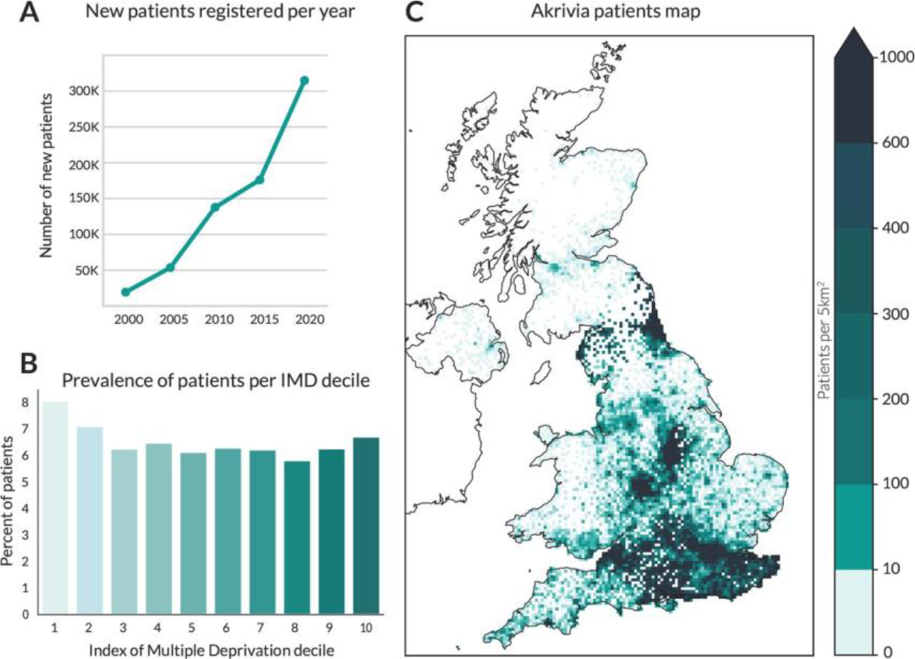
Psychiatry & Mental Health
PT_312
Keywords: Mental Health, Early life stress, Epigenetics, Inflammation, Behaviour problems
Authors: Rebecca Woods, Paranthaman V Kavya, Nicky Wright, Johnathan Hill, Monojit Debnath, Christopher Murgatroyd, Prabha Chandra, Helen Sharp
Early-life stress (ELS) increases risk of poor mental health outcomes in exposed children. Mental health conditions constitute a large healthcare/social burden, demanding improved therapeutic strategies. However, this requires a mechanistic understanding of how ELS exposures induce neuropathogenesis, which remains unknown. We hypothesise that ELS induces ongoing dysregulation of immune/stress response pathways, resulting in altered brain function. To investigate this, we leverage longitudinal data to assess the relationship between altered epigenetic patterning of immune/stress genes and emergence of behavioural problems following ELS.
Participants were members of the Wirral Child Health and Development Study (WCHADS), a longitudinal birth cohort of first-time mothers. Prenatal and postnatal assessment waves occurred periodically during which various parent and child measures were taken at clinic visits/via questionnaires. Demographic confounders at recruitment and during pregnancy/birth included: maternal age; obstetric-risk score; socioeconomic status (English Index of Multiple Deprivation); birthweight for gestational age and child sex. Early-life indices were maternal depression (Edinburgh Postnatal Depression Scale) both prenatally (32wk) and postnatally (2.5yr). Child salivary DNA methylation (genes: NR3C1 (stress response), IL6 (inflammation)) was assessed repeatedly at 14m, 2.5yr, 3.5yr. Child mental health outcome (CBCL) was child externalising behaviours at 3.5yr.
Statistical approach: All variables were log-normalised, then linear regression analyses were used to evaluate associations between ELS and child behaviours at each timepoint, then between gene methylation and both ELS and child outcomes. All analyses controlled for maternal and child demographic confounding variables.
Prenatal and postnatal maternal depression significantly associated with increased rates of externalising behaviours at 3.5yr. Separate assessment of child methylation markers showed that IL-6 and NR3C1 methylation levels significantly increased with age, with the largest increase observed between ages 14mo and 2.5yr. Regression analyses revealed prenatal maternal depression was not significantly associated with either NR3C1 or IL6 methylation at any timepoint. However, higher postnatal maternal depression symptoms at 2.5yr was significantly associated with increased NR3C1 and reduced IL-6 methylation from age 2.5yr, indicating a link between early-life environment and child methylation. Accordingly, by 2.5yr there was a negative association between IL6 methylation and child externalising behaviours at 3.5yr. results were not significant for NR3C1.
Results indicate a relationship between ELS, child methylation of IL6 and emergence of child behavioural problems. Future work will include mediation analyses and trajectory analyses to assess impact of cumulative environmental insults on DNA methylation and behavioural outcomes across the study timeline.
Psychiatry & Mental Health
PT_319
Keywords: Schizophrenia, Phencyclidine, Memroy, Hippocampus, Synaptic plasticity
Authors: Ningyuan Sun, Michael Harte, John Gigg
10 Female Lister Hooded rats were dosed with either saline or PCP (2 mg/kg, i.p.) twice daily for seven days followed by a 7-day washout period. Wireless electrophysiological recordings were conducted in the dorsal CA1 region using chronically implanted, 16-contact linear electrode arrays, allowing real-time assessment of neural activity during spontaneous behaviour and during behavioural tasks. Gamma (30-100 Hz) and theta (4-8 Hz) oscillations—key rhythms implicated in schizophrenia-related cognitive dysfunction—were analysed using MATLAB. A subset of rats was also implanted with a twisted pair stimulating electrode in dorsal CA3. CA1 LTP was then assessed by measuring Schaffer collateral-evoked field excitatory postsynaptic potentials (fEPSPs) in freely moving rats. Data were analysed using mixed-effect model two-way ANOVA.
The scPCP group exhibited significantly reduced LTP compared to controls, consistent with impaired object recognition task performance and previous findings in anaesthetised scPCP rats. Preliminary analyses of spontaneous activity support alterations in gamma and theta power and TG-PAC, suggesting disrupted oscillatory dynamics in the hippocampus. These findings indicate a broader impairment in hippocampal network function beyond synaptic plasticity deficits. Full spontaneous activity results will be presented in the poster.
Psychiatry & Mental Health
PT_320
Keywords: C-reactive protein, cohort study, Patient Health Questionnaire (PHQ-9), antiretroviral therapy, immunopsychiatry
Authors: Arish Mudra Rakshasa-Loots, Shalena Naidoo, Kaylee S. van Wyhe, Mark F. Cotton, Barbara Laughton, Richard H. Glashoff
Despite early antiretroviral therapy (ART), young people living with HIV are at increased risk for depressive symptoms. Early-life inflammation has been linked to the risk for depression in young people. In this exploratory study involving young people with perinatally-acquired HIV, we investigated correlations between inflammatory biomarkers measured at multiple time-points in childhood with the severity of depressive symptoms in adolescence.
Participants were 39 young people living with HIV (56% female) from the Children with HIV Early antiRetroviral (CHER) trial in Cape Town, South Africa. We quantified 40 inflammatory biomarkers in blood plasma using validated ELISA and Luminex™ multiplex immunoassays from retrospective samples at multiple time-points: before ART initiation (n = 20), one (n = 14) and two (n = 13) years following primary ART commencement (“post-ART”), and at age 8 years (n = 36). At age 16, we measured depressive symptom severity using the Patient Health Questionnaire (PHQ-9). Spearman’s rank coefficients (ρ) were calculated with pairwise complete observations for correlations between inflammatory biomarker concentrations at each time-point with PHQ-9 score at age 16.
Figure: Spearman’s correlation coefficients for inflammatory biomarkers measured in blood plasma at multiple childhood time-points in participants with perinatally-acquired HIV and their depressive symptom scores at age 16. Post-ART: following commencement of primary ART.
Psychiatry & Mental Health
PT_322
Keywords: Schizophrenia, sigma-1, NMDA, interneuron, neuroinflammation
Authors: Bethany Dennis, Gavin Clowry, Fiona LeBeau
Schizophrenia is a neuropsychiatric disorder marked by cognitive deficits, including impairments in working memory, attention and executive function. The anterior cingulate cortex (ACC) plays a key role in these processes. N-methyl-D-aspartate receptor (NMDAR) hypofunction is implicated in schizophrenia-related cognitive impairments, associated with aberrant beta (20-30 Hz) and gamma (30-80 Hz) oscillations. Increased neuroinflammation and a loss of fast-spiking parvalbumin (PV)+ interneurons have been observed in post-mortem brain tissue from patients. However, the link between cognitive dysfunction and neuroinflammation remains unclear. Sigma-1 receptor (σ1R) activation has been suggested to counteract these deficits.
ACC slices were prepared from male Lister Hooded rats. For electrophysiology experiments, the NMDAR antagonist PCP and σ1R agonist PRE-084 were bath-applied, separately and together, to kainate-evoked beta and gamma oscillations. For immunofluorescence staining, ACC slices were incubated in artificial cerebral spinal fluid for 4 hours in an interface chamber, with kainate or kainate and PCP. To activate σ1Rs, slices were pre-incubated with PRE-084 before kainate and PCP incubation. Slices were fixed, re-sectioned and immunostained for microglia (Iba1) and reactive astrocytes (GFAP), PV and somatostatin (SST) interneurons, and perineuronal nets (stained with Wisteria floribunda agglutinin; WFA).
Statistical analysis was performed using a One-way ANOVA (Graphpad, Prism).
In the ACC, PCP significantly increased the power of beta oscillations (216 ± 23%) and caused gamma oscillations to switch to a beta frequency (41 ± 1.3 to 27 ± 1.5 Hz). Co-application of PRE-084 does not significantly attenuate the effect of PCP on KA-evoked beta oscillations (165.5 %). However PRE-084 attenuated the effect of PCP on KA-evoked gamma oscillations, blocking the shift to, and significant increase in, beta activity. Immunohistochemistry showed that ACC slices exposed to kainate and PCP exhibited a small increase in % area of Iba1 expression (2.4 ± 0.6%) compared to kainate only (2.0 ± 0.7%), across the ACC. Prior activation of σ1Rs reduced Iba1 % area of kainate and PCP exposed slices (1.6 ± 0.3%). There was no effect on GFAP expression. Interestingly exposure to kainate decreased median PV integrated density compared to control (602 vs 1030 AU), where SST % area was significantly increased when exposed to kainate compared to control (313 vs 230 AU).
Our data suggests that kainate drives beta frequency oscillations in the ACC, possibly via increased SST interneuron activity. PCP induces aberrantly large beta oscillations that increase microglia. Activating σ1Rs could play a protective role against the effects of PCP. Further investigation is required to better understand the interplay between NMDAR and σ1Rs.
Psychiatry & Mental Health
PT_323
Keywords: Nutricosmetic, Wellbeing, Supplement, Healthy adults
Authors: Isobel Kevan, Timo Giesbrecht, John Tyson-Carr, Connor O'Reilly, Giovanni Sala, Nicholas Fallon, Carl Roberts
Orally administered nutricosmetic supplements are putative enhancers of psychological wellbeing. However, efficacy has yet to be fully established. We therefore assessed the effects of the following interventions on outcomes related to psychological wellbeing in healthy adults between the ages of 18-65: probiotics, prebiotics, synbiotics, omega-3 fatty acids, multivitamins and minerals (MVM), and vitamin D. This was conducted using a series of meta-analyses using a random-effects model and subgroup analysis by psychological wellbeing domain. We adhered to the PRISMA reporting guidelines and used the CASP RCT checklist for quality appraisal. We obtained a total of k = 34 studies. We found significant improvements in stress for MVM interventions, and significant improvements in overall wellbeing for probiotic and synbiotic supplements, however the former results lost significance following outlier removal, and the latter were derived from only three studies and should therefore be interpreted cautiously. There was no overall effect found for any other intervention type. Our findings offer support for the therapeutic potential of probiotics, MVM and synbiotic supplements to promote optimal psychological wellbeing. However, effect sizes are small and are susceptible to instability with high heterogeneity across intervention types (probiotics), or are domain specific (MVMs), or have limited number of studies (synbiotics). Potential therapeutic mechanisms of action are discussed but require confirmation with well-designed mechanistic studies exploring effects on the human gut-microbiome.
Psychiatry & Mental Health
PT_324
Keywords: ADHD, serotonin, 5-hydroxytryptophan,
Authors: Eleanor Jackson, Tim Riley, Paul Overton
Objective: Although several pharmaceutical therapies exist for ADHD, current medications carry a high risk for misuse, evidencing a need for alternatives. One possible avenue is serotonergic intervention; serotonin is critical in the regulation of arousal, mood and orienting attention to novel stimuli. Furthermore, serotonin synthesis appears to be impaired in ADHD, with significant reductions in levels of serotonin and its metabolites observed. One option could be intervention within the serotonin synthesis pathway; this would also provide an alternative to pharmaceutical intervention, something often sought after by individuals for whom stimulant treatment is inappropriate. We aim to assess the efficacy of 5-hydroxytryptophan, a serotonin precursor, in reducing distractibility, a core feature of ADHD.
105 non-clinical participants participated in a randomised, double-blind trial in a 2 ( intervention: placebo or 200mg 5-hydroxytryptophan), x 2 (group: high ADHD traits vs low ADHD traits) x 2 (time: pre and 90 minutes post administration) design. Participants were selected using the Adult ADHD Self-Report scale screener (ASRS v1.1) and partitioned into high (score ≥ 4, n = 56) and low (scores ≤ 1, n = 49). Distractibility was assessed using reaction time and accuracy in a task-relevant (Eriksen flanker) and task-irrelevant (n-back coupled with auditory stimulus) paradigm. Intervention efficacy was analysed with 5% two-tailed significance using a 2 x 2 x 2 ANOVA.
Contrary to widespread previous reports, flanker performance produced only small, non-significant performance differences between high and low ADHD groups pre-intervention, as did the n-back. Whilst 5-hydroxytryptophan administration produced a small decrease in reaction time, that occurred in both ADHD groups and was matched by similar reductions in placebo. Overall, the three-way interaction between intervention, group, and time was non-significant (F(1,210) = 0.412, p = 0.522, F(1,210) = 0.06, p = 0.936).
5-Hydroxytryptophan supplementation was not found to selectively improve or impair distractibility in any measure for individuals with high or low ADHD traits. Further work needs to be conducted with measures more sensitive to ADHD status.
Psychiatry & Mental Health
PW_325
Keywords: EEG, time-domain, frequency-domain, mental health and well-being, adolescents
Authors: Miftah Faizah, Sophie Höfels, Faisal Mushtaq
Adolescent is critical period of human development, characterized by substantial, biological, social, and psychological changes. These transformations increase vulnerability to mental health challenges such as depression, anxiety and behavioural disorders. Electroencephalography (EEG) is a neuroimaging tool that has the potential to be applied to study individuals at risk of mental health difficulties and could be leveraged to inform early intervention. Despite its potential, a comprehensive synthesis of EEG correlates linked to adolescent mental health encompassing both positive aspects (e.g., mental well-being) and negative aspects (e.g., diagnosable conditions), is currently lacking. This scoping review seeks to map these associations, to provide a comprehensive mapping of EEG features and related to mental health outcomes.
The scoping review is employing the Joanna Briggs Institute (JBI) methodology and PRISMA-ScR guidelines. Eligible studies will include adolescents aged 10-19 years, with EEG employed as one of the tools to measure brain activity and assess mental health outcomes. Only studies published in English will be included. Studies focusing on medical conditions or motor dysfunctions (e.g., brain injury, tics, epilepsy) and those primarily addressing treatment effects will be excluded. Searches will be conducted across PsycINFO, Medline, Pubmed, and Web of Science with no publication year restrictions. Data synthesis will include key study characteristics including, population details, mental health outcomes and predictors, EEG measures, relevance to Research Domain Criteria (RDoC) framework. A narrative synthesis and descriptive layout will be used to identify trends and gaps in the existing literature, supporting future research on neural correlates of adolescent mental health.
Psychiatry & Mental Health
PW_326
Keywords: Neurofibromatosis-1, Myelin, Grey/White matter, T1W/T2W ratio, Working memory performance
Authors: Varun Arunachalam Chandran, Nils Muhlert, Stavros Stivaros, Caroline Lea-Carnall, Shruti Garg
NF1 is an autosomal dominant neurodevelopmental condition commonly associated with learning difficulties in school-aged children. It has been hypothesised that children with NF1 show an aberrant brain-behaviour relationship linked to Autism Spectrum Conditions (30%) and Attention Deficit Hyperactivity Disorder (50%) (Garg et al., 2013). T2-white matter hyperintensities are commonly seen in the thalamus and basal ganglia in NF1 (Feldmann et al., 2010). In addition, diffusion MRI studies showed widespread white matter microstructure differences in NF1 suggesting aberrant myelination (Karlsgodt et al., 2012). However, no studies have investigated quantitative myelin in NF1 yet. Objectives (i) Compare quantitative myelin levels across the grey and white matter at whole brain and lobar level between NF1 and neurotypicals, and determine (ii) Relationship between estimated myelin levels and working memory performance in children with NF1.
Participants include forty-eight children and adolescents with NF1 (age: 11-18 years; gender: 25 males and 23 females). T1-weighted and T2-weighted structural images were acquired using a Philips 3T MRI scanner. One hundred and sixty-eight (age and gender-matched) control structural MRI images were collected from the Human Connectome Project. Visuospatial n- back tasks were used to ascertain working memory performance. The image preprocessing pipeline includes bias-field correction, coregistration, calculation of T1W/T2W maps, segmentation, spatial normalisation and calculation of GM/WM metrics. ANCOVA and Pearson’s partial correlation was used to test the whole brain and lobar level GM/WM myelin in the case-control and correlation analysis in NF1 respectively, while the effects of age and gender were controlled.
Our results showed significantly reduced grey matter/white matter at whole brain level (F=28.165, p<0.001), and the frontal (F=6.906, p<0.009), temporal (F=27.548, p<0.001), parietal (F=7.440, p<0.007) and occipital (F=28.906, p<0.001) lobes in NF1 relative to neurotypical controls. However, no significant correlation was found between myelin and working memory performance at either the whole brain or lobar level in NF1.
Our findings show that quantitative GM/WM myelin differences are consistent with previous animal model studies in NF1 (López-Juárez et al., 2017). Reduced GM/WM myelin in the whole brain and the frontal, temporal, parietal and occipital lobes suggest that the loss-of-function mutation in the NF1 gene may affect oligodendrocytes producing myelin in NF1. Low quantitative myelin may manifest as a consequence of loss of neurofibromin in NF1 leading to reduction in mature oligodendrocytes/decompaction of myelin (Mayes et al., 2013). T1W/T2W ratio is a useful neuroimaging biomarker to identify quantitative myelin differences in NF1.
Psychiatry & Mental Health
PW_328
Keywords: PTSD, stress-enhanced fear learning, cytokines, mitochondrial dysfunction, oxidative phosphorylation
Authors: Charlotte Rye, Clara Velazquez Sanchez, Jeffrey Dalley, Amy Milton
In addition to severe and debilitating psychological symptoms, individuals with PTSD experience high levels of physical comorbidities such as cardiovascular disease, arthritis, and asthma, which may be linked to elevated levels of proinflammatory cytokines and impaired mitochondrial functioning. Indeed, chronic inflammation has emerged as a key factor in the pathophysiology of several psychiatric conditions, including depression, and can disrupt neuroplasticity, neurotransmitter systems, and blood-brain barrier integrity. PTSD patients, particularly those with chronic or severe symptoms, exhibit heightened levels of inflammatory cytokines, including IFN-γ, IL-6, IL-1b and TNFa, and the overlap in inflammatory pathways between depression and PTSD presents an opportunity for cross-disease insights that could improve both early diagnostics and therapeutic strategies. This study aims to explore the link between inflammation and mitochondrial dysfunction in PTSD and identify plasma markers that distinguish those at risk of developing PTSD from resilient individuals. By leveraging insights from depression research, we may be able to identify common mechanisms and better target interventions that could improve clinical outcomes, particularly for patients with treatment-resistant forms of either condition.
Adult male (n=76) and female (n=76) Lister Hooded rats will be divided into six squads (n=24-28 per squad). Two squads will consist of males assigned to the stress enhanced fear learning (SEFL) procedure – a robust rodent analogue of PTSD - receiving massed shock exposure on either day 1 or 13, while a third squad will undergo chronic, unpredictable shock exposure (a model of depression). The remaining three squads will replicate the procedure for female rats. The manipulated variable will be the number of shocks (0 or 13) delivered during trauma exposure (Figure 1). Fear responses will be assessed by measuring freezing behaviour. Plasma markers of inflammation (IL-1β, IL-6, TNF-α, IL-10, IFN-γ) and mitochondrial bioenergetics (lactate, pyruvate, citrate) will be assessed at baseline and post-test to determine whether any alterations are pre-existing or a consequence of susceptibility/shock exposure. Western blotting will quantify expression of glutamate receptor subunits, ADAR2 enzyme, and oxidative phosphorylation markers.
We will determine i) whether PTSD and depression share common inflammatory and mitochondrial mechanisms or exhibit distinct pathophysiology; ii) whether plasma biomarkers can reliably distinguish those who are susceptible from those who are resilient. Key outcomes will include correlatory analyses between plasma biomarkers and protein expression related to mitochondrial function and glutamatergic signalling. ANOVA will be used to analyse group differences in inflammatory and mitochondrial markers based on sex and shock exposure.

Psychiatry & Mental Health
PW_329
Keywords: childhood trauma, inflammation, brain blood barrier, white matter,
Authors: tenghuan xu
Individuals with a history of trauma are 1.66 to 2.73 times more likely to develop depression in adulthood, with the risk of treatment-resistant depression doubling (Nelson et al., 2017; Ju et al., 2020). Evidence suggests that low-grade inflammation and endothelial dysfunction contribute to depression (van Dooren et al., 2016), but the role of childhood trauma in this process remains unexplored. Individuals with a history of childhood maltreatment exhibit widespread abnormalities in white matter (WM) microstructure (Lim et al., 2020). However, studies examining the relationship between blood-brain barrier (BBB) integrity and WM in depression, particularly in those with a history of childhood trauma, are limited. This study aims to investigate whether white matter structural changes caused by chronic inflammation in individuals with childhood trauma are associated with BBB damage.
Data will be collected from 60 participants divided into three groups: childhood trauma with (n=20) or without depression (n=20), and no childhood trauma (n=20). Clinical assessments will include the Childhood Trauma Questionnaire (CTQ) and Adverse Childhood Experience Questionnaire (ACE).Each participant will provide a 30ml blood sample and undergo MRI using a Philips 3.0T scanner with a 64-channel head coil. Diffusion tensor imaging (DTI) and neurite orientation dispersion and density imaging (NODDI) parameters will be analysed alongside plasma inflammation markers measured via ELISA.
Descriptive statistics for demographic and scale data will be analysed using SPSS 25. Group differences in inflammatory biomarkers and imaging indicators will be tested using chi-square or t-tests. Correlation analyses will assess relationships between inflammatory markers, blood-brain barrier indicators, and imaging data. A mediation model will explore whether the CTQ score or inflammatory markers (independent variables) affect imaging data (dependent variables) via BBB indicators (mediator).
Ju Y, Wang M, Lu X, et al. (2020) The effects of childhood trauma on the onset, severity and improvement of depression: The role of dysfunctional attitudes and cortisol levels. J Affect Disord 276: 402-410.
Lim L, Howells H, Radua J, et al. (2020) Aberrant structural connectivity in childhood maltreatment: A meta-analysis. Neurosci Biobehav Rev 116: 406-414.
Nelson J, Klumparendt A, Doebler P, et al. (2017) Childhood maltreatment and characteristics of adult depression: meta-analysis. Br J Psychiatry 210(2): 96-104.
van Dooren FE, Schram MT, Schalkwijk CG, et al. (2016) Associations of low grade inflammation and endothelial dysfunction with depression - The Maastricht Study. Brain Behav Immun 56: 390-396.

Psychiatry & Mental Health
PW_330
Keywords: metabolomics, methamphetamine, MALDI-MSI
Authors: Ming Lin, Jiamin Xu
Methamphetamine (METH) has already been a serious problem all over the world. The identification of related biomarkers and pathways is helpful to evaluate the degree of METH addiction, develop appropriate treatment during abstinence, and explore the mechanism.
Metabolomics profiling of METH addicted human serum and three regions of METH-induced conditioned place preference (CPP) rat brain has been performed by using UHPLC-MS/MS and matrix-assisted laser desorption/ionization-mass spectrometry imaging (MALDI-MSI), respectively.
Unpaired t test in GraphPad Prism 6.01 was used for statistical analysis.
Untargeted metabolomics analysis demonstrated clear differences between METH abusers and the healthy control by finding 35 distinct expressed metabolites in serum, including 5 TCA intermediates, 17 amino acids and 13 other biomolecules, 15 of which were newly identified following METH exposure. By using MALDI-MSI, the relative quantification and distribution of 14 metabolites were investigated in the nucleus accumbens (NAc), dorsal hippocampus (dHPC) and ventral hippocampus (vHPC) of CPP rat brain. Taken together, METH addiction could influence energy metabolism, amino acids metabolism, and phospholipids metabolism. A multi-parameter model consisting of these related metabolites can be established as a METH addiction biomarker in the future. The mapping of phospholipids provided new insights into the mechanism of METH addiction. Notably, the trend of metabolite changes in NAc and dHPC was almost the same, while it was opposite between dHPC and vHPC. It seems that NAc and dHPC were the two regions more susceptible to METH administration in the brain. And dHPC and vHPC play different roles in METH addiction proved by metabolites mapping.
Training & Education
PM_332
Keywords: Neuroanatomy, Mental health, Focus, Inclusivity, Sustainability
Authors: Daria Pak, Deepanvita Koppada, Vera Bowerman, Sara Chaudhury, Laura Laura Boubert, Phoebe Cummings, Joan Liu
Teaching neuroanatomy is challenging due to the complexity of the human brain and the large student cohort. While animal/cadaver dissections are helpful, practicals are associated with higher cost, and social, ethical and sustainability concerns (McLachlan et al. 2004). Here, we aim to explore the effectiveness of clay sculpting as an innovative educational approach in neuroanatomy to support diverse learners in higher education. Clay is a sustainable medium known to promote focus, tactile learning, social engagement and wellbeing (Penzes et al., 2023).
The study has approval from the University of Westminster Research Ethics Committee. 47 participants (mean, 19years) attended nine two-hour sessions to engage in instructed or freeform clay sculpting to create brain models. Bio- and neuro-feedback measurements were recorded using a pulse oximeter and modern EEG devices (Neurosity, MuseTM), and quizzes, questionnaires and interviews were conducted. T-tests and ANOVA were applied using SPSS_v28.
Participants have varied learning styles, with over half preferring learning through art and physical activities. All participants enjoyed the sessions, with 97% indicating a definite improvement in their well-being. A significant decrease in heart rate between the start to end of the session was noted (mean bpm; 83 start, 76 end; p<0.001). Electrophysiological data showed a slight increase in the percentage of time spent in focus in instructed sessions than freeform (mean %; 61% instructed, 56% freeform; p=0.096). Participants performed better in post-sessional quizzes than pre-quizzes in instructed clay sessions (mean score, 75% post, 50% pre; p<0.05)
Clay sculpting supports neuroanatomy learning by enhancing learners’ understanding, satisfaction, inclusivity and mental health. Thus, creative educational methodologies should be encouraged in higher education to engage diverse learners.
McLachlan JC, Bligh J, Bradley P et al (2004). Teaching anatomy without cadavers. Medical Education.38: 418-424.
Pénzes, I, Engelbert, R, Heidendael, D et al (2023). The influence of art material and instruction during art making on brain activity: A quantitative electroencephalogram study. The Arts in Psychotherapy.83:102024.

Training & Education
PT_333
Keywords: Hemianopia, fMRI, Audiovisual, Functional connectivity, Plasticity
Authors: Fahad Alharshan, Georg Meyer, Fiona Rowe, Kholoud Alwashmi
Post-stroke hemianopia impairs visual processing and daily functioning. Audiovisual integration training may aid rehabilitation. This study investigates the effects of a six-week home-based virtual reality (VR) audiovisual training programme on behavioural performance and functional brain connectivity in chronic hemianopia patients.
Fifteen patients with chronic post-stroke hemianopia (>3 months post-stroke) underwent a home-based immersive audiovisual training programme using VR for six weeks. Pre- and post-training MRI scans, including task-based and functional connectivity (FC) analyses, were conducted alongside behavioural assessments to evaluate changes in visual performance.
Behavioural assessments revealed significant improvements in visual performance post-training, as indicated by faster response times. Task-based fMRI (task vs. rest) showed increased activation in key regions associated with visual processing in the unaffected hemisphere, as well as auditory and multisensory integration during the trained task. FC analysis further confirmed a significant post-training increase in correlational connectivity between visual and auditory processing areas.
These findings suggest that VR-based audiovisual training enhances neuroplasticity in post-stroke hemianopia, improving multisensory processing and visual performance. The observed changes in functional connectivity and brain activation highlight the potential of immersive VR interventions as an effective rehabilitation approach for hemianopia patients.
Training & Education
PW_170
Keywords: joint honours degree, neuroscience, psychology, transdisciplinary education, careers
Authors: Donna Lloyd, Ruth Ingram, Ellen Poliakoff
Background & aims
Joint honours degrees are growing in popularity, due to enhanced career opportunities afforded through acquiring a greater breadth of knowledge and unique transferable skills. Such ‘transdisciplinary education’ (Russell et al., 2008, Higher Education) is highly sought-after by employers, but a holistic view of student perceptions of these degrees is lacking. We aimed to better understand the experiences of neuroscience and psychology joint honours students, to provide more effective support and recommendations for the delivery of joint honours degrees in neuroscience as a growth sector in UK HE.
First year students on the BSc Cognitive Neuroscience and Psychology degree at the University of Manchester completed an online questionnaire (respondents: 19/62 students in the cohort). Free-text responses to questions around reasons for choosing the degree, sense of identification with each discipline, advantages and disadvantages, and suggestions for key areas of improvement were analysed using thematic analysis. Themes were informed by the National Student Survey and Teaching Excellence Framework including teaching quality, academic support, assessment and feedback and employability.
The choice of this joint honours degree was driven by the desire to mix hard sciences with a more applied discipline, and the interconnectedness of these. The perceived benefits were a broader knowledge base and skillset when looking for graduate opportunities, echoing the employer perspectives reported elsewhere. The opportunity to meet a wider variety of people and experience a variety of teaching and assessment methods was valued, as students felt this developed their adaptability. Higher workload was a perceived disadvantage.
Recommendations for improving joint honours degrees include: 1. Support students to manage their workloads and deadlines by highlighting discipline- and department-specific differences in assessment and feedback. 2. Foster a sense of identity and community for joint honours students by providing dedicated learning opportunities, events, workshops, and outreach activities. 3. Raise awareness of the flexibility, adaptability and interdisciplinarity developed through a joint, rather than single, honours degree and how desirable this is for employers.
Future neuroscientists will probably have an unexpected learning journey combining diverse experiences and, as transdisciplinary specialists, be able to work across/outside their discipline. Yet in UK HE, despite increasing in popularity, many joint honours degrees are still seen as the educational anomaly. It is time for greater intentionality to the joint honours degrees we offer and the graduates we produce to meet the challenges of the future.
Training & Education
PW_335
Keywords: Neuroscience, Education, Core modules, Optional modules, Accreditation
Authors: Isabel Logan, Charlotte Mosley, Thomas Malcomson, Emma Yhnell
Considering the broad scope covered by the field of neuroscience, this study compared neuroscience undergraduate degree programmes across the United Kingdom, with a focus on the distribution of core and optional neuroscience-specific (NS) modules.
Publicly available data considering core and optional modules from 13 universities were analysed. The results revealed significant variation in the proportion of NS module credits acquired by graduation, ranging from 28% to 100% across the institutions considered.
The findings highlight particularly low core NS content in Year 1 of undergraduate degrees, potentially affecting informed choice of subsequent modules. The observed flexibility in module selection throughout a neuroscience undergraduate degree is a promising opportunity for students to explore their interdisciplinary interests. However, in response to the high variability in NS core and total credits demonstrated by this research, this study calls for further discussion on establishing an accreditation framework to ensure consistency in neuroscience undergraduate degrees across the United Kingdom.
Training & Education
PW_336
Keywords: Competency, Education, Training, Rehabilitation
Authors: Sarah Flynn
Cheshire and Merseyside Rehabilitation Network (CMRN) is a collaborative partnership, with multiple provider and commissioning organisations to deliver specialist rehabilitation, from hyper acute to community. The largest client group is those with brain injury requiring a high level of MDT complex rehabilitation. Specialist nurses are a key resource in rehabilitation, however, CMRN recognised a gap in competencies to define the key role of the specialist rehabilitation nurse and support the development of a highly skilled nursing workforce. The framework is endorsed by The British Society of Physical and Rehabilitation Medicine who support the advancement of knowledge and practice of neurorehabilitation medicine. This framework addresses workforce development by identifying proficiency levels for non-registered and registered neurology nursing staff and supporting skills gaps to ensure practitioners can apply the competencies to clinical practice.
Enhanced staff knowledge and skills improves patient care. The Framework was developed by MDT experts in neurology across sixteen domains that encompass the key clinical areas relating to specialist rehabilitation nursing. It provides a common language, benchmark, and clarity of expectations in the provision of rehabilitation nursing. Divided into three competency levels from competent to highly specialised, the Framework applies to nurse clinicians at all career stages. Each competency details knowledge, skills and attributes required for a rehabilitation setting. The Framework supports the development of clinical practice, leadership, engagement in quality improvement/ research, career progression, education programmes alongside workforce planning.
The framework will be launched across CMRN in March 2025 and ensures a robust approach to nursing education in neurorehabilitation. The impact of the Framework will be evaluated in enhancing competence and confidence in the application of knowledge and skills into clinical practice, job satisfaction and retention rates, quality of care and organisational benefit. This will be measured through questionnaires, focus groups and targeted data collection and analysis across a number of key matrices.
A standardised approach to rehabilitation neurology nursing education and training via a Competency Framework will ensure a replicable and robust application to workforce development, supporting delivery of high-quality, safe, and effective specialist rehabilitation care. The Framework will provide the assurance to service users, families, commissioners and providers that the specialist rehabilitation nursing team have the knowledge, skills and MDT approach, through consistent job roles, definitions and related competencies, to provide people with neurological conditions the high quality, evidence-based care needed to optimise rehabilitation outcomes.

Treatments & Translational Neuroscience
PM_338
Keywords: Alzheimer's Disease, Neurofilament light chain, Antisense oligonucleotides
Authors: Sebastian Monge, Miwei Hu, Kathleen Schoch, Holly Kordasiewicz, Frank Bennet, Timothy Miller
Alzheimer’s disease (AD) is a neurodegenerative disorder characterized by brain atrophy, cognitive decline, and memory impairment. Neurofilament light chain (NfL), essential for neuronal stability and axonal transmission, is elevated in the cerebrospinal fluid of AD patients and correlates with disease severity. Previous studies suggest that NfL knockout provides neuroprotective effects in other neurodegenerative diseases, but its modulation in AD remains unexplored. We aim to knockdown NfL using antisense oligonucleotides (ASOs)—short nucleic acid sequences that bind to mRNA to reduce protein expression. We hypothesize selective NfL reduction in AD models will mitigate neurodegeneration.
An NfL-lowering ASO was administered intracerebroventricularly to PS19 tauopathy mice (n=8) and wild-type (WT) littermates (n=7) at 9 months. Nontargeting ASOs served as controls in PS19 (n=4) and WT (n=3) mice. PS19 mice exhibit neuronal loss due to mutant tau protein expression. NfL serum levels were measured weekly, with tissues collected one month post-injection. Immunohistochemistry (IHC) was performed on the dentate gyrus and lumbar spinal cord for protein expression and regional assessment, and NfL mRNA levels were quantified by qPCR. A one-way ANOVA with Šídák’s post-hoc test was conducted.
IHC showed significant NfL reduction in the dentate gyrus and lumbar spinal cord following ASO treatment (dentate gyrus: 35%, p<0.0001; spinal cord 60%, p<0.05). ASO treatment also significantly reduced NfL mRNA levels in brain and spinal cord tissues (brain: 75%, p<0.00001; spinal cord: 50%, p<0.05), confirming transcriptional suppression.
Targeted NfL reduction via ASOs effectively modulates NfL levels, with implications for investigating AD-related neurodegeneration. Future studies will examine the impact of NfL reduction on hippocampal CA3-CA1 connectivity using PSD-95 and synaptophysin staining to correlate structural changes with functional outcomes.
1. Agrawal S (1996) Antisense oligonucleotides: Towards clinical trials. Trends in Biotechnology 14(10): 376–387. https://doi.org/10.1016/0167-7799(96)10053-6
2. Mattsson N, Andreasson U, Zetterberg H, Blennow K (2017) Association of plasma neurofilament light with neurodegeneration in patients with Alzheimer disease. JAMA Neurology 74(5): 557. https://doi.org/10.1001/jamaneurol.2016.6117
3. Williamson TL, Bruijn LI, Zhu Q, Anderson KL, Anderson SD, Julien J-P, Cleveland DW (1998) Absence of neurofilaments reduces the selective vulnerability of motor neurons and slows disease caused by a familial amyotrophic lateral sclerosis-linked superoxide dismutase 1 mutant. Proceedings of the National Academy of Sciences of the United States of America 95(16): 9631–9636.
Treatments & Translational Neuroscience
PM_339
Keywords: olfaction, olfactory therapy, Mild Cognitive Impairment, Cognition, neurodegeneration
Authors: Rona Graham, Melissa Lessard-Beaudoin, Gemma Camara, Tamas Fulop, Dominique Lorrain, Christiane Auray, Kevin Whittingstall, Joseph DeSouza
Aim: There is a substantial gap between the burden of neurodegenerative diseases and the resources available to prevent and treat them. Evidence suggests that olfactory therapy (OT) may improve olfactory functions and importantly affect functional connectivity networks and lead to restoration of neural circuits. The aim of our study was to evaluate the olfactory system and cognition levels in control and MCI individuals at baseline, one-year later (pre-OT), and post-OT.
Olfactory tests (UPSIT and ODMT), cognitive tests (MoCA, HVLT-R, BVMT-R, Tail Making Test, Stroop D-KEFS, Boston Naming Test) and MRIs were performed at each time point in seniors and individuals with MCI (60-75 years, both sexes).
Baseline measurements reveal the presence of deficits in olfaction, odor memory and several cognitive functions in the MCI individuals. In contrast, post-OT, our data show improvements in smell identification, verbal learning, visuospatial memory, and cognitive flexibility scores. Differences were also observed in cerebral artery diameters between pre and post-OT. An important question also to be addressed is whether there is specificity within the olfactory dysfunction. Our study confirms that olfactory deficit patterns are observed in MCI and that misidentification of certain odors correlated strongly with cognitive scores. This lead to the development of the Cognitive Associated Diagnostic Odours (CADO) signature. An analysis of the physicochemical characteristics of the CADO is currently underway.
Despite extensive evidence showing olfactory system dysfunction is observed in MCI there has been limited investigation of olfactory memory and studies of olfactory training as a therapy. Our results show that OT may provide benefit for individuals with MCI and we identify a tool that may help to provide early diagnosis for individuals with impending cognitive decline.
Treatments & Translational Neuroscience
PM_340
Keywords: post-operative pain, chronic pain, opioid usage, Peak alpha frequency (PAF), pain sensitivity
Authors: Roksana Markiewicz, Amélie Grandjean, Samuel Hannon, Fang Gao Smith, Ali Mazaheri
The opioid crisis is a global public health emergency, with millions affected by addiction, rising healthcare costs, and overdose deaths. Among the 310 million annual surgeries worldwide, opioids are frequently prescribed for post-surgical pain1. While most patients discontinue use, some develop persistent opioid use (>3 months post-surgery). Early identification of high-risk individuals is crucial for reducing dependency and exploring alternative pain management strategies.
Pain sensitivity, a key predictor of pain chronification and opioid persistence, varies among individuals due to neurological and psychological factors. Recent research highlights Peak Alpha Frequency (PAF) from resting-state EEG as a potential biomarker for severe post-surgical pain and prolonged opioid use. Slower PAF (the strongest signal power within the alpha rhythm, 8–14 Hz)2 has been linked to heightened pain sensitivity and chronic pain predisposition3–6. Initial validation in thoracotomy patients demonstrated its predictive value for post-surgical pain and opioid consumption4. This study aims to use resting-state EEG, focusing on PAF, to stratify patients at risk for severe post-surgical pain and prolonged opioid use. Additionally, we will examine whether post-surgical alpha frequency slowing predicts persistent opioid use and pain chronification.
Resting-state EEG data will be collected preoperatively and 72 hours post-surgery from 500 patients at Birmingham University Hospitals. Pain severity and functional impact will be assessed using the Brief Pain Inventory, and opioid consumption will be measured via urine samples at baseline, 72 hours, three months, and six months post-surgery. Weekly pain check-ins will be conducted via telephone for the first 12 weeks.
Statistical Analysis PAF will be calculated using the Centre of Gravity method, which provides a weighted estimate within the 8–14 Hz range (8–14 Hz). Pain will be categorized as moderate (4–6) or severe (≥7), with statistical analyses adjusted for preoperative pain, age, sex, surgery type, and analgesic regimen.
Preoperative PAF is a promising biomarker for predicting acute and chronic post-surgical pain. Its efficient integration into routine assessments could enable early identification of at-risk patients, improving recovery and reducing opioid dependency.
1 Dobson, G.P. International Journal of Surgery 81, 47–54 (2020) 2 Van Diepen, R.M. et al. Curr Opin Psychol 29, 229–238 (2019) 3 Furman, A.J. et al. Cerebral Cortex 30, 6069–6082 (2020) 4 Millard, S.K. et al. Br J Anaesth 128, e346–e348 (2022) 5 Furman, A.J. et al. Neuroimage 167, 203–210 (2018) 6 Mazaheri, A. et al. Neuroimage 262, 119560 (2022)
Treatments & Translational Neuroscience
PM_341
Keywords: ATP, stroke, cerebral ischemia, neuroprotection, brain injury
Authors: Paulina Michor, Johannes Boltze, Andrew Bottrill, Bruno Frenguelli
Cerebral ischaemia is associated with ATP depletion and loss of ATP metabolites into the blood stream. This renders ATP metabolites unavailable for re-synthesis into ATP via the purine salvage pathway (PSP), the primary route of adenine nucleotide synthesis in the brain, prolonging the ATP deficit. Wishing to accelerate ATP recovery after cerebral ischaemia, we have shown in brain slices that ribose and adenine (“RibAde”) elevates depressed cellular ATP levels to those found in vivo. We have also provided compelling
evidence that our RibAde approach reduced brain lesion volume and accelerated recovery after middle cerebral artery occlusion (MCAO) in rats. We have now examined the mechanism of ATP elevation by RibAde and its effects in the immediate aftermath of stroke.
We incubated 400 µm brain slices (7-10 slices/condition) in standard aCSF supplemented with either stable isotope-labelled ribose (13C5; 1 mM) and adenine (15N5; 50 µM) or unlabelled ribose and adenine at the same concentrations for 1–4 hrs at 33 C. We used mass spectrometry (MS) to identify endogenous ATP, ribose- or adenine-labelled ATP (+5 mass units) or ribose- and adenine-labelled ATP (+10 mass units). In separate studies, anaesthetised adult male SD rats (~300 g) were subjected to 2 hour MCAO (filament model), after which an intravenous infusion of saline (n=15) or RibAde (200 mg/kg/hr and 10 mg/kg/hr, respectively; n=12) was administered for two hours. Thereafter, rats were humanely euthanised under continued anaesthesia and brains were processed for TTC staining.
MS analysis of brain slices incubated in heavy ribose and adenine revealed ATP incorporating either 13C5 or 15N5, and ATP that incorporated both labels. This indicates that brain tissue is capable of directly converting ribose and adenine into ATP, overcoming the loss of these substrates after ATP depletion. In the in vivo studies, the area of TTC staining was similar between treated and untreated animals. However, the intensity of the staining significantly differed (Mann-Whitney U test; U=140; Z=2.415; p=0.016), with stronger staining (reflecting greater mitochondrial activity) in RibAde-treated animals compared to saline-treated animals.
These observations indicate that ribose and adenine are directly converted into ATP by brain tissue, most likely via the PSP, and that RibAde may support cellular viability after cerebral ischemia through enhanced provision of ATP. Given the previous and current use of ribose and adenine in humans, RibAde-based approaches may represent a readily-translatable hyper-acute point-of-care treatment for brain injuries such as stroke.

Treatments & Translational Neuroscience
PM_342
Keywords: Epilepsy, Gene Therapy, Autoregulatory, GPCR,
Authors: Olivia Goff, Steven Devenish, Sahil Patel, Amy Richardson, Laura Ussingkaer, Arki Tian, Dimitri Kullmann
Genetic therapies are emerging as a promising treatment of epilepsy, fuelled by the fact that approximately one-third of individuals with epilepsy are resistant to conventional anti-epileptic medication. Chemogenetics is an appealing approach because the therapeutic effect can be modulated, on-demand, to suppress seizure activity. Conventional chemogenetic tools, such as hM4Di, rely on exogenous drugs for activation. An alternative closed-loop or autoregulatory chemogenetic approach relies on endogenous neurotransmitters to activate a modified receptor. An example is eGluCl, a chloride channel that is of invertebrate origin and whose inhibitory effect relies in part on the chloride transmembrane gradient. We have created an autoregulatory, chimeric receptor that exhibits ‘inhibitory’ Gi/o signalling during periods of excessive neuronal firing. After the generation of our chimera using site-directed mutagenesis, we probed its G-protein specificity using the BRET-based TRUPATH assay. G-protein-coupled inwardly rectifying potassium (GIRK) channel activation was measured through whole-cell patch clamp recordings to further interrogate the chimera’s signalling transduction beyond the G-protein level. When compared to a Gi/o-coupled GPCR from the same family, we found our chimera exhibited the same signalling profile: full activation of Gi/o proteins but no Gq activation, and GIRK-mediated potassium conductance of the same magnitude (Gi/o-coupled GPCR -36.39±2.872pA/pF n=7; GPCR -42.16±10.53pA/pF n=6; p=0.5818, t-test). AlphaFold2 predictions provided support that the chimeric receptor maintains the functional site of Gi/o interaction. Importantly, we showed our chimera can modulate activity in a hyperexcitable network using cortical cultures in multi-electrode arrays. The chimera reduced the normalised mean firing rate both before (GFP 1.413±0.1903 n=28; chimera 2.432±0.2373 n=30; p=0.0014, t-test) and after (GFP 3.415±0.2899 n=28; chimera 1.741±0.2235 n=30; p<0.0001, t-test) the addition of pro-convulsant pentylenetetrazole. We have confirmed that AAV-mediated expression of the autoregulatory gene therapy does not disrupt physiological synaptic transmission. We used horizontal hippocampal slices to measure field excitatory postsynaptic potentials (fEPSP) in the Schaffer collaterals. During baseline and under specific agonist, there was no significant difference in the averaged fEPSP slope between chimera-expressing slices and GFP-expressing slices (GFP n=6 vs. chimera n=7 baseline: p=0.4619; agonist: p=0.3613, two-way ANOVA). Preliminary results indicate the chimera is protective against seizures in the repeated low-dose kainic acid acute mouse model of epilepsy. This novel closed-loop chemogenetic tool could, in principle, be implemented not only for the treatment of epilepsy, but also in other neurological and neuropsychiatric disorders where the balance between excitation and inhibition is disrupted.
Treatments & Translational Neuroscience
PM_356
Keywords: Chronic pain, Pain management, Prediction modelling, Predictors, Patient-reported outcomes
Authors: Andrew Palmer, Laura Bonnett, Katie Herron, Christopher Brown
Chronic pain affects a significant portion of the UK population and is increasingly understood to involve biopsychosocial factors. Pain management programmes are group-based rehabilitation programmes that help patients manage and modify the impact of living with chronic pain. These programmes are the preferred intervention for improving quality of life and biopsychosocial function, yet patient outcomes vary widely, influenced by a range of demographic, clinical, psychosocial, and potentially neurological factors. This study aims to develop and validate multivariable prediction models to identify prognostic factors associated with pain management programme outcomes for chronic pain patients, using data from the Walton Centre NHS Foundation Trust's Pain Management Registry. The models will serve as a baseline for future testing of the additional predictive value of neurological predictors.
This retrospective study utilized data from 2,116 chronic pain patients (74.5% female) who completed a pain management programme at the Walton Centre since 2014. Thirty prognostic factors, including demographic, clinical, and psychometric variables, were analysed to predict patient-reported outcomes. The primary outcome of interest is pain self-efficacy, measured using the Pain Self-Efficacy Questionnaire (PSEQ).
The statistical analysis involves using multivariable regression methods to identify significant prognostic factors. Calibration of the models will be evaluated using bootstrap resampling.
The preliminary full model included 14 out of the 30 prognostic factors evaluated. Preliminary analysis suggests that baseline scores in kinesiophobia and self-compassion are important predictors of improvements in pain self-efficacy. Full results will be presented in the poster. The developed models will enhance the ability to prospectively identify chronic pain patients who are most likely to benefit from pain management programmes, optimizing treatment allocation and patient outcomes. Future research could explore the integration of neurological assessments, such as markers of central sensitization, neural plasticity, and functional connectivity, to improve prediction and treatment personalization further. Incorporating such factors may yield a deeper understanding of the interplay between neurological and psychosocial contributors to chronic pain. This could ultimately advance prognostic modelling and the design of evidence-based pain management programmes.
Treatments & Translational Neuroscience
PT_344
Keywords: hiPSC, reactive astrocytes, drug discovery
Authors: Anna-Lena Zepernick, Eve Corrie, Matthieu Trigano, Tara J. Bowen, Maike Langini, Emma Jones
Astrocytes are one of the key glial types of the central nervous system (CNS) and play a critical role in maintaining brain homeostasis. Following CNS injury or in disease, astrocytes undergo a transformation to become ‘reactive’ and change their morphology and molecular profile, adopting different phenotypic states to respond to pathological insults. Reactive astrocytes participate in neuroinflammatory processes occurring in neurodegenerative diseases through secretion of neuro- and immune-modulatory factors. Recently, an in vitro reactive astrocyte model was developed using a stimulation cocktail of TNF-α, IL-1α, and Complement 1q (‘TIC’), which drives a neurotoxic state. TIC-induced astrocytes have been shown to secrete growth factors, chemokines and pro-inflammatory cytokines, as well as neurotoxic lipids. At MDC, we have developed assays to model this aspect of neuroinflammation using iPSC-derived human astrocytes. This enables analysis of this astrocyte proinflammatory phenotype and potential modulation drug discovery projects.
iPSC-derived human astrocytes were cultured for one week prior to TIC - stimulation. Astrocyte phenotype was evaluated using immunocytochemistry and high content imaging, and media was removed and analysed using multiplex cytokine analysis (Luminex). Semi-targeted liquid chromatography-coupled mass spectrometry analysis was also performed to explore the secretion of neurotoxic lipids.
Where appropriate, data have been analysed with t-test or two-way ANOVA with post-hoc tests.
Here, we show characterisation of a human iPSC-derived model of reactive astrogliosis for drug discovery research. We demonstrate that TIC stimulation induces an increase in pro-inflammatory cytokines, chemokines and growth factors and changes in morphological features including GBP2 expression. In addition, using mass spectrometry we show that the secretion of saturated free fatty acids is increased in TIC-stimulated astrocytes. Together these innovative tools provide a platform to study reactive astrocytes, and their contribution to neuroinflammation, and to assist with development of novel therapeutics to target astrogliosis in disease.
Treatments & Translational Neuroscience
PT_346
Keywords: Schizophrenia, Cerebellum, Kv3.1, Kv3.1 positive modulator, AUT1
Authors: Mahfuza Maisha, Lan Zhu
Schizophrenia is a debilitating brain disorder that imposes tremendous global socioeconomic burdens associated with health and social care. The disorder is characterised by positive, negative, and cognitive symptoms.
Current medications face many challenges, including their ineffectiveness in treating outcome-determining negative and cognitive symptoms. Research on drugs modulating Kv3.1 is promising. This voltage-gated potassium channel enables rapid action potential firing and regulation of neuronal firing patterns, thus contributing to the orchestration of neuronal network activities. Kv3.1 is reduced in the brains of schizophrenia patients (Yanagi et al., 2014).
Past research on Kv3.1 and schizophrenia has mainly focused on the cerebrum; however, growing evidence shows that the cerebellum plays a major role in schizophrenia (Mothersill et al., 2016). Kv3.1 has the highest level of expression in the cerebellum. Studies aimed at modulating cerebellar activity found significant improvements in schizophrenia symptoms. Kv3.1 positive modulators have been shown to boost Kv3 channel activity, rescue Kv3.1 deficiency associated dysfunction of GABAergic interneurons, restore gamma oscillations, and reverse cognitive deficits (Musselman et al. 2023).
This project aims to investigate how Kv3.1 is dysregulated in the cerebellum using an animal model of schizophrenia and whether a Kv3.1 positive modulator (AUT1) can reverse cerebellar-related behavioural deficits and restore cerebellar dysfunction. This research will provide preclinical evidence on the effectiveness of the Kv3.1 modulator in schizophrenia and guide future cerebellum-targeted treatment.
This project is permitted by our Home Office personal, project, and establishment licences. This project complies with the ASPA and local ethics guidelines. Male CBA mice (Charles River, UK) will be obtained at the age of 5 weeks, and a sub-chronic phencyclidine mouse model will be used in two sets of experiments.
Experiment 1 will assess cerebellar motor coordination (on 5 different timepoints/days), protein expression level, and cellular/subcellular expression patterns in the cerebellum using western blot, immunofluorescence, and confocal microscopy. Experiment 2 will examine the reversal effects of AUT1 on the above parameters.
Two-way mixed ANOVA (2x5) will be used for behaviour analysis. The 2-sample independent t-test or non-parametric equivalent will be used to test the difference of means between 2 groups for western blot and immunofluorescence analyses.
Yanagi M et al. (2014) Mol Psychiatry, 19(5)
Mothersill O et al. (2016) Cerebellum, 15(3)
Musselman M et al. (2023) Expert opinion on investigational drugs, 32(6)
Treatments & Translational Neuroscience
PT_347
Keywords: Epilepsy, Keypoint-MoSeq, Connectivity Map, Drug repurposing, Genetic mouse model
Authors: Nika Balkic, Cavan Bennett-Ness, Grant Marshall, Alfredo Gonzalez-Sulser, Catherine Abbott
Epilepsy affects 50 million people, yet one-third remain drug-resistant with over 25 anti-seizure medications available. Commonly used induced mouse models fail to replicate human epilepsy, while genetic models lack spontaneous seizures. EEF1A2-Related Neurodevelopmental Disorder is most commonly associated with heterozygous de novo E122K mutation. Eef1a2E122K/+ mouse model shows EEG abnormalities but lacks behavioural seizures. This study aims to use a novel non-invasive method to detect behavioural phenotype in Eef1a2E122K/+ mice and use translatome and proteome data to identify potential therapeutic candidates.
Top 150 upregulated and downregulated genes from E122K/+ translatome and proteome were analysed using Connectivity Map to identify drugs with potential to reverse mutation effects. Proteomic data from E122K/E122K and D252H/D252H mice were also assessed. Drugs with profile similarity closest to -100% and most selective for E122K/+ genotype were selected for further analysis.
C57BL/6J mice were recorded in open field arena for five minutes for behavioural analysis. Videos were analysed using DeepLabCut for pose estimation and Keypoint-MoSeq (Weinreb et al., 2024) for behavioural syllable classification. Six experiments (n = 6 wild-type and 6 Eef1a2E122K/+ mice) were conducted, comparing syllable frequencies using either wild-type or Eef1a2E122K/+ mice as the baseline, first with both sexes, then separately for females and males. Syllable frequency differences were assessed via two-sample t-tests (p<0.05).
Keypoint-MoSeq identified behavioural changes in Eef1a2E122K/+ mice, revealing a behavioural phenotype for the first time. Gender-specific analysis showed significant changes in females, while males exhibited no altered syllables, consistent with previous findings in another genetic mouse model of epilepsy (Gschwind et al., 2023).
Connectivity Map analysis identified several drugs as potential treatments to reverse the molecular changes. Some have already had success in reducing seizure severity, while others operate through a mechanism that has been suggested as a novel anti-seizure approach, highlighting these compounds as promising therapeutic candidates.
Gschwind T, Zeine A, Raikov I, et al. (2023) Hidden behavioral fingerprints in epilepsy. Neuron 111(9): 1440-1452.e5.
Weinreb C, Pearl JE, Lin S, et al. (2024) Keypoint-MoSeq: parsing behavior by linking point tracking to pose dynamics. Nature methods 21(7): 1329–1339.
Treatments & Translational Neuroscience
PT_348
Keywords: ketamine, antidepressant, psychedelic, medial prefrontal cortex, electrophysiology
Authors: Joe Flanagan, Emma Robinson, Jack Mellor, Matthew Claydon, Judith Schweimer
The NMDA receptor antagonist, ketamine is a rapid acting antidepressant with sustained antidepressant effects that has shown promise as a treatment for major depressive disorder (MDD). The medial prefrontal cortex (mPFC) plays a key role in emotional regulation via top-down inhibition of subcortical limbic brain regions, such as the basolateral amygdala (BLA), and is known to be structurally and functionally altered by chronic stress in rodents and human MDD patients. mPFC infusions of ketamine in rats have been shown to attenuate negative affective biases in the rodent affective bias test, a translationally model of neuropsychological mechanisms in MDD. Despite this, the mechanisms underlying the sustained effects of ketamine within the mPFC remains elusive. This study investigated the acute and sustained effects of ketamine on the intrinsic and extrinsic properties of retrograde-labelled BLA-projecting neurons and unlabelled neurons from the prelimbic mPFC, using in vitro and ex vivo male rat brain slice electrophysiology (strain: HsdOla:LH). Data was statistically analysed using two-way and three-way ANOVAs, or general linear models, as appropriate. Acute bath application of ketamine for 30 min at 1, 3, and 10 μM concentrations had no effect on the strength of electrically evoked excitatory postsynaptic currents at layer I inputs to BLA-labelled and unlabelled layer V prelimbic neurons held at -70 mV. In contrast, slices taken 24-hours post systemic ketamine treatment (1 mg/kg, i.p.) or saline revealed differential effects on the amplitude of miniature inhibitory postsynaptic currents (mIPSC) recorded from BLA-labelled and unlabelled neurons held at +0 mV. A two-way ANOVA revealed a main effect of ketamine treatment on mIPSC amplitude (F (1, 43) = 8.479, p = 0.0057), with multiple comparisons revealing a significant difference (p = 0.0308) between BLA-labelled cells from ketamine-treated (n = 11 cells, 5 animals) versus saline treated rats (n = 13 cells, 6 animals). Ketamine also differentially modulated the intrinsic excitability of these two neuronal populations 24-hours post dosing, as assessed by sequential injections of depolarising and hyperpolarising current steps. This study shows that, at a behaviourally relevant dose, ketamine differentially modulates the intrinsic and extrinsic properties of BLA-projecting layer V prelimbic mPFC neurons 24-hours post dosing. These findings suggest the sustained behavioural effects of ketamine could involve these adaptive changes in inhibitory signalling and are circuit specific.
Treatments & Translational Neuroscience
PT_349
Keywords: Pain, Psilocybin, Inflammatory Pain, Thermal Pain, Psychedelics
Authors: Vivienne Nguyen, Martha Lopez-Canul, Ella Nield, Gabriella Gobbi
1.
Acute pain serves as a sign of disease or threat to the body, and it is caused by injury, surgery, illness, etc. (Prescott and Ratté, 2017). Effective pain treatments are limited and often accompanied by significant side effects, highlighting the need for alternative therapies. 5-HT2A receptor has been implicated in nociceptive pathways (Castellanos et al., 2020), yet the effect of its agonists on pain mechanisms remains elusive. In this study, we examined the effect of psilocybin on persistent pain and thermal acute pain.
2.
Mice (C57BL/6) were randomized and either vehicle (saline i.p.) or psilocybin (3 mg/kg i.p.) was administered. Acute thermal nociception was measured using the Hot Plate Test (HPT). Initial temperature was set at 37°C with a near linear increase of 3°C per min. Nociception was established as the temperature eliciting a fast hindpaw lick and/or paw withdrawal. For inflammatory pain, formalin (1%) was administered in the mid-plantar surface of the hindpaw 10 min after vehicle or psilocybin. Pain-related behavior was measured at 5-min intervals for 60 min and separated in phase 1 (0-10 min) and 2 (15- 60 min). Hallucinogenic behavior and motor coordination were assessed after treatment administration. The data is reported as the mean ± standard error (SEM). A t-test or two-way ANOVA RM followed by Bonferroni post-hoc was performed.
3.
Analysis for the HPT showed no difference in the thermal withdrawal threshold (T-test, t=1.618, p= 0.127, vehicle: 38.2 ± 0.21; psilocybin: 38.6 ± 0.12, n=10 per group). In the formalin test, the Area Under the Curve for both phases showed a significant reduction of nocifensive behavior induced by psilocybin vs vehicle-treated animals (phase-1 p<0.01; phase-2 p<0.001, n=10 per group). Psilocybin increases head-twitch response 0-20 min after administration (p <0.05). No deficits in Rota-rod test were detected at all time points (p >0.05).
4.
This study demonstrates that acute systemic administration of psilocybin has an analgesic effect on chemically-induced persistent pain but not acute thermal nociception. Therefore, psilocybin may be a potential treatment in certain pain conditions. Furthermore, this suggests that the action of psilocybin on pain reduction may specifically target persistent and long-lasting pain, rather than acute pain.
1. Prescott, Steven & Ratté, Stephanie. (2017). Somatosensation and Pain. 10.1016/B978-0-12-802381-5.00037-3.
2. Castellanos JP, Woolley C, Bruno KA, et al. (2020) Chronic pain and psychedelics: a review and proposed mechanism of action. Regional Anesthesia and Pain Medicine 45(7): 486–494.
Treatments & Translational Neuroscience
PT_350
Keywords: Acid Ceramidase, Krabbe Disease, Lysosomal Storage Disorder, Lysosome, Autophagy
Authors: Ellis Jones
The project considers the question: does inhibiting the target enzyme implicated in a rare neurodegenerative disorder have a detrimental impact on key cellular processes?
To elaborate, Krabbe disease is a rare lysosomal storage disorder affecting 1 in every 100,000 live births. The disease’s severe neurodegenerative symptoms present themselves at an early age, resulting in a reduced life expectancy of 2 - 4 years.
The enzyme Acid Ceramidase exacerbates the disease by working on the accumulated substrate molecule producing Psychosine; a toxic molecule which accumulates within lysosomes. Consequently, diseased neuronal cells swell and demyelinate which disrupt cellular function and results in cell death.
This work represents part of a wider project at the Medicines Discovery Institute, targeting Acid Ceramidase with inhibitor compounds to reduce the production of toxic Psychosine. It is intended to establish whether the inhibition of Acid Ceramidase adversely affects the cellular process responsible for recycling and degrading cellular material, known as autophagy.
The main methods that will be used involve the culture of human fibroblast cell lines derived from patients suffering from lysosomal storage diseases Niemann-Pick C1 (NPC1), Mucopolysaccharidosis type 1 and Faber disease and healthy fibroblast control. Subsequently, these cells will be treated with known Acid Ceramidase inhibitor Carmfour for periods of 24 and 48 hours. Finally, Western Blotting and quantitative fluorescent microscopy will quantify levels of the protein LC3B I and II which are markers for the autophagy process. Statistical analysis will be carried out on these datasets; namely students t-test and ANOVA analyses. (Feltri et al., 2021; Esch et al., 2003; Yang et al., 2013)
Esch, S. W., Williams, T. D., Biswas, S., Chakrabarty, A., & Levine, S. M. (2003). Sphingolipid profile in the CNS of the twitcher (globoid cell leukodystrophy) mouse: a lipidomics approach. Cellular and Molecular Biology (Noisy-Le-Grand, France), 49(5), 779–787. https://europepmc.org/article/med/14528915
Feltri, M. L., Weinstock, N. I., Favret, J., Dhimal, N., Wrabetz, L., & Shin, D. (2021). Mechanisms of demyelination and neurodegeneration in globoid cell leukodystrophy. Glia, 69(10), 2309. https://doi.org/10.1002/GLIA.24008
Yang, Y. P., Hu, L. F., Zheng, H. F., Mao, C. J., Hu, W. D., Xiong, K. P., Wang, F., & Liu, C. F. (2013). Application and interpretation of current autophagy inhibitors and activators. Acta Pharmacologica Sinica 2013 34:5, 34(5), 625–635. https://doi.org/10.1038/aps.2013.5
6. Figure 1: Schematic Diagram depicting key pathways implicated in Krabbe Disease, including our target for inhibition: Acid Ceramidase.
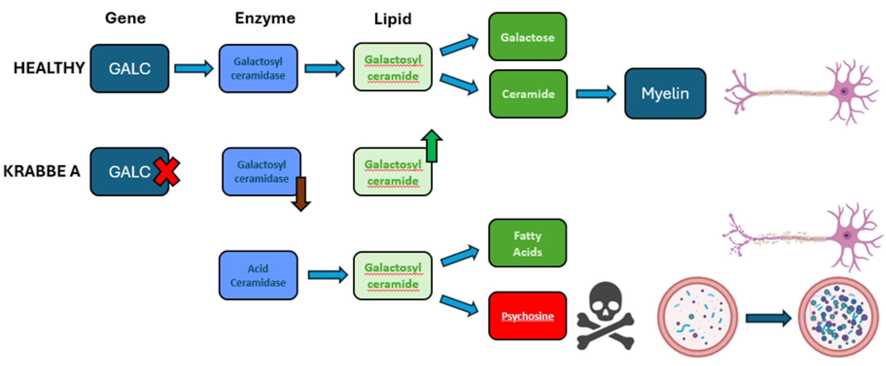
Treatments & Translational Neuroscience
PT_351
Keywords: multiple sclerosis, disease progression, neurodegeneration, kinases, kinome
Authors: Kelly Hares, Thomas Minton, Neil Scolding, Claire Rice
Multiple sclerosis (MS) is an immune-mediated, neuroinflammatory disease characterised by demyelination and neurodegeneration. It affects >2.5 million people worldwide. Although disease modifying therapies are available, these primarily target the inflammatory component of the disease and have limited effect on progressive disability, which is driven by neurodegeneration. Identification of improved biomarkers and treatments for progressive disease are an urgent unmet clinical need.
There are >500 protein kinases encoded in the human genome (the kinome) that play critical roles in cellular processes. Aberrant protein phosphorylation is a hallmark of neurodegenerative diseases, including MS. Although Bruton’s tyrosine kinase inhibitors are showing promise as treatments for MS (Krämer et al., 2023), (Fox et al., 2024), total kinase activity has not been quantified in MS cells or tissues. The aim of this study was to map the kinome in peripheral blood mononuclear cells (PBMCs) isolated from people with secondary progressive MS (SPMS) and determine if kinase activity correlates with clinical measures of disease severity.
The kinome was compared in PBMCs isolated from people with SPMS (n=6) and controls (n=6), using PamGene™ technology. Total kinase activity in PBMC protein lysates was measured using tyrosine kinase (PTK) and serine-threonine (STK) PamChip® arrays. Each PamChip® contains pre-designed peptide sequences against 196- PTK or 144- STK unique phospho-binding sites. Active kinases in the sample lysates phosphorylate their target and are fluorescently visualised. Images are captured, quantified and deconvoluted using the Pamstation12® with BioNavigator software®.
Primary comparisons of kinase activity were performed using unpaired t-test and SPMS clinical associations analysed using one-way ANOVA. PamGene™ upstream kinase analysis (UKA) algorithm was applied to score predicted kinases against peptide signal.
Kinase analysis identified differential activity in 141 kinases in SPMS PBMCs compared with controls, involved in pathways including leucocyte migration, lipid metabolism, oxidative stress, axonal transport and autophagy. In addition, select phospho-peptide activity correlated with clinical measures of disease severity including disease duration and multiple sclerosis functional score (MSFC).
With additional validation and interrogation of the underlying pathways, analysis of the kinome has the potential to identify novel disease biomarkers and therapeutic interventions in progressive MS.
Fox, R.J. et al. (2024) ‘Efficacy and Safety of Tolebrutinib Versus Placebo in Non-Relapsing Secondary Progressive Multiple Sclerosis', Multiple Sclerosis and Related Disorders, 92, p. 106114.
Krämer, J. et al. (2023) ‘Bruton tyrosine kinase inhibitors for multiple sclerosis’, Nature Reviews Neurology, 19(5), pp. 289–304.

Treatments & Translational Neuroscience
PT_360
Keywords: Translational neuroscience, Spatial transcriptomics, Variational autoencoder, Cross-species anatomical mapping, Disease prediction
Authors: Chloe Jaroszynski, Mohammed Amer, Antoine Beauchamp, Jason Lerch, Stamatios Sotiropoulos, Rogier Mars
In translational neuroscience, studies rely on the assumption that model species are similar enough to the human that they can provide insight into the structure and function of the human brain. Today, the translation to the human of findings from studies in the mouse remains under the 10% mark. In our study, we develop a method to translate between the mouse and the human brains, based on the latent space of a variational autoencoder trained on spatial transcriptomics. We first show that our approach retrieves essential anatomical correspondences between species. Our second aim was to explore quantitative translations across species: we show that the translation of plaque deposition images in a mouse model of Alzheimer’s disease (AD) predicts human AD-related cortical atrophy, and that the translation of a subcortical map of Parkinson’s disease (PD) predicts effects of PD in the human.
Using a set of ortholog genes between the mouse and the human, we trained an unsupervised variational autoencoder to reconstruct the mouse gene expression data, combined with a classifier trained in the latent space to distinguish mouse brain regions. We then used the encoder part of the model to embed both species’ gene expression data into the latent space. We computed the latent cross-species similarity, which provided us with a cross-species mapping at the voxel level to use for translations. We used this latent matrix to (1) directly compare brain organisation across species and (2) translate quantitative maps of disease-related brain changes in the mouse to predict the effects of disease in human data. We used the mouse APP-PSI model of AD, and a mask of findings related to mouse models of PD. The human maps were taken from neuromaps.collection:3273 and 2694. Translated maps were compared to real data using Pearson’s correlation and significance was assessed using spin tests accounting for spatial autocorrelation.
The cross-species anatomical comparison showed high similarity between major anatomical areas, and reflected overall principles of organisation with a gradient of primary-to-association cortex, as well as modality specific similarities.
Strikingly, the translated cortical AD map correlated significantly with the human cortical atrophy map (r=0.22, p<0.05) and the translated subcortical PD map correlated significantly with the human subcortical PD map (r=0.56, p<0.001).
By effectively translating quantitative maps of statistical effects of disease, our model holds promise to help improve translation rates of findings from mouse to human.
Treatments & Translational Neuroscience
PW_343
Keywords: Tactile, Dynamic allodynia, Meissner corpuscle, Post-surgical pain, Skin
Authors: Kirsten Wilson, Ying Sze, Anna Regan, Chunyi Zhu, Katarzyna Mazur, Atanaska Velichkova, Carole Torsney
Millions of patients undergo surgery each year with many developing post-surgical pain. A common yet disturbing symptom is when tactile stimuli such as clothing close to the surgical site elicits pain sensation. Tactile-evoked pain involves cross-talk between tactile inputs and pain circuits in the spinal cord, but the peripheral tactile drive has not been defined. To investigate, this study characterised skin innervation and tactile-evoked pain behaviours in a rat model of post-surgical pain (Brennan et al., 1996).
Hindpaw skin-muscle heel incision was performed under isoflurane anaesthesia in 7-8 week old Sprague-Dawley rats. Immunostaining: Biopsies were collected (post-surgical day (PSD)s 1,2,3,& 7) from incision heel, contralateral heel and control pad. Tissue was sectioned and immunostained for PGP9.5 to quantify nociceptive innervation; NF200 and S100 to quantify tactile innervation; and Collagen IV to delineate the dermal-epidermal junction. Behaviour: Rats underwent behavioural assessment of tactile sensitivity (paintbrush stroking) and thermal sensitivity (Hargreaves) at baseline and at PSD 1,3,& 7. BDNF-TrkB requirement was explored using a TrkB-Fc chimera: 100ng/50ul of TrkB-Fc dissolved in PBS was injected subcutaneously around the incision following surgery and on PSD2. Behaviour and immunostaining on PSD3 was compared between TrkB-Fc, vehicle and no injection groups. All groups n≥6.
Behaviour and postsurgical immunostaining data comparing contralateral and ipsilateral hindpaws across PSDs was analysed using repeated-measures (RM) 2-way ANOVA followed by Sidak multiple comparisons post-tests. BDNF-TrkB experiment: Behaviour data was analysed using 2-way ANOVA (treatment and hindpaw as factors), immunohistochemistry of the ipsilateral hindpaw was analysed using one-way ANOVA.
In the incision tissue surround, there is a reduction in nociceptive innervation (PGP9.5) and the development of dermal protrusions, reminiscent of dermal papillae, oriented towards the incision site. These dermal papillae are innervated with putative tactile corpuscles (S100+ bulb) with a time course matching tactile-evoked pain behaviours. Increased epidermal BDNF immunostaining intensity was also observed in the incision site surround. TrkB-Fc administration did not impact nociceptive innervation, thermal hyperalgesia or wound area. However, TrKB-Fc reduced tactile-evoked pain behaviours and the size of the novel tactile corpuscles (S100+ bulb area). In conclusion, following surgery, novel tactile corpuscles develop in a BDNF-TrKB–dependent manner around the incision site, contributing to postsurgical tactile-evoked pain.
Treatments & Translational Neuroscience
PW_352
Keywords: Neurodegeneration, Alpha synuclein, Microfluidics, Seeding, hiPSC-derived neurons
Authors: Eve Corrie, Rebecca Kelly, Matthieu Trigano, Emma V. Jones
Many neurodegenerative diseases are associated with the presence of misfolded, aggregating proteins within the brain, leading to cytotoxicity and cell death. The prion hypothesis states that these toxic species spread through the brain via anatomically connected regions, leading to eventual widespread neurodegeneration.
iPSC-derived neurons with or without iPSC-derived astrocytes were cultured in microfluidics devices consisting of two chambers connected by 10µm wide microchannels. These channels are wide enough to maintain separation of cell bodies but allow projection and growth of neurites through into the adjoining chamber. Fluidic isolation can be maintained by asymmetric volume loading to ensure a unidirectional flow of fluid.
Fluorescently labelled α-synuclein preformed fibrils (PFFs) were added to a “donor” neuron chamber, and the movement of these fluorescent PFFs to an “acceptor” chamber investigated with confocal microscopy. The presence of aggregated α-synuclein was also assayed using immunocytochemistry of α-synuclein phosphorylated at serine 129. Cells were also lysed and the phosphorylated and total α-synuclein content was analysed with a Luminex multiplexed immunoassay approach.
Where appropriate, quantitative data has been analysed with ANOVA or t-test.
We can visualise uptake of α-synuclein PFFs into neurons following direct application to the donor chamber. Progressive accumulation of PFFs is then detected in neurons in the acceptor chamber over time as they are trafficked through the neurites within the channels and released into the acceptor compartment. Exposure to exogenous PFFs also induces phosphorylation and perinuclear aggregation of endogenous α-synuclein. We found that disease-causing mutations in the SNCA gene led to a higher rate of α-synuclein aggregation and phosphorylation in the neurons within the donor and acceptor chambers following PFF application. Phosphorylated serine 129 α-synuclein could also be detected from lysate using Luminex technology.
These experiments model the pathogenic propagation of α-synuclein in disease via seeding of misfolded protein, uptake, and subsequent misfolding and aggregation of healthy α-synuclein. This microfluidics system could be adapted to model the spread of other misfolded proteins associated with neurodegeneration, such as β-amyloid, Tau or Huntingtin and be utilised in drug discovery to develop compounds to inhibit the spread of toxic species through the brain.
Treatments & Translational Neuroscience
PW_354
Keywords: Multiple Sclerosis, NHS Lanarkshire, Patient-Initiated Follow Up, Patient-Initiated Contact, Home Visits
Authors: Ginevra Bubani
Multiple Sclerosis (MS) is a chronic autoimmune disorder of the central nervous system. In Lanarkshire, many MS patients receive annual home visits with few clinical outcomes, while consuming significant resources. It is proposed to implement Patient-Initiated Follow Up (PIFU) to reduce unnecessary home visits and missed appointments, and improve MS service capacity.
This audit reviews demographics, health challenges, and care needs of MS patients under home visit care and explores potential benefits and risks of introducing a PIFU system.
Data includes all patients (n=184) receiving home visits from MS Specialist Nurses (MSSN). Data were extracted from NHS Lanarkshire (NHSL) Clinical Portals and TrakCare.
Lanarkshire has a low incidence of MS, and the highest number of diagnostic investigations compared to other Scottish NHS boards. However, patients in Lanarkshire experience longer wait times for diagnosis after primary referral, and fall below national standard for MSSN contact and discussion of Disease-Modifying Therapies. [1]
MS patients receiving home visits in Lanarkshire differ from the broader MS population across Scotland: they have higher rates of physical disability, co-morbidities, polypharmacy, socioeconomic deprivation, mental health challenges and cognitive decline.
89% have adaptations to property and mobility equipment. 71% have a Package of Care. 51% are under the care of District Nurses. 54% receive daily family support.
62% live in deprived areas, and 26% contribute towards their healthcare costs.
50% report mental health issues, cognitive decline and pain. One third have vision, sleep, speech and swallowing issues. Over two thirds report inability to walk, impaired manual dexterity, bowel/bladder issues, fatigue and sensory symptoms.
14% are regular smokers, 63% take over 6 regular medications, 44% have 3 or more co-morbidities. 53% had an ED admission in the past 12 months.
Only 65% had their last MSSN visit in person. The most common MSSN referrals were Occupational Therapy, Physiotherapy, Social Work, and Speech and Language Therapy. 24% has a record of not attending neurology services.
Introducing Patient-Initiated Contact can alleviate MS staff shortages and resource constraints. PIFU is beneficial for a subset of patients with stable disease (e.g. RRMS). Implementation requires communication strategies to ensure patients understand the changes, a risk assessment, phased implementation strategy, and a feedback loop to address any concerns.
MacDougall N, Harden J. Scottish Multiple Sclerosis Register figures from January to December 2023 [Internet]. Scottish Multiple Sclerosis Register (SMSR). 2024 [cited 2024 Jun 6]. Available at: https://publichealthscotland.scot/publications/scottish-multiple-sclerosis-register-smsr/scottish-multiple-sclerosis-register-smsr-report-2024-figures-from-january-to-december-2023/

Treatments & Translational Neuroscience
PW_355
Keywords: stroke, biosensors, diagnostics, point-of-care, adenosine
Authors: Harriet Allgrove-Ralph, Johannes Boltze, Martin Peacock, Bruno Frenguelli
Reducing door-to-needle time (DTNT) is key for improving outcomes in stroke patients1. Currently, there is a lack of point-of-care stroke diagnostics, leading to delays in diagnosis and treatment. A portable device that can assist clinicians with the differentiation of strokes from conditions with similar symptoms (‘mimics’) and provide information as to stroke severity or timing could reduce DTNTs.
That cerebral ischaemia induces the release of adenosine and its metabolites inosine, hypoxanthine, and xanthine (the ‘purines’) has been demonstrated previously in vivo2 and in rat brain slices deprived of oxygen and glucose3. Peripheral blood purine levels are also elevated in human patients undergoing carotid endarterectomy4, and in stroke patients compared to controls and stroke mimics5. Purines therefore have potential as stroke biomarkers. However, little is known about the specific pattern of their release during the time course of a stroke. Therefore, we plan to use screen-printed electrochemical purine sensors to characterise this in rats during experimental stroke.
The carbon working electrodes (WE) of our biosensors, which also comprise Ag/AgCl counter and pseudoreference electrodes, are functionalised with adenosine deaminase, nucleoside phosphorylase, and xanthine oxidase immobilised in a tetramethoxysilane-based sol-gel6. These enzymes catalyse the production of hydrogen peroxide and urate from purines. A ferric hexacyanoferrate mediator is used to permit the reduction of hydrogen peroxide at a potential of around 0 V vs Ag/AgCl, decreasing interference.
We will model ischaemic stroke by stereotaxically injecting endothelin-1 around the middle cerebral artery (MCA) of anaesthetised rats. Peripheral blood purine readings will be taken before and after experimental stroke induction using amperometric purine biosensors. Blood samples (around 50 µL) will be taken via a tail vein cannula, from which purine concentrations will be measured. Cerebral blood flow through the MCA will be recorded and used along with stroke lesion volume (quantified via TTC staining) to correlate stroke severity with purine release.
Presently, our biosensors provide linear responses to adenosine in PBS up to ~30 µM, though altering enzyme concentrations could further optimise this. Within this linear range, they exhibit good sensitivity, giving readings of ~1 nA/µM.
Electrochemical purine sensors are useful for both research and clinical applications, as they are portable and allow real-time measurements to be taken from small volumes. Elucidating the release profile of purines during stroke and their association with stroke severity will facilitate their future use as biomarkers to accelerate the treatment of this medical emergency.
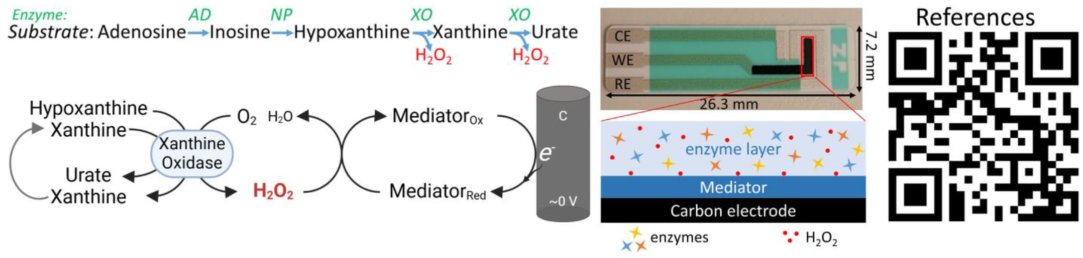
Treatments & Translational Neuroscience
PW_357
Keywords: Viral Infection, Blood-brain barrier, HSV-1, Microfluidic chip model, Cytokines
Authors: Sarah Boardman, Claire Hetherington, Franklyn Egbe, Cordelia Dunai, Benedict Michael
Herpesviruses can damage the blood-brain barrier (BBB). Herpes simplex virus-1 (HSV-1) is a leading cause of infectious encephalitis, which has limited effective treatments (Michael et al., 2020). This project utilises a human 3D microfluidic BBB model with the main aim of elucidate key chemoattractant proteins that enable BBB breakdown to identify potential therapeutic targets.
A microfluidic chip (Brown et al., 2019) was utilised, incorporating human endothelial cells (hCMEC/D3) and primary human astrocytes within separate channels. A syringe pump was used to induce shear stress on the hCMEC/D3 monolayer.
Barrier integrity was assessed by transendothelial electrical resistance (TEER) and expression of tight junction proteins using immunofluorescent microscopy.
The BBB chip model was infected with HSV-1 at varying MOIs (0.05, 0.5) or uninfected control medium. TEER was measured over 48hrs. Cell medium from channels containing hCMEC/D3 were collected at corresponding time-points.
For the Maestro platform, hCMEC/D3 cells, human primary astrocytes, and their co-cultures were cultured in a 96-well plate and impedance was measured over 3 days to assess BBB integrity.
Cultures were incubated with HSV-1 at varying MOIs (0.05, 0.5, 5) or uninfected control medium. Additionally, hCMEC/D3 were exposed to astrocyte-conditioned medium (ACM) from HSV-1-infected astrocytes or inactivated HSV-1 (MOI=5). Impedance was measured over 48hrs. Supernatant was collected for Luminex analysis and cells harvested for RNA analysis.
Data were analysed using repeated-measures ANOVA or appropriate non-parametric tests.
The BBB chip model effectively recapitulates key features of the BBB, exhibiting high ZO-1 expression and a dynamic response to shear stress exhibited by increased TEER over time.
The Maestro platform revealed a dose-dependent decline in impedance following HSV-1. Higher viral loads caused earlier impedance drops, indicating faster BBB disruption (Figure 1). Astrocytes exhibited greater sensitivity to HSV-1 compared to endothelial cells. Furthermore, exposure to ACM induced endothelial cell damage, but at a slower rate than direct viral infection. Luminex analysis identified the production of various pro-inflammatory and chemotactic cytokines, including IL-6 and CXCL1.
Microfluidic chips provide a robust platform for in vitro BBB modelling, enabling precise control and measurement of physiological parameters. Our findings demonstrate distinct patterns of HSV-1 infection in endothelial cells and astrocytes.
Future work will expand these investigations to include VZV, utilising the Maestro platform to establish a time-course of infection-induced damage. This data will guide subsequent experiments within the microfluidic chip model, with a focus on assessing the efficacy of cytokine blockade strategies against both HSV-1 and VZV-induced BBB damage.

Treatments & Translational Neuroscience
PW_358
Keywords: Diffuse Intrinsic Pontine Glioma, Palliative Care, Quality of Life, Symptom reduction
Authors: Alice Ramsey, Daveena Thakkar, Kim Bull
Diffuse Intrinsic Pontine Glioma (DIPG) is an aggressive paediatric brainstem tumour. There are no therapeutic options for improving survival and proliferation of cancer cells causes many distressing end-of-life symptoms. Palliative care is vital for reducing symptoms and improving quality of life (QOL). There have been no previous systematic reviews in this area, therefore this review will investigate all research on palliative care in DIPG and determine its impact on QOL.
4 databases (MEDLINE, CINAHL, PsycINFO, AMED) were searched for articles on palliative care in DIPG. A combination of synonyms and subject headings were included, connected by Boolean operators. Two reviewers were involved in the screening process to ensure reliability. Studies were evaluated against eligibility criteria, with the outcome of interest being impact of palliative care on QOL. Relevant data from studies were extracted using a narrative synthesis approach, followed by quality assessment using the CASP checklist.
Database searching revealed ten possible studies. After screening the full text articles, five were included. Studies were heterogenous but comparison was possible. Three studies agreed on the use of analgesics, anti-emetics, anxiolytics, laxatives, anti-secretory drugs, and anticonvulsants to improve symptoms. One study reported that paracetamol or opioids effectively managed headaches in all patients. All studies included radiotherapy to reduce end-of-life symptoms, thus improving quality of life. Children in one study reported improvements from baseline functioning assessments, reductions in anxiety, and improved health-related QOL following radiotherapy. Two studies reported that children and families preferred end-of-life to be at home and they appreciated support with Do Not Resuscitate (DNR) discussions. One study reported that 91% of families signed a DNR after discussions with the palliative care team.
The use of medications, medical aids, and radiotherapy to reduce symptom burden led to significant improvements in QOL. Furthermore, psychosocial support, particularly with making end-of-life decisions, can have a positive impact on children and their families. This review highlights the need for further research, specifically guidance for healthcare practitioners supporting patients with DIPG is needed.
Treatments & Translational Neuroscience
PW_359
Keywords: Meningoencephalomyelitis, Vimentin, Autoantibodies, Single-cell RNA sequencing,
Authors: Junwei Hao, Dongshan Wan, Shufang Zhao, Chen Zhang, Guoliang Chai
Autoantibodies targeting astrocytes, such as those against glial fibrillary acidic protein (GFAP) or aquaporin protein 4 (AQP4), are crucial diagnostic markers for autoimmune astrocytopathy among central nervous system (CNS) autoimmune disorders. Nevertheless, the diagnostic remains challenging for patients lacking specific autoantibodies.
This retrospective case-series study included samples collected from April 2021 to May 2024. Single-cell RNA sequencing (scRNA-seq) was performed on cerebrospinal fluid (CSF) cells of two index patients to identify the putative target antigen of the clonally expanded B cells. A comprehensive screening for additional patients was conducted using a blinded cell-based and tissue-based assay. Candidate patients were followed up with a median (range) duration of 23 (5-31) months. This study was conducted at a tertiary referral hospital. Fourteen patients with uncharacterized CNS autoimmune disorders and similar clinical and radiological features were identified by CSF autoantibody testing.
All data analysis and visualization were conducted using R (V4.3.2) or GraphPad Prism 10. In descriptive statistics, count and percent were used for categorical variables, whereas median and range or IQR were used for continuous variables. The Mann-Whitney U-test (two-tailed) was used for nonparametric comparison of continuous value between two groups, while the Kruskal-Wallis test was employed for comparing continuous values among multiple groups. Dunn's method was used for post-hoc analysis. P values less than 0.05 were considered significant.
Fourteen candidate patients (10 [71%] female; median [IQR] age, 33 [23-41] years) were identified. Initially, CSF from two women with unknown encephalomyelitis showed astrocytic reactivity on rat tissue but negative for GFAP IgG. 17 (46%) of 37 clonally expanded B cell clonotypes in their CSF expressed IgG autoantibodies targeted astrocytic intermediate filament protein-Vimentin. Subsequent screening identified 12 additional patients. These 14 patients shared a unique clinical profile characterized by relapsing courses and symptoms prominently involving the cerebellum, brainstem, and corticospinal tract (CST). All patients also exhibited elevated CSF protein and cells, intrathecal immunoglobulin synthesis, and magnetic resonance imaging (MRI) of bilateral lesions on CST. Notably, 8 (67%) of 12 patients who received first-line immunotherapy at their first episode responded well. At the last follow-up, 11 (79%) patients experienced significant disability (modified Rankin Scale ≥ 3). In this case series, autoantibodies targeting astrocytic intermediate filament protein Vimentin were identified in patients with previously undifferentiated meningoecnephalomyelitis and common radiographic features.
Treatments & Translational Neuroscience
PW_372
Keywords: dementia, cognitive rehabilitation, proper name anomia
Authors: Aygun Badalova
Proper name anomia is a common experience that can become unpleasantly amplified in people with dementia (PWD). The Gotcha! app aims to provide practice-based therapy for PWD enabling them to spontaneously retrieve the names of key people in their lives. It has been developed using the principles of errorless learning and spaced retrieval pioneered by Clare et al, (2000, 2003), but packaged in an app to support self-management.
Gotcha! is a digital confrontation naming therapy app. PWD supply images and names of the people they want to be able to name and train on one face per day for six weeks. We employed a single-case experimental design with weekly testing of free-naming in both six-week blocks (pre therapy and during therapy). A novel speech verifier was used to provide real-time feedback (Barbera et al. 2020). PWD also had an MEG scan before and after the therapy block where they attempt to name pictures of familiar (trained) and famous (untrained) faces. We interrogated the behavioural data in two ways: 1) a within-subject non-parametric analysis using Tau-U metric (Parker et al. 2011); 2) a parametric group analysis using an ANOVA. MEG data were analysed in SPM. We measured source localised gamma-band (30-80 Hz) power 0-1000 ms after the onset of a face. We ran a group-based 2x2 factorial analysis on the resultant images (familiar vs. famous; pre- vs. post-therapy) using a repeated-measures ANOVA to look for changes in power.
The trial is ongoing (target = 45 PWD). results from the first 20 PWD to complete the trial demonstrate:
1) Tau-U. 80% showed a positive trend with better naming during the training phase with 8/20 reaching statistical significance.
2) ANOVA showed a significant effect at the group level of training>baseline phase, F (1,19)=13.18, p=0.01
Results from the MEG analysis of 14 PWD: We identified a large cluster of 813 voxels situated in the left ventral temporal lobe (MNI: -50 -28 -26, F=9.19, p=0.004) where gamma reduction was associated with training (pre-post) of familiar faces, but not (untrained) famous faces.
Gotcha! app-based therapy for proper name anomia works for the majority of PWD in our trial thus far. This is the first study to demonstrate that the left ventral temporal lobe region supports practice-based retrieval of familiar face-name associations in PWD. Being able to freely produce the name of a relative or loved one has a big impact on people’s lives.
Footnotes
Correction (July 2025):
The author byline in the abstract “PW_149” has been updated to include Aline Nixon and Helen McGettrick.