
Abstract

23 - 26 April 2023 | Brighton, UK
Book of abstracts
Table of Contents


Cognitive Processes & Decision-making
Measuring attentional bias to emotional and illness-related stimuli in functional neurological disorder: A pilot study using modified dot-probe tasks
Introduction: Previous research has suggested potential dysfunction in attentional allocation in functional neurological disorder (FND), including attentional bias (AB) to salient environmental stimuli. We aimed to test the hypotheses that individuals with FND would display enhanced AB towards emotional faces and FND-relevant information, compared to healthy control participants (HCs).
Methods: Fourteen individuals with FND and 16 HCs completed modified dot-probe tasks, a test of general intellectual functioning (WASI-II) and a CANTAB test of sustained attention (Rapid Visual Information Processing, RVIP). An emotional dot-probe task assessed AB to standardised (Karolinska Directed Emotional Faces) facial expressions of happiness, anger, and disgust, compared to neutral. An FND-word dot-probe task measured AB for FND-relevant words that were generated by our FND Patient and Carer Advisory Panel, relative to neutral words.
Statistical analysis: AB scores were analysed with mixed ANOVAs and t-tests. Effect sizes were calculated using partial eta-squared and Hegdes’ g.
Results: On the emotional dot-probe, there was no significant group difference in AB scores (F(1, 28) = .70, p = .41, ηp2 = .02) and no group x expression interaction (F(2, 56) = 1.56, p = .22, ηp2 = .05). Similarly, the effect of group on AB scores was not significant in the FND-word task (t(18.69) = .08, p = .93, g = .03). The groups also did not differ in estimated full-scale IQ scores (p = .57), or in performance on the RVIP (p = .43).
Conclusions: In contrast to previous findings in functional seizures samples, this study did not provide evidence of attentional hypervigilance for facial emotion or FND-relevant information in this mixed FND symptom group.
However, this pilot study was limited by a small, heterogeneous sample and low power. Our future research will investigate attentional functioning in larger samples of participants with specific FND symptom presentations including functional motor symptoms and functional seizures.
Investigating the influence of affective stimulation and experiential detachment on subjective functional neurological symptoms - a pilot study
INTRODUCTION: We aimed to test the hypothesis that exposure to highly arousing affective stimuli would provoke increased FND and associated symptoms. We also explored the effects of experiential detachment on FND symptoms during affective stimulation.
METHODS: Participants were 14 individuals with FND (10F:4M; 10 motor symptoms, 4 motor/seizures) and 13 age- (p=.71) and sex-matched (p=.72) healthy controls (HCs; 11F:2M). We used validated affective stimuli in 12 blocks of 10 images (four Positive, four Negative, four Neutral blocks). Participants were instructed to passively observe the images (Watch) or detach from the experience (Distance) in separate blocks. Momentary assessments of FND symptoms, dissociation, affect, pain and fatigue were obtained at baseline and immediately after each block (Likert- scale, 1-7).
STATISTICAL ANALYSIS: Data were analysed using within-groups or mixed ANOVAs. Post-hoc tests were Bonferroni- corrected.
RESULTS: At baseline, the FND group reported elevated pain (p<.001), fatigue (p=.001), and derealisation (p=.015), compared to HCs, but subjective arousal (p=.83), positive affect (p=.11), negative affect (p=.08), dissociative amnesia (p=.052), and depersonalisation (p=.87) did not differ. During the task, there was a main effect of image type on FND symptoms (p=.002) and an image type x task instruction interaction (p=.020). There was a main effect of image type in the Watch condition (p=.008), but not in the Distance condition (p=.12). In the Watch condition, FND symptom ratings were highest following Negative images (M=3.61, SD=1.73), relative to Positive (M=2.79, SD=1.44) and Neutral (M=2.79, SD=1.33). During the task, there were group main effects (FND>HCs) for derealisation (p=.037), depersonalisation (p=.046), dissociative amnesia (p=.016), pain (p<.001), and fatigue (p=.004), but not for positive (p=.94) and negative affect (p=.20), or subjective arousal (p=.85).
CONCLUSIONS: Exposure to affectively arousing negative stimuli resulted in increased FND symptom severity, but this was mitigated when participants voluntarily detached from their experiences. The influence of affective stimulation on FND symptoms was not mediated by heightened subjective negative affect or perceived physiological arousal
Objective and subjective neurocognitive functioning in functional neurological disorder
INTRODUCTION: Previous studies indicated possible neurocognitive deficits in functional neurological disorder (FND), but findings were variable. We aimed to investigate both objective and subjective neurocognitive functioning in FND, with standardised measures.
METHODS: Individuals with FND (n=16) and healthy controls (HCs, n=17) completed a neurocognitive battery (CANTAB), testing motor response speed, sustained attention, working memory, attentional set-shifting, response inhibition and social cognition. Participants rated their performance on all tasks (1-7 Likert) and completed the Wechsler Abbreviated Scale of Intelligence (2nd ed, WASI-II), Medical Symptom Validity Test (MSVT), Cognitive Failures Questionnaire (CFQ), Multiscale Dissociation Inventory (MDI), Somatoform Dissociation Questionnaire (SDQ- 20), Patient Health Questionnaire-9 (PHQ-9) and Generalized Anxiety Disorder-7.
STATISTICAL ANALYSIS: Data were analysed with t- and Mann-Whitney U tests. Effect sizes were ‘Hedge’s g’ or ‘r’.
RESULTS: There were no group differences on the WASI-II (p=.57), MSVT (ps>.49), and most CANTAB tests. On the CANTAB Emotion Recognition Test, FND participants showed superiority in identifying facial sadness compared to HCs (p=.042, r=.37). On two Emotional Bias Tasks, the FND group displayed shorter reaction times than HCs when selecting happy (p=.013, g=.94) and angry (p=.02, g=.89), and happy (p=.021, g=.87) and disgusted (non-significant trend, p=.054, g=.72) faces. The groups did not differ in their subjective performance ratings for any task (ps>.15). Nevertheless, the FND group reported more daily cognitive complaints than HCs (CFQ, p=.01), which were positively correlated with SDQ-20 (r=.75, p=.001), MDI (disengagement r=.88, p=<.001; memory disturbance r=.69, p=.003) and PHQ-9 (r=.64, p=.007) scores (all elevated in the FND group, p-values <.01-<.001).
CONCLUSIONS: Contrary to previous findings, this FND sample did not display objective deficits in neurocognitive functioning or differences in task-related metacognition, but they exhibited superiority in aspects of social cognition. Daily cognitive symptoms in the FND group were elevated but were related to psychological symptom burden rather than objective neurocognitive impairments.
Investigating the Relationship between Sleep Characteristics, Fatigue and Executive Functioning (an fMRI study)
Aim: Many neurological disorders present cognitive dysfunction along with high levels of fatigue. However, our understanding of the neurological effects of fatigue on cognition is poor. Generally, research informs of a disconnect between reported measures of objective and subjective sleep, such that individuals are not commonly capable of accurately predicting their own sleep quality and quantities. Here we explore the role of sleep characteristics and fatigue using objective and subjective measures alongside fMRI investigation of executive functioning.
Method: Participants (n=34) wore a wrist accelerometer device (GENEActiv) for 7-days to obtain objective measures of sleep and completed the Pittsburgh Sleep Quality Index as a subjective measure of sleep. An MRI protocol consisting of a T1 anatomical scan and task-based fMRI consisting of an n-back and go/no-go task was acquired. A general linear model analysis was used to determine the relationship between fMRI haemodynamic responses, cognitive performance and sleep characteristics.
Results: Here we demonstrate the relationship between objective and subjective measures of sleep quality and quantity, and functional performance in the n-back and go/no-go tasks (accuracy and reaction times). Furthermore, we highlight the corresponding neural network changes associated with perceived and actual sleep quality and subsequent impact on measures of executive functioning.
Conclusion: Demonstrating the relationship between objective and subjective sleep measures, functional performance and subsequent neural correlates evidences the role of sleep characteristics on executive functioning in a healthy population. Through this understanding, we can expand further into the role that fatigue plays in cognitive symptoms of neurological disease.
A retrospective, observational study of outcomes for neurosciences patients transferred to intensive care before and during the COVID-19 Pandemic
The outbreak of SARS_cov_2/ COVID 19 led to the prioritisation of care pathways for patients hospitalised with this infection. Although there is emerging understanding about these pathways, the impact on non-COVID patients during this time frame is poorly understood.
We retrospectively considered the change in utilisation of services for neurological patients transferred from a tertiary neurosciences ward to ITU pre and during the COVID pandemic. Outcomes were measured using the Modified Rankin Scale (MRS) on discharge from hospital. Outcomes were also compared between different neurological diagnoses at the time of ICU admission, patients who had a single versus multiple ICU admissions during a single inpatient stay, the length of stay on ICU and the length of stay on the neurosciences ward prior to being admitted to ICU. Data were collected from a total of 72 inpatient admissions over two 14 month periods before and after the onset of the increase in COVID hospital admissions (defined as 1/3/20) by review of electronic patient records. There was no significant difference in MRS outcomes prior to versus during the pandemic (p = 0.203, using Wilcoxon Rank Sum). The time spent on the neurosciences ward prior to transfer to ITU did not correlate with outcome. The number of days spent on ICU and the number of re-admissions to ICU positively correlated with poor outcome, though these correlations were not significant (p= 0.112 and p=0.081 respectively, using Spearman Rank Correlation).
It is potentially reassuring that there was a consistency of outcomes for patients pre- versus during the COVID-19 pandemic. We hope that these results can begin to guide the utilisation of ITU care by critically ill neurosciences’ patients, especially at a time when there are competing demands for beds.
What matters to people affected by MND? Integrating and valuing the perspectives of people affected in MND Scotland’s research, strategy and funding
As part of MND Scotland’s new strategy, Making Time Count, one of our commitments is to make sure that the views of people affected by motor neuron disease (MND) are central to the development, and ongoing evolution, of the charity. Funding and supporting research is an important part of our work. Over the past 15 years, we have invested around £6 million into a range of projects from basic science and clinical trials, to research improving quality of life. Here we outline how we are integrating the perspectives of those affected into the research we fund and support.
Methods
We have used three separate approaches to bring the perspectives of people affected by MND into our funded research. First, we have embedded lay reviewing into our grant review process. Second, we have introduced a request and support system for researchers seeking input from those affected. Third, we have undertaken a national survey asking people what issues are important to them and what questions they’d like research to address. The findings will help to inform the work we invest in and support in the future.
Results and conclusions
MND is a rapidly progressing terminal condition. Those affected by the disease face innumerable challenges: they also have expertise and often an interest in research. With careful attention to accessibility, we have found that people have much to bring to lay reviewing grant applications. Working in the context of a terminal condition has nuances, but this process has brought a valuable new perspective to funding
As a result of our new involvement request system, we have begun offering researchers bespoke assistance in tailoring their research, so it effectively and appropriately engages those affected.
Through our survey, people affected by MND are also having a wider impact on the charity’s development. The findings cover aspects from social care and bereavement, to a desire for research which focuses upon improved diagnosis, troublesome symptoms, and understanding disease progression.
These three components have enriched our organisational processes and continue to inform future decisions.
This abstract does not include a section on statistical analysis due to its nature (qualitative), and focus on research in practice.
Determining whether dysfunctional checking is habitual in a rodent analogue of compulsive-like checking in obsessive-compulsive disorder
Introduction. A major subtype of obsessive-compulsive disorder involves excessive and maladaptive checking behaviour. This maladaptive, dysfunctional checking can be modelled using the Observing Response Task (ORT). The ORT distinguishes between functional and dysfunctional checking shown by the same individuals, in both rodents and humans. However, although dysfunctional checking on the ORT serves no purpose, it has not yet been formally tested whether it is habitual.
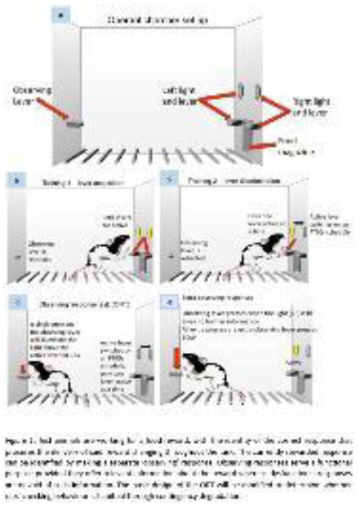
Methods. 24 male Lister Hooded rats will be trained on the ORT (Figure 1). Briefly, rats learn to respond on one of two levers to receive reinforcement, with the correct lever changing unpredictably throughout the session. Rats can identify the currently correct lever by pressing a third, ‘observing’ lever, illuminating a light cue over the correct lever. These functional Observing Lever Presses (OLPs) can be distinguished from dysfunctional Extra Observing Lever Presses (eOLPs), with the latter having no programmed consequences. As we have previously observed individual variation in dysfunctional checking, with sign-tracking rats showing elevated levels, rats will separately undergo classification as sign-trackers or goal-trackers on a pavlovian autoshaping task. Contingency degradation will be used to determine whether dysfunctional checking is habitual. This will weaken the contingency between pressing of the observing lever and light cue presentation. Based on previous reports that sign-trackers rely more on model-free learning, it is predicted that the dysfunctional checking of sign-trackers will be insensitive to contingency degradation. In contrast, goal-tracking rats are predicted to reduce any dysfunctional checking following contingency degradation.
Approach for statistical analysis. The ORT provides a rich data set, including data on OLPs and eOLPs, and secondary measures of generalised task performance (e.g. rates of lever pressing, lever discrimination). Baseline responding on the ORT (prior to contingency degradation) of sign-trackers and goal-trackers will be compared for each measure using mixed-model ANOVAs. Checking following contingency degradation will be analysed using mixed-model ANOVAs, comparing pre-degradation and post-degradation levels of OLPs and eOLPs.
Determining the nature of the relationship between sign-tracking and dysfunctional checking on the Observing Response Task
Introduction
Compulsive behaviours occur in a variety of psychiatric disorders including addiction and obsessive-compulsive disorder. It has been proposed that individual differences in the propensity to respond to reward-related cues is associated with compulsive behaviours. For example, "sign-tracking” (engagement with the reward-related cue rather than the reward) is associated with deficits in attention and impulse control and is resistant to extinction. Indeed, in both humans and animals, sign-trackers tend to exhibit more compulsive-like behaviour. However, it remains unknown whether there is a causal relationship between sign-tracking and compulsive behaviour. If so, then treatments and prevention protocols for compulsive disorders could specifically target the sign-tracking trait. By contrast, if sign-tracking and compulsions are both underpinned by the appetitive motivational system but occur independently, sign-tracking may be a diagnostic marker of disorders to achieve prevention and early intervention.
This study aims to determine the nature of the relationship between sign-tracking and compulsive-like behaviour to expand on our previous observations that sign-tracking rats show higher greater dysfunctional checking on the Observing Response Task (ORT; Figure 1). The causal nature of this relationship will be tested by reversing the order of training, such that rats are first trained on the ORT prior to classification using a pavlovian autoshaping task.
Method
Subjects will be 24 male Lister Hooded rats, trained on the ORT. To induce dysfunctional checking behaviours, rats will undergo ‘uncertainty training’ in which the correct response switches unpredictably, which increases functional checking in all rats and dysfunctional checking in sign-trackers. Rats will then be classified as sign-trackers or goal- trackers using pavlovian autoshaping, and the relationship between high levels of dysfunctional checking and sign- tracking assessed.
Approach for statistical analysis
Logistic regression will be used to determine whether increased dysfunctional checking will make more rats to become the sign-tracking phenotype. Other secondary measures (e.g. rates of lever pressing) will be examined to determine any generalised differences in motivate.

Is motivation toward a reward a predictor of dysfunctional behavior?
When presented with a reward-related conditioned stimulus, individuals vary in their responses. Depending on whether they approach the location of a reward delivery or the cue itself, they are classified as goal- or a sign- trackers respectively. We have previously observed a correlation between sign-tracking and dysfunctional, compulsive-like checking on the Observing Response Task (ORT). As sign-trackers have also been reported to show more rapid progressions to compulsive-like drug-seeking and to rely more upon habitual-like model-free learning, then this may indicate that sign-trackers rely more on habitual learning. Whether this association is specific to sign- tracking, or could be observed with other measures of motivated behaviour, such as pavlovian-instrumental transfer (PIT) remains unknown. Thus, we aim to test whether sign-trackers and goal-trackers show differential performance on both specific PIT (which relies on model-free learning) and general PIT (which relies on model-based learning), and whether PIT performance can be similarly related to dysfunctional checking on the ORT.
Rats will be trained on a Pavlovian-Instrumental Transfer task with two separate reinforcers, two responses and two reward-associated cues (and a third, neutral cue). General and specific PIT will be assessed in a probe test, before rats are subsequently trained on both pavlovian autoshaping (to classify the animals as goal- and sign-trackers) and the ORT.
We will analyze the mean lever presses from the PIT test data using an ANOVA and the ORT task using mixed 2x2 ANOVAs. In our study, we expect that ST will have a greater general PIT effect than GT, which will positively correlate with dysfunctional checking during the ORT task.
Seeking certainty in an uncertain world
To cope with uncertainty, humans often use goal directed behaviours, such as checking, to increase their knowledge about the environment. To date, research on attitudes and behaviours relating to coping with uncertainty have largely relied on self-report. This study investigated the tendency to use cues even when they no longer provide useful information to reduce uncertainty.
Using the translational Observing Response Task, participants could earn rewards (50p) by pressing one of two buttons on a variable ratio schedule. However, at any given time, only one of the buttons was active. This introduced uncertainty, in that participants did not know which button to press to earn rewards. Pressing a third button revealed an observing (certainty) cue that showed which of the two buttons was currently active (meaning that pressing it would lead to a reward). However, pressing this cue incurred a small cost (5p). After six minutes, unbeknownst to participants, the observing cue was extinguished, but continued to incur a cost. Participants also completed a battery of self-report questionnaires which included questions about uncertainty and obsessive compulsive tendencies as well as anxiety levels before and after the task.
A sample of 82 participants was analysed using repeated measures ANOVAs and bootstrapped regression. Overall, observing behaviour within individuals was stable during the first six minutes. Following cue extinction, there was a clear reduction in observing behaviour (η²p=.27, p<.001), however a proportion of participants continued to press the observing cue, with 40% of participants pressing as late as 6-8 minutes after cue extinction. Observing during extinction, but not prior, was a significant predictor of self-reported levels of obsessive-compulsive checking (assessed using the Obsessive Compulsive Inventory). Further, the task was experienced as a mild stressor, with a rise in self-reported anxiety (d=0.29, p<.01).
These findings indicate that certainty seeking is persistent, regardless of its potential to become maladaptive. This provides insight into the mechanisms underlying the development and maintenance of certainty seeking habits.
Distributed neural population encoding during a perceptual decision confidence task: A dynamic causal modelling study
Perceptual decision confidence is the metacognitive ability to internally evaluate the accuracy or correctness of one’s own perceptual decisions. Several studies on humans have identified neural activities in different brain regions during perceptual decision formation (stimulation) phase and decision confidence rating phase. However, it is unknown whether neural encoding is more prominent in decision formation or confidence rating phase. Further, as reaction times (RTs) are related to decision confidence levels, it is unclear how differently slow and fast decisions are encoded within neural populations. We addressed the above by using dynamic causal modelling (DCM) on an open dataset based on a classic random-dot kinematogram with confidence rating task (Gherman & Philiastides, 2020).
We chose 16 participants with significant Spearman’s correlations (p<0.05) between RT and rated confidence level from the dataset. We compared neural activities between stimulation and rating phases as well as between fast and slow responses in stimulation phase. Using T-contrast analysis in SPM12 toolbox, we isolated brain regions with significant activity differences for these two conditions from functional magnetic resonance imaging (fMRI) data. We then created and estimated a DCM model space between extracted brain regions for each condition from electroencephalogram (EEG) data using canonical microcircuit neural mass model and selected the winning model using random-effects Bayesian model selection. For stimulation-vs-rating condition, the right superior and inferior parietal lobule (SPL and IPL), left and right precuneus (PrC), left cingulate gyrus, left and right insula, and right superior frontal gyrus were more active. Compared to rating phase, we found in stimulation phase higher neural populations’ activities averaged over all participants in the winning model, indicating stronger neural encoding. Next, we extracted activities from the right SPL, left PrC, right supramarginal gyrus, left precentral gyrus, and left medial frontal gyrus and found, for slow responses, the winning model exhibiting higher activity for all neural populations in the left PrC, suggesting the encoding of decision uncertainty.


A Systematic Review of M-EEG evidence on value-based decisions in humans: experimental paradigms and spatiotemporal characteristics
Decision-making is an integrative process that is crucial for humans and animals on a daily basis, and it has been at the centre of a long tradition of psychological and economic research. This systematic review focuses on the spatiotemporal dynamics of value-based decision-making. Since there is an abundance of fMRI, neuropsychological, and animal studies on the topic, we instead examined evidence collected from 100 magnetoencephalography and electroencephalography studies, which are known for their high temporal resolution. Additionally, we classified value-based decisions into ‘external’ (EDM) and ‘internal’ (IDM) and used this division as the theoretical framework of the present review. In ‘external’ decisions, the values of different options are objectively defined, whereas said values in ‘internal’ decisions are defined by the individual. The review aims to assess whether there is a convergent pattern of event-related potentials or fields (ERP/ERF) findings and how EDM and IDM processes have been studied so far with these methods. Based on precedents in the literature, we extracted statistically significant time intervals that result from contrasts between experimental conditions that are sensitive to differences in (external or internal) value. We also examined the topographical and source space distribution of these intervals. Finally, we classified the paradigms into specific clusters of experimental designs, an approach that will guide future research and inspire the development of novel tasks to study value-based decisions. Due to the nature of the data, we could not apply typical statistical analyses and, instead, looked at the proportion of papers that reported significant intervals for the aforementioned contrasts. Overall, our findings show that there are similarities as well as differences in the time course and the topography of EDM and IDM processes. To our knowledge, the current review is the first to directly contrast these two types of decision-making and to provide an in-depth description of the paradigms and ERP/ERF components that are most consistently employed in the field.
The interplay between internally- and externally-guided decision-making: evidence from three online behavioral experiments
Value-based decision-making is vital for our interactions with the external environment, and the nature of the values that guide our decisions can originate from different sources, e.g., external (objective rewards) or internal (connected to our preferences). However, how these different values interact with each other has not been addressed in the literature, so far.
In the current study, we designed a series of three experiments where sources relating to internal values (i.e., subjective preferences for specific food items) and to external values (i.e., the reward probability associated with the different options) are presented simultaneously, with the aim to examine whether the presence of task-irrelevant value information influences human behaviour.
In Experiment 1, after the stimulus display, participants were cued with a dollar sign or a heart-shaped symbol, indicating whether they had to perform a reward-based or a preference-based decision. In Experiment 2, the task cue was presented before the stimuli, removing the uncertainty component. In Experiment 3, we introduced a delay between the cue and the response, to investigate how the removal of temporal constraints affected the participants’ behaviour.
Results from linear mixed models show that, across all experiments, there is evidence of a spill-over effect on accuracy and RTs from the preference-based domain into the reward-based one, as indicated by significant interactions between value conflict (i.e., the absolute difference between the average ratings of the two food items shown on the screen) and congruency (i.e., whether the preferred item was associated with advantageous or disadvantageous border colour), with odds ratio value and confidence intervals of: OR = .65 [.58, .72], p < .001; OR =.66 [.59, .74], p < .001; OR = .74 [.66, .83], p < .001 for Experiment 1, 2, and 3, respectively.
Overall, the data suggests that a value-based decisional conflict emerges, hampering the participants’ performance, and it could be explained in turn by factors like selection and reward history, value-directed attentional capture, and conflict monitoring.
Comparing the neural correlates of reward processing during Becker–DeGroot–Marschak and Vickrey auctions: an ERP study
In value-based decision-making research, auctions are widely used as a tool to quantify subjective values (SV) of goods in the form of willingness-to-pay. The Vickrey auction (VA) and Becker–DeGroot–Marschak auction (BDM) are the most well-established of these, being strategically equivalent demand-revealing mechanisms that are differentiated only by a human opponent in the VA, and a random-number-generator opponent in the BDM. Game parameters are such that players are incentivised to reveal their true private SVs. Behaviour should be identical in both tasks, but this has been repeatedly shown not to be the case.
Methods
The neural correlates of outcome feedback processing during the VA and BDM were directly compared using EEG. 28 healthy participants completed both tasks, bidding for household products which were then divided into high- and low-SV categories. Both tasks used a random-number-generator opponent, with the VA including a human opponent deception to induce a social environment.
Approach for statistical analysis
EEG data were spatially transformed to reference-free using the common average reference method, and artefacts were removed with principal component analysis. Selected EEG epochs were averaged for each condition, and analysed using repeated measures ANOVAs.
Results and conclusions
A P3 component peaking at 336 ms over midline parietal sites showed more positive amplitudes for high-SV outcomes, and for win outcomes in the VA but not the BDM. A N170 potential in the right occipitotemporal electrodes and a vertex positive potential component were larger in the VA than the BDM. Both auctions elicited a Reward Positivity potential, maximal at 275 ms along the central midline electrodes, that was not modulated by auction task or SV. Results point to an enhanced cortical response to bid outcomes during VA task in a potential component associated with emotional control, and to the occurrence of enhanced face-sensitive potentials in the VA. These findings suggest modulation of bid outcome processing by the social-competitive aspect of auction tasks. This study progresses the neural characterisation of the impact of social context on reward processing in risky environments.
Can electrophysiological effects of reward during encoding explain the context dependence of reward-enhanced memory?
Introduction. Selective encoding can be studied by manipulating how valuable it is for participants to remember specific stimuli, for instance by varying the monetary reward participants receive for recalling a particular stimulus in a subsequent memory test. Reward-enhanced memory may be driven by automatic dopaminergic interactions between reward circuitry and the hippocampus and thus be insensitive to context; or it may be driven by meta- cognitive strategies, and thus context-dependent. It would be reasonable for participants to strategically attend to items that predicted high reward more than to items that predicted low reward in mixed lists, but to attend both types of items equally in pure lists, as this incurs no tangible cost.
Method and statistical analysis. We contrasted these alternatives by using a list-composition manipulation, and tracked selective encoding through multiple EEG measures of attention. The sample size (N=43) was determined a priori based on effect sizes in Hajcak et al. (2013). The pre-registered analyses of memory performance and extracted ssVEP amplitudes utilised a generalised mixed effect model with the lme4 package in R. We complemented the focal analysis with a pre-registered global data-driven analysis of all time points of the trial and all analysed electrodes, controlling for multiple comparisons with nonparametric cluster-based permutation tests.
Results. Reward-enhanced memory depended on list context, such that recall of high-reward items was increased in mixed, but not pure, lists. This result and aspects of the recall dynamics confirm predictions of the eCMR (emotional Context Maintenance and Retrieval) model. The amplitude of steady-state visual evoked potentials was lower for high-reward items regardless of list context, suggesting that high reward decreased visual processing of the stimuli and that SSVEP may index the modulation of context-to-item associations predicted by eCMR. By contrast, reward modulated the amplitude of Late Positive Potentials in mixed lists, mimicking the memory results.
Conclusions. Taken together, the results provide evidence for the joint functioning of both automatic and strategic processes in how selective encoding shapes eventual memory.
Too little and too much: balanced hippocampal, but not prefrontal, neural activity is required for novel object recognition in rats
Impaired GABAergic inhibition, so-called neural disinhibition, in the prefrontal cortex and hippocampus has been linked to cognitive deficits in a range of disorders, including schizophrenia (Bast et al., 2017, Br J Pharmacol). The novel object recognition (NOR) task has been used widely to study cognitive deficits in rodent models. Rat models of NMDA receptor hypofunction consistently show NOR deficits at 1-min retention delays, suggested to reflect reduced prefrontal and hippocampal GABA function (Cadinu et al., 2018, Neuropharmacology). However, the contribution of GABAergic inhibition in these regions to NOR capacity has not been established. Here, we investigated the effects of neural disinhibition or functional inhibition on NOR using local infusions of the GABA-A receptor antagonist picrotoxin or agonist muscimol, respectively. Our infusion targets were the medial prefrontal cortex (mPFC), dorsal hippocampus (DH) and ventral hippocampus (VH).
Three cohorts of male Lister hooded rats were used, with one cohort (n=16) for each brain region (mPFC, DH and VH). The impact of bilateral regional saline, picrotoxin or muscimol infusions on NOR was compared using a within- subjects design. The NOR task consisted of 3-min acquisition and retention trials, separated by a 1-min retention delay. Object exploration times and discrimination index were analysed using ANOVA, with infusion condition and object (novel vs familiar) as within-subject factors.
In the mPFC, neither functional inhibition by muscimol nor disinhibition by picrotoxin affected NOR. In both DH and VH, neural disinhibition impaired NOR. Functional inhibition in the DH impaired NOR, whereas there was limited evidence for VH functional inhibition to affect NOR.
Overall, our data suggest that hippocampal, but not prefrontal, GABAergic function contributes to NOR at 1-min retention delays. In addition, results indicate that balanced neural activity in the DH is required for NOR, with both too little and too much activity causing NOR deficits. NOR deficits following VH disinhibition may reflect an aberrant drive of projections to other brain regions, which may include the DH. Moreover, our data suggest that hippocampal GABA deficits may contribute to NOR deficits caused by NMDA receptor hypofunction.
Ventral hippocampal functional inhibition disrupts repeated reversal learning, whereas disinhibition disrupts expression of the previous response
Reversal learning is a form of cognitive flexibility, and involves switching from one response to another when the reward contingencies of the responses are reversed. Recently, we found that medial prefrontal cortex (mPFC) disinhibition by the GABA-A receptor antagonist picrotoxin markedly impaired repeated reversal learning performance in rats. mPFC functional inhibition by the GABA-A receptor agonist muscimol did not impair repeated reversal learning performance, although it impaired acquisition of reversal learning. The ventral hippocampus (VH) strongly projects to the mPFC, and VH disinhibition may disrupt functions depending on appropriate mPFC activity (McGarrity et al., 2017, CerebCortex). However, little is known about how changes in hippocampal activity affect reversal learning.
Here, we examined how VH functional inhibition or disinhibition, by muscimol or picrotoxin infusion, affected repeated reversal performance on a two-lever reversal task in male Lister hooded rats. Rats were trained to acquire a spatial discrimination (right or left lever) and then completed four reversals, to achieve relatively stable reversal performance levels. Then, the impact of saline, muscimol and picrotoxin infusion into the VH on repeated reversals was compared within-subjects. Retraining days, without infusions, were interleaved between reversal days to reinforce the previous rule before the next reversal; on infusion days, 20 ‘reminder’ trials to test expression of the previous rule preceded reversal trials. Data were analysed by repeated-measures ANOVA, using infusion as within- subjects factor.
Repeated reversal learning was impaired by VH functional inhibition (increased responses to criterion), but unaffected by disinhibition. This indicates that repeated reversal learning requires VH activity, but not balanced levels of VH activity. In contrast, VH disinhibition, but not inhibition, impaired expression of the previous response during reminder trials (reduced % of correct responses, increased omissions). This suggests that such expression does not require VH activity, but is disrupted by aberrant activation of VH projection sites. Bayesian trial-by-trial analysis will be used to examine how VH manipulations affected strategies underlying reversal performance.
Too little and too much: The effects of prefrontal inhibition and disinhibition on early reversal learning and established reversal performance
Schizophrenia has been associated with both hypofrontality (i.e., reduced prefrontal cortex (PFC) activation) and prefrontal disinhibition (i.e., reduced GABAergic neural inhibition). Additionally, schizophrenia is characterised by marked reversal learning deficits. Reversal learning has mainly been found to require the orbitofrontal (OFC), but not PFC However, the PFC may still be required for reversal learning if the reversal is particularly demanding.
Additionally, even if the PFC is not required, local disinhibition may impair reversal performance, as such disinhibition causes aberrant prefrontal neuron firing and may, thus, also disrupt processing in projection sites, including the OFC.
Here, we examined the impact of medial PFC inhibition and disinhibition in rats, by infusion of the GABA-A receptor agonist muscimol or antagonist picrotoxin, respectively, on an operant two-lever reversal task. In two cohorts, we studied the impact on (1) early reversals (task naïve rats with significant between-session performance improvements) and on (2) later serial reversals (when rats showed relatively stable performance indicating reversal proficiency).
In addition to classical performance measures, including responses to criterion, we used a Bayesian trial-by-trial analysis to examine strategies underlying reversal performance and how these were affected by prefrontal manipulations. Data was analysed using ANOVAs with infusion, trials and task stages as variables.
PFC inhibition impaired early, but not late, reversal performance by increasing perseveration and impairing ‘lose- shift’ behaviour. In contrast, PFC disinhibition disrupted late reversal performance, characterised by impairments in both exploitatory and exploratory strategies, resulting in slower switching to the new response-reward associations.
Results indicate that PFC hypoactivity impairs exploration during early reversal stages, which disrupts early reversal acquisition. At later reversal stages, hypoactivity does not impair performance, indicating the PFC is not required when task proficiency is high. In contrast, PFC disinhibition impaired later serial reversal performance by impairing both exploration and exploitation.
Contribution of medial prefrontal cortex D1 receptors to early and repeated reversal learning in rats
Cognitive flexibility allows individuals to adjust their behaviour in response to changes in their environment. A form of cognitive flexibility is reversal learning: the ability to switch between responses when the reward contingency of the responses is reversed. Impaired in Schizophrenia and other neuropsychiatric disorders, reversal learning depends on frontal cortex function. The prefrontal cortex receives midbrain dopamine projections, which convey information about reward feedback signals. Moreover, dopamine D1 receptors in the medial prefrontal cortex (mPFC) have been implicated in learning and attention. However, whether mPFC D1 receptors play a role in the acquisition of reversal learning or in flexibly switching between response rules after repeated exposure is not fully understood.
To address this, we examined the impact of mPFC infusion of the D1 receptor agonist SKF 81297 or the antagonist SCH 23390 on reversal performance on a two-lever task in male Lister hooded rats. In one cohort, we tested the impact on early reversals, when rats were naïve and showed significant between-session performance improvements. In another cohort, we tested the impact on later repeated reversals, when rats had ‘learnt to reverse’ and their performance was relatively stable across reversals.
In both experiments, mPFC SCH, but not SKF, increased response omissions and latencies compared to saline infusions. Furthermore, mPFC SCH impaired the expression of the previous rule and decreased perseverative errors during early reversals, but not later reversals. Moreover, a Bayesian trial-by-trial analysis revealed that mPFC SKF increased perseveration, whereas SCH decreased perseveration, during later reversals. These findings suggest that mPFC D1 receptors modulate the expression/exploitation of recently learnt rules and inhibit exploratory behaviour. Specifically, D1 antagonism reduces expression of a previously learned rule and promotes exploration, whereas D1 agonism promotes exploitation at the expense of exploration. Additional Bayesian trial-by-trial analyses are on the way to examine the emergence of exploratory versus exploitative behaviour across reversals.
Further characterisation of the rat model of Tourette-related striatal disinhibition: in vivo electrophysiological and behavioural studies
Tourette’s syndrome is characterised by loss of GABAergic inhibition, so called neural disinhibition, in the striatum. Dorsal-striatal microinjection of GABA-A antagonists, including picrotoxin, produces tic-like movements in rodents and primates that resemble motor tics in Tourette’s.
Here, we unilaterally infused picrotoxin (300ng/0.5ul) or saline (0.5ul) into the anterior dorsal striatum of young adult male Lister hooded rats and characterised further the neuro-behavioural impact of striatal disinhibition by electrophysiological and behavioural measurements.
Electrophysiological recordings in the striatum under isoflurane anesthesia showed that disinhibition via picrotoxin, apart from evoking large LFP spike-wave discharges, markedly enhanced multi-unit burst firing. In freely moving rats, striatal picrotoxin reliably induced tic-like movements, which involved lifting the contralateral forelimb, which resulted in rotation of head and torso, before returning to normal body posture. Some of these movements lasted for several seconds and led to the whole body rotating around its long axis. Automated photobeam measurements in an open field revealed that striatal disinhibition increased locomotor activity and fine motor counts. The time course of the latter matched that of tic-like movements, suggesting a simple automated way to measure these. Striatal disinhibition did not affect prepulse-inhibition (PPI) of the acoustic startle response, but tended to reduce startle. Data was analysed using ANOVAs.
Striatal disinhibition caused striatal spike-wave discharges in anaesthetised rats, similar to previous findings in freely moving rats, and enhanced burst firing of striatal neurons. In freely moving rats, striatal disinhibition reliably produced tic-like movements, and we characterised the time course and key features of these movements. Striatal disinhibition increased locomotor activity suggesting such disinhibition may contribute to hyperactivity, which is often comorbid with Tourette’s. Contrasting with PPI disruption in Tourette’s, striatal disinhibition in rats did not affect PPI, suggesting GABAergic inhibition is not required for intact PPI and deficits in PPI are not necessary for tic- like movements.
Hindbrain clocks: a timely appearance in postnatal development
1. Introduction
Our recent work has highlighted that the dorsal vagal complex (DVC) is a surprisingly robust circadian timekeeping centre functioning semi-autonomously from the master clock, the suprachiasmatic nucleus. The DVC is a collection of brainstem nuclei involved in energy balance, ingestive processes, and feeding. Despite dramatic changes in feeding behaviour in postnatal development, when this brainstem clock begins to ‘tick’ and how it may change in postnatal ontogeny remains unknown. The aim of this study was to explore how the DVC clock develops at a molecular level in the first four weeks of postnatal life.
2. Methods
The rhythms of the clock gene Per2 were examined in the DVC of PER2::LUCIFERASE (PER2::LUC) reporter mice. 7- day bioluminescence recordings were taken from ex vivo brain slices in four developmental timepoints: postnatal day (P)7, 14, 21, 28, and compared to adults (over 2 months old). The intensity of bioluminescence over time was analysed using the ImageJ Stacks T-functions plugin. Period, phase, and robustness of the rhythms were then determined using the PyBoat package in Python.
3. Approach for statistical analysis
Differences in the period of PER2::LUC expression in distinct subregions of the DVC across developmental time points were analysed using a two-way ANOVA followed by Tukey’s multiple comparison. The rhythm robustness in these subregions over development was analysed using a repeated measures two-way ANOVA followed by Sidak’s multiple comparison. ANOVAs were conducted using GraphPad Prism. Phase was analysed using the MATLAB circular statistics toolbox (Watson-Williams test, circular analogue of ANOVA).
4. Results and conclusions
Here we show that across development and as early as P7, the DVC circadian oscillators exhibit robust rhythms in PER2::LUC expression. Sub-regional differences in the phasing of these rhythms mirror that of the adults. This investigation was limited to broad regional populations so future studies can assess cellular level synchrony across different sub-populations. Our results suggest DVC circadian oscillators are functional in early postnatal ontogeny and could potentially participate in the control of rhythmic behaviour in ontogeny.
Enhancing Learning Outcomes through Multisensory Integration: An fMRI and DTI Study of Audio-Visual Training in Virtual Reality
The integration of information from different sensory modalities is a fundamental process in the brain that enables behavioural and perceptual enhancement. Virtual reality (VR) technology presents a unique opportunity for the provision of immersive and realistic audio-visual (AV) environments. Understanding the neural mechanisms underlying multisensory integration in virtual environments is crucial for the development of VR applications, particularly in the field of rehabilitation.
This study aimed to investigate the effects of multisensory training utilizing VR on brain activity and microstructure, as well as cognitive performance. Twenty healthy participants were recruited and instructed to complete a 30- minute daily training program on VR for four weeks. The task was a ‘scanning training’ paradigm that is commonly used in hemianopia rehabilitation. Neuroimaging data, including functional magnetic resonance imaging (fMRI) and diffusion tensor imaging (DTI), as well as performance data, were collected at baseline, after two and four weeks of training, and four weeks post-training.
The behavioural data were analysed using repeated measures ANOVA, which revealed significant improvements in behavioural performance (faster response time and higher scores over time). fMRI and DTI were analysed using paired t-tests to compare pre- and post-training results. The results revealed increased BOLD signal activation in multisensory brain regions involved in early-stage AV processing during the bimodal fMRI task as compared to the unimodal task. Additionally, DTI data revealed changes in microstructural indices in the optic radiation and superior longitudinal fasciculus II, which are key white matter tracts involved in visual processing, spatial attention, and multisensory integration.
The results of this study demonstrate that incorporating spatial auditory cues to voluntary visual training in VR leads to augmented brain activation and microstructural changes in multisensory integration, resulting in measurable performance gains that apply to involuntary and visual search conditions. This research underscores the potential of VR-based multisensory training as an efficacious method for enhancing cognitive function and as a valuable tool in rehabilitative programs.
Uncovering Microstructural Changes in the Brain with Diffusion Kurtosis Imaging: A Study on Visuomotor Training
DKI (Diffusion Kurtosis Imaging) is a novel MRI technique that examines the non-gaussian diffusion of water molecules in body tissue. It has been proven to be more sensitive in detecting microstructural changes in the brain comparing to traditional DTI (Diffusion Tensor Imaging) in detecting microstructural change in a variety of white and gray matter regions.
This study evaluates the sensitivity of DKI to microstructural alteration induced by visuomotor training: 14 healthy participants underwent a home-based eye movement training programme, consisting of 30 daily 30-minute sessions, done 5 days a week for 6 consecutive weeks (Aloufi et al., 2021 https://doi.org/10.1016/j.neuroimage.2020.117673)
DKI images were collected pre and post the intervention. A whole-brain white matter analysis was performed using ExploreDTI software to measure the DKI parameters, including: axial kurtosis (AK), mean kurtosis (MK), radial kurtosis (RK), and fractional anisotropy (KA). A paired sample t-test was applied over all regions for each parameter with a 95% confidence level.
Compared to conventional DTI analysis, additional brain regions were found to show a significant change in response to training: the DKI analysis shows increasing KA (in 6 regions) and decreasing in AK and MK (in 2 and 4 regions respectively) after the training, while no significant changes RK were found.
Overall, the findings suggest that DKI may provide more sensitive information about microstructural changes in the brain induced by visuomotor training since DTI findings were not identified these regions.
Investigating speed and vitality form perception in autistic and non-autistic individuals
Introduction:
Autistic individuals have difficulties with social interaction. Vitality form describes how actions are performed (e.g., rude or gentle) and is crucial for interpreting the actions of other people. It is important to understand whether vitality discrimination is altered in autism and whether this is distinct from more basic speed discrimination. This study aims to investigate vitality form discrimination in autistic adults and will be the first to compare speed and vitality form discrimination within the same participants. The main hypotheses are that autistic individuals may have 1) poorer performance in vitality form than speed discrimination 2) shorter eye gaze duration in the intransitive (gesture) than transitive (cup) condition.
Methods:
The experiment is a within-participant design. Participants will be asked to complete the speed and vitality form discrimination tasks while their eye movements are recorded. The two tasks share the same structure and participants are asked to rate how fast (speed task) or gentle (vitality form task) the movement is. Video stimuli consist of one actor using their right hand to present an action to another actor (Fig. 1). The four transitive conditions involve passing a glass cup (with or without a blue or green lid) to increase variety and interest in the stimuli. The two intransitive actions are pointing and giving gestures. Eight execution times 500, 700, 900, 1000, 1100, 1200, 1400, 1600 mm, were decided after a pilot session showed significantly different vitality form ratings from 5 neurotypical participants.
Planned analysis:
Dependent variables are slopes, response values and response times. Slopes are calculated from response values across the execution times to measure discrimination ability. Eye data will include duration on and distance from a pre-defined interest area. Primary analyses are 1) A mixed effect ANOVA with factors of action (Object/gesture), task (speed/vitality form) and group (autistic/non-autistic) for each dependent variable. 2) A mixed ANOVA of action and group to test the mean percentage time of eye gaze spent in the interest area.

Gotcha! A trial of an app-based therapy for proper name anomia in people with Dementia: clinical effects and MEG correlates
Proper name anomia is a common experience that can become amplified in patients with a diagnosis of dementia (PWD). The Gotcha! app aims to provide practice-based therapy for PWD to relearn the names of key people in their lives. It has been developed according to the principles of errorless learning, which have previously been shown to improve the remembering the familiar people’s names and benefit the relationship between the PWD and their loved ones. (Clare et al, 1999, 2000, 2003).
Methods:
Gotcha! is a digital confrontation naming therapy app which enables patients to train one face per day by using photos that the app represents. During the development phase we carried our qualitative research (thematic analysis) on why PWD get involved in research projects such as ours. Gotcha! therapy block lasts for six weeks and prior to the therapy patients complete a multiple baseline paradigm with eight weekly tests of free naming of the to- be trained faces. During the therapy, a novel speech verifier is used to provide real-time feedback (Barbera et al. 2020). Two analyses method is used to investigate the behavioural data: 1) within-subject non-parametric analysis using Tau-U metric (Parker et al. 2011); 2) a parametric group analysis using an ANOVA.
Results:
The thematic analysis revealed four themes that will be discussed in more detail on the talk.In terms of the quantitative data, our results from the first 16 subjects showed: 1) Tau-U. 73% showed a positive trend with better naming during the training phase with 5/10 reaching statistical significance. 2) ANOVA demonstrated a significant effect at the group level of training>baseline phase, F(1,9) = 6.68, p = .029.
Memory & Learning
Distinct types of memory produce different activation patterns across the cortical layers of lateral entorhinal cortex
Introduction: Object-in-place (OiP) memory, our ability to associate an object with a specific location, requires a network of brain regions which includes the perirhinal cortex (PRH) and lateral entorhinal cortex (LEC).
The Targeted Recombination in Active Populations (TRAP2) mouse allows neurons activated by a specific event to be labelled (DeNardo et al. 2019). The TRAP2 mouse was used to investigate activation patterns within PRH and LEC following OiP learning. In a second set of experiments the necessity of neurons activated in LEC during memory encoding for memory retrieval was tested.
Methods: Experiment 1: TRAP2 x Ai14 mice were assigned to one of four behavioural conditions; OiP, novel arena, familiar arena or home cage. Following habituation animals conducted the assigned behavioural task, immediately after the behavioural task animals received an injection of 4-hydroxytamoxifen (50mg/kg i.p.). After a 7 day delay animals were re-exposed to the behavioural task and 90min later were perfused. Brains were sectioned, imaged and the number of cells expressing tdtomato in the superficial and deep layers of PRH and LEC were counted using imageJ.
Experiment 2: TRAP2 x cre-iDREADD mice were implanted with guide cannula aimed at the LEC. Following recovery animals were habituated to handling and the arena. Mice performed an OiP task and immediately after the task animals were injected with 4-OHT (50mg/kg i.p.). Following a forty-eight-hour delay mice were infused with either vehicle or CNO (30µM) and performed a second test phase.
Statistical analysis: Experiment 1: A two-way ANOVA was used, post-hoc comparisons used the student-Newman- Keuls test.
Experiment 2: A two-way ANOVA was used to compare performance in the OiP task, simple main effects were used for post-hoc analysis.
Results and conclusion: Following the OiP task, the number of tdtomato positive cells was significantly increased in the deep layers of LEC.
Infusion of CNO into LEC before test phase 2 significantly impaired performance compared to vehicle infused animals.
These results provide evidence that object-in-place memory formation selectively activates deep layers of LEC and that cells activated in the LEC during object-in-place memory encoding are essential for memory retrieval.
So excited to see you! Object in place learning increases neuronal excitability in lateral entorhinal cortex
Learned information is thought to be stored by material changes in the brain. This information is hypothesised to exist as plastic changes in a sparse and widely distributed subpopulation of brain cells which are activated during memory encoding and then reactivated upon retrieval, the so-called memory engram. Here, we examine such plastic changes in neurons activated during learning of an associative recognition memory task, focussing on cells in the lateral entorhinal cortex (LEC).
Methods: TRAP2 x Ai14 mice underwent object-in-place learning: mice explored 4 objects in an arena for 10 min. The position of 2 of these objects was exchanged during a 5-minute delay, before replacement into the arena for a further 10 min (test 1). Mice were immediately given a 50 mg/kg I.P. injection of 4-OH-tamoxifen to enable labelling of active neurons with tdTomato (tdTom). Following a delay of 46h, mice were exposed to a further 10 min behavioural test (test 2): here the pair of objects which had remained static in test 1 exchanged positions. Brain slices containing LEC were prepared 10 min after test 2. Patch-clamp recordings were made to assess neuronal excitability in tdTom expressing neurons and their unlabelled neighbours.
Statistical analysis: Individual electrophysiological parameters were assessed using Mann-Whitney tests, action potential firing was assessed by 2-way ANOVA.
Results & conclusions: In slices of LEC taken from mice which had received test 2 46h after labelling we observed an increase in firing in tdTom-labelled layer 5 pyramidal neurons compared to their unlabelled neighbours, this was associated with an increase in input resistance. By contrast, firing of tdTom+ layer 2 fan cells was not significantly different to unlabelled neurons. tdTom+ layer 5 pyramidal cells did not have increased excitability in animals that did not undergo test 2, or those where only familiar object-place configurations were presented at test 2. These data suggest that L5 pyramidal engram cells in LEC increase excitability following reactivation, and that this increase in excitability is associated with the encoding of novelty within the object-place associations, which may serve to update information at fast timescales.
The Functional Role of Layer 1 in the Medial Entorhinal Cortex
Introduction
There has been significant recent progress in understanding of the medial entorhinal cortex (MEC) and its functionality in spatial memory. However, roles of its most superficial layer, layer 1 (L1), which is comprised of a sparse population of interneurons that receive inputs from outside of the MEC (Vandrey et al., eLife., 2022), are largely unclear. Here, we investigate the connectivity of MEC L1 by mapping out its inputs and establishing its influence on postsynaptic cells.
Methods
Neuron-derived neurotrophic factor (NDNF) has been shown to be a specific marker for the majority of neocortical L1 cells (Schuman et al., J. Neurosci., 2019). We used a NDNF-cre mouse line to genetically target a large portion of the MEC L1 population.
We recorded L1 NDNF cell electrophysiology using whole-cell current clamp recordings in acute brain slices and then visualised their morphology with biocytin labelling. Their outputs were established by optogenetically activating the NDNF L1 population whilst recording principle neurons across the MEC. GABA receptor antagonists were applied to identify receptors that were active during neurotransmission.
Inputs to neurons in L1 were visualised by using a rabies virus tracing approach (Callaway and Luo, J. Neurosci., 2015). A helper virus with an optimised glycoprotein (G) was injected into MEC L1 and then, after 5 weeks, G-deleted rabies virus was injected. This virus spread transynaptically to neurons presynaptic to infected NDNF cells.
Statistical analysis
To compare IPSP responses in current clamps experiments, a Kruskal-Wallis H-test with Dunn’s multiple comparison was used.
Results and Conclusions
MEC L1 cells inhibited principle cell types across layers 1 to 5 by activating GABAA and GABAB receptors. MEC L1 receives brain-wide sources of synaptic inputs, with most local presynaptic cells located in MEC L3 and most long- range afferents originating in areas involved in episodic and spatial memory (lateral EC, subiculum and hippocampus). Many input neurons were also found in thalamic nuclei, such as the reuniens nucleus. Since the cell types that L1 inhibits may be important for spatial memory, we are now testing roles of L1 neurons in spatial tasks.
Does deletion of Fmr1 affect spatial coding in the medial entorhinal cortex?
The medial entorhinal cortex (MEC) has established roles in location memory and path integration (McNaughton et al., Nat Rev Neurosci, 2006). Impaired memory and navigation have been reported in ASD mice and humans (Consortium, Cell, 1994; Lind et al., J Abnorm Psychol, 2013). Here, we aim to test if Fragile X (FXS) mice show altered spatial representation in the MEC during a goal-directed virtual location memory task.
We recorded with tetrodes from the MEC of mice and trained them to locate reward zones (RZ) on 200-cm virtual tracks (Tennant et al, Cell Rep, 2018). In stage 1, mice were repeatedly exposed to 4 consecutive trials with a visible RZ and 1 trial with an invisible RZ. In stage 2 every second uncued trial was replaced by a probe trial with no visible RZ and no reward. Stage 3 introduced extra tracks including the same RZ, a different RZ and two groups of tracks with distinct wall contexts and RZs.
Experiments compared Fmr1y/- mice with littermate controls. Non-parametric Aligned Rank Transform and Tukey HSD tests were applied to reward rate, percentage of trials with rewards given, and first stop locations averaged over trials. To assess ramp-like firing, we generated the firing rate map and fit linear models as a function of position across regions (Tennant et al, Current Biology, 2022). Cells were classified as ramp cells if the model was significant (corrected p < 0.01, Benjamini & Hochberg method). We used generalized linear mixed effect (GLME) to assess influences of position, speed and acceleration on firing rate. Cells were classified based on which coefficient(s) better predict firing rate (ANOVA for coefficient significance; corrected p < 0.01).
Both groups learned the task with similar reward rate and spatial stopping profiles even after we extended tracks or moved RZ. With 2 alternating tracks, Fmr1y/- mice were less accurate initially after introduction of a novel track. In both groups over 45% of cells had ramp-like firing, with position as the most dominant coefficient to predict firing in more than 50% of cells. Our initial results suggest that encoding of location in the MEC is maintained in Fmr1y/- mice. We are now testing whether firing rates are modified at later stages when reward location and track context are modified
Anchoring of grid cell firing predicts behavioural outcome in path integration-dependent location estimation
Grid cells of the medial entorhinal cortex (MEC) have been long thought to provide the neural substrate for path integration. Performance in path integration-dependent location estimation has been shown to be diminished in mice with disrupted grid firing however it is unclear whether this is due to a direct consequence of altered grid cell firing. Here, we address this by asking does grid cell firing covary with path integration-dependent behaviour? Grid cells were recorded from mice performing a virtual reality (VR) linear location task that can be solved using either beaconing or path integration strategies. We demonstrate grid cells adopt two distinct coding schemes during the task, either a position code that reflects periodic firing fields anchored to salient features of the track, or a distance code that reflects periodic firing fields that are independent of track location. Grid cells switched between these coding schemes within sessions. When grid cells were encoding position, mice performed better in trials that required path integration but not on trials that required beaconing. This result provides direct evidence linking grid cell activity to path integration-dependent location estimation and is consistent with models of location estimation that utilise anchored grid codes such as the grid-phase vector models of location estimation.
Representation of Space in Rats performing the Episodic Memory using Egocentric and Allocentric Strategies with Miniscope Ca2+ imaging
Introduction: The hippocampus is crucial to encode episodic memories. Our aim is to understand how spatial information is represented and accessed. Tasks can be solved using egocentric or allocentric coordinates, and the strategy may change with time (Packard&McGaugh Neurob.Learn.Memory1996).
Hypothesis: We aim to test if the representation of space is affected by the navigational strategy used by rats to learn and remember the reward position during exploration (Q1) or the execution of the task (Q2).
Methods: We train 10 male LH rats in the everyday memory task using an egocentric or an allocentric protocol (Broadbent et al Eur.J.Neurosc2019). GCaMP6f is expressed virally in rat’s CA1 and neuronal activity is recorded with miniature microscopes. Rats learn to retrieve food from one of 6 possible sandwells, whose position changes every session, and are tested for memory at 60minutes. After 27 training sessions with controls without visual cues, 7 consecutive sessions are recorded for each group. Rats enter the arena from four possible locations, generating a number of trajectory combinations.
Analytical and statistical approach: The behavioural performance is measured as number of errors. And analysed with RM 2-way ANOVA. Several spatial parameters are considered in Q1: place field number and stability, directionality, information content. We adopt a rigorous statistical definition of place cells, comparing their mutual information with the randomised null distribution. Wilcoxon or t-test compare measurables between groups (animals are experimental points).
In Q2, the neuronal representation of matched spatial trajectories and goals is compared between the two groups. Cosine distance and multidimensional analysis are used (Gobbo et al PNAS2022) to parametrize neural representations and compare symmetrical (identical in egocentric but distinct in allocentric terms) and opposite- direction trajectories. This provides a robust way to identify relative differences and make comparisons across animals and between groups (RM 1-way ANOVA).
Implications: Goals and paths can assume different meanings depending on the spatial framework. Our data provide new information on how the hippocampus represents them at the neuronal level in egocentric and allocentric terms
Pattern separation deficits in the 3xTgAD mouse model of Alzheimer's disease
The 3xTgAD mouse model of Alzheimer’s disease (AD) exhibits pronounced deficits in episodic-like memory encoding, akin to AD patients. Pattern separation (PS) is a dentate gyrus-dependent computational process that is required to encode similar episodic memories as distinct neural representations within the brain, making impaired PS a likely contributor of episodic memory deficits in AD. Therefore, this study aimed to determine whether impaired PS underlies the episodic-like memory deficit in 3xTgAD mice. To achieve this control and 3xTgAD mice performed a four-trial continuous novel object recognition task. Each trial comprised a sample phase where the mouse was familiarised to identical sample object pair followed by a two-minute inter-trial interval away from the objects and a test phase during which the mouse was exposed to the sample object paired with a novel object of either high or low similarity to the sample object. Two-way RM-ANOVA found there was a significant effect of group but not trial on the novelty discrimination ratio (D2) for high similarity objects, with Šídák’s post-hoc revealing that control mice D2s were significantly greater than those of 3xTgAD mice at trial 4. There was no significant effect of group or trial on D2s for low similarity objects. Furthermore, proactive interference did significantly affect D2s across trials for both groups and novel object types. Overall, these findings indicate that 3xTgAD mice had a moderate PS deficit; such that they preferentially explored low similarity novel objects (which requires less PS) but only control mice had the PS capacity to detect novelty in high similarity objects.
Investigating Object-in-Place memory in the Fmr1-/y rat model of Fragile X Syndrome
Fragile X Syndrome (FXS) is a leading monogenic cause of inherited intellectual disability and autism. It is caused by disruption of the FMR1 gene, leading to loss of its protein product FMRP. To study the effects of FMRP loss on brain development, mouse and rat models have been created by knocking out the Fmr1 gene. Previous data from the lab (Till et al, 2015; Asiminas et al, 2019) has shown that Fmr1-/y rats are selectively impaired in an Object-Place-Context (OPC) associative recognition memory task, but not in simpler object- and object-context recognition tasks. As memory in the OPC task requires interactions between the hippocampus (HPC) and medial prefrontal cortex (mPFC), we reasoned that such interactions might be impaired in the Fmr1-/y rat. A prediction of this hypothesis is that Fmr1-/y rats should show impairments in other tasks that rely on HPC-mPFC interactions. One such task is the Object-in-Place (OiP) recognition memory task (Barker & Warburton, 2011).To test OiP memory, rats (n=13 Fmr1-/y, 14 WT) were placed in an arena with 4 different novel objects and allowed to explore freely during three 10min sample sessions (ITI 3min). After 3min or 1h delay, the rat was placed back in the arena with the same 4 objects, 2 of which had swapped locations. Time spent exploring each object was used to calculate a discrimination index (DI) reflecting preference for the swapped versus stationary objects. OiP memory is inferred if rats preferentially explore the swapped objects. With a 3min delay between sample and test, both WT and Fmr1-/y rats showed significant preference for the swapped objects (mean DI significantly >0; one-sample t-tests p<0.05).With a 1h delay, WT rats showed significant preference for the swapped objects (p<0.05) whereas Fmr1-/y rats did not (p=0.57). These data suggest that adult Fmr1-/y rats have intact OiP memory at a short (3min) delay, but do not show long-term (1h) OiP memory. Our findings are consistent with the hypothesis that the circuitry underpinning OiP memory is disrupted in Fmr1-/y rats. However, as even short-term (5 min) OiP memory requires HPC-mPFC communication (Barker & Warburton, 2011), FMRP loss may affect processes within the circuitry that are required for memory consolidation but not for short-term OiP memory
Stabilization of the medial entorhinal head direction signal by visual input during learning
Introduction
Head direction (HD) cells provide a compass-like signal to mice during navigation. HD cell activity is thought to be primarily driven by vestibular inputs, updated by sensory information from external visual cues to stabilize the HD system and anchor it to the external allocentric frame of reference. The interaction between internal vestibular cues and external visual inputs is poorly understood. Here we investigated how visual signal stabilizes HD cell activity with and without vestibular input during learning.
Methods
To address this question, we recorded HD cells from the medial entorhinal cortex (mEC) in 16 C57BL/6J male mice while they were freely foraging for food pellets in familiar or novel rectangular enclosures for three consecutive days. To investigate how visual inputs drive HD cell firing in the absence of vestibular inputs, the mice were also recorded head-fixed navigating in the novel and familiar 1D visual-input-based virtual enclosures. HD firing fields were constructed by dividing the number of spikes by the total number of visits occurring at each position. We conducted quantitative analyses to compare firing fields.
Statistics
We statistically compared measures in novelty experiments (day 1-3) and familiar baseline experiments in both real and virtual protocols. The mean value of a given measure was calculated for each experiment and all combined means were compared using two-way ANOVA with Tukey-Kramer post hoc test, except when comparing event frequencies, where we computed the Bayes factor.
Results and Conclusions
In contrast to previous studies, we found that HD cell firing fields become larger and less stable in novel enclosures. Removing vestibular input did not affect these novelty-induced changes, suggesting that anchoring by the external visual inputs is not instantaneous and is strengthened with increased familiarity. The nature of changes in firing field properties and the mechanisms of spatial learning were dependent on the cell’s directional tuning selectivity. Our findings highlight the dynamic changes in signal processing during learning in the mEC HD network.
Memory and emotion in the human brain: Connectivity
The effective and functional connectivity and diffusion tractography of 360 Human Connectivity Project-Multimodal Parcellation cortical and 66 subcortical regions at 7T in 171 HCP participants shows the following:
A Ventromedial Visual ‘Where’ Stream for scene representations has effective connectivity in the pathway V1 > V2 > V3 > V4 > Ventromedial Visual regions VMV1-3 and medial parahippocampal regions PHA1-3 which is the Parahippocampal Scene Area (PSA or PPA) where spatial view cells are found. It is proposed that scene representations are formed in this ventral pathway by overlapping visual features in scenes that form a continuous attractor network. These cells are different to place cells in rodents.
A Dorsal Visual Stream connects via V2 and V3A to MT+ Complex regions (including MT and MST), which connect to intraparietal regions (including LIP, VIP and MIP) involved in visual motion and actions in space. This stream performs coordinate transforms for idiothetic update, and has effective connectivity to the Parahippocampal Scene Area, where it is proposed to implement the idiothetic update of spatial view cells as discovered in macaques.
A Ventrolateral Visual ‘What’ Stream for object and face recognition projects hierarchically from V1 > V2 > V3 > V4 > FFC (Fusiform Face Cortex) > inferior temporal cortex TE regions, and has effective connectivity to the human hippocampus via lateral parahippocampal cortex TF.
The human hippocampal system can then form combinations of these ‘What’ inputs from the ventrolateral stream with ‘Where’ inputs from the Ventromedial Visual Stream and Reward Inputs from the orbitofrontal cortex to implement episodic memory, and navigation from landmark to landmark.
The orbitofrontal cortex, vmPFC and anterior cingulate cortex involved in emotion provide reward inputs to the hippocampal memory system, and influence memory consolidation via connectivity to the basal forebrain and septal cholinergic systems.
Rolls (2022) The hippocampus, ventromedial prefrontal cortex, and episodic and semantic memory. Progress in Neurobiology 217: 102334.
Rolls, Deco, Huang and Feng (2023) Human amygdala compared to orbitofrontal cortex connectivity, and emotion. Progress in Neurobiology 220: 102385.
Analysis of the subiculum network in humans and macaques
Introduction: Preclinical studies suggest that the subicular complex, a hippocampal (HC) subregion, has a privileged role within a wider network for spatial/event memory. Despite its importance in this system, details of this networks connections and how these brain regions work together is relatively unknown. Addressing this, we will apply a range of structural and functional analysis approaches to a large sample of 7T MRI data to examine the properties of this subicular network. Findings will be contrasted with those of another HC subfield - CA1. Further, we will use novel 7T MRI macaque analysis and historical anatomical tract-tracing data to compare the networks of humans with those of monkeys. This cross-species approach will help us to establish the anatomical validity of MRI-constructed structural connections, which are prone to false-positives. Finally, we will use graph theoretical (GT) techniques to describe various properties of the network.
Methods: Subjects are 173 adults from the Human Connectome Project and 9 macaques from The Virtual Brain Macaque MRI repository. Samples have task-free functional, diffusion and submillimetre structural scans. HC segmentation will be performed using gold standard automated approaches. Other spatial network ROIs will be taken from a functionally-derived atlas. Functional connectivity (FC) will be BOLD correlations and structural connectivity (SC) will be estimated with probabilistic tractography. Tract tracing data will be used as a reference for the exclusion of anatomically implausible tracts created during the SC analysis. Derivative data and scripts will be published on Github and the OSF.
Statistical analysis: Connectivity comparisons will consist of two 3-way mixed ANOVAs with IVs: Species (human vs. macaque), HC ROI (CA1 vs. subiculum), and Extra-HC ROIs. The DVs will be: FC (z-transformed correlations), and SC (streamlines between ROIs). SC and FC connectivity measures will be used as weighted inputs for GT analysis. GT- derived measures will be compared to density-matched null models to assess statistical significance. Additionally, we will simulate a virtual lesion of each ROI. Following each knockdown, global efficiency will be recalculated and corresponding drops in network efficiency measured.
Connectivity of the Human Cerebellar Cortex
KA and IA are joint first authors
Introduction: The cortico-cerebellar system is topographically-organised and is one of the largest long-range systems in the primate brain (Ramnani, 2006). Neurons in the cerebral cortex send outputs to the cerebellar cortex via the pontine nuclei, and these return connections to the cerebral cortex via the cerebellar nuclei and the thalamus. Understanding the contributions of this system to behaviour requires a detailed understanding of how parts of the cerebellar and cerebral cortices map to each other. Previous work has shown that in the frontal lobe, cortical motor areas communicate with Lobules HIV-HVI and Lobules HVIIB, while prefrontal cortex communicates with Lobule HVIIA and Lobule IX (Kelly & Strick, 2003). However, much of this system remains unmapped. We will fractionate the cerebellar cortex into evenly spaced components, and use resting-state fMRI to identify the connectivity of each with the cerebral cortex.
Methods and Statistical Analysis: We will use previously acquired and preprocessed resting-state human fMRI data (CamCAN Data Repository, Taylor et al., 2017; Shafto et al., 2014). Our analyses will compare the results based on primary and replication samples (N=50 each, representative of the age and gender distributions in the repository). Seed-to-voxel connectivity analyses will be conducted using the CONN Toolbox and SPM12. Connectivity analyses will be run on 182 seed voxels (radius, 3mm) across the left cerebellar cortex. First-level, subject-specific general linear models will be estimated (GLMs will incorporate regressors from 182 seed voxels, in which timecourses from head motion, white matter and CSF will represent confounds). SPM{t} tests will generate a contrast image for each seed voxel which will then be incorporated into a second level group analysis. We will test the hypothesis that seed voxels in Lobules HIV-HVI and Lobules HVIIB will show connectivity with areas of the cortical motor system, and that Lobules HVIIIA and IX will show connectivity with parts of the prefrontal cortex. We will also explore the connectivity of all cerebellar cortical areas with those in the cerebral cortex, including those beyond the frontal lobe.
The Vestibulocerebellum: New Insights from Large Scale Resting State Connectivity
Introduction: Cells in Lobules IX and X of the cerebellar cortex are known to connect with the vestibular nuclei and are widely considered to form the ‘vestibulocerebellum’. In humans and non-human primates, studies have identified a set of neocortical areas that respond to vestibular stimulation. A meta-analysis that included 28 functional images studies, identified a network of human brain areas involving the parietal operculum, central and posterior insula, inferior parietal cortex and lateral and medial parts of the premotor cortex (zu Eulenburg et al., 2012). The anatomical relationships between this network and the cerebellar cortex remains unclear. The aim of our study was to investigate anatomical patterns of connectivity between this cortical vestibular network and areas of the human cerebellar cortex. We tested the hypothesis that each element of the cortical vestibular network influences activity in Lobules IX and X.
Methods and Statistical Analysis: We used resting state fMRI data in humans (N=514; Cam-CAN Data Repository, Taylor et al., 2017; Shafto et al., 2014). We conducted a seed-to-voxel analysis using the CONN Toolbox and SPM12. EPI scans were realigned, normalised to the MNI template, smoothed with a Gaussian kernel of 4mm and the time courses denoised. First level analysis: the coordinates of 13 members of the cortical vestibular network (zu Eulenburg et al., 2012) were used as seeds in our analysis. The time courses of these voxels were used to construct general linear models (GLMs) which were estimated for each subject. After estimation, contrast images were taken to a second level analysis. Following this, t contrasts and a conjunction analysis were performed.
Results and Conclusions: We were unable to reject our null hypothesis because we were not able to find effects in Lobules IX and X, even at very low thresholds. Surprisingly, we found effects in adjacent left Lobule HVIIIa (FWE; p<.05). We conclude that classically defined vestibulocerebellar areas do not appear to come under the converging influence of areas in the cortical vestibular network, but there does appear to be such a convergence in HVIIIa.
Investigating hippocampal-cerebellar functional connectivity using the Cambridge Center for Ageing and Neuroscience (CamCAN) dataset
Introduction: Evidence from nonhuman species suggest that a close functional interaction exists between the hippocampus and the cerebellum. Rodent studies show that cerebellar disruption interferes with the firing of hippocampal place cells – an important property of hippocampal function. This is consistent with evidence that cerebellar lesions affect performance on navigational tasks which are thought to depend on hippocampal spatial representations. Electrophysiological and anatomical work suggest that such functional interactions may be mediated by direct and indirect pathways, with the hippocampus receiving input from disparate parts of the cerebellar cortex, including lobule VI, HVIIA (Crus I) and paraflocculus. Despite this emerging evidence in nonhuman animals, very little is known about hippocampal-cerebellar connectivity in primates. A deeper understanding of this interaction will be important for refining neurobiological models of spatial learning and memory.
Methods: To address this, we will use task-free fMRI data from around 700 individuals from the Cambridge Centre for Ageing and Neuroscience (CamCAN) dataset. We will also use task-free fMRI data from around 184 subjects from the Human Connectome Project (HCP) dataset to investigate the potential influence of age on this interaction.
Approach for statistical analysis: MRI data will be (1) quality assessed to identify potential exclusions based on motions, and (2) pre-processed using the default fMRIprep pipeline. Primary seed-based functional connectivity analysis will be conducted using the CONN toolbox (Nieto-Castanon, 2021). The BOLD time series of anatomically- defined bilateral hippocampal seeds (Harvard-Oxford atlas, 50% threshold) will be used as regressors to examine correlations (Fisher-transformed correlation coefficient) within a target ROI encompassing the whole cerebellar cortex (SUIT). Follow-up analyses will examine differential connectivity along the hippocampal long-axis and, cerebellar connectivity with an extended memory system, incorporating the thalamus, retrosplenial and parahippocampal cortices.
Hypothesis: We hypothesise a preferential connection between the hippocampus and cerebellar lobule VI and lobule HVIIA (Crus I).
Are inter-hemispheric shape asymmetries of white matter tracts related to language lateralization
Interhemispheric anatomical asymmetries have long been thought to be related to language lateralization. Some studies have examined whether interhemispheric asymmetries of fundamental diffusion scalar metrics of white matter tracts known to be essential for language are consistent with language lateralization in healthy and brain- damaged populations, with inconsistent results. In the present study, we examined whether asymmetric morphometric features of white matter tracts are related to the side of language lateralization in a large cohort of healthy individuals. We investigated 1049 healthy participants from the Human Connectome Project (HCP) database. All participants underwent diffusion and functional MRI. Functional activation associated with language comprehension was determined using a story-math contrast. Hemispheric language comprehension lateralisation was determined using a laterality index (LI) for each participant’s activated regions. The structural asymmetry index of the several shape metrics of the white matter tracts was calculated. The relationship between structural and functional asymmetry indices was assessed using a linear regression model. The results demonstrated no significant associations between various tract characteristics along the white matter tract, indicating that interhemispheric shape asymmetries of white matter fibre tracts are not related to the lateralization of language comprehension functions. On the one hand, this is consistent with other studies that indicate no relationship between language lateralization and interhemispheric asymmetries of white matter and grey matter structure, suggesting that the lateralization of language functions may not have a gross morphological basis. On the other hand, it may be that such interhemispheric structure-function relationships exist in a white matter not classically considered to comprise language neural networks.
Expectation and memory encoding: How object-location expectancy in visual scenes influences memory formation
We typically encounter familiar objects in certain locations within a visual scene (e.g, a toaster on the kitchen sideboard rather than on a chair). The presentation of a scene induces a prior expectation that facilitates subsequent memory for such location-congruent objects. However, objects in unexpected locations elicit a prediction error that may also facilitate their encoding into memory. Taken together, this suggests memory performance should be a U- shape function of expectancy, with better memory for either highly congruent or highly incongruent object-locations (supported by dissociable brain systems according to SLIMM [1]). This was confirmed by Quent et al. [2] using naturalistic stimuli in immersive virtual reality (iVR). The aim of the present study is to replicate these findings using instead 2D images of scene-object locations, which are better suited for future MRI investigations.
Participants will study a series of scenes and will be asked to locate an object in either a semantically expected (i.e., congruent), unexpected (i.e., incongruent) or neutral location. Participants will rate expectancy on a sliding scale from -100 (highly unexpected) to +100 (highly expected). At test, memory will be probed, first by cued recall for objects followed by 3 alternative forced choice (3AFC) images, each with the object in a different location (only one of which was studied), where expectancy of the locations is approximately matched (to control for a congruency bias). In addition, we will test memory for incidental information, for which SLIMM predicts only a linear increase with incongruency.
The effect size by Quent et al. [2] will be used to power a preregistered study. The predicted U-shape will be tested by a Bayes Factor (BF) for a positive quadratic component of a second-order polynomial fit of individual expectancy ratings. Interrupted regression will check for opposite slopes on either side of a breakpoint. For incidental information, the predicted linear increase effect is supported if the BF for the linear component is large, but that for the quadratic component is small.
References:
1. Van Kesteren, M.T.R., et al. (2012). Trends in Neurosciences, 35, 211-219.
2. Quent, A., Greve, A. & Henson, R.N. (2022). Psychological Science
SENTENTIAL PREDICTION AND MEMORY ENCODING: how word expectancy influences memory formation
1. Introduction:
Events that are congruent with expectations made by the current context or “schema”, are better remembered than unrelated events. Yet, events that conflict with expectations can also be better remembered, resulting in memory performance exhibiting a “U-shaped” function of schema (in)congruency. We have recently confirmed these predictions, made by the SLIMM framework [1], using experimentally-trained rules [2] and more naturalistic stimuli in Virtual Reality [3]. Interestingly, a recent study [4] reported a trend for a similar U-shape using linguistic material, where sentential gist modulated the encoding of expected and unexpected words. The aim of this study is to replicate these findings, and extend to memory for words that are incidental to the gist, for which SLIMM predicts only a linear increase with incongruency.
2. Methods:
Participants study a series of sentences which render upcoming words more or less predictable. The end of each sentence presents a “critical” word that is either congruent, unrelated or incongruent with the preceding gist. In addition, each sentence contains a “secondary” word that is incidental to this gist. Memory for critical and incidental words will be tested using 3 alternative forced choice with semantically-similar lures.
3. Approach for statistical analysis:
Our pilot data and the data reported by [4] will be used to power a preregistered study to confirm whether or not the SLIMM framework holds for expectations of words generated by sentential context. We will also test a quadratic component to test for the U-shaped function across the three levels, followed by one-sided, paired sample t-tests to establish whether memory is better for congruent and incongruent trials, relative to unrelated trials.
4. References:
[1] Van Kesteren, M.T.R., et al. (2012). Trends in Neurosciences, 35, 211-219
[2] Greve, A. et al. (2019). Journal of Experimental Psychology: General.
[3] Quent, A., Greve, A. & Henson, R.N. (2022). Psychological Science.
[4] Höltje G, Mecklinger A. (2022). Brain Res. 2022 Aug 1, 1788:147942.
What’s in a word? Memory for expected and unexpected sentence endings
1. Introduction
Schema theory predicts that information consistent with previous knowledge is more easily incorporated and remembered. However, prediction error accounts in psychology and neuroscience suggest that novelty or surprise is a key driver of memory. The SLIMM model [1] integrates these contradictory theories by proposing a U-shaped function for memory, driven by dissociable memory systems, with greater recall for expected and unexpected information compared to a neutral condition. Additionally, the model proposes greater recall of task-incidental information in the unexpected condition compared to the expected and neutral conditions. These predictions have been validated after training simple rules [2] and using pre-experimental knowledge in more naturalistic VR environments [3]. In the current project, we test SLIMM’s predictions for verbal memory.
2. Methods
A behavioural experiment will be carried out with healthy adult participants. Materials consist of sentences which end with words that are either expected, unexpected, or neutral. Sentences will be paired with famous people as purported speakers. Participants will be exposed to the materials in the form of sentence + speaker and asked to indicate how much they expected the sentence-final word. After a brief distractor task, participants will complete two surprise retrieval tasks: first for the task-relevant target words, and then for task-incidental speaker identity.
3. Approach for statistical analysis
The primary outcomes of this experiment are memory performance in the two retrieval tasks. For the first task/hypothesis, we will fit a quadratic component to test for the U-shaped function across the three levels, followed by one-sided, paired sample t-tests to establish whether memory is better for expected and unexpected trials, relative to neutral trials. For the second task/hypothesis, we will use a linear contrast to test whether task-incidental memory is better in the unexpected condition compared to other conditions.
4. References
[1] van Kesteren, Ruiter, Fernández & Henson (2012). Trends in Neurosciences
[2] Greve, Cooper, Tibon & Henson (2019). Journal of Experimental Psychology: General
[3] Quent, Greve & Henson (2022). Psychological Science
Schema and memory selection: How external cues support internal control in memory selection?
Introduction. Remembering everything is often not optimal. When only certain past events are relevant to their goals, people try to remember ‘target’ events selectively, avoiding retrieval of others. Selective retrieval is thought to be achieved via both internal control and external cues[1]. We will test whether selective retrieval is more effective when internal cues are supported by prior knowledge schemas relating external cues to study episodes.
Methods. Participants will study pictures of object-scene pairings of different schema-expectancy. In each test block two scenes will be targeted. They will decide whether object names shown as retrieval cues refer to target episodes. Congruency between objects and targeted scenes is derived from independent ratings. N=32 is based on Quent et al.’s schema expectancy effect on object memory (β=0.51,α=.05,power>.90)[2].
Analysis. We will use generalized linear mixed models to assess the effect on memory of congruency between test cues and targeted scenes. Predictors will be object-scene congruency at test(continuous), trial type at test(target/non-target/new) and their interaction. Multiple logistic regressions will predict binary target/non-target judgments and correct RTs at test with random intercepts of participant and item. Inclusion of random slopes will be decided based on model fit. Subsidiary analysis will check if findings hold for confident responses(required in future imaging studies). Inclusion criteria are overall target hits – non-target false alarms>0.1 and <20% no-responses at test.
Hypotheses. Target accuracy should be higher when targeted objects are congruent with their scenes if internal control reinstates these studied contexts at test(positive linear and/or quadratic effect). However, for congruent non-targets there is a potential conflict. Where objects were studied with non-targeted scenes but are semantically congruent with the currently targeted scene, we expect more false alarms and/or slower RTs than for cross- congruent non-targets that have high schema-expectancy with their studied scene but not with the currently targeted scene.
1. Moccia,A, Morcom,AM(2021). Cognitive Affective & Behavioral Neuroscience,22,492
2. Quent,JA, Greve,A, & Henson,RN(2022). Psychological science,33,2084

The effect of semantic processing and control demand on subsequent memory
Introduction - It is well established that semantic compared to perceptual processing during events benefits subsequent memory. An important mechanism of this benefit may be semantic control. Episodic encoding and semantic control activate overlapping brain regions, and recent lesion data support a critical role of control in episodic memory with one key impact being to help resolve interference at retrieval (Stampacchia et al., 2018). We will focus on encoding, examining the effects of semantic control and task demand during encoding on subsequent memory. This study will help us understand if controlled semantic retrieval and selection during encoding contributes to subsequent memory.
Methods - Participants will complete study and test phases online. At study they will complete 90 trials in each of two different encoding tasks involving semantic control in which an image is presented along with 3 different words. They will select either which word is most related in meaning to the probe image (global association) or which is most similar in size (feature selection). Both require controlled semantic retrieval but feature selection also involves competing associations. At test participants will make remember/familiar/guess/new judgements on 270 images individually. Sample size will be determined via a priori power analysis using the simr package in R (alpha=.05, power=.80, effect size will be determined from a review of previous research).
Analysis approach - We will use a generalised linear mixed model to examine the effect of semantic task (global association/feature selection) and control demand (continuous, calculated using word2vec scores reflecting existing word-probe associations) on whether studied items are remembered or forgotten. This model will include standardised predictors with random intercepts of participant and item. We will set alpha at .05 and use Holm- Bonferroni to adjust for multiple post-hoc comparisons.
Hypotheses - If engaging semantic control benefits encoding, we expect greater control demand will lead to higher subsequent memory performance across tasks. Alternatively, or in addition, if semantic selection benefits encoding, we predict that trials encoded through the feature selection task will be better remembered.
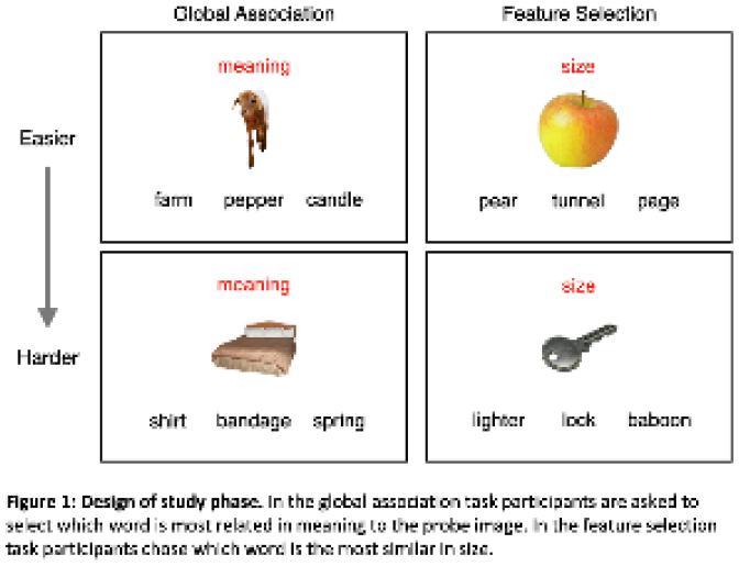
Role of midline thalamus in synchronising sleep oscillations
Introduction
Memory consolidation is a process of stabilisation of information in two stages: synaptic consolidation and system consolidation. Evidence point to sleep, and in particular slow wave sleep, as an ideal stage for memory consolidation, when information is reorganised and transferred from hippocampus to neocortex. Indeed, information transfer could occur through the temporal coupling of two sleep oscillations: the hippocampal ripples and cortical spindles. We recently showed that midline NECAB-1 thalamic neurons synchronise neocortical slow oscillations during slow wave sleep in mice. Here, we hypothesise that through synchronising cortical activity, midline thalamus mediates coordination of spindles and ripples.
Method
To investigate the role of midline NECAB-1 thalamic neurons in regulating cortical spindle activities and hippocampo- cortical coupling, we optogenetically inhibited NECAB-1 thalamic neurons while recording the local field potential signals in multiple cortices and hippocampus. Baseline sleep and NECAB-1 inhibited sleep were recorded in the same animal in an interleaved order to study how the properties of sleep oscillations and their temporal coupling were affected by NECAB-1 neuron activities.
Statistics
Bonferroni corrected Wilcoxon signed rank test with 95% confidence level was used to assess the changes in parameters when optogenetics was used in NECAB-1 inhibition group and in negative control group. Two-way ANOVA was used to compare the difference in the changes between NECAB-1 inhibition group and negative control group.
Results and Conclusion
We report that inhibiting NECAB-1 thalamic neurons during sleep reduced the peak power of cortical spindles by 6.4% ± 2.7%, with 95% confidence level (n = 8 mice, p=0.03), while the there was no effect on that in negative control animals (n = 5, p=0.63). The percentage of ripple before spindle incidence was used as an indicator of hippocampo-cortical coupling sequence, which did not show consistent changes in the 7 animals analysed, and there was no significant difference between NECAB-1 inhibition group and negative control group (p=0.26). In conclusion, NECAB-1 inhibition reduced cortical spindle power but its effect on hippocampo-cortical coupling was unclear.
Does Theta Synchronicity of Sensory Information Enhance Associative Memory?: Replicating the Theta-Induced Memory Effect
The binding of information coming from different sources is critical for associative memory. Previous animal research suggested that the timing of theta oscillations in the brain supports the binding of different information. Clouter et al. (2017) investigated this in humans by modulating the intensity of video (luminance) and sound (volume) clips so that they ‘flickered’ at certain frequencies. They manipulated the flicker frequency and synchronicity between the visual-auditory stimulus pairs in a within-subjects design. The results indicated better memory for pairs flickering synchronously at theta frequency compared with no-flicker, asynchronous theta, and synchronous alpha and delta conditions. This suggests that particularly theta synchronicity can enhance memory. Additionally, a source localization analysis with electroencephalography showed entrainment in the visual and auditory cortical regions for the video and sound clips, respectively, suggesting that flickering indeed modulated brain activity. The large effect size and the feasible applicability of the paradigm to daily life demonstrate that the replication of the method would be highly valuable. The present study aims to replicate the findings in a magnetoencephalography (MEG) experiment. Participants will be presented with the flickering stimuli during scanning. There will be four conditions: (1) 0° phase offset (synchronous) at theta, (2) 180° phase offset (asynchronous) at theta, (3) 0° phase offset at delta, and (4) no-flicker. MEG recording will allow the confirmation of oscillatory entrainment in respective cortical areas. Three pairwise comparisons between the theta 0° phase offset condition against the other three conditions will be tested according to the Bayesian evidence. A Bayes Factor (BF) of 6 in favor of the alternative hypothesis for all the comparisons will be the criteria to conclude that the theta-induced memory effect is replicable. Whereas, a BF of 6 in favor of the null hypothesis in any of the comparisons will lead to the conclusion that the effect is not replicable. The results will inform us about the relationship between associative memory and brain oscillations, as well as evaluating whether this paradigm offers an effective tool for improving memory.
Investigating meditation's potential to enhance working and episodic memory performance using EEG
Introduction
Meditation practice has been shown to act on various cognitive functions. Improvements in attention have been the most consistent research findings, whilst enhancements in working memory performance can be seen even after a single session of meditation. Given attentional control is key component of models of WM, and the link between WM and encoding/retrieval in episodic memory (EM) theoretically, meditation should also affect EM. The planned study will investigate this supposition.
Additionally, meditation can be categorised into operationally distinct types however, no studies to date have looked specifically at the potential differential effects on WM and EM. Furthermore, few studies have included neural measures to investigate meditation's effects on memory function. The planned study will investigate the effects of three types of meditation on WM and EM using both behavioural and EEG measures.
Methodology
Participants (N=60, aged 18-45) will be randomly assigned to one of four groups: focused attention meditation (FAM), open monitoring meditation (OM), loving kindness meditation (LKM) and a control sham meditation active control (SM).
Procedure: Participants will complete baseline demographic questionnaires and self-report measures of anxiety, positive and negative affect, state mindfulness and depth of meditation. EEG will then be recorded during 10 minutes resting state, followed by completion of an Episodic Memory Task and N-back Task, a meditation or mind wandering session (SM group) and a repeat of self-report measures and the Episodic Memory and N-back Tasks. Procedure protocol is presented in the image attachment included with this submission.
Analysis
For all measures, mixed measures ANOVA will be performed; meditation type X time (i.e., pre and post intervention measures). Time- frequency analysis will be performed on EEG recorded at resting state, during meditation and tasks. Theta, beta, gamma, alpha and delta power will be used for comparison analysis for resting state versus during meditation. For the EM task, theta power will be used for comparison analysis, with increased theta power as an indication of memory performance.

Proactive and Retroactive Effects of Novel Exploration and Wakeful Rest on Long-term Memory
Exploration of a novel environment seems to enhance memory for unrelated information encoded shortly before or afterwards. Behavioural tagging theory (BTT) proposes this is due to neurochemical changes induced by novelty, benefitting weak encoding occurring in close temporal proximity. BTT predicts that novel exploration may proactively or retroactively benefit memory, yet evidence of retroactive effects in humans is lacking – something the present study will address. Furthermore, interfering stimuli or effortful tasks following encoding impede memory by interrupting consolidation processes. Therefore, novel exploration may not enhance memory as much as simply resting after encoding – this study will directly compare these interventions.
The present study will adapt a virtual-reality paradigm, developed by Schomaker, Baumann and Ruitenberg (2022), to compare exploration of a novel environment, a familiar environment, and wakeful rest. All participants will undergo each intervention, on separate days. Half the participants will encode word lists shortly before each intervention (retroactive group), and half shortly after (proactive group). Participants will have same-day and next- day free recall tests, and finally a delayed remember/know recognition test for all words.
Directional, paired, Bayesian t-tests will assess whether recall is better following novel versus familiar exploration in the proactive and retroactive groups (as predicted by BTT), and whether recall is better following wakeful rest versus novel exploration (predicted to be true for the retroactive group only). Non-directional, unpaired, Bayesian t-tests will compare recall between retroactive and proactive groups following novel exploration and wakeful rest. These analyses will be repeated with delayed recall data, to investigate the effect of a delay and thus consolidation processes, and recognition test data to compare familiarity-based recognition versus recollection.
Both novel exploration using virtual-reality and wakeful rest are potential interventions for improving memory, with implications for clinical and ageing populations. Direct comparison of these will prove valuable because wakeful rest has no associated cost or set-up time, unlike virtual-reality exploration.
Conflict of interest: Co-author Prof Rik Henson is BNA President
Depotentiation of emotional reactivity using Targeted Memory reactivation during Rapid-Eye movement sleep
Introduction: Emotional reactivity has been shown to habituate across a night of sleep. This is thought to be mediated by memory reactivation in REM sleep. Such reactivation can be intentionally triggered by targeted memory reactivation (TMR) technique, in which a tone previously associated with a memory during wake is re- presented during subsequent sleep. We have previously shown that REM TMR leads to the habituation of responses to negative stimuli1. Here, we build on this prior work by repeating our original study with measures of heart rate deceleration (HRD) and brain activity.
Methods: Seventeen participants rated the arousal of 48 affective pictures paired with semantically matching sounds on a 1-5 scale. Half of the sounds were cued during REM in subsequent overnight sleep. Following a 48-hour delay, arousal ratings and HRD were recorded in a magnetic resonance imaging (MRI) scanner.
Approach for statistical analysis: Functional images were analysed to compare responses to cued and un-cued images. HRD was measured as the maximum R-R interval in the 5s following each picture onset, subtracted from the mean R-R interval during the 1.5s baseline period before each picture onset.
Results and Conclusions: TMR during REM led to a reduced activation of insula, subgenual anterior cingulate, amygdala, and orbitofrontal cortex (p <= 0.001 uncorrected). Heart rate deceleration was also significantly reduced for cued compared to un-cued items (p = 0.02). These preliminary findings support the notion that REM TMR fosters a decrease of the emotional tone over time and can lead to important implication for the early-treatment of psychiatric conditions.
Hutchison, I. C., Pezzoli, S., Tsimpanouli, M. E., Abdellahi, M. E. A., & Lewis, P. A. (2021). Targeted memory reactivation in REM but not SWS selectively reduces arousal responses. Communications Biology, 4(1), 1–6. https://doi.org/10.1038/s42003-021-01854-3

The involvement of the subiculum in reference memory: behavioural and electrophysiological data
The dorsal subiculum (dSub) represents a major output region of the hippocampus and an important conduit of spatial information to the rest of the brain. Despite this key position in the hippocampal network, the precise role of the dSub in spatial memory has yet to be established. The dSub is known to be required for spatial working memory (WM). Conversely, its role in long-term spatial ‘reference’ memory (RM) remains unclear. Here, we examined the dSub’s involvement in RM by inhibiting it during memory formation and characterising its network activity during learning.
Spatial memory was assessed using an eight-arm radial maze task (RAM) task, in which adult male rats learned the location of three static rewards, across 7 days. To test the role of the dSub in the task we infused the GABA-A agonist Muscimol into the dSub before learning on the first two days of the task. A mixed ANOVA analysis revealed an increase in RM and WM errors, compared with saline infused rats. However, dSub inhibition during learning may solely impair WM, which could, in turn, impact RM formation. To account for this, we infused Muscimol after learning in order to disrupt RM consolidation, leaving WM intact during the task. The same analysis revealed a significant effect on performance, consistent with a role of the dSub in spatial long-term memory formation.
Next, we characterized changes in dSub neuronal activity across days. Neuropixel probes were implanted in the dSub of rats that first explored a familiar arena and then performed the RAM task. Most cells maintained stable action potential wave forms across the first 5 days. At the assembly level, joint firing patterns of cells remained similar in the open field (where no learning took place), while neuronal activity reorganised between day 1 and 5 of learning on the RAM (Fishers Z).
Taken together, this data provides supporting evidence for the dSub’s involvement in long-term memory and a method for recording the same cells across multiple days.
Circuit dissection of state-dependent shifts in perception
Introduction: Learning and long-term memory formation are important but energetically costly processes. Neural strategies that could help identify the most relevant or fruitful associations are therefore likely to be beneficial for an animal’s survival. Neuronal mechanisms to support such guided learning, however, are not well established. One candidate to help instruct new memory formation is a shift in perception induced by an animal’s prior experience. Here, we used an established molluscan system (Lymnaea stagnalis) to directly probe the relationship between past memory, perception, and new learning.
Methods: Adult (3-5 month) snails were behaviourally trained using a single-trial appetitive conditioning paradigm. The central nervous systems of naïve and trained animals were removed and the activity of individual identifiable neurons of known function were recorded using intracellular electrophysiology.
Approach for Statistical Analysis Two-group statistical comparisons were performed using two-tailed t-test statistics (either paired or unpaired as stated in the text) or a Mann Whitney test or Wilcoxon signed-rank test for non- parametric data. Data with more than two groups were first analyzed using a one-way ANOVA or Kruskal-Wallis test followed by Tukey’s or Dunn’s post-hoc tests with Bonferroni sequential correction.
Results and Conclusions: Strong classical conditioning drives a positive shift in perception which facilitates the robust learning of a subsequent and otherwise ineffective weak association. Circuit dissection approaches reveal the neural control network responsible, characterized by a mutual inhibition motif. This both sets perceptual state and acts as the master controller for gating new learning. Pharmacological circuit manipulation in vivo fully substitutes for strong-paradigm learning, shifting the network into a more receptive state to enable subsequent weak-paradigm learning. Our study reveals a key mechanism for coupling past and future learning through changes in perception. We hypothesise that this serves to signal to the animal a potentially learning-rich environment, allowing new positive associations to form to cues that would otherwise be ignored.
A parsimonious neuronal mechanism for a learning-induced switch in an innate sensory response
Introduction: Sensory cues in the natural environment predict reward or punishment, important for survival. While some sensory responses are innate, they can undergo fundamental changes due to prior negative experience associated with the stimulus. However, the neuronal mechanisms underlying such behavioral switching of an innate sensory response require further investigation. We used the model learning system of Lymnaea stagnalis to address the question of how an anticipated aversive outcome reverses the behavioral response to a previously effective feeding stimulus, sucrose.
Methods: Adult (5-6 months old) snails were trained using a multiple-trial aversive conditioning paradigm (sucrose paired with a strong tactile US to the head) and tested for a conditioned response 24 hours later. Learning-induced changes in neuronal and muscle activity were recorded in semi-intact preparations containing the CNS and attached sensory regions (lips and tentacles) and feeding muscles, respectively. Photoinactivation of single identified neurons was used to test their necessity for circuit-level changes induced by the aversive training.
Approach to Statistical Analysis: Two-group statistical comparisons were performed using two-tailed t-test statistics. Data with more than two groups were analyzed using a one-way ANOVA followed by Tukey’s or Dunn’s post-hoc tests with Bonferroni sequential correction. When comparing time-dependent changes in the same group of animals under two different conditions, a two-way ANOVA with repeated measures was used, followed by paired t-tests.
Results and Conclusions: Key to the switching mechanism is an extrinsic inhibitory interneuron of the feeding network, PlB (pleural buccal), which is normally inhibited by sucrose to allow a feeding response. After multi-trial aversive associative conditioning, PlB's firing rate increases rather than decreases in response to sucrose application to the lips and the feeding response is suppressed; this learned response is reversed by the photoinactivation of a single PlB. A learning-induced persistent change in the cellular properties of PlB that results in an increase rather than a decrease in its firing rate in response to sucrose provides a neurophysiological mechanism for this behavioral switch.

Enhanced activity of parvalbumin neurons and protein phosphatase calcineurin in mesocrotciolimbic brain areas following appetitive extinction learning
Reward-associated memories play a key role not only in behaviours necessary for survival such as nutrient procurement, but also for disorders such as drug addiction and overeating. Elucidating the neural mechanisms supporting reward memory persistence and inhibition can help us better understand these disorders. In animals trained with an environmental cue (conditioned stimulus, CS) followed by delivery of food reward, subsequent exposure to the CS triggers a conditioned response in anticipation of reward delivery. Repeated presentation of the CS alone (no food delivery) weakens the conditioned response through a process called memory extinction. Previous studies have demonstrated the roles of parvalbumin (PV)-expressing interneurons and the protein phosphatase calcineurin in fear extinction learning. However, these studies examined a limited number of brain areas and the extent of activation across a wider neuronal network involved in reward and motivation have not been described. Thus, we investigated the activation patterns of several mesocorticolimbic brain areas implicated in reward and motivation including the medial prefrontal cortex (mPFC) nucleus accumbens shell and core (NAcS and NAcC) and the basolateral amygdala (BLA).
To this end, we trained rats in autoshaping by contingent presentations of a compound CS (light + lever) paired with food-reward delivery, followed by extinction learning and performed immunohistochemistry for the activity marker ‘Fos’, PV, and calcineurin.
Data revealed that extinction of food-seeking behaviour increased activation of Pv+ interneurons in the BLA and mPFC, and increased calcineurin levels in the mPFC, NAcS and BLA. We also examined whether increased motivational significance or ‘sign-tracking’ modulated these activity patterns, but we did not observe any activity changes.
Together, these results suggest that parvalbumin- and calcineurin-mediated inhibitory signalling especially across a widespread mesocorticolimbic neuronal network underlies the extinction of food-seeking behaviour in rats.
Influence of threat cue detection sensitivity on extinction in adult male rats
Introduction
Hypervigilance, a state of sensitivity to threatening stimuli, is an attentional bias symptomatic of many anxiety disorders. The development of new therapeutics for anxiety disorders is highly dependent on the characterisation of animal models of anxiety. As such, this research intends to develop and refine a rodent test of hypervigilance that could be used to screen new therapeutics.
Methods
Rats were trained in a fear-conditioning procedure, in which a tone predicted an unavoidable footshock. Ultrasonic vocalisations and freezing were measured across sessions. To test sensitivity to the threat cue, the salience was modified at test. Inter-individual differences in responses were examined in subsequent extinction of the conditioned responses. Pharmacological manipulation of NMDA co-agonists was investigated as a potential enhancer of extinction consolidation.
Approach for statistical analysis
In order to analyse the effects of group (cue salience, or drug treatment depending on the experiment) and CS presentation on freezing repeated measures two factor ANOVAs with post-hoc Sidak (cue salience) or Dunnett (compare to control drug vehicle) correction tests were performed. Investigation into the association between freezing and ultrasonic vocalisation levels were conducted using Pearson’s correlation coefficient. Statistical significance was taken to be at P<0.05.
Results and conclusions
A drop in cue salience reveals individual differences in vigilance to a threat cue. The findings suggest the ability of NMDA-receptor co-agonists to influence extinction may depend on the extent of learning in the extinction session. The relation of cue sensitivity to subsequent extinction, and reacquisition of conditioned freezing are currently being investigated.
Conflict of interest: This work was funded by Boehringer Ingelheim Pharma GmbH & Co. Funding comprised of consumables and animals (CNS Diseases Research). The funder did not have any additional role in study design, data collection and analysis, decision to publish or preparation of the article. There are no patents, products in development, or marketed products to declare.
Sleep’s Contributions to the Development of PTSD-like Symptomatology in Lewis Rats
Introduction:
Around 70% of people experience a traumatic event in their lifetime, but only a subset (around 10%) develop Post- Traumatic Stress Disorder (PTSD). Given the crucial role of sleep in the processing of daily experience, we hypothesise that the processing of a traumatic experience during the first subsequent sleep bout is critical to future outcomes and development of PTSD. This ongoing study aims to explore whether pre- and post-traumatic sleep physiology predicts development of PTSD-like symptoms in rats.
Methods
Male and female Lewis rats (n=28) were individually housed with infrared cameras installed above their homecages to continuously monitor sleep/wake behaviour. After one week, rats were exposed to a feline predatory odour (or unscented cat litter for controls) placed in their cage for 10 minutes, with a camera to monitor behaviour. They were subsequently tested a week later on the elevated plus-maze (EPM) to measure long-term anxiety, in addition to the open field test, recordings of ultrasonic vocalisations (USVs), and context re-exposure.
Approach for Statistical Analysis
Animals were grouped into those with high-anxiety, characterised by spending zero time on the open arms of the EPM, those with low-anxiety, and controls. T-tests assessed group differences in freezing during exposure and a one- way ANOVA compared total sleep across groups.
Results and Conclusions
The high-anxiety group showed significantly less sleep during the day of predatory threat exposure compared to both control and low-anxiety groups (F=6.15, p=0.013). No other days differed in estimated total sleep between groups (p>0.05). Comparisons between adjacent days within groups revealed a significant increase in amount of sleep in the high-anxiety group the day after exposure to the predatory threat (t=-6.13, p=0.004) with no other differences across days.
These changes in sleep/wake behaviour during the day of predatory threat exposure support our hypothesis that the development of PTSD-like symptoms is influenced by sleep-related memory processing within the first 24 hours.
Humans and mice integrate sensory evidence and temporal expectation in similar ways when making decisions about when to act
Introduction
Animals process a constant flow of sensory input and combine it with prior knowledge to make choices. When relevant sensory events occur around particular times, animals become capable of expecting the timing of upcoming events. Here, we investigated how humans and mice use uncertain sensory information to decide when to act, and how their decisions are influenced by their temporal expectations, by comparing the performance and strategies of humans and mice carrying out an identical behavioural task.
Methods
We adapted a visual change detection task previously developed for mice (Orsolic et al., 2021, Neuron). 10 humans (7 male & 3 female, 18-50 years old) were instructed to observe a drifting grating fluctuating in speed and to press a keyboard when they detected a sustained change in the drifting speed (temporal frequency: TF). We varied the magnitude and timing of the changes in each trial and when the changes happened in blocks: 3-8 (early) or 10-15 seconds (late) after trial onset. At the end of each trial, participants were asked to report their confidence. 5 mice carried out the same task.
Approach for statistical analysis
We compared the task performance of humans and mice using a two-way repeated measures ANOVA. Both species sometimes reported before the changes happened (early reports). We compared their proportion and average TF prior to early reports with a t-test and Pearson correlation coefficient, respectively. We also compared when they made early reports between early and late blocks using t-tests. For humans, we used a one-way repeated measures ANOVA to compare standardized confidence levels of early reports and correct hits.
Results and conclusions
We found humans and mice had similar psychometric curves and reaction times (Fig. 1A, B). While humans had lower early report rates, both species had an increase in report-triggered TF averages ~0.5 seconds before early reports (Fig. 1C, D), suggesting they continuously monitor noisy sensory evidence in similar ways. Humans were less confident when they made early reports compared to rewarded reports (Fig. 1E). Early reports happened earlier in early blocks (Fig. 1F), implying humans and mice combine priors about temporal expectation with sensory evidence to decide when to act.

Trial-resolution neural representations of behavioural strategies in a tactile reversal learning task
Animals continuously track past and current sensory and contextual information to make appropriate decisions in changing environments. Such a process is an essential component of flexible behaviour. However, how animals employ specific behavioural strategies during sensory-guided task learning and rule switching remains understudied.
We trained mice on a tactile reversal learning task and implemented a trial-by-trial Bayesian evidence accumulation model to track the probability of strategies used during behaviour. We measured multiple exploratory and explicit strategies across key task learning and reversal phases and confirmed significance using two-sided t-tests and ANOVA. To study neural representations of behavioural strategy switching, we measured functional responses from excitatory layer 2/3 neurons in the primary somatosensory cortex (S1) using two-photon Ca2+ imaging. Furthermore, we employed tensor component analysis (TCA) to reveal neural population structure and aligned single-trial behavioural strategies with TCA trial factors (using Pearson correlation and a change-point detection algorithm).
During initial task learning, mice showed an increase in the cue-driven strategy (e.g., win-stay-cue) compared to the choice-driven strategy (e.g., win-stay-choice). Following the rule-switch, mice reused learnt strategies to dynamically adapt their behaviour. Silencing of a key prefrontal brain structure, the lateral orbitofrontal cortex (lOFC), during reversal, resulted in delayed and decreased choice-to-cue strategy transition and impairments in flexible behaviour. Our neural analysis reveals several components of S1 responses related to relevant trial phases (sustained ITI activity, stimulus presentation, action and outcome) shared across mice. Several of these components correlated with specific strategies during relevant task phases of learning and following the rule-switch. Our study sheds light on strategies animals employ during flexible behaviour, their cognate neural signatures in S1, and further highlights the role of lOFC in leveraging prior knowledge supporting error-guided learning.
Brainstem control of state-dependent motor response reversal
Resting Xenopus laevis tadpoles start swimming when appropriately stimulated, but halt if the very same stimulation is applied during ongoing locomotion [1; Li lab unpublished data]. This opposing behaviour can be explained by a reconfiguration of the sensorimotor circuit, so that the same sensory pathway can exert either an excitatory or inhibitory effect on locomotion, depending on the current motor state. Studying this motor response reversal means understanding a mechanism of neuronal network reconfiguration, which could be shared by different neuronal networks and across different species.
To understand where the reconfiguration happens, brain activity will be characterised during behaviour by means of either 2-photon imaging or single-cell electrophysiological recordings, and labelling using neurobiotin for morphological reconstruction. A group of midbrain cholinergic cells are prime candidate sensors of the motor state and for the neuronal network reconfiguration [1]. Immunohistochemistry will therefore be used to locate these cholinergic cells. Paired whole-cell electrophysiological recordings and pharmacology will then be used to characterise the connectivity of these cells in relation to the locomotor central pattern generator.
3D imaging of GCaMP expressing tadpole brains will be analysed by linear regression analysis. A series of parameters descriptive of the neuronal physiology and morphology will be extracted from single-cell electrophysiological recordings and neurobiotin staining respectively, and used to run a cluster analysis to identify cell subtypes in the tadpole midbrain and hindbrain. Finally, the effects of drugs altering the cholinergic transmission will be assessed by means of paired t-tests.
1. W.-C. Li, X.-Y. Zhu and E. Ritson. Mechanosensory Stimulation Evokes Acute Concussion-Like Behavior by Activating GIRKs Coupled to Muscarinic Receptors in a Simple Vertebrate. eNeuro, 2017
Cellular and molecular mechanisms of age-related changes in a defined neuronal network encoding associative memory
Introduction: Due to the complexity of the central nervous system (CNS), studying the aging processes at the level of neural circuits and individually identified neurons is not an easy task in vertebrates. As a result, aging research heavily relies on invertebrate model organisms. One such invertebrate model is the great pond snail (L. stagnalis), which were used here as a versatile aging model to investigate the molecular mechanisms of aging and age-related memory impairment.
MM: The neural transcriptome of adult (5-6 months) snails was sequenced and assembled. The ‘molecular footprint of aging’ was investigated between young (3-4 months) and aged (>12 months) snails in the whole CNS and in an identified key interneuron of implicit learning, the Cerebral Giant Cell. Whole-mount in situ hybridization was performed for four homolog sequences to genes involved in aging and age-related memory impairment of vertebrates in the CNS between young and aged snails. To genetically modify the expression of the four chosen sequences, we applied the method of single-cell embryo manipulation and CRISPR/Cas9-mediated genetic modification.
Statistical Analysis: After normalization and quality control, the differential gene expression analysis was performed with the Limma R package. Linear models were fitted for each gene in each sample and F test was used to assess differential expression between the samples (1.2 fold change and 0.05 adjusted P value).
Results and Conclusions: Several evolutionarily conserved homolog sequences were identified in the neural transcriptome to genes involved in aging and age-related memory impairment of vertebrates. Both at the system and single-cell levels, several transcripts showed significant changes during aging which may contribute to age- related memory impairment. The expression of klotho, huntingtin, presenilin, and RbAp48 was visualized in the CNS of young and aged animals. Based on the preliminary results, the involvement of the genetic modification method can open avenues for the investigation of molecular processes underlying age-related memory decline in more detail leading to the discovery of novel mechanisms operating not just in molluscs but also in higher organisms.
This work was supported by NAP2022-I-10/2022 and OTKA138039
Researching the ability of prior experience to influence subsequent learning in early amyloid pathology and healthy aging
Previous experiences affect how subsequent learning is acquired. For example, a spatial or contextual learning that is initially dependent on N-methyl-D-aspartate receptors (NMDAR) can be become independent of NMDAR after previous acquisition of a similar task (Finnie et al., 2018). To date this phenomenon of switch in the receptor mechanism of learning has been studied in young animals. Here, we ask if this switch is affected by early aging or by an amyloid precursor protein gene knock-in (APPKI) mouse model of Alzheimer’s disease. If any impairment of the switch would be seen, we ask if improving temporal memory association (Cai et al., 2016) could rescue this function. Mixed male and female young wildtype mice, middle-aged wildtype mice, and young APPKI mice received 2 contextual fear conditionings in distinct contexts. Systemic injections of the NMDAR antagonist or saline was applied before conditioning to determine if the learning required NMDA receptors. The animal holding and transportation between the 2 learnings were either the same or different to assess if these cues were critical for the switch of learning mechanism. The time between recall of the first learning and encoding of the subsequent learning were altered to assess the role of temporal association in the switch. The effect of NMDAR antagonism was analysed via unpaired t-tests if the data adhered to normality or Mann-Whitney U if not. All data was analysed with SPSS 24 (IBM). We first replicated the NMDAR-independent second learning in young mice as seen in previous studies when the holding and transporting cues were matched between 2 learnings. If these cues were mismatched, the second learning remained NMDAR-dependent. The switch in the receptor mechanism in the second learning was not seen in middle-aged mice or in young APPKI mice. Reducing the delay between recall of the first memory and the second conditioning rescued the switch in APPKI mice. While reducing the delay between the two conditionings rescued the switch for the middle-aged mice. The holding cues of the learning event are important for inducing experience- dependent changes in NMDAR-dependent learning. Early amyloid pathology and middle-age also impact switches in learning mechanisms that reflect prior experience.

Naturalistic assessments of inhibitory control across the lifespan: Systematic review of inhibition measures in different settings
Inhibitory control is essential in our daily lives and refers to the ability to suppress or delay responses to achieve a goal. It emerges in the first year of life, rapidly developing throughout the toddler and preschool periods, before continuing to mature in adolescence and early adulthood (Whedon, 2020). Despite measurement advancements, constrained, non-naturalistic experimental paradigms remain the norm in cognitive neuroscience (Hartley, 2022; Nastase et al., 2020). However, these methods fail to account for the complexities of everyday life (Munakata et al., 2011). It is proposed that to advance our understanding of cognition we must take an ecological approach (Hartley, 2022), using naturalistic settings. Nonetheless, to date, we lack a comprehensive review of studies measuring inhibition using such methods, which are defined as methods using dynamic, contextualised, continuous and often multisensory stimuli with high ecological validity (Aliko et al., 2020).
To address this gap, we reviewed published studies using naturalistic assessments to measure inhibition across the lifespan. Studies needed to include at least one naturalistic method of assessing inhibition, such as self-reported, smartphone- or external sensor-assessed inhibition (e.g., through ecological sampling methodologies; ESM); or a laboratory assessment of inhibition capturing naturalistic behaviours (e.g., gamified tasks, virtual reality). Electronic searches were conducted from November 2022 to January 2023 using Pubmed, PsychINFO and Web of Science. The search identified 8003 studies, with 5649 currently screened for title and abstract.
Data will be extracted for several parameters, including setting, method of assessment and task characteristics. Because no fit-for-purpose quality appraisal tool could be identified for the reporting of naturalistic or real-world methodologies, two quality appraisal tools will be used: a quality appraisal tool developed by Liao et al. (2020) and adapted by Kwasnicka et al. (2021) for ESM studies, and The Appraisal tool for Cross-Sectional Studies (AXIS; Downes et al., 2016) for non-ESM studies. Findings from the review would a valuable resource for researchers interested in both the development and application of naturalistic paradigms.
Mental Health & Neuropsychiatric Disorders
Exploration heuristics during anxiety – an online study
Every day, we may choose something new randomly (random exploration) or select something new with no prior information (de-novo exploration). The link between exploration and anxiety has only been studied using trait-like anxiety questionnaires, but an experimental manipulation of anxiety could have different results. Individual differences (e.g., sex or novelty-seeking (NS) trait) also impact specific exploration strategies. Thus, we examined if anxiety manipulation in a task would influence different exploration strategies while also looking at sex, NS bias and trait anxiety (from TPQ (TPQ-NS) and STAI questionnaires). 117 healthy subjects (58 female) performed Maggie’s farm task online, which allows to review different exploration strategies, promoting exploration via the number of available choices (horizon). The threat of aversive stimuli (loud noises appearing randomly) emulated anxiety, in a between-subject design. Comparing computational models of exploration, the best-fitting model (by BIC criterion) in our data was a Thompson model with an ε-greedy element (random exploration) and a novelty bonus η (de-novo exploration). We used repeated-measures ANOVA, comparing the effect of horizon on the ε and η parameters with the anxiety category as a between-subject factor. We used partial Pearson’s correlations of ε and η derivatives (mean and standardised-difference (SD) across horizon) with STAI and TPQ-NS measures correcting for participant’s reported stress levels and anxiety category. Partial correlations analyses were repeated after splitting the data by sex. There was no between-subject effect of anxiety category on the horizon of either ε (F(1,1)=0.253, p=0.6) or η (F(1,1)=0.305, p=0.58). SD of ε was negatively correlated with TPQ-NS (r=-0.184, p=0.050) but no other partial correlation was significant. When splitting by sex, SD of ε was negatively correlated with the STAI score (r=-0.341, p=0.01) in females and the TPQ-NS score in males (r=-0.275, p=0.038). The mean η positively correlated with the STAI score (r=0.318, p=0.016) in males. While the experimentally modulated anxiety did not affect the exploration parameters, individual differences in NS and trait anxiety are suggested to affect random and de novo exploration in a sex-dependent manner.
Evaluating the long-term outcomes of a school-based intervention for depression in older adolescents
Introduction. The leading cause of disability in youth are neuropsychiatric disorders, accounting for 45% of years lost because of disabilities. More than 75% of them arise by mid-twenties, positioning adolescence as a key period for early intervention. Concerningly, symptoms of depression and anxiety are on the rise amongst 16–18-year-olds and there is a lack of interventions targeting older adolescents. Schools are a universal access point for prevention and early intervention; however, recent meta-analyses of school-based mental health programs suggest that less than a third of interventions target older adolescents and there is scant evidence of their effectiveness in the longer term.
Methods. To test the long-term clinical effectiveness of a school-based intervention specifically developed for stressed 16–18-year-olds, a long-term follow-up of a cluster randomized controlled trial (BESST Trial) will assess outcomes at 12 months and 18 months post-randomization. Primary outcome data on depressive symptoms, as well as secondary outcome data on anxiety symptoms, wellbeing, sleep and resilience will be collected between October 2022 and May 2024. A mixed-methods approach will explore quantitative differences in outcome data between students who received the intervention and controls who received the normal school provision, as well as qualitative data on the perceived usefulness of the intervention during the transition to young adulthood.
Statistical analysis. Outcomes will be analysed using multilevel linear mixed-effects models to account for the clustered structure of the data. A treatment group by time interaction term will be included to allow for extracting comparisons at both follow-up times. A random intercept model will be fitted for each school and student, and the difference between the intervention and control scores will be estimated, alongside the 95% confidence interval and p-value. Additionally, a sensitivity analysis will be undertaken using intention-to-treat principles, whereby multiple imputation will be used to assess sensitivity of the results to missing data under the missing at random assumption for students lost to follow-up. Qualitative data will be gathered in interviews and analysed using reflexive thematic analysis.
The effects of negative trait affect on decision-making under ambiguity
Introduction
Affective states can influence decision-making. For example, people in negative affective states tend to make more ‘pessimistic’ decisions than those in positive affective states, particularly in response to ambiguity. Here we aimed to show that individuals with self-reported negative trait affect make more ‘pessimistic’ decisions when faced with ambiguous stimuli. This study was preregistered here: https://osf.io/shvfr
Methods
A total of 540 participants (final sample of 509; 273 female, aged 18-73, median age = 36) were recruited to an online experiment where they completed questionnaires assessing trait anxiety, depression and pessimism, as well as taking part in a judgement bias task. The judgement bias task is a cognitive task designed to look at decision- making under ambiguity.
Statistical analysis
To determine whether demographic and questionnaire variables were significantly associated with judgement bias, we used a general linear model (GLM) containing the following independent variables: age, sex, anxiety score, depression score and pessimism score. As the questionnaire scores were highly correlated with each other we also ran three further GLMs, each containing one of the questionnaires as well as age and sex.
Results and conclusions
We found that higher trait anxiety and depression scores were significantly associated with a lower proportion of ‘optimistic’ responses to ambiguous stimuli, i.e. a ‘pessimistic’ judgement bias. Interestingly, this was not true for trait pessimism scores. We also found significant effects of age and sex on judgement bias; males were significantly more ‘optimistic’ in the task than females and ‘optimistic’ judgement bias responses significantly decreased with age.
In conclusion, self-reported trait anxiety and depression, but not trait pessimism, are associated with ‘pessimistic’ decision-making under ambiguity. Next, we plan to apply computational modelling to this dataset to investigate how negative trait affect influences underlying decision-making processes at the individual level.
Using zebrafish as an in vivo model of the human eye movement disorder Duane’s Retraction Syndrome
Introduction: The ocular motor system (OMS) consists of 3 cranial nerves interacting with 6 extraocular muscles to control eye movement. This organisation is well conserved, including in zebrafish. Miswiring of these nerves can result in Duane’s Retraction Syndrome (DRS), manifesting in limited unilateral or bilateral abduction of the eye. A number of autosomal dominant missense mutations have been identified in CHN1, encoding the signalling protein α2-CHN, leading to DRS. Previous work in our lab has demonstrated that α2-CHN plays a crucial role in axon guidance within the OMS by integrating extracellular signals to contribute to remodelling of the cytoskeleton.
Methods: In order to further investigate the role of α2-CHN in the OMS, we established three transgenic zebrafish lines harbouring either loss-of-function (LOF) or gain-of-function (GOF) mutations in chn1. We verified the presence of the mutant alleles using Sanger sequencing and expression of α2-CHN at 3 days post-fertilisation (dpf) by Western blot. Using transgenic lines expressing GFP to visualise motor neurons, combined with wholemount immunostaining of muscles and acetylcholine receptors, we imaged the OMS at 3dpf. Additionally, we used a custom-built setup to analyse the optokinetic reflex (OKR) at 5dpf. We varied a number of parameters to assess horizontal eye movement fitness.
Statistical Analysis: Analysis of the OMS anatomy was performed on at least 3 clutches of larvae, with a minimum of 19 individuals per genotype and compared using Mann-Whitney. For the OKR, at least 2 clutches were analysed, with a minimum of 17 individuals and compared using one-way ANOVA followed by Tukey test.
Results/Conclusions: Both LOF and GOF α2-CHN alleles increased defasciculation in the OMS and decreased synapse formation. In a subset of homozygous GOF individuals, we observed stalling of the oculomotor nerve. This suggests that both loss of α2-CHN or constitutively activated protein affect microtubule stability and impair axon guidance in the OMS. Consistent with these findings, we observed a reduced OKR fitness in both GOF and LOF individuals, indicating that these anatomical differences lead to functional defects in horizontal eye movements and establish these zebrafish lines as models for DRS.
A homozygous loss-of-function mutation in DRD1 causes disrupted cyclic AMP homeostasis in a patient with severe dystonia-parkinsonism
Introduction: The human dopaminergic system is vital for a broad range of neurological processes, including the control of voluntary movement. Here we report a proband presenting with clinical features of dopamine deficiency, in whom we identified a homozygous loss-of-function DRD1 variant.
Methods: Triome whole-genome sequencing and analysis was undertaken using the Illumina NovaSeq 6000 platform, Qiagen QCI Interpret Translational and Sanger sequencing. Mutant constructs for overexpression in HEK293T cells were generated. Gene and protein expression, localisation and function were assessed using qPCR, Western blotting, biotinylation, immunofluorescence, ligand binding and cAMP assays. Molecular modelling was also undertaken.
Approach for statistical analysis: Unpaired parametric Student’s t tests were used when comparing 2 data sets. Two- way ANOVAs with multiple comparisons were performed when comparing more than 2 data sets. Statistical significance was considered on both occasions where p<0.05.
Results and conclusions: A novel homozygous variant c.110C>A (p.Thr37Lys) was identified in DRD1 encoding dopamine receptor D1, in a patient presenting with severe infantile dystonia-parkinsonism, with frequent oculogyric crises, dysautonomia, hypomimia, bradykinesia and global developmental delay. CSF neurotransmitter analysis was normal. This variant was absent in the homozygous state in gnomAD with a CADD score of 27.5. Structural modelling of the mutant protein suggested an alteration in substrate binding. Although this variant did not affect protein expression and subcellular localisation, cAMP levels were significantly reduced in response to the selective D1 dopaminergic agonist Chloro APB (p<0.0001), and ligand binding was significantly reduced. Numerous D1 agonists failed to rescue the reduced cAMP levels, and the patient had no clinical response to dopaminergic therapy. Our study identifies DRD1 as a novel disease-associated gene with disruption of downstream cAMP signalling likely to affect key corticostriatal neuronal networks involved in motor control. A better understanding of the underlying molecular genetic mechanisms may facilitate future development of targeted precision therapies.
Conflict of interest: MAK is a co-founder of Bloomsbury Therapeutics.
Variants in MED27, SLC6A7 and MPPE1 in severe complex dystonia: oligogenic inheritance
Introduction: Despite advances in next generation sequencing technologies, the identification of variants of uncertain significance (VUS) can often hinder definitive diagnosis in patients with complex neurodevelopmental disorders. The objective of this study was to identify and characterize the underlying cause of disease in a family with two children with severe developmental delay associated with generalized dystonia and episodic status dystonicus, chorea, epilepsy, and cataracts.
Methods: Whole-exome sequencing was performed followed by functional characterization of candidate genes using structural homology modelling, patient-derived primary cell lines, in vitro overexpression systems, and a zebrafish model.
Approach for statistical significance: Comparisons between two group of data (functional activity of hPROT, expression and localization of hPROT and expression of GPI anchored proteins) an unpaired parametric Student’s t- test was performed using GraphPad Prim V9.0.0. Statistical significance was considered where P < 0.05.
Results and conclusion: Homozygous variants were found in: MED27 - c.839C>T (p.Pro280Leu), SLC6A7/PROT - c.1186G>A (p.Gly396Ser), and MPPE1/PGAP5 - c.985A>T (p.Arg329*). The patients had many features of MED27- related disorder, however the SLC6A7 and MPPE1 variants were investigated to determine whether these genes also contributed to the phenotype. Homology modelling predicted SLC6A7 p.Gly396Ser to result in impaired proline recognition; reduced cell-surface expression, decreased proline transport, decreased proline affinity and reduced maximal currents were observed. Zebrafish morpholino knockdown of slc6a7 revealed developmental delay and fragile motor neuron morphology that was rescued by PROT-WT, but not PROT-G396S RNA. Finally, analysis of patient fibroblasts indicated a reduction in cell-surface expression of glycophosphatidylinositol (GPI) anchored proteins, linked to PGAP5 dysfunction. We report a family harboring a homozygous MED27 variant with additional loss-of-function SLC6A7 and MPPE1 variants, highlighting the potential for blended oligogenic phenotypes.
Molecular mechanisms of the cognitive impairment associated with Duchenne Muscular Dystrophy
Introduction: Boys with Duchenne Muscular Dystrophy (DMD) suffer from a progressive muscle-wasting rendering them wheelchair bound by 12, and causing death due to cardiac or respiratory failure in the second/third decade of life. Moreover, they present with non-progressive neuropsychological abnormalities severely affecting them and their families’ quality of life, relationships, and compliance with their medications. DMD is caused by mutations in the DMD gene encoding dystrophin. Every DMD patient suffers the loss of the full-length dystrophin and has an IQ one standard deviation lower than the mean (Rae et al., 2016). Specific dystrophin isoforms are expressed in distinct brain regions and one unique full-length dystrophin (Dp427p) is present only in the Purkinje cell (Gorecki et al., 1992), the cerebella’s major output neuron, and cerebellum mediates motor control but also cognitive function, including short-term, working, and verbal memory, which are impaired in DMD. While some specific deficits, primarily altered functions of the GABA synapses were found in DMD brains, these abnormalities do not explain all the symptoms. The aim of this study is to uncover the molecular mechanisms behind the DMD neuropsychiatric impairment.
Method: Total RNA was extracted from the cerebella of five mice lacking full-length dystrophin (mdx) and the control C57BL/10 mice. At different timepoints RNA sequencing was performed on stranded libraries using Illumina. The software R was used to analyse significantly differentially expressed transcripts. Functionally enriched pathways in mdx mice were uncovered using EnrichR.
Statistical Analysis: Genes with a p-value < 0.05 and a false discovery rate < 0.1 analysed using EdgeR’s QLF tests were considered significantly altered. Enriched pathways had an adjusted p-value < 0.05 calculated by the Fisher exact test with Benjamini-Hochberg correction, and at least 3 genes altered.
Results and Conclusions: There were significant transcriptomic alterations identified in the dystrophic cerebella. The altered transcripts were found to be enriched in specific pathways and these will be discussed in detail. Current data indicates that some alterations in dystrophic cerebella contributing to the cognitive impairment in DMD is treatable.
A recurrent de novo MAX p.Arg60Gln variant causes a syndromic overgrowth disorder through differential expression of c-Myc target genes
Introduction: Cyclin D2 (CCND2) stabilisation underpins a range of macrocephaly-associated disorders through de novo mutation of CCND2, or through activating mutations in upstream genes encoding PI3K-AKT pathway components. Here we describe three individuals with overlapping macrocephaly-associated phenotypes who each carry the same recurrent de novo variant in Myc-associated factor X (MAX) and characterise the effect of the mutation on cMyc and E-box promoter binding and the transcriptome.
Methods: Patients were sequenced, following informed consent, as part of the Deciphering Developmental Disorders (DDD) study or as part of an ongoing research study in Nijmegen. Cyclin D2 protein and RNA was assessed by Western Blot and quantitative real-time PCR, respectively. Cyclin D2 degradation was assessed using a cyclohexamide assay. All modelling and molecular rendering was done using the open source version of Pymol. Circular dichroism measurements were performed on a Jasco J-810 spectropolarimeter equipped with a Peltier-type thermostat. RNA sequencing libraries were created using the Illumina Truseq library preperation kit and sequenced on a NextSeq500.
Statistical analysis: Statistical analyses were performed using Prism 9 software. Statistical calculations were performed using 3 independent biological replicates unless stated. All statistical tests performed were unpaired two- sided and a P-value of < 0.05 was considered statistically significant.
Results: Through analysis of two large sequencing cohorts, we identified three individuals who each shared the same recurrent p.Arg60Gln variant in MAX. The variant, located in the b-HLH-LZ domain, leads to increased levels of CCND2 through increased transcription, and not stabilisation of CCND2. We show the purified b-HLH-LZ domain of MAXR60Q (Max*R60Q) dimerizes more readily as a heterodimeric and specific DNA complex with c-Myc than Max*WT. Furthermore, Max*R60Q binds its target E-Box sequence with a lower apparent affinity than Max*WT resulting in an increase in transcriptional activity of c-Myc in patients carrying this mutation. The recent development of Omomyc, a c-Myc inhibitor, provides a possible therapeutic option for MAXR60Q patients, and others carrying similar germline mutations.
Variant G155D associated with autosomal dominant nocturnal frontal lobe epilepsy alters Calcium Binding Protein 4 structure-function relationship
Introduction – Autosomal dominant frontal lobe epilepsy (ADNFLE) is a partial epilepsy disorder with a usual age of onset between 10 and 14 years of age. The point mutation G155D in Calcium Binding Protein 4 (CaBP4), which is a voltage-dependent calcium channel modulator expressed in photoreceptor synaptic terminals, has been linked to ADNFLE. However, the effects of this mutation on CaBP4’s structure-function relationship are still unknown.
Methods – G155D-associated structural changes were examined using a combination of computational modelling (AlphaFold, 2Struc and DynaMut) and circular dichroism (CD). Susceptibility to protease digestion (trypsin) was determined using SDS-PAGE and densitometry analysis. The effect of G155D on calcium binding was assessed using intrinsic fluorescence spectroscopy (tyrosine).
Approach for statistical analysis – To compare wild type and mutant in both calcium-free (apo) and calcium-bound conditions, two-way ANOVA tests have been performed on trypsin digestion data and the circular dichroism data. For the analysis of calcium binding, t-tests have been performed.
Results and conclusions – Computational predictions and CD data showed that the G155D point mutation does not cause large changes to the secondary structure. However, we observed a significant gain in flexibility within close proximity to the G155D mutation and an increase of predicted intra-molecular interactions compared to CaBP4. Both DynaMut and trypsinisation highlighted that G155D destabilised CaBP4 structure, with a higher susceptibility to protease digestions, in both apo and calcium conditions. Using Calcium binding titration and fluorescence spectroscopy, we demonstrated significant differences in affinity between CaBP4 and the G155D variant. Therefore, at this stage in the investigation, we showed that the ADNFLE-associated mutation G155D significantly affects both stability and functionality of CaBP4.
The development and characterisation of cellular assays for measuring the effects of LIMK1 inhibitors
The LIM domain Kinase isoforms (LIMK1 and LIMK2) are dual-specificity kinases that regulate actin dynamics through the phosphorylation of the actin depolymerising factor/ cofilin (ADF/CFL) protein family. CFL is a crucial downstream effector in the Rho/Rac/Cdc42 signalling pathways. The dysregulation in pathway activity drives several diverse pathological processes, including the intellectual disability disorder fragile X syndrome (FXS). FXS is characterised by an abnormal synaptic morphology resulting from trinucleotide expansion of the CGG sequence in the FMR1 gene, which results in the loss of the protein expression causing dysregulation of signalling pathways that ultimately impacts ADF/CFL activity. Inhibition of LIMK1 in-vitro and in-vivo has restored synaptic morphology and alleviated associated symptoms of FXS. Herein, we describe the development of indication-specific cellular assays to profile novel LIMK1 inhibitors and facilitate the optimisation of such inhibitors in the context of FXS.
A compound series was screened and pharmacologically profiled using a NanoBRET target engagement intracellular kinase assay. HEK-293 cells were transfected with LIMK1-NanoLuc fusion vector and then a cell-permeable tracer was added to bind to the LIMK1-NanoLuc. Competitive displacement due to compound binding results in a loss of measured signal. This enabled a direct assessment of the compound effect towards LIMK1/2 and their ability to selectively bind in a live-cell system. AlphaLISA SureFire assay was used to quantify the functional target phospho- CFL following concentration-dependent inhibition. Finally, western blot analysis investigating levels of LIMK1, p-CFL and total CFL comparing control and treated cells. The efficacy of each compound was evaluated using a non-linear regression model to generate IC50 curves (GraphPad Prism 9.3.1). For protein analysis the data was normalized to the control, semi-quantified using FIJI software (ImageJ V.2.1) and a two-sample t-test was applied.
The biochemical and functional aspects of LIMK1 inhibition can be effectively assessed with reliable cell-based technology. Current compounds have demonstrated promising PK profile, with potent inhibition, good target engagement and no observed toxicity to healthy cells.
Liver X receptor-agonist treatment rescues degeneration in a Drosophila model of hereditary spastic paraplegia
Hereditary spastic paraplegias (HSPs) are a group of inherited, progressive neurodegenerative conditions caused by a length-dependent degeneration of the longest upper motor neurons. While more than 80 spastic paraplegia genes (SPGs) have been identified, many cases arise from mutations in genes encoding proteins which generate and maintain tubular endoplasmic reticulum (ER) membrane organisation. However the mechanisms by which mutations in these genes cause the axonopathy observed in HSP have not been elucidated.
This study aims to further our understanding of ER-shaping proteins through the study of novel in vivo and in vitro models, using CRISPR/Cas9-mediated gene editing to knockout the ER-shaping protein ARL6IP1, mutations in which give rise to the HSP subtype SPG61.
Loss of Arl6IP1 in Drosophila results in progressive locomotor deficits, emulating a key aspect of HSP in patients. ARL6IP1 interacts with ER-shaping proteins and is required for regulating the organisation of ER tubules, particularly within long motor neuron axons. Unexpectedly, we identified physical and functional interactions between ARL6IP1 and phospholipid transporters in both human and Drosophila model systems, pointing to a conserved role for ARL6IP1 in lipid homeostasis. Furthermore, loss of Arl6IP1 from Drosophila neurons results in a cell non-autonomous accumulation of lipid droplets in axonal glia. Importantly, treatment with lipid regulating liver X receptor-agonists blocked lipid droplet accumulation, restored axonal ER organisation, and improved locomotor function in Arl6IP1 knockout Drosophila.
Our findings indicate that disrupted lipid homeostasis contributes to neurodegeneration in this model of HSP, identifying a potential novel therapeutic avenue for the treatment of this disorder.
Selective effects of psilocin on layer V neurons in prelimbic cortex
Introduction
Recent phase 2 clinical trials have shown promise for the treatment of depression with a single dose of psilocybin. Secondary outcomes indicated that psilocybin may be more efficacious than the commonly used medication escitalopram. Despite their promise, preclinical research is still limited in this field, and a mechanistic understanding of the actions psychedelics in the brain has yet to be elucidated.
Behavioural evidence has highlighted that the medial prefrontal cortex (mPFC) plays an important role in mediating the hallucinogenic and therapeutic effects of psychedelic drugs. In vivo research has described variable changes to neuronal firing rates in the anterior cingulate cortex in response to psilocybin exposure, but how specific cell types and neuronal networks respond to these drugs is unclear.
We hypothesize that psilocybin exerts specific effects on neuronal circuits associated with the processing of affect, enabling the remodelling of synaptic connections and facilitating the reconsolidation of memories with a more positive valence. We aim to address this by assessing the electrophysiological properties of specific neuronal populations and their response to the acute application of psilocin, the active metabolite of psilocybin.
Methods
Whole-cell patch clamp recording in ex vivo brain slices of cells labelled by retrograde fluorescent tracer.
Approach for statistical analysis
Unpaired t-tests to test for significance between two groups. One way ANOVA to compare groups with one variable (i.e. concentration). Two-way ANOVA to compare effects with two variables (i.e. concentration and labelling).
Results and conclusions
We find that psilocin has variable effects on evoked synaptic currents such that amplitudes are increased, decreased, or unchanged in a cell-dependent manner. We also report that psilocin extends the decay of evoked currents in a subset of cells, which could underlie changes to bursting seen in some neurons in vivo following psilocybin administration. Our data indicate that fluorescently labelled cells projecting from prelimbic cortex to basolateral amygdala may be biased to inhibition following psilocin exposure.

Pharmacological characterizations of a psychedelic derivative 2-oxo-PCE
Introduction
Many recent studies have reported the positive effects of psychedelic and dissociative drugs in the treatment of psychiatric disorders such as depression, mood disorders, and addiction. Recent publications highlighting such therapeutic promise (i.e. the recent study by Berman et al (2020)), have led to the approval by the US Food and Drug Administration (FDA) of esketamine for the treatment of depressed patients (2019). However, there is still a need for the development of drugs with better therapeutic effects.
Synthetic psychoactive substances (SPS) have been designed to mimic naturally occurring psychoactive substances and despite the increasing volume of research into their effects, there is not definitive consensus as to whether they live up to their therapeutic potential. On the other hand, there is clear acceptance that their misuse can cause more harm than benefits. Therefore, the search for a safer and more therapeutically effective drug remains challenging.
One candidate drug is 2-oxo-PCE (a derivative of Ketamine, a N-methyl-D-aspartate receptor [NMDAR] antagonist). The work presented here was designed to pharmacologically characterise the effects of 2-oxo-PCE on NMDAR- and AMPAR-mediated fEPSPs and synaptic plasticity in area CA1.
Methods
fEPSP recordings in ex-vivo hippocampal slice preparations. The NMDAR mediated component of fEPSPs were isolated by the perfusion of antagonist drugs to block AMPA, GABAA and GABAB receptors. Under these conditions the DE50 of 2-oxo-PCME on NMDAR-mediated fEPSPs was established.
Approach for statistical analysis
t-tests used to compare between two groups and ANOVA used to compare data across groups with one factor (i.e. concentration).
Results and conclusions
We report here that bath application of 2-oxo-PCE: (a) rapidly and more potently than Ketamine reduced NMDAR- mediated synaptic transmission in a concentration dependent manner; (b) blocks the induction of Long-Term Potentiation (LTP); (c) reduces the level of Long-Term Depression (LTD) and (d) does not affect AMPA receptor mediated neurotransmission.
This dataset suggests that 2-oxo-PCE is fast-acting and more potent than ketamine. However, more research is necessary to advance the understanding of the effects of 2-oxo-PCE in the brain.
Preclinical characterisation of SPL028: a deuterated derivative of N,N-dimethyltryptamine, developing a treatment for mental health disorders
Introduction
SPL026 (N,N-dimethyltryptamine (DMT) fumarate) is a short-acting psychedelic that is being developed for the treatment of major depressive disorder. Its rapid metabolism via MAO-A correlates with an intense and transient psychedelic experience. The profound subjective effects, due to its complex receptor pharmacology, are thought to be associated with therapeutic efficacy. Prolonging the pharmacokinetic (PK) profile to give a longer psychedelic experience may be clinically beneficial for certain psychiatric disorders.
Isotopic substitution of protium (i.e. hydrogen, H) with deuterium (D) offers an approach to prolong PK due to a higher bond strength of C-D, relative to C-H, while retaining the same physical properties. D substitution of H at the α-carbon of DMT is predicted to have a direct effect on metabolism, slowing oxidative deamination by MAO-A, while retaining the same pharmacological profile of DMT.
Methods
The PK and pharmacology of SPL028 (α,α-bisdeutero-N,N-dimethyltryptamine (D2DMT) fumarate) [di-deuterated analogue of SPL026] was investigated through a series of preclinical studies. The PK was assessed in vitro via metabolic stability and in vivo following intravenous and intramuscular dosing in rats. The receptor pharmacology of SPL028 was compared with SPL026 & vehicle through in vitro receptor profiling and ex vivo receptor binding at on- target 5-HT2A & 5-HT1A receptors, and off-target D2 receptors. Finally, a drug discrimination study using rats trained to recognise SPL026 vs vehicle, assessed if SPL028 generalises to SPL026 training cue over a time course & dose range.
Statistical analysis
Where sufficient data was collected, SPL028 & SPL026 were compared using ANOVA or t-test. For ex vivo binding and drug discrimination, data were also analysed by one-way ANOVA followed by appropriate post hoc tests comparing SPL028 & SPL026 to vehicle.
Results
SPL028 exhibited prolonged PK and pharmacodynamic effects compared to SPL026. In vitro & in vivo studies found the metabolic stability of SPL028 increased relative to SPL026. The in vitro & ex vivo receptor profiles of SPL028 and SPL026 were equivalent. These findings were consistent with drug discrimination study: rats were not able to distinguish SPL028 from SPL026.
Conflict of interest: M.G., T.B., Z.J., C.R. and E.J. are all currently paid employees of Small Pharma and have owned stock in the company, S.C., I.D. and H.R., are all paid employees of contracted research organisations engaged by Small Pharma.

Comparing and profiling the effects of novel treatments for alcohol use disorder
Background and purpose: There is considerable evidence to suggest that early life adversity is central to the development of alcohol addiction in humans and rodents. There is therefore the potential to use a rodent model to study and compare treatments. The current best treatment for alcohol use disorder (AUD) is Naltrexone, however this is largely ineffective.
Drug-assisted psychotherapy in early phase 3 clinical trials has shown clear benefit to patients with AUD and has highlighted the need to elucidate the mechanism of the drugs used. In this study a comparison will be drawn between an entactogen (MDMA), against a psychedelic (Psilocybin), Naltrexone, and a control (saline). The aim is to define the neural basis of these drugs, and measure quantitatively if MDMA reduces alcohol consumption.
Methods: Naltrexone, MDMA and Psilocybin will be subcutaneously given, and their effects on alcohol drinking levels measured using an operant drug administration system. These results, in combination with behavioural assays such as the elevated plus maze and a three-chamber maze measuring sociability, will allow insight into the behavioural phenotypes that align with alcohol preference.
The integration of chronic in vivo electrophysiological recordings will allow neural network activity to be quantified in brain regions central to addiction aetiology, and subsequently map the neural signatures of drug action.
Specifically, electrodes will be inserted into the dorsal Central Accumbens, Basal Lateral Amygdala, posterior Dorsomedial Striatum, anterior Dorsolateral Striatum, Nucleus Accumbens, and the Pre-Frontal Cortex where local field potentials will be recorded.
Analysis: This experiment will use 36 Sprague-Dawley rats, half male half female, and the data will be analysed using a two-way analysis of co-variance (ANOVA). The mean alcohol intake between drug groups will be analysed to see if there is a significant reduction in alcohol intake, and to see if this effect is specific to animals who have experienced early life adversity (early maternal separation).
Implications: The neurological basis of addiction is increasingly well-defined, however, compounds need to be rationalised at a pre-clinical level to allow further clinical trials to ensue.
The impact of the atypical antidepressant tianeptine on the behavioural consequences of Early Life Adversity
Tianeptine is an atypical antidepressant and opioid receptor agonist. Chronic administration reverses depression-like behaviours in adult mice that have experienced chronic stress/corticosterone administration in a mu opioid receptor (MOR)-dependent manner. Due to the link between adverse childhood experiences and depression, we examined the impact of tianeptine on the depression-like behaviours caused by early life adversity (ELA) in mice using the limited bedding/nesting model. This model induces fragmented maternal care during a key point in murine development.
From P2-P9, C57BL6/J ELA dams and pups were placed into cages containing reduced bedding on a raised steel-mesh platform (ELA group, n=5 dams); control dams and pups were housed in standard cages (control group n=4 dams).
Dam sortie frequency was monitored. From 10 weeks of age, blinded subcutaneous injections of tianeptine (30mg/kg) or vehicle (saline) were administered twice daily for 7 days to control and ELA male and female mice (control tianeptine n=13; ELA tianeptine n=14; control vehicle n=11; ELA vehicle n=13). Additional ELA groups received tianeptine (30mg/kg) co-administered with the MOR-selective antagonist cyprodime (10mg/kg; n=13) or tianeptine only (n=12). Behavioural testing was then conducted over 4 weeks comprising social interaction and recognition memory tasks, the elevated plus maze anxiety test, and a water maze spatial learning task. All data were analysed using unpaired t-tests or repeated measures ANOVA.
ELA increased dam sortie frequency. Adult ELA mice that received vehicle showed reduced social interaction and recognition memory. Tianeptine reversed these social deficits to control levels, where there was no difference between tianeptine and vehicle groups. This effect was blocked by cyprodime co-administration. Anxiety and spatial learning were unaffected by ELA. In all tests, no sex differences were observed.
Our results indicate that a 7-day period of chronic tianeptine administration can reverse the anhedonic effects of ELA on social interaction and recognition memory in mice in a MOR-dependent manner. These findings extend previous work by demonstrating the effects of tianeptine in a model of developmental adversity and confirms the role of MORs in this process.
Does proteinase-activated receptor 2 (par2) inhibition impair pharmacologically-induced depression-like behaviour in mice?
Introduction: Depression is a major contributor to the global disease burden, with current treatments having limited efficacy in many people with major depressive disorder (MDD). Thus, it is clear that the treatment of depression is an unmet clinical need and there is a profound need to improve our understanding of the aetiology of depression so that new, better-targeted interventions can be developed. We have recently shown that PAR2 deletion impairs LPS- induced sickness behaviour and that direct PAR2 activation with AC264613 (AC), reduces locomotor activity and induces anhedonia in mice, both of which are core symptoms associated with MDD. Strikingly, AC-induced behavioural changes correlate with altered cytokine levels, with these changes being analogous to those reported in MDD. Hence, we hypothesise that PAR2 inhibition will impair depression-like behaviour in mice. To test this hypothesis, we will characterise a recently developed PAR2 antagonist, AZ8838, in vitro and in vivo to determine whether PAR2 is a novel target for developing novel antidepressants.
Methods: AZ8838 will initially be characterised in rodent primary hippocampal cultures and cultured human astrocytes using Ca2+ imaging techniques. To ensure the validity of using AZ8838 in vivo to examine depression-like behaviour, we will undertake pharmacokinetic profiling to determine the bioavailability of AZ8838 in the brain over a 24h period. Behavioural tests, including locomotor activity, sucrose preference, and the splash test, will be used to investigate whether AZ8838 impairs depression-like behaviour in mice pharmacologically induced by AC and LPS.
Power calculations & statistical analysis: Power calculations indicate that for Ca2+ imaging and AZ8838 PK profiling, with a 90% power and assuming a 5% significance level, the sample size should be at least 5 per treatment to observe a 50% change. For AZ8838 to cause a 30% change in pharmacologically-induced depression-like behaviour, the sample size should be at least 9 for each group in the behavioural experiments. Statistical analysis will be performed on treatment and control groups by performing one-way and two-way ANOVAs with Tukey’s post-hoc tests where appropriate.
The importance of dose: is poor dose selection undermining the translational validity of preclinical behavioural neuroscience?
Rationale: Animal behavioural research represents a critical component of psychiatric drug development but there has been poor translation of findings from animal studies into clinical benefits. Although many possible reasons may underlie these failures, one issue we have identified is inappropriate dose selection. Following studies in a novel translational model of affective biases in depression, the affective bias test (ABT) we observed that our maximal effective doses were surprisingly similar to clinical doses and much lower than those reported in the literature for a conventional model of depression, the forced swim test (FST). Methods: We undertook two systematic reviews to investigate the range of doses and rationale for dose selection provided in preclinical studies associated with depression research. Dose ranges were compared against animal equivalent doses (AED) calculated based on allometric scaling and receptor occupancy data where available. Results: 232 ketamine and 195 fluoxetine papers were included in the first systematic review. The median dose, 10mg/kg, exceeding the AED by 1.6-3.2x for fluoxetine and 3.2-6.5x for ketamine. Pharmacokinetic studies indicate that a 10mg/kg dose of ketamine or fluoxetine elicits plasma concentrations greater than would be achieved with clinical doses, while in pharmacodynamic studies 10mg/kg fluoxetine elicits higher serotonin transporter occupancy than clinical doses and greater engagement of 5-HT2C receptors. In the second review, 3 ABT and 150 FST studies were included for 11 drugs. Although 60.5% of FST doses were effective in eliciting a behavioural effect, median doses exceeded the relevant AED by up to 26x. ABT doses did not exceed AEDs for 9/11 drugs. 43.3% of FST papers did not justify their dose choice and the majority only referenced previous publications with significant findings as their rationale.
Conclusions: Our research found a pervasive use of inappropriate doses in preclinical depression research. High doses will result in saturating levels of receptor occupancy and engagement of off target receptors which are not relevant to the clinic and may engage different underlying mechanisms.

Conflict of interest: ESJR has received research grants from pharmaceutical companies with research programmes in psychiatry. These companies have not been involved in the work presented in this abstract.
Evaluation of the neurocognitive effects of ketamine in an animal model of depressive disorder
Depression is a mental disorder of global concern and it is caused due to psychological stress. Ketamine is an N- Methyl D-Aspartate (NMDA) receptor antagonist and reviewed to have potential as a neurocognitive drug. This study is aimed to assess the antidepressant potential of ketamine by evaluating neuro-behavioral changes associated with neurocognitive function. A forced Swim Test (FST) induced model of depression was adopted. 20 mg/kg (IV) ketamine and 20mg/kg oral fluoxetine were given. Thirty adult Wistar rats used were divided into six groups (n=5): A= Control, B= FST, C= ketamine, D= fluoxetine, E= FST + ketamine and F= FST + fluoxetine. The excised brain tissues were fixed in 4% paraformaldehyde, and prefrontal-cerebellar cortices were processed and stained. The treatment was for three days. Statistical significance was set at (p < 0.05) using the Tukey post-hoc test. This study showed that the ketamine-treated FST group had a significant increase in brain weight, improved anhedonia, reduced immobility time, and improved synaptophysin expression when compared to the FST group at p<0.05. Also, neuropathology of the prefrontal-cerebellar cortex of the FST group showed degeneration of the pyramidal and Purkinje cells, and the marked proliferation of reactive astrocytes, chromatolysis, and necrotic neurons which was averted by ketamine treatment. Hence, ketamine has a rapid antidepressant potential that acts on synaptophysin and astrocytes by the fast reversal of neuroinflammation and increased synaptogenesis that rapidly improves neurocognitive function in the prefrontal-cerebellar cortex
Exploring the effects of ketamine on resting-state functional connectivity in remitted depression
Ketamine has been shown to have rapid acting antidepressant effects that can be detected within hours of a single intravenous infusion. Studies have shown that ketamine infusions can lead to a normalisation of the functional connectivity (FC) alterations observed in depression including in the ventral striatum, amygdala, hippocampus, posterior cingulate cortex, and subgenual anterior cingulate cortex (sgACC).
Two hours after being administered ketamine or placebo in a double-blind, crossover design, resting state fMRI was acquired and blood samples collected from 33 healthy participants with remitted depression. FC was investigated using a seed-based connectivity (SBC), ROI-to-ROI, and an exploratory resting state network (RSN) approach. Blood samples were analysed for ketamine’s metabolites.
Non-parametric paired T-tests compared the ketamine and placebo conditions for each set of the SBC and RSN connectivity maps. A SBC cluster was significant if pFWE < 0.005 with Bonferonni correction. The exploratory RSN analysis had a significant cluster if pFWE < 0.05. A paired T-test compared the FC between two pairs of ROIs under ketamine and placebo with a Bonferroni corrected p < 0.025 considered significant. Correlation analyses assessed linear relationships between any significant SBC and RSN findings and metabolite levels.
The ROI-to-ROI analysis revealed significantly decreased correlation in FC between the sgACC and amygdala when comparing ketamine with placebo (p = 0.0003). The ventral-striatum seed had increased FC following ketamine when compared to placebo in both subcortical and cortical regions however these did survive Bonferroni correction.
Within the executive control RSN two clusters in the left hippocampus and right amygdala had significantly decreased FC following ketamine when compared to placebo (p < 0.05). There were no significant correlations between the observed FC changes and metabolite levels. These changes, which occurred in a remitted depressed cohort and were not the secondary effects of symptom improvements, support a model in which ketamine alters brain connectivity in areas and networks important for reward processing and emotional regulation that are implicated in depression.
Dopamine D3 receptor modulation of stimulated dopamine release in nucleus accumbens of rat brain slices in vitro
Dysregulation of dopamine signalling in the mesolimbic pathway, projecting from ventral tegmental area to nucleus accumbens (NAc) may be critical in schizophrenia, a debilitating mental illness affecting around 0.5% of the population. Drugs used to treat schizophrenia target dopamine receptors, particularly D2. However, recent interest has been directed at other receptors belonging to the D2-like dopamine receptor subfamily, including D3 receptors (D3R). Importantly D3R have relatively high density in limbic brain areas, including NAc, and may play a pivotal role in modulation of dopamine release. Changes in D3R density has been reported in schizophrenia, and drugs affecting D3 signalling may provide potential novel antipsychotic drugs. Despite this little is known about the function of D3R, particularly in modulating dopamine release. Therefore, this research aimed to characterise the effects of D3R agonists on stimulated dopamine release in NAc of brain slices taken from normal rats and rats pretreated with phencyclidine, modelling schizophrenia.
Fast-scan cyclic voltammetry was used to measure electrically-stimulated dopamine release from NAc in male and female rat brain slices (400µM). Repeated trains of pulses (10 x 1ms pulses; 800µA at 3min intervals) were delivered at high (60Hz) or low frequency (10Hz), resembling phasic or tonic firing in neurones, respectively. The effect of PD128907 (D3R agonist: 0.1, 1, 10μM) was tested by applying the drug in the superfusate during four consecutive stimulations (12min).
The release of dopamine provoked by both high and low frequency stimulation showed concentration-dependent attenuation by PD128907. At high frequency stimulation all three concentrations caused significant decreases, whereas at low frequency the changes were more variable, and were non-significant at the low concentration.
Therefore local modulation of dopamine release by D3R in NAc. Since the experiments used brain slices, the effects are mediated through local circuits, probably via autoreceptors on dopamine terminals, although effects via intermediaries cannot be ruled out. A better understanding of D3R modulation of local dopamine release may provide a stronger theoretical basis for development of potential novel treatment for schizophrenia.
Effect of alpha-4 beta-2 nicotinic acetylcholine receptors on L-DOPA-induced dyskinesia in 6OHDA rat model of Parkinson’s Disease
Introduction: Accumulating evidence suggests that CNS α4β2 nicotinic acetylcholine receptors (nAChRs) are important targets for the development of therapeutic approaches for Parkinson’s disease (PD). L-DOPA is the gold standard treatment for PD but within a few years, dyskinesia presenting as abnormal involuntary movements (AIMs) becomes a debilitating side effect of treatment. Extensive pre-clinical work using a wide variety of rodent and primate models shows that nicotine not only protects against damage to nigrostriatal and other neuronal cells, but is also antidyskinetic. This suggests that nicotine or sub-unit selective nAChR agonists may be disease modifying and antidyskinetic. Studies in several parkinsonian animal models including hemi-parkinsonian rats show that nicotine reduces L-DOPA-induced AIMs but the role of α4β2 nAChRs has not been previously investigated in dyskinesia. The aim of this study therefore, is to investigate whether α4β2 nAChRs possess therapeutic antidyskinetic potential .
Method: This study evaluated the role of A85380, an α4β2 agonist and dihydro-β-erythroidine hydrobromide (DHβE), an α4β2 competitive antagonist in a hemi-parkinsonian rat model. Rats were unilaterally lesioned with 8µg 6-hydroxydopamine then primed with 8mg/kg L-DOPA plus 15 mg/kg benserazide given orally once daily for 33 days until stable AIMs developed. The effect of A85380 at doses 0.001- 0.01mg/kg and DHβE at doses 1-10mg/kg were examined on AIMs induced by L-DOPA/benserazide treatment by blinded observers in a latin-square design.
Approaches for statistical analysis: The AIMs scores for each experiment were analysed using a two-way ANOVA (time x concentration) and p≤ 0.05 was considered statistically significant.
Results and conclusion: Treatment with 0.003 and 0.01mg/kg A85380 significantly reduces total ALO (axial, limb and orolingual) AIMs (n=12; *p≤ 0.05) but has no significant effect on locomotive AIMs. In addition, treatment with DHβE did not significantly affect L-DOPA induced ALO AIMs or locomotive AIMs. Therefore, the α4β2 agonist, A85380 was extremely potent in reducing AIMs in the rat hemiparkinsonian model suggesting that α4β2 nAChR drugs may be useful in reduction of established dyskinesia in man.
Alpha7 nicotinic receptor- mediated reduction of L-DOPA-induced dyskinesia in 6OHDA rat model of Parkinson’s Disease
Introduction: In Parkinson’s Disease (PD) induction of involuntary movements (AIMs) or dyskinesia, after long-term L-DOPA therapy is a major problem. Evidence shows that nicotine can protect against nigrostriatal damage and reduce L-DOPA-induced dyskinesia, and studies in transgenic mice suggest that deletion of α7 nAChRs leads to increased L-DOPA-induced AIMs in animals with nigrostriatal lesion compared to lesioned wild-type littermates.
Similarly, nicotinic agonists selective for the α7 nAChRs have been shown to reduce dyskinesia. The discovery of positive allosteric modulators (PAMs) such as PNU-120596 selective for nAChRs has extended the repertoire of therapeutic strategies for targeting various subtypes of nAChRs. PNU-120596 increases both agonist efficacy and open time of the α7 nAChRs channel, resulting in extended nicotinic stimulation. The aim of this study was to assess the role of α7 nAChRs selective PAMs in dyskinesia reduction using rat AIMS models, and to make direct comparison to nicotine.
Methods: Rats unilaterally lesioned with 8 µg 6-hydroxydopamine were primed with oral L-DOPA (8 mg/kg) plus benserazide (15 mg/kg) once daily for 3 weeks until AIMs were fully developed.
Approach for statistical analysis: AIMs experiments were analysed using a repeated measure two-way ANOVA followed by Dunnett’s post-hoc tests. *p≤ 0.05 was considered statistically significant, data are presented as mean ± SEM.
Results and conclusions: Treatment with 0.01 or 0.03mg/kg nicotine reduced AIMS by 33%, whereas 0.3mg/kg nicotine (n=16; *p≤ 0.05) reduced AIMS by 34% (n=16; *p≤ 0.05). Treatment with 1mg/kg or 3mg/kg PNU-120596 reduced AIMS by 34% and 44% (n=16; *p≤ 0.05); respectively. Combined treatment with 0.01 or 0.03mg/kg nicotine and 1mg/kg PNU-120596 enhanced the anti-dyskinetic effects by 57% (n=12; *p≤ 0.05) compared to each drug alone, resulting in an additive effect. These findings provide an essential role for α7 nAChRs in the anti-dyskinetic effect of nicotine. Targeting α7 nAChRs which has a rapid desensitization kinetic using PAMs, presents advantages such as reduced toxicity, improved pharmacokinetics, and better selectivity for the receptor target.
Voice Hearing in Borderline Personality Disorder: From Ongoing Thoughts to Connectivity Gradients
Auditory verbal hallucinations (AVH) in patients with borderline personality disorder (BPD) are a common yet misunderstood phenomenon. Their phenomenology and neural underpinnings are highly heterogeneous and these features are often studied in an isolated manner, with little consideration given to co-occurring mental states. To gain a more integrated understanding of hallucinatory experiences, this study assessed their relationship to experiential states identified using experience sampling. During a symptom-capture fMRI paradigm and using a button box, sixteen patients with a clinical diagnoses of BPD reported instances of AVH. After each three runs, they rated a series of statements describing their ongoing thoughts (e.g., 'My thoughts were about other people') and features of their hallucinations (e.g., 'The voices were distressing'). Dimension reduction (principal components analysis) was conducted on these data separately to reveal multivariate patterns of experience related to thoughts and AVH. First, using a series of linear mixed models, we assessed the relationship between these patterns of subjective experience. We then assessed how patterns of thought related to AVH-evoked BOLD activity, with respect to several cortical connectivity gradients (Margulies et al, 2016). At the subjective level, thoughts involving negative memories were linked to more distressing, loud and prevalent hallucinations [b = 2.07, 95% CI (0.82, 3.31), t(40.3) = 3.36, P < 0.002], while more spontaneous and intrusive thoughts were associated with hallucinations that were perceived as coming from more 'outside' the head than 'inside' [b = -2.83, 95% CI (-4.4, -1.26), t(37.2) = -3.7, P < 0.001]. At the neural level, negative memories were associated with more transmodal (e.g. default mode network) activation during AVH [b = -0.05, 95% CI (-0.09, -0.02), t(24.0) = -3.14, P = 0.004], while temporally-distant thoughts about other people were associated with more motor activation during AVH [b = -0.06, 95% CI (-0.11, -0.02), t(22.7) = -2.62, P = 0.015]. This study demonstrates that features of ongoing experience interact with subjective and neural features of hallucinations, establishing a more integrated approach to studying how ongoing mental states contribute to hallucinations.
A Picture or a Thousand Words? Characterizing the Thoughts of Autistic Adults
Autistic people may be distinguishable from non-autistic individuals in the form and content of their thoughts. Such differences potentially underlie both psychological vulnerability and strengths including creative originality motivating the need for better understanding of autistic thought patterns. In a non-clinical population of undergraduates, self-reported autistic characteristics were recently linked to the tendency to think more in words than images during a working memory task (Turnbull et al., 2020). However, it is unclear if people with a clinical diagnosis of autism express this verbal dominance or manifest other differences in thought. The current study therefore applied the same methods (multi-dimensional experience sampling of thoughts during 0- and 1-back tasks) to examine differences in ongoing thoughts between adults with and without a clinical diagnosis of autistic spectrum condition. Principle components analysis was conducted on the experience sampling data to reveal 'patterns' of thought. Using linear mixed models (where individuals' scores on these thought patterns were dependent variables), autistic individuals’ thought was characterised by less variability in the modality of thought, with a tendency to think in the form of words during both conditions [b = -0.07, 95% CI (-0.12, -0.03), t(1125) = -3.47, p = 0.001]. In contrast, the non-clinical comparison group demonstrated a preference for verbal thinking only during the harder 1-back task. In addition, autistic individuals show a decoupling between task performance and off-task social episodic thinking since, unlike non-autistic individuals, an increase in on-task thought was not associated with improvements in performance [b = -0.06, 95% CI (-0.10, -0.02), t(345.538) = -2.74, p = 0.006], a finding that may be related to differences in social processing during the off-task state. Overall, our results provide an important clinical replication and extension of previous work, highlighting that autistic individuals show a preference for consistently thinking in words despite changing task demands.
Determining and understanding the behavioural and physiological effects of drugs of abuse using zebrafish larvae
In 2020 over 350 million people abused drugs globally with approximately 50% of the population over 12 having used illicit drugs at least once. Evidently, substance abuse is a global health concern costing England approximately £19.3 billion and the United States of America (USA) $78.5 billion annually. Moreover, one in five deaths between the ages of 24 to 35 in the USA are attributed to opioid misuse with nearly 80% involving poly-drug use. This highlights the dangers substance abuse has on public health. Here, we examined the effects induced by substances of abuse by performing light/dark assays in 4 days post fertilisation zebrafish (Danio rerio) larvae. We exposed fish to abuse substances and recorded their locomotion for three hours which included 30-minutes of 5-minute alternating light changes followed by a 30-minute break in the light, repeated three times. The data was analysed by averaging fish movement in millimetres (mm) for light and dark phases and performing a two-way ANOVA on the results. The Dunnett’s post-hoc comparison test and unpaired student’s t-test were used where appropriate. Data was excluded from analysis if the fish moved 50 mm or more than the average for that time bin. Control fish (DMSO only) produced predicted hypolocomotion in light conditions and hyperlocomotion in dark conditions. Low concentration (0.5–5 mM) diazepam (Valium) exposure induced dark phase hyperlocomotion with subsequent dose-dependent hypolocomotion. These findings were replicated with other GABAA-interacting sedatives including midazolam, flunitrazepam (Rohypnol) and sodium thiopental. GHB exposure had no locomotor changes, which corresponds with the lack of GHB receptor characterisation in the fish to date. Ketamine and tiletamine reversed the light/dark response with light phase hyperlocomotion and hypolocomotion in the dark (5-50 mM). Opioid exposure induced dose-dependent hypolocomotion with our evidence indicating comparable potencies to humans. Our findings demonstrate the suitability of the light/dark assay for assessing drugs of abuse and therefore we will perform additional light/dark assays for understanding the effects of poly-substances and novel synthetic drugs.
Rapid screening for social and sleep phenotypes in novel schizophrenia model zebrafish
Schizophrenia is a complex mental health problem, which affects about one in every 100 people. There are a wide range of symptoms including changes in thinking, emotions and actions. Social dysfunction is a core element in the diagnosis and experience of schizophrenia and is often the first sign of the illness. Similarly, disturbances to sleep patterns have are also a key hallmark of schizophrenia and is thought to be predictive and perhaps causative of future disease severity.
While we know schizophrenia has a large genetic component (c.80% heritability by some estimates), the mutations identified thus far generally effect entire regions of the genome and changes expression of many genes, making the mechanism of schizophrenia pathology and behavioural changes extremely difficult to study. Recently, a large collaboration of research teams, including the UCL Molecular Psychiatry Laboratory, identified specific changes in multiple, individual genes that greatly increase the risk of developing schizophrenia. Although these changes are rare in the population, understanding the mechanism of action of these genes will be fundamental in understanding the neural basis of schizophrenia.
Here we use the zebrafish model organism and developed an efficient pipeline rapidly generating hundreds of Crispr KO animals, which are then assessed through behavioural assays to analyse larval sleep behaviour and juvenile social behaviour. Several candidate genes show a significant (corrected p<0.05, MWU), marked and heterogenous reduction in social drive, increase in anxiety-type behaviours and disturbances to sleep patterns; hallmarks of schizophrenia and other mental health disorders. In the future we aim to use imaging and neural circuit mapping and breaking to understand the neural basis for these changes in behaviour and establish stable, mutant, schizophrenia model fish lines for further study by ourselves and the wider community, providing critical insight to the mechanisms and potential therapeutic targets for mental health disorders.
Social Interaction and Pain Tolerance in Zebrafish
We cope better when we are not alone. Supportive social environments are known to have an analgesic effect, significantly reducing the perception of pain. Functional and anatomical studies in humans have indicated that the nociceptive and social circuits share overlapping areas of activity in the brain. However, the underlying circuitry as well as its molecular mechanisms are poorly understood.
To better understand how pain and social circuits modulate one another, I study Juvenile Zebrafish. Zebrafish are highly social, show stereotyped responses to noxious stimuli, and allow whole brain imaging with single cell resolution.
I have developed a new behavioural assay where fish are exposed to both social and noxious heat stimuli simultaneously. This assay has provided evidence that the mere sight of social cues, other conspecific zebrafish, is enough to drive zebrafish into swimming in higher water temperatures, which they would normally avoid. This is characterized by a significant shift of their relative position in the gradient, one-sample t-Test (n=73, p-value<0.01). These data demonstrate that social context can modulate thermal noxious tolerance in zebrafish through a descending pain modulatory pathway, similar to humans. To gain insights into the mechanisms of pain modulation in zebrafish I have generated whole-brain activity maps to identify anatomical correlates as well as specific neuronal populations involved in the pathway.
These data will provide invaluable insights on the mechanisms of pain modulation, and potentially identify new treatments for people suffering from chronic pain.
Developing the rapidly aging African turquoise Killifish (Nothobranchius furzeri) as a model for neurodegenerative disorders
The short-lived (~20 weeks) African turquoise Killifish (Nothobranchius furzeri) is an emerging vertebrate model for studying aging and age-related disorders. Commonly used vertebrate animal models typically have relatively long lifespans of up to 3.5 years while invertebrate models with short lifespans are not optimum as they do not share the same physiology.
Aging Killifish exhibit cognitive decline, cellular senescence, neurodegeneration, decline in proteostasis, telomere attrition and mitochondrial dysfunction. These hallmarks of aging together with the short lifespan, the availability of a sequenced and annotated genome and transgenic technologies make the Killifish an attractive model for aging and age-related diseases. However, not much is known about the anatomy of the Killifish brain or how it is affected during aging.
Methods
We propose to map the Killifish brain using histochemical and immunohistochemical approaches. Brains from Killifish at various timepoints, from hatching through to juvenile, matured adult and aged fish (5,8,12 and 20 weeks) will be fixed, embedded in wax and sectioned or processed for cryo-sectioning and stained. This will allow for the determination of the histology of the brain, as well as allowing for the elucidation of the changes during aging.
Alongside standard histological staining such as haematoxylin and eosin, we will perform silver and Luxol fast blue staining to characterise the changes in the organisation of nerve fibres. Thioflavin S and Congo red staining will also be performed to monitor the incidence of amyloid plaques in the Killifish brain. In addition, immunohistochemistry will be carried out to stain for Aβ40, Aβ42 and for phosphorylated tau which are markers for Alzheimer’s disease.
Approach for Statistical Analysis
Each of the stainings will be performed on sections from brains obtained from three separate fish for each timepoint. Staining will be carried out and data will be acquired from multiple adjacent sections. We will perform unpaired T-tests and ANOVA if data is normally distributed or non-parametric tests including Mann-Whitney test if non-normally distributed.
A deep learning toolbox for decoding behaviour in larval zebrafish
Introduction: The actions and behaviour of animals provide a window into how their minds work. Efficiently decoding animal behaviour from video recordings is an important problem in neuroscience, since it allows us to correlate neural activity with specific movements and therefore understand how the brain produces behaviour. The manual study and classification of thousands of frames of filmed animal behaviour is time intensive. Recent advances in deep learning have made identifying an animal’s position and pose in video recordings a more efficient process, but recognising animal movement or sequences of poses as meaningful behaviour is a fundamental tool missing from the toolkits of neuroscientists. Here, we trained and applied a neural network tool (Spatial Temporal Graph Convolutional Networks, ST-GCNs) to decode a range of zebrafish behaviours from sequences of poses.
Methods: Freely swimming zebrafish larvae (5-7 dpf) were video recorded at 330 frames per second. Zebrafish larvae coordinates were first extracted using a DeepLabCut ResNet model trained to recognise key landmarks along the zebrafish head, trunk and tail. Sequences of poses were manually behaviourally coded and used to train an ST- GCN consisting of three layers. We tested the decoder to classify three simple swim types (forward, left and right) from unseen behavioural data as well as larger datasets containing a more diverse range of behaviours. Statistical approach: To avoid bias, we ensured training, testing and validation datasets were well balanced and data was completely unseen in the test set. ANOVA was used to evaluate significance (p <0.05) in comparison of heading angles between different behaviours. Results and conclusions: The network was able to correctly classify behaviours with high accuracy (~90 %). Training the model on a larger zebrafish behaviour dataset containing 36 different behaviours demonstrated that ST-GCNs can be applied to rapidly decode a wide range of zebrafish behaviours (75% accuracy for ~200,000 swim bouts), greatly improving the efficiency of behavioural analysis.

Exploring Neuromuscular Function in Drosophila Models of C9ALS/FTD
Introduction: Amyotrophic lateral sclerosis (ALS) and frontotemporal dementia (FTD) are adult-onset, neurodegenerative diseases that exhibit significant pathological and genetic similarities. One common genetic cause of both ALS and FTD is an expanded GGGGCC (G4C2) hexanucleotide repeat in C9orf72; the disease resulting from this is referred to as C9ALS/FTD. Accumulating evidence suggests that toxic dipeptide repeats (DPRs), produced via the repeat-associated non-ATG (RAN) translation of repeat-expanded sense and antisense RNAs, drive C9ALS/FTD pathogenicity. Models of C9ALS/FTD (C9 models) have previously been well described in Drosophila, with larvae exhibiting reduced locomotion and body contractions, and reduced synaptic activity and neurotransmission at the neuromuscular junction (NMJ). This study aims to identify genetic and pharmacological modifiers of neuromuscular function in Drosophila models of C9ALS/FTD, in the hope of revealing a novel therapeutic target.
Methods: Pharmacological screens were conducted by adding various compounds to standard fly food and feeding them to C9 larvae, whilst genetic screening involved deleting a random portion of the C9 larval genome. In all cases, the behavioural phenotype in C9 larvae overexpressing G4C2 or poly-DPR constructs in the nervous system was characterised by measuring crawling speed and body contraction frequency.
Statistical analyses approach: Statistical analyses were performed using GraphPad Prism 9 software. One way ANOVA tests were used for comparisons between two or more groups, followed by a Tukey-Kramer post-hoc test.
Results and conclusions: The genetic screen identified 33 genes that, when deleted, rescue impaired larval performance, suggesting they are negative regulators of the C9-associated behavioural phenotype. Pharmacological screening revealed two compounds, nonanoic acid (NA) and 4-methyloctanoic acid (4-MOA), that are able to improve the behavioural phenotype in C9 larvae. Future work aims to elucidate the mode of action of the compounds and negative regulators that were identified. Our results are the first step in identifying novel therapeutic targets and potential treatments for C9ALS/FTD in humans.
Mutant forms of FUS are mislocated in the cytoplasm in the mouse primary motor cortex and hippocampus in vivo
Introduction: Mutations in the Fused-in-sarcoma (FUS) FUS gene have been found to cause an inheritable form of amyotrophic lateral sclerosis (ALS) or motor neuron disease, associated with cytoplasmic aggregation. FUS has also emerged as a significant disease protein in a subgroup of frontotemporal dementia (FTD). However, the role of FUS in the pathophysiology of ALS and FTD is not fully known, and most of the work has been done in vitro. This project aims to explore further the role of FUS in vivo, looking at the subcellular location of wildtype and mutated forms of FUS in neurons in the motor cortex and the CA1 area of the hippocampus.
Methods: Stereotaxic surgery was performed on 8-12 week-old mice for viral injection of wildtype FUS-GFP and two mutant forms of FUS P525L-GFP and FUS-16R-GFP reported in ALS patients. Three weeks after viral injection, mice were transcardially perfused with PBS followed by 4% paraformaldehyde (PFA). Coronal slices were prepared for immunohistochemical analysis. Preparations were co-stained with DAPI and imaged with a confocal microscope. We then determined whether FUS was localised to the nucleus or the cytoplasm of neurons. The next steps will involve electrophysiological characterisation of neurons expressing mutated FUS. Approach to statistical analysis: FIJI imaging software will be used to quantify FUS in the motor cortex and CA1 in the hippocampus. The effects will be analysed using an ANOVA with Bonferroni’s multiple comparisons correction (p value<0.05 considered statistically significant).
Results and conclusions: Wildtype FUS was localised in the nucleus of neurons in both hippocampus and the motor cortex (figure 1), as previously reported in the literature. The FUS mutants were found to be mislocated in the cytoplasm in neurons in the hippocampus and the motor cortex, further supporting the role of mutant FUS aggregation in ALS pathophysiology.

Brainwide Alterations in levels of FOXP2 in an Idiopathic Model of ASD
The gene encoding the fork-head box p2 (FOXP2) transcription factor is commonly understood to be a potential Autism Spectrum Disorder (ASD) candidate gene, reportedly expressed in multiple cortical and subcortical areas such as the globus pallidus external (GPe) and the basolateral amygdala (BLA). Morphological and functional alterations within these regions may be associated with the repetitive motor behaviours and social communication deficits observed in ASD, respectively. Though the potential role of altered FOXP2 expression in relation to alterations in these regions have not yet been fully elucidated. The aim of the current study is to explore population number of FOXP2 expressing neurons and investigate the relative concentration of FOXP2 within these neurons in the GPe and BLA of an idiopathic model of ASD (BTBR T+tf/j). To explore this, tissue from adult C57BL/6J mice (n=4) and BTBR T+tf/j mice (n=4) was processed using immunohistochemical techniques and fluorescent microscopy. The number of FOXP2 expressing neurons, and their relative levels of expression, were compared between animal model and brain region. Significant main effects were seen in relative expression levels of FOXP2 between brain regions and animal models (F(1,14)=15.30, p=.002, Eta2=.552(52.2%);F(1,14)=5.62, p=.003, Eta2=.287(28.7%)). BTBR animals displayed significantly lower levels of FOXP2 expression in all regions, compared to controls. Whereas no significant differences in the number of FOXP2 expressing neurons were noted between animal models in both regions. This may suggest that alterations in FOXP2 observed in ASD may relate to altered neuronal functioning on a molecular level, rather than a circuit level with alterations localised to specific brain regions/components of circuits, with downstream effects potentially contributing to the observed behavioural phenotype of ASD.
Adiponectin mimetic molecule shows cognitive enhancing effects in rat hippocampal neurons
Introduction
Alzheimer’s disease (AD) is a neurodegenerative disease affecting 4% of the over 65 population in the UK as of 2020, with no disease-modifying treatment currently available. Adiponectin is the most abundant adipokine secreted by the white adipose tissue and is postulated to be part of the molecular link between obesity and disorders such as AD. Adiponectin plays role in metabolism and obesity, however little is known about its involvement in brain health and AD. The present study aims to explore whether adiponectin has cognitive enhancing effects and investigate the underlying cellular mechanisms in hippocampal neurons. To this end, we used AdipoRon, an adiponectin receptor agonist which shows central activity following peripheral administration.
Methods
Hippocampi were dissected from P1-P4 Sprague Dawley rat pups and used to generate neonate primary hippocampal cultures. During the second week of in vitro culture, neurons were exposed for 15 min to the compounds indicated in each experiment and then immunocytochemistry was performed. Images were acquired using SOM Leica SP5 confocal imaging system, and surface immunolabelling of proteins of interest in hippocampal dendrites was quantified using Zeiss ZEN 3.4 Lite.
Approach for statistical analysis
All data are expressed as means ± SEM, and statistical analyses were performed using either t-test for pair-wise tests, or ANOVA on ranks for comparisons between multiple groups. * p ≤ 0.05, **p ≤ 0.01, ***p ≤ 0.001, with n representing the number of analysed dendrites.
Results and Conclusions
AdipoRon increased the surface expression of the AMPA receptor subunit GluA1 in a time-dependent (n=48; p=0.001) and dose-dependent manner (n=60; p=0.002). This effect took place via the activation of AdipoR receptors (n=48; p<0.001), with the requirement of NMDA receptor activation (n=48; p<0.001). PI3K and AMPK-dependent pathways underlie the AdipoRon effects (n=48; p<0.001), with a possible involvement of GSK-3β (n=48; p<0.001).
Moreover, AdipoRon promoted the trafficking of GluA1 (n=60; p<0.001) and phosphorylated AMPK (n=48; p<0.001) into synapses. Therefore, AdipoRon shows cognitive enhancing effects in rat hippocampal neurons, effects that are mediated by PI3K and AMPK-dependent pathways.
The effect of acute exercise on Novel Object Recognition and hippocampal BDNF in the scPCP mouse model for schizophrenia
1. Introduction
Treatments for Cognitive Impairments Associated with Schizophrenia (CIAS) are an unmet clinical need, with the severity of these symptoms remaining a strong predictor of functionality for patients. Sub-chronic phencyclidine (scPCP) treatment models CIAS in mice, causing impairments in behavioural tests including Novel Object Recognition (NOR). Here, we tested whether rescue of NOR deficits by acute exercise in scPCP mice correlates with elevated brain-derived neurotrophic factor (BDNF).
2. Methods
48 adult female C57BL/6J mice were divided into scPCP and Vehicle groups. Each of these cohorts was divided further into exercise or sedentary subgroups. Mice were dosed daily for 10 days with s.c. 10mg/kg PCP or 0.9% saline, followed by a seven-day washout. Exercised mice were habituated with home cage running wheels for 96 hours prior to testing. During testing, mice were given individual wheel access for 1 hour and then subjected immediately to an NOR task followed by tissue collection. Sedentary mice underwent the same NOR and tissue collection protocol with no prior wheel access. Hippocampal BDNF will be measured using ELISA.
3. Approach for statistical analysis
Discrimination index (DI) values were calculated using object exploration durations. Mixed effects ANOVA analysis with post hoc Sidak’s tests compared DIs between groups. One-sample t-tests compared each group’s DI to zero (chance). Two-way ANOVA will compare levels of hippocampal BDNF.
4. Results and Conclusions
There was an effect of PCP treatment on NOR (ANOVA result) and scPCP sedentary mice had significantly lower DIs than Vehicle sedentary mice. Although there was no effect of exercise (ANOVA), there was a trend for scPCP exercised mice to perform better. Thus, when compared to chance (DI=0), both Vehicle groups showed significant novelty preference, while the scPCP sedentary group did not, and the scPCP exercised group trended towards significance (p=0.05). This suggests that acute exercise may produce a modest improvement in NOR performance in the scPCP model. Ongoing analyses to investigate the link between exercise, BDNF and cognition in the model will be presented.

Characterisation of prefrontal hypofunction in the subchronic phencyclidine model
Background:
The subchronic phencyclidine (scPCP) model induces robust and long-term cognitive deficits relevant to schizophrenia. A recurrent finding in patients and scPCP is reduced parvalbumin (PV) interneuron density, a GABAergic interneuron critical for excitatory and inhibitory (E/I) balance. It has been suggested that E/I imbalance contributes to sustained behavioural and brain deficits in scPCP, although how this imbalance persists in the absence of PCP remains elusive. Here, we aimed to characterise changes in the prefrontal cortex (PFC) of scPCP-treated animals to understand the nature of chronic brain alterations and investigate how changes perpetuate pathology.
Methods:
54 female Lister Hooded rats were dosed with vehicle (0.9% saline) or PCP (2mg/Kg, bidaily, 7 days), then underwent a 12-week washout. Brains were collected, and the PFC was analysed using quantitative PCR (qPCR) (n=7), simple western analysis (WES) (n=10), and immunohistochemistry (IHC) (n=10). As the brains used for each technique were from a different animal, and we had two treatment groups, we assessed differences in normally distributed data with unpaired t-test, with Welch correction where appropriate. Not normally distributed data were analysed with the Kruskal Wallis test.
Results:
Key to E/I balance are glutamate and GABA. Here we saw reductions to VGlut1, NMDA receptor subunit 2B, PSD95 and SNAP25 (p<0.05) in scPCP-treated rats. Investigations on GABAergic pathology focused on PV interneurons. We saw increased PV mRNA and protein while cell density was reduced. There was further GABAergic dysregulation, with reductions to GAD67, the enzyme that synthesises GABA, and reduced density of perineuronal net (PNN), critical for protecting the PV interneuron. Together, these changes suggest a reduced number of impaired PV interneurons (p<0.05, t-test).
Conclusions:
The scPCP model results in a sustained and profound alteration of proteins relevant to glutamatergic and GABAergic output. These changes could result in impaired cognition and PFC hypofunction, reflecting clinical data. Studies understanding the long-term brain environment after scPCP and the effects of novel pro-cognitive compounds are ongoing.

Neurodegeneration & Aging
Inhibition of NMDA receptors rescues intrinsic neuronal excitability impaired by ApoE4
The APOE4 allele is associated with an increased risk of developing late-onset Alzheimer’s disease. How this allele confers a greater disease risk is poorly understood and no dedicated therapeutic interventions exist for APOE4 carriers. Signalling through the ApoE receptor can regulate aspects of neurophysiology, and is implicated in some actions of ketamine. We tested whether APOE genotype alters intrinsic neuronal excitability and modifies the action of ketamine, and another NMDA receptor antagonist, memantine.
Organotypic hippocampal slices were prepared from P6-8 homozygous E3 and E4 mouse pups and cultured for 21 days, before performing whole-cell recordings at room temperature. The extracellular solution was supplemented with either 20 µM ketamine, 10 µM memantine or vehicle (control) and action potential firing rate was assayed by depolarising current steps. Firing rates were fit by a linear model with genotype and drug as predictors. 95% confidence intervals for the model parameters were computed from 10,000 bootstrap resamples. Hypothesis testing was achieved using 2-way ANOVA (type III).
Depolarizing current injection into CA1 neurons of APOE3 slice cultures caused neurons to fire at frequencies upto 11.00 Hz [9.31, 12.31]. Reduced intrinsic excitability was evident in APOE4 neurons, where the firing rate was lower by -2.94 Hz [-5.06, -0.78]. While acute application of either ketamine or memantine had very little effect in APOE3 neurons (+0.08 Hz, [-2.94, +2.72] and +0.71 Hz [-0.98, +2.65] respectively), both drugs increased firing rates more in APOE4 (than in APOE3) neurons by +3.53 Hz [+0.15, +7.18] and +4.27 Hz [+1.00, +7.24], effectively rescuing firing rates to APOE3 control levels with the interaction between genotype and drug being statistically significant (F(2,70) = 3.99, p = .023).
Preliminary results indicate the effects seen on firing rates may be underpinned by APOE4-specific impairments to NaV current densities that similarly proved rescuable by both drugs. Further, these effects appear to be independent of any changes in excitatory or inhibitory synaptic transmission onto the CA1 neurons. These data demonstrate impaired intrinsic membrane excitability caused by APOE4 and its rescue with clinically relevant NMDAR antagonists.

The effect of APOE isoform on early life hippocampal activity following naturalistic behaviour
Introduction
APOE4 (Apolipoprotein E, variant E4) increases risk of late-onset Alzheimer’s disease and drives dysfunction in multiple cellular pathways, including synaptic function. However, the phenotypic effects of APOE isoforms on neuronal ensemble activity during behaviour is not known. Therefore, we investigated the effects of two major APOE isoforms (APOE3, APOE4) on hippocampal neuronal ensemble recruitment.
Methods
APOE3 and APOE4 targeted replacement mice (APOE-TR, C57BL/6J background) were tested at early to mid-life (3- 12-months) across three between subjects’ experiments (E1: N = 81, E2: N = 20, E3: N = 12).
Animals were exposed to environmental novelty to activate an acute hippocampal ensemble (home cage control were used in E1), and brains were extracted one hour later. Fixed brain sections were labelled using c-Fos immunofluorescence with neuronal markers. Subsequently, we reconstructed dendrites of c-Fos+ and c-Fos- neurons using carbocyanine dyes. In final experiments, CA1 Fos+ neurons were selectively identified using an inducible c-Fos-tTA-GFP double viral labelling approach for ex vivo current and voltage clamp recordings.
All procedures performed in line with UK Home Office Animals (Scientific Procedures) Act 1986 (revised 2013) following review by the University of Sussex AWERB; PPL: PP3378340).
Statistical analysis
Statistical approaches included general linear models (robust parameter estimation), between/mixed analysis of variance (ANOVA) using the Sidak method for post-hoc tests or simple main effects analysis when appropriate. Standard alpha criterion was set at 0.05 and standardised effect sizes were calculated. Experimenters were blind to condition at the data analysis stage.
Results and conclusions
Compared to APOE3-TR mice, APOE4-TR mice showed an increased hippocampal ensemble size at young age particularly in CA1, which was modified by sex but not associated with dendritic structural differences in c-Fos+/c- Fos- neurons. We predict that APOE4 may promote increased intrinsic excitability in APOE4 which could increase probability of ensemble recruitment. Together our results suggest APOE4 may promote a state of hippocampal hyperactivity early in life prior to gross neuropathology.

The influence of APOE isoform on rapid ‘everyday memory’ across the lifespan
APOE4 (Apolipoprotein E, variant E4) increases risk of late-onset Alzheimer’s disease (LOAD) and late-life episodic reference memory deficits. However, the phenotypic effects of APOE isoforms on rapid everyday memory across the lifespan is unknown. Therefore, we investigated the effects of two major APOE isoforms (APOE3, APOE4) on rapid everyday memory across the life span in APOE targeted replacement mice (APOE-TR).
Methods
Female and male APOE3 and APOE4-TR mice (C57BL/6J background) were trained and tested in a novel rapid everyday memory maze task across their lifespan in a longitudinal design (3-18-months, 3-month intervals, total N = 36).
The novel paradigm involved a delayed-match-to-place (DMTP) appetitive maze task in which location of a hidden food reward was shifted daily. Acquisition performance and rapid recall accuracy was measured at each timepoint.
All procedures performed in line with UK Home Office Animals (Scientific Procedures) Act 1986 (revised 2013) following review by the University of Sussex AWERB (PPL: PP3378340).
Statistical analysis
Mixed general linear models (robust parameter estimation) were used to compare APOE genotypes, sex and age on all performance measures. Standard alpha criterion was set at 0.05 and standardised effect sizes were calculated. Experimenters were blind to condition at the data analysis stage.
Results and conclusions
Mice quickly acquired task rules, showing within day learning in navigation metrics and a spatially graded preference for the target location during unrewarded probe trials as an indicator of memory recall accuracy.
APOE-TR mice demonstrated a complex set of genotype, age, and sex-dependent effects on rapid everyday learning and memory. Despite mild acquisition deficits at 3-months, APOE4 males exhibited advantages in single trial recall accuracy. At mid-late life only female APOE4 mice demonstrated deficits in rapid everyday learning, while overall single trial recall accuracy was poor. This was rescued by additional encoding opportunities but did not distinguish APOE genotypes. Together these results suggest only mild early life defects in rapid everyday learning associated with APOE4 and which may be superseded by defects in long term reference memory.
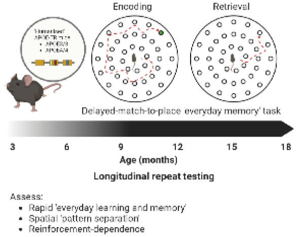
Testing attention in APOE-TR mice from young adulthood to old age
Introduction
Human studies have demonstrated that carrying the Apolipoprotein 4 (APOE4) version of the APOE gene (a risk factor for Alzheimer’s disease) can be beneficial for components of memory and attention in young adults. To begin dissecting the neurobiology underlying these cognitive benefits we tested targeted replacement mice carrying the human APOE3 and APOE4 gene in place of mouse APOE in a modified 5-Choice Serial Reaction Time Task (5-CSRTT), where mice can be trained to criteria in less than two weeks. We re-tested the mice at four timepoints to determine how APOE impacts cognition across the lifespan. We hypothesised we would see cognitive benefits in young APOE4- TR mice, but a faster rate of cognitive decline with old age when compared to APOE3-TRs.
Methods
We assessed the attention of young APOE3 and APOE4-TR mice (N=31, 8 weeks) in a modified 5-CSRTT paradigm (adapted from Remmelink et al. 2017). Using this modified paradigm allows mice to perform the task 24 hours a day. Training was completed within 10 days, and the assessment of attention took place within 3 weeks. We retested the same mice at 6, 12 and 18 months of age. Attention was tested by manipulating the stimulus duration and inter-trial intervals.
Statistical analysis
Mixed general linear model ANOVAs, with Tukey’s and pairwise comparisons as post-hoc tests, were used in each case. Standard alpha criterion was set at 0.05.
Results and conclusions
As predicted with traditional 5-CSRTT, shortening the stimulus duration (from 1s to 0.2s) caused lapse of attention, and increasing the inter-trial interval caused more premature responses in all mice. However, there were no clear genotype differences in performance either in young mice, across age, or with sex. The lack of genotype differences and deterioration in performance with age may be due to the life enrichment of repeat testing across the lifespan, or a loss of sensitivity in the translation of the task from traditional to 24 hours. We are currently rebuilding the apparatus (using open-source hardware) and coding a new modular program to give us more flexibility in testing, and a greater sensitivity to detect behavioural changes.
Neurovascular contributions to Alzheimer's disease: the effects of human APOE3 or APOE4 genotype on an early stage AD mouse model
The APOE4 allele confers an increased risk of cardiovascular disease and is the leading genetic risk factor in sporadic Alzheimer’s disease, suggesting a link between vascular dysfunction and a disease characterised by progressive cognitive decline. Amyloid β accumulation, strongly linked to AD, appears to start earlier in certain brain regions such as the hippocampus.
We crossed transgenic mice strains; those with homozygous targeted replacement of the mouse APOE gene with either the human APOE3 or APOE4 allele to mice carrying a doxycycline responsive APPSwe/Ind operon. APP expression is suppressed with doxycycline containing diet and is triggered when mice are switched to normal chow. We are using this novel model to interrogate vascular function in these mice as well as cognitive performance over a time course of Aβ accumulation.
Surgically implanted cranial windows allow us to investigate neurovascular function in either the hippocampus or visual cortex. Mice are subject to a behavioural testing protocol including Barnes Maze, Object-in-Context Recognition and Y-Maze paradigms aiming to test visual and spatial memory function. Testing is carried out at three timepoints: prior to expression of Aβ (on Dox diet), 6-8 weeks and 16-18 weeks after Dox removal.
Preliminary results indicate vascular changes over the time course associate with Aβ accumulation and may differ by APOE genotype and brain region. On the Barnes Maze APOE4 mice have slightly worse performance at 16-18 weeks off Dox compared to the APOE3 mice and the anticipated learning effect (due to repeated testing) also appears to be reduced in this group.
Overall, however, mice appear so far to have no substantial cognitive deficit even at the latest time point, suggesting they're representative of a pre or early symptomatic AD model. Thus, we can identify early vascular changes in this model, likely preceding onset of cognitive decline, opening up the potential for identification of reliable neurovascular AD predictors.
Using linear mixed modelling and Principle Component Analyses we hope to identify specific neurovascular deficits associated with Aβ accumulation and how these changes may correlate to the trend of mild cognitive decline we are starting to see in these mice.
Sex and interferon gamma signaling regulate microglia migration in vivo in the adult mouse cortex
Microglia are morphologically dynamic immune cells in the brain. Although some, but not all studies indicate these cells are capable of migration, especially in neonates or after injury, there has been little direct study in the adult cortex. Further, whether this mobility is evident in all microglia, is sex dependent, and what molecular mechanisms drive this, are not well understood. One issue with previous studies was the dependence on myeloid based reporters that do not discriminate between microglia and macrophages. Furthermore because most cells were labeled, unambiguously resolving long-distance movement in vivo was not possible, especially after injury when cells cluster. Here, we performed time lapse in vivo imaging of sparsely labelled microglia using adult, tamoxifen inducible microglia reporter mice. In the absence of injury, we find a relatively small population of microglia migrate over a 24h period. Following the induction of a cerebral microbleed (CMB), the population of mobile microglia increases significantly. However microglial mobility was dependent on sex, where microglia in male mice migrated significantly greater distances towards the bleed whereas female microglia did not. In order to understand the signaling pathways that regulate migration, we then interrogated the role of interferon gamma (IFNγ), a putative chemotaxic signaling pathway, in both male and female mice. Our data show that in male mice, stimulating microglia with IFNγ or inducible microglial knockdown of IFNγ receptor 1 stimulates or inhibits migration, respectively, whereas female microglia were generally unaffected. These findings highlight the diversity of microglia responses to injury and it’s dependence on biological sex. Further we provide insights into the signaling mechanisms that drive this unique migratory capability in the mature brain.
Drug discovery tools to examine neuroinflammation signalling in human iPSC-derived microglia
Introduction
The translation of therapies targeting CNS disorders from preclinical drug discovery to the clinic is complex, with a high attrition rate often due to a lack of efficacy. Identification of efficacious therapeutics will require the evaluation of potential new disease pathways and different mechanisms of action to uncover novel disease targets.
Neuroinflammation is proposed to play a major role in across the spectrum of neurodegenerative diseases, and neuroinflammatory mediators are receiving increasing interest as potential drug targets. Microglia, the resident immune cells of the CNS, are key mediators of neuroinflammation in the CNS. In recent years, there has been a significant effort directed towards developing human in vitro CNS cell models, with the aim to improve the understanding of disease mechanisms and to increase clinical translation, with more relevant models.
Methods and Results
Here, we demonstrate human iPSC-derived microglia are functional and respond to inflammatory stimuli using gene expression (Nanostring) and cytokine analysis. To examine the activation of neuroinflammation signaling pathways in these cells, lentiviral-based fluorescent reporters were used to evaluate the two phases of NLRP3 inflammasome activation; the priming (signal 1) and activation (signal 2) phases. First, we show NFĸB reporter activation in real-time in response to inflammation and the priming (signal 1) of inflammasome in iPSC-microglia. A second reporter assay examining the formation of ASC-specks was employed to analyse activation of NLRP3 inflammasome. Together, these innovative tools provide an opportunity for novel drug discovery and to further understanding of microglia in neuroinflammation and CNS disease.
Statistical Analysis
Where quantitative data is shown, we used Anova with post-hoc tests to analyse differences between conditions. For analysis of gene expression data, the differential expression analysis calculated log2 fold change and adjusted P values between control and treated conditions.
Cellular glial models for drug discovery in neurodegeneration
Introduction
Neuroinflammation, mediated by microglia and astrocytes, contributes to pathological processes in neurodegenerative diseases including Alzheimer’s disease (AD). Exposure of microglia and astrocytes to disease relevant cellular insults, leads to their activation and release of neuroinflammatory mediators that promote neurodegeneration. Modulation of glial cell function represents a viable therapeutic approach in AD. Drug development for glia-targeted therapeutics is limited by a lack of disease-relevant models that recapitulate important aspects of disease biology and are suitable for high-throughput drug discovery. We have developed and characterised quantitative and reproducible primary rodent microglia, astrocyte, and triculture systems for pharmacological studies. We are now testing disease-relevant insults with the goal of having a physiologically relevant glial cell models for drug discovery.
Methods
We previously established a suite of functional assays with glia and neurons including microglial phagocytosis and cytokine production (qPCR) and neuronal electrophysiology in the presence of glia (MEA). We characterised the cellular responses to proinflammatory factors i.e. lipopolysaccharide (LPS) and proinflammatory factors TNF, IL-1α, and C1q (“TIC”) in these assays. We are building on these experiments by characterising glial and neuronal responses to other disease-relevant challenges, such as amyloid beta (Abeta) exposure.
Approach for statistical analysis
We aim to compare the response of glial cells stimulated with disease-relevant insults and appropriate controls using either t-test or One-way ANOVA (or their nonparametric equivalents), based on the data distribution predetermined by Shapiro-Wilk normality test and number of conditions tested.
Results and conclusion
Our preliminary results suggest that glial cells show various physiological responses to a variety of insults. As an example, treating primary rat microglia with synthetic S26C Abeta 1-40 dimer caused no change in cytokine expression, morphology or phagocytosis in microglia. We are in the process of exploring glial cell response patterns with human brain extracts from disease and control brains.
Astrocyte assay development for target validation of astrocytic cAMP signalling in Alzheimer’s disease
Introduction
Astrocytes play a fundamental role in pathological processes of neurodegenerative diseases, including neuroinflammation, impaired glutamate uptake, reduced neurotrophic support and defective metabolism. Activation of cAMP signalling in astrocytes elevates glycolytic rate, increases glutamate transporter and neurotrophic factor expression, and suppresses the immune response. Molecules that regulate astrocytic cAMP signalling are therefore potential therapeutic targets for neurodegenerative diseases such as Alzheimer’s disease (AD).
Methods
To aid astrocyte-targeted drug discovery, we developed an astrocyte-focused in vitro platform to be used for target validation and drug screening. Using primary rat and human iPSC-derived healthy and familial AD astrocytes in monoculture and in co-culture with neurons, we have optimised pharmacological assays including cAMP, Ca2+, RNA- seq, multi-electrode array, and seahorse assays which can quantitatively and reliably measure changes to astrocyte function and neuronal activity.
Approach for statistical analysis
qPCR and seahorse data analysed using one-way ANOVA; Western-blot data analysed with Mann-Whitney test; MEA data analysed using unpaired T-test.
Results and conclusion
We validated the potential of astrocytic cAMP signalling as a therapeutic target for AD in our astrocyte assays. We showed that the activation of adenylyl cyclase using forskolin or stimulation of a Gs-coupled GPCR using a tool compound induces cAMP signalling in rat and hiPSC-derived astrocytes. Alongside increased cAMP levels, application of forskolin and a tool compound led to elevated glycolytic rate, increased glutamate transporter expression, and downregulation of pro-inflammatory pathway genes in astrocytes. Finally, addition of a tool compound in rat primary astrocyte and neuron co-culture induced an acute increase in neuronal excitability.
In summary, we showed that cAMP activation had positive effects on metabolism, glutamate transporter levels and inflammatory gene expression, providing evidence that activation of astrocytic cAMP signalling may translate to therapeutic benefit in AD. Our astrocyte platform can be applied to future novel drug discovery programs targeting astrocytes.
Evaluation of Serum/Glucocorticoid Regulated Kinase 1 as a potential target for treatment of dementia
Introduction
Neuroinflammation is a hallmark of neurodegenerative diseases. Genome wide association studies (GWAS) have identified an enrichment of microglial genes associated with altered risk of Alzheimer’s disease (AD) which could be potential new targets for the treatment of AD and positioning experimental studies of microglial cells at the centre of exploratory drug discovery programs for neuroinflammatory targets. Serum/Glucocorticoid Regulated Kinase 1 (SGK1), has emerged as a potentially interesting target for AD being upregulated under certain pathophysiological conditions, including inflammation. SGK1 activates a range of ion channels, transporters, enzymes, and transcription factors such as NfkB, thus regulating cytokine production, and, indirectly, tau hyperphosphorylation. SGK1 is upregulated in AD brain, Tau P301S mice and 5xFAD mice. In this target validation study, we investigated the expression of SGK1 and developed an in vitro model of inflammation in microglia to look for effects on SGK1 with the goal of validating SGK1 as a putative therapeutic target for drug discovery.
Methods
We confirmed SGK1 expression levels in various microglial data sets and developed a rat primary microglial cell culture system in vitro that models different physiological states of microglia: serum-free microglia and classical serum-containing media. We miniaturised and automated an inflammation assay (based on LPS addition) and multiplexed our cytokine secretion assay (MSD technology) and qPCR endpoints to generate target validation data for SGK1.
Approach for statistical analysis
For rtqPCR and MSD data ACTB expression was used for normalization. N= 3 per condition, one-way ANOVA Bonferroni multiple comparisons; mean ± sem.
Results and conclusions
We integrated our high-throughput microglial platform using acute LPS stimulation with compound administration to measure effects of SGK1 modulation with small molecules. Pre-treatment with an SGK1 inhibitor reduced cytokine production dose dependently, recapitulating the effect described in Kwon et al., 2021 in glial cultures and in vivo.
Evaluation of the modulation of the inflammatory response provided key information on how targeting SGK1 with a therapeutic may impact inflammation associated with AD.
NETSseq platform identifies THIK1 as a target to modulate neuroinflammation in Alzheimer’s disease
Neuroinflammation is a common underlying pathological feature of many neurodegenerative diseases. Thus, understanding the gene expression profile of microglia and how these change in disease will provide critical insight. Cerevance’s proprietary Nuclear Enriched Transcript Sort Sequencing (NETSseq) platform allows for deep RNA-seq expression profiles (~12,000 genes detected per cell type) of distinct purified cell types (> 60 to date), including microglia, from human post-mortem brain tissue. To better understand the temporal changes that occur during Alzheimer’s disease (AD), machine learning approaches, including a trajectory-based analysis to map high- dimensional gene expression data into a one-dimensional quantity called “pseudotime”, have been utilized.
Differential expression analysis against pseudotime has identified genes that are associated with disease progression including KCNK13, a gene coding for the 2-pore K+ (K2P) channel THIK1. NETSseq demonstrated highly specific expression of KCNK13 in microglia, which progressively increases with AD progression. These data and subsequent literature evidence have suggested a role for THIK1 in modulating the NLRP3 inflammasome.
Herein, the discovery and pharmacological characterisation of C101248, the first potent and selective small-molecule inhibitor of THIK-1 is described. C101248 showed a concentration-dependent inhibition of both mouse and human THIK-1 (IC50: ~50 nM) and was inactive against K2P family member TREK-1, the closest homologue to THIK-1, TWIK-2 and Kv2.1. THIK-1’s constitutive activity was confirmed by patch clamp electrophysiology and blocked by C101248 at a similar potency. In isolated microglia C101248 prevented NLRP3-dependent release of pro-inflammatory cytokine IL-1β to the same degree as a genetic depletion of the channel. C101248 showed no reduction of IL-1β when treating THIK-1-depleted microglia, underscoring its selectivity and mode of action.
In conclusion, NETSseq demonstrated the ability to identify novel microglia targets that are involved in the pathological regulation of neuroinflammation. Targeting these genes may be a means of modulating microglial function and ongoing neuroinflammatory processes in disease, as demonstrated by the KCNK13 / THIK1 example described herein.
Assessing neuroinflammatory signalling in a human tissue model of traumatic brain injury
Background
Despite in vitro and in vivo research into the mechanisms underlying traumatic brain injury (TBI)1, few agents have successfully translated to clinical practise. We demonstrate a human tissue, explant model of TBI and its use to identify the neuroinflammatory pathway instigated by the molecule High Mobility Group Box Protein 1 (HMGB1) as a potential therapeutic target in TBI.
Methods
Normal human cortical tissue samples were collected from appropriately consented patients undergoing resections of non-traumatic lesions, such as sclerosis or neoplasia. Tissue was plated as 3D cultures, and after ten days in vitro underwent weight-drop injury to simulate neurotrauma. Cells were fixed 1-7 days hours post-injury and immunostained for DAPI (total cells), IBA1 (Microglia), NG2 (oligodendrocyte precursors), neurones (TUJ1) and HMGB1. HMGB1-axis genes were assessed in control and injured cultures 1-7 days post injury via qPCR. Cell death up to one week after injury was quantified using ATP and LDH cytotoxicity assays. Cell death was also assessed after control and injured cultures were treated with the HMGB1-signalling antagonist FPS-MZ1.
Results
Microglia and NG2 cell counts were significantly increased in injured cell cultures, whereas neurone counts were significantly reduced. Localisation of HMGB1 within NG2, microglial and neuronal cells was statistically significantly greater following injury compared to control cultures. HMGB1 and its associated downstream neuroinflammatory proteins were significantly upregulated up to seven days post-injury. Cell death was statistically significantly increased after injury, and continued to accrue up to seven days post-injury. Cell death was not affected by anti- HMGB1 treatment in control cultures, whereas cell death was significantly reduced when injured cultures received the treatment.
Conclusions
We demonstrate use of a human tissue explant model of TBI to identify co-localisation of HMGB1 within NG2 cells following simulation of traumatic brain injury. This work supports recent animal tissue data suggesting that HMGB1 pathways are upregulated following TBI, and that it may influence human brain cell responses to trauma2. The reduction in cell death post injury after treatment with an HMGB1-antagoist in vitro highlights this signalling pathway as one of interest for potential clinical drug development for traumatic brain injuries.
How does focused ultrasound modulate brain microglial function?
Introduction
Focused ultrasound (FUS) has gained much attention as a non-invasive technique that uses acoustic energy to modulate neuronal activity. However, the mechanisms by which FUS affects the functions of glial cells, specifically microglia, are poorly understood. Microglia play a pivotal role in protecting the central nervous system (CNS) against pathogens and inflammation. They exhibit diverse functions, including surveillance and phagocytosis and are implicated in neurological and neurogenerative diseases. Previous reports have shown FUS to activate microglia in mouse models, however, the cellular and molecular mechanisms are unknown [1]. This study aims to understand and evaluate the role of FUS stimulation of microglia and how it influences their functions in homeostatic and inflammatory states.
Methods
Experiments will be performed using in vitro BV-2 cell lines. The methodology we will employ to understand the effect of FUS on microglia includes cell cytotoxicity using lactate dehydrogenase (LDH)-cytotoxicity assay, quantifying microglial inflammatory activation by enzyme-linked immunosorbent (ELISA) and phagocytosis assays. Bulk RNA- sequencing will be performed to investigate changes in gene expression. To test these factors in an inflammatory state, cells will be first treated with lipopolysaccharide (LPS).
Approach for statistical analysis
We will test the underlying mechanisms of FUS in modulating microglial function by using one-way analysis of variance (ANOVA) with Dunnett’s post-hoc test to compare means from multiple conditions against the control. When comparing the difference between two conditions student’s t-test will be used accordingly. To ensure statistical significance and reproducibility, experiments will be performed in triplicates. Statistical analysis will be conducted using GraphPad PRISM software with a p-value <0.05 considered statistically significant.
[1] Bobola et al. (2020). Brain Stimulation, 13(4), 1014–1023.
The role of CD44 in neurodegeneration
Prion diseases are prototypic protein-misfolding neurodegenerative disorders. Prion disease are infectious, progressive and invariably fatal. The earliest neuropathological changes of prion disease include synaptic & dendritic spine loss and reactive astrocytosis concurrent with the accumulation of misfolded prions within the central nervous system. CD44 is a multifunctional protein that has roles in dendritic spine & synaptic morphology and plasticity and also astrocyte morphology and reactivity. During prion disease increased expression of CD44 has been identified as a specific marker of heterogeneity within prion-disease reactive astrocytes. Both prion strain and host specific patterns of CD44 upregulation are observed commencing half-way through the disease incubation period.
To determine the role of CD44 in neurodegeneration we challenged groups of both male and female CD44 knockout and C57Bl/6 control mice intracerebrally with ME7 prions. CD44 knockout mice are viable but experience deficits in spatial learning and memory. Mice were subject to weekly automated CatWalk gait analysis and clinical scoring for symptoms of prion disease. Uninfected mice of both genotypes were also monitored for comparison. Pre-clinical and terminal brain samples will be collected for routine histopathological, protein (Western) and gene expression (RT- QCPR) analyses to determine the impact of the absence of CD44 on prion neuropathogenesis, prion disease incubation period and gait aberration. Statistical analyses will be performed by one or two-way ANOVA where appropriate.
At the time of abstract submission these experiments are still in progress, however the results should reveal if CD44 has a role to play in the maintenance of synaptic integrity and astrocyte reactivity during prion-induced neurodegeneration. A greater understanding of the initial events and their molecular regulation during neurodegeneration will direct novel therapeutic approaches to modulate astrocytes and prevent synaptic loss across a wide array of neurodegenerative conditions.
The Neuroinflammatory Environment in Depression of Alzheimer’s Disease: A Human Post-Mortem Study
Introduction: Depression in those with Alzheimer’s disease (AD) results in a worsened prognosis than either condition alone. Comorbid depression and AD complicates both diagnosis and treatment, with current limited options highlighting the need for further understanding and research for biomarkers and therapeutic targets in this combination of diseases. Neuroinflammation has been implicated in the pathophysiological progression of both depression and AD, but little is known about the neuroinflammatory environment when these conditions occur simultaneously. This study aims to explore the neuroinflammatory environment in AD brains comparing those with and without depression.
Methods: Post-mortem brain tissue from cases with AD who also suffered from depression (n=20) and AD cases with no history of psychiatric illness (n=24) were studied for a range of inflammatory markers. Markers of microglial function, Iba1, P2RY12, CD64 and CD68 were assessed by immunohistochemistry. Endothelial inflammatory markers ICAM-1 and VCAM-1 were measured by ELISA, and brain cytokine levels of IFN-γ, IL-1β, IL-2, IL-4, IL-6, IL-8, IL-10, IL- 12p70, IL-13, and TNF-α were measured using multiplex assays. Multiple linear regression analysis was used to assess differences between groups, where possible. If variables could not be transformed to a normal distribution, then Mann-Whitney test was used.
Results: No significant differences were found between AD cases with or without depression for any of the markers of microglial function or endothelial inflammation. However, a significant increase was found in the level of anti- inflammatory cytokine IL-4 in the depression and AD cases compared to the AD only controls (P=0.037). No other cytokines had significant differences between groups.
Conclusion: This study suggests a more anti-inflammatory environment in the brains of those with depression in AD compared to AD alone. This may represent reduced inflammation, or it could represent a compensatory anti- inflammatory response to the presence of depression-related changes in AD. This raised anti-inflammatory cytokine goes against the general evidence in the literature for increased inflammation in depression and AD, warranting further investigation into potential mechanisms involved.
Investigating changes in interneurons and perineuronal nets in a rodent model of alpha-synucleinopathy
Introduction: Dementia with Lewy Bodies (DLB) is characterized by the accumulation of aggregated insoluble α- synuclein (α-syn) protein. Patients with DLB exhibit impaired higher cognitive functions which involve the prefrontal cortex (PFC). Parvalbumin-expressing (PV) interneurons, which are critical for normal cognitive function, are often surrounded by a protective extracellular matrix –the perineuronal nets (PNNs). Recent evidence suggests changes in both PV cells and the PNN could play important roles in neurodegeneration.
Methods: Using transgenic mice that express human mutant (hA30P) a-syn we are investigating the impact of α-syn pathology on PV interneurons and PNNs in the PFC at different disease stages in A30P compared to control mice. We used immunofluorescence (IFC) in coronal frozen sections containing PFC from paraformaldehyde fixed brains of male and female A30P and age-matched C57BL/6 wild type (WT) mice. IFC was conducted against PV to investigate the interneurons and lectin and avidin staining was performed to detect PNNs. Sections were imaged using fluorescence confocal microscopy and densitometric analysis was conducted using FIJI software. We compared PV and PNN integrated density between medial PFC (mPFC) including anterior cingulate, prelimbic and infralimbic cortices and lateral orbitofrontal cortex (LOFC) in both A30P and WT mice.
Results: In aged (10-12 months old) A30P and WT mice most PV cells in mPFC and LOFC were surrounded by PNNs. Integrated density measurements were significantly higher for both PV immunofluorescence (n=4 WT, n=5 A30P; p<0.05, unpaired t-test) and PNN staining (p<0.05, unpaired t-test) in LOFC compared to mPFC in both WT and A30P mice. Interestingly, we also found significantly reduced integrated density of PV expression only in the mPFC in the aged A30P mice compared to WT mice (n=4 WT, n=5 A30P; p<0.05, unpaired t-test).
Conclusions: We found significant regional differences in PV cell and PNN distribution across PFC regions in both WT and A30P mice. Importantly, we also found that abnormal human a-syn expression in A30P mice caused a reduction in PV expression which may represent a downregulation of expression or a loss of PV neurons or processes. This may have functional implications in this model of
Evidence for prodromal neuroinflammation in a rodent model of alpha-synucleinopathy
Toxic aggregation of the presynaptic protein alpha-synuclein is a key feature of several neurodegenerative diseases, including dementia with Lewy bodies (DLB) and Parkinson’s disease. Recent studies suggest neuroinflammation early in the disease may be a prodromal change occurring in DLB patients. Transgenic mice expressing mutant human alpha-synuclein (hA30P) show early increases in cortical network hyperexcitability. The study aims to investigate whether this early abnormal cortical network activity may be linked to neuroinflammatory changes occurring prior to the onset of cognitive dysfunction.
Fixed, frozen sections from the hippocampus of 2-4 month old hA30P and age-matched wild-type (WT) mice were immunofluorescently stained for GFAP, Iba-1, and c-Fos, which reveal reactive astrocytes, reactive microglia, and newly upregulated neuronal activity, respectively. Sections were imaged and densitometric analysis of the images performed using FIJI software. A comparison of Z-scores of percentage area stained was conducted using unpaired Mann-Whitney t-test.
Results showed an interesting differential laminar distribution of astrocytes and microglia in the CA3 region of hippocampus. Very few GFAP+ astrocytes were localized within stratum pyramidale, while reactive microglia were mainly localized within it. There was a significant increase in % area of CA3 region of the hippocampus of hA30P occupied by reactive astrocyte and microglia compared to WT (GFAP+ astrocytes 27 sections/8 WT mice and 27 sections/8 hA30P mice, P<0.0001 and Iba-1+ microglia 29 sections/8 WT mice and 27 sections/8 hA30P mice, P<0.05). In addition morphological changes including an enlargement of soma with elongation of processes of reactive astrocytes and enlargement of reactive microglia soma and decreased length of the processes in hA30P mice in comparison to WT. The results also showed a significant decrease in c-Fos+ neurons/area in hA30P compared to WT mice (15 sections/4 hA30P mice, 15 sections/4 WT mice, p<0.011).
We have found evidence of early neuroinflammation in the hippocampus in young male hA30P mice at 2-4 months of age with an increase in gliosis. This is accompanied by down-regulation of c-Fos expression consistent with chronic network hyperexcitability.
Patient-based modelling of alpha-synuclein pathology in the murine brain
Parkinson’s disease (PD) is a neurodegenerative disorder characterised by motor dysfunction and α-synuclein (α-syn) pathology. Patients can however show diverse symptoms of variable severity, leading to an uncertain prognosis. To study PD, toxin-based (e.g. MPTP) mouse models of PD can be used to induce motor dysfunction, but lack the disease aetiology of PD. In contrast, seeding models of PD are based on the prion-like function of α-synuclein. When pathological α-syn is injected into the brain of a C57Bl6J mouse, it can act as a template for murine α-syn, causing spreading, aggregation and neuronal dysfunction, closely mimicking the actual disease processes.
Here, we investigated whether differential synuclein profiles might correlate with individual disease progression.
To test this hypothesis, we designed a new seeding model of PD based on human α-syn infusion into the dorsal striatum of 12-week-old male C57Bl6J mice. Α-syn derived from post-mortem tissue from four male PD patients using a sucrose-gradient based extraction to identify high vs low amounts of toxic and aggregated α-syn using western-blotting. A randomized, blinded study was set up to assess behavioural and molecular changes in this model six months post-striatal infusion in a cohort of 82 mice, where mice received α-syn from a patient with high or low amount of α-syn (n=12 per patient) or one of two control buffers (n=6 per patient and n=10).
Presence of phosphorylated α-syn with IHC (DAB, pSer129, 1:5000) in the striatum as well as other areas, such as the S1BF area in the cortex, highlighting a successful introduction of toxic α-syn stable for 7 months as well as seeding activity of α-syn. Quantitative analysis of western blots showed an increase of phosphorylated α-syn (pSer129, 1:5000, Mann Whitney test) in the groups that received patient α-syn. Interestingly, further analysis of inflammatory markers showed differences in astrogliosis between the different patient groups (GFAP, 1:1000, Kruskal-Wallis test). Analysis of behavioural tests such as home cage activity, nestbuilding and balance beam (2-way ANOVA for all tests) showed none to minor changes in the behaviour. Home cage activity was reduced during the dusk and dawn period in all mice that received α-syn, while their overall ability to build nests and maintain their balance on the balance beam seemed unchanged.
This model features aggregated α-syn and a patient-specific inflammatory response, while only expressing minor behavioural changes. We propose this model would be an ideal candidate for testing drugs aiming to intervene with PD prior to onset of behavioural changes.
Gut-Brain Axis in a viral model of Parkinson’s Disease: Characterising alpha-synuclein pathology and bacterial interventions
Parkinson’s disease (PD) is the second most common neurodegenerative disease in the aging population. PD primarily affects dopaminergic neurons of the substantia nigra and presents with alpha-synuclein (αsyn) protein aggregations spreading through the brain. Motor symptoms (postural instability and tremor) accompany disease progression. Gut effects are observed up to 20 years before other complaints, alongside αsyn expression in the intestine and colon, inflammation, and dysbiosis. Microbiome changes have been catalogued for PD patients. The aim of this study is to define gut bacterial candidates and metabolites that can improve neuronal health and affect the spread and/or aggregation of αsyn.
Here, virus-based gene delivery (human-A53T-αsyn, AMS Biotechnology) was used to model early events of synuclein pathology. An empty virus served as control. Preparations comprise rat hippocampal cultures and neuroblastoma SH-SY5Y cells, incubated post viral exposure for multiple days. Toxicity was monitored using propidium iodide and CCK-8 viability assays (Merck). SH-SY5Y cells were differentiated and treated with a panel of anaerobic bacterial supernatants (SN; e.g., Blautia coccoides, and Fecalibacterium prausnitzii, identified as being reduced in PD patients) to determine protective or damaging effects.
A53T-αsyn toxicity is calculated based on at least 3 independent replications and expressed relative to empty virus (mean ± SEMs). Confidence intervals determine variance, and one-way ANOVAs followed by Bonferroni post-tests will reveal differences between treatments.
Human-A53T-αsyn viral tag expression confirmed early cellular uptake (day 3) in all cell types, followed by αsyn protein emergence (days 4-5) progressing over time, alongside toxicity. A 5.1E10 vg/ml viral titre caused >25% ± 5% cell toxicity by day 6. In differentiated SHSY5Y, a loss of ~45% ± 10% was observed by day 7 (CCK-8 assay). Bacterial SNs are currently being tested vs vehicle (media) at 10% as preliminary bacterial testing results showed poorer efficacy at 5% and 15%. Ongoing work expands on the bacterial panel before co-treatment with virus.
In conclusion, our in vitro αsyn models show cell loss and αsyn protein expression, thus offering a test bed for gut bacterial preparations.
Effects of soluble ?-Synuclein oligomers on dopaminergic neurons in substantia nigra and ventral tegmental area using whole-cell patch clamp recording
Parkinson’s disease (PD) is a neurodegenerative condition that leads to motor dysfunctions such as rigidity, bradykinesia, and postural instability. A hallmark of PD is the early degeneration of dopaminergic neurons (DNs) in the substantia nigra pars compacta (SNc). It is unclear why these DNs are so vulnerable and other DNs, such as those in the ventral tegmental area (VTA) are only affected later in the disease.
The causes of PD remain still elusive, but protein aggregation probably plays a key role. Alpha synuclein is a small native protein that can misfold and aggregate in PD, and its deposition in Lewy bodies closely correlates with disease progression. There is strong evidence that small soluble aggregates of alpha synuclein are toxic to neuronal function. For example, it has been demonstrated that in SNc DNs, alpha synuclein aggregates open KATP channels, reducing neuronal excitability (Hill et al, 2019). We are investigating whether similar effects occur in VTA DNs.
Method
Acute coronal brain slices were prepared from P15-21 C57/BL6 mice. Whole-cell patch clamp recordings were made from SNc and VTA DNs. DNs were identified by their electrophysiological profile, position in the slice and hyperpolarising response to dopamine. Alpha synuclein oligomers were prepared from pre-formed fibrils (PFFs) (Abcam, ab218819) using a published protocol (Kumar et al, 2020). Alpha synuclein oligomers were introduced into DNs via the patch pipette as in Hill et al., (2019) and were recorded for 30 minutes to monitor changes to neuronal properties.
Preliminary results and approaches for statistical analyses
Alpha synuclein oligomers had similar effects on SNc DNs to those reported by Hill et al, (2019) such as increasing whole cell conductance. Ongoing work is investigating the time-dependent effects of alpha synuclein oligomers on ventral tegmental area (VTA) DNs.
All statistical analyses will be completed using non-parametric tests, tests such as Mann-Whitney and Kruskall-Wallis. Hill, E., et al (2021), eneuro, 8(1).
Investigating Selective Neuronal Vulnerability in Alzheimer’s and Parkinson’s Disease With Patient Induced Pluripotent Stem Cells
- Department of Physiology, Anatomy, and Genetics University of Oxford, Kavli Institute; Nora Bengoa-Vergniory - Basque Centre for Neuroscience Achucarro; Richard Wade-Martins - Department of Physiology, Anatomy, and Genetics University of Oxford, Kavli Institute
Introduction:
Alzheimer’s and Parkinson’s disease feature progressive neurodegeneration associated with the accumulation of protein aggregates in a remarkably regionally selective manner. The cortical neurons that are relatively vulnerable in Alzheimer’s Disease (AD) are only affected late in Parkinson’s Disease (PD), whereas midbrain dopaminergic neurons exhibit striking vulnerability in PD, but are relatively spared in AD. Rodent and human post mortem studies have posited a role for cell autonomous mechanisms driving this, but having a live human cell model that can replicate the phenomenon of selective neuronal vulnerability can help to better determine common and contrasting disease mechanisms and identify therapeutic targets.
Methods:
Here, we used induced pluripotent stem cell (iPSC) derived neurons as they offer a rare opportunity to examine cell autonomous vulnerability in live human cells. iPSCs from patients with AD-related presenilin-1 mutations (n=3), PD- related leucine rich repeat kinase 2 mutations (G2019S n=3, R1441C n=3), and isogenic corrected (n=3) and healthy controls (n=4) have been differentiated into both cortical neurons and midbrain dopaminergic neurons to enable comparison of vulnerability phenotypes in different neuronal subtypes from the same patient.
Approach for Statistical Analysis:
All statistical comparisons are two-way ANOVA with Bonferroni correction for multiple comparisons. Results and Conclusions:
AD cortical neurons insulted with alpha-synuclein pre-formed fibrils (PFFs) have impaired neurite outgrowth, reduced synaptic density, and extensive aggregate formation. Meanwhile, PFF insulted PD cortical neurons exhibit normal neurite outgrowth and relatively little aggregation, whereas PD dopamine neurons readily produce aggregates. These preliminary results show relative vulnerability of AD and resilience of PD cortical neurons to alpha synuclein aggregates for the first time. This suggests the selective vulnerability to proteinopathy exhibited in these diseases may be replicated by the iPSC neuronal model, and additionally supports the notion that cell intrinsic factors may partly determine this vulnerability.
Human LUHMES neurons as an attractive cellular system to investigate Parkinson’s Disease related pathologies affecting endo-lysosomal network
1. Introduction
LUHMES is a conditionally immortalised human, mesencephalon-derived cell line, which can be quickly differentiated to a homogenous population of dopaminergic-like neurons upon tetracycline addition. This, together with an amenable to transduction nature, makes them an attractive cellular system to study PD-related pathologies.
Endolysosomal network dysregulation is an underlying factor in PD development, and proteins part of the pathway are attractive candidates for therapeutic intervention. We aim to establish LUHMES-based reporter cell lines for PD target validation and high-throughput monitoring of endolysosomal parameters.
2. Methods
LUHMES were transduced with lentiviruses for expression of genetically encoded fluorescent reporters, monitoring luminal lysosomal pH. After selection, cells were FACS sorted to obtain homogenous levels of reporter expression. Following differentiation into dopaminergic-like neurons, cells were challenged with different treatments and reporter signal measured using a high-content imaging platform. LUHMES neurons were also treated with cell- permeable fluorescent probes (e.g. Bodipy-FL pepstatin A) to assess lysosomal parameters, as well as changes in pH upon treatment with bafilomycin and chloroquine.
3. Statistical analysis
Input images were processed using Harmony/Columbus Image Analysis Software. Nuclei, cytoplasmic regions and fluorescent puncta were identified under control and treatment conditions. Puncta were segmented and the number per cell and fluorescence intensity quantified. Data were presented as a mean ±SD (n=3-5) and analysed using one-way ANOVA or t-test.
4. Results and conclusions
We showed that LUHMES neurons express neuronal markers of dopaminergic lineage and exert spontaneous neuronal activity. Fluorescent probes were successful in assessing pH-induced changes in fluorescence intensity and distribution of endolysosomal parameters. In conclusion, differentiated LUHMES cells have proven to be a robust and attractive cell system for the study of PD-relevant endolysosomal functions, as well as early drug discovery target validation.
α-synuclein expression in L62 mice, before and after HMTM treatment
Introduction: α-synuclein (α-syn) aggregation in fibrils is a hallmark of synucleinoapthies such as Parkinson’s Disease. The role of α-syn in the development and progression of these disorders has been studied using mouse models, e.g. the Line 62 model (L62), in which full-length human α-syn fused to a membrane-targeting signal sequence is overexpressed under the control of the mouse Thy1-promotor.
The first aim of this study was to re-establish behavioural and histopathological phenotypes in L62 mice (Frahm et al., 2018), following the transfer of these mice to a new breeding and laboratory facilities. Secondly, we wanted to confirm the inhibitory effects of HMTM on α-syn levels (Schwab et al., in 2018).
Methods: Both male and female mice (16 controls; 37 L62) were used for this study. At 6 months of age, mice were administered HMTM orally (gavage) at a dose of 15mg/kg/day, for 6 weeks. After this, brain tissue was harvested, embedded and sectioned at selected areas, and α-syn levels quantified immunohistochemically.
Results and conclusions: Qualitative and quantitative analysis showed heightened accumulation of α-syn in L62 brains, while control brains were void from such labelling, confirming the results reported earlier (Frahm et al., in 2018). The analysis of the effect of HMTM on α-syn levels is ongoing.
HMTM has the potential to be a disease-modifying treatment for synucleinopathies, and L62 mice offer a model for screening other compounds for the potential treatment of these disorders.
References:
Frahm, Silke, et al. "Alpha-Synuclein transgenic mice, h-α-SynL62, display α-Syn aggregation and a dopaminergic phenotype reminiscent of Parkinson’s disease." Behavioural Brain Research 339 (2018): 153-168.
Schwab, Karima, et al. "A protein aggregation inhibitor, leuco-methylthioninium bis (hydromethanesulfonate), decreases α-synuclein inclusions in a transgenic mouse model of synucleinopathy." Frontiers in Molecular Neuroscience 10 (2018): 447.
Investigating the Effects of Chronic Pharmacological Inhibition of Glymphatic Function on an Animal Model of α- synuclein Propagation
Introduction :
The glymphatic clearance system is a brain-wide pathway responsible for the removal of waste solutes. Facilitated by astrocytic aquaporin-4 channels (Aqp4), this pathway has been shown to effectively clear proteins prone to aggregation in neurodegenerative diseases including amyloid-β and tau. The extracellular space, which is cleared by the glymphatic system, defines the major conduit for cell-to-cell propagation of such proteins in the brain due to their ‘prion-like’ characteristics. Here, we will test whether modulation of glymphatic function affects α-synuclein (αS) propagation in an animal model of Parkinson’s Disease.
Methods :
To study the impact of glymphatic function on synuclein pathology, αS propagation will be initiated in young transgenic mice (M83 line) overexpressing a mutated (A53T) form of human αS, by intrastriatal injection of fibrillar recombinant αS or monomers as controls. Animals will be treated with a pharmacological Aqp4 inhibitor, TGN-020 or vehicle for 6 weeks. Motor behavioural performance will be accessed every 2 weeks. At the end of the study in vivo structural whole-brain MRI scans will be acquired, as well as immunohistochemical characterisation of αS pathology in the striatum and connected brain regions. All the work will be performed in accordance with the UK’s Animals (Scientific Procedures) Act 1986.
Statistical analysis:
Data will be tested for normality and, if normally distributed, comparisons between groups will be made via either a repeated measures two-way ANOVA followed by post hoc Bonferroni tests for multiple repeated comparisons (behaviour data), or regular one-way ANOVA followed by post hoc Bonferroni tests (MRI and histology). If not normally distributed, non-parametric tests (e.g. Kruskal-Wallis) will be used. Power calculations indicate that n=8 will be sufficient to allow us to determine if synuclein propagation and its resultant neurodegeneration is exacerbated as a result of glymphatic function
Investigating the Role of Aquaporin-4 Mediated Glymphatic Clearance in Parkinson’s Disease
Introduction
The glymphatic system is a brain pathway involved in clearance of cellular waste. This system relies on aquaporin-4 (AQP4) water channels present on astrocytic endfeet, which ensheath the cerebral vasculature and allow cerebrospinal fluid to move across the interstitium. Studies show that this pathway effectively clears aggregation- prone proteins in neurodegenerative disease, e.g. amyloid-β and tau, and AQP4 expression changes have been observed with Alzheimer’s disease (AD) progression. Evidence suggests that α-synuclein (αS) is also cleared by this system, so we hypothesise that, like in AD, AQP4 expression may also change throughout development of Parkinson’s disease (PD), effecting the ability of the brain to clear pathogenic αS. Here, we will study post-mortem PD brain tissue, to investigate AQP4 expression and localisation throughout disease progression. Further, we will look at AQP4’s membrane binding partners, the dystrophin associated complex (DAC), which ensures AQP4’s polarisation at the astrocytic endfeet; to determine if dysregulation of these proteins also becomes affected in disease.
Methods
To study the pathological significance of AQP4 mediated glymphatic clearance of αS in PD, post-mortem tissue from three brain regions (locus coeruleus, substantia nigra, and prefrontal cortex) from both early and late-stage PD cases, and age-matching controls, will be examined. Tissue sections will be stained for AQP4, αS, GFAP (astrocytic marker) and IsolectinB4 (blood vessel marker), for quantification of AQP4 vessel polarisation and study of the relationship between AQP4 expression and αS deposition. Further, qPCR and Western blotting will be used to evaluate gene and protein expression levels of DAC elements for study of the integrity of this complex throughout PD progression.
Analysis
Statistical comparison between group means will be made using a two-way ANOVA with Bonferroni Multiple Comparison Test, and regression analysis with Pearson correlation coefficient used to detect correlative association between continuous variable datasets. For all experiments, n=15/group will be used; calculated to provide >95% power in detection of significant differences (p<0.05) between groups means based on previously published control data.
Early Changes in Network Dynamics in Primary Hippocampal Neuronal Cultures Derived from the AppNL-G-F Mouse Model of Alzheimer’s Disease
Introduction
Amyloid β (Aβ) pathophysiology is an early and persistent feature of Alzheimer’s disease (AD), developing many years before symptomatic onset. Preceding the formation of amyloid plaques, extracellular Aβ has been shown to induce synaptic dysfunction and subsequent hyperexcitability in the hippocampus.
Methods
The humanised AppNL-G-F knock-in mouse model contains genetic mutations, found in patients with familial AD, that increase the expression of pathogenic Aβisoforms and increase Aβ oligomerisation. Utilising primary hippocampal neuronal cultures derived from AppNL-G-F or WT mice, grown on 60-channel multi-electrode arrays (MEAs), this work aimed to elucidate the earliest signs of network disruption due to Aβ-induced pathophysiology by performing regular recordings of spontaneous activity over a 56 days-in-vitro culture maturation period.
Approach for Statistical Analysis
Assumptions for statistical testing were checked using the Shapiro-Wilk and Bartlett’s tests for the normality of distribution and equality of variance, respectively. Student’s t-test and Mann-Whitney U test were used for comparing parametric and non-parametric data as appropriate, with the Benjamini-Hochberg correction implemented to control for the false discovery rate.
Results and Conclusions
Mean firing rates increased for both genotypes as cultures matured but were broadly comparable. However, a higher proportion of activity occurred within bursts for AppNL-G-F cultures. Furthermore, they demonstrated comparative within burst hyperexcitability via several indicative metrics. We next plan to evaluate whether novel pharmacological interventions at an early stage may prevent such hyperexcitability from developing.
In conclusion, AppNL-G-F MEA cultures demonstrate early and progressive hyperexcitability and network dysfunction. Indeed, such early aberrations may predispose networks to subsequent pathogenic mechanisms and pathological spread in AD. Although this AppNL-G-F model may be used to gain such mechanistic insights, it also has further utility for the pre-clinical testing of novel treatment strategies that may benefit patients with AD.
Induced Epileptiform Activity is Increased in Ex Vivo Hippocampal Slices Derived from Humanised Knock-In Mouse Models of Alzheimer's Disease
1. Introduction
Hyperexcitability of neurons in the hippocampus and entorhinal cortex has been demonstrated in Alzheimer’s disease.
2. Methods
We performed multi-electrode array (MEA) recordings of hippocampal slices from young wild-type mice, and the AppNL-F and AppNL-G-F knock-in (App-KI) mouse models of Alzheimer’s disease amyloidopathy. To investigate the potential for amyloid pathology to drive early hyperexcitability, epileptiform activity was induced in these slices by treatment with aCSF supplemented with high K+ concentrations. A novel automated analysis pipeline was designed to detect epileptiform activity.
3. Approach for Statistical Analysis
One-way ANOVA with post hoc Holm-Šídák tests comparing WT mice to AppNL- F and AppNL-G-F mice
4. Results and conclusions
We found an increased frequency of induced epileptiform activity in the hippocampus of AppNL- F mice (n = 8, 0.42
± 0.04 Hz, Holm-Šídák: t(24) = 2.75, p = 0.022) and AppNL-G-F mice (n = 11, 0.38 ± 0.05 Hz, Holm-Šídák: t(24) = 0.026, p = 0.026) treated with a high K+ concentration of 7.5 mM from as early as 4–5 weeks old compared to WT mice (n = 8, 0.22 ± 0.05 Hz, One-way ANOVA: F(2,24) = 4.34, p = 0.025) — the earliest that hyperexcitability has been observed in these mouse models to date. At this age, amyloid deposition has not yet begun, thus suggesting soluble amyloid oligomers are the driving factor behind increased epileptiform activity. In 12–13 week-old AppNL- F mice, where amyloid plaque deposition is still yet to occur, increased induced epileptiform activity was maintained, however, slices derived from AppNL-G-F mice, which have plaques at this age, had comparable levels of epileptiform activity to WT. These findings suggest that inducible hyperexcitability occurs at an early age in App-KI mouse models of Alzheimer’s disease amyloidopathy, driven by soluble amyloid oligomers.
Exploring different plaque environments in Alzheimer’s disease using spatial transcriptomics
Previous research has described a plaque induced gene (PIG) response to amyloid pathology in an animal model of Alzheimer’s disease (AD). These genes suggest an involvement of microglia and astrocytes in how cells in the brain respond to pathology, however the lack of resolution in the study did not allow for specific intercellular interactions to be studied. New advances in the field of spatial transcriptomics now allow us for the first time to study changes in the cellular neighbourhood in response to a build-up of pathology and specifically investigate how different cell types contribute.
Using state-of-the-art spatial transcriptomic platforms such as CosMx and Stereo-Seq, we investigated cellular responses in the APPNL-G-F animal model of AD.
We explored changes in the neighbourhood of plaque niches by calculating the number of different cell types present within a 40 m radius around plaques. The subsequent dimensionality reduction highlighted differences in microglial density around plaques, which showed a positive correlation with an increase in disease-associated microglial (DAM) genes in microglia, analysed using Pearson correlation . Using SENIC co-expression analysis, we identified Irf8 as a key driver of these microglia changes around some plaques . In line with an increase in DAM, we also identified an increase in reactive astrocytes in the same plaque niches, which was driven by Sox9.
Using continuous differential gene expression using the quasi-likelihood F-Test, we investigated changes in neuronal transcriptome. We identified changes in neurons as microglia activation increased in the plaque niche s. The proposed function of these genes, analysed using gene set enrichment analysis, showed increases in neuronal stress and downregulation of synaptic function.
These findings suggest that cells have differential response to different plaques, highlighted in differences seen in microglia, astrocytes and neurons.
Platelet-activating factor receptor (PAFR) modulation as potential strategy to reduce astrocyte pro-inflammatory signalling in Alzheimer’s disease
The platelet-activating factor (PAF) molecule is a pro-inflammatory phospholipid mediator that functions by binding its receptor PAFR. PAF is a key player in the mechanism of chronic inflammation. The pathology of disorders such as cardiovascular disease, cancer, rheumatoid arthritis, but also neurodegenerative diseases has been linked to such chronic inflammation [1].
PAF signalling has been associated with cardiovascular diseases, however its role in neurodegenerative disorders is less clear. The amyloid-beta hypothesis of Alzheimer’s disease (AD) suggests that neuroinflammation due to amyloid-beta in the brain is a driving cause behind this disorder [2].
Here, we investigate whether amyloid-beta driven PAF signalling has implications for AD.
Our preliminary results show that PAFR expression is significantly upregulated after treatment with Aβ and LPS (as positive control) both on gene and protein levels. Protein levels of PAFR are significantly decreased after inhibiting PAF receptors following treatment with Aβ. Inhibiting PAF receptors also significantly reduces levels of oxidative stress, as determined by fluorescent staining. Additionally, inhibiting PAF receptors also decreases astrocyte activation, determined by staining with anti-GFAP, a marker for astrocyte activation.
Thus, PAF signalling seems to control the majority of neuroinflammation in our in vitro model. Further assays will model AD pathology in neurons and analyse AD hallmarks in neurons treated with medium from astrocytes exposed to our treatment conditions. We conclude that PAFR inhibition is a strategy in AD and identified molecules with potent PAFR inhibitory activity.
1. Furman, D., et al., Chronic inflammation in the etiology of disease across the life span. Nat Med, 2019. 25(12): p. 1822-1832.
2. Heneka, M.T., et al., Neuroinflammation in Alzheimer's disease. The Lancet Neurology, 2015. 14(4): p. 388- 405.
SVCA approach analysis for TSPO PET is sensitive to LPS-induced neuroinflammation in humans
Background: The development of a robust, clinically-viable imaging marker of neuroinflammation is currently an unmet need in neuroimmunology drug discovery. The current gold standard is TSPO PET due to its relative molecular specificity for microglia. However, the lack of a reference brain region devoid of TSPO expression and consequent need for invasive and methodologically challenging arterial blood sampling has hindered its wider clinical applicability. A supervised clustering algorithm (SVCA)-based technique, that extracts a pseudo-reference region from PET voxels with minimal specific binding, has recently been introduced to enable non-invasive quantification of neuroinflammatory processes using TSPO PET. Here we present the first direct demonstration that SVCA can also enable non-invasive assessment of experimentally-induced neuroinflammation in the human brain. Methods: 19 healthy volunteers (mean age: 23.7±4.4 (std) years, mean BMI: 24.9±2.4 (std) Kg/m2) underwent PET scanning using the 2nd generation TSPO tracer [18F]-DPA714, on two separate sessions: Once 3½ hours after receiving LPS endotoxin (1ng/kg i.v.) and once 3½ hours after placebo (saline) in a repeated-measures within-subject study design. We applied SVCA to estimate [18F]-DPA714 DVR (distribution volume ratio) as a proxy of TSPO density. Our primary outcome measure was %deltaDVR, i.e. the increase in TSPO binding induced by LPS compared to placebo [(DVRLPS – DVRPLAC)/DVRPLAC x 100], which reflects LPS-induced microglia activation. Peripheral inflammatory response was measured as LPS-induced increases in total and differential white blood cell (WBC) count relative to placebo Results: LPS acutely increased WBCs as anticipated. LPS increased DPA714 DVR in the whole brain (p=0.007) and total cortical grey matter (p=0.02) but had no effect in subcortical areas or total white matter. We observed a negative correlation between brain deltaDVR and reduction in lymphocytes count at 3hr post injection (Pearson’s r=- 0.59 p=0.024). Conclusions: Our findings demonstrate the viability of a novel, non-invasive approach to TSPO PET for assessing human neuroinflammation. Our data also shed light on the correspondence between peripheral and central inflammatory responses.
The neuroprotective properties of NLX-112 in MPTP treated mice are mediated by reactive gliosis and astrocytic GDNF
Introduction: Preventing dopaminergic (DA) neuronal death is key to halting the progression of Parkinson’s Disease (PD). There is evidence for serotonin 5-HT1A agonists having neuroprotective effects on DA neurons. NLX-112 is a potent and selective 5-HT1A agonist which is currently undergoing clinical development for L-DOPA-induced dyskinesia in PD. Therefore, we examined the neuroprotective potential of NLX-112 in a mouse MPTP model of PD.
Method: Mice were divided into four groups and received either saline, MPTP, NLX-112 or MPTP+NLX-112 (s.c). Immunoreactivity (-ir) for tyrosine-hydroxylase (TH, expressed in DA neurons), glial fibrillary associated protein (GFAP, an astrocytic filament protein), ionized calcium-binding adapter molecule 1 (Iba1, a microglial phagocytotic protein) and glial derived neurotrophic factor (GDNF, promotes neuronal survival) was investigated in the substantia- nigra (SN) and striatum. Cell number, co-localisation and optical-density was used to determine the extent of -ir. All experiments complied with ARRIVE guidelines.
Approach for statistical analysis: Group comparisons were carried out using one-way ANOVA followed by Fishers LSD and performed on the statistical software package GraphPad Prism 9.0.
Results and conclusions: Compared to saline treated mice, MPTP caused a loss of TH+ve neurons in the SN (-29%) and TH+ve fibre density in the striatum (-55%), with both effects attenuated by NLX-112 (-3% in the SN and -30% in the striatum). In the striatum, MPTP increased GFAP-ir, an effect which was reduced by NLX-112. Co-localisation of GFAP-GDNF in the striatum was increased by MPTP (110%), and was increased even further by NLX-112+MPTP (333%). In the SN, GFAP-GDNF co-localisation was unchanged by MPTP, but was increased by NLX-112+MPTP (173%). Furthermore, MPTP increased Iba1-ir (117%) in the SN, an effect almost abolished by NLX-112. These data show that NLX-112 exhibits neuroprotective properties in MPTP treated mice. In this model, NLX-112’s protective effects are likely mediated through reversal of MPTP induced inflammation as shown by attenuation of astrogliosis and inhibition of microgliosis but also by upregulation of the neurotrophic factor, GDNF in astrocytes.
Transglutaminase 2 in Parkinson's disease model
Transglutaminase 2 (TG2) is a ubiquitous enzyme that belongs to the transglutaminase family, which consists of nine multifunctional proteins evolved from the papain family and is expressed in various cells and tissues. It acts by catalyzing the Ca2+-dependent covalent crosslinking between γ-carboxamide group of peptide-bound glutamine and a free primary amine group of lysine residue bound to a polypeptide or protein, producing a proteolytically-resistant isopeptide bond. TG2 has been shown to be upregulated in various types of neurodegenerative diseases including Parkinson’s disease (PD). This association between TG2 and neurodegeneration has focused on TG2 structure and mechanism of action, highlighting the importance of TG2 activity and post-translational modifications. Our aim is to use neuronal cell culture models Differentiated SH-SY5Y that simulate various molecular lesions that occur in a parkinsonian nervous system, such as proteasomal inhibition, mitochondrial complex 1 inhibition, and lipid peroxidation, all of which eventually result in protein aggregation in those neurons. Identifying the transamidating activity of TG2 was achieved by quantifying incorporated FITC-cadaverine under PD-mimetic conditions. Our results showed increased TG2 activity (p-value < 0.05 ) after inducing mitochondrial complex 1 inhibition which manifested in decreased cell viability and increased oxidative stress. Moreover, Western blot analyses also showed an increased expression of TG2 in the same model (p-value < 0.05 ). In addition, our results show colocalization of TG2 and Aggregates of α-synuclein which have previously been found in tissues of patients with PD, as well as in other neuronal aging-related diseases. The results suggest the role of TG2 can be further investigated to identify protein substrates that might contribute to the progression of PD.
Do histological abnormalities precede early behavioural alterations observed in LRRK2 Knock-In G2019S mouse model of Parkinson’s?
Introduction –
Despite the common association of Parkinson’s (PD) with motor dysfunction, the earliest manifestations are non- motor in nature (anosmia, alterations to speech, and problem-solving issues). Despite major histopathological changes being hallmark to PD (aggregated protein inclusions, synaptic loss, and neuronal death) it is unknown whether they contribute significantly to the earliest changes observed in PD. Understanding this would inform upon the temporal order of the pathophysiology of PD thus aiding in therapeutic design prioritisation.
Methods –
Here we will use C57Bl6/J mice carrying a knock-in (KI) LRRK2 G2019S mutation, a genetically faithful model of PD. The LRRK2 G2019S mutation is the most common genetic cause of PD and presents similarly to the majority of sporadic PD cases. Histological, morphological, and behavioural measurements will be assessed in the LRRK2 G2019S KI mice and their wildtype littermates at ages 4, 8, 12, and 24 weeks. Behavioural assessments including ultra-sonic vocalisation (USV) frequency, duration and repertoire, olfactory discrimination, light-dark box and elevated zero maze tests will be used to investigate potential changes to vocalisation, olfaction and anxiety-like behaviours respectively, all functions affected early in PD. DiOlistic labelling will be used to assess changes in the number and morphology of dendritic spines in the striatum, cortex and olfactory bulb. Vasculature interactions between pericyte and endothelial cells and their impact on the Blood-Brain Barrier (BBB) will be determined using histological techniques. Finally, Proximity Ligation Assay will be used to detect the level of soluble aggregates of PD-associated protein alpha synuclein.
Approach for statistical analysis –
For each measure, a two-way ANOVA test will be carried out between WT and LRRK2 KI mice, and between male and female mice.
Our data will aid in determining whether histological abnormalities contribute to the earliest changes observed in PD, as well as clarifying whether our model is appropriate for early pathophysiological assessment of Parkinson’s.
RNA binding protein Rbfox1 and its regulation of synaptic function is required for normal nociception and pain processing
Recent studies suggest that splicing factor Rbfox1 is involved in mediating maladaptive plasticity in a range of neurological conditions associated with abnormal neuronal activity and maladaptive plasticity. Rbfox1 is an RNA binding protein (RBP), which regulates genetic output through alternative splicing (nuclear function) and regulation of translation (cytoplasmic function). Rbfox1 regulatory networks comprise wide range of genes that are essential for neuronal excitability and activity as well as trans-cellular signalling. Significant number of target genes which are regulated by Rbfox1 are also associated with chronic pain and abnormal nociception.
To address the role of Rbfox1 in the nociceptive system and pain processing mechanisms, we used Drosophila and mouse genetics combined with behavioural, molecular and imaging techniques. We provide a significant evidence that Rbfox1 expression is required for normal nociception in sensory neurons of both species. Functional assessment of somatosensory neuron function in mice with sensory neuron specific deficiency in Rbfox1, indicates normal activation of nociceptive neuron stomata in the DRGs following mechanical and thermal stimulation, excluding any defects in excitability following sensory neuron specific knockdown. However, transcriptomic analysis combined with functional ex vivo assessment depolarisation dependent vesicle release at the presynapse suggest that Rbfox1 deficiency leads to increased drive at the first synapse in the spinal cord. Further analysis of the Drosophila nociceptive system using translational reporter tools we demonstrate that Rbfox1 activity adapts to noxious stimulation and is upregulated following sensitisation. Indeed, overexpressing Rbfox in Drosophila nociceptors prevents injury induced sensitisation of nociceptive pathways as evidenced through behavioural assessment of heat evoked jump behavioural – which is robust measure of nociceptive sensitisation in flies. We hypothesise that regulation of expression of RBPs from the Rbfox family following injury is a compensatory mechanism, that is required for adaptation of the circuit in the process of resolution of pain and regeneration following injury.
Dissecting the role of plasmalogen lipids in synapse function
Intro/Aims:
This research aims to reveal fundamental mechanisms of neurodegenerative disorders by studying the consequences of disrupted lipid homeostasis in neurons by means of functional synapse assays and protein expression analyses. In neurodegeneration, the physiology of synapses is altered. Here, plasmalogen lipids are abundant and their peculiar conical shape makes them ideal in supporting vesicle fusion. In addition, plasmalogen levels decrease with pathological progression in Alzheimer’s disease. Since current evidence is correlative, we aim to provide tools to directly test whether plasmalogens support synaptic transmission for normal neuronal cell function. Specifically, we seek to prove that plasmalogens are required for synapse function and later explore their potential for neuro- regenerative supplementation therapies.
Methods:
My work will include western blot and ICC/IF detection of markers of mature neurons (NeuN, beta3-tubulin) alongside established synaptic and vesicular markers (Synaptophysin1, PSD95, SV2, VAChT), and an assay for neurotransmitter release (Ach). Wet-lab assays will be performed on otherwise untreated differentiated cells alongside differentiated cells genetically silenced by using siRNA for plasmalogen biosynthetic enzymes FAR1 and PEDS1. Specifically, we will modulate plasmalogen levels in differentiated human SH-SY5Y cells as they embody a convenient model for developing assays. This work will be complemented with lipidomic analyses and will be soon translated to relevant iPSC-derived neurons.
Statistics:
Based on our preliminary data and previous literature, we expect the data from lipidomic and synaptic assays to be normally distributed. Comparison of multiple groups (n=6) at one timepoint (e.g., normal vs plasmalogen-deficient) will use one-way ANOVA with Bonferroni correction (GraphPad Software).
Dissecting the role of plasmalogen lipids in synapse function and neurodegeneration by interdisciplinary lipidomics
Introduction
More than 520,000 people in the UK suffer from dementia caused by Alzheimer’s Disease (AD), a number set to rise towards 1 million by 2025 with the resultant cost to the economy expected to surpass £25 billion. With currently no effective treatments for patients with AD or dementia, it is crucial that research advances our understanding of these conditions with an aim to bring therapies to the fore.
Synaptic dysfunction develops early in neurodegeneration, driving the gradual impairment of memory, sensation, and cognition. This correlates with the loss of plasmalogen phospholipids, which are enriched at synapses where their conical geometry is believed to ease synaptic vesicle fusion. Direct assessment of the role of plasmalogens at synapses has yet to be carried out, which is our goal.
Methods
We have developed a neuronal model using the neuroblastoma cell line, SH-SY5Y, to study the effects of plasmalogen levels on synapse formation and function following post-transcriptional silencing of plasmalogen biosynthetic enzymes. We are developing a novel quantitative shotgun lipidomics approach for the study of plasmalogens in neuronal cells, and with this data in parallel with immunocytochemistry, Western-blot and neurotransmitter assays, we will determine and characterise the role of plasmalogens at synapses.
Furthermore, we will assess the capacity of plasmalogen precursor supplementation to restore plasmalogen levels and hence to abrogate disruption of synapse formation and function, endeavouring to open a future treatment direction for AD.
Approach for Statistical Analysis
Based on our preliminary data and previous literature, we expect the data from lipidomic and synaptic assays to be normally distributed. Comparison of multiple groups at one time point (e.g., normal vs plasmalogen-deficient vs plasmalogen-supplemented groups) will use one-way ANOVA with Bonferroni correction (GraphPad Software).
Estimating a 50% decrease of plasmalogen levels and/or synaptic puncta upon KD and a 50% increase upon supplementation, SD=20%, equivalent to an effect size of 3.16, we should be sufficiently powered (β=0.95) with n=6 (α<0.05) (GPower software).
Anatomical brain lipid profiling identifies gangliosides and sphingomyelin as altered in Parkinson’s disease
Parkinson’s disease (PD) is a progressive, neurodegenerative condition affecting roughly six million people worldwide. Most PD cases are sporadic with unknown underlying causes. Many studies have looked at transcriptomic, proteomic and metabolomic brain changes in neurodegeneration, but considering that the brain consists of nearly 65% lipids, there have been few studies aimed at understanding the lipid distribution across the human brain and the relation to PD. Addressing this, we developed targeted panels to measure over 140 lipids related to ceramide metabolism and applied these to several brain regions from unaffected controls and individuals diagnosed with PD.
Targeted and multiplexed LC-MS/MS lipid analyses were performed on post-mortem brain tissue from the temporal cortex, frontal cortex, parietal cortex, cerebellum, parahippocampus, putamen, cingulate cortex and caudate from healthy controls (n=19) and individuals diagnosed with early or late stage PD (n=17). We evaluated the data for normal distribution and compared the expression between PD and controls using Student’s t-test or Mann- Whitney’s U-test, adjusted for multiple testing by the Benjamini-Hochberg procedure with 5% false discovery rate. Overall differences between PD and controls, and region-specific expressions, were evaluated by multivariate analysis, correlation analysis, hierarchical clustering and UMAP.
We observed that different regions of the brain demonstrated different lipid profiles, with the cerebellum being the most distinct. We identified an increase of gangliosides in PD, observed in the majority of the regions. We also detected increased levels of sphingomyelin, especially elevated in the putamen. Notably, several lipids were differentially expressed in the cerebellum – a region usually assumed unaffected by PD pathology. Our results suggest that there may be a disruption in the processing of gangliosides in the glycosphingolipid pathway and further that sphingomyelins may accumulate in PD. This atlas of brain lipid expression in PD and healthy controls provides insights into differences related to PD, but also to age, sex, and between brain regions and thus adds to a body of knowledge necessary for the understanding of health and disease in the human brain.
Imaging sterols in the multiple sclerosis (MS) brain by matrix-assisted laser desorption/ionisation (MALDI)
BNA abstract
Cholesterol and oxysterols are important to the fundamental aspect of both neuronal and glial cell populations, with cholesterol being a major lipid component of myelin, with its dysregulation has been linked to several neurodegenerative diseases including MS, where analysis of biofluid has shown altered levels of cholesterol and oxysterols, however these analytes have not been investigated in the MS brain. MALDI - mass spectrometry imaging (MSI) is fast becoming a popular method for lipid analysis, allowing for precise quantification in pathological and anatomical regions of interest. MSI of cholesterol has not fully been explored primarily due to the difficulty of ionizing sterol molecules.
We have optimised a method to visualise and quantify cholesterol on human MS brain tissue using MSI with enzyme assisted derivatisation. We have also applied a pioneering technique (liquid extraction solvent analysis, LESA) for the analysis of other sterol derivatives (i.e., oxysterols) across those tissue sections.
Statistical analysis implied comparison of multiple group of non-normally distributed data, which required the use of the Kruskal-Wallis ANOVA accompanied by Dunn's multiple comparisons test (GraphPad software).
In this pilot study, we identified areas of chronic active white matter (WM) lesions, cortical grey matter (GM) lesions and normal appearing (NA)WM and GM in four post-mortem cases of MS (1 PPMS, 3 SPMS; age range: 38-54; 3F; REC: 08/MRE09/31+5) and 3 controls (age range: 60-68; 2F). Our data highlighted a general reduction of cholesterol in MS compared with control. In MS tissue, a significant 16-fold decrease in cholesterol was seen in WM lesion centre compared with NAWM (P<0.0001). WM lesion edge showed a 5-fold increase in cholesterol compared with WM lesion centre (P=0.0004), with remyelinated WM also showing a marked increase in cholesterol compared with the lesion (P=0.045). A similar trend was observed in the GM, but no significance was observed. Data from LESA analysis also showed differences for oxysterols and cholesterol precursors; however, this data was based on n=1 and therefore did not show significance.
Our findings suggest major changes in the cholesterol and sterol makeup in the brain of progressive MS.
In silico modelling human VPS13 proteins associated with donor and target membranes suggests lipid transfer mechanisms
The VPS13 protein family constitutes a novel class of bridge-like lipid transferases. Autosomal recessive inheritance of mutations in VPS13 genes is associated with the development of neurodegenerative diseases in humans.
Bioinformatic approaches previously recognised the domain architecture of these proteins. In this study, we model the first ever full-length structures of the four human homologs VPS13A, VPS13B, VPS13C, and VPS13D in association with model membranes, to investigate their lipid transfer ability and potential structural association with membrane leaflets. We analyse the evolutionary conservation and physicochemical properties of these proteins, focusing on conserved C-terminal amphipathic helices that disturb organelle surfaces and that, adjoined, resemble a traditional Venetian gondola. The gondola domains share significant structural homology with lipid droplet surface-binding proteins. We introduce in silico protein-membrane models displaying the mode of association of VPS13A, VPS13B, VPS13C, and VPS13D to donor and target membranes, and present potential models of action for protein-mediated lipid transfer.
Phospholipase C-gamma 2, PLCɣ2, From Gene to Screen to Hit
Introduction
Genome Wide Association Studies (GWAS) identified a novel coding variant (P522R) in the immune gene Phospholipase C-gamma 2 that is protective against the cognitive decline associated with late onset Alzheimer’s Disease (LOAD). PLCɣ2 is a membrane-associated signalling enzyme predominantly expressed in immune cells that hydrolyses the 1-phosphatidyl-1D-myo-inositol 4,5-bisphosphate to secondary messengers’ inositol-3-phosphate (IP3) and diacylglycerol. The protective PLCɣ2 variant has a mild hypermorphic effect on enzymatic activity, and therefore identifying small molecules that mimic this moderate potentiation represents a novel therapeutic strategy for Alzheimer’s Disease. To facilitate the identification of modulators of PLCɣ2 we developed a B cell RAMOS assay, since the PLCɣ2 pathway downstream of the B cell receptor is well characterised and complemented this with the Cellular Thermal Shift Assay, CETSA® technology in the THP-1 macrophage cell line.
Methods
Reaction conditions were optimised to identify small molecule modulators of the B cell pathway using a biochemical IP1 HTRF endpoint as a readout of IP3 production. In the THP-1 cells, where PLCɣ2 is highly expressed, the remaining intact PLCɣ2 protein was measured after heat shock in the presence of compounds to determine that the compounds were either on target or proximal to PLCɣ2 whilst eliciting the desired pharmacological profile. We used these approaches in parallel to screen a library of 20000 diversity compounds and identified potentiators that were concordant across both.
Approach for statistical analysis
The compounds were screened in single-point and selected compounds were run to determine EC50. Cut-off criteria for failing plates were Z prime < 0.5 and CV>10%. B scoring normalisation was used to correct for plate and positional effects. % Effect was calculated over DMSO controls.
Results and conclusions
We used functional and biophysical cell-based approaches to identify positive modulators of PLCɣ2. This offered the advantage of identifying compounds engaging with the activated form of PLCɣ2 in the cellular environment where the membrane, native substrate and other interacting partners of the complex are present. The combination of these approaches allowed identification of novel modulators of the PLCɣ2 pathway thus demonstrating the feasibility of identifying small molecules that mimic the hypermorphic effect of the P522R variant.
Investigating the effects of soluble recombinant Tau aggregates on hippocampal synaptic plasticity
Introduction
Memory loss and progressive cognitive decline are key symptoms of Alzheimer’s disease. Long-term potentiation (LTP), a form of synaptic plasticity, provides a good neuronal correlate for learning and memory. One of the key pathological hallmarks of Alzheimer’s disease is the accumulation of misfolded proteins like tau into insoluble aggregates (Neurofibrillary tangles). There is strong evidence that early soluble tau aggregates disrupt neuronal function and synaptic plasticity. We aim to investigate the role of soluble tau aggregates on the dysregulation of synaptic plasticity to determine its mechanism of action.
Methods
Acute hippocampal brain slices were isolated from male and female 5–6-week-old C57BL/6J. Field excitatory postsynaptic potentials (fEPSPs) were evoked and recorded in the CA1 hippocampal region. Soluble tau oligomers (diluted in aCSF) were sonicated from pre-formed fibrils (rPeptide, TF-1001-1). Slices were incubated in soluble recombinant tau solution (444 nM or 133 nM) for 1 hour prior to recording. Input-output and paired-pulse data were collected to identify changes in basal synaptic transmission. Following a 20-minute baseline, slices were exposed to high-frequency stimulation (100 Hz, 1 s) to induce LTP.
Preliminary results, statistics, and future approaches
Preliminary data suggest that slices pre-incubated in recombinant tau did not have significantly different input- output responses but showed elevated paired-pulse facilitation. Slices pre-incubated in recombinant tau also showed significantly reduced LTP. To continue this work, we aim to test the effect of soluble tau truncations on synaptic plasticity. Non-parametric statistical tests such as Mann-Whitney and Krushall-Wallis will be applied to electrophysiological data.
Imaging Mass Cytometry of post-mortem human brains reveals that most vulnerable neurons in Alzheimer’s Disease do not accumulate pTau or β-amyloid
Introduction
Neurodegeneration in Alzheimer’s disease (AD) selectively affects a subset of “vulnerable neurons” initially. Here we sought to define intrinsic cell characteristics and local pTau and β-amyloid (Aβ) pathology associated with vulnerable neurons with the long-term objective of developing cell type-specific neuroprotective therapies.
Methods and statistical analysis
I analysed 43 post-mortem human middle temporal gyri from non-disease controls and AD donors with either the common (CV) or the AD high-risk TREM2 variants (TREM2var; Fancy et al., 2022). To distinguish neuronal subpopulations, define their distribution and local pTau/Aβ pathology, I developed a panel of 31 antibodies for imaging mass cytometry for visualising excitatory and inhibitory neurons, glial cells, Aβ and pTau. The SIMPLI pipeline (Bortolomeazzi et al., 2022) was used for image processing and analysis. Statistical analysis was performed using Dirichlet model for testing variation in cell proportions and Kruskal-Wallis plus Wilcoxon tests or ANOVA plus Tukey tests for paired group analysis of non-normal or normal distributed data, respectively. All p values were corrected for multiple testing.
Results
While extracellular Aβ (representing plaques) increases 2.5x in AlzCV and 3.5x in AlzTREM2var compared to their CV or TREM2var controls (p=0.01, 0.0001), intracellular signal for Aβ (intraAβ) is higher in CtrlCV (13.76±5.95% intraAβ+ cells) and significantly decreases in AlzTREM2var cases (6.4±2.82%; p=0.0035). The neuronal subtypes accumulating intraAβ are L3-6 GAD1+ and L5-6 RORB+, which, compared to CtrlCV, are selectively reduced in AlzCV (35.2% fewer RORB+, p=0.0002) and in AlzTREM2var (58.5% fewer GAD1+, p=0.045; 55.5% fewer RORB+, p=0.0000). Conversely, pTau accumulated mostly in L3 RORB+ neurons, which increased in AlzCV and AlzTREM2var samples compared to non-disease controls (23.9% more, p=0.02; 44.5% more, p=0.02).
Conclusions
My results indicate RORB+ and GAD1+ neurons are vulnerable in AD and that pathological intraAβ accumulation, rather than pTau, may be initiating early neurodegeneration. I will next explore which Aβ fragment accumulates in vulnerable neurons and its relation to defective autophagy, as well as the proximity of vulnerable neurons to activated glia.
Conflict of interest: This study also was supported by an investigator-initiated grant from Biogen IDEC to PMM and JSJ. PMM has received consultancy fees from Roche, Celgene, and Neurodiem. He has received honoraria or speakers’ fees from Novartis and Biogen and has received research or educational funds from Biogen and Novartis.
Tau-containing Alzheimer’s disease cerebrospinal fluid induces neuronal hyperexcitability and alters hippocampal theta oscillations
Introduction:
Alzheimer’s disease (AD) is the leading cause of dementia worldwide. In AD animal models, soluble tau aggregates have been shown to disrupt neuronal function, alter synaptic plasticity and impair cognitive function in animal models. In humans, a fraction of these toxic tau species are secreted into cerebrospinal fluid (CSF), some of which can be measured as diagnostic and prognostic biomarkers, starting from early stages of disease. However, the mechanisms of how these tau forms alter neuronal and network function are not fully understood.
Methods:
Here, we have developed and applied a novel approach to examine the electrophysiological effects of CSF from AD patients with a tau-positive biomarker profile. The method involves incubation of acutely-isolated wild-type mouse hippocampal brain slices with small volumes of diluted human AD-CSF, followed by a suite of electrophysiological recording methods to evaluate their effects on neuronal function from single cells through to the network level.
Approach for statistical analysis:
Comparison of the effects of the same CSF samples, with and without immuno-depletion for tau, enabled a pioneering demonstration that AD-CSF potently modulates neuronal function, and that tau is central to these effects. Due to small sample sizes (n < 15), statistical analysis was performed using non-parametric methods; Kruskal–Wallis analysis of variance (ANOVA), Mann–Whitney and Wilcoxon signed-rank tests as required.
Results and conclusions:
We demonstrate that tau-containing AD-CSF mediates an increase in neuronal excitability in single cells. We then observed, at the network level, increased input-output responses and enhanced paired-pulse facilitation as well as an increase in long-term potentiation. Finally, we show that tau-containing AD-CSF modifies the generation and maintenance of hippocampal theta oscillations, which have important roles in learning and memory and are known to be altered in AD patients.
Together, we describe a novel method for screening human CSF to understand functional effects on neuron and network activity, which could have far-reaching benefits in understanding pathological mechanisms of AD, thus allowing for the development of better targeted treatments.
Investigating the electrophysiological properties of tau on OLM cells in the hippocampus
Introduction
Alzheimer’s disease pathology is characterised by the accumulation of misfolded proteins. Tau, a soluble microtubule stabilising protein, which is normally bound to microtubules, detaches, and can aggregate to form oligomers and then further to neurofibrillary tangles (NFTs). While the deposition of NFTs correlates well with disease progression, there is growing evidence to suggest that soluble tau oligomers have toxic effects on neuronal properties. Previously it has been shown that introducing full-length oligomeric tau-441 aggregates into excitatory pyramidal cortical neurons altered neuronal function, disrupted synaptic transmission and plasticity1. In our study we will quantify the time dependent effects of tau oligomers on individual subclasses of interneurons in the hippocampus.
Methods
Acute Parasagittal hippocampal slices were obtained from P15-21 C57/BL6 mice and transferred into the recording chamber individually. Whole-cell patch-clamp recordings were made from Oriens-lacunosum moleculare (OLM) cells in the hippocampal CA1 region which were identified by their characteristic current-voltage relationship, morphology, and position in the slice. Recombinant tau aggregates were produced by sonicating tau preformed fibrils (rPeptide; TF-1001-1) and are added to the filtered intracellular solution (444 nM). Tau aggregates were introduced into single neurons via the patch pipette. Effects on neuronal properties were recorded over a 40-minute time period.
Preliminary results, statistics & future approaches
OLM cells were identified and recorded stably over a 40-minute period. Currently, tau aggregates are introduced into single interneurons, and the electrophysiological effects are measured. In future studies, how tau aggregates alter the synaptic connections between interneurons and pyramidal neurons in the hippocampus will be investigated using paired recordings. Due to small sample sizes, the data obtained will by statistically analysed using Mann- Whitney, Kruskall-Wallis, or ANOVA.
1. Hill, E., Karikari, T.K., Moffat, K.G., Richardson, M.J. and Wall, M.J., 2019. Introduction of tau oligomers into cortical neurons alters action potential dynamics and disrupts synaptic transmission and plasticity. eneuro, 6(5).
Effects of recombinant soluble tau aggregates on the generation of oscillations within the cortico-thalamic network
Introduction
The thalamic cortical network (CTC), centred in the thalamus, generates rhythmic oscillations which are pivotal to normal sleep and brain activity. Dysregulation or disruption to these oscillations promotes several pathophysiological states, including absence epilepsy and a range of sleep disorders. Sleep abnormalities are common and often a highly disruptive symptom of Alzheimer’s Disease (AD), reported in up to 45% of patients.
Whether, and to what degree, dysregulation of the CTC loop occurs in AD and the impact it evokes on sleep abnormalities is unknown.
Methods
Horizontal brain slices (400 um) that retained sufficient intrathalamic circuitry to support oscillations were obtained from p12-21 wild-type rats. These preparations were incubated in diluted recombinant soluble tau aggregates to model AD. Oscillations were evoked via stimulation of the internal capsule and were recorded in the fibres of the ventral basal thalamus via electrical field recordings in an interface chamber. Several parameters were observed, including minimum stimulus strength required to evoke oscillations, oscillation number per stimulus and oscillation latency following stimulation.
Preliminary results, statistics & future approaches
Preliminary data suggests that slices incubated with recombinant tau aggregates have greater excitability, in that the stimulus strength required to generate oscillations is, on average, lower than that of control slices incubated in aCSF only. Due to limited sample sizes (n < 10), statistical analysis will be completed using non-parametric tests, for example, signed-ranked tests such as Mann-Whitney and Wilcoxon or analysis of variance (ANOVA, Kruskal-Wallis), selected as appropriate. To further this work, we hope to establish the effects of soluble tau aggregates on the CTC network on a cellular level using whole-cell patch clamp recordings from different regions within the CTC loop. From this, we hope to ascertain whether tau aggregates cause dysregulation of the CTC generated oscillations and the mechanisms by which this dysregulation occurs.
Behavioural outputs of tau spread in drosophila clock neurons
Introduction
Alzheimer diseases is one of the leading causes of dementia. This represents a huge burden in terms of health and social cost. In 2019 alone, the United Kingdom had over 850, 000 people living with AD. Disruptions in normal circadian rhythm physiology has been observed in AD. However, it is unclear what precise mechanisms are responsible for these behavioural changes. Previous studies have shown that expression tau in Drosophila could give rise to circadian disruptions similar to those observed in AD.
Methods
Using the Drosophila Activity Monitoring system, I will investigate the phenotypes that result from expressing phosphorylated tau and various isoforms of wild type human tau in the whole brain, in various subsets of neurons including clock neurons. In meeting these goals, I will include male and female flies to identify sexed linked differences. I will also compare flies with adult-onset expression of tau (using an inducible expression system) to flies which had embryonic expression of tau (using Gal 4 drivers). I will compare expression of tau within synaptically connected neuronal subpopulations.
Approach for statistical analyses
Previous observations have shown that tauopathic flies show increased locomotion and decreased sleep. I our study, ShynyR-DAMs app will be used to extract CSV files from the DAMs monitor files and an R-script will be used to analyse Drosophila locomotor activity, sleep and circadian rhythms. After testing for normality test, data will be analysed by 1-way ANOVA (for a single factors) or 2-way (for two factors). Multiple comparisons after ANOVA will be performed by Tukey HSD test.
An improved Caenorhabditis elegans model of Alzheimer’s Disease to monitor neuronal signalling activity
Introduction
Alzheimer’s disease (AD) is the most prevalent form of neurodegenerative disease and is characterised by the presence of Aβ plaques and neurofibrillary tangles which mediate memory impairments due to loss of neurons and impairments in neuronal plasticity. One subset of neurons greatly affected by Aβ toxicity are the glutamatergic neurons. Oftentimes transgenic rodent animals are used to study the toxic effects of Aβ causing these animals to develop unpleasant cognitive impairments. Thus, to reduce and replace the rodent animals used, we are developing a C. elegans neuronal model of Aβ1-42 which will allow simultaneous glutamate and calcium imaging in specific populations of glutamatergic neurons.
Methods
C. elegans bicistronically expressing RCaMP and iGluSNFR in glutamatergic neurons will be immobilised in microfluidic devices and stimulated by odorants. The effect of Aβ1-42 on sensory responses will be determined to reveal the progression of its toxic effects.
Approach to statistical analysis
The data will be analysed using the Suite2P python package. An ANOVA or t-test will be carried out where relevant to identify statistical significance in any differences between the wild type and Aβ expressing strains. The identity of all traces will be blinded prior to their analysis.
Fibrillar amyloid-beta decreases protein synthesis in microglia
Alzheimer’s disease (AD) is characterised at the cellular level by neuronal dysfunction and loss, as well as increased glial reactivity. At the molecular level, it presents protein aggregates composed of extracellular amyloid-beta (Abeta) plaques and intracellular hyperphosphorylated microtubule-associated Tau protein. We have shown previously that Abeta enhances Tau expression in neurons (Li & Götz, EMBO J 2017) leading to Tau-dependent dysregulations in the de novo protein synthesis (Evans et al., EMBO J 2019) and ribosomal functions (Evans et al., Acta Neuropathol Commun 2021). However, the effect of Abeta on protein synthesis in microglial cells (that do not express Tau) remains to be investigated.
To address this, we have used bioorthogonal labelling, click chemistry and other classic biochemistry techniques in vitro to tag, visualise and quantify the newly synthesised microglial proteins following Abeta stimulation in at least three biological replicates per experimental group.
Our results revealed that the internalisation of Abeta fibrils (fAbeta) induces a fast overall decrease in the microglial protein synthesis rates. In contrast, scrambled or monomeric Abeta show less pronounced effects. To investigate if fAbeta triggers the integrated stress response (ISR), an evolutionarily conserved signalling pathway activation by cellular stress, we have next probed the levels of phosphorylated eukaryotic translation initiation factor 1 alpha (P- eIF1a). Our results revealed a marked increase in the levels of P-eIF1a within microglia following fAbeta stimulation, but not with scrambled or monomeric Abeta.
In conclusion, we have revealed that fAbeta induces a global decrease in the protein synthesis of microglial cells, leading to the activation of the integrated stress response. We next aim to validate these results in vivo and investigate if pharmacologic inhibition of the ISR leads to an amelioration of the AD phenotype.
Characterising truncated tau self-assembly and morphology to assess its potential as an in-vitro model of tau aggregation in alzheimer’s disease
protein Tau into intracellular neurofibrillary tangles (NFTs). These NFTs are composed of β-sheet rich paired helical and straight filaments (PHFs and SFs, respectively). A major limitation in developing PHFs and SFs in-vitro is the addition of cofactors to promote Tau self-assembly and generate fibrils that are morphologically similar to those found in AD. As a result, there is an active interest in developing in-vitro models of self-assembling Tau that can be used to provide results that are physiologically relevant to AD.
We have previously developed an in-vitro model for Tau assembly using the dGAE fragment that encompasses Tau297-391, which was recently shown to assemble into AD-like PHFs under certain conditions. More recently, to include the second imperfect repeat region, we have been characterising Tau186-391. The self-assembly of this fragment into fibrils is being characterised in terms of protein concentration, pH, temperature, reducing agents and agitation time. This will be measured primarily through changes in the secondary structure of the fragment, using circular dichroism spectroscopy and thioflavin S fluorescence and kinetics assays.
Transmission electron microscopy will be used to investigate the morphology of the fibrils derived from this Tau fragment, with a particular focus on the periodicity and width of the generated fibrils. These measurements will be compared to those from AD brain tissue-derived PHFs (obtained and characterised by Al-Hilaly et al. 2020) and the data will be analysed using a one-way ANOVA with multiple comparisons and Sidak’s multiple comparisons test. The characterisation of fibril assembly from this truncated Tau protein will also provide insight into the contribution of the N-terminal region of the Tau protein in fibril assembly and provide a promising new model for future tauopathy research.
The involvement of disulphide bonding in truncated tau 297-391 assembly and seeding capability
Introduction: Tau protein undergoes aberrant self-assembly in Alzheimer's disease (AD) to form insoluble amyloid fibril aggregates, which are associated with neurodegeneration and cognitive dysfunction. Understanding the mechanisms involved in the assembly of these fibrils will help in our understanding of the initial stages of the disease and for potential therapies inhibiting this assembly. Disulphide bonding between cysteine residues has previously been thought to be essential for tau self-assembly using other models of tau assembly, such as K18/K19 and T40 with heparin. However, recent cryo-electron microscopy (cryo-EM) studies have suggested disulphide bonding is not involved in the structure of AD fibrils.
Methods and Materials: We use a truncated form of tau, corresponding to 297-391 of full-length tau (termed dGAE) as an in vitro model, which has a cysteine residue and allows us to investigate the involvement of disulphide bonding. We have investigated the assembly of dGAE using Thioflavin-S kinetics assays and using Amylofit modelling, been able to look at the specific assembly mechanism. We then studied the seeding capabilities of these structures in in vitro kinetic assays and in FRET Biosensor cells.
Approach for Statistical Analysis: Investigating the assembly mechanism was done using Amylofit modelling software, in which the best model is fitted visually with no statistical analysis needed. An ordinary one-way ANOVA was used to look at the lag phase time between conditions. Statistical differences between seeding ability in FRET Biosensor cells was done using 2way ANOVA.
Results and Conclusion: Inhibiting disulphide bonding favours the pathological self-assembly of dGAE and allows the production of fibrils that exhibit characteristics expected from AD fibrils, such as seeding and protease stability. This suggests that disulphide bonding is not involved in tau pathology, which is supported by recent cryo-EM that indicate the cysteine residue is not available for bonding in AD fibrils. This data also further establishes dGAE as a reliable tau aggregation model.
Investigating Synaptic Tau Pathology and Propagation in Progressive Supranuclear Palsy
Pathological forms of tau protein accumulate in the brain and induce neurodegeneration in tauopathies including Progressive supranuclear palsy (PSP). In Alzheimer’s disease, the most common tauopathy, tau pathology accumulates in synapses and in model systems this is associated with synaptic dysfunction and the trans-synaptic spread of tau pathology throughout the brain. Much less is known about the synaptic accumulation of tau and tau propagation in primary tauopathies, such as PSP. PET data have demonstrated an association between synaptic density, tau burden and disease severity in PSP patients and that tau pathology progresses through functionally connected brain regions in PSP. However, there is currently a lack of data from human brain tissue examining whether tau accumulates in, and spreads via, synapses in PSP.
Here we use a sub-diffraction-limit resolution microscopy technique called array tomography, to characterise synaptic tau in the frontal cortex (BA9) of post-mortem human brain samples from 7 PSP and 7 age, sex and PMI matched control patients. We further study the engulfment of synapses and synaptic tau by GFAP positive astrocytes. Using linear mixed effects modelling of the data, we show for the first time that tau colocalises to pre- synaptic vesicles (synaptophysin) and post-synaptic vesicles (PSD95) in PSP, and that AT8-positive tau accumulates in pre- and post- synaptic pairs, supporting the role of trans-synaptic tau propagation in PSP disease progression.
Finally, we show that GFAP positive astrocytes engulf a greater proportion of synapses and tau containing synapses in PSP. These data support that in PSP 1) tau accumulates in pre- and post-synapses, 2) tau may spread trans- synaptic, and 3) that astrocytes may contribute to synapse degeneration in PSP.
Total and Phospho-Tau Levels in an Animal Model of Alzheimer's Disease: A Cross-Platform Comparison of Biomarker Measurements
Neurodegenerative disorders are multifactorial with unpredictable onset and rate of progression. Therefore, biomarkers that allow detection and tracking of disease progression and therapeutic efficacy are of significant medical value. We investigated changes in protein levels of total Tau (tTau) and phospho-Tau (pTau) species in different brain regions of a 5.6 month-old wild-type (WT) and P301S transgenic mouse model of Alzheimer`s Disease. Cortex, midbrain, and hippocampus, were evaluated using both a Jess-based western blotting platform and ELISA assay. Using Jess, protein normalization was carried out using several different approaches, including measuring β- actin levels, Protein Normalization and Replex/Total Protein Normalization modules (Bio-Techne). Results were analysed by 2-ay ANOVA followed by Tukey`s test. β-actin protein levels varied across the different brain regions examined, with midbrain showing lowest expression (p<0.05). By contrast, Protein Normalization and Replex/Total Protein Normalization modules showed similar results, across regions, although Total Protein Normalization showed less variation. After Total Protein Normalisation, tTau levels in P301S mice were significantly higher than in WT mice in all brain areas (p<0.05). For clinically relevant pS396 and pT181 biomarkers, P301S mice also showed significantly higher levels in all regions of the brain evaluated when compared to WT. In corroboration, we also observed higher levels of pS396 and pT181 in the P301S in relation to WT mice using an orthogonal ELISA assay. Notably, the levels of pTau varies among the brain areas, with cortex presenting the highest concentrations (p<0.05). Our data highlights the importance of validating different sample normalization techniques and corroborates the fact that pTau can be differentially expressed in the brain, which could implica
Preregistration poster
Effect of age on the dynamics and proportion of neurotransmitter released from single vesicles
Introduction: Ageing is associated with cognitive decline, attributed to a decreased ability of neurons to transmit and sense signals. Despite this, little work has explored how ageing affects neurotransmitter release. This study explored how increasing age affected vesicular neurotransmitter content and release from the cerebral giant cell (CGC), a key neuron involved in long-term memory formation in the pond snail L. stagnalis.
Methods: Micro and nano-tip carbon fibre electrodes in combination with single cell amperometry (SCA) and intracellular vesicle impact electrochemical cytometry (IVIEC) were used to monitor somatic vesicular 5- hydroxytryptamine (5-HT) release and intracellular vesicle content of the CGCs. An unsupervised machine learning clustering algorithm was applied to the collected data to identify different population of vesicles with different modes of release.
Approach for statistical analysis: Intracellular vesicle content and extracellular release events from young and old CGCs, were analysed using an IgorPro 6 routine from the Sulzer laboratory. Statistical analyses between groups were performed on the pooled data of all recorded events or on the mean of the medians from each individual cell using the appropriate t-test.
Results and conclusions: Age significantly decreased the intracellular vesicular 5-HT content of the CGCs. However, there was a significant increase in the number of molecules released per vesicular release event by the CGCs. This was due to increases in the half width, rise time and fall time of the amperometric events. The proportion of transmitter released by the CGCs increased from 46% in the young to 78% in the old. Two clusters of release events were predicted by the clustering algorithm, suggesting the existence of two distinct populations of vesicles. Age significantly increased the number of molecules released by the CGCs from both pools of vesicles. The increased proportion of transmitter released from the old vesicles suggests a reduction of plasticity at the level of individual vesicles.
Computational modelling of mitochondrial trafficking in long-range axons in health and neurodegenerative disease
Introduction: Axons have complex morphologies with branches reaching over long distances to form synaptic connections in distant brain regions. Mitochondria, which are mostly synthesised at the soma, are transported throughout the axon to supply ATP and maintain calcium homeostasis. It remains unclear how neurons effectively deliver mitochondria across their axonal arbour. We propose a novel computational approach to investigate properties of axonal mitochondria trafficking based on our in vivo two-photon imaging studies of long-range cortical axons. The aims are first, to extrapolate imaging observations in small axon portions to entire axonal arbours, second, to investigate how branching morphology affects trafficking, and third, to predict how changes in mitochondria motility associated with neurodegenerative tauopathy affect overall mitochondria distribution.
Methods: We designed a model to simulate mitochondria movement within axonal structures as individual and non- interacting particles that move stochastically between discrete nodes and at discrete timesteps according to their state (using custom Python code). Individual mitochondria switch between four possible states: anterograde moving, retrograde moving, transient pause, or long-term pause. The parameter values of probabilities of transition between states were fitted to imaging data using the optimisation algorithm CMA-ES.
Approach for Statistical Analysis: Statistical analyses are not applicable to our model outputs. Computational simulations allow to reach arbitrary statistical power by running sufficient iterations to accurately quantify results.
Results and Conclusions: Our simple model can replicate mitochondrial movement characteristics from imaging data and extend them to reconstructed neuronal morphologies. Initial results reveal that branching morphology impacts on the availability of mitochondria at different locations along the axon. Changes in motility parameters, such as those associated with neurodegenerative disease, affect mitochondria distribution. The generic model we developed, along with the associated procedure for fitting the model parameters to data, is highly adaptable for future data-driven computational modelling studies on mitochondrial trafficking in neurons.
Meta-analysis of presynaptic protein loss in rodent models of Alzheimer’s disease: Preliminary results
Introduction: Synaptic protein loss is a known early feature of Alzheimer’s disease (AD) and presynaptic proteins are reported to be most affected. Previous reviews found presynaptic protein loss to be area- and protein-specific in human patients. There have been no attempts to systematically appraise presynaptic alterations in animal models. Therefore, a meta-analysis was performed to characterize presynaptic protein changes in rodent AD models. Data represent an initial analysis.
Methods: A systematic literature search restricted to publications since 2015 was conducted on Pubmed, Medline and Embase, using search terms presynaptic marker or presynaptic protein or synaptic marker or synaptic protein or proteome and AD or Alzheimer. After removal of duplicates, title and abstracts were screened for eligibility. Full texts for eligible publications were retrieved and screened. Data and study characteristics for studies meeting criteria were extracted. Standardized mean difference with small sample size adjustment was calculated from extracted data. For primary analysis, multilevel meta-analysis was conducted on all available effect sizes for an overall effect of presynaptic proteins. Subgroup analysis was only performed if five or more independent studies were available.
Approach for statistical analysis: Three-level random effects meta-analysis with effect sizes clustered within studies was performed in R studio. Heterogeneity was calculated using Q-test and I2 statistic.
Results and conclusions: Both amyloid- and tau-based animal models showed significant presynaptic protein loss, while models aiming to recapitulate both neuropathological hallmarks did not show significant protein reduction. Most models analysed showed substantial reductions in protein levels compared to wildtype animals. Overall effects were strongest in P301S tau transgenic mice and animals with induced pathology by injection of pathological amyloid-beta into the brain. Like human AD patients, animal models show reduced levels of presynaptic proteins, however, effects differ depending on the type of model. Further analysis will be performed with particular emphasis on brain regions of interest and protein categories and functions.
A novel therapy for the treatment of TLR mediated neuroinflammation
Alzheimer’s disease is a neurodegenerative disease characterised by progressive memory loss. Accumulation of amyloid β (Aβ) plaques and tau tangles in the brain are primary pathological hallmarks. However current disease- modifying therapies which target these aspects in isolation show limited clinical efficacy. Aβ aggregation is promoted by interaction with free metals including Cu2+, which in turn determines its capacity for oxidative damage and excitotoxic neuronal death. Aβ is also the primary inflammatory stimulus in the AD brain. We have previously shown that much of its detrimental effects are mediated through activation of toll-like receptor (TLR)2, leading to uncontrolled microglial activation and neuronal dysfunction. Coumarin derivatives are widely known for their antimicrobial properties. Our collaborators have developed a novel suite of coumarin-derived Schiff base compounds, with the ability to both regulate oxidative capacity in cancerous cells and chelate free Cu2+. Therefore we sought to further explore their potential as novel multifunctional neurotherapeutic agents.
Coumarin derivative L4 was selected for investigation based on its previously-determined solubility and cellular tolerability. BV2 microglia were incubated with the TLR2 agonist lipoteichoic acid (LTA; 5µg/ml) in the presence and absence of L4 (0-50µM; 24h). Microglial activation was determined by the production of proinflammatory cytokines (ELISA) and nitric oxide (NO; Western immunoblot and Griess assay). Two-way ANOVA revealed a significant, concentration-dependent attenuation in TNFα and IL-6 release from LTA-stimulated cells. Moreover, L4 significantly reduced the LTA-induced expression of iNOS and nitrite. Further analysis in THP-1 monocytes indicates that L4 likely mediates these effects via inhibition of NF-κB. To explore its neuroprotective effects, cell viability (CCK-8, LDH) and reactive oxygen species (ROS; DCFDA assay) were evaluated in H2O2-stimulated N2a neuroblastoma cells. Two-way ANOVA revealed that incubation with L4 significantly reduced ROS production, and mitigated neuronal death in response to oxidative stress. Taken together, our findings support the further investigation of coumarin-derivative L4 as a strategy to target multiple facets of AD pathology.
Investigating a novel coumarin-derivative in models of Alzheimer’s disease in vitro and in vivo
Alzheimer’s disease (AD) is the most common age-related dementia worldwide. However, there are currently no disease-modifying therapies for AD that provide significant clinical benefit. The accumulation of β-amyloid peptide (Aβ), and free Cu2+ ions which promote its aggregation, contribute to the build-up of neuritic plaques characteristic of the AD brain. Aβ evokes the uncontrolled pro-inflammatory activation of microglia and induces oxidative damage in neurons which accelerate neuronal death. L4 is a novel coumarin-derived Schiff base molecule, designed to sequester Cu2+ with high affinity. In addition, it is known to regulate the production of reactive oxygen species in a concentration-dependent manner. This study explores L4 as a potential therapeutic for Aβ-related impairment in models of AD in vitro and in vivo.
Human SH-SY5Y neuroblastoma cells were exposed to H2O2 (0.5, 1mM) or Aβ1-42 (50µg/ml) ± CuCl2 (100µM; 24- 72h). Cell viability (CCK-8, LDH assays) was examined in the presence and absence of L4 (25µM). Two-way ANOVA revealed a significant attenuation in oxidative-mediated neuronal death, following exposure to L4. Wildtype N2 C. elegans were incubated with bacterial-derived stimuli (lipopolysaccharide (LPS): 100μg/mL, lipoteichoic acid (LTA): 1mg/mL; 24h) to assess inflammatory-mediated changes in well-established neuron-controlled behaviours in vivo (roaming, reversal, omega bend assays). One-way ANOVA identified significant alterations to behaviour in response to LTA but not LPS. However, these LTA-induced changes were not restored by exposure to L4 (50µM). Transgenic C. elegans CL4176 express human Aβ1-42 in their muscle cell wall and are a widely used in vivo model of amyloidosis. Temperature-dependent aggregation of Aβ (25°C) results in complete paralysis of the nematodes within 24-30h. Our finding revealed that long-term exposure to L4 (50µM; 3 days) significantly mitigated Aβ1-42-mediated paralysis, compared with vehicle treated controls (two-way ANOVA). Together, our findings report L4 as a novel neuroprotective compound, and highlight its potential as a neurotherapeutic for AD.
Neuroprotective properties of arylpiperazine-sulphonamides in in vitro models of Parkinson's disease
Introduction: Parkinson’s disease (PD) is a neurodegenerative disease characterised by the degeneration of dopaminergic neurons in the substantia nigra. While incompletely understood, mitochondrial dysfunction, oxidative stress, neuroinflammation and α-synuclein accumulation through impaired protein clearance have been implicated in PD pathogenesis. Current treatments for symptom management do not target the underlying pathophysiology, thus provide no cure or prevent disease progression. Compounds with arylpiperazine moieties have been shown to possess neuroprotective properties and may provide a valuable treatment for PD. As such, this programme of work sought to investigate the neuroprotective potential of novel arylpiperazine-sulphonamides in an in vitro model of PD.
Methods: Retinoic acid differentiated SHSY5Y cells were incubated with neurotoxins that cause mitochondrial dysfunction (MPP+) and impaired protein clearance (lactacystin) to model PD in vitro. MTT assays were used to assess cell viability following 24 hours of co-incubation with neurotoxins at IC50 concentrations, and compounds 4206, 4207, 4298 and 4133 at concentrations ranging from 0-10µM.
Approach for statistical analysis: One-way ANOVAs with Dunnett’s multiple comparisons were used to compare the effects of each compound at a range on concentrations with a toxin-treated control group.
Results and conclusions: Treatment with compounds 4206, 4207, 4298 and 4133 improved cell viability by 58.25% (p<0.05), 78.95% (p<0.001), 75.25% (p<0.01) and 82.55% (p<0.001), respectively, at the optimum doses, compared to MPP+ treated cells. Compounds 4207 and 4133 were also successful at improving cell viability by 99% (p<0.01) and 80% (p<0.01), respectively, compared to lactacystin treated cells. That these compounds display neuroprotective properties against multiple pathogenic mechanisms is extremely encouraging for their potential as treatments in neurodegenerative disease, since these mechanisms occur simultaneously and can act on one another. Additional work to further examine the neuroprotective properties and investigate the mechanisms of action of these compounds will be completed in due course.
Targeting Intracellular amyloid-beta (A?) peptide by Neprilysin in a neuronal cell line
Introduction
Alzheimer’s disease (AD) is a neurodegenerative disorder characterised by the presence of plaques and neurofibrillary tangles for Aβ and tau respectively in the brain leading to cognitive decline, a reduction in the quality of life and eventually death. With an increase in aging populations worldwide, there is a pressing need for effective AD treatment. The ‘Amyloid beta cascade hypothesis’ suggests that the parenchymal deposition of aggregated Aβ is primarily responsible for the development of AD. Aβ is a potential target for intervention. Amyloid degrading enzymes (ADEs) such as Neprilysin (NEP) control the degradation of Aβ in the brain and maintain its homeostasis. We propose to use the catalytic domain of NEP to target the degradation of Aβ.
Methods
We will create a recombinant Pichia pastoris yeast expression vector pPICZα A containing NEP in fusion with a cell penetrating peptide (CPP) and a haemagglutinin (HA) tag for imaging, using standard molecular cloning approaches. HA tagged CPP-NEP will be expressed and purified using the yeast expression system and assayed for activity.
We will also create mammalian expression plasmids with Aβ40, Aβ42, with a myc epitope tag and introduce these into SH-SY5Y cells and create stable lines. Expression will be confirmed by immunostaining for the myc tag in the transfected cells and by western blots of cell extracts. To test the ability of the recombinant HA tagged NEP-CPP to cleave Aβ40/42 we will use differentiated SH-SY5Y cells stably expressing Aβ40 or Aβ42. Cells will be exposed to the purified recombinant HA-NEP-CPP and uptake will be monitored by immunostaining staining for the HA tag. Cell lysates of treated and untreated cells will be analysed by western blots for degradation of Aβ40 and Aβ42.
Statistical analysis
Each experiment on cells will be performed a minimum of 3 times with at least two independent stable cell lines. Data will be tested for normality. If the data is normally distributed, a T-test or an ANOVA test will be performed. If data is non-normally distributed, an appropriate corresponding non-parametric test will be used. For experiments with a given or expected effect size, sample size will be adjusted according to a statistical power calculation to avoid false negatives.
Acid ceramidase inhibition as a mechanism to treat lysosomal storage disorders
Lysosomal storage disorders (LSDs) are complex neurodegenerative disorders characterised by an abnormal build-up of toxic materials within lysosomes as a result of lysosomal enzyme defects. Due to the complex nature of these disorders, currently, there are no available treatments for the majority of LSDs. Acid ceramidase (ACase), a lipid hydrolase that cleaves ceramide, contributes to the pathology of LSDs by deacylating accumulating glycosphingolipids into lyso-glycosphingolipids, which are potent signalling lipids and are toxic to cells. Therefore, ACase makes a promising therapeutic target as a potential treatment for LSDs.
The chemotherapeutic drug Carmofur is a potent, covalent inhibitor of ACase, however it is unsuitable as a treatment for LSDs as over-inhibition of ACase causes the development of Farber disease – an aggressive LSD. By western blot analysis, our data showed that ACase has a relatively slow turnover time of 24 h, further corroborating our aim to find a non-covalent inhibitor of ACase. Therefore, our aim is to inhibit ACase to an extent that can alleviate the lysosphingolipid-induced pathology, but not induce Farber disease. We aim to do this by developing a non-covalent inhibitor of ACase.
After establishing a fluorescent ACase activity assay, we tested numerous in house compounds for ACase inhibition. Currently, our most promising compound (compound A), inhibits ACase with an IC50 of 343 nM. Preliminary biophysical characterisation has also shown that compound A is a non covalent inhibitor of ACase. IC50 values were determined using a non-linear regression model (GraphPad Prism 9.3.1). For protein analysis, data was normalised to GAPDH as a control, and the results were semi quantified using the FIJI software (ImageJ 1.52d). Statistical differences were determined using one-way ANOVA.
Compound A provides a promising starting point to find a compound that will inhibit ACase via a mechanism suitable for treating LSDs. Further work, including crystallisation of ACase and compound A, will help discover more potent and specific inhibitors of ACase for onward development towards the first therapy for several life-shortening and life-limiting LSDs.
A small molecule drug discovery approach to inhibit Notum and activate Wnt signaling in the brain
Introduction
Notum, a carboxylesterase enzyme that depalmitoleoylate Wnt, is a key negative regulator of Wnt signalling. Notum expression is upregulated in postmortem brain tissue from Alzheimer’s disease (AD) patients. Wnt signalling is an important pathway regulating several cellular functions, such as embryonic development, and tissue homeostasis and regeneration and is downregulated in AD. Notum inhibition could be a therapeutic intervention in neurodegenerative diseases where Wnt signalling is downregulated. We are developing small molecule Notum inhibitors with the goal of restoring Wnt signalling in AD.
Methods
We ran a screening campaign to identify potent, selective and brain penetrant inhibitors of Notum activity suitable for oral dosing in rodent models of disease to run proof of concept and in vivo safety assessment of Notum inhibition. Our screening strategy identified hits that confirmed binding to the Notum active site and 20 of the hits had IC50 <100 μM. X-Ray crystallographic fragment screening delivered a lead compound that restored Wnt signalling in the presence of Notum in a cell-based TCF/LEF reporter assay. Assessment in pharmacology screens showed our lead compound to be selective against serine hydrolases, kinases, and other drug targets.
Approach for statistical analysis
Statistical analysis is not applicable for this study, as no comparisons were made between groups of samples. Results and conclusions
Using a multimodal medicinal chemistry and fragment-based screening approach we could identify a potent, selective and brain-penetrant Notum inhibitor. Using structural approaches, we confirmed active site binding. We confirmed on target activity and downstream modulation of Wnt-signalling in a cell-based assay and could progress the lead compound to in vivo pharmacology and safety studies.
Characterising adaptive immunity in Parkinson’s disease – a potential immune modulatory role for the stomach hormone, ghrelin
Parkinson’s disease (PD) is the second most common neurodegenerative disease. A neuropathological characteristic is an altered immune response that is characterised by chronic microglial activation, increased expression of microglial genes and the infiltration of circulating immune cells into the central nervous system (CNS). The role of the adaptive immune system is less well characterised compared to the innate immune system in PD. To address this, we characterised the presence of T-cells in post-mortem midbrain tissue from PD and control subjects. In addition, we questioned whether the neuroprotective effect of the stomach hormone, ghrelin, in models of PD, were associated with altered T-cells in the adult mouse midbrain.
Human brain tissue was obtained from the PUK Brain Bank at Imperial College London with ethical approval (07/MRE09/72). Briefly, post-mortem midbrain tissue included controls with no indications of degenerative disease and people diagnosed with PD. Additionally, adult WT and DAT-AMPK-ko mice were treated with either the PD- linked mitochondrial neurotoxin, MPTP (30 mg/kg), or saline. In addition, mice were treated with daily injections of ghrelin (1 mg/kg) or saline prior to perfusion and tissue collection. Brains were cryo-sectioned, placed onto SuperFrost+ coated slides, and analysed by IHC with anti-CD3 antibody.
Statistical analyses were performed on CD3+ T cell counts in the hippocampus and midbrain of mouse and human tissue. Student’s t-test or one-way ANOVA with appropriate post-hoc comparisons were performed with P <0.05 considered significant.
We report a significant increase in the number of CD3+ T cells in human PD midbrain compared to control. However, no differences were observed in the hippocampus. These findings are consistent with previous reports and suggest that further analysis of T-cell subtype are warranted.
We confirm an immune modulatory role for acyl-ghrelin in the context of PD, as the MPTP-induced increase in midbrain CD3+ cell number was inhibited by treatment with acyl-ghrelin in WT mice. Notably, the reduction in T-cell infiltration was absent in acyl-ghrelin treated DAT-AMPK-ko mice.
These data support a role for the adaptive immune system and ghrelin-signalling in PD.
Detection of ghrelin species through mass spectrometry, novel biomarker for diagnosing dementia?
Introduction/Aim:
Parkinson disease (PD) is a chronic progressive age-related neurodegenerative disease. The majority of patients begin having physical symptoms of PD between the ages of 50-85 and of which up to 80% of people with PD develop dementia (PDD).
Currently, biomarkers for diagnosing neurodegenerative disorders are limited. The ‘hunger hormone’ ghrelin has previously been linked to calorie restriction and the protection of nerve cells in dementia models. Ghrelin exists in various forms, including acylated ghrelin (AG) and unacylated ghrelin (UAG). AG has been shown to stimulate neurogenesis whilst UAG hinders the process. Whilst the plasma ratio of AG:UAG is reduced in PDD and therefore may be a potential biomarker of dementia a major challenge is measuring both ghrelin peptides simultaneously from the same sample. We aim to develop a mass spectrometry (MS) technique with high sensitivity and the ability to detect the different species of ghrelin from a single plasma sample. Validation of the MS technique will allow investigation of whether circulating AG:UAG is altered between healthy controls and people with dementia.
Methods:
We have optimised methodology for analysing ghrelin using multiple MS approaches including liquid chromatography with tandem mass spectrometry (LC-MS/MS), matrix-assisted laser desorption of flight (MALDI- TOF) and bead-assisted mass spectrometry (BAMS). We will apply these approaches to analyse the ratio of AG:UAG in plasma from people with PD and PDD.
Approach for statistical analysis:
Analysis of ghrelin ratio from the plasma will be performed using 1-way ANOVA with post-hoc testing. P<0.05 will be considered statistically significant.
Results and conclusion:
Optimized LC-MS, MALDI-TOF and BAMS protocols have enabled the detection of exogenous ghrelin peptides. These methods will be used to analyse AG and UAG ratios in patient plasma samples to determine whether ghrelin may be a potential biomarker for PDD.
Effects of mildly reducing inspired oxygen on mouse hippocampal oxygenation and function
Baseline cerebral blood flow (CBF), oxygen saturation (sO2), red blood cell velocity (RBCV) and capillary density are lower in the hippocampus than other brain regions with similar neuronal activity; likely making it especially vulnerable to even a mild decrease in blood/ oxygen supply, as seen in early Alzheimer’s Disease (AD). However, little is known about how such a mild decrease in blood/ oxygen supply affects hippocampal neuronal and neurovascular function, and whether this may lead to subsequent hippocampal dysfunction and cognitive decline in AD.
We modelled this in 6 adult male and female mice of a C57BL/6J background. Cortex overlaying the hippocampus was surgically ablated, 2µl AAV1.CaMKII.GCaMP6f.WPRE.SV40 was infused 300µm below the hippocampal surface, and custom-made cannula were implanted over the hippocampus. Net haemodynamic measures, including CBF and sO2 were recorded using a combined laser doppler flowmetry/haemoglobin spectroscopy probe (Oxy-CBF probe). The cerebral metabolic rate of oxygen consumption (CMRO2), a proxy for neuronal activity, was calculated. Changes in response to lowering the fraction of inspired oxygen (FiO2) via a nose cone from 21% to 19%, 17%, 15%, 13% or 11% for 1 hour were recorded in awake head-fixed mice. Subsequently, mice were subcutaneously injected with 3000m.w. Texas Red to fill the blood vessels, and 2-photon imaging of neuronal calcium signalling and neurovascular coupling was performed. The effects of reducing the FiO2 from 21% to 15% or 11% were recorded.
Linear mixed effect models with post-hoc estimated marginal means were performed in R to determine the effect of FiO2 on resting hippocampal sO2, CBF and CMRO2, and the size of haemodynamic responses to fluctuations in CMRO2.
Reducing the FiO2 to 17% or lower for 30-45 minutes significantly reduced hippocampal sO2 (p<0.05*), despite increased CBF (p<0.05*). The CMRO2 was increased during hypoxia (p<0.05*), despite no significant change in the size of haemodynamic responses to fluctuations in CMRO2 (p>0.05). This suggests excitatory hippocampal neurons may become hyperactive during mild hypoxia, despite no significant change in neurovascular coupling. However in vivo 2-photon imaging data will be extracted and analysed to directly test this.
Investigating tissue oxygen deficits in an A?-producing mouse model of Alzheimer’s
Decreased blood flow occurs early in Alzheimer’s disease (AD), preceding the onset of pathology and cognitive symptoms. A healthy blood supply to the brain is important for delivering oxygen to active neurons, a process termed neurovascular coupling. A number of genetic and lifestyle factors can predispose a person to reduced brain blood flow, including diet, exercise and APOE genotype. A compromised blood supply is unhealthy for the brain as it causes tissue hypoxia, creating an environment where toxic proteins such as amyloid beta (Aβ) can build up and neurons die.
We crossed APOE3 or APOE4 mice with app/tta mice, which develop Aβ plaques after the cessation of a doxycycline diet. This allowed us to assess blood flow changes before and after the onset of Aβ accumulation, and also in the presence of the genetic risk factor APOE4. Our mice followed an experimental timeline whereby they underwent a surgery to implant a cranial window over CA1 hippocampus (HC) or visual cortex (V1) at 8-12 weeks old, and then were subject to a battery of imaging procedures at 3 time points: baseline (on doxycycline diet, no Aβ produced), and then 4-8 weeks off and >12 weeks off the doxycycline diet (Aβ produced). We measured neuronal and vascular activity in awake mice using laser Doppler flowmetry or two photon imaging at each of these timepoints.
We show that as the amount of time off doxycycline increases and Aβ accumulates, the oxygen saturation (SO2) of the tissue decreases in both HC and V1. Deficits in tissue oxygenation occur when there is a mismatch between the brain’s energy demand by neurons and energy supply by the vascular network. Whilst we observed mild vascular deficits in the dilation properties of individual vessels (slower and less frequent responses), these did not summate to cause an overall reduction in net blood volume in either HC or V1. The net oxygen metabolism of neurons was increased by time off doxycycline, and the duration and magnitude of calcium events from individual cells increased. We speculate that the deficits in tissue oxygenation observed here are as a result of increased energy demands by neurons which are not being met with an overall increase in blood supply.
Type 1 respiratory failure-induced hypoxemia causes hypoxia and apoptosis and triggers complex signaling pathways in the rat brain
Introduction. Type 1 respiratory failure (T1RF) is associated with secondary acute brain injury, which could be caused by primary hypoxic injury, increased intracranial pressure, or mediated by lung injury-triggered inflammation. This study aimed to elucidate the effects of T1RF-induced hypoxemia on the brain in the absence of lung injury.
Methods. SD rats were exposed to 8%O2 in N2 for up to 48h. Tissue partial pressure of O2 (PtO2), cerebral blood flow (CBF) and lactate concentration in the cerebral cortex (CC) were measured. Apoptosis in the CC, ependymal layer (EL) and choroid plexuses (CPs) was estimated using the TUNEL assay. Cerebrospinal fluid (CSF) samples were collected at various time points and the concentrations of 32 signaling molecules were estimated in these samples by multiplex assays. Expressions of 84 signaling molecules and 5 housekeeping genes at the transcript level in the CC were explored by RT2 Profiler PCR Array.
Statistical analysis. Data was compared with one-way ANOVA and the Kruskal-Wallis test. The Mann-Kendall test was used to assess if the CBF was increasing or decreasing during hypoxemia. Fold changes in the expressions of the tested mRNAs were estimated using GeneGlobe software. Statistical significance was set at p<0.05.
Results & conclusions. Hypoxemia exerted a significant effect on PtO2 in the CC (p<0.01) (Figure 1A). This was accompanied by a significant increase in lactate concentrations in the CC and CSF (p<0.01). Hypoxemia exerted significant effect on the number of apoptotic cells in the CC, (Figure 1B), EL and CPs (p<0.01). Any monotonic increase or decrease in the CBF during hypoxemia could not be detected (p>0.05). There was a significant effect of the duration of hypoxemia (p<0.01) on the concentrations of 7 signaling molecules in the CSF. Expression of mRNA for 13 mainly proinflammatory cytokines decreased >2 folds after 6h, which was followed by a >2-fold increase in expression of VEGFa and interleukin 7 after 24h, and VEGFa, interleukin 7, platelet factor 4, lactate dehydrogenase and Secreted Phosphoprotein 1 after 48h hypoxemia. Analysis of these data by Ingenuity Pathway Analysis software revealed possible activation of pathogen induced cytokine storm signaling pathway and erythropoietin signaling pathway.

Targeting oxidative stress as neuroprotective strategy against hypoxia in isolated rat hippocampal slices
The contribution of hypoxia and oxidative stress to the pathophysiology of acute ischemic stroke (AIS) are well established and can lead to disruptions in synaptic signaling. Hypoxia and oxidative stress lead to the neurotoxic overproduction of reactive oxygen species (ROS). Antioxidant compounds have been shown to have a preconditioning and neuroprotective effect against an ischemic insult. Therefore, this study explores the effects of the ROS scavenger, MnTMPyP, on synaptic transmission post hypoxia in isolated rat hippocampal slices using electrophysiological techniques and organotypic hippocampal slice cultures. Field excitatory postsynaptic potentials (fEPSPs) were evoked by stimulation of the Schaffer-collateral pathway of the CA1 region in the hippocampus using aCSF-filled monopolar glass electrodes. fEPSPs were acquired at 20 kHz. Stimulus strength was adjusted in order to give 40 % of the maximal response, determined by input/output curves. Paired fEPSPs were stimulated every 30 s separated by a 50 ms interval. Organotypic slices were kept in culture for at least 8 days in vitro (DIV) prior to experimentation. Comparisons of controls and drug treated groups were carried out using Student’s t-test using Prism Software (Ver 9.0 GraphPad). All results are presented as mean ± SEM. A minimum of P < 0.05 was considered to be statistically significant. We report a novel modulatory effect of MnTMPyP on synaptic transmission post hypoxia in the CA1 where fEPSP slope failed to recovery post-hypoxia in the presence of MnTMPyP. This reduction of the fEPSP by MnTMPyP post hypoxia (24.6 ± 7.1 %, n = 5, at 20 min post hypoxia) in the CA1 compared to controls (125.1 ± 12.4 % ; n = 8) was attenuated through the co-application of the adenosine A1 receptor antagonist, DPCPX (200 nM)(96.6 ± 30.5 %, n = 6), and the NMDA receptor antagonists, AP-5 (10 µM) (64.6 ± 11.2 %, n = 5) and DCKA (5 µM)( 57.62 ± 13.9 %, n = 5). Additionally, our organotypic data demonstrated that MnTMPyP treated slices (20.06 ± 2.85 %; n=9) had a similar amount of cell death compared to controls (26.9 ± 8.5 %; n=9). Taken together, our results suggest a complex role for MnTMPyP on both synaptic signaling in an hypoxic environment and cell viability.
Altered cerebral neurometabolic response to methylene blue in Bipolar Disorder
Introduction: Electron transport chain (ETC) abnormalities have been reported in the frontal lobe of patients with Bipolar disorder (BD). Methylene blue (MB) is proposed to affect neuronal metabolism by providing an alternative electron transfer pathway at mitochondrial ETC level. We used quantitative neuroimaging to study the effects of an acute MB administration on regional cerebral oxygen metabolism in BD.
Methods: Fifteen BD Type 1 patients (10 F, age 41.4±11) and 15 controls (10 F, age 37±9.7) underwent two separate MRI sessions in a single-blinded randomised cross-over study, each after IV infusion of either MB (0.05 mg/kg) or placebo. MR Imaging data were acquired in pre-selected ROIs using Arterial Spin Labelling and Asymmetric Spin Echo to estimate Cerebral Blood Flow (CBF) and Oxygen Extraction Fraction (OEF). Cerebral Metabolic Rate of Oxygen (CMRO2) was derived from CBF and OEF via the Fick’s principle. We explored correlations between CMRO2 and mood stability scores assessed by inter-daily variability of one week-PANAS scores.
Approach for statistical analysis: We used ANOVA repeated measures to test the effect of the interaction between administration of MB, and Groups (Bipolar vs Controls). PANAS scores were then added as covariate to the model
Results and conclusions: We observed an altered CMRO2 response to MB in BD relative to controls in frontal lobe (p=.033), cingulate (p=.017), hippocampus (p=.031): in those regions MB reduced CMRO2 in BD patients (from - 46.3% to -66%) but there was no significant change in controls. PANAS-derived mood instability measures were positively correlated to baseline regional CMRO2 values (p<0.012). Our observation of an altered CMRO2 response to MB in BD patients is consistent with post-mortem reports of altered ETC function in BD brain. Our experimental paradigm reveals a novel in vivo imaging biomarker of altered neurometabolism in BD.
Characterising cerebral hemodynamics in adults with Down syndrome
Background:
Adults with Down syndrome (DS) have a high risk of developing Alzheimer’s disease (AD) due to the triplication of chromosome 21. By age 40, most adults with DS show AD neuropathology [1], mainly atrophy and amyloid-β deposits in the parenchyma and blood vessels [2].
Cerebral blood flow (CBF) decrease has been suggested as one of the early neuroimaging biomarkers for AD in the general population [3]. However, little is known about CBF changes in DS individuals and their correlation with cognitive scores and AD symptoms.
In this work, I characterise cerebral hemodynamics across the lifespan of adults with DS.
Method:
Arterial Spin Labelling (ASL) data was acquired from a total of 33 individuals with DS with ages between 17 and 58. CBF maps were generated from ASL images using the ASAP toolbox [4]. Mean CBF values were extracted for grey and white matter.
First, correlations between CBF and age were studied in DS. Then, the relationship between CBF and cognitive assessments was analysed via Spearman’s pairwise correlation, controlling for age and sex.
Results:
CBF is inversely associated with age in DS in both grey and white matter. CBF grey vs white ratio was about 2.5 by age 18 and was reduced to less than 2 at around 55 years, coinciding with the mean age of AD diagnosis in DS [5].
Associations with cognitive scores showed strong negative correlations between mean grey and white matter CBF and Simple Reaction Time (SRT) correct responses (Spearman’s r= -0.693, p=0.003 and r= -0.537, p=0.032 respectively. This measure of general alertness and motor speed has been suggested to be sensitive to early cognitive changes in AD in the general population [6].
The relationship between CBF and the development of AD symptoms was further analysed.
Conclusion:
Cerebral perfusion in adults with DS showed a marked decrease with age, as well as a correlation with a measure of response time. CBF and SRT could be used as early biomarkers of AD in DS.
[1] Strydom, A. et al., J. Policy Pract., 2010
[2] Lott and Head, Nat. Rev. Neurol., 2019
[3] Korte, N. et al., Acta Neuropathol., 2020
[4] Abad, V. M. et al, Magn. Reson. Imaging, 2016
[5] Sinai, A. et. al., J. Alzheimer's Dis., 2018
[6] Andriuta, D. et al., Dement Geriatr Cogn Disord.,2019

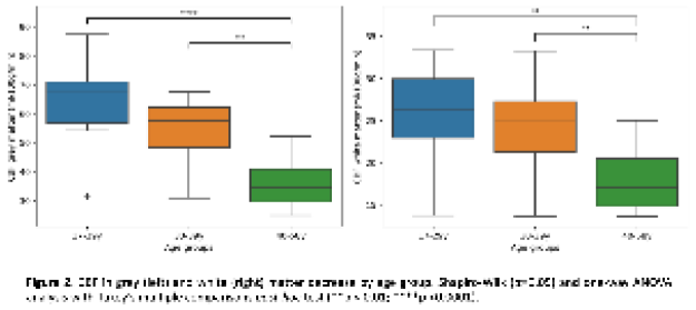
Pulse Pressure Impairs Cognition via White Matter Disruption
We previously found that pulse pressure (systolic – diastolic) predicted cognitive decline. Here, we hypothesized that the effects of pulse pressure on cognition are partly explained by white matter microstructure impairment, independently to other cardiovascular risk factors. We tested this in a cross-sectional population-based cohort (n=615, age 18-88). We associated pulse pressure, total blood pressure and heart rate variability with an indicator of white matter microstructure which is relevant to cerebrovascular health (Peak Width of Skeletonised Mean Diffusivity, PSMD, calculated on diffusion weighted imaging), and with processing speed and fluid intelligence. In robust multiple linear regressions, we found that pulse pressure significantly predicted white matter microstructure, above other vascular measures. We also found that white matter microstructure significantly predicted processing speed. This motivated testing whether white matter mediates the effects of pulse pressure on cognition, using structural equation models. White matter significantly and substantially mediated the effect of pulse pressure onto processing speed. This was moderated by age, such that older individuals with higher pulse pressure had lower white matter integrity and reduced processing speed. We expanded the model to show that vascular-related changes in processing speed drive changes in higher cognitive abilities. In conclusion, we found that pulse pressure acts through white matter microstructure to reduce cognition, particularly in older individuals. Better managing pulse pressure may help to preserve cerebrovascular health, and to support cognitive abilities throughout life.
Loss of parvalbumin interneurons and decreased colocalization with perineuronal nets in the dorsomedial striatum of BTBR, idiopathic ASD mouse model
Autism Spectrum Disorder (ASD) is a predominantly idiopathic condition characterised by restrictive, repetitive behaviours and interests (RRBIs) and a deficit in social interaction and communication. Solely from parvalbumin (PV) immunohistochemistry (IHC), the number of parvalbumin-immunoreactive (PV+) GABAergic interneurons has been reported to be decreased across several mouse models of ASD. However, more recent literature disputes for the downregulation of the Ca2+ binding protein PV, not the loss of neurons. These alternatives are anticipated to have opposing effects on excitation/ inhibition (E/I) balance. Although convincingly, PV downregulation has only been reported in transgenic and environmental mice models of ASD. The study of an idiopathic mouse model of ASD, BTBR, which may be considered to have a more human-like aetiology of ASD, will continue to identify convergent molecular endpoints across ASD models and biomarkers which would allow for an earlier diagnosis and intervention. This study used IHC techniques to quantify PV+ cells in the dorsomedial (DMS) and dorsolateral striatum (DLS), regions that were shown to govern goal-directed and habitual behaviour, respectively. Vicia Villosa Agglutinin (VVA) a lectin recognising perineuronal nets (PNNs) which ensheath parvalbumin interneurons was used as a second marker. Student t-tests were carried out and a value of p < .05 was deemed significant. The findings show that female BTBR mice have significantly fewer PV+, and VVA+, cells in the DMS compared to the controls. The decreased counts of PV+ and VVA+ cells suggest that BTBR mice indeed have a loss of parvalbumin interneurons. Consequently, the E/I balance is likely shifted towards diminished inhibition, at least in the DMS region, which may be responsible for symptoms of RRBIs in BTBR mice. Additionally, compared to the controls, female BTBR mice have a significantly reduced PV+VVA+ percentage colocalization in the DMS. Altered colocalization of PV+ cells with PNNs may equally be a result, or the consequence of PV interneuron loss, but may also contribute to symptoms of RRBIs. ASD research should investigate the DMS and DLS as heterogeneous structures.
Mitochondrial dysfunction in parvalbumin cells triggers juvenile-onset severe neurological disorder in vivo
Introduction: Mitochondrial diseases comprise the largest group of inherited metabolic disorders. Neurological symptoms include epilepsy, ataxia, and cognitive impairment. Previous studies implicated severe oxidative phosphorylation (OXPHOS) deficiencies in GABAergic inhibitory neurons accompanied by neurodegeneration in mitochondrial disease. This study aims to test the hypothesis that metabolically demanding fast-spiking parvalbumin-expressing (PV+) neurons are highly susceptible to mitochondrial dysfunction.
Methods: A novel mouse model of mitochondrial DNA (mtDNA) depletion selectively within the PV+ cells was generated by a conditional knockout of mitochondrial transcription factor A (Tfam) gene via cre-loxP system. Mice were characterised at behavioural, electrophysiological, neuropathological, and molecular levels.
Approach for statistical analysis: Shapiro-Wilk normality test was carried out. Unpaired Student’s t-test was selected for parametric data and Mann-Whitney U test for non-parametric data.
Results and conclusions: Mutant mice exhibited a progressive phenotype, initiating at 8 weeks of age with tremor, and neuropsychiatric features including cognitive impairment and anxiety-like behaviour. Hyper-locomotion and stargazing (absence-like seizures) were detected at 10 weeks, with severe ataxia observed by 12 weeks.
Hippocampal electrophysiology demonstrated a deficit in gamma oscillations in the knockout group upon cholinergic agonism, and aberrantly high area power of gamma oscillations upon stimulation with glutamatergic agonist kainate. A concomitant loss of calbindin-expressing inhibitory interneurons was detected in hippocampal CA3 region.
OXPHOS complexes I, III and IV within the PV+ cells of the knockout mice had differential deficiency levels which were brain region dependent. PV+ neurons demonstrated an upregulation of anaplerosis enzyme pyruvate carboxylase. PV+ Purkinje neurons showed a reduction in mtDNA copy number and modest cell loss. Cerebellum exhibited reactive microgliosis and astrogliosis. Knockout mice had reduced weight and severely shortened lifespan. The novel mouse model recapitulates key features of neurological phenotype associated with mitochondrial dysfunction and could be used as a powerful translational model.
Delineating the neurodegenerative mechanisms underpinning epilepsy in Alpers’ syndrome
Background: Alpers’ syndrome is a paediatric mitochondrial disease typically caused by bi-allelic pathogenic variants in the mitochondrial DNA polymerase gamma gene (POLG), and is characterised by super-refractory epilepsy which is associated with severe neurodegeneration, neurological decline and premature death. The precise neurodegenerative mechanisms underpinning the occipital-predominant epilepsy in Alpers’ syndrome remain unclear. However, the dysfunction and degeneration of inhibitory interneurons, in conjunction with astrocytic abnormalities, are hypothesised to contribute to seizure-associated alterations in cortical activity.
Methods: We performed a quantitative neuropathological investigation of inhibitory interneurons and reactive astrocytes in post-mortem cortical tissues from 13 patients with Alpers’ syndrome, 9 neurologically-normal controls and 5 sudden unexpected death in epilepsy (SUDEP) patients, controlling for generalised epilepsy-associated pathology.
Results: Alpers’ syndrome tissues demonstrated a severe loss of parvalbumin-expressing interneurons, particularly within the occipital cortex, with decreased abundance of mitochondrial oxidative phosphorylation (OXPHOS) proteins in those remaining. Reactive astrogliosis was prominent in occipital tissues and was characterised by an accumulation of abnormal hypertrophic astrocytes which demonstrated OXPHOS protein deficiencies and altered expression of key astrocytic proteins including glutamine synthetase, Kir4.1 and aquaporin-4.
Conclusions: The occipital cortex has a particularly high abundance of parvalbumin-expressing interneurons, which are strikingly lost in Alpers’ syndrome. This is accompanied by changes to crucial astrocytic proteins suggesting that dysfunctional astrocytes have a role in the pathophysiology by exacerbating neuronal hyperexcitability in Alpers’ syndrome. Our current research is now focussed on modelling these neuropathophysiological features of Alpers’ syndrome using human and rodent organotypic cortical slices.
Gene expression alterations in cortical neurons carrying autosomal dominant mutations affecting MAPT and APP dosage
Autosomal dominant mutations affecting the dosage of APP or MAPT are causal for early-onset forms of Alzheimer’s disease (AD) and frontotemporal dementia (FTD). I aim to identify changes in gene expression in the cortical neurons carrying such mutations that could explain the onset of familial dementia phenotype decades later. For this, I created single-cell RNA-Seq datasets from in vitro cortical neurons derived from patients with familial AD and FTD mutations. I generated 10X datasets from cortical neurons carrying APP duplication and MAPT E10+16 mutations and their isogenic controls, derived from patient-derived iPSCs using two differentiation protocols: induced cortical neurons via transgene introduction (Pawlovski et al., 2017) and directed differentiation protocol (Shi et al., 2012).
Preliminary results suggest that in vitro iPSC-derived cortical neurons carrying APP duplication and MAPT E10+16 mutations display gene expression changes relevant to the known disease mechanisms underpinning dementia. The results from in vitro cortical neurons are compared to the post-mortem samples from patients carrying APP duplication and MAPT E10+16 mutations, and to publicly available transcriptomics datasets from cortical neurons carrying APP or MAPT mutations that affect their dosage. The overarching goal of this project is to identify the pathways reflective of a changed cellular status in developing cortical neurons carrying mutations linked to changed APP or MAPT dosage, that could shed light on the disease processes leading to familial dementia decades later.
A deep sequencing investigation of mitochondrial DNA damage in cholinergic neurons of the Pedunculopontine Nucleus
In Parkinson’s disease (PD) patient brains, higher levels of somatic mitochondrial DNA (mtDNA) damage, manifesting as large-scale deletions, are frequently seen. Such mtDNA damage associates with neuronal loss, particularly the nigral dopaminergic neurons. However, cholinergic neuronal loss also occurs, particularly within a brainstem structure termed the Pedunculopontine Nucleus (PPN). We previously showed that, in contrast to nigral dopaminergic neurons, which show reduced mtDNA copy number (mtCN) in the presence of mtDNA deletions, PPN cholinergic neurons attempt to maintain the pool of wild-type mtDNA mtCN by increasing mtCN.
Here we will isolate single cholinergic neurons from post-mortem PPN tissue of aged controls versus PD patients followed by ultra-deep sequencing. At a single-cell resolution, we will consider the location of mtDNA deletions, their size, the nature of deletion breakpoints and their heteroplasmy level. We will also compare how the number of point mutations, their locations and heteroplasmy levels vary between PD and control cases. We will compare observations of these genomic features between PD patients and controls, as well as with published data on PD- affected nigral dopaminergic neurons. Our study aims to understand how mtDNA deletions are generated and explain the different compensatory responses by mtDNA in nigral (reduced mtCN) versus PPN cholinergic (increased mtCN) neurons to the accumulation of mtDNA deletions. Finally, we will use single cells qRT-PCR to determine expression levels of critical nuclear genes involved in mitochondrial biogenesis, mitophagy and mtDNA maintenance.
Preliminary results support earlier observations of higher levels of large deletions in PD compared to controls. The deletions range between 5-140bp (small) and >1,400bp (large). Analysis of large deletion location ‘hotspots’ is consistent with generation due to errors during replication. We find no significant difference in the levels of point mutations between cases and controls. The low concentration of small deletions suggests that oxidative damage is not a significant driver of excess mtDNA damage affecting PPN cholinergic neurons in PD patients.
Mitochondrial DNA Maintenance Failure in Degenerating Pontine Neurons Differs Between Lewy Body Disease Clinical Phenotypes
Introduction: Lewy Body Disease (LBD) entails Dementia with Lewy Bodies (DLB) and Parkinson’s Disease with Dementia (PDD) distinguished via the 'one year rule' between onset of dementia and parkinsonism. Consensus exists that phenotypes converge end stage, suggesting similar pathological mechanisms i.e. type-specific neuronal loss and protein aggregates in remaining neurons. Deletions in mitochondrial DNA (mtDNA), encoding OXPHOS components, may underlie the neurodegeneration, as we previously showed for Parkinson’s disease. Methods: We quantified mtDNA deletion (mtDel) levels and mtDNA copy number (mtCN) in single acetylcholine (ACh)- and noradrenaline- (NA) producing neurons from two degenerating pontine nuclei, the pedunculopontine nucleus (PPN) and locus coeruleus (LC) in post-mortem DLB, PDD and neurologically-normal control brains. We also quantified mRNA expression of nuclear encoded genes regulating mtDNA repair, biogenesis and mitophagy; for statistics we used ANOVAs. Results/conclusions: DLB cases harboured greater mtDel burden in both PPN ACh and LC NA neurons compared to PDD, frequently surpassing the 60% threshold for inducing mt functional defects. In DLB cases, mtCN was also greatly reduced in both neurotypes. Our data indicates PDD is an mtDNA depletion syndrome, with both neurotypes showing intact mtDNA but severely reduced copies; female PDD patients showed most severe mtCN depletion. In DLB-affected PPN ACh neurons, nuclear genes regulating mtDNA failed to respond to the clonal expansion of mtDNA deletions, displaying reduced expression; such pathways were largely intact in PDD patients.
Disease stage and patient age correlated with mtCN in PPN ACh neurons and with both mtCN and mtDel in LC NA neurons, highlighting mtDNA changes as potential factors in DLB’s progression. Our results indicate that LBD- affected brainstem neurons fail to maintain sufficient copies of wild-type mtDNA, the degree of pathological burden being neurotype and clinical subtype dependent. We reveal distinct mtDNA patterns in DLB vs PDD and impaired mtDNA-regulating RNA transcription in DLB-impacted degenerating pontine neurons. Our findings identify mtDNA maintenance pathway enrichment as a therapeutic target to promote pontine neuronal survival in the DLB subtype.
Transforming diagnostic yield of motor neurone degenerative diseases through empowering variant interpretation in underrepresented populations
Introduction
Motor neurone degenerative diseases (MNDs) are an extraordinarily heterogeneous group of conditions associated with progressive deterioration of motor neurone function and survival. The hereditary spastic paraplegias (HSPs) are a large subgroup of MNDs characterised primarily by upper motor neurone degeneration. Due to their highly monogenic nature HSPs comprise excellent study models to define crucial motor neurone survival-degeneration molecular pathways.
In genetically isolated populations recessive monogenic HSP subtypes may increase in frequency due to community cultural marriage preferences. The Exeter Rare Disease research group has investigated these conditions in Amish and Omani communities, leading to the discovery of novel monogenic causes of disease and detail community HSP/MND genetic architecture.
Methods & results
Exome-genome sequencing data from multiple Amish/Omani individuals with HSP/MNDs was filtered to exclude small variants/CNVs at >0.1% allele frequency or >1 homozygotes (gnomAD/in-house databases). Likely/pathogenic variants present in ClinVar/HGMDpro or within exons/±6 intronic nucleotides were evaluated using variant appropriate statistical tools (REVEL/CADD/SpliceAI). This defined specific founder gene variants associated with multiple novel (eg B4GALNT1, EPT1, TMEM63C) and previously described (eg SPAST) monogenic forms of HSP, and additional candidate new genetic causes of MNDs/HSP.
Discussion
In isolated populations the founder effect and large family size empowers genetic studies, enabling new disease gene discovery. Evidencing this our work in such settings has defined 8 novel monogenic causes of HSP/MNDs, facilitating diagnostic provision for families worldwide. Importantly this work increases representation of populations currently underserved in genomic databases, empowering rare disease variant interpretation.
Ultimately the scientific insights provided by new disease gene discovery aids the definition of novel disease biomarkers and potential therapeutic targets, alongside enabling new rapid, low-cost approaches to target pathogenic founder gene variants. Together this works provides crucial new pathomolecular scientific insights, alongside transforming diagnostic provision for affected families.
Patient-derived cellular models as tools to elucidate the pathophysiology of Hao-Fountain syndrome
Introduction:
Synaptic plasticity (SP) plays a key role in the human ability to adapt to environmental input, along with the processes of learning and memory. Altered SP significantly contributes to neurological and psychiatric disorders, including Autism Spectrum Disorders (ASD). Mutations in essential proteins facilitating SP have also been identified in human patients with intellectual disabilities. Recently, loss of function mutations in ubiquitin-specific protease 7 (USP7, also called herpes virus-associated ubiquitin-specific protease, HAUSP), have been identified as a disorder- causing variant, linked explicity to Hao-Fountain syndrome, a neurodevelopmental disorder manifesting intellectual disability, ASD, and seizures. Located at chromosome 16p13.2, USP7 encodes a deubiquitinating proteolytic enzyme that can cleave multiple ubiquitin chain linkages. Previously, USP7 was shown to regulate the ubiquitination of proteins, including the MDM2-p53 pathway, which is vital for DNA repair, transcription, and cancer. However, so far, the precise mechanisms of how USP7 mutation causes the clinical phenotype on a cellular level are missing.
Methods:
In this collaborative project, using patient-derived human induced pluripotent stem cells in combination with omics approaches (proteomic and transcriptomic analysis) and targeted biochemical analyses (qrt-pcr and western blot analysis), we aim to understand the functions of USP7 in neuronal development and SP by recapitulating neurodevelopmental processes using in vitro model systems including 2D models from iPS cells to differentiated mature neurons along with 3D models systems such as cerebral organoids.
Approach for statistical analysis:
Throughout our approaches following each phase of differentiation we will utilise primarily one-way ANOVA analysis along with appropriate post-hoc tests to provide information at an individual level whereby we monitor alterations at a protein and mRNA level when comparing each sample individually, while also combining patient versus control to identify potential alterations due to the USP7 deletion when compared to neurotypical controls.
High resolution respirometry to assess SH-SY5Y differentiation methods and their suitability for Alzheimer’s modelling
Alzheimer’s disease (AD) is a common neurodegenerative disease characterized by gradual onset cognitive decline that affects millions of people globally. During AD, β-amyloid (Aβ) proteins misfold and progressively aggregate. These proteins exert neurotoxic effects towards cholinergic neurons. Mitochondrial dysfunction is thought to be one of the first signs of this toxicity.
SH-SY5Y cells, a neuroblastoma derived cell line, are commonly used for research into AD and other neurodegenerative diseases. When undifferentiated, these cells display an immature catecholaminergic neuron phenotype. Protocols involving retinoic acid (RA) and RA with brain derived neurotrophic factor (BDNF) differentiate SH-SY5Y cells into a mature cholinergic phenotype; but unclear and conflicting results exist as to the effect of differentiation on mitochondrial function.
High resolution respirometry (HRR) involves the continual measurement of oxygen flux in a closed chamber containing living tissue. Addition of substrates, uncouplers and inhibitors (SUIT protocols) allows for assessment of individual respiratory enzymes consecutively.
This study aims to use HRR to investigate the effect of RA and RA/BDNF protocols on mitochondrial function of SH- SY5Y cells, specifically with regard to AD.
HRR will be performed using O2K-Resirometer (Oroboros). SUIT-003-D020 will be performed on undifferentiated, RA differentiated and RA/BDNF differentiated SH-SY5Y cells to measure proportional contributions of electron chain complexes to overall respiratory capacity. Longer SUIT protocols can then be used to further assess any differences identified.
Then, each cell type will be challenged with Aβ at progressive points throughout the aggregation process and effect on mitochondrial function will be measured, to assess both which stage/stages in the aggregation cause mitochondrial dysfunction and the mechanism behind the dysfunction.
A one-way ANOVA statistical test will be used to analyse significance between undifferentiated, RA differentiated and RA/BDNF differentiated cells.
A one-way ANOVA analysis test will be performed between control cells and cells exposed to monomeric Aβ; misfolded Aβ; oligomeric Aβ and proto-fibrillar Aβ within each of the differentiation types.
Testing carbonic anhydrase inhibitors for treating mitochondrial dysfunction in neurodegeneration
Mitochondrial dysfunction is a key feature of neurodegeneration. Identifying the components of ageing that promote mitochondrial dysfunction may lead to the development of therapeutics to counteract neurodegenerative decline. Carbonic anhydrases (CA) play a role in maintaining cellular pH by catalysing the reversible reaction that balances levels of carbon dioxide, bicarbonate and carbonic acid. pH is particularly important for mitochondrial respiration due to the proton cycling that occurs in the electron transport chain to produce ATP. Proteomic analysis of the ageing mouse brain revealed a significant increase in expression of CA, isoforms CAII and CAIII, in aged mice compared to young mice. Elevated levels of CAII were also found in younger brain tissue of the neurodegeneration mouse model pcd5J, potentially identifying CAII as a specific marker of neurodegenerative decline. Treating C. elegans with CAII (after maturity was achieved) reduced their lifespan by up to 58% compared to controls. CA inhibitors are already used to treat glaucoma and have been suggested as a potential therapeutic to treat Alzheimer’s disease. Further research is required to identify the specific role CAs play in mitochondrial dysfunction and whether this is a mechanism that can be targeted to treat neurodegenerative decline in ageing.
The SH-SY5Y human neuroblastoma cell line will be treated with CAs and CA inhibitors. High-resolution respirometry (HRR) using the Oroboros O2K will be utilised to observe their effect on mitochondrial respiration. Differentiated SH-SY5Y cells will be exposed to 1-Methyl-4-phenyl pyridinium (MPP+). MPP+ inhibits complex 1 of the electron transport chain in mitochondria and induces dopaminergic cell death providing a useful model of Parkinson’s disease. Differentiated SH-SY5Y cells treated with MPP+ will be used to observe whether CA inhibitors can restore mitochondrial function. HRR data will allow accurate delineation of mitochondrial dysfunction and precise assessment of amelioration effects of the CA inhibition.
Results from the HRR in control cells and treated cells will be analysed for statistical significance using a one-way ANOVA statistical test.
Application of imaging mass cytometry on post-mortem brain tissue to understand mitochondrial pathology on Parkinson’s
Mitochondrial dysfunction is associated with neuronal loss in Parkinson’s. However, the exact mechanism behind this loss remains unclear. To further our understanding of the nature of this dysfunction and its impact on neuronal health, we utilised imaging mass cytometry (IMC) to perform complex proteomic profiling of mitochondrial and associated proteins. IMC is a powerful technique that combines microscopy and mass spectrometry to simultaneously detect ~50 protein targets at the single cell level. We developed and applied large antibody panels comprising key components of key mitochondrial pathways in addition to several important brain cell markers. We used these panels to analyse changes in mitochondrial OXPHOS, mitochondrial fatty acid metabolism and mitochondrial quality control in Parkinson’s midbrain.
FFPE midbrain sections (5um thickness) from Parkinson’s cases (n=15) were subjected to IMC for the measurement of protein abundance and compared to healthy controls (n=15).
Statistical analysis was performed using R. Differences between groups were described using Bayesian estimation, Mann-Whitney U test, Wilcoxon signed-rank tests and multiple linear regression modelling.
Profiling of the mitochondrial protein abundance highlighted the heterogeneity between individuals within each group. We found evidence of deficiencies involving multiple respiratory chain subunits in both dopaminergic neurons and astrocytes (PMID: 33980828 &34779538). Additionally, we identified a synergistic decrease of Parkin, PINK1, Phospho-Ub, HSP60, TFAM and SIRT3 in Parkinson’s neurons, suggesting the regulatory machinery of mitochondrial quality control including mitophagy, mitochondrial proteases and biogenesis might be impaired.
Analysis of the proteins responsible for fatty acid oxidation (particularly the acylcarnitine pathway, PMID: 28394042) identified several differences between Parkinson’s and control cases, and appeared correlated to OXPHOS dysfunction.
Our studies highlighted the use of IMC in the assessment of mitochondrial protein expression in Parkinson’s, providing important post-mortem evidence to support the complexity of impaired mitochondrial protein homeostasis in Parkinson’s, which may increase the neuronal vulnerability to age-related oxidative stress.

EBP1 ubiquitination by PARKIN promotes mitophagy protecting neuron death in brain disease
Cerebral ischemia induces massive mitochondrial damage, leading to neuronal death. The elimination of damaged mitochondria via mitophagy is critical for neuroprotection. Here we show that the level of ErbB3-binding protein 1 (EBP1) was notably increased early during transient middle cerebral artery occlusion and prevented neuronal death by eliciting cerebral ischemia reperfusion (IR)-induced mitophagy. Neuron-specific knockout of Ebp1 increased infarct volume and aggravated neuron loss with impaired mitophagy and was rescued by introduction of adeno- associated virus serotype 2 expressing EBP1. We identified that EBP1 is ubiquitinated on lysine 376 by PARKIN on the damaged mitochondria and interacts with adaptor protein P62 for mitophagy induction. Thus, our study suggests that EBP1 ubiquitination following cerebral IR-injury promotes mitophagy induction, which may be implicated in neuroprotection.
MSK1 is required for the beneficial synaptic and cognitive effects of enriched experience across the lifespan
Introduction: Normal human ageing and age-related dementias impair cognition. Positive experiences, such as social interactions, cognitive training and physical exercise, have been shown to ameliorate some of the harms to cognition associated with ageing. Similar positive interventions in animal models, known as environmental enrichment, strongly influence neuronal morphology and synaptic function and enhance cognitive performance. However, little is known as to how the environment influences neurons to respond and adapt to these positive sensory experiences. Previously we showed that the BDNF-activated CREB kinase, MSK1, is necessary for the expansion of the synaptic dynamic range and the full complement of cognitive enhancement provoked by enrichment in young mice. Here we report that MSK1 continues to be important for synaptic plasticity and cognitive improvement in response to enrichment well into old age.
Methods: We used aged male wild-type and MSK1 kinase dead (MSK1 KD) mutant mice housed in either standard housing throughout the study or moved into and remaining in environmental enrichment from 78 weeks of age. Behavioural testing in the elevated plus maze, spontaneous alternation task and Morris water maze was conducted in weeks 88-92. Between weeks 94 and 100 animals were culled and tissue removed for ex vivo analyses of area CA1 synaptic transmission and long-term potentiation, CA1 spine density and hippocampal RNA-seq.
Statistics: Statistics were computed by IBM SPSS 27 using two-tailed one or two-way analysis of variance (ANOVA). The level of significance was taken to be p<0.05 (α=0.05). Exact p values are reported where p<0.0001. For lower values, p< 0.0001 is reported. Data are reported as mean ± SEM and bar graphs display individual data points.
Results and conclusions: We show that aged male wild-type mice that underwent environmental enrichment showed less anxiety-like behaviour, improved performance in spatial working and spatial reference memory tasks, and an enhancement in hippocampal LTP. Many of these benefits were absent in MSK1 KD mice. We conclude that enrichment is beneficial across the lifespan and that MSK1 is required for the full extent of these experience-induced improvements of cognitive abilities and synaptic plasticity.
Mef2c transcription factor is required for development of the medium spiny neurons of the mouse striatum
Introduction: Medium spiny neurons (MSNs) are the major projection of the striatum and are the neurons predominantly degenerating in Huntington’s disease. Understanding normal striatal MSN development is important for understanding the pathological conditions affecting this area of the mouse brain. We showed previously that the transcription factor myocyte enhancer factor c (Mef2c) is significantly upregulated in the striatum during embryonic development. This study investigated the role of Mef2c in the developing mouse striatum.
Methods: A conditional knockout (CKO)of the Mef2c gene in the striatum was made using the Gsx2-cre-loxp system to generate Gsx2-Cre Mef2c-/- mice. A total of 80 mice were used, with equal numbers of males and females. Mice lacking Gsx2-Cre served as controls. A range of assays were undertaken in a developmental series: stereological quantification of striatal volume and striatal cell counts for NeuN, and MSNs markers; Golgi-cox based dendritic tracing, proliferation using BrdU and Edu, apoptotic assays e.g. TUNEL, cell culture and motor behavioural testing.
Approach for statistical analysis. three tests were used: Two-way ANOVA followed by Bonferroni post hoc test when a significant interaction between the two main factors exists; Two-way repeated measure ANOVA test to analyse dendritic arborization and behavioural data; and Multiple t test with Holm Sidak correction (significance set at 0.05).
Results and conclusions: Mef2c CKO animals showed a significant impairment in exploratory behaviour of new environments compared to controls. Striatal volume and MSN numbers were significantly reduced in CKO striatum from postnatal day 14 (P14) onward, accompanied by a relative decrease of the striatal matrix compartment, and there was a mild, yet significant, reduction of dendritic intersections in 12-month-old CKO striatae. A significant increase in apoptotic activity was observed in P3 CKO striatum accompanied by a significant reduction in the anti- apoptotic factor Bcl-xl, but proliferation indices were unchanged. In conclusion, we showed that Mef2c is required for the survival of a subpopulation of matrix MSNs and proper development of dendrites and suggest that these changes underlie the abnormalities in motor functioning.
Long-interspersed element 1 mobile DNA activity is prominent in parvalbumin interneurons
Introduction. The retrotransposon Long Interspersed Element 1 (LINE-1, L1) is a mobile genetic element that can autonomously mobilise from one location in the genome to another via a “copy and paste” mechanism involving an RNA intermediate. Due to its replicative nature, L1 copies have accumulated in mammalian genomes over evolutionary time, making up over 17% of human DNA. L1 insertions within and proximal to genes provide a significant source of regulatory sequences that can impact gene expression in various ways. Somatic L1 insertions have been detected in humans, macaques, and mice neuronal cells. Previous studies suggest that early brain development and neurogenesis provide a milieu for L1 mobilisation. However, whether the L1 activity is restricted to specific neuronal subtypes is unknown, and its impact on neuronal function remains unclear.
Methods. To address this knowledge gap, we have used single-molecule RNA in situ hybridization and immunohistochemistry, as well as functional assays using L1 mobilization reporters in both primary neuronal cultures and mice. In addition, we have used long-read Oxford Nanopore DNA sequencing to profile methylation at L1 loci in sorted neuronal populations from mice.
Approach for statistical analysis. Significance testing of cell counts in mice was done using one-way ANOVA with Tukey’s multiple comparison test or two-tailed t-test using GraphPad Prism. Methylation comparisons were performed by Fisher’s Exact Test using methylated and non-methylated call counts, with significance defined as a Bonferroni corrected P value of less than 0.01.
Results and conclusions. We have found that L1 activity is stimulated by the SRY-box transcription factor 6 (SOX6), a key player in the neuronal-type specific transcriptional program of parvalbumin-expressing (PV+) interneurons. PV+ neurons harbour unmethylated L1 promoters, express higher levels of L1 mRNA than other neurons, and support L1 transgene mobilisation in mice. Additionally, we identified unmethylated L1 promoter loci driving the expression of PV+ function-related genes, such as calcium-dependent activator protein for secretion 2 (CAPS2). In conclusion, our findings reveal that L1 activity is an integral part of the transcriptional program of PV+ neuron generation.
Selenium Deficiency and Increased Sodium/Potassium Ratios are Widespread in the Huntington’s Disease Brain
Background: Huntington’s disease (HD) is an autosomal dominant neurodegenerative disease caused by mutations in the Huntingtin (Htt) gene. This disease leads to progressive motor dysfunction, neuropsychiatric abnormalities, and cognitive deterioration. Although primarily characterised neuropathologically by neuronal loss and the presence of mutant Htt aggregations in the brain, metallomic perturbations may also be present in the HD brain and contribute to pathogenesis. This study aimed to identify what such, if any, perturbations may be and where they are localised.
Methods: Changes in eight essential metals and selenium were investigated in eleven brain regions in nine genetically confirmed HD cases and nine age, post-mortem delay, and sex-matched controls using inductively coupled plasma mass spectrometry (ICP-MS).
Statistical Analysis: Significant case–control alterations were identified using non-parametric Mann–Whitney U tests; sodium/potassium ratios (Na/K) were also analysed in the same manner. p < 0.05 was considered significant. Risk ratios, E-values, and effect sizes were also determined to evaluate the robustness of significant case–control alterations.
Results: The HD brain showed significantly decreased selenium in every investigated region, as well as increased Na/K in 10 out of 11 regions. Multiple regions also showed increased calcium and zinc levels, with localised decreases in iron, copper, and manganese.
Conclusions: Selenium deficiency and increased Na/K appear to be a widespread feature in the HD brain and may reflect mitochondrial dysfunction and oxidative stress as well as blood–brain barrier breakdown; these changes may contribute to pathogenesis in HD and may represent potential therapeutic targets.
Cranial irradiation induces age-dependent cognitive deficits and hippocampal marker expression indicative of synaptic and GABAergic dysfunction
Radiotherapy is the mainstay of brain cancer treatment, in both adult and paediatric patient populations. Although effective in tumour control, exposure to therapeutic radiation can induce progressive cognitive decline that affects domains including memory and social functioning. We investigated the cognitive ability of mice irradiated at different ages to correlate behavioural changes with well-established markers of cognitive function. Male C57BL/6 mice aged 8- and 26 weeks were subject to a single dose of 20 Gy delivered to the right hemisphere. The novel object recognition (NOR) test was performed 7- and 50-days post-IR, and the 3 Chamber Social Interaction (3-CSI) Test 50 days post-IR. Synaptic (SNAP-25, PSD-95) and GABAergic (PV, GAD-67) markers in the hippocampus were analysed with simple westerns. NOR data was analysed using an unpaired Student’s t-test. 3-CSI data was analysed using a repeated measure 3-way ANOVA. Molecular data was analysed using a one-way ANOVA. Irradiation at 8 and 26 weeks significantly impaired NOR test performance at day 7, as indicated by decreased discrimination indices compared to controls (8w, p<0.001; 26w, p<0.01) and day 50 (8w, p<0.01; 26w, p<0.05). Neither treatment nor age had an effect on social preference. Mice were unable to complete the social memory element of the test at 8 weeks old, thus no effect of IR was seen. 26-week old control mice were able to discern the two mice, and spent more time interacting with the stranger mouse, an ability lost in the IR mice (p<0.05). 8 week old IR mice showed significant hemispheric decreases in PV (right (R), p<0.05) and GAD-67 (left (L), p<0.05), and an increase in PSD-95 (R, p<0.01). When delivered to 26 week old mice, IR reduced levels of PV (L, p<0.01), GAD-67 (L, p<0.01; R, p<0.05) and SNAP-25 (L, p<0.001; R, p<0.001). Our findings in this study reveal clear IR-induced impairments in visual-spatial memory, and a vulnerability of older populations to social memory losses. Markers relevant to the GABAergic inhibitory system and synaptic connectivity were altered by IR in a hemispheric- and age-dependent manner, indicating that neural networks relevant to cognitive functioning are disturbed even 50 days after exposure.

Endocannabinoid neurotransmitter abnormalities in Parkinson’s Disease Psychosis and grey matter loss: A meta- regression
Introduction
There are extensive grey matter abnormalities in Parkinson’s Disease (PD) patients with visual hallucinations and delusions (i.e., PD psychosis, PDP), as well as dysfunction of serotonergic receptors. Evidence from clinical trials has shown the effectiveness of cannabinoid-based medications in alleviating psychosis in PD patients. However, the relationship between endocannabinoid system and grey matter atrophy in PDP has not been examined. Here, we aimed to investigate the relationship between grey matter volume loss in PDP and endocannabinoid receptors.
Approach for statistical analysis
Systematic searches were conducted on PubMed, Web of Science, and Embase. Peak coordinates were extracted from magnetic resonance imaging (MRI) studies for PDP and PD patients without psychosis (PDnP) and were entered in Seed-based d Mapping with Permutation of Subject Images (SDM-PSI, v6.21). PD medications expressed in Levodopa equivalent daily dose (LEDD) was entered as covariate. Gene expression data for endocannabinoid (i.e., CB1/CB2) receptors were extracted from the Allen Human Brain Atlas, and parcellated across the 78 regions of the Desikan-Killiany brain atlas. Unadjusted and LEDD-adjusted Hedges’ g effect-size estimates were extracted from the SDM-PSI analysis as a measure of grey matter loss in PDP patients for these 78 regions and entered in multiple regression models. Analyses were conducted on R.
Results and conclusions
Results from 10 MRI (PDP patients, n=211, mean age = 69.01 years; PDnP patients, n=298, mean age = 67.34 years) showed reduction in grey matter was observed in parieto-temporo-occipital regions in PDP patients in both LEDD- adjusted and unadjusted analyses (uncorrected, p<0.05). Local expression of CB1 receptor was significantly associated with whole-brain grey matter volume loss (unadjusted, b=0.130, p=0.004; LEDD-adjusted, b=0.146, p=0.002). Entering fatty acid amide hydrolase enzyme in the model did not change these results. Extensive decrease in cortical volume within regions involved in high order visual information processing and information integration in PDP was observed. Abnormalities in CB1 receptor may contribute to better understanding cannabinoid-based medications as a treatment for psychosis in PD patients.
Photobiomodulation therapy (PBMT) 1068 nm for the treatment of neurological complications of COVID-19: molecular and cellular effects
Introduction: COVID-19 can result in severe complications, such as the pro-inflammatory cytokine storm, acute respiratory distress syndrome, thrombosis or neurological issues. PBMT, using near-infrared light with the wavelength 1068 nm, could be used as a non-invasive, drug-free treatment for COVID-19. PBMT acts via the mitochondrial enzyme cytochrome c oxidase to induce inflammatory changes, cytoprotection, nitric oxide release and increase blood flow. It is hypothesised that these effects will benefit the respiratory system and other organs targeted by SARS-CoV-2, including the brain (with neurological symptoms, ranging from anosmia to stroke and delirium, becoming increasingly common.)
Methods: Human SHSY5Y neuronal and rat C6 glioma cells were stressed with SARS-CoV-2 spike peptide, lipopolysaccharides or hydrogen peroxide, and treated with 1068 nm. Responses were measured with MTT assays, lactate dehydrogenase (LDH) release, immunohistochemistry and immunofluorescence. Initial experiments were performed on human skin cells (HDFs).
Approach for statistical analysis: SPSS and Microsoft Excel were used to perform statistical analysis with t-tests and one-way ANOVAs (using * p < 0.05, ** p < 0.01, *** p < 0.001 and **** p < 0.0001). ImageJ’s ‘Analyze particles’ feature was used to count cells.
Results and conclusions: PBMT improved (*) mitochondrial viability in HDFs under inflammatory and oxidative stress, obeying the cytochrome c oxidase theory of activation. In C6 cells, PBMT reversed the S-peptide-induced redistribution of the SARS-CoV-2 receptor ACE2 to the cell membrane, which may be useful in reducing viral attachment. Starvation with serum free media reduced the number of glioma and neuronal cells, and PBMT increased cell number (**** and *** respectively) back to control levels. The basis of this was explored; PBMT had no effect on the cytotoxicity marker LDH but increased the percentage of proliferating cells expressing Ki-67 (**).
PBMT shows promising effects against SARS-CoV-2 infection and neurological complications. In future experiments, techniques will be repeated with microglia to assess their response to PBMT. ATP determination and calcium imaging will be used on all cells to further understand the mitochondrial effects of 1068 nm.
Synaptic resilience in Lothian Birth Cohort 1936 participants is associated with maintained cognition during ageing
Many people experience declining cognitive function as the brain ages. Age is the most important risk factor for cognitive decline and dementia, but the degree to which individuals experience these aspects of ageing is hugely variable. Understanding the neurobiological bases of brain ageing can offer key insights into individual differences in cognitive ageing and dementia risk. Region-specific synapse changes observed in the ageing brain and in Alzheimer’s disease (AD) correlate with cognitive decline; thus, we hypothesize that synaptic resilience contributes to healthy cognitive ageing.
In this study, we test this hypothesis through observing human post-mortem brain tissue from people without dementia donated by participants in the Lothian Birth Cohort 1936 alongside young control subjects and people who died with AD. We used array tomography imaging to examine synapse density and the accumulation of Alzheimer’s- related pathological proteins within synapses in two brain regions (inferior temporal cortex and visual cortex).
Further, we use proteomics and RNA sequencing to characterise molecular changes in biochemically enriched synaptic fractions and in total brain homogenates from these brain regions. We observe a stepwise decline in synapse density between young controls, ageing participants with normal cognition, and people with AD and a stepwise increase in the proportion of remaining synapses containing amyloid beta or tau proteins. Molecular analyses indicate decreases in synaptic signalling in ageing compared to young controls, which are exacerbated in AD. The Lothian Birth Cohort 1936 participants took an intelligence test at age 11 and multiple cognitive tests through their 70s and 80s allowing us to compare people with lifetime cognitive decline compared to those with maintained or resilient cognition over their lifetimes. Although our participant numbers are modest when split by lifetime cognition (n= 7 participants who experienced lifetime cognitive decline and 8 with lifetime cognitive resilience), we observe some differences, including increased gliosis in people with lifetime cognitive decline and paradoxically gene changes indicating decreased synaptic signalling in people who were cognitively resilient despite no change in synapse density.
Population-Level Genomic Variation and Childhood Structural Brain Organisation
We do not understand the mechanisms that shape emergence of brain organisation and how this variability might arise. To address this, we integrated computational models of structural brain development, genomic variation, and population-level cognitive variation in 2154 children aged between 9 and 11 years old from the Adolescent Brain Cognitive Development study (Casey et al., 2018).
Using streamline counts from deterministic tractography, we simulated structural connectivity development using generative network modelling (Akarca et al., 2021; Betzel et al., 2016; Kaiser & Hilgetag, 2004; Vértes et al., 2012). Starting from a sparse seed network, this relatively novel technique iteratively adds edges until the simulated connectome has the same number of edges as the observed connectome. The probability of a new edge being added is proportional to its metabolic wiring cost eta and the value gamma it brings to network topology. This relationship is governed by different wiring rules, which add new edges based on high clustering, degree, shared neighbours, or spatial proximity. We performed group-level model selection and selected the model whose simulated connectome had the smallest dissimilarity in capturing local and global connectome properties. To probe common genetic influences on structural brain development and cognition, we computed polygenic scores (PGSs) for cognitive ability for 1461 children with high-quality genomic data.
PGSs accounted for 4% of variance in cognitive ability. A homophily wiring rule which adds edges to nodes with similar neighbours provided the best fit. Across individuals, this translated to large negative wiring cost penalties and smaller positive wiring value terms. We extracted 3 significant components from a PLS of PGSs and individual optimal eta-gamma combinations as predictors of cognition. All 3 predictors loaded positively onto the first component, suggesting that linear combinations of the predictors accounted for linear variation in cognition. Using pathway enrichment of the 3 predictors ranked by predictive value, we found unique and converging pathway enrichments.
This suggests that common and distinct biological pathways underpinned structural brain development parameters and cognitive ability PGSs.
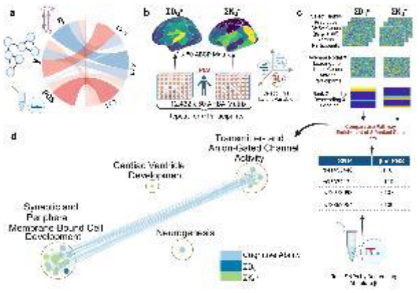
Shared Transcriptomic Signatures between Neurodegeneration and Psychiatric Diseases
Introduction
Neurological conditions present a wide spectrum of symptoms, which often exhibit considerable overlap. These similarities pose a challenge to clinicians for the correct diagnosis of these conditions, which are often observed to co-occur, as for example in the case of depression in patients with Alzheimer’s disease. To investigate these co- morbidities, we start from the observation that many brain disorders share genetic architectures, which suggests that related genes and pathways may be implicated in onset and progression of co-occurring conditions. To quantify these observations, we leverage recent advances in transcriptomics of single-cell data to understand the role of different cell types in the context of molecular events which may underlie disease-associated susceptibility. Our aim is to perform a meta-analysis to identify shared disease associated transcripts across different brain conditions.
Methods
Brain derived single-nucleus RNA sequencing (snRNA-seq) data were collected for a range of neurological diseases, including Alzheimer’s disease, Parkinson’s disease, depression, schizophrenia, and others. These data were pre- processed for quality control, mapped to the telomere-to-telomere genome assembly, and quantified to generate expression matrices. These matrices were then processed using custom R scripts built using the Seurat, and WGCNA packages. We are currently performing differential expression analysis for these samples. We will quantify these transcriptome changes and calculate a transcriptomic-based distances, which we refer to as ‘omic distance’. We will use this distance to cluster these samples on a transcriptomic space of reduced dimensionality to assess shared transcriptomic changes among these disease conditions.
Approach for Statistical Analysis
Principal component analysis was employed for dimensional reduction followed by UMAP and tSNE projections. Wilcoxon rank-sum test was used for differential expression analysis along with Benjamini-Hochberg to calculate false discovery rate in multiple hypothesis testing. The log-fold change and the p-values obtained from differential expression were used to calculate the omic distance and k-means clustering was employed with hypergeometric test for functional annotations.
Cell Type Specific Transcriptomic Signatures of the Ageing Brain
Introduction
There have been great advances in understanding cellular ageing processes. However, the selective vulnerability of brain cell types to these ageing processes is yet to be characterised in detail. This knowledge is important since ageing is the primary risk factor for many neurodegenerative disorders. The investigation of molecular changes that underlie brain ageing will help to identify distinctions between healthy and disease-associated ageing brains. The application of single-nucleus transcriptomics derived from brain tissue is generating a wealth of gene expression profiles at a transcriptome-wide scale for individual cell nuclei. With this information our aim is to perform a meta- analysis to identify cell type specific transcriptomic signatures across the lifespan of neurologically healthy donors.
Methods
Single-nucleus RNA-seq data derived from the prefrontal cortex of neurologically healthy donors across human lifespan was processed and samples categorised into 5 age groups. Data were processed through quality control, genome mapping and transcript quantification to create gene-cell matrices for each sample. These matrices were then input to an automated single-cell/nucleus pipeline based on the Seurat package in R. Within this pipeline nuclei were clustered and annotated to specific cell types using a multiple marker gene expression approach. Cell type specific nuclei were then integrated across samples within age groups. Next, differential expression analyses (DEA) will be performed across age groups to identify age related transcriptome alterations at a cell type resolution.
Metrics from the DEA will be used to calculate omic distances, which enable the projection of samples/age groups onto a 2D transcriptomic space, to globally assess the transcriptome variability across the lifespan.
Approach for Statistical Analysis
In this workflow dimensional reduction was performed using PCA based on the top 3000 variable genes per sample. Using 30 dimensions from PCA, data were then clustered using UMAP and tSNE approaches. For DEA, Wilcoxon rank- sum test with FDR multiple-testing correction will be utilised. The associated p-values and fold changes form the basis of omic distance calculations for generating pairwise distance matrices.
Synchrotron XRF Metal Distribution in Neurodegenerative Disease: A Correlated Multimodal Imaging Registration System
Introduction: Transition metals have important roles in brain structure and function, and they have been connected to detrimental mechanisms leading to neurodegenerative diseases. Synchrotron x-ray fluorescence (SXRF) measurements are increasingly used as a non-destructive method to acquire sensitive and specific data on metal element distributions in biological tissue such as post-mortem brain tissue [1]. Registration systems, enabling the integration of SXRF-acquired elemental maps into histological evaluations, will advance what can be learned from these data. It important to address this for SXRF mapping in metallomics investigations of tissues, as traditional approaches (e.g. staining, or deposition of metal grids for alignment) can be vehicles for metal contamination, creating issues of sample integrity that limit interpretation. Our method will allow registration of data from two or more complementary modalities, delivering composite views to enable specific questions about the role of metals in neuropathology to be addressed.
Method: Samples from two age-matched cases (disease and control) will be cryosectioned with a sapphire blade and mounted onto a grid-etched quartz slide, minimising scope for metal contamination. Light microscopy of ~1mm diameter regions of interest (ROI) will be imaged prior to SXRF analysis, and aligned with serial sections stained to establish tissue structure and targets of interest. For SXRF mapping, beamline I18 at the Diamond Light Source synchrotron will be used: a microfocus beamline with a 2µm resolution. The pre-identified ROI in cryosectioned tissue will be mapped in order to determine chemical element distributions at parts-per-million concentrations.
Approach for statical analysis: Spectral fitting to precisely identify elemental peaks, and integrate the area under each element peak, will be performed using PyMCA to extract maps of concentration distribution [2]. Non- parametric testing of the null hypothesis (no difference in absolute and relative metal element levels for cells and adjacent extracellular matrix) will be demonstrated utilising the multiple ROI, aided by the new etched-grid registration system.
[1]F. Lermyte et. al., Cells 2019 8:1231
[2]V. Solé et. al., Spectrochim. Acta Part B 2007 62:63-68
Machine Learning Techniques for Biomarker Discovery of Depression to Investigate its Effect on the Brain and Explore Associations with Dementia
The use of machine learning algorithms, such as random forest classifiers, has the potential to greatly improve the discovery of biomarkers for mental health conditions, such as Major Depressive Disorder. Biomarkers are measurable indicators of a biological process or condition that can be used to diagnose and monitor diseases. In the case of depression, biomarkers could help identify individuals at risk for the condition, as well as monitor the effectiveness of treatments. This study aimed to use random forest classifiers, to discover potential biomarkers for patients with depression using structural and functional imaging derived data from magnetic resonance imaging (MRI) scans. By applying machine learning algorithms to MRI data, this study sought to identify specific patterns or features that could be used as biomarkers for depression. Once potential biomarkers were discovered using random forest classifiers, the next step was to assess their potential association with dementia. This was done using a statistical method called structural equation modelling, which allowed evaluation of the relationship between the biomarkers and dementia. The results of this analysis could provide valuable insights into the potential overlap between these two conditions and the potential for using discovered biomarkers as a diagnostic tool for both depression and dementia.
Imaging neuroinflammation and blood-brain barrier permeability in people living with HIV and severe depression
Introduction
People living with HIV experience an increased risk for depressive symptoms, though it is unclear whether any biological mechanisms contribute to this risk. HIV infection in the central nervous system may elicit chronic neuroinflammation and concomitant blood-brain barrier (BBB) dysfunction. Neuroinflammation is, in turn, associated with depressive symptoms in the general population. Thus, it is possible that neuroinflammation contributes to the increased risk for depressive symptoms amongst people living with HIV. However, participants with severe depressive symptoms are often excluded from studies of HIV co-morbidities. In this proof-of-concept study, we aim to explore whether neuroinflammatory signatures can distinguish between people living with HIV who experience severe depression and those who experience mild depression.
Methods
For this cross-sectional study, virally-suppressed adults living with HIV will be recruited in Brighton, UK. Depressive symptoms will be measured using the Patient Health Questionnaire (PHQ-9) and the South African Depression Scale (SADS). We will seek to recruit equal numbers of participants with high (n = 10; PHQ-9 > 15) and low (n = 10; PHQ-9 < 9) depression severity. All participants will undergo diffusion-weighted magnetic resonance spectroscopy (DW-MRS), a novel and sensitive technique for imaging neuroinflammation. Using DW-MRS, Apparent Diffusion Coefficients (ADCs) of the neurometabolites myo-inositol and choline, which are considered markers of glial cell activation, will be quantified in the anterior cingulate cortex (ACC). BBB permeability (leakage rate, Ktrans) will be measured using dynamic contrast-enhanced magnetic resonance imaging (DCE-MRI). Absolute concentrations of inflammatory biomarkers will be quantified in blood serum using a Luminex assay.
Statistical Analysis Approach
Linear regression models will be used to test for associations between measures of neuroinflammation (ADC[mI], ADC[Cho], Ktrans, and absolute concentrations of blood biomarkers) and depression severity (PHQ-9 and SADS scores). Regression models will be adjusted for covariates including participant age, sex, ethnicity, and HIV plasma viral load. We will report effect size estimates (regression coefficients), p-values,
Mission Lucidity: decoding brain disease
More than 60 million people worldwide suffer from neurodegenerative conditions such as Alzheimer’s and Parkinson’s disease, and that number continues to rise dramatically as life expectancy increases. The diseases are devastating for patients and their families, and have enormous socio-economic costs.
Over the past decades, scientific research has provided many important insights towards understanding, diagnosing, and treating neurodegeneration, but therapeutic breakthroughs are still lacking. A long history of clinical trial failures has discouraged industry investment, while the need for effective treatments has become a medical emergency.
To lift the hurdles and roadblocks that are slowing down progress, we need to mobilize state-of-the-art technologies such as nanoelectronics, brain-machine interfaces, single-cell biology and genetic engineering.
In 2018, Mission Lucidity was launched as a joint initiative between VIB, KU Leuven, UZ Leuven and imec. It combines the expertise of best-in-class partners in biomedical research, clinical care and nanotechnology, who share a common vision of a future without neurodegenerative diseases. Our mission is to create new tools to accelerate global scientific discovery and medical progress, transform neurodegeneration research and pave the way for prevention and cures.
Our project portfolio addresses major challenges in various stages of neurodegenerative diseases: from screening and risk prediction to better patient cell-based models for research and drug development, early diagnosis based on affordable and sensitive biomarkers, stratification of patients into subgroups for personalised medicine, and non- invasive interventional therapies based on neuromodulation.
Mission Lucidity is a proud member of the European CURE-ND alliance, founded in 2020 together with the UK Dementia Research Institute, the Paris Brain Institute, and the German Centre for Neurodegenerative Diseases.
The priority index pipeline recapitulates known ALS biological pathways and suggests novel drug targets
Introduction
The priority index (Pi) pipeline [Fang et al., 2019], originally developed and tested on rheumatoid arthritis (RA), is a genetics-led pipeline that integrates functional data from GWAS, ontologies and biological networks to produce a ranked list of genes with evidence for disease association. We modified the Pi pipeline to predict novel target genes associated with Amyotrophic Lateral Sclerosis (ALS).
Methods
575 SNPs from an ALS GWAS (p-value threshold <1 x10⁻⁷) were used as initial input [van Rheenen et al., 2021]. These SNPs were mapped to 44 genes utilising gene proximity, physical interactions with chromatin in microglia, neurons and oligodendrocytes [Nott et al., 2019], and association with eQTLs expression in spinal cord and brain cortex (GTEx v8) [The GTEx Consortium, 2020].
Ontology information was incorporated based on manually curated lists of pathways involved in ALS function (GO) [Ashburner et al., 2000; Gene Ontology Consortium, 2021], ALS associated genes (DO) [Schriml et al., 2018] and ALS related phenotypes (HPO) [Kohler et al.,2021].
A STRING network (v11.5) [Szklarczyk et al., 2019] of 5183 genes with strong experimental evidence (score >800) was used to identify genes that interconnected the GWAS-implicated genes in a random-walk with a restart network approach.
Approach for statistical analysis
The Pi R package was used in discovery mode. Random walk with restart was used to identify genes related to GWAS-implicated genes based on connectivity. The gene ranking was achieved by integrating all predictors (gene proximity, chromatin interactions, eQTL associations, ontology associations (function, disease and phenotypes) and network associations) by Fisher’s combined meta-analysis. For each predictor, the scores were converted to P-like values, and aggregated for each gene using a Fisher’s combined method. The combined p-values were rescaled to a Pi rating (0–5).
Results and conclusions
We obtained a ranked list of 5211 genes associated with ALS. Several genes not present in the GWAS had high connectivity to GWAS-implicated genes, and were enriched in ALS relevant disease processes such as autophagy. We prioritised potential target genes by integrating structural information and cell-type expression data.
The Impact of a High-Fat Diet on Locomotion and Olfactory Learning in Wild-Type and Triosephosphate Isomerase- Deficient Drosophila melanogaster
Introduction: Alzheimer’s Disease (AD) affects 520,000 people in the UK, with approximately 40% of cases attributable to modifiable risk factors. Memory impairment, a hallmark symptom of AD, was modelled using triosephosphate isomerase (TPI)-deficient Drosophila melanogaster. The gut-brain axis (GBA) links the gut microbiome to neuroinflammation, which exacerbates AD pathophysiology. Alterations to the gut microbiome were modelled using a high-fat diet (HFD), and its effect on behavioural and neuronal phenotypes were assessed.
Objectives: To investigate whether hTPII170V and hTPIwstd mutations, alongside a HFD, cause reduced locomotion and olfactory learning in Drosophila melanogaster larvae. Thus, making TPI deficiency appropriate for modelling memory impairment.
Methodology: Larval locomotion was assessed by measuring the distance travelled using ANY-maze software. Olfactory learning and memory were assessed using Pavlovian conditioning to train the larvae to associate an odour (distilled water/n-amyl acetate) with a fructose reward. A preference index was calculated and analysed to determine the extent of larval conditioning.
Statistical Analysis: One- and two-sample t-tests were utilized to analyse olfactory learning/locomotion within and between groups. A two-way analysis of variance (ANOVA) and post hoc Tukey test were conducted to compare the data. Values of p<0.05 were accepted as significant.
Results: There was significant learning in w1118 normal diet (ND) (p=0.0051) and hTPIwstd HFD (p=0.0411), with no significant learning in all other groups. Compared to w1118 ND, there was significantly reduced learning in hTPIwstd ND and w1118 HFD (p=0.0215). A significant increase in locomotion was identified between w1118 ND, hTPII170V ND and hTPII170V HFD (p=0.0010).
Conclusion: TPI deficiency (hTPIwstd) is appropriate for modelling memory impairment in Drosophila melanogaster. A HFD impairs olfactory learning and memory in w1118 larvae. Locomotor activity is increased by a hTPII170V mutation, and a HFD in hTPII170V larvae when compared to w1118. A larger sample size is required to validate these results; further research assessing the relationship between other dietary modifications and AD may yield more clinically relevant findings.
Triose-phosphate isomerase (TPI) dysfunction alters synaptic vesicle release mechanisms and reduces life span
Neurodegeneration is linked to aberrant production of redox active molecules, e.g. nitric oxide (NO). One target of NO-mediated posttranslational modifications is the glycolytic enzyme triose-phosphate isomerase (TPI) the activity of which is impaired by 3-Nitrotyrosination. In parallel, reported mutations within the protein render it inactive and are associated with disease.
We used Drosophila melanogaster expressing wstd1 (hTPIM81T) to investigate the effects of mutant TPI on neuronal function, morphology and longevity linking its compromised activity to synaptic defects at the glutamatergic neuromuscular junction (NMJ). Electrophysiological recordings in two-electrode voltage-clamp, provided evoked and spontaneous excitatory junctional currents (e/sEJCs) to assess synaptic release, depletion, and recovery. Confocal images of NMJs were taken on a Zeiss LSM 880 confocal microscope to assess NMJ morphology. Longevity studies were performed for both genotypes.
Data is expressed as mean±SEM (n=no. of muscles/flies). Student’s t-test and Log-rank (Mantel-Cox test) were used for comparisons with p<0.05 being significant. Amplitudes of s/eEJC were unaffected by wstd1 expression, however, sEJC frequencies were reduced in wstd1 (1.29±0.18s-1 and 0.83±0.08s-1 (p<0.05, n=14, 13)). eEJC amplitudes showed enhanced depression at 60Hz stimulation (950ms) and plateaued at 35±13% vs 53±4% of initial values in wstd1 and in W1118, respectively (p<0.05). Exponential fits to the decaying amplitudes revealed tau values of 202±64ms vs 286±70ms for W1118 (p>0.05, n=4 each). Subsequent recoveries from depletion were significantly slower in wstd1 (tau W1118: 13353±3436ms, wstd1: 4773±391ms, p<0.05, n=4 each). Calcium dependency of release (0.25-3mM) was unaltered in wstd1. Preliminary confocal images showed unaltered NMJs morphology in wstd1 larvae. Wstd1 flies showed reduced longevity, median spans of 40 days vs 60 days in W1118.
Our data suggests that TPI-induced synaptic dysfunction is in part due to altered vesicle dynamics, possibly reflecting a reduced endocytosis. Supressed TPI activity enhances protein glycation and redox stress which could impact on protein function within the vesicle recycling machinery presenting a possible link between glycation and synaptopathology.
Investigating the therapeutic effects of plant-based compounds on ageing and neurodegenerative disease in Drosophila
Introduction
Ageing is the gradual deterioration of cells, tissue, and organs, leading to a decline in body functions and an increased risk of numerous diseases. Pharmaceutical approaches have been attempted to slow ageing. Various plant-derived compounds have been identified as bioactive, including decanoic acid (DA), a medium-chain fatty acid and methyl-jasmonate (MJ), a cyclopentanoic compound. DA has been used against epilepsy, and recent studies have also shown it inhibits the mTOR pathway, a nutrient-sensing pathway known to promote longevity when suppressed. Conversely, MJ can reduce neural damage in mice and has an anti-inflammatory effect. The previous finding suggests that DA and MJ may improve the symptoms of ageing and provide a therapeutic effect on ageing- related neurodegenerative disease.
Methods
The main aim of this project is to identify the effect of DA and MJ on the lifespan, and physical performances of wild- type (WT) flies and Parkinson’s disease (PD) model flies with a non-functional PLA2G6 gene resulting in a reduced lifespan and age-dependent locomotion deficit. 50 μM, 250 μM and 1 mM of DA and MJ were administrated orally by adding to the standard fly food. The effect of the compounds on the lifespan was examined with lifespan assay, and the effects on the healthspan were investigated by locomotion assay.
Statistics:
The lifespans were analysed by log-rank test, which compares the survival distribution of two or more independent samples. The locomotion assay was tested with two-way ANOVA with Tukey's multiple comparisons test to examine the influence of two independent variables (Compound treatment and Age). In both cases, the result was considered significant if p<0.05.
Results and conclusion
The results showed that DA and MJ promote longevity in both WT and PD model flies in a concentration- and gender-dependent manner. Furthermore, the optimum concentration was identified to be 250 μM for both compounds. Although no significant improvement in locomotion was found in flies treated with DA, both WT and PD flies fed with MJ displayed increased locomotion compared to non-treated animals. In conclusion, DA showed a beneficial effect on the lifespan, and MJ improved both lifespan and healthspan.
Maternal high fat diet consumption during only preimplantation or whole gestation/lactation affects locomotory, exploratory and anxiety-like behaviour
Introduction:
Both human and rodent studies have demonstrated that altered maternal environment influences foetal development, causing impairments in offspring brain. We hypothesise, in the absence of obesity, a maternal high-fat diet (HFD) during gestation/lactation or only during the preimplantation period affects offspring locomotor, explorative and anxiety behaviour.
Methods:
After mating, female-MF1 were allocated to one of three groups: Embryonic-HFD (EHFD: HFD up to E3.5, chow diet after); HFD and control group (NFD), consuming HFD and chow diet throughout pregnancy/lactation periods, respectively. Open field test (OFT) for 10min at 4 (OFT1) and 10 (OFT2) weeks old and elevated plus maze test (EPM) for 5min at 5 (EPM1) and 11 (EPM2) weeks old were performed. OFT data were analysed as the first-5 and last-5min. Comparisons were made for male and female offspring from HFD(n=9), EHFD(n=8), and NFD(n=9) groups using a hierarchical linear regression model.
Results:
EHFD males but not females, significantly travelled less (p=0.043), rested more (p=0.026), and had fewer ambulatory (p=0.023) and jump counts (p=0.018) than controls in the first 5min of OFT1. HFD males had fewer jump counts (p=0.027) while HFD females spent more time rearing (p=0.001), compared to respective controls in the second 5min of OFT1. Moreover, the number of closed arm entries was higher in HFD females (p=0.020) compared to NFD females in EPM1. There were no significant differences observed in OFT2 or EPM2.
Conclusions:
Maternal HFD exposure during only the preimplantation period particularly decreased juvenile male offspring locomotor and exploratory behaviour. Juvenile female offspring from mothers who consumed HFD for the whole of pregnancy and lactation, showed anxiety-like behaviour. This confirms that diet changes during critical developmental periods can affect offspring locomotor, explorative and anxiety behaviour.
Neurodevelopment & Early Life Stress
Investigating brain region connectivity and biobehavioural markers of psychiatric susceptibility following early life stress
Early life stress (ELS) is a primary risk factor to various physical and mental health disorders. ELS also results in increased susceptibility to negative outcomes following adult stress. Being able to model ELS in rodents allows us to probe the mechanisms behind susceptibility to psychiatric disorders to identify novel therapeutic targets. Repeated maternal separation (MS) is a validated model of ELS resulting in neurological and biobehavioural stigmata of a depressive phenotype. We will use a longitudinal design to investigate the behavioural, biochemical and neuroanatomical impacts of MS and later life stress.
Complete litters (males and females) of Lister-hooded rats will be separated from their dam for 6 hrs/day from P5-19. Control litters will have standard animal husbandry and both groups will be weaned at P21.
At P56 all animals will undergo sucrose preference tests and then training and testing for two behavioural tasks. The first is a spatial probabilistic reversal learning task. The main outcome measures can be used as a metric of behavioural flexibility. The second is an ambiguous cue judgement bias task. The main outcome measures here will indicate positive or negative bias and the relative proportions of rats with these biases if they have been subject to MS. Both tasks will build a picture of altered feedback sensitivity in MS rats and will be retested following an adult stressor involving unpredictable, inescapable mild footshock.
Blood samples will be collected following significant stressful events across the lifespan of the rats and analysed for quantification of corticosterone and the cytokines IL6, IL1-b and TNF-a using IQELISA. Ex vivo diffusion tensor imaging will be used for tractography to further investigate the previous in vivo result of a larger amygdala in MS rats only after adult stress.
Estimated marginal means and standard errors will be generated for descriptive statistics. Cohen’s d will be calculated to report the effect size for changes in main outcome variables. Hypothesis testing will primarily use linear mixed effects models including continuous predictors such as sex, session and age. Neuroimaging data will be regressed with behavioural and immunological readouts.
Conflict of interest: JWD acknowledges funding from a GlaxoSmithKline (GSK)-Varsity award. OS acknowledges funding from the MB/PhD programme at Cambridge University. JWD has received funding from GSK and Boehringer Ingelheim Pharm GmbH & Co.
MURIDAE: Modalities for Understanding, Recording, and Integrating Data Across Early life
Early in life is critical in development of many neuropsychiatric disorders. Yet, crucial gaps in our understanding of this remain - exactly when brain development is perturbed early in life, and why it can lead to mental health problems. MURIDAE (Modalities for Understanding Recording and Integrating Data Across Early life) will address these challenges by fully characterising these critical time windows in the mouse. MURIDAE is a cluster within the newly established MRC National Mouse Genetic Network and brings together expertise from around the UK with the MRC Mary Lyon Centre (MLC).
First, we will establish new, integrated approaches for studying the early life (pre-adult) period in the mouse. Using machine learning analysis of home cage video data we aim to link the emergence of changes in behaviour in early life with changes in brain development and connectivity. We will then apply this platform to new translational genetic mouse models of schizophrenia that are guided by the latest genomic discoveries.
All testing and analysis will be carried out blind to genotype/treatment, using the blinding function in the MLC LIM system. Data will be independently reviewed by data analysts and any outliers queried, however these will not be removed without clear experimental reason e.g. equipment failure. Data will also reviewed for other factors such as user variability and batch effects, metadata splits are put in place where appropriate. The most appropriate statistics will be selected based on evidence in literature and input from statisticians. Where a test is new and there is no precedence the most appropriate model will be chosen through assumption testing and model building in open source tools such as python and R script with input from statisticians. Reporting of results will be aligned with the ARRIVE guidelines and where appropriate, statistical models will be made available through the open source platforms such as github.
Although MURIDAE is initially centred on schizophrenia, once established our early life phenotyping platform will be of relevance to all mouse models of neurodevelopmental / neuropsychiatric disorders. We aim to engage with the wider neuroscience community in order to provide robust and translationally relevant research for users.
Smart-Kage: a fully automated system for life-long continuous phenotyping of mouse cognition and behaviour
Comprehensive ethologically-relevant behavioural phenotyping in rodent experiments is essential for deciphering the neural basis of animal cognition. Automated home-cage-based testing platforms present a valuable tool to fulfil this need. However, they often involve complex animal training routines, water or food deprivation, and probe a limited range of behaviours. Here, we present a new fully automated AI-driven home-cage system for cognitive and behavioural phenotyping in mice ("smartKage"). The system incorporates spontaneous alternation T-maze, novel- object recognition and object- in-place recognition tests combined with monitoring of an animal’s position, water consumption, quiescence and locomotion patterns, all carried out continuously and simultaneously in an unsupervised fashion over long periods of time (>8 months). Mice learnt the tasks rapidly without any need for water or food restrictions. We applied an ethomics approach to show that combined statistical properties of multiple behaviours can be used to discriminate between mice with hippocampal, medial entorhinal and sham lesions and accurately predict genotype of Alzheimer’s disease mouse model (AppNL-G-F) on an individual animal level, surpassing the performance of several gold standard cognitive tests. This technology could enable large-scale behavioural screening for genes and neural circuits underlying spatial memory and other cognitive processes.
Conflict of interest: M.B. and J.K. are co-founders of a startup company, Cambridge Phenotyping Limited, offering related technology products to the neuroscience community. M.B. is CEO and CTO, J.K. is CSA, N.K. is the lead software developer and H.H. is an advisor of the company. Other authors declare no competing interests.
Early Adversity Shapes the Wiring Economy of the Brain Across Species
Early adversity is robustly associated with a range of neural and psychological differences. However, the mechanism through which it might causally contribute to these outcomes remains unclear, in part due to largely correlational research that focuses on isolated brain regions. One possibility is that early adversity may shift the economic constraints that govern the development of macroscopic structural connections across the whole brain (Bullmore & Sporns, 2012). To test this possibility, we used generative network modelling (Vertes et al. 2012) to simulate the formation of the structural connectomes of a sample of mice, half of which had been randomly assigned to a paradigm of unpredictable postnatal stress. The imposition of early life adversity significantly shifted model parameters toward zero, heightening the stochastic nature of connectome formation. We then conducted a parallel analysis in a sample of young adults from the Avon Longitudinal Study of Parents and Children (ALSPAC). Using a data-driven approach, we identified the forms of early-life deprivation and threat that most strongly predict the wiring economy of the brain in young adulthood. Results revealed both similarities and differences to the development of the mouse connectomes. We explored the implications of adversity-induced shifts in generative parameters using targeted attacks on network hubs, as well as assessments of the cognitive and socioemotional functioning of the subjects. We conclude that, while greater stochasticity in the structural development of the brain may constrain the development of cognitive abilities, it could also reflect an adaptive mechanism that facilitates effective responses to future adversity.
Neurodevelopmental Changes after Adverse Experiences in Early Adolescence
Adverse childhood experiences (ACEs) are common and include experiences such as abuse and neglect (Felitti et al., 1998). Previous research has shown that there is a compelling link between youth adversity and a variety of detrimental outcomes, including reduced mental health, socioeconomic status, and even life expectancy (Felitti et al., 1998; Hughes et al., 2020; Liao et al., 2021). However, the pathways by which adversity shapes development are still to be understood (Huang et al., 2012). In the psychopathology literature on early adversity, neurological changes have been hypothesized to follow adverse life experiences (Short & Baram, 2019). This study aims to contribute to our understanding of the associations between adversity and brain development in adolescence by determining subpopulations of brain connectivity and investigating their connection with adverse experiences during early adolescence.
This study used data from the prospective, longitudinal Adolescent Brain Cognitive Development study (ABCD, N ≈ 12.000, participants aged 9-12). Structural brain connectivity was assessed using fractional anisotropy (FA) data in canonical white matter tracts. Adverse life experiences modelled included family conflict and school satisfaction. Our analysis followed a two-step approach: We used K-Means clustering to determine subgroups based on height (size) and developmental (shape) patterns of brain connectivity. Using multinomial regression, we used these clusters to predict brain development subgroups from adverse experiences.
We found subpopulations based on the height and development of FA data, indicating individual differences in brain development trajectories. Family conflict, neighbourhood safety and socioeconomic status were predictive of these brain connectivity subgroups. Across the different regions, it appears that family conflict may be predictive of the height of FA. At the same time, socioeconomic status and neighbourhood safety may be relevant for brain connectivity development across the two-time points. Therefore, there is an association between brain connectivity and adverse experiences during early adolescence. This shows that neurodevelopmental changes may be a pathway by which adversity shapes developmental outcomes.
Does Early Life Adversity Predict Reduced Social Regulation of the Neural Threat Response in Adulthood?
Introduction
Across the lifespan, positive social relationships predict better health and well-being. While the mechanisms underlying the benefits of social connectedness are not fully understood, the physical presence of a social support figure reliably regulates neural responses to threat. However, to date little attention has been paid to whether individual differences exist in sensitivity to the threat regulating benefits of social support. Attachment specific internal working models (IWM) of self and others may explain variation to threat sensitivity. Namely, hypervigilance to threat in adults may reflect IWM of inadequate care provision in infancy. Thus, the aim of the present study is to test the hypothesis that a history of unreliable or disrupted caregiving is predictive of reduced social regulation of the neural threat response.
Methods
A cross-sectional sample of 80 adults (aged 18-25) will be recruited to a laboratory study where they will complete a threat task. Participants will be presented with 24 visual cues that either predict threat (20% chance of receiving an electric shock, 20 ms at 4 mA) or safety (no chance of shock). During the task, participants will either hold the hand of a close friend (1 block) or a stranger (1 block). Functional near-infrared spectroscopy will be used to quantify the neural threat response. Specifically, relative oxygenated and deoxygenated haemoglobin concentration changes in medial and lateral prefrontal regions, in friend versus stranger conditions, will be the primary measure of interest, as the presence of social support reliably regulates threat-evoked neural activity in these brain areas.
Participants will also complete the Childhood Trauma Questionnaire (Bernstein et al 1998) and the Relationship Structures Attachment Questionnaire (Fraley et al 2011).
Approach for statistical analysis
Multiple linear regression will be used to test the hypotheses that 1. The presence of a close friend will predict reduced neural responses to threat. 2. Childhood Trauma will predict less social regulation of the neural threat response. 3. Attachment style will moderate the relationship between childhood trauma and social regulation of the neural threat response.
Strikingly different neurotransmitter release strategies amongst interneuron subtypes of the olfactory bulb
Neurons establish morphological polarity by specifying two different compartments: the axon and the somatodendritic domain. This polarity is also functional, because in most neurons dendrites receive the majority of inputs while axons generate output signals via neurotransmitter release. However, many exceptions to this dogma occur in the olfactory bulb (OB), where most GABAergic interneurons are anaxonic and can only release neurotransmitters from their dendrites. OB glutamatergic cells, however, have classic morphological polarity but release neurotransmitters from both axonal and somatodendritic domains. Dendritic release is, therefore, a common feature of OB neurons.
A subset of GABAergic interneurons in the OB also release dopamine. These dopaminergic (DA) cells comprise two groups – axonic and anaxonic – depending on the presence or absence of an axon. Here, we provide structural and functional evidence showing that, unlike their anaxonic counterparts, axon-bearing DA neurons rarely if ever release GABA from their dendrites. We injected a Cre-dependent AAV in embryonic VGAT-Cre mice to obtain sparse cell morphology (GFP) plus structural evidence for putative neurotransmitter release sites (synaptophysin-mRuby), finding dendritic mRuby puncta almost exclusively in anaxonic cells. We then obtained electrophysiological recordings in acute slices from DAT-tdT mice, using an auto-evoked inhibition (AEI) protocol to detect dendritic GABA release. All anaxonic neurons displayed an AEI response, while almost all axonic DA cells (Mann-Whitney test, ****p<0.0001) did not. Our results suggest that axon-bearing DA neurons are the only OB cell type to not effect dendritic neurotransmitter release, placing a key spatial constraint on their ability to shape olfactory sensory processing.
Mapping the emergence of a subcellular balance between excitatory and inhibitory synapses along dendrites
Understanding the spatial organisation of synapses is essential for comprehending how neurons integrate and compute information. The aim of this project was to map the distribution of excitatory and inhibitory (E/I) synapses along pyramidal cell dendrites in the hippocampus throughout development. Our goal was to uncover the logic of how E/I synapses are arranged and chart the emergence of a balance between the two. We used fibronectin intrabodies (FingRs), expressed in individual pyramidal neurons and delivered by in utero electroporation, to fluorescently label E/I postsynaptic compartments. Confocal microscopy of large, tiled image series was used to reconstruct the entire basal dendrites of pyramidal neurons in CA1 of the hippocampus at four developmental periods: P7, P10, P14 and P21. This enabled us to map the spatial distribution of synapses across complete dendritic branches. We used serial block face scanning EM (SBFSEM) at these same developmental periods to assess synaptic distribution with high resolution. Confocal imaging of FingRs along basal dendrites revealed that the density of excitatory synapses doubled from P7 to P21, while the density of inhibitory synapses remained consistent, suggesting differences in the timeline of E/I synapse formation. Regardless of this difference, we observed a similar balance between the excitation and inhibition already present within short stretches of dendrite at all developmental timepoints (n=2764 excitatory and 619 inhibitory synapses across 51 dendrites from 13 cells, p<0.05, Spearman’s rank). SBFSEM confirmed this sub-branch balance (n=889 excitatory and 75 inhibitory synapses from 8 dendrites, p<0.05, Spearman’s rank). However, throughout the entire lengths of dendritic branches, excitation and inhibition were most correlated at P7. Our results indicate that the organisation of excitation and inhibition throughout the dendritic arbours of hippocampal pyramidal neurons is non-random and established early in development. Specifically, our results demonstrate that excitation and inhibition are proportional to one another at a sub-branch level. Basal dendrites of CA1 pyramidal neurons integrate synaptic inputs locally and our findings suggest that they are well equipped to do so in a balanced manner.
Early postnatal development of neocortex-wide activity patterns in GABAergic and pyramidal neurons
Introduction: Before the onset of sensory experience, developing circuits spontaneously generate synchronised activity that will not only influence and guide its wiring, but ultimately contribute to behaviour. These complex functions are thought to rely on widely distributed cortical networks that simultaneously operate in multiple spatiotemporal scales. Particularly, the timing of GABAergic maturation seems to be aligned to the different developmental trajectories of each cortical region, playing a key role in development of function within individual brain areas. Although various studies have looked at the of local connectivity of these cortical microcircuits, the dynamics of the functional maturation of GABAergic cortical networks at the large-scale level have never been investigated.
Methods: We used macroscopic widefield imaging of head-fixed, behaving neonatal mice expressing genetically- encoded calcium indicators in GABAergic interneurons and in excitatory neurons to visualise cortex-wide activity patterns across postnatal development.
Statistical analysis: Excitatory and inhibitory activity patterns are assessed using time-averaged correlation coefficients. Generalised Linear Mixed Models are used to assess effects of age on correlations between excitatory and inhibitory population activity.
Results: This study provides the first broad description of inhibitory population activity across the developing cortex, and its cross-talk with excitatory dynamics. The activity maps of inhibitory networks observed across the first two postnatal weeks show a complex mixture of small events, generally confined to a single brain region, and larger events, which include simultaneous patterns across distant cortical areas. These postnatal activity patterns vary with age and cortical region, becoming rapidly more complex at later stages of development. We found dynamic spatiotemporal fluctuations in the relative activation of excitatory and GABAergic neuronal populations during bouts of spontaneous cortical activity. In vivo manipulation of inhibition disrupts these fluctuations, affecting not only the local activity, but also the wider cortical functional network.
A cortex-wide map of interneuron distribution and developmental programmed cell death
Consisting of many functionally distinct areas, the cortex is one of the most complex regions of the mammalian brain. Understanding how this diversification occurs during development remains a major goal. Interneurons make up 1/6 of the neuronal population in the adult mouse cortex. They are diverse cells, grouped into broad populations based on developmental origin (MGE or CGE), and discrete populations based on expression of molecular markers, physiological properties, and morphology. A cortex-wide map of GABA populations is currently missing from the literature. The final number of cells in the cortex is fine-tuned via programmed cell death, a mechanism that occurs during early postnatal development. This process has been studied in the mouse somatosensory cortex but has not yet been examined across the entire cortex.
We developed a comprehensive map of interneuron distributions across the mouse cortex. We used various cre/flp mice crossed with reporters to label (i) all interneurons (ii) MGE and CGE derived (iii) and discrete populations (eg. martinotti, bi-polar and basket cells). We counted cells from each population and established cell densities (comparing regions with one-way ANOVA and Fisher’s LSD for multiple comparisons). By labelling pyramidal cells, we also established E/I ratios. We used transgenic knock-out mice in which GABA cell populations are ‘immortalised’ to prevent programmed cell death. We then compared densities to those in control tissue (t-test) to determine the degree of cell death and create an atlas of programmed cell death.
Interneuron distribution across the cortex is extremely heterogeneous. These differences remain at the level of broad populations (MGE vs CGE) but also when looking at discrete cell-types (bipolar, basket cells, martinotti cells). This suggests certain inhibitory motifs may be present at varying levels across cortical regions which could be in part, due to differences in cell death (ranges from 0-40%). This atlas of interneuron distribution will be of use to those studying the cortex; a region that should not be assumed as uniform in cell populations. Further investigation could establish whether these differences in inhibitory populations are responsible for the functional specialisation of cortical areas.
Anatomical characterization of synapse development in prefrontal and somatosensory cortex
During postnatal development, the brain undergoes a series of processes that bring about its mature architecture. The timeline of cortical maturation is thought to be hierarchical, with primary sensory areas maturing earlier than higher-order areas, such as the prefrontal cortex (PFC). The cortex is a laminar structure with distinct synaptic inputs targeting specific layers. Within each cortical area there is also evidence for layer-specific development. For example, sensory critical periods in the mouse somatosensory barrel cortex (S1BF) follow an “outside-in” pattern, occurring first in thalamo-recipient layer (L)4 followed by superficial L2/3. The maturation of the PFC is thought to be delayed to that of S1BF. However, less is known about the timeline of this maturation, particularly with respect to the development of synaptic connectivity within specific layers. This project aims to determine similarities and differences in the synaptic maturation of a primary sensory barrel cortex (S1BF) and the PFC.
We used a knock-in transgenic mouse line where two endogenous postsynaptic proteins, PSD-95 and SAP102 are fused with EGFP and mKO2 respectively. Coronal sections of PFC and S1BF were collected at 8 different timepoints, spanning birth to adulthood. Confocal imaging allowed quantification of the raw signal intensity profile across layers and captured individual synaptic puncta. Analysis of the density, size, and intensity of PSD95 puncta allows us to draw comparisons across different lamina and brain areas throughout the milestones of mouse sensory and cognitive development.
Analysis was performed using ANOVA with Turkey correction (n=6 mice per age group). Significance was defined by p < 0.05.
Our findings reveal considerable maturation in the density of PSD95 expression between birth and adulthood. These changes follow a similar pattern in S1BF and PFC. However, we also observe interesting differences in the timeline and laminar sequence through which these two brain regions mature. These data provide insights into synaptic developmental trajectories throughout the brain. Although there are many similarities between cortical areas, there are also key differences that may give rise to their unique connectivity and properties.
Neurexin 1 expression in the early human fetal forebrain
Introduction. Neurexin1 (NRXN1) is a presynaptic terminal protein and candidate neurodevelopmental disorder susceptibility gene; mutations may upset synaptic stabilization and function. We previously showed by tissue RNAseq, qPCR and immunohistochemistry (IHC) that NRXN1 expression was high in the fetal human cerebral cortex between 8-12 PCW when synaptogenesis is limited. IHC suggested that protein expression was highest in the cortical plate (CP) and progenitor zones, not the synapse containing subplate. In this study, we re-examined the localisation of NRXN1 expression.
Methods. Transcriptomics data was taken from the following databases (solo.bmap.ucla.edu/shiny/webapp/ and NeMO Analytics - Main page ). Thin paraffin sections 8-21 post-conceptional weeks (PCW) were obtained from the Human Developmental Biology Resource (hdbr.org) with maternal consent and ethical approval and used for RNAScope in situ hybridization (ISH) against NRXN1 mRNA.
Results. In both cortex and thalamus scRNAseq at 18PCW showed highest NRXN1 expression in more mature glutamatergic neurons, some expression in GABAergic neurons, migrating glutamatergic neurons and non-dividing radial glia, and lowest expression in intermediate progenitors and cycling cells. For ISH, at 8-10 PCW, Expression was strong in the CP and low in the cortical progenitor zones, except for the boundary with the ventral telencephalon, where expression was also high in progenitor cells. In the ganglionic eminences, expression was higher in the proliferative zone than in the post-mitotic compartments. In the thalamus, expression was high in both proliferative and post-mitotic compartments and higher than in pretectum and hypothalamus. This pattern was maintained at older stages (15-21 PCW) but, in the cortex, expression increased in proliferative zones. Relatively stronger expression of NRXN1 in was seen in some emergent thalamic nuclei than others.
Conclusion. This study confirms that, in the developing human forebrain, NRXN1 is likely not to just sub serve synapse formation. It is expressed quite strongly in certain proliferative zones but not others. It is most strongly expressed by glutamatergic neurons of the CP and thalamus. NRXN1 may have roles in cell-cell recognition, migration and axon guidance.
Chemogenetic targeting of neurogliaform interneurons to control hyperexcitability
Seizures are thought to be due to a breakdown of the GABAergic inhibitory system allowing excessive excitation to spread across the brain. Recent work has shown that dendritic inhibition provided by somatostatin-positive (SOM+) interneurons, via GABAA receptor activation, is too weak to control hyperexcitability. Furthermore, parvalbumin- positive (PV+) interneurons, similarly acting via GABAA receptors, can become paradoxically pro-epileptic during seizures due to chloride loading of excitatory neurons. Another class of interneurons could, however, be more powerful for controlling network excitability during seizures. Indeed, neurogliaform (NGF) interneurons, which work by ‘volume transmission’, indiscriminately inhibit most neuron types within a 100µm radius. They also act via both GABAA and GABAB receptors, thus their inhibitory action is both long-lasting and not fully reliant on transmembrane chloride gradients.
Here, we propose to target and activate NGF neurons using chemogenetics to restore GABAergic inhibition and prevent focal seizures as a treatment for drug-resistant epilepsy.
For this, we will use an Ndnf-cre mouse line to specifically target the excitatory hM3Dq actuator to either NGF neurons in layer I of the cortex or to the stratum lacunosum-moleculare (SLM) of the hippocampus. Using this mouse line, we have already shown that optogenetic activation of layer I NGF neurons in vivo can reduce seizure duration, even a few seconds after seizure onset, suggesting that they retain their inhibitory power during seizures unlike SOM+ and PV+ interneurons. We are now testing whether a more translatable, chemogenetic approach to activate NGF neurons, is sufficient to reduce seizure severity. Our preliminary data suggest that we can specifically target chemogenetics to NGF neurons in the SLM of the hippocampus using the Ndnf-cre mouse line. In addition, we confirm that chemogenetic activation of SLM NGF neurons has an anti-epileptic effect in ex vivo hippocampal- entorhinal cortex slices. We are now testing this approach in vivo using a chronic mouse model of temporal lobe epilepsy to understand whether it is a viable therapeutic strategy.
Characterization of local field potential activity in a cell-based neuronal assay for neurotoxicity and disease modelling
The flexibility and accessibility of induced pluripotent stem cell (iPSC) technology has allowed complex human biology to be reproduced in vitro at high throughput scales. Indeed, rapid advances in stem cell technology have led to widespread adoption for the development of in vitro models of neuron electrophysiology to be used in screening applications in drug discovery and safety. As in vitro neuronal models become more mature, the functional electrophysiological activity may begin to reproduce low-frequency brain rhythms, in addition to the spiking, bursting, and synchronization already observed.
The objective of this work is to develop and validate a functional neuronal assay that quantifies the relationship between spiking activity of individual neurons and the network activity embedded in the local field potential (LFP) and utilizes metrics associated with discrete LFPs to characterize neural activity. A planar grid of microelectrodes embedded in the substrate of each well of a culture plate interfaces with cultured cellular networks to continuously monitor broadband (1 – 5000 Hz) electrophysiological data from rodent cortical neuronal networks during maturation and before and after drug treatment. Spikes were detected for spike train analysis and LFPs were detected and extracted during network bursts.
Descriptive metrics such as mean firing rate are averaged first across electrodes, then across wells in a multiwell plate. The mean power spectral density (PSD) is computed from individual LFPs within a well, and bandpower calculations are performed on this mean PSD.
Spectral power within the LFP was correlated with the emergence of bursting activity within the rodent cortical neurons when monitored over 28 days in culture. LFP event frequency, amplitude, and spectral power in were quantified and compared across the rodent models over the culture period. A known reference compound, Amoxapine, was then added to the cultures, which eliminated alpha (Hz) band oscillations from the network burst triggered LFPs. These results highlight the utility of low frequency signals in neuronal cultures and support the continued development and use of neural assays for high throughput drug discovery and safety assessment.
Conflict of interest: The Authors are or have been full-time employees of Axion BioSystems.
Topographic variation in neurotransmitter receptor densities explain differences in intracranial EEG spectra
The causes of epilepsy and its defining symptom – seizures – lie in cellular and architectural features of brain tissue. Though, for clinical diagnoses, electrophysiological measurements, e.g., intracranial electroencephalography (iEEG), are used to infer causes of seizures. Therefore, understanding how brain tissue generates (pathological) electrophysiological signals is essential. We aim to bridge this explanatory gap using biophysically informed meso- scale neural mass models (NMM) and dynamic causal modelling (DCM). In the first part of our project, we developed the approach and derived a normative parameter map.
We asked how neuroreceptor density data from autoradiography studies are related to ‘healthy’ cortex iEEG signals from individuals with epilepsy. We first tested if a canonical microcircuit NMM replicates electrophysiological (iEEG) data using DCM. We then asked if receptor densities can be predicted by iEEG signals, and if regional receptor compositions (‘fingerprints’) can explain regional variation in iEEG spectra.
First, our DCM replicated ongoing awake cross spectral densities of iEEG signals (1770 data series) highly accurately; with 40 exceptions (≅ 2.3%) DCM was able to explain key components of regional cortical signal variability.
Second, using both correlation between DCM parameters and receptor densities, and fitting DCM parameters with AMPA, NMDA and GABA regressors combined with Parametric Empirical Bayesian (PEB), we found that receptor densities are only predictable collectively but not individually.
Third, using PCA we captured regional receptor composition variability and showed that the principal components of receptor density fingerprints can explain regional variation in the generation of SEEG spectra, i.e., including receptor density fingerprints improves model evidence (free energy ≈ accuracy – complexity).
In summary, we show how tissue characteristics can be incorporated to improve biophysically grounded models and explain regional variations in electrophysiology. The results will be part of a toolbox (published on GitHub and EBRAINS) that enables integration of normative datasets as prior information to generate patient specific models of (pathophysiological) cortical dynamics, e.g., in epilepsy.
Electrical conductivity of whole brain tissue, grey and white matter post-mortem, in vivo and in silico
Introduction:
Electric brain tissue properties, particularly conductivity, are a key parameter in computational modelling of bioelectric fields. But conductivity estimates are inconsistent across literature and available values are mostly based on post-mortem recordings, often neglecting the dispersive nature of biological tissue. For example, a conductivity of 0.2S/m is typically used in clinical deep brain stimulation(DBS) models and 0.3S/m in epileptic source location while accurate models are highly sensitive to the selected electrical tissue properties.
The aim of this study was to develop a method to record rodent dielectric brain properties in vivo using 4-terminal measurements. The influence of bulk tissue conductivity in computational modelling of DBS was also examined.
Methods:
Linear 4-terminal measurement probes for whole brain tissue (interelectrode distance 3mm, electrode Ø100µm) and white/grey matter (2mm, Ø2µm) were developed. Cell constants for each probe were calculated based on complex conductance in 0.1, 0.01 and 0.001 M KCl standard solution at 25°C with a conductivity of 1.41, 0.141 and 0.0141 S/m, respectively.
8 adult male and female rats were used for post-mortem (<1 hour) whole brain recordings and 1 male rat for whole brain, white and grey matter in vivo (Isoflurane anaesthesia) between 20Hz–300kHz.
A sensitivity analysis of bulk tissue conductivity was conducted using a finite element model of DBS electrode surrounded by encapsulation and brain tissue.
Approach to statistical analysis: Descriptive statistics and ANOVA(GraphPad Prism).
Results & conclusions:
Standard solution conductivity was frequency independent with cell constants of 0.0303 (large probe) and 0.0182 (small probe). Whole brain conductivity was frequency dependent but approximately constant from 2-20kHz. The mean conductivity post-mortem was 0.121S/m in this range (SD 0.06 5).
In vivo recorded conductivity was 0.216S/m in the 2-20kHz range in one rat.
In silico, lower conductivity led to higher electrical potentials in the vicinity of the lead and a change from 0.2S/m±0.1S/m results in an up to 53% change in electric potential.
In conclusion, physiological values of dispersive electrical brain tissue properties in vivo are required for accurate models.

The genetic basis of functional subpopulations in the Inferior Olive
1. Introduction
Our ability to learn new movements and adapt them to a relentlessly changing environment is an essential task for navigating the world. The inferior olivary nucleus (IO), which sits in our brainstem, is essential for this motor learning process. The IO is thought to detect mismatches between intended and actual movements; the IO then “teaches” the cerebellum by signalling the occurrence of movement errors to correct future movements. In spite of its importance, the exact function of the IO is still unclear. Due to its optical transparency, together with the ease of genetic and pharmacological manipulation, zebrafish represents a powerful model for in vivo imaging studies.
Preliminary imaging evidence shows that IO cells have highly diverse neural activity profiles and encode selective representations of the sensory environment and motor output. The diversity in spatial location, dendritic morphology, axonal projection patterns and anatomically-defined classes of inferior olive neurons suggest a specific role of each cell subtype. How are these specified?
2. Methods
We used scRNA-seq to disentangle the genetic complexity of the IO and delineate its single-cell expression profiles in 5 days old zebrafish, corroborated by fluorescent in situ hybridisations to confirm the scRNA-seq data and reveal cluster marker genes.
3. Approach for statistical analysis (e.g. ANOVA, confidence intervals, t-test etc)
Candidate gene markers for each cluster were identified by MAST test for differential expression. MAST employs a hurdle model tailored to scRNA-seq data by addressing specific characteristics such as stochastic dropout and bimodal expression (Finak et al., 2015).
4. Results and conclusions
We found a number of cell clusters in the Inferior Olive of 5 days old zebrafish, and identified a number of differentially expressed genes. These newly acquired expression data will help to explore the neuronal diversity and functions of IO subtypes in more detail. Our study provides insight into the distinctive characteristics of the cells in the IO nucleus, allowing us to relate their genetic makeup to their activity patterns and motor learning. This is the first step towards the genetic characterisation of IO subpopulations to clarify their role in
Dissecting the contribution of astrocytes and upper layer neurons to human cortical circuit dynamics in Down Syndrome
INTRO
Human cortical circuit assembly is critical to neurodevelopment, but our understanding of this process in Down Syndrome (DS) remains incomplete. Previously, we transplanted induced pluripotent stem cells (iPSCs) and used multiphoton microscopy to find that DS grafts have increased synaptic structural stability and decreased neuronal oscillatory activity. Our project investigates the mechanisms behind these cellular alterations, focusing on the effect of major cortical cell types and microenvironment on axonal bouton stability and strength.
METHODS
Patient-derived iPSCs and isogenic controls were differentiated into neural progenitors and injected into the cortex of immunodeficient mice with cranial windows. We altered the differentiation protocol to normalize graft size differences and GFAP+ and SATB2+ cell numbers across genotypes, as these were overproduced and underproduced, respectively, in previous DS grafts compared to controls. 2-photon images were taken of axonal en passant boutons (EPB) and of somatic calcium signals over time. EPB turnover rate (TOR) and EPB size (strength) changes were quantified using EPBscore software. Calcium imaging was analyzed via MATLAB/Fiji scripts to determine event frequency, integral, and amplitude. Immunohistochemistry was performed on perfused brains to characterize cell identity.
STATISTICS
Normality tests were performed on each dataset. Data was analysed using t-tests, ANOVA, or Pearson correlation with GraphPad Prism. Significance was set at p<0.05.
RESULTS
No significant difference was found in EPB TOR and EPB strength or somatic calcium signals between DS and control grafts. Therefore, differences in synaptic stability and somatic activity outcomes have been normalized across genotypes. Correlative analysis was used to see if the normalization of SATB2+ and GFAP+ cell levels contributed to this rescue. SATB2+ cell densities were not found to be significantly correlated to any outcomes. GFAP+ cell numbers significantly correlate with synaptic stability outcomes (p=0.0253), but not with neural activity outcomes.
CONCLUSIONS
Trisomy 21-related dysregulation is not sufficient to alter axonal bouton stability and neuronal activity compared to control neural networks of similar scale and cellular composition.
Using CCND2 genetically modified cortical spheroids as disease models of neurodevelopment
Mutations in genes encoding PI3K-AKT-mTOR pathway components result in a group of megalencephaly-associated overgrowth conditions, including megalencephaly-polymicrogyria-polydactyly-hydrocephalus syndrome (MPPH).
Children with MPPH and related overgrowth syndromes present with delayed development, uncontrollable seizures and a lower life expectancy, thought to be caused by de novo frameshift mutations (Mirza et al, 2014).
Cyclin D2 (CCND2) is a downstream component of this pathway and plays a crucial role in neuronal development. Using CRISPR/Cas9 we created 2 independent induced pluripotent stem cell (iPSC) lines with truncating mutations in the final exon of CCND2, along with a heterozygous and homozygous line containing a previously identified frameshift patient mutation (c.842C>G, p.Pro281Arg) (Mirza et al, 2014). Western blot confirmed successful truncation of CCND2 in all crispant cell lines compared to the wild-type. CHX assays confirmed CD2 expression is stabilized in both heterozygous and homozygous c.842C>G lines. Furthermore, we observed altered expression of other PI3K-ATK-mTOR pathway markers such as retinoblastoma (RB) and S6 Kinase (S6K) phosphorylation.
We subsequently differentiated both crispant and a non-mutated iPSC line into human cortical spheroids (hCS) to better understand the role of Cyclin D2 in neuronal development and investigated spheroids at key time points for 60 days post-differentiation. We found little difference in the early stages of neurogenesis but by day 25, truncation crispant spheroids contained more PAX6+ neural progenitor cells (NPC’s) compared to WT. By day 60 these crispant hCS were larger in size, had an increased cortical thickness and had altered neuronal patterning, consistent with a cortical migration defect.
In summary, we have generated crispant iPSC lines harbouring CCND2 mutations and show they mimic the neurodevelopmental phenotypes associated with MPPH. Further characterisation of these models is underway to better understand the mechanisms underpinning this group of overgrowth disorders.
High glucose impacts the proliferation and differentiation of neural progenitors in TSC stem cell model
Introduction: Tuberous sclerosis complex (TSC) is a genetic disorder caused by mutations in either TSC1 or TSC2, genes responsible for regulating cellular growth and tumour suppression. TSC mutations lead to hyperactivation of the mTOR pathway and clinically patients can present with epilepsy and autism spectrum disorder. The mTOR pathway plays a key role in cell growth in response to amino acid availability, cellular energy status, and nutrient provision including glucose. AMPK is another nutrient sensor and upstream regulator of mTOR, which is activated in low glucose conditions via phosphorylation of TSC2 followed by mTOR inhibition. Conversely, under high glucose conditions, AMPK is suppressed via inhibition of TSC and activation of mTOR. Cell culture media typically has very high glucose concentrations to support cell viability and growth. However, given the interaction of glucose and other nutrients with the mTOR pathway, the investigation of TSC disease mechanisms is potentially confounded.
Methods: Here we have generated cortical neurons from human Embryonic Stem Cells (hES) using Dual SMAD inhibition. The cell lines used were hES harbouring a TSC2 deletion (TSC2−/−) and an isogenic control line (TSC2+/+). The neural differentiation was carried out under a constant physiologically correct glucose (5mM) or high glucose (25mM) media condition. ICC, qRT-PCR, and molecular assays were performed at different time points throughout differentiation.
Approach for Statistical Analysis: For statistical analysis, three independent sets of differentiation were used as biological replicates (N=3) and each experiment was performed with three technical replicates (n=3). The data are shown as mean with SEM and analysed using One-way ANOVA or Two-way ANOVA with multiple comparisons.
Result and Conclusion: Our results suggest that neural differentiation is highly sensitive to glucose concentrations in the cell culture media, affecting neural progenitor gene expression markers and the later neuro-glial switch.
Furthermore, our studies highlight an important gene-environment interaction that may explain the heterogeneity of clinical presentations of patients with TSC mutations.
Investigating Type II Diabetes (T2DM) in the Blood Brain Barrier in Mouse and Human Platforms
The ability to research neurovascular coupling in vitro is highly dependent on platforms that accurately mimic the in vivo blood-brain-barrier microenvironment. Patients with T2DM are more likely to get dementia, but their link is not well understood. Here we describe a murine in vitro model for glycaemic testing, and an ongoing protocol to differentiate brain endothelial cells (BECs) from IPSCs via lentiviral expression of three transcription factors (TFs).
Further, snRNA-seq from fresh-frozen diabetic and control brains will be performed from isolated BECs to validate and identify new targets in T2DM. Mouse capillaries were isolated by centrifugation through dextran and digested prior to seeding. After BEC expansion, the cells were split and seeded for normal (5.5mM, LG) or hyperglycaemic (15mM, HG) conditions. Glucose consumption, TEER, qPCR, and ROS assays were then employed after 4 days.
Human IPSC-brain BECs were produced via lentiviral expression of 3 TFs, and undertaking the IPSC through a mesodermal fate, followed by a BEC specification. Results were obtained via ICC and qPCR for BEC markers. Human BEC nuclei isolation was performed via differential centrifugation, dextran spin, and filtering. The resulting capillaries were digested and homogenised to release nuclei. Parse Biosciences kit was used for fixation and barcoding of the isolated nuclei. Analysis comparing LG and HG conditions will undergo t-tests for all assays (n=3). For the single nuclei human data, 25 diabetic and 25 controls were used. We will construct pseudo-bulk populations of each cell type for each sample to reduce variation and to avoid false positives for differential gene expression. This uses a linear model for determining significance of differential RNAseq read counts. Mouse endothelial cells in HG consume a higher amount of glucose than those in LG conditions, but no phenotypic or inflammatory changes were detected. Although expression of key transcription factors yielded robust expression of key endothelial markers in IPSCs, they lack tight junction proteins specifying them as brain endothelial cells and therefore should not be used to model the blood brain barrier. Future results of snRNA-seq will yield key targets to validate in mouse or other human models.
TGF-β1-mediated senescence of outer blood-retinal barrier is regulated by mTORC1-NOX signaling pathway
The retinal pigment epithelium (RPE), outer blood-retinal barrier (BRB) undergoes characteristic structural changes and epithelial-mesenchymal transition (EMT) during normal aging, which are exacerbated in age-related macular degeneration (AMD). Although the pathogenic mechanisms of aging and AMD remain unclear, transforming growth factor-β1 (TGF-β1) is known to induce oxidative stress, morphometric changes, and EMT as a senescence-promoting factor. In this study, we examined whether intravitreal injection of TGF-β1 into the mouse eye elicits senescence-like morphological alterations in the RPE and if this can be prevented by suppressing mammalian target of rapamycin complex 1 (mTORC1) or NADPH oxidase (NOX) signaling. We verified that intravitreal TGF-β1-induced stress fiber formation and EMT in RPE cells, along with age-associated morphometric changes, including increased variation in cell size and reduced cell density. In RPE cells, exogenous TGF-β1 increased endogenous expression of TGF-β1 and upregulated Smad3-ERK1/2-mTORC1 signaling, increasing reactive oxygen species (ROS) production and EMT. We demonstrated that inhibition of the mTORC1-NOX4 pathway by pretreatment with 5-aminoimidazole-4-carboxamide ribonucleotide (AICAR), an activator of AMP-dependent protein kinase, or GKT137831, a NOX1/4 inhibitor, decreased ROS generation, prevented stress fiber formation, attenuated EMT, and improved the regularity of the RPE structure in vitro and in vivo. These results suggest that intravitreal TGF-β1 injection could be used as a screening model to investigate the aging-related structural and functional changes to the RPE, outer BRB. Furthermore, the regulation of TGF-β-mTORC1-NOX signaling could be a potential therapeutic target for reducing pathogenic alterations in aged RPE and AMD.
Enhanced Sestrin expression through Tanshinone 2A treatment improves PI3K-dependent inhibition of glioma growth
Introduction: Tanshinone 2A (T2A) is a diterpenoid quinone compounds used in a Traditional Chinese Medicines that exhibit potential anti-cancer effects, but the mechanism of action remains unclear. Here we investigate the molecular mechanisms of T2A in tractable model and apply insight to develop a potential new approach to the treatment of Glioblastomas multiforme, a highly aggressive brain cancer which responds poorly to current chemotherapeutic intervention.
Methods: We employ the model system Dictyostelium discoideum to provide insight into the mechanism of T2A and apply this insight to reduce 2D cell proliferation and tumour (spheroid) expansion in both rodent- and human- derived glioblastoma cell lines.
Approach for statistics Mann Whitney tests were carried out to determine significance of unpaired and non- parametric data.
Results and conclusions: We show that T2A potently inhibits Dictyostelium cellular proliferation in a dose-dependent manner, supporting the use of this model in identifying specific molecular mechanisms. Acute T2A treatment reduced phosphoinositide 3 kinase (PI3K) and protein kinase B (PKB) activity, however only chronic treatment inhibited downstream mechanistic target of rapamycin complex 1 (mTORC1). The chronic effect of T2A was independent of mTORC1 regulators including PKB, tuberous sclerosis complex (TSC), and AMP-activated protein kinase (AMPK), to suggest that these components are not responsible for the effect of T2A on reducing mTORC1 activity. In contrast, chronic T2A treatment enhanced expression of the negative mTORC1 regulator, Sestrin, to inhibit mTORC1 activity, and combinatory treatment using T2A and a PI3K inhibitor (LY294002) caused a synergistic inhibition of cell proliferation. This effect was validated in translational studies using human- and mouse-derived glioblastoma cell lines, where combinatory T2A PI3K inhibitor (Paxalisib) treatment provided a synergistic inhibition of glioblastoma proliferation in both 2D monolayer and 3D spheroid cultures. Thus, we propose a new approach for cancer treatment, including glioblastomas, through combinatory treatment with T2A and PI3K inhibitors.
Automated Optimisation of Stem Cell Differentiation into Neurons
Stem cells are unique in their self-renewal and differentiation capacity into many cell types. They are an attractive platform for modelling human tissue in vitro but it can be difficult to produce homogenous cultures due to the limited control of the microenvironment. Automating the differentiation protocols can reduce human error and increase the efficiency and yield of the differentiation protocol. There are commercial systems for automated imaging and treatment of cells, but they are expensive to purchase and maintain.
Here we show an open-source robotic platform for cell culture maintenance and stem cell differentiation that can boost the reproducibility of protocols and promote the accessibility of this type of stem cell research. The robot comprises four NEMA-17 stepper motors which control the movement of the XYZ axis, and application manifold. The Z axis can autofocus on cells and includes a camera with a fluorescent blue light filter.
Using this platform, we aim to automate the differentiation of Ntera-2 cells (NT2Cs) into functional GABAergic and glutamatergic neurons via the robotic platform. NT2Cs are a pluripotent embryonic carcinoma cell line with similar surface antigen and protein expression to embryonic stem cells (ESCs). Differentiation of NT2Cs can be induced by retinoic acid (RA) application however cell populations exhibit significant phenotype heterogeneity. Optimised differentiation will be achieved by automated testing of thousands of conditions with variations of exposure time, concentration, and application dynamics of RA.
Cell differentiation will be monitored by automatic immunostaining for surface antigens and intracellular proteins related to pluripotency (TRA-1-60 and Oct-4) and neuronal differentiation (A2B5 and NeuroD1). The statistical model Bayesian optimisation will be used to evaluate variables in real-time in conjunction with the experiments. All hardware, software analysis, and optimisation tools used in this project are published at https://github.com/frescolabs/FrescoM and made available for other researchers to use in similar studies.
Elevated Glucocorticoid alters the trajectory of hypothalamic development and function
1. Introduction
Exposure to excess glucocorticoid (GC) during early development is implicated in disease risk in later life, including psychiatric disorders and age-related diseases. GC-induced acceleration of growth-rate leading to adverse phenotypes later is widely observed at the organism level, but cellular and molecular understanding of this process is currently lacking. One of the most consistent long-term effects of elevated GC exposure on the brain is reduced adult neurogenesis in the hippocampus. Despite this, the effects of GC exposure on neurogenesis during development, and effects on other brain regions have not been well studied.
2. Methods
To address this, we use an optogenetic zebrafish model to analyse the effects of GC exposure on neurogenesis during development in the whole brain. In our model, we non-invasively elevate the endogenous cortisol level during early life. We analyse effects on neurogenesis using transcriptomics and confocal brain imaging, as well as functional consequences for behaviour and animal physiology.
3. Approach for statistical analysis
Prior to testing for statistically significant differences between groups, data were tested for normality and variance. Where data did not fit the assumptions for t-testing or ANOVA, non-parametric alternatives were used. Where appropriate, Bonferroni correction for multiple testing was used.
4. Results and conclusions
We identify that the hypothalamus is a highly GC-sensitive region where elevated GC causes accelerated development of the hypothalamus and precocious feeding behaviour, suggestive of GC-induced adaptive developmental plasticity. However, in later development this is followed by failed hypothalamic maturation and early decline accompanied by impaired feeding, growth, and longevity. This rapid decline is suggestive of a state of allostatic overload, induced by the cumulative burden of GC. In GC-exposed animals, the developmental trajectory of hypothalamic progenitor cells is strikingly altered, potentially mediated by direct regulation of transcription factors such as rx3 by GC. Our data provide cellular and molecular level insight to GC-induced adaptive plasticity leading to allostatic overload in a developing brain, a process crucial for health across the life-course.

Neuromodulation & Receptor Targeting
Acute stress activates 5-HT-glutamate co-releasing neurons in mice
Introduction
5-hydroxytryptamine (5-HT) is a key regulator of emotional behaviour and is implicated in disorders such as anxiety and depression. A recent breakthrough is the discovery that many 5-HT neurons express the vesicular glutamate transporter 3 (VGLUT3) and co-release glutamate. Global VGLUT3 deletion in mice increases anxiety-related behaviours potentially implicating 5-HT-glutamate co-release in anxiety and stress mechanisms. Here we examined the impact of acute stress on 5-HT-glutamate co-releasing neurons.
Methods
Adult C57BL/6J mice (6-7/group) were injected with saline or fluoxetine (10 mg/kg i.p.), exposed to 6-min swim stress, and then transcardially perfused 90 mins later. Coronal brain slices were cut at the level of dorsal and medial raphe nuclei (DRN and MRN), processed for triple-label immunohistochemistry, and visualised by epifluorescence microscopy. Colocalization of TPH2 (5-HT neuron marker), VGLUT3 and c-Fos (marker of neuronal activation) identified activated 5-HT-glutamate neurons. Cell counting was made blind to treatment with the mean count of 3 sections per mouse being used for quantification.
Approach for statistical analysis
All data distributions passed the Shapiro-Wilk test. Groups were compared by Tukey’s post-hoc test following one- way ANOVAs. P<0.05 was considered statistically significant.
Results and Conclusions
Acute swim stress increased c-Fos in the DRN but not MRN. Numerous c-Fos/TPH2 co-labelled neurons were evident within some DRN sub-regions (ventral DRN) but not all (lateral DRN). Interestingly, in the ventral DRN swim stress increased c-Fos in TPH2/VGLUT3 co-expressing neurons (F(2,17)= 4.896, p=0.021, post-hoc p=0.036; see Fig 1), and this effect was reduced by fluoxetine (post-hoc p=0.042). Taken together, the current data suggest that an acute stressor activates subsets of DRN 5-HT neurons, and this includes 5-HT-glutamate co-releasing neurons. The co- release of 5-HT-glutamate in the forebrain may contribute to the emotional response to stress.

The impact of corticotrophin releasing hormone and urocortins on the viability and proliferation of different types of brain tumours
Background:
Malignant brain tumours invariably correlate with poor patient survival. High-grade gliomas (HGGs), the most common form of tumour in the central nervous system (CNS), have a low median survival and medulloblastomas (MBs), the most common paediatric brain tumour, lead to severe complications in survivors. These poor outcomes stem in part from a lack of efficacious therapies.
The family of corticotropin-releasing hormone (CRH) stress neurohormones, which includes urocortin I-III (UCN I-III), contribute to a range of neuroplastic processes, as well as glial proliferation. This raises the prospect that stress- induced cell division underpinning such glial proliferation could promote oncogenic processes and tumorigenesis. If so, this could elucidate distinct pathways to target tumoral development or progression. Therefore, the broad aims of this project were to determine the comparative expression profiles of the CRH family in different brain tumours and the impact on their viability and proliferation.
Methods:
The mRNA and protein expression levels were assessed in HGG and MB cell lines using qRT-PCR and confocal immunocytochemistry. The effects of applied CRH and UCN peptides, and synthetic agonists and antagonists, were assessed through viability and proliferation assays.
Statistical significance of viability and proliferation assays (n=3) was calculated using a two-way ANOVA approach and of qRT-PCRs (n=5) through a Kruskal-Wallis test. Values of P < 0.05 were accepted as significant.
Results and Conclusion:
The qRT-PCRs revealed that MBs express CRH and UCN mRNA at higher levels than HGGs. Immunocytochemistry and confocal microscopy confirmed CRH and CRH-R1 expression on plasma membranes and cytoplasmic compartments, with UCN predominantly located within the nucleus of both HGG and MB cell lines. Applied CRH and UCN ligands had contrasting effects on the viability and proliferation of different patient-derived HGC and MB cell lines. This suggests functional CRH-UCN-R pathways within different types of brain tumours, in a patient-specific manner. This raises the prospect of repurposing available CRH-R ligands for targeted treatment of susceptible tumour types.
The effects of methamphetamine with reuptake inhibitors on primary cell cultures of chromaffin cells
Introduction: Amphetamines can induce release of norepinephrine (NE), epinephrine (Epi), and dopamine (DA) in the brain. Aversive effects of methamphetamine (MA) use are similar to Epi responses, suggesting a possible MA- stimulated Epi secretion in the peripheral nervous system via adrenal chromaffin cells. Given that chromaffin cells are derived from the neural crest, it is possible they respond to MA similar to the known mechanisms in the brain.
The study proposed here seeks to explore if MA stimulates catecholamine (CA) release from adrenal chromaffin cells. Understanding the role that chromaffin cells have in eliciting the aversive side effects of MA use have a crucial role in understanding MA addiction. Further, lack of CA secretion is known to be associated with various diseases, including Parkinson’s disease (PD). Providing the detailed mechanisms involved in peripheral CA secretion could increase the potential of autologous transplants for patients with PD.
Methods: This research will employ fresh primary bovine chromaffin cells in culture treated with exogenous MA. Acetylcholine (Ach) will be used as a positive control. Cells will be treated with varying levels of MA to represent therapeutic, abuse or lethal doses (0.2 µM, 2 µM and 20 µM, respectively), and aliquots of media will be extracted at various time points (0.25, 1 or 24 hours) for ELISA and LC/MS analysis of CA (Epi, NE, DA) concentrations. Cells will be treated with Ach, MA or a combination of MA and Ach. Additionally, inhibitors of Epi reuptake or Epi degradation will be added to each treatment. Finally, mRNA analysis of samples before and after treatment will be performed.
Comparison of transcriptional changes with CA secretion will be used to elucidate the mechanism of any CA release.
Approach for statistical analysis: All treatments will be performed in quadruplicate for each experiment performed for this project, with each experiment repeated at least three times leading to at least 12 replicates for each treatment type. Data will initially be analyzed using confidence intervals and T-test. This will be followed by one-way ANOVA for each CA plus a MANOVA for all CA (Epi, NE and DA), using MA treatment as the independent variable and CA concentrations as the dependent variables.
Defining the clinical benefit of dual activation of M1/M4-muscarinic receptors in the treatment of neurological disease
Introduction
Polypharmacy at the M1 and M4 muscarinic acetylcholine receptors (mAChRs) has been shown to have clinical
benefits in the correction of behavioural abnormalities in both schizophrenia and Alzheimer’s disease. The contribution of each receptor, and the interaction between M1-M4, for clinical efficacy remains unclear. Therefore, proof-of-concept behavioural studies were designed to begin to address this question.
Methods
Chinese hamster ovary (CHO) cells stably overexpressing the human M1 or M4 receptor or M1-DREADD (Designer Receptor Exclusively Activated by Designer Drugs) were treated with increasing concentrations of acetylcholine, clozapine-N-oxide (CNO) or HTL9936, and receptor signalling assessed by assays measuring IP1 accumulation or levels of ERK phosphorylation.
Male and female mice expressing M1-DREADD received vehicle (10% tween-80), CNO (0.3mg/kg) or HTL9936 at 10, 30 or 100mg/kg by intraperitoneal injection (n=5 per group), 30 mins prior to being placed in an open field (OF) arena for 10 mins. Animal movements were recorded and analysed using AnyMaze software. Animal data are presented as mean ±SEM and were analysed with one-way ANOVA followed Sidak’s correction for multiple comparisons where *p<0.05 compared to vehicle and #p<0.05 compared to CNO.
Results
In vitro cell assays demonstrated activation of both the M1 and M4 receptor with HTL9936 and acetylcholine but activation of M1-DREADD by CNO only.
In vivo, M1 agonism with CNO had little effect on distance travelled in OF, but M4 agonism with HTL9936 decreased locomotion in a dose dependent manner (Distance travelled (m): Veh; 39.4 ±4, CNO; 45.0 ±8, HTL 10mg/kg; 24.9 ±5, 30mg/kg; 18.2 ±5#, 100mg/kg; 8.3±2*#). Using thigmotaxis measures in the OF, HTL9936 treated mice spent less time in the OF centre than CNO treated mice, suggesting increased anxiety with M4 agonism and decreased anxiety with M1 agonism (%time in centre: Veh; 6.9 ±1%, CNO; 12.2 ±2%, HTL 10mg/kg; 6.8 ±1%, 30mg/kg; 5.0 ±2%#, 100mg/kg; 5.1 ±2%).
Conclusions
The M1-DREADD mouse provides a unique opportunity to investigate the polypharmacy of M1/M4 ligands. Preliminary results suggest differing effects of M1 and M4 on anxiety and locomotion. Further work will expand the group sizes and incorporate additional
The effect of transcranial direct current stimulation on motor responsiveness: a potential intervention for prolonged disorders of consciousness
BACKGROUND: The therapeutic options for patients with prolonged disorders of consciousness (PDOC) are limited and management is challenging. Some patients with PDOC have retained awareness but cannot perform voluntary movements. It is suggested that this motor deficit is caused by reduced functional coupling between the primary motor cortex (M1) and the thalamus (1). Transcranial direct current stimulation (tDCS) is a form of non-invasive brain stimulation gaining increasing interest as therapeutic tool. It is proposed that tDCS to M1 may have the potential to strengthen thalamo-cortical connections and restore some voluntary motor control. We paired tDCS to M1 with passive thumb movements in healthy participants, to test our protocol and investigate effects in healthy subjects.
We hypothesised that anodal stimulation would increase the speed of active thumb movements and reduce reaction time, and that cathodal stimulation would have the opposite effect.
METHODS: 19 participants received five 20-minute sessions of anodal, cathodal and sham tDCS to M1, paired with passive movement of the right thumb. We measured mean velocity of active movement, peak acceleration, and reaction time, for active thumb movements before and after single and repeated sessions of each stimulation type.
STATISTICAL ANALYSIS: We conducted 2x3 repeated measures ANOVAs to assess the effects of time (pre- and post- stimulation) by stimulation type (anodal, cathodal, sham) for single and repeated sessions.
RESULTS: A significant interaction between stimulation type and time was found for mean velocity following repeated intervention sessions. Both anodal and cathodal stimulation produced a decrease in velocity of thumb movement compared with sham.
CONCLUSIONS: Our findings suggest that tDCS paired with passive thumb movement can influence the targeted thalamo-cortical pathway, which is extremely promising for PDOC patients. Functional magnetic resonance imaging data for our population could provide further information on the effects of this intervention on neural structures.
(1) Stafford CA, Owen AM, Fernández-Espejo D. The neural basis of external responsiveness in prolonged disorders of consciousness. Neuroimage: clinical. 2019 Jan 1;22:101791
Comparable reduction in Parkinson-like motor symptoms with conventional and adaptive deep brain stimulation in Parkinsonian rats
Introduction:
Closed-loop deep brain stimulation (DBS) which adjusts stimulation parameters in real time can potentially improve efficacy and reduce side effects in the treatment of medically refractory Parkinson’s disease (PD). The hemiparkinsonian rat model of PD can be used for testing different aDBS algorithms and establishing efficacy before clinical trials. In this study we compare on-off and proportional closed-loop DBS, to conventional DBS in hemiparkinsonian rats.
Methods:
A stimulation and recording multi-electrode array was implanted into the left subthalamic nucleus (STN) and 15 ug 6- OHDA (n=7), or vehicle only (n=3), were injected into the left medial forebrain bundle in 10 rats. Starting 3 weeks post-surgery, open-loop DBS, on-off or proportional closed-loop DBS based on recorded STN beta-power and control algorithms not based on beta activity were applied using W2100 system (Multichannel systems). Behaviour was assessed during the cylinder and stepping tests. Successful model creation was confirmed via apomorphine-induced rotation test and TH-immunocytochemistry, and electrode location was histologically confirmed.
Approach to statistical analysis: Groups were compared using t-test and the influence of stimulation algorithms within groups was analysed using linear mixed models taking the specific animal into account as a random intercept term (R Studio).
Results and conclusions:
Hemiparkinsonian rats had reduced contralateral paw use (20% in cylinder and 25% in stepping tests, p<0.001). Conventional, on-off and proportional closed-loop DBS, but not randomly applied on-off nor low amplitude continuous stimulation, restored contralateral paw use significantly in both behavioural tests to ~45% (P<0.001). STN relative beta power was suppressed during DBS (P<0.001). Both closed-loop algorithms used 40% less energy than conventional DBS.
Hemiparkinsonian rats can be used to test closed-loop DBS in freely moving animals. Closed-loop DBS, using both on- off and proportional control, is as effective as conventional DBS in reducing PD motor symptoms in hemiparkinsonian rats. Relative beta power is a feasible biomarker for closed-loop DBS in rats and substantial energy savings can be achieved. More complex closed-loop algorithms could be studied.

NMDA and sigma-1 receptor modulation of network oscillations and neuroinflammation in the rodent hippocampus and anterior cingulate cortex
Introduction: Aberrant beta and gamma oscillations and impaired working memory are reported in schizophrenia patients. Increased neuroinflammation is reported in patient post-mortem tissue. Beta and gamma can be recorded from rat anterior cingulate cortex (ACC) and hippocampus (HPC) in vitro. Cognitive and network dysfunction is modelled in rodents using the NMDA receptor antagonist PCP. In vivo studies suggest sigma-1 receptor (σ1R) activation may reverse deficits. The mechanism by which PCP disrupts oscillations, and effects of PCP and σ1R modulation on network activity and neuroinflammation are unknown.
Methods: Rat ACC/HPC slices were prepared and placed in an interface chamber for electrophysiological recording. Oscillations were evoked using kainate. PCP was applied to stable beta/gamma oscillations followed by co- application of σ1R agonist PRE-084. Peak frequency and area power were measured. To assess the effect of PCP and σ1R activation on glial activation ACC/HPC slices were transferred to an interface chamber, pre-incubated for 30 minutes with PRE-084 then incubated for 4 hours with KA and/or PCP, and compared against slices not pre- incubated with PRE-084. Slices were then fixed and immunohistochemistry was conducted using astrocyte marker GFAP and microglia marker IBA1. The %area stained was measured.
Statistics: One-way ANOVA and repeated-measures ANOVA were used where appropriate.
Results and conclusions: In the ACC, PCP significantly increased the power of beta oscillations (216±23%) and caused gamma oscillations to switch to a beta frequency (41±1.3 to 27±1.5 Hz). In contrast, PCP had no effect on HPC oscillations. PRE-084 had no effect on oscillations in either brain region. GFAP expression in HPC slices exposed to KA alone was 6.78 ± 1.3%, which significantly increased to 8.93 ± 0.92% when exposed to KA and PCP (p = 0.022).
Preliminary data suggests the PCP-evoked neuroinflammatory response was reduced by pre-incubation with the PRE-084 (6.08 ± 1.17%; p = 0.025). PCP induces an abnormally large beta oscillation in rat ACC, but not the HPC, and increases neuroinflammation. The σ1R agonist PRE-084 reduces PCP’s neuroinflammatory effect. Further investigation is needed to better understand these two receptors and their effects on cognition.
Conflict of interest: S. Neale: E. Ownership Interest (stock, stock options, royalty, receipt of intellectual property rights/patent holder, excluding diversified mutual funds); Neurexpert Limited.
T. Salt: E. Ownership Interest (stock, stock options, royalty, receipt of intellectual property rights/patent holder, excluding diversified mutual funds); Neurexpert Limited.
Modulation of beta and gamma frequency oscillations in the rat anterior cingulate cortex (ACC) in vitro by the mGlu2 metabotropic glutamate receptor
Introduction: The anterior cingulate cortex (ACC) plays a role in memory, attention and executive functions. These functions are associated with beta (20-30 Hz) and gamma (30-80 Hz) network oscillations. We have demonstrated that both beta and/or gamma oscillations can be recorded in vitro from rodent ACC. Aberrant beta and gamma frequency activity occurs in schizophrenia patients, and Group II mGlu receptors (mGlu2Rs & mGlu3Rs) are potential targets for treating schizophrenia. Cognitive and network dysfunction can be modelled in rodents using phencyclidine (PCP). It is important to determine whether these receptors affect cortical network oscillations.
Methods: Coronal ACC slices (450µm) were prepared from rats. Slices were transferred to an interface chamber for recording. Network oscillations were evoked using kainate (800 nM). Stable beta (20-33 Hz) and gamma (33-80 Hz) oscillations were observed in ACC slices.
Statistics: Repeated-measures ANOVA and paired t-test analysis were used where appropriate.
Results and conclusions: Both beta and gamma activity were attenuated in the presence of mGlu2/3 receptor agonist LY354740 in a concentration depend. After 60 mins application, LY354740 reduced the area power of beta and gamma activity, with 3µM LY354740 reducing beta and gamma area power by 52±5 and 66±9%, respectively. The effects of LY354740 were occluded by bath application of the antagonist LY341495 (300nM). To distinguish between mGlu2 and mGlu3 activation, the mGlu2-agonist/mGlu3-antagonist LY541850 (1µM) was applied, and this was found to reduce beta and gamma oscillations by 38±9 and 58±8%, respectively. Application of PCP (10µM) to a stable oscillation induced a significant increase in beta frequency oscillations (216±23%). Subsequent application of LY354740 (3µM) or LY541850 (1µM) significantly reduced this effect, returning oscillation power to levels not significantly different from pre-PCP application. Our results show that Group II mGluRs modulate network oscillations and point to an involvement of mGlu2Rs. In addition, attenuation of the effect of PCP points to a mechanism by which mGlu2Rs may stabilise aberrant network activity. These results underline the importance of mGlu2Rs as targets for the treatment schizophrenia.
Conflict of interest: S.A. Neale: E. Ownership Interest (stock, stock options, royalty, receipt of intellectual property rights/patent holder, excluding diversified mutual funds); Neurexpert Limited.
T.E. Salt: E. Ownership Interest (stock, stock options, royalty, receipt of intellectual property rights/patent holder, excluding diversified mutual funds); Neurexpert Limited.
Activation of axonal Group 1 metabotropic glutamatergic receptors persistently enhances hippocampal granule cell action potential firing
Group 1 metabotropic glutamate receptors (mGluRs), consisting of mGlu1 and mGlu5 receptors, play a critical role in the induction and maintenance of synaptic plasticity in the hippocampus. Within the hippocampal dentate gyrus, these are located on hippocampal granule cell soma and dendrites. Yet, little is known about how Group 1 mGluRs affect their intrinsic excitability. This was the aim of our work.
To investigate this, we obtained hippocampal brain slices from 4-6-week-old wildtype mice and made electrophysiological recordings from granule cell soma before and after pharmacological agents were applied. Statistical significance between control and pharmacological treatments was determined using paired Student's t- tests with Prism 8.
We found that when application of Group 1 mGluR agonist, DHPG (100 μ M), onto granule neurons that had a long axon (length > 35 μ m) depolarized resting membrane potential (RMP), increased input resistance, reduced threshold of single action potentials and generated an afterdepolarization following multiple spikes (n=7). 30 min washout of DHPG reversed all effects apart from changes in the input resistance and action potential threshold.
Consequently, depolarization-induced action potential firing was persistently enhanced. The long-term changes in the input resistance and action potential threshold were mGlu1 receptor-dependent as they were blocked by the mGluR1 antagonist, LY456236 (10 μ M, n=6), and not the mGluR5 antagonist, MPEP (30 μ M, n=6). The sustained decrease in the action potential threshold caused by DHPG was axon-dependent as DHPG did not alter the action potential threshold in short axon (< 35 μ m) neurons (n=7). Immunogold labeling together with electron microscopy showed that mGlu1β and mGlu5 subunits were expressed on dentate gyrus granule neuron soma-dendrites present in hippocampal sections. Interestingly, in support of the axonal effects of DHPG, mGlu1β subunits were also present on granule neuron axons. These findings strongly suggest that, in addition to soma and dendrites, mGluR1s are located in axons where they affect information processing. Further, these results indicate that the diverse effects of mGluR1s on neuron intrinsic excitability are dependent on their subcellular localization.
Control of NPY/AGRP neuron firing and body weight by synaptic GABAA receptor α3 subunits
Introduction: Activation of NPY/AGRP neurons in the arcuate nucleus of hypothalamus elicits feeding behaviour and acts as a negative valence signal for learning food-seeking behaviours. NPY/AGRP neuron activity is rapidly suppressed upon exposure to food-related sensory cues and food consumption, suggesting that inhibitory control of AGRP neurons is important for regulation of feeding behaviour. Here we used electrophysiology and in vivo knockdown methods to investigate properties of GABAergic inputs onto NPY/AGRP neurons in mice.
Methods: NPY-GFP transgenic mice were euthanised and their brains were rapidly removed to prepare hypothalamic slices. Spontaneous synaptic activity was recorded with whole-cell patch clamp methods to isolate GABAergic inhibition onto NPY/AGRP neurons. GABAAR subunits were individually knocked down with a viral shRNA strategy targeted to NPY/AGRP neurons using AGRP-Cre transgenic mice. Knock down efficacy was assessed by in situ hybridization and the effects on synaptic currents and body weight were measured.
Approach for statistical analysis: Two group comparisons used two-tailed t-tests for parametric data or non- parametric equivalents (Mann Whitney or Wilcoxon signed-rank test). For datasets with more than two groups, statistical comparisons used a one-way ANOVA or Kruskal-Wallis test followed by post-hoc tests for paired comparisons.
Results and conclusion: We found that synaptic inhibition mediated via GABAARs is sufficient to generate prolonged inhibition of NPY/AGRP neuron spiking within milliseconds. Electrophysiological recordings in acute brain slices show that NPY/AGRP neurons receive fast and strong synaptic GABAAR inputs that are markedly biphasic, showing distinct fast and slow components. In vivo RNA knock-down of specific GABAAR subunits in NPY/AGRP neurons showed that knock-down of gabra3 expression selectively disrupts the slow component and causes a significant increase in body weight. Taken together, our findings suggest that GABAAR α3 subunits play a key role in the regulation of feeding behaviour through the generation of long-lasting inhibitory synaptic currents.
A Novel Type of Selective Modulation of GABAA Receptors
This work develops our understanding of gamma-aminobutyric acid type A (GABAA) receptor diversity and our ability to selectively modulate receptor sub-populations implicated in neurological disorders such as ataxia, epilepsy, and autism.
The laboratory previously raised a panel of ~10 nanobodies against the β3 subunit of GABAA receptors. This work assessed these nanobodies for their binding and modulation characteristics and explored two different formats:
1) Conjugation of two identical nanobodies by a fragment crystallisable region (Fc), increasing their concentration at the receptor binding site.
2) Conjugation of strongly, silently binding nanobodies to weakly binding but strongly potentiating nanobodies (“concatemers”).
Recombinant DNA was generated using restriction-digest cloning and transfected for expression in EXPI293 cells. Protein was purified using Ni-NTA beads against a hexahistadine tag. Purified nanobodies were screened for binding against HEK293 cells expressing β3 homomer or heteromeric ⍺1β3 or ⍺1β3ɣ2 GABAA receptors. Binding was assessed by secondary staining with fluoro-goat-anti-human IgG or fluoro-streptavidin. Purified nanobodies were also tested by electrophysiology for their ability to modulate GABAA receptors.
For simple binding, fold-gain values (binding value over background fluorescence on control cells) were calculated and Kruskall-Wallis and Dunn tests were used to compare between nanobodies and against a realistic null distribution. For binding curves, a model of nonlinear regression of specific binding was fitted to the data.
A range of distinct nanobody binding patterns and modulation effects were observed. Competition experiments were able to discriminate distinct binding locations between nanobodies. In addition, some required the presence of a β-β interface only present on extrasynaptic GABAA receptors, whereas others could bind to heteromeric ⍺1β3ɣ2 receptors.
This work paves the way for further development of these nanobodies into novel binders and/or modulators as important research tools to characterise behaviour and disease, and with the potential for therapeutic translation for the treatment of disorders such as ataxia, epilepsy and autism.
Exploring the therapeutic potential of protein PAMs of GABAA receptors
Aberrant γ-aminobutyric acid (GABA) signalling is associated with a range of central nervous system (CNS) disorders, including epilepsy, depression and anxiety. Unfortunately, classical benzodiazepines and other small molecule pharmaceuticals target a broad range of ionotropic type-A receptors (GABAARs) and produce many side effects.
Antibody-based therapies can overcome this by targeting specific GABAAR isoforms and could revolutionise existing treatments. Here, immunogenically-raised nanobodies with promising subtype-selectivity and pharmacological activity [1] were explored. Specifically, strategies were employed to aid transport of nanobodies across the blood brain barrier (BBB) – a major obstacle preventing many larger biological drugs from accessing the CNS. By engineering and genetically fusing these nanobodies to systems that aid BBB trafficking [2], we aim to enhance their delivery. Using mammalian expression systems, antibody-based molecules were generated in a range of formats, purified and then characterised. This included measuring target affinities for the BBB receptor and GABAAR in cell- based assays. Retention of nanobody binding for GABAAR was orientation specific, although all formats retained binding affinities for the BBB receptor in the nanomolar range. C57BL/6 mice were IP injected with generated molecules for pharmacokinetic analysis. Plasma and brain tissue samples were tested in an ELISA to measure CNS penetration. Results were analysed using 2-way analysis of variance; where appropriate, Tukey test was used for pairwise comparisons. Post-injection, molecules were successfully detected in brain tissue by ELISA and binders were compared to a non-binding isotype control. Such work provides an exciting foundation for the future development and further characterisation of these molecules in order to unlock the therapeutic potential of subtype-selective GABAAR protein modulators.
1) Miller, P. et al., Heteromeric GABAA receptor structures in positively-modulated active states. bioRxiv: 2018.
2) Webster, C. I. et al., Enhanced delivery of IL-1 receptor antagonist to the central nervous system as a novel anti-transferrin receptor-IL-1RA fusion reverses neuropathic mechanical hypersensitivity. Pain 2017, 158 (4), 660-668.

Elucidating the interaction between GABA-A receptors and Phospholipase-C delta-1
Benzodiazepines are amongst the most widely prescribed medications for the management of anxiety and the relief of insomnia. They produce their effects through positive allosteric modulation of the GABA-A receptors, the main inhibitory receptors in the brain. Previous studies have demonstrated that dissociation of phospholipase-C delta-1 (PLCD1) from the GABA-A receptor upon chronic administration of diazepam in-vitro, triggers a cascade of events leading to the loss of GABAergic inhibitory synapses and the development of pharmacological tolerance to this drug. In this study, we aim to define the specific amino acid residues involved in the interaction between GABA-A receptors and PLCD1. Using Modeller 9v21, computational homology modelling was employed to assign structure to the intracellular loop (ICL) of the beta-3 subunits TM3-4 domain where this is lacking in existing GABA-A receptor crystal structures including beta-3 homopentamer (PDB ID: 4COF), heteropentamer (PDB ID: 6HUG), as well as refining a PLCD1 crystal structure (PDB ID: 2ISD). Protein-protein docking approaches using HADDOCK and ClusPro determined five residues (Q716, K717, Q778, Y779, R780) within the ICL likely to directly mediate the interaction with PLCD1. These residues were mutated to alanine using site-directed mutagenesis and individual beta-3 subunit mutants were co-immunoprecipitated with PLCD1 to determine which of these residues had the most prominent effect. The functional implications of these mutations are currently being tested with cell-surface ELISA and electrophysiology. Determining the specific sites of interaction of PLCD1 with GABA-A receptors, will help direct the ongoing research efforts aimed at the prevention of tolerance development to benzodiazepines.
Development of a screening assay to support the drug discovery of GABAAR Positive Allosteric Modulators, for the treatment of Post-Partum Depression
Introduction: Postpartum depression (PPD) is an extreme form of the “baby blues” with serious consequences and an unmet need for treatments. γ-aminobutyric acid type A receptors (GABAARs) are implicated in PPD pathophysiology, very relevant given the recent approval of Brexanolone; a proprietary formulation of the neurosteroid allopregnanolone, a GABAAR positive allosteric modulator (PAM). δ-GABAARs are more sensitive to the hormonal changes of pregnancy, being downregulated to counteract enhanced GABAAR activity that presumably occurs due to increased allopregnanolone production. We propose that drugs which enhance the function of the δ subunit-containing GABAARs (δ-GABAARs) will be efficacious in PPD without the side effects commonly attributed to treatment with Brexanolone, such as sedation and unconsciousness. The Medicines Discovery Institute is working to identify such compounds. This work describes the development of a functional assay to serve as a primary screen for the efficacy novel compounds on GABAARs.
Methods: Using Fluorescent Imaging Plate Reader (FLIPR) technology, an assay was developed in house to characterise various compounds against δ-GABAARs-overexpressing HEK293 cells (α4β3δ cell line). This expression was achieved through use of a doxycycline controlled transcriptional activation system. FLIPR was used to evaluate the magnitude of GABA receptor activation for a compound series through measurement of the changes in membrane potential δ-GABAARs to validate our assay.
Statistical analysis: The fluorescence readings for each compound condition were normalised and analyses were conducted using a nonlinear regression model against log[GABA], to generate EC50 curves (GraphPad Prism 9.3.1).
Results and Conclusions: Known PAMs of δ-GABAARs, consistently produced data in-line with the literature . Our data supports the concept of a δ-GABAAR PAM, which can be used to generate optimised compounds. The results support the validity of the in-house FLIPR assay and are comparable to electrophysiological data. Novel compounds can therefore be screened and considered for the next stage of the drug discovery process. A collective data set including functional affinity, aqueous solubility and cell permeability can be generated.
Generation of thermostable, high yield a3b glycine receptor chimeras for raising antibody modulators with analgesic properties
Inhibitory neurotransmission in the spinal cord is mainly governed by glycine receptors (GlyR). There are four a subunit subtypes (a1-4) and a b-subunit, which assemble into pentameric rings typically comprising 4 a-subunits and 1 b-subunit. Of the receptor types, the a3b GlyR is of particular interest due to its role in nociception and inflammatory pain making it a potential therapeutic target to treat chronic pain. However, due to the remarkably high sequence homology between the a subunits, it is challenging to target the a3b GlyR. Selective targeting is crucial, as a1b plays a pivotal role in spinal motor control and off-target modulation will lead to severe side effects. To date, only a few small molecules against GlyRs exist, but these lack subunit selectivity. Consequently, this project aims to generate high affinity antibody (Ab) modulators which bind unique 4 – 8 residue epitopes, thereby, achieving higher selectivity. Typically, phage display requires large amounts of antigen, which presents a challenge for GlyRs.
Therefore, engineering of the GlyR to improve protein yields would be advantageous.
Here, the a3 and b subunits were modified via overlap PCR and assessed using an array of screening methods (in vitro screening and purification, and fluorescent imaging) to determine expression, and yields. As this is a pilot study, statistical analysis was not required until follow-on work. Initial data of the full-length a3, as well as GlyRs with engineered intracellular loops, exhibited low yields. Surprisingly, however, modifications of the transmembrane domain resulted in highly stable chimeras with 15-fold higher yields than the full-length a3 and giving rise to large- scale yields of 0.3 mg/litre for the engineered a3 GlyR. Despite the GlyR being non-functional as assessed by electrophysiology, cryo-EM data showed the extracellular domain taking on a twisted, open conformation making it a useful target for Ab generation. Additionally, the engineered GlyR represents a useful platform for cryo-EM, as even short data acquisition resulted in the highest published resolution for GlyRs of 2.2 Å. All in all, the chimera represents a thermostable option to increase yields laying a solid foundation for raising antibody modulators with AstraZeneca.
Dopamine D1-like receptors modulate synchronized oscillations in the hippocampal-prefrontal-amygdala circuit in contextual fear
Introduction: Contextual fear conditioning (CFC) is mediated by a neural circuit that includes the hippocampus, prefrontal cortex, and amygdala, but the neurophysiological mechanisms underlying its regulation by neuromodulators remain unclear. Dopamine D1-like receptors (D1Rs) in this circuit regulate CFC and local synaptic plasticity, which is facilitated by synchronized oscillations between these areas. Therefore D1R signalling may regulate CFC by modulating this oscillatory synchrony.
Methods: We determined the effects of systemic D1R blockade on CFC and synchronized oscillations between dorsal hippocampus (DH), prelimbic (PL) prefrontal cortex, basolateral amygdala (BLA), and ventral hippocampus (VH), which sends hippocampal projections to PL and BLA. Male Lister hooded rats were implanted with electrodes into all of these areas. The D1R antagonist SCH23390 or vehicle was injected (i.p.) before CFC (i.e. unsignalled footshocks in a novel context) and retrieval was tested drug-free the next day. Local field potentials from each area were recorded during CFC and at retrieval.
Approach for statistical analysis: Freezing at retrieval was analyzed via unpaired t-test (5 min test) and two-way ANOVA (1 min bins). Theta and gamma coherence before CFC and at retrieval were analyzed via a comparison of coherence test. Theta-gamma coupling was analyzed via two-way ANOVA. Post-hoc comparisons were conducted using Tukey’s test.
Results and conclusions: SCH23390 altered DH-VH and reduced VH-PL and VH-BLA synchrony before CFC, as inferred from theta and gamma coherence and theta-gamma coupling. SCH23390 impaired CFC, as indicated by decreased freezing at retrieval, which was characterized by altered DH-VH and reduced VH-PL, VH-BLA, and PL-BLA synchrony. This reduction in VH-PL-BLA synchrony was not accounted for by non-specific movement effects, as revealed by comparing between epochs of movement and freezing in the vehicle-treated controls at retrieval. These results suggest that D1Rs regulate CFC by modulating synchronized oscillations within the hippocampus-prefrontal- amygdala circuit. They also confirm and add to growing evidence indicating that this circuit synchrony at retrieval reflects a neural signature of contextual fear memory.
The effects of 40 Hz ultrasound stimulation on neuronal function
Periodic fluctuations of electrical activity in the central nervous system are known as neural oscillations. Gamma oscillations, between 30 and 100 hertz (Hz), play a crucial part in the communication of cortical signals and cognitive brain processes [1]. Importantly, exogenous induction of gamma oscillations by sensory stimulation – termed gamma entrainment – has beneficial effects in rodent models of neurodegenerative disease [2]. Accordingly, establishing an efficacious method to induce gamma rhythms in the human brain could represent a novel therapeutic intervention. Transcranial focused ultrasound (tFUS) is a non-invasive neuromodulatory technique that harnesses the unique properties of ultrasonic waves, allowing for spatially and temporally precise brain stimulation [3]. In this study, we aimed to explore the potential neurotrophic effects of ultrasound stimulation. Acute rat hippocampal slices were stimulated with 40 Hz ultrasound (or sham stimulation) for 1 hour, and then processed for Western blotting.
Statistical significance between experimental groups was determined by unpaired t-tests, where we found a range of differences in protein expression, including an increase in brain-derived neurotrophic factor (BDNF) expression following ultrasound exposure. This is particularly interesting given the role BDNF plays in promoting regenerative processes [4]. Together our data indicate that ultrasound stimulation at gamma frequencies induces neurotrophic signaling in hippocampal neurons, highlighting the possibility of using this approach for therapeutic means.
1. Adaikkan, C. and L.-H. Tsai, Gamma Entrainment: Impact on Neurocircuits, Glia, and Therapeutic Opportunities. Trends in Neurosciences, 2020. 43(1): p. 24-41.
2. Manippa, V., et al., An update on the use of gamma (multi)sensory stimulation for Alzheimer's disease treatment. Front Aging Neurosci, 2022. 14: p. 1095081.
3. Darmani, G., et al., Non-invasive transcranial ultrasound stimulation for neuromodulation. Clinical Neurophysiology, 2022. 135: p. 51-73.
4. Shi, X.-J., et al., Effects of brain-derived neurotrophic factor (BDNF) on the Schizophrenia model of animals. Journal of Psychiatric Research, 2022. 156: p. 538-546.
L-aspartate signalling in the brain
Introduction: While the role of L-glutamate as an excitatory neurotransmitter is well established, the role of L- aspartate (L-Asp) remains uncertain. L-Asp is a specific NMDA receptor agonist, and although it can be found in, and released from, synaptic vesicles, a vesicular transporter for L-Asp has not been identified.
Methods: Immunofluorescence was used to localise asparagine synthetase (ASNS) with markers of GABAergic neurons: VGlut3 and glutamate decarboxylase (GAD). L-Asp release was evoked via theta-burst stimulation (TBS) and studied in mouse hippocampal brain slices via extracellular recording and microelectrode L-Asp biosensors.
Approach for statistical analysis: The electrophysiology experiments had three conditions: baseline, drug application and wash and were evaluated by means of the Friedman test. The effect of NMDA receptor antagonist DAP5 on L- alb-induced synaptic transmission depression and seizure-like activity was determined using Mann-Whitney U test and Chi-squared test respectively.
Results and conclusions: ASNS was localised in Vglut3 and GAD positive neuronal cell bodies and synaptic terminals around the pyramidal cell bodies. The intensity of ASNS staining was higher in the hippocampal CA3 region compared to CA1 region. Blockade of ASNS with 1mM L-albizziine (L-alb) inhibited synaptic transmission and induced epileptic activity in 5/7 slices. Synaptic transmission of hippocampal brain slices was reduced by 50% after 15-minute in 1mM L-alb (n=7). DAP5 significantly blocked the effect of L-alb on synaptic transmission (n=4, p=0.0061) and seizure-like activity (0/3 slices, X2 = 4.29, p=0.038). The release of L-Asp was significantly enhanced in the presence of L-alb in hippocampal CA3 region (average asp concentration: baseline = 106±74 μM, alb = 192±143 μM, wash = 126±92 μM; n=5, mean±SEM) (X2=7.6; p=0.022). Whereas in CA1 region L-Asp release evoked by TBS was only observed in 2/10 slices. Our findings suggest that ASNS is a key determinant of extracellular L-Asp concentration. The actions of L-alb are indirectly via the NMDA receptor. Excessive accumulation of extracellular L-Asp lead to inhibition of synaptic transmission and induce seizure-like activity. Disorders of L-Asp transmission could contribute to chronic epilepsy.
ASC mutations separate Shank3 N-terminal domains and induce protein aggregation
Introduction
Shank3 is an integral scaffolding protein located within the postsynaptic density (PSD) of excitable glutamatergic neurons. Mutations found within Shank3 have been linked with Autism Spectrum Conditions (ASC). Crystallisation of the highly conserved N-terminal region (1-348) revealed a Shank/ProSAP N-terminal (SPN 1-99) domain linked to an Ankyrin Repeats (Ank 99-348). ASC patient mutations have been mapped to these domains with significant effects on binding partners, yet the effect on the stability of the protein is widely unknown. Here we investigated how ASC mutations affect Shank3 stability and structure.
Methods
Shank3 and mutants were expressed using E. coli and purified using an automated AKTA Pure system. Purified samples were assessed via A280, SDS-Coomassie, Nuclear Magnetic Resonance (NMR) and monomeric state was shown via Size Exclusion Chromatography-Multi-angle light scattering (SEC-MALS). Concentration was monitored via measuring A280 and A595 at several time points. Melting temperature was assessed via Differential Scanning Fluorimetry (DSF). Data were collected in triplicate for A280 and DSF and tested for significance via One-way and Two-Way ANOVA, Students T-test and Tukey’s multiple comparisons’ test.
Results/Conclusion
We expressed N-terminal domains SPN and Ank separately and introduced ASC mutations into different Shank3 fragments. Firstly, we tested the SPN, Ank and SPN-Ank wild type (WT) for long-term stability. The SPN domain decreased significantly in concentration when compared to Ank and SPN-Ank. NMR was then conducted on these same fragments between 0-20 hours. These spectra also showed a decrease in peak intensity for the SPN, and no reduction in peak intensity for Ank domain. From this we concluded the SPN was intrinsically most unstable alone and suggests a regulatory mechanism between the SPN and Ank. NMR spectra of ASC mutation P141A (1-348) taken after 20 hours showed increased domain separation and decreased peak intensity when compared to the WT (1- 348). This suggests the ASC mutation decreases domain association and increases aggregation. We hypothesise Shank3 N-terminal is intrinsically unstable upon separation of the SPN and Ank domains and ASC mutations disrupt this association.
Mutations in Kv3.3 increase vesicle density at the release site of hippocampal excitatory synapses
Excitability changes in epilepsy have been well characterised in postsynaptic neurons, but potential presynaptic contributions are uncertain. We have observed hyperexcitability and seizures in a mouse model of spinocerebellar ataxia type 13 (SCA13) where presynaptic Kv3.3 potassium channels are impaired. Here we measure the proximity of presynaptic synaptic vesicles (SV) to the release site of hippocampal synapses.
The R420H SCA13 mutation in TM4 of Kv3.3 subunits blocks channel opening, increasing action potential duration and transmitter release, but non-ionic mechanisms have been observed for other SCA13 mutations. The objective here was to study hippocampal synapses across 3 genotypes: WT, Kv3.3KO and the R420H mutation. I compared SV density at excitatory synapses from CA1 (possessing little Kv3.3) and CA3 (with high Kv3.3). Mice were killed and perfused with low calcium aCSF followed by glutaraldehyde fixation. Osmium-thiocarbohydrazide was employed as a staining and contrast agent, and sectioned tissue was imaged on a JEOL JEM-1400 electron microscope.
Both the CA1 and CA3 showed increased SV density at release sites in excitatory synapses of Kv3.3 mutants, and showed increased docking of vesicles to the release site: WT control 0.1404 ± 0.3482 (n = 235, ±SD); Kv3.3KO 0.0.9433 ± 0.08487 (n = 247) R420H 0.9553 ± 0.8487 (n = 246) of CA1; WT control 0.4016 ± 0.6408 (n = 249, ±SD); Kv3.3KO 0.5917 ± 0.7543 (n = 240) R420H 0.4280 ± 0.6539 (n = 243) of CA3. Statistical significance at the 95% level of this Gaussian distributed data confirmed by a Mann-Whitney test for median differences. CA1 showed increased SV density towards the release site in both R420H and KO mutants, as well as plasticity changes already found in other hyperexcitability research, however, CA3 showed increased SV density only in the KO.
This data shows that Kv3.3 channels play a role in regulating SV density at release sites in excitatory presynaptic terminals. Further work is required to determine if this is a direct or indirect effect which could be mediated by either ionic or non-ionic mechanisms.
Investigating the Cerebellar Potassium Channel Mechanisms of Schizophrenia Using a Pharmacological Mouse Model
Schizophrenia is a debilitating neurodevelopmental disorder. Current antipsychotics face challenges due to incomplete pathophysiological understanding. The cerebellum is now known to be involved in all brain functions including higher cognitive functions. Cerebellar structural and functional abnormalities are associated with schizophrenia. Kv3 channels (Kv3.1-3.4), a subfamily of voltage gated potassium channels, enable neuronal fast spiking. Kv3.1 reduction is seen in the cerebral cortex of patients and in an animal model of schizophrenia and may contribute to disrupted gamma oscillations and hence cognitive impairment. Different Kv3 channel subtypes show overlapping yet distinctive cerebellar neuronal expression patterns. Another Kv channel type, Kv2.1, is important for homeostatic regulation of neuronal excitability. The Kv2.1 gene is a schizophrenia vulnerability risk gene; Kv2.1 reduction is linked to schizophrenia-like behaviour in animals. We previously found downregulation of Kv2.1 in the main cerebellar neurons in a subchronic phencyclidine (PCP) mouse model of schizophrenia (unpublished). This project aims to investigate changes in the detailed cerebellar morphology, the cerebellum-related behaviour, cerebellar expression patterns and levels of Kv3 and Kv2.1 in this mouse model.
Behavioural tests, immunofluorescence staining, fluorescence and confocal microscopy are employed in this study, using a PCP mouse model. T-test or Mann-Whitney U test and ANOVA will be used for quantitative analysis.
Our preliminary data shows reduced cerebellum-related motor coordination in this PCP model (N = 12 for each group is needed to achieve a power of 0.80), it also shows cerebellar Kv3.3 and Kv3.4 cellular expression patterns are consistent with literature. Cerebellar morphology, including the molecular and the granule layer thickness and the linear density of Purkinje cells, is not changed in this PCP model. Significant Kv3.1b downregulation (control, N = 3; PCP, N = 3) is seen in part of the cerebellum in this PCP model. Subcellular expression patterns, potential changes to these patterns and levels of Kv3.x and Kv2.1 are being explored.
Kaczmarek and Zhang (2017) Physiological Reviews, 97(4) Jones et al. (2011) British Journal of Pharmacology, 164(4)
Violet light modulates mouse hippocampal function via the non-visual retinal opsin OPN5
Introduction
The purpose of this study is to establish a novel neuronal circuit by which violet light (VL) improves memory. Light therapy using blue or bright white light has been shown to modulate neurological functions such as learning and mood by activating RGCs to transmit electrical information to various brain regions. OPN5 is a non-visual photoreceptor expressed in a population of RGCs and activated by VL. It has recently been demonstrated that the axons of OPN5-expressing RGCs project to the LGN, a brain region known to connect to the hippocampus to improve spatial memory. Therefore, VL is a promising light therapy to improve memory by activating a neuronal circuit connecting OPN5-expressing RGCs to the hippocampus.
Methods
To test the hypothesis that activation of OPN5 by VL modulates neuronal activity in the hippocampus, RNA sequencing was conducted in the hippocampus of aged wild-type and OPN5 knockout mice following VL treatment compared to the control ambient white light (WL). Memory behavioral tests were also conducted in aged mice treated with VL compared to control WL. A transgenic mouse model of Alzheimer’s disease, P301S, which show pathological hippocampal accumulation of phosphorylated Tau and memory deficits, was also exposed to VL and WL. Tau phosphorylation, neuronal activation, microglial phenotype, and oligodendrocyte markers were quantified in the P301S mice using Western blot, immunostaining, and qPCR.
Approach for statistical analysis
One-way ANOVA with multiple comparisons correction was used to compare the different lighting conditions against continuous white light control.
Results and Conclusions
VL improved memory and increased transcription of genes involved in oligodendrocytes in the hippocampus of aged mice. In the Alzheimer’s disease model, VL increased numbers of microglia, modulated microglial morphology, and delayed accumulation of phosphorylated Tau. Additionally, VL activated regions in the brain involved in visual information processing. Our results suggest that VL treatment improves memory through retinal OPN5, and the downstream visual circuit activates glial cells in the hippocampus toward a neuroprotective phenotype.
Engineering a novel platform to boost expression of voltage-gated sodium channels for applications such as structural biology and antibody generation
Nine isoforms of voltage-gated sodium channels (Navs), Nav1.1-Nav1.9, are essential for the propagation of neuronal action potentials. Particularly, Nav1.7-Nav1.9 have been implicated in the conduction of nociceptive signals and chronic pain. Despite Navs being established analgesic drug targets, the development of selective and efficacious modulators is hampered by a lack of rational approaches to drug discovery. Robust protein production methods would lower the barrier for structure-based drug design, which would produce novel academic tools to study the role of Navs in pain transduction and provide a translational route to generate analgesics. Notably, protein modulators, including antibodies and nanobodies, have recently risen to prominence due to their ability to bind target molecules with exquisite selectivity, suggesting that protein modulators of Navs could be efficacious and selective. However, efforts to develop these have been limited by technical challenges associated with poor expression of mammalian Navs.
We set out to address this issue by engineering a novel platform to boost mammalian Nav yields during heterologous expression and purification. We trialled and verified an alternative higher-yielding, non-mammalian Nav homologue as a scaffold, and produced several structure-guided chimeras that include elements of mammalian Navs. We hypothesised that the Nav modular design with four homologous domains would provide a way to embed mammalian Nav domains onto the non-mammalian scaffold to test their effect on protein yield without compromising folding. We found that the chimeric Navs can be purified with encouraging protein yields due to the retention of domains from the more stable, non-mammalian homologue. Importantly, we have produced chimeras with extracellular regions of mammalian Navs that represent useful antigens against which to raise protein modulators using animal injections or synthetic platforms. These results are a solid foundation for follow-on work aiming to generate and characterise novel protein modulators of mammalian Navs with analgesic properties. As this study involves protein module design and pilot screening of yields and monodispersity to identify initial hits from low n numbers, the use of statistics is not appropriate.
Psychiatric risk gene Cyfip1 and the effect on the multiprotein complexes at the synapse
Cyfip1 (cytoplasmic Fraglie X interacting protein) is a neuropsychiatric risk gene relevant associated with autism spectrum disorder, schizophrenia, and cognitive impairment more generally. The Cyfip1 protein has numerous biological functions at the synapse, including mRNA translation (via complexing with the Fragile X protein FMRP) and actin cytoskeletal regulation via through its presence in the WAVE complex.
Cyfip1 is one of a 1,000 postsynaptic proteins that partake in functionally-relevant multiprotein complexes at the synapse. However, there is little understanding on precisely how reduced Cyfip1 protein might impact larger, common multiprotein complexes at the synapse.
Two molecular candidates were investigated here to understand multiprotein dynamics at hippocampal synapses: the hub protein and master regulator of synaptic plasticity Arc and the structural Post Synaptic Density-95 (PSD95) protein.
Methods
Microdissected hippocampal brain regions were obtained from naïve, adult male transgenic Cyfip1 KO mice and wildtype (WT) littermates (n=9/genotype). Subsequently, synaptically-enriched cellular fractions, known as synaptoneurosomes (SNS’s), were created from these samples (via centrifugation and Syn-PER). Standard immunofluorescence-based semi-quantitative western blotting was performed and densitometric analysis performed (outliers >2.5 STD were removed).
Results & Conclusions
Proteins levels for PSD95 and Arc remained unaltered by the deletion of Cyfip1 normalised to Gapdh. Given that Cyfip1 and Arc both bind to the structural protein PSD95, we examined their ratios to PSD95. Here we found that the ratio of Cyfip1/PSD95 was reduced in the hippocampal synapses of KO’s compared with WT’s ((F(1,15)=7.27, P=0.015, 1-way ANOVA)), which were not observed in animal-matched cell-whole hippocampal homogenate samples. Arc/PSD95 ratios did not alter in either synaptic or in whole-cell fractions. Further, WT’s displayed strong positive correlations between Arc/PSD and Cyfp1/PSD, which was somewhat abolished in KO’s (linear regression).
Additional protein and immunoprecipitation assays are ongoing.
In conclusion, risk genes can impact the fine balance of protein stoichiometries in multiprotein complexes at the synapse.
Neuroplasticity & Regeneration
Psychoactive compounds with different mechanisms of action predominantly modulate slow-firing cortical neurons independently of consciousness
A change in brain state causes sub-populations of cortical neurons to be up and downregulated. We find that the log firing rate distribution of upregulated neurons narrows whilst the distribution of downregulated neurons widens.
This implies that in both cases the rates of slow-firing neurons are modulated considerably more than the fast-firing neurons, suggesting that such neurons are more flexible. This can be seen in both natural and artificial changes in brain state, and is suggested to be a general structure across multiple categories of brain state transitions. This work aimed to further test this prediction by comparing the firing rate modulation caused by psychoactive drugs with different mechanisms of action, and test whether this structure exists in both awake and anaesthetised brain state transitions.
Neuropixels probes were used to record from the medial prefrontal and primary visual cortices of awake and anaesthetised mice administered with either a 5-HT2a receptor agonist (TCB-2, a classical psychedelic) or an NMDA receptor antagonist (ketamine). A combination of statistical tests was used in the analysis of the neural recordings including Pearson’s correlation and the Brown-Forsythe test for equality of variance.
The same structure of increased modulation of slow firing neurons compared to fast firing neurons was observed with both classes of psychoactive compounds as well as in both awake and anaesthetised conditions. This supports the hypothesis that this structure is a general phenomenon across brain state transitions and does not depend on a specific mechanism of drug action or a conscious awareness. These results provide further evidence that neuronal networks are reliant on flexible slow firing neurons and more rigid fast firing neurons to adapt to changes in brain state.
Brain state transitions primarily impact the rate of slow-firing neurons
Introduction
The spontaneous firing rate of forebrain neurons spans at least five orders of magnitude (0.001—100 spk/s), yet across distinct brain states the rate of individual neurons typically does not change more than 2-3 fold. Given this relatively fixed position of each neuron on the rate spectrum, are there consistent distinctions in the way fast and slow-firing neurons multiplicatively modulate their rates across brain states?
Methods
To address this question, we analysed neuronal population recordings from rodent cortex, hippocampus and thalamus across transitions between wakefulness and sleep, changes in the level of arousal during wakefulness, and following administration of psychoactive drugs.
Statistical analysis
We used Pearson correlation, Brown-Forsythe test for equality of variance, and ad hoc methods, e.g., spike-train thinning.
Results
A brain state transition typically both elevated and suppressed a substantial percentage of neurons. Therefore, we considered the up- and downregulated subpopulations separately. Consistently across all the considered brain areas and states, we observed that the bell-shaped distribution of log-rates of an upregulated subpopulation could get narrower but not wider. Conversely, the log-rate distribution of a downregulated subpopulation only got wider. This implies that in both up- and downregulated subpopulations, the rates of fast-firing neurons were modulated substantially less than of the slow-firing neurons. Further analysis of the modulation variance indicated that generally, fast neurons were only weakly modulated, whereas slow neurons exhibited high variability in their strength of modulation. The subpopulation of ‘malleable’ slow neurons was unique to each category of brain state transition. This empirical modulation structure was recapitulated by a correlated bivariate log-gamma distribution, whose marginals have long left tails, but not by a bivariate Gaussian distribution.
We suggest that this modulation structure generalises to other brain areas and categories of brain state transitions and supports the view that forebrain neuronal networks rely on hubs of ‘rigid’ fast neurons while allowing subpopulations of ‘malleable’ slow neurons to flexibly adapt to specific brain states.
Chemogenetic activation of preoptic area torpor circuits recapitulates only some features of natural torpor
Recent studies indicate that neurons of the preoptic area of the hypothalamus (POA) play a role in natural torpor in mice. We investigated the extent to which the hypothermic state induced by targeted reactivation of POA neurons that are active during torpor (‘synthetic torpor’) recapitulates the full characteristics of natural torpor.
Homozygous TRAP2 mice underwent bilateral injection of AAV2-hSyn-DIO-hM3Dq-mCherry into the POA (n = 3, females). Natural torpor was induced by 24-hour fast, and 4-hydroxytamoxifen (50mg/kg i.p.) was given, to drive DREADD expression in active neurons. TRAPed POA neurons were reactivated by administering clozapine-N-oxide (CNO; 2 mg/kg i.p.), resulting in synthetic torpor. To assess whether synthetic torpor drives bradycardia independently of body temperature, mice were anaesthetised with isoflurane and ECG recorded. Body temperature was either maintained via a heat pad (temperature-clamped), or allowed to become hypothermic (n = 3, repeated measures). In separate experiments, surface temperature was recorded with a thermal camera during natural and synthetic torpor in unanaesthetised mice (n=2, repeated measures). Heart rate data was analysed using a 2-way ANOVA.
Following CNO administration, heart rate dropped in both groups. When body temperature was allowed to drop, there was a 36% reduction from baseline at 20 min post-CNO (p < 0.0001). When body temperature was clamped, there was an 11% reduction from baseline at 25 minutes post-CNO (p < 0.01). During the onset of synthetic torpor, mice displayed profound vasodilatation in the tail. Vasodilatation was not observed in the tails of mice during natural torpor.
These data suggest that neurons in the POA contribute to both the hypothermia and the bradycardia observed during daily torpor. Further, these data indicate that the bradycardia observed during natural torpor is the result of both an active process, driven by neurons in the POA, and a passive component secondary to the hypothermia. Our observation that synthetic torpor is accompanied by tail vasodilatation, which is not seen during natural torpor indicates that activity in the POA may not be sufficient to recapitulate all aspects of natural torpor.
Opioidergic neurotransmission in the nucleus of the solitary tract brainstem circuitry
The nucleus of the solitary tract (NTS) is the first central site for integration of autonomic sensory input arriving from the vagus. NTS neurons excite preganglionic cardiac vagal neurons (CVNs) in the nucleus ambiguus (NA), to slow the heart and reduce cardiac output. This cardioinhibitory response is sensitive to exogenous and endogenous opioids such as β-endorphin, a product of the precursor proopiomelanocortin (POMC). The NTS contains POMC neurons which project toward the NA. Activation of these neurons evokes a rapid bradycardia which is abolished by opioid receptor antagonism. This establishes an apparent paradox whereby rapid excitation of CVNs involves a slowly acting inhibitory opioid.
We hypothesise that NTS POMC neurons are 2nd order glutamatergic neurons, forming monosynaptic connections with vagal motor neurons in the medulla, and that released β-endorphin modulates NTS POMC glutamatergic neurotransmission.
NTS POMC neurons were optogenetically activated using a Cre-dependent Adeno-Associated Viral vector (AAV2- EF1α-DIO-ChR2(H134R)-mCherry) injected into the NTS of POMC-Cre mice. Fast opto-evoked excitatory postsynaptic currents (EPSCs) could be recorded in the NTS, NA and dorsal motor nucleus of vagus. Opto-evoked EPSCs were abolished by ionotropic glutamate receptor antagonists. Together this establishes NTS POMC neurons as 2nd order NTS neurons in vago-vagal reflexes.
NTS POMC neurons were found to spontaneously fire at rest (4.1±1.3 Hz, n=14). A prolonged light stimulus, increased the excitatory postsynaptic potential (EPSP) frequency in NA neurons from 2.2±1.3 Hz to 23.6±3.1 Hz (n=6) (repeat measures one-way ANOVA, p<0.001. Bonferroni post hoc test, control vs during p=0.015). Application of an opioid receptor antagonist (CTAP) completely abolished the rapid excitatory drive by POMC neurons (control pre 4.1±3.9 Hz, vs during 25.9±4.8 Hz, n=2) (CTAP pre 1.4±0.8 Hz, vs during 1.6±1.0 Hz, n=2).
We propose that activating NTS POMC neurons evokes the release of β-endorphin in the NA, increasing NTS POMC excitatory drive to putative CVNs. β-endorphin mediated disinhibition of excitatory POMC terminals, via inhibitory interneurons, likely accounts for the apparently paradoxical, rapid, opioid-sensitive bradycardia.
Spinal cord circuitry reorganisation after acute and delayed treatment with Fortasyn® Connect in a rat model of cervical contusion spinal cord injury
Introduction
Spinal cord injury (SCI) is a leading cause of disability worldwide. Damage to the cervical corticospinal tract (CST) can lead to paralysis of the limbs and trunk. Fortasyn® Connect (FC) is a multi-nutrient that supplements phospholipid precursors and co-factors essential for membrane synthesis. We hypothesized that acute and delayed administration of FC will be neuroprotective and promote regeneration and neuroplasticity of the CST after cervical contusion SCI (CCSCI).
Methods
Following injury, 60 adult Sprague-Dawley rats were treated with either a control diet (control group (CG); n = 20), FC for 18 weeks then a control diet for 14 weeks (acute treatment group (AG); n = 20), or a control diet for 18 weeks then FC for 14 weeks (delayed treatment group (DG); n = 20). At 30 weeks, biotinylated dextran amine (BDA) was injected into the motor cortex contralateral to the lesion. The injured sections from animals with left-sided lesions were processed with toluidine blue or BDA staining (CG: n = 7; AG: n = 5; DG: n = 6).
Approach for statistical analysis
All data sets were tested for normality using the Shapiro-Wilk Test. If the data were normally distributed, one-way or two-way ANOVAs were used to compare the means of the three treatment groups, followed by a Dunnett’s multiple comparisons test. A Kruskal-Wallis test was performed when data were not normally distributed, followed by a Dunn test. All test outcomes were considered statistically significant if P < 0.05.
Results
Neither acute nor delayed administration of FC led to increased neuronal survival or decreased lesion size or cavitation in the CCSCI model. However, the AG showed increased sparing of CST tracts and both the AG and DG showed increased CST sprouting in the grey matter.
Conclusion
FC was not notably neuroprotective in a CCSCI model but significantly promoted the CST reorganisation, suggesting that FC can modulate neuroplasticity following cervical contusion SCI, even with delayed treatment.
Acute baclofen administration promotes functional recovery after spinal cord injury in mice
Traumatic Spinal Cord Injury (SCI) leads to severe motor and sensory functional impairments. Medical advancements have improved supportive therapeutic measures for SCI patients, but no effective therapeutic options exist to date that can promote neurological recovery after SCI. Deficits in motor function are the most visible consequence of SCI. However, other complications, as spasticity, produce a significant impact on SCI patient’s welfare. baclofen, a GABA agonist, is the most effective drug for spasticity treatment. Interestingly, emerging data reveals that baclofen can also play a role on neuroprotection and regeneration after SCI. The goal of this study was to analyse the potential of baclofen as a treatment to promote recovery after SCI. For this, we used a pre-clinical compression SCI mouse model with the administration of baclofen 1mg/Kg at different time-points after injury. Behaviour analyses (locomotor and bladder function) were performed during nine weeks after the SCI. Afterwards, spinal cords were collected and processed for histological and molecular analysis. An RNA-seq study was also performed 7 days after the SCI for one of the treatment conditions. Our data showed that baclofen leads to locomotor improvements in mice when it is administered acutely after SCI. Moreover, baclofen administration also led to improved bladder function in all experimental groups. Interestingly, acute baclofen administration modulates microglia activation state and levels of circulating chemokines and cytokines, suggesting a putative role of baclofen in the modulation of the immune response. Transcriptomic and pathway enrichment analyses also revealed that many of the differentially expressed genes after the baclofen treatment are related to calcium homeostasis, ion transport, lysosome activity, ubiquitination, kinase activity or cell adhesion. Although further studies must be performed to understand the mechanisms that underlie the functional improvements produced by baclofen, our data shed light into the pharmacological potential of baclofen to promote recovery after SCI. Our outcomes revealed that baclofen, a well- known drug used for spasticity management, improves motor performance and bladder control after SCI in a pre- clinical animal model.
Anti-obesity compounds upregulate adult neurogenesis in the hypothalamus of mice
Introduction
Adult neurogenesis (AN) has been implicated in regulating energy homeostasis. Adult-generated neurons and adult Neural Stem Cells (aNSCs) in the hypothalamus were found to control food intake and body weight. Conversely, Diet Induced Obesity (DIO) by High Fat Diets (HFD) has adverse effects on hypothalamic AN (hAN). However, the effects of anti-obesity compounds on hAN are not known.
Methods
To determine the effects of anti-obesity compounds on hAN, mice were administered two compounds that reduce body weight under HFD. First, we used a lipidized analogue (called LiPR) of an anorexigenic neuropeptide, Prolactin Releasing Peptide (PrRP). Second, we used Liraglutide, an agonist of the Glucagon-like Peptide 1 receptor (GLP-1RA). We determined the effects on these compounds on aNSCs and new neurons using metabolomics, stereology, time- lapse imaging, and RNAseq. In addition, we performed calcium imaging of human neurons derived from the induced Pluripotent Stem Cells (iPSCs).
Approach for statistical analysis
All data were tested for normal distribution. Normally distributed data were analysed by ANOVA or unpaired T-Test. Non-normal distribution data were analysed by Mann-Whitney Test (unpaired) or Wilcoxon rank test. Data are presented as mean ± SEM (normally distributed) or median ± interquartile range, IQR (non-normal).
Results and Conclusions
We have found that both anti-obesity compounds influenced hAN but with different effects. LiPR rescued HFD- decrease in aNSC number and survival of adult-born neurons in the hypothalamus and reduced cell proliferation, which was also observed with Liraglutide. Importantly, LiPR reduced activation of aNSCs by influencing their cell cycle as determined by in vitro time-lapse imaging and RNAseq. In the adult hippocampus, LiPR but not Liraglutide rescued HFD-induced decrease in number of immature neurons. In human hypothalamic iPSC-derived neurons, LiPR increases intracellular calcium suggesting it may act also in the human brain. These results show for the first time that anti-obesity compounds differentially influence adult neurogenesis and suggest that the neurogenic process can serve as a target of anti-obesity pharmacotherapy.
Effects of Diet and Exercise on Adult Neurogenesis in the Hypothalamic Neurogenic Niche
Affiliations:
1 School of Biosciences, Cardiff University, Cardiff CF10 3AX, UK Abstract Body
Introduction
Diet induced obesity (DIO) causes long term adverse effects to adult neurogenesis (AN), the process of generating new neurons from adult Neural Stem Cells (aNSCs) in the adult brain. Impairment of AN by DIO exacerbates the progression of obesity. In contrast, physical exercise restores normal AN, which can prevent the development of obesity. Determining which mechanisms are diet and exercise responsive in hAN is important as this can lead to the development of new methods to prevent development of obesity.
Methods
To determine the effects of DIO and voluntary exercise on hypothalamic AN (hAN), mice were exposed to High Fat Diet (HFD) or control diet (CD) and to voluntary running or sedentary conditions. Immunohistochemistry, stereology, time lapse imaging, RNAseq and neurosphere assays were used to determine the effects of HFD and exercise on cell proliferation and neurogenic differentiation.
Approach for statistical analysis
All data were tested for normal distribution. Normally distributed data were analysed by ANOVA or unpaired T-Test. Non-normal distribution data were analysed by Mann-Whitney Test (unpaired) or Wilcoxon rank test. Data are presented as mean ± SEM (normally distributed) or median ± interquartile range, IQR (non-normal).
Results and Conclusions
WT mice were exposed to HFD or CD for 2 weeks with concurrent access to running wheels. Exposure to 2 weeks of running increased cell proliferation in the Subgranular Zone (SGZ), the neurogenic niche of the adult hippocampus, as reported previously but did not change proliferation in the Hypothalamic Ventricular Zone (HVZ). In contrast, we observed a decrease in cell proliferation in HFD runners compared to all other treatment groups and a trend in upregulation of doublecortin-positive cells in the hypothalamic parenchyma of HFD runners compared to HFD sedentary mice. To resolve whether longer period of exercise is required to induce neurogenic effects in the HVZ, we are in the process of exposing animals to 3 months of running.
GHSR expressing neurones within the rostral dentate gyrus regulate hippocampal neurogenesis in adult mice
The production of new neurones in the adult mammalian hippocampus that contribute to learning and memory is known as Adult Hippocampal Neurogenesis (AHN). An important characteristic of these cells is that they separate highly similar memories into distinct memory representations that are unique and less easily confused, a function called ‘pattern separation’. The gut hormone acyl-ghrelin has been reported to improve spatial memory and AHN via binding to its receptor, GHSR, within the hippocampus. Despite acyl-ghrelin-mediated AHN being widely reported, the role of hippocampal GHSR in this function is still not clear. For this reason, we aimed to evaluate the effect of ablating hippocampal GHSR on AHN in adult mice.
24 8-week-old male GHSR-Cre mice on C57BL/6 background were bilaterally injected with AAV-Cre dependent Caspase (AAV-flex-taCasp3-TEVp) into the rostral dentate gyrus of the hippocampus (rDG) (AP = -1.8mm, ML: +/- 0.8mm, DV: -2.1mm from bregma) to induce cell-autonomous apoptosis (GHSRKOrDG mice), or with saline solution (GHSRWTrDG mice). All results were expressed as mean±SEM. Statistical analysis was performed using unpaired t- test or 2-way ANOVA with Tukey’s post hoc multiple comparisons test.
Data showed that GHSR ablation in the rDG decreased the survival of new adult-born cells (BrdU+) and cell proliferation (Ki67+) in GHSRKOrDG mice compared to GHSRWTrDG mice. Moreover, the ablation of GHSR+ neurones reduced the number of immature neurones and increased the number of aberrant neurites (Dcx+), specifically in the rDG. Interestingly, the neural stem and progenitor cell (NSPCs) population (Sox2+) was increased in GHSRKOrDG mice. Analysis of the astrocyte population (GFAP+) demonstrated that they were not altered in GHSRKOrDG mice. Collectively, these data suggest that selective ablation of rDG GHSR+ neurones in adult mice impaired AHN in that region but increased the number of quiescent NSPCs across the rostro-caudal axis of the DG. These studies extend our knowledge of the ghrelinergic system and our understanding of GHSR-regulated AHN. The spatio-temporal ablation of GHSR demonstrates that GHSR+ neurones of the rDG regulate AHN and suggest that further research into the role of GHSR in regulating AHN and higher brain function is warranted.
Mineralocorticoid receptor expression in adult neurogenesis in the rat dentate gyrus
Introduction: The mineralocorticoid receptor (MR), is implicated in the adult neurogenesis in the dentate gyrus, but its exact role is unknown. By determining MR expression across the various cell stages of adult neurogenesis, we will obtain insight into the cell stages at which MR is expressed thereby helping to elucidate the function of MR in the adult neurogenesis process.
Methods: We used immunofluorescence staining on adult male rat brain sections of the dentate gyrus to investigate the expression of MR alongside four markers of distinct stages of adult neurogenesis (sox2, DCX, Tuj1, NeuN). The level of MR expression as well as its overlap with staining of these markers were quantified. Additionally, the levels of expression were further categorised into specific dentate gyrus subregions to assess whether there were any differences in expression patterns across their hippocampal region.
Statistical analysis: One-way ANOVA tests were conducted to compare expression levels across all MR and neurogenesis marker groups and between dentate gyrus regions. In appropriate cases, a post-hoc Bonferroni test was performed.
Results and conclusion: Our data show that MR is very rarely expressed in Sox2+ progenitor cells. Expression of MR substantially increases during neuronal differentiation and is most abundant in the final stage of neurogenesis, i.e. in mature (NeuN+) neurons. Further analysis into subregion-specific expression of MR revealed higher expression levels in the tip and the dorsal blade of the dentate gyrus than observed in the ventral blade of the dentate gyrus. These results are consistent with a role of the MR in neuronal differentiation as part of adult neurogenesis. Furthermore, the higher abundance of MR in the dentate gyrus tip and dorsal blade suggests that neuronal activity-related factors may play a role in the expression levels of MR and, thus, possibly, the course of the local neurogenesis process.
Studies are now proceeding with female rat tissue to enable comparisons with our current results in males.
Omega-3-derived endocannabinoids prevent cytokine-induced decreases in human hippocampal neurogenesis and astrogliogenesis, and increases in apoptosis
Introduction
Eicosapentaenoyl ethanolamide (EPEA) and Docosahexaenoyl ethanolamide (DHEA) are two principal omega-3- derived endocannabinoids (eCBs) that are synthesised from eicosapentaenoic acid (EPA) and docosahexaenoic acid (DHA), respectively. This study investigated whether treatment with EPEA and DHEA can prevent the detrimental changes induced by treatment with interleukin-1 beta (IL-1β) or interleukin-6 (IL-6) on neurogenesis, astrogliogenesis, and apoptosis.
Methods
Human hippocampal progenitor cells were treated with IL-1β (10000pg/ml) or IL-6 (50pg/ml) either alone or with EPEA (300pM) or DHEA (700pM) for 48hrs during differentiation. Immunocytochemistry was used to detect doublecortin (DCX)+ neuroblasts, microtubule-associated protein-2 (MAP2)+ neurons, S100 Calcium Binding Protein β (S100β)+ astrocytes and caspase3 (CC3)+ apoptotic cells.
Results
Treatment with both EPEA and DHEA prevented IL-1β-induced decreases in DCX+ cells (IL-1β+EPEA vs IL-1β: +73.36%, p<0.0001; IL-1β +DHEA vs IL-1β: +70.78%, p<0.0001) and MAP2+ cells (IL-1β+EPEA vs IL-1β: +89.32%, p<0.0001; IL-1β +DHEA vs IL-1β: +81.38%, p<0.0001), and IL-1β-induced increase in CC3+ cells (IL-1β+EPEA vs IL-1β: - 53.05%, p<0.0001; IL-1β +DHEA vs IL-1β: -54.67%, p<0.0001). However, treatment with EPEA and DHEA did not prevent IL-1β-mediated decrease in S100β+ cells (IL-1β+EPEA vs IL-1β: -3.6%, p>0.9999; IL-1β +DHEA vs IL-1β: -1.6%, p>0.9999). Similarly, treatment with EPEA and DHEA prevented IL-6-induced reduction in DCX+ cells (IL-6+EPEA vs IL- 6: +123.4%, p<0.0001; IL-6+DHEA vs IL-6: +124.18%, p<0.0001) and S100β+ cells (IL-6+EPEA vs IL-6: +108.4%, p<0.0001; IL-6+DHEA vs IL-6: +114%, p<0.0001). However, only DHEA prevented IL-6 induced reduction in MAP2+cells (IL-6+EPEA vs IL-6: +4.8%, p>0.9999; IL-6+DHEA vs IL-6: +67.12%, p<0.0001). In contrast, only EPEA prevented IL-6-induced increase in CC3+ cells (IL-6+EPEA vs IL-6: -40.27%, p<0.0001; IL-6+DHEA vs IL-6: +2.85%, p>0.9999).
Conclusion
Our findings demonstrate the anti-inflammatory and neuroprotective effects of EPEA and DHEA against IL-1β and IL- 6. Further investigations would help identify the molecular mechanisms underlying their beneficial mode of action.
Remote and Selective Control of Astrocytes by Magnetomechanical Stimulation
Introduction
Astrocytes play crucial and diverse roles in the brain. To understand their functions and translate such knowledge into new therapies, the ability to selectively control astrocytes in a live brain is highly desirable. While optogenetics and chemogenetics have enjoyed great success in research, the need for genetic modification has impeded their clinical application. This has motivated us to develop the magnetomechanical stimulation (MMS) technology that enables remote and selective control of astrocytes without genetic modification. MMS triggers endogenous mechano-gated ATP and Ca2+ signalling with targeted magnetic forces created by magnets acting on magnetic particles attached to astrocytes.
Methods
Two devices (“yoke” magnet and “Magnetic Mangle”) were made for in vitro and in vivo experiments respectively. The fringe magnetic field of an MRI scanner was also used. To assess force generation with different particles, Ca2+ signals and ATP release were measured in astrocyte cultures. Then the chosen type was injected into the brain and its fate was investigated with MRI and histology. Lastly in vivo MMS was performed on astrocytes in the ventrolateral medulla (VLM), which are known to modulate the central sympathetic drive. The arterial blood pressure (ABP) was monitored as a readout.
Approach for statistical analysis
ATP release measurements and ABP changes were subjected to statistical analyses (t-test and linear regression).
Results and conclusions
With the yoke magnet and Fe3O4 particles, the mechanosensory threshold of astrocytes was determined (0.32 Pa). SiMAG particles (500 nm) coupled with the anti-GLAST antibody bound selectively to astrocytes in vitro and, when actuated by the Magnetic Mangle, induced ATP release. After injection into the rat VLM, anti-GLAST-coupled SiMAG particles bound to astrocytes and were still present 7 days later. Lastly, these particles induced ABP rises upon actuation by either the MRI scanner fringe field or the Magnetic Mangle.
This work is the first to demonstrate the in vivo feasibility of MMS as a neuromodulation method with spatial, temporal and cell-type specificity. Because no device implantation or genetic modification is required, MMS is a promising candidate for clinical translation.

Methods to examine morphology and function in human iPSC-derived CNS disease models
Introduction
The ability to reprogram somatic cells to stem cells has revolutionised the modelling of human diseases in vitro. Induced pluripotent stem cells (iPSCs) can be used to generate the various cell types found in the central nervous system (CNS), including diverse types of neurons, astrocytes, and microglia. MDC has expertise in the generation of iPSC-derived monocultures, cocultures and tricultures of neurons, astrocytes and microglia. These can be applied to the modelling of neurodegenerative diseases (for example, via disease-causing mutations or exposure to toxic aggregates), and used in a variety of assays relevant to drug discovery. One focus of this poster is the characterisation of neurons which have been engineered to have the mutation associated with Huntington’s disease, as an example of a specific disease model.
Methods
At MDC, we use a number of techniques to examine morphological and functional changes that occur due to disease phenotype or drug treatment. This includes Incucyte live-cell imaging to track the morphology and viability of disease-state neurons over time. The functional activity of neurons can be assayed with calcium imaging approaches. This poster presents a method for calcium imaging utilising lentiviral transduction of a genetically encoded calcium indicator driven by a synapsin promoter, allowing long-term Incucyte imaging of action potential firing. In addition, more traditional calcium dyes can be used, for example by loading with the calcium indicator Fluo4, allowing analysis at the single neuron level. We also use immunocytochemistry and advanced microscopy to investigate changes in structural markers of neurons, astrocytes and microglia in triculture in response to disease-associated states such as neuroinflammation.
Approach for statistical analysis
Where appropriate, data have been analysed with two-way ANOVA with post-hoc tests.
Results and conclusions
Here we present a variety of techniques to probe the function of iPSC-derived CNS cell models in control and disease states. These methods are invaluable to drug discovery as they further the understanding of disease phenotypes and provide a way to investigate therapeutics in a human system in vitro.
Inducing fast-spiking neurons from glia in the postnatal cerebral cortex
Direct lineage reprogramming of resident glia into induced neurons (iNs) is an emerging concept for the remodelling and restoration of diseased circuits. Here, we aimed at testing whether the proneural transcription factor Ascl1 in combination with Bcl2 (Gascón et al 2016) can reprogram glia undergoing developmental expansion into functional iNs in the early postnatal cortex. For this, we transduced neonatal (P5) proliferating glia with retroviruses encoding the reprogramming factors and explored the electrophysiological properties of these cells in acute brain slices. The significance of the differences between groups was analysed by independent t-test (for samples with normal distribution) or using the Mann–Whitney U test (for samples without normal distribution). The number of independent experiments (n), number of cells analysed, and the number of cells recorded for electrophysiology are reported in the panels legends.
We found that cells transduced with Ascl1 and Bcl2 acquired membrane properties similar to immature neurons, displaying transient inward currents and fired single action potentials. Intriguingly, forced co-expression of the phospho-deficient variant Ascl1SA6 (Ali et al., 2014) and Bcl2 resulted in the generation of iNs capable of repetitive action potential firing. At 4 weeks post injection, Ascl1SA6-Bcl2-derived iNs developed fast-spiking (FS) properties characterized by sustained high-frequency firing (>150 Hz) and received excitatory synaptic inputs. Consistent with this, a substantial portion of Ascl1SA6-Bcl2 iNs also expressed mRNA and protein of the calcium binding protein parvalbumin.
Taken together, our data show the potential of a phospho-deficient mutant of Ascl1 to induce FS-interneuron specific features from glia in vivo.
Testing bioimplant modulation of regenerative responses in injured organotypic spinal cord slices derived from the chick embryo
Effective repair of spinal cord injury sites remains a major clinical challenge. One promising strategy is the implantation of multifunctional bioscaffolds for enhancing nerve fibre growth, guiding regenerative tissue and modulating scarring/inflammation processes. Given their multifunctional nature, such implants require testing in models which replicate the complex neuropathological responses of spinal injury sites. This is often achieved using live, adult animal models of spinal injury. However, these have substantial drawbacks for developmental testing, including the requirement for large numbers of animals, costly infrastructure, high levels of expertise and complex ethical processes. As a partial replacement strategy, organotypic slices of spinal cord derived from rodents have previously been shown to replicate neuropathological responses to injury and have the capability to test bioscaffold implantation. Whilst promising, such systems are restricted to institutes with dedicated rodent infrastructure. As an alternative, we show that organotypic spinal cord slices can be derived from the E14 chick embryo and cultured with high viability for at least 28 days, with major neural cell types detected. A transecting injury could be reproducibly introduced into the slices and neuropathological responses associated with adult spinal cord injury observed at the lesion margin. This included characteristic ‘palisading’ astrocyte morphologies and upregulation of glial fibrillary protein in astrocytes, microglial infiltration into the injury cavity and limited nerve fibre outgrowth. Bioimplantation was able to modulate these responses, disrupting the astrocyte barrier, enhancing nerve fibre growth and supporting immune cell invasion. Chick embryos are simple and cost-effective to house and need only limited technical skill in handling. Therefore, the data show the chick embryo spinal cord slice system could be a highly useful model for laboratories developing new tissue engineering solutions for spinal injury but which do not have routine access to neural tissue for testing.
The image shows a bioimplant (dotted outline) within the injury cavity of a spinal cord slice, with invading microglia and astrocyte processes.
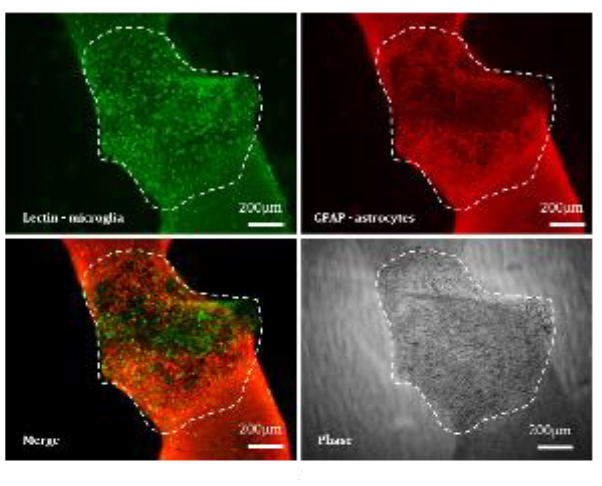
Uncrossing The Wires Of Human Specific Neural Network Development Using Multi-Electrode Arrays In Organoid Models
The mechanisms underlying human brain and network development, and how the processes leading to network establishment differ from other species, remain poorly understood. Human brain development is a protracted process compared to other apes and mammals and this may have implications for the efficiency of networks generated. This project aims to better understand whether a delayed neuronal maturation in humans facilitates the establishment of more sophisticated or efficient neural networks, and what cellular mechanisms may underlie this.
Methods:
Brain organoids were generated from ESCs or iPSCs of human or mammalian origin and cultured in suspension before stage-matched slicing for prolonged slice culture at the air-liquid interface (Air-Liquid Interface Cerebral Organoids). ALICOs were either placed on high resolution multi-electrode arrays (MEAs) for acute recording of electrical activity at different timepoints, or were recorded in a longitudinal fashion with custom built MEAs optimised for compatibility with ALICOs in their usual culture environment.
Long-term MEA-ALICO culture was evaluated for health and morphology using brightfield microscopy and immunohistochemical techniques. Organoid morphology with respect to neuronal tracts and connections was compared between species. Electrical activity was recorded and spike activity morphology, rate, and amplitude analysed and compared between species across time. Low frequency waves were recorded and mapped temporally.
Statistics:
In order to validate long term culture health, non-parametric t-tests were carried out on quantifications of morphological parameters between MEA-cultured ALICOs and controls. For quantifications of spiking data across time between species, multiple factor ANOVAs have been utilised. Statistical significance was marked at P<0.05. Similarly stringent tests are used in ongoing experiments.
Results and Conclusions:
Our novel designs permit longitudinal recording from the same samples, optimised for in-situ ALICO recording in their usual culture environment. Human organoids exhibit slower maturation compared to other species. Ongoing experiments seek to analyse and quantify connectivity and inter-species differences correlated with maturational delays.
Listening to the Experience of participants on Neurosurgical trials: outcomes of the LEARN-GDNF and LEARN- TransEuro studies
Introduction: Clinical trials involving direct intra-cranial delivery of novel therapeutics (including growth factor, cell and gene therapies) are filling the horizon for Parkinson’s disease. To give these trials the best chance of success participant recruitment and retention need to be maximised. Understanding the specific needs and lived experience of participants and their support network in coping with the significant burdens of neurosurgery, brain imaging and repeated ‘off’ assessments will serve to enhance development of acceptable, patient-centred trial designs.
Methods: Semi-structured online interviews were conducted with participants and their family members/ care partners following a trial of Glial Derived Neurotrophic Factor (GDNF) infused via intra-cranially implanted drug delivery catheters (Whone et al.) and also with participants of the TransEuro, a foetal cell transplantation trial (Barker et al.). Interviews were conducted alone or in dyads, observed by a second non-participatory researcher.
Analytical Approach: Interviews were transcribed verbatim and analysed using a reflexive thematic approach supported by NVivo software (statistical analysis is not applied to qualitative data).
Results: The experiences of participants were largely positive, expressing strong feelings of collegiality and altruism. Specific issues surrounding the nature and timing of assessments and post operative care were identified. A common thread relating to negative experiences lay with the difficulties participants faced once the trial had ended and the transition from participant to patient. This included the impact of learning about the negative outcome of the trial and decreased interactions with health care professionals following an intensive study period.
Conclusions: Listening to the voice of lived experience from complex trials is vital to enable trial design for maximum impact. Simple modifications to trial conduct can significantly improve the participant experience and study data have been used to generate resources to support this. Further, our data advocates for the inclusion of broad and diverse patient experience in the development of future studies.
Whone et al. Brain. 2019:142: 512–25 Barker et al. Nat Med. 2019;25(7):1045-53
Novel Approaches & Interdisciplinary Perspectives
The Perception Census: An extremely large-scale online study of perceptual diversity
Introduction: We all experience the world differently, but the nature and extent of perceptual diversity remain largely unknown. Here we introduce The Perception Census: an online research program part of the Dreamachine project, which itself was part of the UK Unboxed festival. The Perception Census aims to capture individual differences across different perceptual domains. Having already reached over 20,000 participants, the Census provides a unique opportunity to shed new light onto questions that have so far been examined in relatively small, highly specific samples. Methods: The Perception Census consists of 58 tasks divided into 11 sections investigating topics like the perception of colour, music, and time, as well as sensory precision, imagery, and susceptibility to illusions. Participants also complete tasks measuring traits including synaesthesia, phenomenological control, and sensory sensitivity. Completing the first section of the Census is mandatory, after which participants can freely explore the other sections. Data collection began in July 2022 and will continue until at least May 2023. So far, the first section has been completed by 20,219 participants, and each of the other sections by an average of 4,290 participants. In this presentation, we focus on the scope of the census and present preliminary participant statistics. Approach for statistical analysis: Some tasks were included in the Census with specific goals for confirmatory research while others were included for exploratory purposes to generate predictions for future studies. Due to the large scope of the project, we were unable to prepare preregistrations for confirmatory research before data collection. However, the predictions and statistical analysis plans for confirmatory research will be preregistered before carrying out analyses. Together with collaborators for each task, we will analyse a small random sample of the data related to each research question, to act as pilot data to form specific analysis pipelines. After submitting a pre-registration of predictions and analyses, full analysis can proceed. Upon completing preregistered analyses, we will make the data from the Census widely available, providing a valuable resource for researchers interested in perceptual diversity.
Dreamachine – a journey into the extraordinary potential of the mind
Introduction. Dreamachine is a major interdisciplinary programme bringing together neuroscientists, philosophers, artists, composers, and technologists, created by Collective Act. Its goal is to take audiences on a magical journey to reveal the power of their own minds and brains, to ignite mass curiosity about neuroscience and philosophy, and to generate a sense of awe, wonder and connection. The foundation of Dreamachine is that bright stroboscopic light on closed eyes can generate vivid visual and emotional experiences, as explored by the scientist Grey Walter and the artist Brion Gysin decades ago. Our Dreamachine reimagined Gysin’s original invention by creating a collective experience (20-30 people), utilising spatial sound, and situating the core experience in a neuroscientifically-informed audience journey. The programme also includes an (ongoing) study of perceptual diversity (The Perception Census) and a major learning programme, which has reached over 1 million young people in the UK.
Methods. We created Dreamachine using stroboscopic light sequences, fine-tuned to generate immersive visual hallucinations in most people without being overwhelming. We developed a pre-screening procedure to minimise risk (e.g. from undiagnosed photosensitive epilepsy), and we involved over 2,200 people in focus group testing (including groups of blind/partially-sighted people). We developed an interactive ‘sensory tool’ tablet-based interface surveying audience members about their Dremachine journey, and we provided opportunities to draw, write, and talk about their experiences.
Analysis. Full analysis is underway. Analysis will cover sensory tool data (linking to The Perception Census), image analysis of drawings, analysis of wider impact through longitudinal follow-up, and more.
Results. "One of the most indescribably profound experiences of my life." This testimony from one audience member echoes thousands of others. Over 30,000 people experienced Dreamachine, with audience reports being overwhelmingly positive. More than 15,000 drawings were created, showcasing a striking diversity in experience. Dreamachine is a unique science-art collaboration that has had transformative effects on thousands of people. It was commissioned by UNBOXED: Creativity in the UK.

Exploring the recognition of emotion from dynamic and static faces by people with Parkinson’s
Introduction
People with Parkinson’s (PwP) have been found to have impairments in recognition and production of emotional facial expressions. Although most studies use static stimuli to test for emotion recognition, dynamic faces are more realistic and allow for a natural display of emotion from neutral state. Previous research has shown that healthy participants are better at recognising emotion from dynamic faces compared to static faces; and preliminary work has suggested that this may also be the case for PwP (see Bek et al., 2020), despite evidence for general difficulties in emotion recognition. Therefore, we investigated emotion facial recognition from dynamic versus static faces by PwP and healthy controls.
Methods
We compared 42 PwP (mild-to-moderate severity) and 42 healthy controls (50 females and 34 males; age mean 65 years, SD=7.39). All participants performed an online computerised emotion facial recognition task. Nine different facial emotions (anger, contempt, disgust, embarrassment, fear, joy, neutral, sad, surprise) were presented in dynamic and static form in separate blocks of trials, at low and high levels of intensity.
Statistical analysis
A two-way mixed ANOVA was used [Group (Parkinson’s, controls) X Stimulus (dynamic, static)]. Results
There was a main effect of Group and a Group x Stimulus interaction. Controls had higher recognition accuracy than the PwP, but this was only significant for the dynamic stimuli. However, PwP had significantly higher recognition accuracy for static stimuli than dynamic; unlike the control group who had significantly higher recognition accuracy for dynamic than static stimuli.
Conclusion
PwP did not show the typical pattern of better recognition of emotions from dynamic stimuli, as seen for the controls. This suggests that PwP are more impaired in recognising dynamic facial expressions, which may relate to their difficulties in producing facial expressions. This is likely to have negative consequences for their everyday social interactions.
Investigating the effects of COVID-19 and the first UK lockdown on anxiety and mood in people with Parkinson’s
Anxiety is common in people living with Parkinson's (PwP) and a key contributor to poor quality of life. Anxiety has been related to the emotional and physical stress of living with Parkinson's. In this study we examined how anxiety and mood were affected in PwP during the UK’s first COVID-19 lockdown.
Methods
166 PwP (79 female) and 50 healthy controls (HC; 32 female) participated in an online survey. Standardised scales included anxiety during (state) and prior to (trait) lockdown, apathy, and depression. In addition, qualitative data from open questions about lockdown experiences was analysed using thematic analysis.
Statistical analysis
A two-way mixed ANOVA, a three-step hierarchical regression analysis was administered, and thematic analysis.
Results
Both groups reported significantly higher anxiety during compared to before lockdown; however, PwP had significantly higher anxiety overall (state and trait) and a higher proportion isolated early compared to HC. Depression and trait anxiety were both significant predictors for state anxiety in PwP. Additionally, PwP reported feeling vulnerable and experienced increased motor symptoms during lockdown, sometimes linking these effects to increased anxiety. Many described being negatively affected by lockdown, attributing this to low mood as well as reduced access to medical support, physical activities, personal space, and physical and social contact. Notably, some reported a positive experience during the lockdown due to fewer social obligations, and others reported no change in their lifestyle.
Conclusion
Both PwP and controls experienced higher anxiety during than before lockdown and some PwP thought this exacerbated their motor symptoms. While many PwP described negative effects during the lockdown, others
reported positive experiences or described little change since living with Parkinson’s was already a lockdown of its own. This highlights the need for remote support in general as well as in the case of another lock-down.
Origin of seizures in a mouse model of focal cortical dysplasia
Focal cortical dysplasia (FCD) is a leading cause of pharmacoresistant epilepsy, and is often associated with cognitive impairments and autistic features. It is characterised by cortical dyslamination and the presence of cytomegalic/dysmorphic neurons, and is caused in most cases by somatic mutations of genes encoding members of the mTOR signalling cascade. FCD-associated epilepsy is often a poor candidate for resective surgery because the dysplastic region typically occurs in an eloquent cortical territory, highlighting the need for an improved understanding of epileptogenic mechanisms in FCD, and the need for novel therapeutic strategies. Despite the overt structural abnormality, whether seizures arise from dysplastic neurons and their connections remains unclear.
Here we present electrophysiological and imaging data dissecting the contribution of heterotopic/dysmorphic and morphologically normal appearing neurons in the dysplastic region. We used a mouse model of FCD generated by in utero electroporation of a constitutive activator of mTOR (RhebS16H), which recapitulates many molecular, morphological, electrographic, and behavioural features of FCD type II in patients, including subclinical electrographic abnormalities.
Whole cell patch-clamp recordings of dysplastic neurons obtained in acute brain slices from FCD mice revealed profound intrinsic hypoexcitabilty, while neighbouring, morphologically normal cells show reduced rheobase, increased membrane resistance, and higher stable firing rates than control cells. However, dysmorphic neurons functionally integrate in the cortical network receiving spontaneous excitatory and inhibitory input. Following a test for normal distribution and prior assumptions, parametric statistical tests were used to assess difference and significance. We also recorded spontaneous activity in both dysplastic and non-dysplastic neurons using single-cell calcium fluorescence imaging in freely moving animals, during both interictal and ictal periods. Here we compared basic excitability in the populations by measuring calcium event frequency and AUC. Deconvolutions of the calcium signal were used to more accurately compare internal synchrony and responsivity of the populations during different behaviours. This experiment allows us to capture, in a naturalistic setting, the involvement of both dysmorphic and normal-appearing neurons in seizure generation and maintenance, and to infer the cellular and network basis of seizures in FCD.
Gene Therapy for Focal Cortical Dysplasia type II
The Focal Cortical Dysplasias (FCDs) are a group of malformations of cortical development, which are frequently associated with drug-resistant epilepsy in children. FCD type II is the commonest pathology found at epilepsy surgery in children. It is typically caused by somatic mutations resulting in the mammalian Target Of Rapamycin Complex 1 (mTORC1) hyperactivation. Epilepsy surgery is not always effective and is often precluded by proximity to eloquent brain regions. Gene therapy is thus currently the most promising candidate replacement for surgical treatment of FCD.
We tested a gene therapy approach based on viral expression of a modified KCNA1 gene (which encodes the potassium channel Kv1.1). We chose Kv1.1 as a therapeutic protein not only because it attenuates neuronal excitability, but also because mTOR hyperactivation is associated with downregulation of KCNA1 expression.
The therapy tested in a mouse model of FCD type II generated by in-utero electroporation of neuronal progenitors with a constitutively active RHEB plasmid, an activator of mTORC1. A battery of molecular biology and behavioural tests confirmed that the model recapitulates the pathology seen in humans, including cognitive comorbidities.
Continuous EEG recordings from mice before and after local injection of AAV9-KCNA1, showed a 64% decrease in seizure frequency over 4 weeks (n=13 mice), which was not seen in mice injected with a control AAV encoding a fluorescent reporter alone (n=11 mice). We observed no worsening of performance in tests sensitive to frontal lobe function, including spontaneous alternation in T-maze and olfactory habituation and discrimination.
In conclusion, KCNA1 gene therapy is a promising genetic approach for the treatment of FCD type II.
Neuroprotective effect of prolyl hydroxylase inhibition in an in vitro rat oxygen-glucose deprivation model
Stroke is one of the leading causes of death and disability worldwide. Targeting prolyl-hydroxylase inhibition has been shown to be a successful approach in ischemia. Therefore, this study investigated the effects of the prolyl- hydroxylase inhibitors Roxadustat and JNJ-42041935 in an oxygen-glucose deprivation (OGD) rat stroke model. Field excitatory post-synaptic potentials (fEPSPs) were elicited by stimulation of the Schaffer collateral pathway in rat hippocampal slices. During OGD slices were perfused with glucose-free aCSF bubbled with 95% N2/5% CO2 for 20min. LTP was elicited by high frequency stimulation (HFS) consisting of 3 trains of 100Hz with a stimulus interval of 20s. Organotypic hippocampal slices (P7 rat pups) were exposed to 1 hr of OGD before staining with 2µM Propidium Iodide (PI). All data are presented as mean value±SEM. Analysis was performed using unpaired t-test and one-way ANOVA. Incubation with Roxadustat (100µM) did not modulate the OGD-induced depression (16.5±1.0% vs controls 21.2±3.0%, n=6) but significantly improved the synaptic transmission during 40min recovery (120.3±18.1% vs controls 70.0±11.3%). Since Roxadustat is a potent iron chelator and ferroptosis is a well-known mechanism of neuronal damage in ischemia, we evaluated whether iron chelation was involved in the modulation of synaptic transmission. Application of Roxadustat did not reverse the inhibitory effect of Fe3+ (100µM) in our OGD experiments. However, application of Roxadustat impaired LTP (111.0±7.0% vs controls 138.0±7.0%, n=5, at 60 min). JNJ-42041935 (10µM) did not reverse the synaptic depression observed during OGD (28.0±4.1% vs controls 21.2±3.0%, n=4) but did improve synaptic transmission recovery (116.4±7.0% vs controls 70.0±11.3%). In the organotypic slice cultures, pre-treatment with Roxadustat and JNJ-42041935 improved neuronal viability compared to untreated slices (19.5±3.0% and 11.0±2.0% vs OGD 41.0±7.0%, n=9). In conclusion, our electrophysiological results have demonstrated a modulatory role for the two PHD inhibitors in hippocampal synaptic transmission during OGD. Also, the PI data highlighted a protective effect for both compounds. Further research will be required to elucidate the exact mechanistic action of the compounds during OGD.
The Neuroscience community's attitudes towards green Neuroscience
Introduction
Earth has reached the point of being in a climate crisis, leading to an uninhabitable planet (Ripple, W.J. et al. (2021) “World scientists’ warning of a climate emergency 2021,” BioScience, 71(9), pp. 894–898.). If humans increased sustainable behaviour the negative effects of the climate crisis could be averted or reversed, including the negative impact on mental wellbeing. This project aims to provide insight into the attitudes and behaviours of the neuroscience community with respect to ‘green neuroscience’, i.e. the environmental sustainability of the neuroscience sector.
Methods
An online survey has been designed to collect data from members of the neuroscience community. Requests to complete the survey have been emailed to members of the British Neuroscience Association (BNA) as well as posted on social media. The survey includes collection of demographic data to enable identification and sub-categorisation of respondents. Remaining questions collect information on green neuroscience with respect to (1) place of study/work; (2) sustainability of the research process itself; (3) attending events; (4) the local community; and (5) societies and the BNA; as well as (6) open-ended text questions.
Approach for statistical analysis
Data will be analysed differently depending on question type. Null hypothesis significance testing (NHST) is appropriate for six of the survey questions. For five of these, parametric testing will be used: the independent two- sample t-test, with sample sizes calculated assuming a one or two-tailed hypothesis with an effect size of 0.5, α≤0.05 and power of 0.8. The remaining question will use Pearson’s r formula to uncover any correlations between the variables, with sample sizing decided assuming an effect size of 0.5, α ≤0.05 and power of 0.8. The statistical analysis software will be JASP. The open-ended text questions will require thematic analysis to help identify patterns of behaviour. Chosen codes will include ‘change’, ‘sustainability’ and ‘motivation’. Chosen themes include ‘prevention of change’; ‘motivation for change’. Primary outcome will be different for each question. The survey as a whole will allow better understanding of the neuroscience community towards their sector’s environmental sustainability.
Measuring and reducing the carbon footprint of fMRI analysis
Introduction - Data storage and processing contribute to the climate crisis through the carbon footprint associated with energy use. Neuroimaging research frequently relies on large datasets and computationally expensive analysis. We will investigate how the carbon footprint of fMRI analysis can be measured and reduced, providing tools and guidance to make analysis more sustainable without inconveniencing researchers. Method - We will investigate two strategies. (i) Modifying preprocessing pipelines to remove steps that increase energy consumption but do not significantly improve data quality. In 100 subjects from the Human Connectome Project, using data for a motor and an emotion processing fMRI task, we will quantify such trade-offs for the fMRIPrep pipeline. Multiple variants will be run, with preprocessing steps differentially toggled on or off. (ii) Scheduling analysis to run at times at which the carbon intensity of the UK National Grid is low, using real-time public data. Carbon intensity peaks at the start and end of the working day and is lowest in the middle of the night. Approach for Statistical Analysis - For step (i), task activation in regions typically associated with a given task (e.g., primary motor cortex for the motor task) will be extracted using group-level ROI analysis in FSL following each fMRIPrep variant. Smoothness of resulting fMRI data will also be extracted. These proxies for preprocessing performance will be plotted against estimates of carbon emissions from a given pipeline, measured by CodeCarbon (Goyal-Kamal et al., 2021). ANOVA and post-hoc contrasts will be run between pipelines for performance metrics and carbon emissions. The elbow point, where additional preprocessing steps confer negligible benefits in performance but significant increases in carbon emissions, will be identified. Step (ii) will contextualise the CodeCarbon estimates of a given subject’s data relative to historical carbon intensity data, allowing one to identify time windows at which the carbon intensity of a given analysis step will likely be lowest.
Goyal-Kamal, F. B., Schmidt, V., et al. (2021). CodeCarbon: Estimate and track carbon emissions from machine learning computing. Zenodo. Available at: https://doi.org/10.5281/zenodo.4699491
Aerobic exercise improves dorsal hippocampal CA1 synaptic plasticity and novel object memory in the sub-chronic phencyclidine rat model for schizophrenia
Introduction
Evidence suggests that aerobic exercise improves neurocognition and increases relative hippocampal volume in patients with schizophrenia. Here, we introduced chronic exercise to the sub-chronic phencyclidine (scPCP) rat model for schizophrenia to investigate its effect on the cognitive impairment associated with schizophrenia.
Methods
40 Female Lister Hooded rats were dosed with either saline or PCP (2 mg/kg, i.p.) twice daily for seven days. Running wheels were provided for aerobic exercise, 1 hour daily for 30 days. Novel object recognition memory was measured after 6 weeks of exercise. Synaptic plasticity in the dorsal hippocampus was then assessed via acute in vivo electrophysiology under urethane anaesthesia (30% w.v.; 1.4g/kg i.p.). The hippocampal CA1 Schaffer-evoked field excitatory postsynaptic potential (fEPSP) was analysed to measure short and long-term synaptic plasticity. Data were analysed with Student t-test and mixed-effect three-way ANOVA.
Results
Behavioural testing after exercise showed improved novel object recognition memory in the exercised scPCP group. The nonexercised scPCP group showed smaller potentiation after high-frequency stimulation compared to the other three groups, especially for response amplitude. However, the exercised scPCP group showed similar potentiation to the control group. Effects on short-term plasticity will also be presented. In conclusion, chronic aerobic exercise appears to rescue cognitive and hippocampal synaptic deficits in the scPCP rat model.
Disturbed basal mitophagy and neuronal activity in Parkinson’s patient-derived dopaminergic neurons with an α- synuclein gene triplication mutation
Introduction
α-Synuclein accumulation in Lewy bodies in the brain is one of the hallmarks of Parkinson’s disease (PD), which predominantly affects dopaminergic neurons in the substantia nigra pars compacta. α-Synuclein has been linked to dysfunctional degradation of mitochondria or mitophagy and impaired neuronal activity, however the exact mechanisms how α-synuclein contributes towards these phenotypes are poorly understood. Here, we have characterised induced pluripotent stem cell (iPSC)-derived dopaminergic neurons from a patient with an α-synuclein (SNCA) gene triplication mutation, which causes a rare form of early-onset familial PD, and compared these to neurons from healthy control volunteers.
Methods
All iPSC lines were derived from dermal fibroblasts from healthy donors or PD patients which have been recruited through the Discovery clinical cohort and gave signed informed consent (Ethics committee: National Health Service, Health Research Authority, NRES Committee South Central, Berkshire, UK, REC 10/H0505/71). The live cell mt-Keima mitophagy assay was used to measure basal mitophagy and PINK1-Parkin dependent mitophagy by treatment with the mitochondrial uncoupling agent CCCP. Neuronal activity was assessed from D30-D90 post-differentiation using a multi-electrode array (MEA) system.
Statistical Analysis
mt-Keima results were analysed by one-way ANOVA. MEA results were analysed by two-way ANOVA.
Results
We find that under basal conditions SNCA triplication neurons show an increase in mitochondria located in lysosomes compared to control neurons 35-50 days post-differentiation. No significant changes were seen in mitophagy induced by mitochondrial membrane depolarisation, suggesting that this process is PINK1-Parkin independent. A neuronal activity assay using multi-electrode arrays (MEAs) revealed that SNCA triplication neurons have significantly reduced firing rates and bursting events from D60 post-differentiation onwards to D90. These combined results suggest that mitochondrial dysfunction is likely to precede impairment of neuronal activity. Further investigations are underway to pinpoint how α synuclein deregulates basal mitophagy and to elucidate its connection to the observed reduction in neuronal activity.
Autophagy modulation using small molecules in LRRK2 Parkinson’s patient fibroblast-derived dopaminergic neurons
Parkinson’s Disease (PD) is the second most common neurodegenerative disease, characterised by degeneration of dopaminergic neurons. The causes of PD are not fully understood; however, some familial types of PD are due to mutations in several genes, many of which have a role in autophagy and mitochondria.
For this study we use patient fibroblast-derived dopaminergic neurons (iDANs) harbouring the LRRK2 G2019S mutation, which is the most common inherited, autosomal dominant cause of PD. The iDANs were generated from LRRK2 mutant patient fibroblasts via cellular reprogramming methods which retain ageing features.
We aim to investigate how modulating the autophagy pathway can potentially ameliorate the PD phenotype. Moreover, we are working with an industry partner to validate novel potential therapeutic targets.
We have observed increased autophagosomes and autolysosomes levels in the LRRK2 mutant iDANs compared to control, with an increased number of lysosomes and mitochondria per cell as well. Furthermore, we found decreased mitophagy rates in the LRRK2 mutant iDANs. These data are critical to understanding endogenous functions of LRRK2 in patient derived dopaminergic neurons.
All the data were normalised to the Healthy control vehicle and the means were compared by two-way ANOVA following by the Dunnett's multiple comparisons test.
Furthermore, we sought to investigate the effect PDPK1 and PI3KCB inhibitor compounds on these phenotypes in our patient derived system. PDPK1 and PI3KCB are two proteins that play a key role in the autophagy pathways. We found these two small molecules are able to reduce some of the autophagy and mitophagy alterations seen in iDANs.
Next, we will investigate if these small molecules can also ameliorate the PD phenotype improving the mitochondrial functionality i.e., either increasing the mitochondrial membrane potential or shaping the mitochondrial morphology.
Effect of LRRK2 inhibitors on autophagy and mitophagy in LRRK2 patient fibroblast-derived dopaminergic neurons
Parkinson’s Disease (PD) is a progressive neurodegenerative disease characterised by the loss of dopaminergic neurons in the substantia nigra pars compacta (SNpc). It is the 2nd most common form of neurodegeneration and is multifactorial, having genetic and non-genetic causes. Notable mechanisms involved PD development include the accumulation of misfolded proteins aggregates (alpha-synuclein), autophagy impairment, and mitochondrial and lysosomal dysfunction.
This study used patient fibroblast-derived dopaminergic neurons (iDANs) possessing the LRRK2 G2019S mutation. The iDANs were generated from LRRK2 mutant patient fibroblasts via cellular reprogramming methods which retain ageing features. LRRK2 mutations are one of the most prominent monogenetic risk factors for PD linked to both familial and sporadic forms of the disease. The LRRK2 G2019S “gain of function” mutation occurs in the LRRK2 kinase domain and leads to pathogenic kinase activity via aberrant phosphorylation, which in turn increases lysosome formation and alters autophagy. Therefore, to better understand endogenous functions of this phenotype and cellular pathways, we used three LRRK2 inhibitor compounds on LRRK2 mutant iDANs.
For the parameters we investigated regarding autophagy and mitophagy, data was normalised to the vehicle control and analysed using a two-way ANOVA, followed by Dunnett’s Test for Multiple Comparisons.
Compared to the healthy control, the LRRK2 mutant iDANs have shown increased lysosomal and mitochondrial numbers per cell, decreased autophagy rates and increased LC3 puncta and autophagy levels. We found that LRRK2 inhibitors improve autophagy and mitophagy related alterations within our mutant line, with GSK2578215A having the most significant effects in a dose-dependent manner.
Our findings demonstrate that treatment with LRRK2 inhibitor small molecules reverses pathological phenotypes observed in autophagy and mitophagy. Future work will focus on investigating mitochondrial functionality via membrane potential and utilising live assays to assess the functional properties and morphology of the LRRK2 iDANs treated with LRRK2 inhibitors.
Primary Screen of Novel Compounds for the Treatment of Parkinson’s Disease
Introduction: Parkinson’s Disease (PD) is the second most common neurodegenerative disease worldwide, caused by the loss of dopaminergic neurons in the substantia nigra. There are currently no licensed compounds for PD treatment capable of modifying the disease progression. We have previously identified the naturally occurring bile acid ursodeoxycholic acid (UDCA) as boosting mitochondrial function in PD patient cells, subsequently we screened 122 bile acid metabolites and synthetic derivatives and identified 5 top performing compounds capable of recovering mitochondrial defects seen in PD patient fibroblasts. Here we have performed a primary screen of 82 analogues and metabolites of two of these top five compounds in sporadic (sPD) patient fibroblasts for effects on mitochondria and ATP production.
Methods: Three age- and sex-matched pairs of sPD patient and healthy control fibroblast cell lines were used to screen 82 compounds for restorative activity in mitochondrial membrane potential (MMP) and total cellular ATP production. Compounds were tested at 100nM and 1uM. Lead compounds were tested in an Agilent XF Seahorse MitoStress assay to detect any possible effects on mitochondrial respiration. Each compound was assessed compared to the positive effect of UDCA which we have previously published.
Approach for statistical analysis: All compounds were tested with three independent biological repeats (n=3). Screening data was analysed for compounds restoring mitochondrial parameters at least 1 standard deviation and 2 standard deviations above the vehicle treated mean in each sPD patient line; then assessed for a positive effect across multiple lines. Seahorse data was analysed using 2-way ANOVA with multiple corrections to identify compounds capable of significantly increasing or decreasing respiratory chain parameters.
Results and conclusions: A total of 26 compounds were found to increase total cellular ATP production and 13 to improve MMP, without causing toxicity in the sPD fibroblasts. These compounds improved ATP levels or MMP to above vehicle control sPD levels, with some returning levels to those of control cell lines and will be further assessed in dose-response and downstream assays to identify mechanisms of action. Two priority compounds assessed
Conflict of interest: HM and OB are in receipt of research funding from NZP UK Ltd. which co-funds LH's studentship.
Stratifying People with Sporadic Parkinson’s Disease by Pathological Mechanism in Patient-Derived Fibroblasts
Objectives
To stratify a new cohort of Sporadic Parkinson’s disease (sPD) patients into small, homogeneous subgroups defined by specific mitochondrial and lysosomal dysfunction.
Background
sPD is widely recognised as a heterogeneous disorder, both clinically and mechanistically. Several dysfunctional mechanisms have been identified, including mitochondrial and lysosomal dysfunction. However, clinical trials select participants without considering mechanism heterogeneity and continually fail to reach efficacy outcomes.
Methodology
We investigated mitochondrial and lysosomal dysfunction, two key mechanisms of sPD and promising targets for therapeutics. Imaging and biochemical assays assessed mitochondrial and lysosomal health parameters in fibroblasts from a new cohort consisting of 35 sPD patients and 24 healthy individuals. The sPD population was then stratified by assessing patterns of dysfunction across these parameters. Validation of these subgroups is currently being undertaken in the patient fibroblasts and induced Dopaminergic Neurons.
Statistical Analysis Approach
The results for each parameter were transformed to z-scores in proportion to the entire control population. Composite z-scores across triplicate repeats were calculated, using the sum of the Pearson correlation coefficients between all components, and was repeated for both media conditions to provide a single z-score for each parameter per cell line. Population differences were then interrogated using the T-test and F-test of equality of variance.
Results
The sPD population was heterogeneous in 88% of mitochondrial and lysosomal parameters. Stratification of this cohort by distinct patterns of dysfunction identified four unique subgroups, defined by mitochondrial dysfunction, lysosomal dysfunction or both. The top 3 patients in each subgroup were selected for validation and mechanism studies, which discovered a significant reduction in maximal respiration in the pure mitochondrial dysfunction subgroup.
Conclusion
This study suggests that it is possible to stratify cohorts of sPD patients providing a possible model for clinical trial recruitment in order to aid the effectiveness, and therefore approval, of new therapeutics and support an approach to personalised treatment plans.
MicroRNA and gene expression profiles in a Parkinson’s disease cell model
Introduction: Parkinson’s disease (PD) is a neurological condition that predominantly effects movement. It is estimated that 1% of the population over 65 will develop PD, rising to 3% by the age of 85. Analysis of MicroRNA (miRNA) may highlight the genetic mechanisms that contribute to pathogenesis of PD. It has been suggested that miRNA regulate up to 60% of protein coding genes and the dysregulation may contribute to the pathogenesis of different disease states within the body. Identification of miRNA expression profiles may allow for the early identification of PD, potentially allowing for earlier medical intervention before the disease onset. The aim of this study was to analyse the expression of miRNAs and the possible interaction of genes in cells of dopaminergic phenotype after they have been subjected to a mitochondrial or proteasomal inhibitor.
Methods: The SH-SY5Y cells were exposed to the mitochondrial neurotoxin MPP+ and proteasome inhibitor MG132 for 24 hours to result in 50% cell death. The cells were extracted using TRIzol and purified using the mirvana kit.
Following isolation and purification, reverse transcriptase quantitative polymerase chain reaction was conducted to observe the possible dysregulation of the genes, SNCA, ITPR1 and CACNA1C and the PD associated miRNA: miR-107, and miR-128a, in the absence and the presence of MPP+ or MG132.
Approach for statistical analysis: Paired t-test was used to observe the 2-ΔΔCt for expression variation between
wildtype cells and drug exposed cells.
Results and conclusions: The three genes and miR-107 exposed to MPP+ were all significantly down regulated (p<0.05), SNCA (90.94%), ITPR1 (56.43%), CACNA1C (79.72%) and miR-107 (74.56%). There was a significant
downregulation (p<0.05) of SNCA (87.11%) and miRNAs, miR-107 (95.71%) and miR-128a (62.09%) that had exposure to MG132. Interestingly, both MPP+ and MG132 caused a similar level of decreased expression in SNCA, although both drugs have a different mode of action. Based on current findings, the expression data generated indicate that both drugs may cause significant down regulation of specific genes and miRNAs within the cell potentially identifying a contributing factor associated with the pathogenesis of PD.
Moving and music in my head: Vividness and use of imagery related to music and movement in people with Parkinson’s
Introduction: Parkinson's disease affects multiple aspects of movement, impacting significantly on everyday tasks. Music is used in interventions for people with Parkinson’s, either to pace movements, or as an integral element of activities such as dance. This study explored self-reported vividness of motor imagery (imagined movements) evoked by music and auditory (including musical) imagery in people with Parkinson’s, and whether and how they use these types of imagery in everyday life. We investigated whether vividness and use of imagery were associated with prior music and dance experience.
Methods: Participants (N=199) completed an online survey including: (i) vividness ratings of visual and kinaesthetic music-evoked motor imagery, (ii) vividness of auditory imagery, and (iii) ratings and open questions about their everyday use of these types of imagery.
Approach for statistical analysis: This study followed a pre-registered analysis plan (logged on OSF). We used correlations to explore relationships between imagery vividness and use and (a) severity of Parkinson’s symptoms, (b) musical and dance experience.
Results and conclusions: While most participants reported experiencing music-evoked motor imagery (with more vivid visual than kinaesthetic motor imagery), <20% reported actively using music to support motor imagery in daily activities. In contrast, participants reported a broad range of contexts and uses for musical imagery (imagined music), from supporting movement (e.g., walking or exercise) to emotion regulation and concentration. Correlational analyses associated music-evoked motor imagery with an urge to dance and musical training, while the use of musical imagery was associated with singing ability. A minority of participants reported not experiencing either motor or musical imagery, suggesting that interventions based on imagery may not be suitable for all. Nonetheless, even participants with more severe motor symptoms reported both types of imagery, indicating promise for their strategic use at different stages of Parkinson’s. Indeed, musical and motor imagery have the potential either separately or combined to support rehabilitation strategies for people with Parkinson’s.

Music, Movement, Mood & Parkinson's: A transdisciplinary PPI approach to optimizing the use of music in rehabilitation for people with Parkinson’s
Introduction
Medication/surgical options do not ameliorate all symptoms in Parkinson's so adjunct therapies are needed to improve quality of life (QoL). Therapeutic approaches involving physical activities and music can increase QoL for people with Parkinson's (PwP; Karageorghis et al., 2020). This research study will test a new music-based group intervention (Songlines) designed for and with PwP to improve mood and movement.
Methods
An inclusive approach (Patient and Public Involvement, PPI) working with PwP, practitioners and medical professionals was used to create context appropriate materials for the new intervention. This involved 15 workshops and 28 interviews exploring the use of music in relation to potential rehabilitative mechanisms, and two online surveys documenting the use of music in everyday life by Swiss and British PwP (Rose et al., 2022).
The Songlines intervention using the co-created materials will now be evaluated using a within-subject repeated measures design (N=72). Mixed methods involving a newly developed protocol integrating a pressure sensitive gait mat with motion capture technology will record change over time in functional mobility. Standardized measures (e.g., PDQ-39, MDS-UPDRS) and qualitative methods will track change over time in relation to motivation and mood.
Statistical analyses
The systematic development process has produced a protocol that directly links tasks with outcomes, thereby generating a series of testable hypotheses. Repeated measures ANCOVAs will be conducted on the quantitative data, while thematic analyses of qualitative data will provide insight into participant experiences.
References
Karageorghis, C. I., Rose, D., Annett, L. E., Bek, J., Bottoms, L., Lovatt, P. J., Poliakoff, E., Schultz, B. G., Whyatt, C. P., Young, W. R., & Delevoye-Turrell, Y. N. (2020). The BASES Expert Statement on the Use of Music for Movement among People with Parkinson’s. The Sport & Exercise Scientist, 63, 6-7.
Rose, D.C., Poliakoff, E., Hadley, R., Guérin, S.M.R., Phillips, M., & Young, W.R. (2022). Levelling the playing field: The role of workshops to explore how people with Parkinson's use music for mood and movement management as part of a patient and public involvement strategy. Front. Rehab. Sco, 3:873216.
Impact of global signal regression on connectome predictive modelling in autism spectrum disorder
Introduction
Connectome-based predictive modelling (CPM; Shen et al. 2017) is a recently developed method that enables researchers to establish brain-behaviour predictive relationships based on neuroimaging data. However, there is little consensus on how to best preprocess the input neuroimaging data. One of the most controversial steps is global signal regression (GSR), which involves extracting the shared variance from the voxelwise fMRI time series. Although this step can eliminate artifacts such as head movement, respiration and cardiac cycles, others claim that by removing the global signal, important neural information is lost (Murphy et al., 2017). Our goal is to investigate the impact of GSR on CPM predictive power and predictive networks identified.
Methods
We will use the ABIDE dataset with 573 controls and 539 autism spectrum disorder (ASD) patients (Craddock et al., 2013). Preprocessed resting-state fMRI data with and without GSR will allow for a comparison between models predicting clinical diagnosis based on each type of data. We will apply a modified version of CPM. Traditionally, CPM is used to predict continuous values: we aim to predict diagnostic categories instead.
Approach for statistical analysis
Analysis steps include a) using t-tests to identify edges predictive of clinical diagnosis, b) creating a summary value for each participant with the predictive edge weights, c) fitting a classification model to the training data, d) 10-fold cross-validation in novel subjects, using the model generated in the previous step and e) testing performance, by comparing predicted to actual diagnosis for new participants with accuracy, sensitivity and specificity measures. This procedure will be done across both datasets: once using data with GSR and once using data without GSR. Differences in predictive power between data with and without GSR will be evaluated using permutation testing. Overlap between predictive networks derived from the data with and without GSR will be quantified using the Dice coefficient. For further details, see Fig. 1.
References
Craddock, C., et al. Frontiers in Neuroinformatics, 7, 27. (2013) Murphy, K., & Fox, M. D. NeuroImage, 154, 169-173. (2017) Shen, X., et al. Nat Protoc 12, 506–518. (2017)

The dependence of network organization on overall functional connectivity and its impact on classification of autism spectrum disorder
Introduction
Graph theory is a key approach to study brain connectivity. However, some network measures are strongly correlated with overall functional connectivity, and accounting for this reduces group differences (van den Heuvel et al., NeuroImage 2017). These findings raise questions about the diagnostic and prognostic relevance of graph theory. Individual differences in overall functional connectivity can be removed by applying global signal regression (GSR), a controversial pre-processing method that removes noise but potentially also valuable neuronal information. An alternative method is mean regression (MR) to remove shared variance between mean functional connectivity and edge weight across participants. Our aim is to investigate the dependence of network organisation on overall functional connectivity and its impact on classification of autism spectrum disorder (ASD), a neurodevelopmental condition in which atypical functional connectivity patterns have been observed (Keown et al., Biol. Psych. CNNI 2017).
Methods
Resting state fMRI data (N = 785) from the Autism Brain Imaging Data Exchange (ABIDE) dataset pre-processed with the Connectome Computation System (CCS) pipeline and parcellated using the Harvard-Oxford atlas will be used.
Connectomes will be built using Pearson’s correlation, for both raw (Not Regressed, NR) and GSR data. Mean regression will be applied to NR connectomes. Each individual NR, MR, and GSR connectome will be proportionally thresholded to retain 5%, 15%, or 25% of the strongest edges. Three representative graph theoretic measures – global clustering, assortativity and characteristic path length – will be calculated using each thresholded connectome.
Statistical analysis
Mann Whitney U tests will be used to assess the effect of MR and GSR on group difference in network measures between ASD patients and Typical controls (TC). The proportion of shared variance between NR, MR and GSR measures and mean functional connectivity will be quantified using simple linear regressions. A kernel Support Vector machine (kSVM) applied using 5-fold cross-validation will be used to evaluate the relevance of graph theoretic measures in the phenotypic categorization of ASD patients, comparing NR, MR, and GSR thresholded connectome.

Cerebral haemodynamic correlates of altered consciousness states induced by High-ventilation breathwork
INTRODUCTION
Therapeutic breathwork practitioners who employ hyperventilation-like breathing (HVB) techniques report profound subjective effects resembling aspects of altered states of consciousness (ASC) induced by 5-HT2A agonists such as Psiloycbin. We hypothesize that altered cerebral haemodynamics induced by prolonged voluntary hyperventilation might be implicated in the emergence of these subjective effects.
METHODS
19 healthy participants with 6-months+ experience in HVB were recruited. Participants performed 20 minutes of normal breathing followed by 25 minutes of HVB facilitated by breathing instructions, whilst undergoing MRI scanning. Cerebral blood flow (CBF) was measured using pseudo-continuous arterial spin labelling (pcASL) at baseline (BASE), and twice during HVB, i.e., immediately after starting HVB (START HVB), and after 6 minutes of uninterrupted HVB (SUSTAINED HVB). Capnograph measurements confirmed end-tidal CO2 remained below 20mmHg during SUSTAINED HBV. Subjective ratings of ASC were collected via the 5 dimensions of ASC questionnaire.
ANALYSIS
CBF maps were partial volume corrected and normalised in MNI space. Differences between each of the HBV phase and BASE were tested using paired-sample t-tests, and voxelwise maps of delta CBF were computed. Subjective measures were retrospectively assessed using the ASC domain of the 5-Dimensional ASC Rating Scale and correlated to deltaCBF.
RESULTS AND CONCLUSIONS:
We observed time-dependent reductions of CBF by HBV, relative to BASE. CBF was reduced by 30.6% and 41.9% during START HVB and SUSTAINED respectively.
ASC scores were correlated to deltaCBF in the left posterior insula both at the start of HVB and SUSTAINED HVB, whilst they correlated to deltaCBF in the anterior cingulate only in the SUSTAINED HVB. Our findings indicate that rCBF reductions in areas critically involved in conscious and affective representations of internal states, namely the dorsal and posterior insula and the anterior cingulate cortex, are related to subjective reports of ASC. It is plausible that the progressive disruption of insular - cingulate interactions compromises central aspects of self-representation and ultimately leading to short-lived ASC-like experience.

The Effect of C02-Induced Anxiety on Functional Connectivity
Background
The CO2 challenge is an experimental model of anxiety in which participants breathe in an increased level of CO2 (typically around 5-9%), which induces a temporary state of increased anxiety. While the subjective and cognitive effects of the CO2 model have been well studied, there is limited research showing how the CO2 model affects the brain. Such a model has great potential for advancing our knowledge of the neural basis and treatment targets for anxiety disorders. Here we will characterise the brain networks underlying CO2 -induced anxiety by measuring changes in functional connectivity (FC) using resting-state fMRI during the administration of elevated levels of CO2.
Methods
Participants (n=20) will complete the State-Trait Anxiety Inventory (STAI) to obtain an established measure of trait anxiety. Prior to participation in the MRI component, a brief (2-minute) exposure to the CO2 model will be used to screen for intolerance to the protocol. Participants will then complete the MRI experiment consisting of a high- resolution structural scan (MPRAGE) followed by blinded exposure to medical air and an elevated (6%) CO2 mixture. Gasses will be delivered to a mouthpiece via a Douglas bag. Heart rate and blood oxygen levels will be recorded continuously throughout the scan session, for both participant safety monitoring and physiological analysis.
Subjective rating of state anxiety will also be recorded pre/post-inhalation phase, using the STAI and a standard 10- point rating scale.
Proposed analysis.
A general linear model will be used to determine the relationship between FC, [pre, peri, and post] anxiety measures, and physiological recordings. The analysis will include: (i) whole brain voxel-wise analyses, (ii) Seed-based and ROI-ROI (using limbic and HPA structures such as amygdala and hippocampi), and (iii) Dynamic connectivity measures to observe the evolution of FC over time due to CO2-induced anxiety. These findings will advance our understanding of the neuropsychophysiological mechanisms of acute and pathological anxiety, and further, validate the CO2 experimental model as a valuable research tool.
The Effect of Mindset on Anxiety and Stress Regulation: A Virtual Reality Biofeedback Intervention
As a promising tool to relieve anxiety and stress, game-based virtual reality (VR) biofeedback represents individuals’ real-time physiological data within an immersive, dynamic, and interactive environment. Users are required to learn to monitor and regulate their physiological responses in order to successfully overcome challenges posed by the game environment. While biofeedback aims to facilitate favourable behavioural modification for emotion regulation, performance in biofeedback training is variable across individuals, which could be partly accounted for by implicit theories relating to individuals’ “mindset”: the perceived stability of human attributes. This social-cognitive construct suggests that individuals’ prior experiences, knowledge, and expectations may profoundly shape their perception towards the malleability of emotional regulation skills, and in the context of this study, whether the ability to control physiological responses could be learnt through effort.
This study examined the effect of mindset on the ability to control heart rate and regulate stress levels in a novel VR biofeedback game. Healthy participants (n = 53) took part in a 5-minute biofeedback training and a subsequent 10- minute stress-inducing game in which self-regulation was required to ensure survival. Photoplethysmography was measured throughout the game experience to extract heart rate recordings and stress scores. Mindset scores were assessed by Implicit Theories scales at baseline and post-intervention. Pearson’s correlations were used to examine the relationship among stress and pre-intervention anxiety mindset as well as the overall mindset. Paired-sample t- tests were used to further explore the possibility of a two-way relationship by examining whether experience of the biofeedback training had an impact on mindset. The biofeedback game was associated with a significant shift towards growth mindset. The change in mindset was also associated with lower levels of stress, regardless of their starting mindset. These early results suggest the potential of biofeedback games for improving mindset and anxiety symptoms warrants future investigation
Interoception and a 4-day working week
1) Introduction
Interoception is the ability of the brain to track internal bodily states, with the aim of responding appropriately in order to achieve homeostasis. Interoception has been associated with cognitive and emotional processes, while dysfunction in the interoceptive system plays a central role in the aetiology of mental health disorders. Sleep quality and stress, which are influenced by occupational status and working patterns, have been linked to changes in specific dimensions of interoception. In the current study, we investigated whether a 4-day working week impacted interoceptive function in a healthy population.
2) Methods
20 employees in businesses switching from a 5-day to a 4-day working week with no loss of salary were recruited. Participants performed an interoceptive focus task while undergoing fMRI, in addition to out-of-scanner heartbeat perception tasks and interoceptive questionnaires, before and after a 12-week trial of a 4-day working week.
3) Approach for statistical analysis
The fMRi data obtained during the interoceptive focus task was analysed with first- and second-level models. We assessed which brain areas were active during the interoceptive-focus task and if there was any significant change in task-related activity after the 4-day working week trial period.
T-tests were used to investigate whether there were significant changes in the heartbeat perception tasks and interoceptive questionnaires as a result of a 4-day working week. Linear regression was used to detect the presence of subgroups that might benefit most from a 4-day working week.
4) Results and conclusions
The mid and posterior insula showed significant activity during the interoceptive focus task compared to the control task. However, no significant changes were observed as a result of the 4-day week.
A 4-day week was associated with close to significant changes in interoceptive accuracy and sensibility. Results also suggested the presence of subgroups most likely to benefit from a 4-day week in terms of interoceptive function.
Sample size likely limited statistical power (recruitment continues). Future work can assess the fMRI data on a more fine-grained level, for instance utilising participant ratings obtained during the focus task.

Scene-selectivity in CA1/subicular complex: Multivoxel pattern analysis at 7T
Introduction: Univariate functional magnetic resonance imaging (fMRI) studies in humans suggest that the anteromedial subicular complex of the hippocampus supports conjunctive scene representations (e.g. Hodgetts et al., 2017). However, it is possible that univariate approaches were not sufficiently sensitive to detect scene-related activity in other subfields that have been implicated in spatial processing (e.g. CA1). Further, functional gradients in the hippocampus (which may arise via patterns of extrinsic connectivity) do not respect classical subfield boundary definitions, and it may be that category sensitivity does not adhere to discrete anatomical boundaries but is distributed across anatomical subfields. To address this, we applied searchlight multivariate pattern analysis to 7T fMRI data of healthy adults who undertook a perceptual odd-one-out task for scenes and other categories, hypothesising that scene classification would be possible in multiple hippocampal regions within, but not constrained to, distal subicular complex and CA1.
Methods: 25 young adults completed an odd-one-out discrimination task for scenes, faces, objects, and size during 7T fMRI (voxel size: 1.2mm3; Fig 1A).
Approach for statistical analysis: Support vector machine searchlights (5 k-folds 2 repeats) were applied to each category pair (e.g. scenes vs. faces). Permutation tests (FSL Randomise) were carried out on classification accuracies. Masks of significant classification for scenes vs. non-scene categories (p<0.01) were combined creating a ‘scene- sensitive’ map. To inspect individual differences within subicular complex and CA1, individual searchlight maps were binarized and overlaid, producing probabilistic overlap maps.
Results and conclusions: The scene-sensitive searchlight map overlapped with subicular complex (distal subiculum, pre/para subiculum), proximal CA1, as well as retrosplenial cortex and parahippocampal place area (Fig.1B).
Probabilistic overlap maps converged with searchlight findings (revealing partial overlap in anterior subicular complex/CA1), but the lack of perfect overlap also reveals inter-individual differences in exact locations of scene sensitive regions. (Fig.1C). Our work helps to map the scene processing network within the human hippocampus.

Laminar responses to repeated faces in ventral visual stream using 7T fMRI
Repeated face images cause reduced neural activity in brain regions such as occipital face area (OFA) and fusiform face area (FFA). A predictive coding framework suggests that repetition increases the precision of “top-down” predictions from higher to lower visual regions, reducing prediction errors that are assumed to drive fMRI BOLD signal. Our recent study supports the theory by showing that the effect of repetition modulated effective connectivity between ventral visual regions. Nevertheless, the theory makes additional predictions about the cortical layers involved, proposing that the decreased activity would be observed in the superficial pyramidal cells that encode prediction error. The study aims to test this prediction.
At least 8 participants will be recruited. The stimuli and procedures will be the same with the face repetition paradigm on OpenfMRI (https://www.openfmri.org/dataset/ds000117/), which is a multi-modal neuroimaging dataset and has been widely used for methodological developments. Participants will make left-right symmetry judgments to novel and repeated face images for 9 sessions. Imaging data will be acquired on a Siemens 7T Terra scanner with a 32-channel phased-array head coil. Structural images will be acquired using MP2RAGE T1-weighted sequence. Functional images will be acquired using a multi-band GE sequence, with 0.8mm isotropic voxel sizes, along the ventral visual pathway. We will assign voxels to three layers (superficial, middle, deep) using the equi- volume approach implemented in BrainVoyager. Layer-specific time courses will be extracted from the OFA and FFA, and the GLM used to estimate stimulus-locked fMRI responses to each condition.
We will use repeated-measures ANOVAs to compare across conditions (new vs repeated). We predict greatest RS in superficial layers (after correcting for any superficial haemodynamic bias using Deming regression), since it is the superficial pyramidal cells that encode prediction error in the predictive coding scheme. However, given the metabolic demand of cells in all layers, we also expect some (albeit reduced) RS in middle and deep layers.
Fast Periodic Visual Stimulation – Proof-of-concept for a novel EEG technique for assessing visuoperception in dementia
Introduction
Earlier diagnosis of dementia with Lewy bodies (DLB) requires the discovery of biomarkers that differentiate it from other dementias and healthy ageing. Promisingly, visuoperceptual impairments are more pronounced in DLB than in other common dementias. This study aims to show that Fastball, a novel electroencephalography (EEG) method that objectively and passively assesses an individual’s ability to discriminate between different categories of stimuli can capture visuoperception. This proof-of-principle study will test Fastball visuoperceptual tasks in younger and older adults free of neurodegenerative disease, with the view that they can subsequently be used to diagnose DLB.
Method
As shown in Figure 1, Fastball requires participants to passively view a stream of standard (S) and oddball (0) images that periodically vary in their content whilst their EEG is recorded. If participants can discriminate oddballs from standard stimuli, two neural responses are elicited: F, a visual steady state response, and f+, an oddball response.
Consequently, the presence and magnitude of the oddball response provide a measure of participants’ ability to differentiate the stimulus categories. Based on sample size calculations, an estimated 20 younger (18-40) and 20 older adults (55+) will undergo three Fastball conditions that capture different facets of visuoperception. The conditions will be structured as follows: Line orientation discrimination – Vertical lines (standard) vs rotated lines (oddball); Object perception – Scrambled objects (standard) vs complete objects (oddball); Spatial attention – A triangle in location X (standard) vs a triangle in location Y (oddball).
Statistical Analysis Approach
To determine ageing’s effect (younger vs older) across conditions, we will use a 2x3 mixed ANCOVA on the SNR of scalp-averaged f+, covarying the SNR of scalp-averaged F. Bonferroni corrected post-hoc pairwise comparisons will be performed. In addition, cluster permutation analysis with 1000 permutations and an initial cluster formation alpha of ≤ 0.05 will explore differences in the topographic distribution of oddball responses between younger and older adults. Specifically, the difference in f+ between groups will be assessed across electrodes per electrode.

Reliability and reproducibility of sexual arousal fMRI paradigms in female and male HSDD patients
Introduction
Hypoactive Sexual Desire Disorder (HSDD) is loss of desire for sexual activities causing marked distress and interpersonal difficulty. Understanding the neural correlates of HSDD may improve our knowledge and treatment of the disorder, however studying sexual desire/arousal with fMRI is complex, and few studies exist (particularly in male HSDD). Three recent studies have investigated pharmacological interventions into HSDD in women (x2) and men (x1), using tasks which present sexual stimuli designed to provoke and measure arousal. The aim was to use data from the placebo condition in these studies to investigate the reliability and reproducibility of the tasks across separate samples of females and identify any consistent differences with males.
Methods
Two fMRI tasks were used to measure desire and arousal in all three studies. The first was a standard block design with sexual video clips and control clips. Each clip lasted 20s which may not provide enough time for participants to become aroused. The second was a naturalistic design where participants watched an 8-minute-long sexual film.
Participants continuously rated their arousal throughout this task using a hand-held analogue dial device. This arousal rating was used as a regressor in a general linear model (GLM). Additionally, the long video was analysed using Amplitude of Low Frequency Fluctuation (ALFF) and fractional ALFF (fALFF) analysis which is a model free analysis method (MFAM).
Statistical analysis
Brain activation patterns from the long video (GLM and (f)ALFF analysis) and the short video, from the three studies were compared using dice coefficients (DC).
Results
The DC of the female-female and male-female comparisons were broadly similar. The short video task produced the highest DC across all comparisons (0.57-0.71) and the long video GLM analysis produced the lowest (0.02-0.33). (f)ALFF analyses produced dice coefficients between 0.28-0.66.
Conclusions
When measuring arousal, block designs produce robust and reliable results but may not provide as much ecological validity as naturalistic designs. Analyses using subjective measures of arousal are less reliable. Using a naturalistic design with a MFAM may provide better ecological validity without sacrificing reliability
Predictable task switching recruits task-active and resting-state networks
Switching is a difficult cognitive process characterised by slowed responses and reduced accuracy. It is associated with the recruitment of a large coalition of task-active regions collectively referred to as the Multiple Demand Cortex (MDC), and occasionally, the characteristically task-negative Default Mode Network (DMN). To unpick the role of the DMN during switching we collected fMRI data from 24 participants playing a task switching paradigm that perturbed predictability (i.e., cognitive load) across three switch dimensions – sequential, perceptual, and spatial predictability (Fig 1A). General linear models computed activity maps unique to switch vs. stay trials and all switch dimensions, then computed pairwise mutual information functional connectivity (miFC) between regional timeseries. Full factorial 2x2x2 repeated measures ANOVA with the 3 predictability dimensions as factors assessed the effects of switch dimensions on miFC. Results were FDR-corrected for the number of comparisons. The large (>100) set of results prevents their complete sharing in this abstract.
Linear mixed effects models (LMEM) showed switch trials exhibited an expected cost in reaction time (F(1,5855)=151, p=3.4e-22) and that increased sequential predictability produced a significant benefit to task accuracy (F(1,17996)=4.6, p=0.03). Switch trials recruited a broader activity map than stay trials including regions of the DMN, the MDC, and other task-active networks. The only dimension-specific contrast capturing significant activity showed more sequentially predictable trials recruited significantly increased activity in the somatomotor and salience/ventral attention networks as compared to sequentially unpredictable trials (Fig 2B). Increased sequential predictability influenced higher miFC between regions in the MDC and DMN, as well as control, visual, somatomotor, attention, limbic, and temporal parietal networks.
The improved task performance, unique activity, and increased miFC associated with increased sequential predictability suggest that the DMN may coordinate more strongly with the MDC to generate a temporal schema of upcoming task events, which may attenuate switch costs.
Audiovisual Integration of Social Information Across Space
Introduction
In the macaque, the superior temporal sulcus (STS) is a site of convergence of many different streams of information including auditory, visual, spatial, and social information. In the visual and social domain, the STS contains several face patches, regions which respond more to faces than to non-face objects. We recently reported the visual responses to social stimuli by neurons in the anterior fundus (AF) face patch are modulated by the addition of auditory information. However, the precise acoustic information encoded by this modulation remains unknown.
Here, we investigated whether spatial factors such as sound direction, gaze direction, or their interaction had an impact on neural responses in this region.
Methods
We recorded single unit data from the AF face patch in two macaque monkeys during presentation of audiovisual movies. Movies were presented within a hemispheric virtual reality dome which allowed auditory and visual components to be spatially separated. Subjects were required to fixate their gaze on one of three locations before an auditory only, visual only, or audiovisual movie of a vocalizing monkey was presented. The visual element arose from the fixation location while the temporally coherent acoustic element played either directly from the location of fixation, or at the same elevation, but shifted 30o to the right or left in azimuth, or at the same azimuth, but 45o above or below the position of fixation.
Statistical Analysis
To analyze these data, we obtained the spike rates of each neuron during presentation. To understand the impact of the manipulated spatial factors, we conducted a regression analysis on the spike rates with time window, eye position, sound location, and their various interactions as factors, using LASSO regularization to select variables.
Results and Conclusions
We made three key observations. Firstly, visual responses were strongly influenced by eye position. Second, auditory and audiovisual interactions tended to arise during later time windows. Finally, sound source position influenced the presence and size of audiovisual interactions but the effects across the population were heterogenous. Overall, these results indicate AF neurons incorporate different spatial properties into their responses
Elucidating the role of Secreted Kinase Fam20C in microglia
Introduction
Very little was known about the kinases involved in the secretory pathway until a member of the Family with sequence similarity 20 (Fam20), Fam20C, was found to be secreted into the medium of osteosarcoma cells. Interestingly, phospho-proteomic studies of human cerebrospinal fluid and plasma suggest that two-thirds of phospho-secretome are Fam20C substrates. Since Fam20C is known to be mutated in patients with osteosclerotic bone dysplasia, some strides have been made in understanding the role of Fam20C phosphorylation in biomineralization processes. However, the role of Fam20C in other cell types is largely unknown. Macrophages and microglia express high levels of Fam20C. The function of Fam20C in these cell types has yet to be explored.
Preliminary data shows that induction of macrophages by LPS increases Fam20C expression, and Fam20C KO decreases macrophage phagocytotic capacity. In this study I aim to understand the role Fam20C plays in microglial functions and how Fam20C phosphorylation of microglia secretome may impact neurodevelopmental processes.
Methodology
Two microglia cell lines will be used (HMC3 and BV-2). Fam20C expression will be measured using qPCR and western blot. Data-independent acquisition mass-spectrometry will be used to identify Fam20C substrates. Fam20C KO will be performed using lentiviral GFP-tagged CRISPR constructs in BV-2 cells. These will be used to assess migration, colony formation, phagocytosis, and neurosphere formation. Lastly, immunohistochemistry will be used on sections of embryonic telencephalon and early post-natal cortex of microglia-specific Fam20C knock-out CX3CR1- Cre/Fam20Cfl/fl mice to study microglia population and distribution, neural stem cell numbers and neuronal differentiation, cell death, cortical patterning, gliogenesis and synaptic pruning.
Approach for statistical analysis
Statistical analysis will be performed using GraphPad Prism. Normality will be tested using a Shapiro-Wilks test followed by a t-test or a Mann Whitney test where appropriate.
Sensory Processing & Perception
Two disinhibitory circuits modulate the interaction between stimulus habituation and fast contrast adaptation in primary visual cortex
Detecting novel stimuli among familiar ones is crucial for survival. One adaptive strategy of sensory systems is to suppress responses to common stimuli while enhancing responses to new ones. In primary visual cortex of mice (V1) about half of pyramidal cells (PCs) adapt to a high-contrast stimulus within seconds by reducing their gain (depression) while half become more responsive (sensitize). These opposing forms of adaptation are controlled by inhibitory interneurons. Adaptation in V1 also occurs on timescales of days as PCs habituate to repeated stimuli. We have investigated whether the local inhibitory circuits that generate fast adaptation are also involved in the long- term changes underlying habituation.
We presented awake mice with a 10 s visual stimulus (high-contrast drifting grating) repeatedly during 6 sessions (2 days apart) while monitoring calcium signals using multiphoton microscopy. To test the role of interneurons, we simultaneously modulated neuronal activity with inhibitory or excitatory opsins in one of three types: somatostatin (SST), parvalbumin (PV) or VIP. We compared different sessions using one-way ANOVA, parametric, or Kruskal-Wallis test, non-parametric.
Habituation over days caused a decrease in the number of PCs responding to the stimulus, accompanied by an increase in SST activity and a decrease in VIP and PV. In responsive PCs, fast adaptation across the population shifted from depression to sensitization. Direct interactions between SSTs and PCs were identified by a decrease in PC activity when over-activating SSTs and these were stronger after habituation. Indirect interactions through SST->PV- >PC disinhibition were identified by an increase in PC activity when over-activating SSTs and these were weaker after habituation. Silencing VIPs and monitoring SST activity demonstrated that this interaction was also weakened by habituation, consistent with reduced activity through the VIP->SST->PC disinhibitory pathway also reducing PC responses. Finally, presenting a stimulus with different orientation partly reversed the effects of habituation, demonstrating its stimulus specificity. Together, these results reveal a concerted change of cortical activity during habituation that is controlled by two distinct disinhibitory circuits.
How does stimulus saliency influence stimulus habituation?
Introduction: Neurons in the primary visual cortex (V1) undergo short- and long-term changes in responsivity shaped by context and experience. We have previously shown that on short timescales (seconds) pyramidal Cells (PCs) can decrease (depress) or increase (sensitize) the gain of their response to a high-contrast stimulus and that somatostatin expressing interneurons (SSTs) modulate these fast adaptive properties in two ways: by direct inhibition and by disinhibition via PV interneurons. Here we investigate the relation between these fast adaptive changes and habituation occurring on timescales of days, focusing on the saliency of the habituating stimulus.
Methods: We used two-photon microscopy to image L2/3 of V1 in awake mice expressing GCaMP6f in PCs and ChrimsonR (an optogentic activator) in SST interneurons. Mice head-fixed on a treadmill were exposed to drifting gratings in repeated 10 s trials during 6 sessions over 2 weeks. Every second trial was paired with optogenetic activation of SSTs. To change the relevance of the stimulus from redundant to salient, a second group of mice was rewarded with condensed milk at the end of each stimulus presentation.
Statistical analysis: Significant differences between data groups were tested with a one-way ANOVA test. If the data was not normally distributed a non-parametric Kruskal-Wallis test was used. Significance was defined as p<0.05.
Results and conclusions: Whether or not the stimulus was rewarded, repetitive exposure to the stimulus caused a shift from depressing to sensitizing fast adaptation within the PC population. Simultaneously, the number of stimulus-responsive PCs decreased in the non-rewarded condition, whereas they appeared stable across sessions when the stimulus predicted a reward. Optogenetic activation of SST cells allowed us to separate the population of PCs into 3 groups showing a decrease in initial response to the stimulus, an increase or no significant change. In the rewarded condition, there was a significant increase in the number of PCs that increased their responsivity during SST cell activation. We propose that the saliency of a repeated stimulus weakens direct inhibition to PCs from SSTs and/or increases disinhibition via the SST->PV->PC pathway.
Impact of locomotion on adaptation in the mouse superior colliculus
Visual processing strongly depends on the recent stimulation history, a phenomenon called adaptation, generally observed as decreasing sensitivity to a constant input feature. Adaptation at the first stages of visual processing was mainly studied ex-vivo or in anaesthetised animals assuming that behavioural contexts play no role. However, we recently learned that locomotion and arousal strongly impact visual processing as early as in the retina and its direct target, the superior colliculus (SC), a key subcortical area for sensory integration and orienting behaviour. Here, we aim to understand how locomotion impacts visual adaptation in the SC. We recorded neuronal responses in the SC using acute extracellular electrophysiology (Neuropixels probes) and simultaneously tracked locomotor activity in awake head-fixed mice free to run at will. To assess adaptation to a sustained stimulus, we randomly presented sinusoidal gratings drifting in one of 12 directions for 2 s, with an inter-stimulus interval of 0.5 s. Each trial was defined as active when 90% of the running speed was above 1cm/s and as quiet otherwise. Orientation and direction tuning were computed in collicular neurons by subtracting the baseline from the visual response and then averaging between active or quiet trials. Adaptation index in active or quiet states was calculated based on the average evoked activity during the first and last 0.5 s of stimulus presentation. Collicular neurons exhibited various responses to sustained stimulation: some showed strong onset responses and fast adaptation, some showed slow adaptation, while few neurons showed no adaptation. Neurons in the SC generally presented either stronger and faster adaptation in active than quiet states or remained unaffected by locomotion. As a first approach, we will evaluate statistical differences of the adaptation index in response to the preferred direction between active and quiet states by t-test. We are now acquiring more data and analysing how behavioural states modulate different temporal dynamics observed in the SC. Up to now, our results suggest that adaptation in the SC is affected by locomotion, providing new insights into how organisms navigate their dynamic surroundings.
Funded by: Wellcome Trust and the Royal Society.
The influence of locomotion on the visual tuning properties of neurons in the superficial superior colliculus in mice
Behavioural states influence neural activity in the cortex including sensory areas. More recently, behavioural modulation has also been observed in subcortical areas such as the superior colliculus (SC) . The purpose of this modulation at these early stages of visual processing is largely unknown. To approach a better understanding of the functional relevance of behavioural modulations, we quantified how the tuning of functional cell types in the superficial SC (sSC) of mice changes between locomotion and stationarity. To record neural activity, we expressed GCaMP8f or GCaMP8m in neurons of the sSC and imaged these through two-photon imaging in head-fixed mice free to run on a treadmill while we recorded the mice’s running speed. To determine tuning properties of the neurons, we presented drifting sinusoidal gratings, which varied in temporal, spatial frequency, contrast, direction and orientation. To functionally classify the cells, we presented sparse noise stimuli, frequency and contrast chirps, and black, white, green and blue full field stimuli. For analysis, we separated trials of stimulus presentation according to running speed. Trials during which 90% of the running speed was above 1cm/s were classified as a running state and anything below as a resting state. We determined direction, orientation and frequency tuning curves by subtracting the baseline (values 500ms pre-stimulus) from the visual response and then averaging across trials belonging to running or rest trials. We will test statistical differences using ANOVA (factors: stimulus parameter, behavioural state). Our preliminary data show pronounced changes in responses of individual neurons between running and rest. Some examples show increased responses during high spatial frequencies (>8cycles/degree) and low temporal frequencies (<2 cycles/second) during running. We are now in the process of further data collection, summarizing changes in visual tuning across large populations of sSC neurons, and analysing whether different functional cell types are modulated differently by running. So far, our results show that running affects the visual responses of sSC neurons to all visual features we tested. This research was supported by a Sir Henry Dale Fellowship (grant 220169/Z/20/Z to SS).
Reward modulates visual responses in mouse superior colliculus independently of arousal
The superior colliculus (SC) is a visual area that controls innate approach and avoidance behaviours. Neurons in its superficial layers receive direct retinal input but do not only process visual stimuli. sSC neurons are also modulated by the animal’s running speed and arousal. Here, we asked whether other behavioural variables affect visual activity in the sSC when the animal is performing a visual decision task. We trained mice in a visual detection task that required them to detect a stimulus of varying contrast in the left or right visual field. They had to interactively move the stimulus towards the centre of the visual field (Panel A), or refrain from movement in case of no stimulus.
Correct choices were rewarded with water, incorrect choices were followed by auditory noise. (Panel B). We then imaged the sSC of 4 well-trained mice over 16 sessions, using two-photon calcium imaging.
We quantified the contribution of various task events to the neuronal visual response by their effect on the gain of a hyperbolic ratio function (p<0.05, shuffle test; Panels C,D). We discovered that about 20% of sSC neurons were positively modulated by previous reward (Panel E). Similar to previous studies, we found that pupil size modulated the visual responses of sSC neurons, positively or negatively (Panel F).
We showed that modulation by pupil size and reward act independent of each other. First, the pupil size during stimulus presentation does not change between trials following positive or negative feedback (p=0.76, t-test; Panel G). Second, the modulation of the gain by the two factors is independent (p<0.001;t-test; Panel H).
Lastly, using Neuropixels, we validated our results by recording from the entire depth of the SC. We discovered that most reward modulated neurons were found in the sSC rather than in deeper layers (Panel I).
Our findings show that sSC neurons’ visual response is strongly influenced by two independent state variables: pupil- linked arousal and previous reward. Future studies may reveal how these non-visual modulations help downstream processes in guiding behaviour.
Funded by: European Commission, BBSRC and the Wellcome Trust

Error Detection by the Olivocerebellar System and its Role in Motor Adaptation
Introduction. The ability to refine and learn new movements is fundamental for adapting to an everchanging environment. Crucial to this learning process is thought to be the detection of a mismatch between predicted and true sensory feedback during movements by the inferior olive. But how is sensory information spatially represented in the inferior olive? Methods. To address this, we used two-photon calcium imaging of inferior olive neurons expressing GCaMP6f in 5-7dpf zebrafish larvae during the presentation of different sensory modalities: visual, flow and motion. We produced distinct neuronal maps for each sensory stimulus by regressing neuronal responses with sensory regressors and quantified directional tuning by computing the vector sum of neuronal responses to eight different directions of stimuli. Statistical approach. Neurons were ranked by t-score and slope from regression analyses with the 95th percentile being classified as responsive to a sensory modality. Results and conclusions.
Strong responses were prevalent for flow and motion stimuli presented in directions along the head-tail (anterior- posterior) axis. Interestingly, all directions of visual stimulus elicited robust responses and revealed a highly organised spatial representation and selective tuning of inferior olive neurons in response to visual input. In similarity with flow and motion modalities, the largest visual responses were seen in the direction of the head-tail axis. There was a small degree of convergence between sensory modalities. Simultaneous electrophysiological recordings of motor activity from the tail show increased fictive swim bouts in response to forward (tail-to-head) moving visual stimuli. Neurons which correlated with this motor onset also responded to forward moving visual stimuli in the absence of behaviour. These findings suggest that sensory information is represented in the inferior olive in a highly organised way and that neurons may be biased towards a goal of stabilising the zebrafish’s position in space along the anterior-posterior axis.

Forward inhibition regulates selectivity for sensory inputs in Thalamic Relay neurons
1) In the thalamus, fast & slow forms of inhibition arise from transient activation of synaptic GABA-A receptors whereas a tonic form arises from the activation of extra-synaptic GABA-A receptors. However, it is yet established whether and how these forms of inhibition influence sensory processing. In this study, we use the visual system as a model to study the effects of inhibition on sensory information processing by relay neurons.
2) Dynamic clamp was used to model ON & OFF-type retinal ganglion cell (RGC) inputs to the visual thalamus. Using whole-cell patch-clamp electrophysiology, ON & OFF-type inputs were randomly interleaved at varying conductance levels to mimic fluctuations in synaptic weight associated with RGC inputs. Similarly, the fast, slow & tonic forms of inhibition were interleaved with the RGC inputs at varying conductance levels to mimic physiologically relevant fluctuations in inhibition strength.
3) Data was tested for normality using the Shapiro-Wilks test. Significance testing for normally distributed data was assessed using a One-Way Repeated Measures ANOVA. Post-hoc testing was done using a Bonferroni correction. Non-normally distributed data was tested for significance using a Kruskal-Wallis ANOVA along with post-hoc Dunn’s correction.
4) At baseline, we found OFF-type RGC inputs relaying information more reliably compared to ON-type (p<0.001; z=- 5.78). Input frequency inspection showed ON-type patterns contained more high frequency inputs which may underly this difference.
Introduction of F1 & tonic forms of inhibition showed no significant difference in ONs compared to baseline (p=1; z=- 0.16 & p=0.54; z=-1.68), although, there was a significant decrease with F2 inhibition (p=0.004; z=3.34).
Introduction of F1 & F2 in the OFFs showed no significant difference compared to baseline (p=0.28; z=-1.97 & p=1; z=0.36). However, there was a significant increase in information transfer with the addition of tonic inhibition (p=0.014; z=-3.02).
In sum, our results show thalamic relay neurons can select for various RGCs based upon the specific firing patterns associated with different RGC types. Furthermore, the dynamic-clamp approach reveals how feedforward inhibition generated by local interneurons alters this input selectivity.
Modelling the transformations of sensory information across the processing hierarchy
Introduction
A major challenge for neuroscience is understanding how and where sensory information is transformed as signals are passed along the sensory pathways of the brain. Here, we demonstrate a novel approach to modelling the auditory hierarchy, which substantially outperforms standard spectrotemporal models and reveals how information is transformed at each stage of processing.
Methods
We presented complex synthetic sounds (dynamic random chords) to 31 awake or anaesthetized mice, and extracellularly recorded populations of single neurons in three brain regions: the inferior colliculus (IC, n=440), medial geniculate body (MGB, n=362) and primary auditory cortex (A1, n=639). For each neuron, we constructed a novel inter-area population communication model, which describes the neuron's time-varying response as a linear combination of the responses of neurons in other regions.
Approach for statistical analysis
We measured model performance using the normalized correlation coefficient between model and real neuronal responses on a held-out test set, and compared this with standard linear-nonlinear spectro-temporal receptive field (LN-STRF) models.
Results and conclusions
Our inter-area population communication models provided excellent descriptions of neural responses in all three areas, and substantially outperformed LN-STRF models when applied to ascending communication. This suggests that neurons in higher areas (e.g. A1) can be well described in terms of the population responses of neurons in lower areas (e.g. IC), and that crucial nonlinear transformations of auditory information occur at (or before) both IC and MGB, and persist into A1.
We also built descending inter-areal population communication models, which modelled neuronal responses in lower regions (e.g. IC) in terms of the responses of neurons in higher areas (e.g. A1). Interestingly, we found that the descending models provide relatively poor descriptions of neuronal responses, and are generally inferior to LN-STRF models. This suggests that the transformation of information across the auditory hierarchy is irreversible, and that sound is encoded in a lossy fashion as it ascends the auditory hierarchy.
Computational modelling of age-related delays in audio-visual sensory information processing using MEG
Cognitive ageing in humans is marked by declines in vision and hearing and is associated with changes in amplitude and latency of sensory-evoked potentials. These changes manifest as delays in the neural response to stimulation, and likely reflect a general slowing of information processing due to ageing.
Findings from animal experiments of age-related slowing have identified two key mechanisms—reduced axonal transmission due to demyelination and reduced neural responsiveness due to altered excitability. There has been evidence that both mechanisms may drive delays in distinct ways—for instance, visual-evoked responses are marked by a constant delay—suggesting reduction in transmission mediated by white matter tracts; while auditory-evoked responses are marked by a delay that accumulates over time—likely mediated by deficits in local processing in grey matter [1].
In the proposed study, we investigate the causal role of these mechanisms underlying age-related delays in visual and auditory evoked responses using dynamic causal modelling (DCM) of magnetoencephalography (MEG) data from a large (N=630) healthy population-derived sample from the Cambridge Centre for Ageing & Neuroscience (Cam- CAN) [2]. Data was acquired during an audiovisual task in which participants passively experienced 120 trials with either a visual stimulus consisting of two circular checkerboards, or binaural auditory tone.
We apply DCM to model evoked responses for visual and audio trials from MEG data [3]. We model the bilateral extrastriate and primary auditory cortices as generators of visual and auditory evoked magnetic fields respectively. We hypothesize that age-related delays in transmission will result in reduced strength of inputs to the generators, while age-related deficits in local processing will result in reduced strength of intrinsic connections in the generators. We test these predictions using Bayesian model comparison within the Parametric Empirical Bayes (PEB) framework. Our findings will help elucidate the different neural mechanisms that could result in distinct age-related delays in different sensory systems.
1. D. Price et al, Nat Commun. 8, 15671 (2017)
2. J. R. Taylor et al, NeuroImage. 144, 262–269 (2017)
3. O. David et al, NeuroImage. 30, 1255-1272 (2006)
Acute tryptophan depletion alters affective touch perception
Affiliative tactile interactions help regulate physiological arousal and confer resilience to acute and chronic stress. C- tactile afferents (CTs) are a population of unmyelinated, low threshold mechanosensitive cutaneous nerve fibres which respond optimally to a low force stimulus, moving at between 1 and 10 cm/s. As CT firing frequencies correlate positively with subjective ratings of touch pleasantness, they are hypothesised to form the first stage of encoding affiliative tactile interactions. Serotonin is a key modulator of social responses with known effects on bonding.
The aim of the present study was to determine the effect of acutely lowering central serotonin levels on perceptions of CT-targeted affective touch.
In a double blind, placebo-controlled design, the effect of acute tryptophan depletion (ATD) on 25 female participants' ratings of directly and vicariously experienced touch was investigated. Psychophysical techniques were used to deliver dynamic tactile stimuli; some velocities were targeted to optimally activate CTs (1-10 cm/s), whereas other, faster and slower strokes fell outside the CT optimal range. Discriminative tactile function, cold pain threshold and tolerance were also measured.
Ratings for directly experienced touch and vicarious touch were analysed using mixed-effects models.
ATD significantly increased pleasantness ratings of both directly and vicariously experienced affective touch, increasing discrimination of the specific hedonic value of CT targeted velocities. While ATD had no effect on either tactile or cold pain thresholds, there was a trend for reduced tolerance to cold pain.
These findings are consistent with previous reports that depletion of central serotonin levels modulates neural and behavioural responsiveness to appetitive sensory signals.
Tactile estimation of hedonic and sensory properties during active touch: an EEG study
Perceptual judgements about our physical environment are informed by somatosensory information. In real-world settings haptic exploration involves dynamic hand movements to contact surfaces, termed active touch. Texture is an important surface feature; estimation tasks have previously been used to quantify subjective judgements of surface texture. Magnitude estimations of roughness have been found to alter the haemodynamic response. Current EEG studies collect subjective judgements of texture separately from stimulation tasks. Therefore, it is unknown how subjective judgments modulate the electrophysiological correlates of texture processing.
In the current study, we used EEG to investigate cortical oscillatory changes during active exploration to inform estimation of surface properties and hedonic preferences of two textured stimuli: smooth silk and rough hessian. A purpose-built touch sensor quantified active touch, while EEG recorded oscillatory brain activity from 129 channels. By fusing these data streams at a single trial level, oscillatory changes within the brain were investigated while controlling for objective touch parameters (i.e., friction). Time-frequency analysis was used to investigate changes in cortical oscillatory activity in alpha (8–12 Hz) and beta (16–24 Hz) frequency bands.
The analysis of EEG data was conducted using SPM12. A covariate representing the combination of friction and load was included on a single trial level for each condition, this accounted for potential confounds and allowed for a more precise examination of the relationship between brain activity and texture processing.
Results reproduce findings from our lab, whereby active exploration of rough textures increased alpha-band ERD in contralateral sensorimotor areas (Henderson et al., 2022). Hedonic processing of less preferred textures resulted in an increase in temporoparietal beta-band and frontal alpha-band ERD, suggesting that higher-order brain regions are involved in the hedonic processing of texture. Overall, the current study extends our previous findings and provides novel insight into the neural mechanisms underlying texture perception during active touch and how this process is influenced by cognitive tasks.
Purinergic signalling in the non-sensory cells of the ageing cochlea
INTRO: Hearing depends on the function of a specialised class of sensory cells, the hair cells, located in the organ of Corti of the mammalian cochlea. The unique physiological environment in which these cells operate is maintained by a syncitium of epithelial and supporting cells, collectively termed non-sensory cells. Among the different proteins expressed in the non-sensory cells, metabotropic purinergic receptors (P2Y) have been shown to be critical for cochlear development and function [1]. Considering that purinergic receptors are known to undergo changes in expression during ageing in several tissues, we hypothesized that similar changes could underpin age-related hearing loss (ARHL), the causes of which are still largely unknown. Therefore, we investigated potential progressive changes in the expression and function of P2Y receptors in the non-sensory cells of the mouse ageing cochlea.
METHODS: We investigated the localisation of P2Y1, P2Y2 and P2Y4 receptors in the aged cochlea by performing immunolabelling experiments. We also used ratiometric calcium imaging as a readout of purinergic receptor function in cochlear non-sensory cells. We compared both early-onset and late-onset ARHL mouse strains, and animals of three age groups: postnatal day 7 (P7), adult (P30) and aged (17-24 months).
APPROACH FOR STATISTICAL ANALYSIS: Statistical analysis was made using one-way ANOVA followed by a suitable post hoc test or Student’s t-test; p < 0.05 was selected as the criterion for statistical significance.
RESULTS AND CONCLUSIONS: Immunolabelling and calcium imaging experiments revealed a downregulation of P2Y receptor expression and a decrease of purinergic-mediated calcium responses after the onset of hearing, which in mice occurs at around P12. Conversely, we observed an upregulation of P2Y receptor expression in the non-sensory cells of aged mice when compared to P30 adults. Moreover, aged non-sensory cells had significantly larger calcium responses and displayed calcium oscillations to prolonged purine applications. We conclude that the ageing cochlea undergoes alterations in purinergic receptor expression and function, which may be involved in senescent mechanisms contributing to ARHL.
[1] Housley et al (2009), Trends neurosci 32, 128-141.
In vivo gene-based therapy in mouse models of congenital and age-related hearing loss
Hearing loss is the most common sensory disorder, and it is estimated that approximately 2.5 billion people worldwide will be affected by 2050. Deafness affects both children and adults and despite its clinical prevalence there are currently no approved drugs for hearing loss therapy. Within the inner ear are sensory receptors called hair cells, which transduce auditory information into electrical signals that are transmitted to the brain. Genetic mutations can lead to the loss or malfunction of the hair cells and can disrupt the normal pattern of connectivity between the hair cells and the adjacent neurons. Myosin7a is a key protein expressed in the hair cell body and stereociliary bundles. Mutations in the gene encoding myosin7a (Myo7a) lead to congenital hearing loss observed in patients diagnosed with Usher Syndrome Type I. Mice with mutations in Myo7a show some changes that are similar to progressive or age-related hearing loss and were used in this study as a model for these conditions.
The aim of the present project is to test whether viral mediated delivery of Myosin7a to the dysfunctional hearing organ can aid in the restoration of hair cell function. To address this aim, we first tested different AAV serotypes to identify their transduction efficiency. Secondly, we designed the dual-AAV vector containing Myo7a, and delivered the construct in vivo. Functional recovery was performed using a combination of in vivo electrophysiology (auditory brainstem recordings) and morphological approaches that included immunostaining and scanning electron microscopy.
Statistical analysis of means were made using one-way ANOVA tests followed by a suitable post hoc test; p < 0.05 was considered statistically significant.
Data recorded shows that AAV-gene based therapy leads to the partial restoration of hearing function in murine models for congenital and age-related hearing loss. These results also show that the auditory system retains the ability to be manipulated through the replacement of missing or mutated genes in the inner ear organ. Overall, this study highlights the necessity for further improvement in the efficiency of hair cell transduction prior to the implementation of this therapeutic approach in patients diagnosed with deafness and hearing loss.
A machine-learning-based approach to predict early hallmarks of progressive hearing loss
INTRODUCTION:
Age-related hearing loss (ARHL), the most common health condition in older adults, is usually diagnosed only after patients start losing key hearing abilities, such as being able to distinguish speech in noisy environments. This is usually an indication that some irreversible damage has already happened to the sensory cells and neurons.
Therefore, as we develop therapies to target ARHL, we also need to improve the diagnostic tools to detect the disease at an early stage.
Here, we provide proof-of-principle, using mouse models, that machine learning (ML) techniques applied to non- invasive electrophysiological recordings of hearing function (auditory brainstem responses, ABRs) are well suited to detect early signs of ARHL.
We used the common C57BL/6N mouse strain (6N), which suffers from early-onset ARHL due to an hypomorphic allele of Cadherin23 (Cdh23ahl). The ARHL phenotype becomes evident from around 3 months of age, when these mice develop a threshold shift in ABRs compared to a co-isogenic mouse strain in which the normal Cdh23 has been reinstated (6N-Repaired).
METHODS:
We recorded ABRs from a cohort of 1-month-old 6N and 6N-Repaired mice (104 mice). We trained ML algorithms through supervised learning using the ABR data as input features and the genotype as target. The hyperparameters of the models were optimised using random grid search and the performances were evaluated using repeated k-fold cross validation (10 repeats, k=5).
APPROACH FOR STATISTICAL ANALYSIS:
The statistical significance of the cross-validated scores was evaluated using permutation testing. Statistical analysis of experimental data was made using ANOVA followed by a suitable post hoc test. P<0.05 was selected as the criterion for statistical significance.
RESULTS AND CONCLUSIONS:
Despite no shift in auditory thresholds at 1 month of age, several ML algorithms were successful in identifying the mice that had the Cdh23ahl allele (6N). Interestingly, feature importance analysis indicated that subtle differences in ABR waves I and V were employed by one of the models to discriminate between the two genotypes.
Our strategy highlights the power of ML for the early diagnosis of ARHL and it could dramatically improve the success rate of future treatments.
Is hearing loss a risk factor for Idiopathic Parkinson’s disease? A UK Biobank and English Longitudinal Study of Ageing (ELSA) analysis
Introduction: Hearing loss is oftentimes considered an inevitable sequela of ageing. Additionally, hearing loss is a substantial risk factor for dementia. Specifically, mild hearing loss almost doubles dementia risk, moderate hearing loss dementia risk and severe hearing loss increases risk almost five times (Lin et al., 2011). Whilst the mechanistic foundations of the relation between hearing loss and dementia remains unclear, some suggest that this link may be due to both hearing loss and dementia sharing a common cause (Griffiths et al., 2020). The overproduction of α– synuclein or occurrence of oxidative stress are potential candidates for such a common cause. Specifically, evidence has shown that the occurrence of oxidative stress may be one of the earliest events in dementia pathogenesis (Mao, 2013) and a central factor in acquired hearing loss (Henderson et al., 2006). Moreover, the over-expression of α– synuclein has been shown to lead to mild hearing loss (Akil, 2022) and is now considered to be central in the neuropathology of multiple dementias, including Alzheimer’s disease, dementia with Lewy Bodies and Frontotemporal dementia (Twohig & Nielsen, 2019). Parkinson’s is also associated with the occurrence of oxidative stress and over-expression of α–synuclein. As such, we predict that hearing loss will be intricately linked, and a risk factor, for Parkinson’s.
Methods: This study will employ a secondary big-data analysis methodology, using data held within the UK Biobank and ELSA dataset. The UK Biobank (N= 0.5 million) and ELSA datasets (N = 12,099) are biomedical database which contain in-depth secondary health data pertaining to hearing loss and clinical diagnosis for UK participants. We will include data from individuals diagnosed with Parkinson’s and individuals without Parkinson’s as a control.
Analytical approach: To investigate whether hearing loss is a risk factor for Parkinson’s we will conduct logistic
regressions with Parkinson’s diagnosis as the outcome measure, hearing loss as the predictor variable, and age and sex as covariates. We will make inferences about the association between hearing loss and Parkinson's disease based on the p-values and the size of the regression coefficients of the hearing loss variable (odds ratio).
Neuromodulatory changes in the efficiency of information transmission in retinal bipolar cells
Introduction.
Information processing in the retina is adjusted by neuromodulators (NMD) that act on ion channels and synapses. Two of these are Dopamine (DA) and Substance P (SP), which are regulated diurnally: DA levels peak during the afternoon (PM) whereas SP levels are higher in the morning (AM). Here we investigate how these NMD act on the information transmitted through the retina and their impact on a visually-driven behaviour – the optomotor response (OMR).
Methods.
All visual information used by the brain is transmitted through the synapses of bipolar cells (BCs). We measured this information in larval zebrafish by multiphoton imaging of glutamate release from BC synapses using the reporter iGluSnFR. Separately, the OMR was assayed in free-swimming fish using an IR camera. For both behavioural and synaptic outputs we calculated the mutual information (MI) with a set of 11 stimuli of varying contrast (drifting gratings eliciting the OMR).
Approach for statistical analysis.
Contrast response functions were compared with ANCOVA and synaptic parameters using one-way ANOVA or Kolmogorov-Smirnov test. Significance was defined as p < 0.05.
Results and discussion.
There was an increase in synaptic gain of BCs in the PM compared to AM, which was blocked either by injecting an antagonist of D1 receptors (D1R) or an agonist of neurokinin-1 receptors (NK1R) into the eye. Simultaneously, three aspects of synaptic “noise” were diurnally modulated: (i) spontaneous release of vesicles, (ii) the variability of evoked release and (iii) the distribution of multivesicular events of different amplitude. The combined effects of these changes caused a 4x increase in the MI in the PM compared to AM. A causal relation with the actions of DA and SP in the retina was demonstrated by a 2x increase in the MI when a D1R agonist was injected in the AM while activation of NK1R in the PM decreased MI by 40%. In behavioural experiments, the gain of the OMR increased by a factor of 2.2x in the PM compared to the AM, and these changes were also modulated by D1R and NK1R.
This study demonstrates how neuromodulators acting on information transmission in the retina alter the strength of a visually-driven behaviour.
Diurnal modulation of the information transmitted from the retina to the optomotor response
How much sensory information is extracted from the environment and how much is lost in the generation of a behaviour? We investigate this in the context of the optomotor response (OMR) of larval zebrafish which begins with contrast detection in the retina.
Using multiphoton microscopy and the glutamate reporter iGluSnFR we counted vesicles released from individual synapses of bipolar cells (BCs) and calculated the mutual information (MI) between this output and a stimulus set of 11 different temporal contrasts. To assay the OMR, fish were put in a virtual environment in which moving gratings were presented to elicit turning responses. After each turn of the fish, the grating rotated to be always orthogonal to the body orientation. Behavioural response was quantified as the cumulative angle turned (CAT) by the fish over 2 s trials using the same set of contrasts used to measure MI in the synaptic output. CAT is a continuous measure and the conventional approach to calculate MI with a discrete variable (contrast) would be to discretize by binning. The fundamental drawback of this approach is that MI becomes dependent on the number of bins used. We overcame this issue by calculating MI using the Nearest Neighbours algorithm for comparisons of discrete and continuous variables.
Fish were tested in the morning (AM) and afternoon (PM) and contrast-response functions compared with ANOVA. For testing MI differences, CAT measurements were bootstrapped and compared using t-test. Significance at p<0.05.
The OMR was subject to diurnal modulation, with larger average CAT responses observed in the PM compared to AM. In AM, individual BC synapses transmitted information about contrast at an average of 0.45 bits/s but none of this information was apparent in the behaviour (MI≃0). In PM, the MI in the behaviour was 0.01 bits/s while single BC synapses transmitted 1.94 bits/s. Nonetheless, diurnal changes in the amount of information extracted in the retina did change the gain of the behavioural response (see Moya-Diaz et al.). This study demonstrates the enormous loss of information between the retina and OMR and provides an example of a general approach for how the flow of information controlling behaviour is modulated, extendible to other visuo-motor pathways.
Investigating the role of non-synaptic interactions in the peripheral olfactory system
Introduction: Insects, like many animals, rely strongly on their sense of smell for locating potential food sources, mates and predators. Flying insects such as Drosophila melanogaster possess an exceptional ability to interpret complex odour plumes. In addition to identifying odorants and their intensities, plume navigation also requires differentiating relevant odorant mixtures originating from one source from irrelevant odorants emitted from separate sources. This capacity is typically attributed to lateral inhibition in the antennal lobe. However, we propose that non-synaptic “ephaptic” interactions (NSIs) between olfactory receptor neurons (ORNs) co-located in sensilla on the antennae could also play an important role by pre-processing olfactory information.
Methods: We are investigating the feasibility of this hypothesis using single sensillum recordings and behavioural experiments of free-flying Drosophila in a wind tunnel. In both approaches, we utilise a two-channel high-precision olfactory stimulator to deliver precisely timed pulses of odours selective for each of two co-localised ORNs in Drosophila sensilla.
Approach for statistical analysis: Paired t-test and repeated measures ANOVA were used to compare ORN responses during single and double odour presentation.
Results and conclusion: We demonstrate in two different olfactory sensilla that NSIs can occur on short timescales as seen in natural odour plumes. When a brief pulse of one odorant is delivered at increasing delays from the start of a second odorant, this inhibition increases as the delay increases, and eventually saturates. We are now investigating the extent to which this inhibition can lead to changes in flight behaviour whilst navigating overlapping odour plumes in a wind tunnel. These results provide not only insights into the processing speed of the insect olfactory system but may also elucidate the significance of potential interactions in other examples of compartmentalised sensory neurons, including mammalian taste buds and a wide range of invertebrate sensilla.
This work was funded by a Leverhulme Trust Research Project Grant.

Imaging ORCO-positive olfactory cells in Anopheles gambiae larvae
Anopheles gambiae mosquito is a malaria vector in sub-Saharan Africa. Malaria is responsible for thousands of deaths, especially among children under 5 years old. Currently, there are many strategies of mosquito control, however the increase the increased insecticide resistance and increased malaria incidences require novel interventions. One of the promising approaches is targeting larva, that reduces the number of mosquitoes emerging into adulthood. Mosquitoes depend on the olfactory system to locate their host, therefore, targeting the sense of smell is an effective strategy to prevent host finding. Larval mosquitos detect smells via a sensory cone in the antenna, which houses multiple olfactory sensory neurons (ORNs). ORNs express only 12 different olfactory receptors (ORs). ORs are co-expressed with odorant receptor co-receptor ORCO to form receptors which are either narrowly or broadly tuned to a variety of host-derived odours. Previous studies on An. gambiae larvae have identified, working in a heterologous system, some OR ligands (Xia et al., 2008), and unpublished work from our lab established behavioural valence of these compounds in the larvae. The aim of this project is to characterise in-vivo cellular responses to behaviourally-relevant compounds via calcium imaging, and to ascertain the OR-ligand pairings. I will using the new transgenic line that we developed (Orco-QF2, QUAS-GCamp6s and others) to directly visualise responses of ORCO neurons in the larval antennae. The outcomes of this project will be two-fold. First, we will provide the first comprehensive characterisation of a simple aquatic olfactory system if an insect. Second, understanding f the cellular of molecular basis of odorant perception in An.gambiae larvae may lead to the development of new olfaction-based methods of larvae control.
Direct and indirect olfactory threat cue memory in rats: dominance and oxytocin
Introduction
Social learning occurs when an “observer” animal, naïve to a conditioned stimulus, is in the presence of a “demonstrator” animal that has been conditioned to react to the stimulus. This project aims to elucidate the mechanisms behind olfactory social learning to an aversive cue; namely, how animals observe and internalize fear behaviour of other animals.
Methods
Male Lister hooded adult rats were housed in triads, and dominance was measured through a resource competition task. The demonstrator animal was conditioned to an otherwise neutral odour paired with three foot shocks (0.5mA, 0.5s). The conditioned demonstrator rat was subsequently placed back in the conditioning context with a naïve observer, and the conditioned odour was introduced. All rats were then tested individually and freezing behaviour and ultrasonic vocalizations were measured.
To understand the mechanism behind fear transmission, pharmacological interventions and sensory modality manipulations were employed in different experiments. Observers were injected with an oxytocin receptor antagonist (L-368,899) or saline prior to social learning. Additionally, rats underwent social learning through exposure to the odour in the presence of alarm pheromones of the demonstrator. Conditioned and non-conditioned rat brains were collected, and c-fos expression was analysed as a marker of activity in the amygdala and BNST.
Statistics
In order to analyse the effects of groups and CS presentation on freezing, repeated measures of two factor ANOVAs with post hoc Sidak correction tests were performed. Statistical significance was taken to be at P<0.05.
Results and Conclusions
Rats were successfully aversively conditioned directly and indirectly to odours. Rats were able to learn indirectly regardless of receiving the oxytocin receptor antagonist. Dominance hierarchy was not a limitation on social learning. Ongoing experiments are examining the role of the amygdala and BNST in social learning.
No evidence for goal priming or sensory specific satiety effects following exposure to ambient food odours
Sensory specific satiety (SSS) describes a decline in the hedonic value of a food as it is eaten relative to a food that has not been eaten. Implicit wanting of the consumed food has also been shown to decline. Several studies have reported that brief exposure to food odours can also produce a SSS effect, in the absence of consumption, selectively reducing hedonic ratings and subsequent high calorie food choices. In contrast, other studies have reported goal priming effects of ambient odours.
The aim of the present study was to determine whether exposure to ambient food odours would selectively reduce implicit motivation for associated foods.
Participants took part in either an ambient odour (N=40) or food consumption (N=39) study. They were randomly assigned to an indulgent (chocolate) or non-indulgent (orange) food group and completed two blocks of an incentive-force task. One block was completed immediately before and the other immediately after odour exposure/food consumption. A grip-force transducer was used to measure effort exerted effort to win food prizes. The prizes were depicted in visual images presented at both conscious (200ms) and non-conscious (33ms) levels.
A repeated-measures ANOVA containing three within subject factors: Block (one, two), Image (Orange, Chocolate) and Duration (33ms, 200ms) and one between subject factor: Group (Orange, Chocolate), was used to measure the difference in exerted effort from block-one to block-two.
In both studies, greater effort was initially exerted to win the indulgent than the non-indulgent food. While no significant satiety or priming effect was found following ambient odour exposure, a classic sensory specific satiety effect was found in the food consumption experiment. That is, force exerted for chocolate images declined significantly following chocolate consumption, in the absence of any decline in grip exerted for orange stimuli.
Thus, using an implicit measure of incentive motivation, we found no evidence of either ambient odour induced sensory specific satiety or goal priming effects. While this could be explained by factors such as odour concentration, timing, and nature of exposure, it raises questions about the robustness of previously reported odour induced satiety and priming.
Circadian gate control of pain in the medullary dorsal horn
Introduction
Circadian variation of pain is evident in humans and animals, but the underlying mechanisms remain unclear. Our group has recently shown that mouse trigeminal nociceptor activity and orofacial mechanical pain behaviour vary depending on the time of day, with higher sensory thresholds during the inactive phase.
Methods
Patch clamp recordings were obtained from acute brainstem slices containing the trigeminal nucleus caudalis, also called the medullary dorsal horn (MDH). Animals were 6-12 weeks old and were kept on a 12h light : 12h dark cycle. All mice were bred on a C57 BL/6/J background. Animals were sacrificed 4h after lights on (inactive phase, ZT4) and 4h after lights off for (active phase, ZT16) recordings.
Approach for statistical analysis
Data was assessed for normality using the Shapiro-Wilk test. Normally distributed populations were tested using Student’s t-test or one-way ANOVA. Non-normally distributed data was tested using the Kruskal-Wallis test. Two- way ANOVA was used to test the significance of time point, genotype and their interaction. Post-hoc testing for multiple comparisons was done using the Holm-Sidak test. Data are shown as mean and standard error of the mean.
Results/Conclusions
Voltage clamp recordings from MDH layer I and II neurons revealed differences in excitatory drive between the two time points assessed. We found increased spontaneous input but reduced TRPV1/capsaicin-evoked synaptic activity during the active phase, aligning with the time when animals display decreased pain behaviour. Surprisingly, a third of the neurons at this time point showed reduced EPSC rates in response to capsaicin. This unexpected response pattern to TRPV1 activation was abolished in the presence of GABAA receptor blockade, indicating a role of inhibitory interneurons in the circadian regulation of MDH excitation. Global knockout of the clock genes Cry1 and Cry2, and sensory neuron-specific ablation of Bmal1 abolished the variation of synaptic currents between time points. Collectively, the data supports the role of circadian mechanisms in intraspinal modulation of trigeminal nociceptive traffic. We hypothesise that inhibitory interneurons act as a circadian gate on feedforward excitatory circuits within the MDH.
Cortical and behavioural mapping of somatosensation and pain in awake mice
Introduction
The primary somatosensory cortex (S1) processes sensory information from the body surface. Somatosensory stimuli are diverse, encompassing thermal, mechanical and pain modalities, which often overlap. It is not known whether modality is encoded in S1 by specific areas, dedicated cells or a combinatorial code. To address this question, we used transdermal optogenetics to create a pure noxious stimulus and compared this to a battery of naturalistic somatosensory inputs while monitoring cortical activity and behaviour in awake head-fixed mice.
Methods
High-speed cameras were used to record mice that were awake and behaving. Behavioural features – hind paw withdrawal, whisking, orbital tightening, pupil dilation, and facial expressions – were extracted and compared across a diverse array of hind paw stimuli. At the same time, S1 neural activity was recorded using widefield imaging with CaMK2a-tTA::tetO-GCaMP6s and two-photon imaging with AAV-GCaMP6s in hind limb S1.
Approach for statistical analysis
Data was compared using non-parametric statistical tests, cross-validation, and shuffling. Results and Discussion
All hind paw somatosensory inputs, even weak innocuous stimuli, generated behaviour. The nature of this behaviour depended on the saliency and abruptness of the input. The saliency of input explained the degree of arousal and withdrawal. Abruptness was reflected by the magnitude of whisking and orbital tightening. These graded behavioural responses combined to reflect the overall features of the stimulus. Facial expressions corresponded to general states evoked by the stimulus (‘pain’, ‘non-pain’). Simultaneous S1 recordings showed that input saliency translates into larger neural responses, but diverse inputs generate signals that overlap in S1, even in the absence of movement during anaesthesia. This suggests somatotopic coalescence of different somatosensory modalities. We next examined how S1 layer 4 maps to different somatosensory inputs using two-photon imaging. This demonstrated a lack of modality-specific signals even in the major input layer of S1.
Closed-loop control of cutaneous inputs in freely exploring mice
Introduction
The current understanding of the relationships between somatosensation, pain, and behaviour in animal models is principally derived from methods that use restrictive and simplistic environments, often with rodent models that are restrained or head-fixed. Here, we describe an optical approach that allows us to precisely target cutaneous inputs in mice that are freely exploring within a large environment. We deliver spatiotemporally precise stimulation to map somatosensation and pain to nocifensive and protective behaviours in real-time.
Methods
Behaviour was captured with a camera from below a glass floor using real-time markerless pose estimation. Body part coordinates were converted to spatial locations to target and trigger laser stimulation depending on conditions within a pre-programmed behavioural task. The lasers produced either infrared heat stimulation in wildtype C57BL/6J mice or optogenetic stimulation of ChR2-expressing afferent fibres innervating skin, using TRPV1-Cre::ChR2 mice. Diverse behavioural features were then calculated – from withdrawal reflexes to behavioural motivation, learning and action – to map these to the context of the cutaneous input.
Approach for statistical analysis
We examined mouse position and activity, along with response latency, probability, and magnitude. Non-parametric tests were used to determine changes in behaviour.
Results and conclusions
We first validated the optical approach, finding that real-time body part tracking was accurate to 3.3 pixels. Second, we developed an approach to automatically select and target one animal at a time when multiple animals are present in separate chambers. Infrared heat and optogenetic afferent stimuli caused withdrawals and whole-body behaviours. Third, we targeted mice in an open field arena, finding that single-shot optogenetic stimulations disrupted exploratory behaviour. Finally, we examined motivated exploration in a maze. This makes it possible to establish the relationships between reflexes and free behaviour during motivation, learning and action. The ability to deliver context-dependent cutaneous inputs in freely exploring mice should pave the way to establish neural mechanisms for somatosensation and pain that shape behaviour.
Modulation of spinal somatosensory-evoked potentials by lacosamide pregabalin and tapentadol in anaesthetised rats recorded with multielectrode arrays
Efforts to develop new treatments for chronic pain are hampered by the lack of reliable translation from preclinical models. Translatable biomarkers improve the success rate of the development of new treatments by demonstrating modulation of the nociceptive system in vivo. Spinal somatosensory-evoked potentials (SEPs) are one such class of biomarkers that may enable target engagement within the spinal cord to be assessed. This study will characterise the basic properties of the rodent response and their modulation by 3 “standard-of-care” treatments. Ultimately the translatability will be assessed via comparisons with equivalent clinical data from partners in the IMI-PainCare consortium.
Adult male Wistar rats (n=52, 250-375g) were anaesthetised with isoflurane. Following a spinal laminectomy over L3- 4, a linear multi-electrode probe (64 channel) was inserted into the dorsal horn. Electrical stimuli (4Hz x 250s low- intensity electrical stimuli and 3x stimulus ramps) were delivered to the sciatic nerve in each 10min block, with recordings consisting of a 30min baseline period and up to 90min post-dose. Drugs were administered in a blinded manner following a block-randomised design (vehicle, 3, 10 and 30mg/Kg i.p.).
Low-intensity SEPs within the spinal dorsal horn have a characteristic depth profile (peak amplitude putatively lamina III/IV). The 4ms delay to the principle negative peak (N4) is consistent with peripheral conduction via Aβ- fibres. Waveform and multi-unit activity analyses of the ramp stimuli revealed intensity dependent activation of multiple primary afferent fibres classes (A/C-fibres), each exhibiting distinct dorsoventral distribution patterns within the dorsal horn. 30min post-dose 30mg/Kg lacosamide significantly reduced the amplitude of the N4 peak (p=0.0371, 2-way RM ANOVA, dose x time; primary endpoint). 20min post-dose the N4 SEP was completely abolished by 10mg/Kg tapentadol (P=0.0098, 2-way RM ANOVA, dose x time). Pregabalin did not show any significant effect on the N4 SEP. This study shows peripherally and centrally acting analgesics have a modulatory effect on the amplitude rat lumbar N4 SEP and supports the case of the N4 SEP as a potential pharmacodynamic biomarker that demonstrates mechanistic insight of analgesic action.
Modulation of laser evoked potentials (LEPs) by lacosamide, pregabalin & tapentadol in awake rats
Introduction
Current treatments for pain often provide incomplete symptom relief and can also be accompanied by adverse effects and the potential for addiction, yet efforts to develop new treatments are thwarted by the lack of reliable translation from preclinical models. One approach to improve the success rate is to utilise biomarkers to confirm target-engagement and demonstrate relevant modulation of the nociceptive system in vivo. Laser-Evoked Potentials (LEPs) are one such measure that could fulfil these requirements. Here we profile the effects of 3 standard-of-care compounds on LEPs in awake, behaving rodents in advance of comparable clinical data being made available from the IMI-PainCare consortium.
Methods
12 male, Wistar rats were implanted with multiple EEG electrodes to record resting-state and evoked activity (LEPs/auditory-evoked potentials [AEPs]). For each compound, dosing was performed according to a balanced, cross- over design with a minimum of 5 days between treatments. On each recording day, a baseline session was performed, followed by 3 post-dosing time points (1, 2 & 4hr). At each time point, rats were stimulated with 12 laser pulses delivered to the plantar surface of their hind paws and 60 auditory stimuli.
Approach for statistical analysis
Stimulus response curves were analysed using one-way repeated measures ANOVAs. The time course of pharmacological effects was compared to vehicle using two-way repeated measures ANOVAs. Post-hoc analyses were performed using the Bonferroni adjustment.
Results and conclusions
In response to a 1.5J stimulus, all compounds (30mg/Kg ip) caused a reduction in LEP amplitude relative to the vehicle group. This was typically associated with a concomitant reduction in nocifensive behaviours and AEP amplitude. Notably, the LEP & behavioural changes elicited by tapentadol were evident at both 1 & 2 hour timepoints, while the AEP effects were restricted to the 1 hour session. This suggests that the relative effect of compounds on LEPs Vs AEPs may be able to dissociate between specific effects on the nociceptive system versus more global changes in nervous system function.
Silent cold-sensing neurons underpin cold allodynia in neuropathic pain and are important targets for analgesia
Introduction
Patients with neuropathic pain often experience excruciating cold allodynia symptoms, where even mild cooling is perceived as severely painful. The cell and molecular basis of cold allodynia is still poorly understood, as well as its current treatment on patients has shown very mild results at best.
Methods
We used a combination of behavioural tests to assess cold and mechanical thresholds in mice, as well as in vivo calcium imaging or different neuropathic pain models and analgesic treatment with pregabalin injected intravenously. This research satisfied the ARRIVE guidelines for in vivo experiments on animals.
Statistical Approach
We presented datasets accompanied by raw data points or a representation of the underlying distribution.
For the statistical analysis, we used paired t-tests, Wilcoxon tests, or Kolmogorov-Smirnov tests to compare two groups, and One-Way ANOVA or Kruskal-Wallis tests for multiple comparisons. When comparing two factors over multiple groups we used Two-Way ANOVA with post-hoc tests for multiple comparisons.
Results/Conclusions
We discovered a new population of normally silent, NaV1.8-positive large diameter neurons that becomes sensitive to cooling. Ablating these neurons diminished the cold allodynia symptoms in a model of oxaliplatin-dependent neuropathy. We furthermore examined the effect of analgesic drugs on the oxaliplatin-evoked unmasking of these silent cold sensors: intravenous injection of pregabalin relieved cold allodynia symptoms and significantly decreased the number of cold sensitive neurons by altering their excitability and temperature thresholds. The medium/large unmasked cold sensors were identified as the neurons that were predominantly silenced by the treatment.
Furthermore, deletion of the α2δ1 subunit abolished both the behavioural and functional effects of pregabalin. Taken together, these results underscore the importance of peripheral sensitization and altered neuronal excitability in the genesis of cold allodynia in diverse neuropathic pain states, as well as their potential to be analgesic targets by highlighting a novel, peripheral effect of pregabalin on oxaliplatin-dependent neuropathy.
Adaptive coding of pain prediction error in the anterior insula
Introduction. Understanding the mechanisms behind the influence of and context on pain perception can improve analgesic treatments. Prediction error (PE) signals how much a noxious stimulus deviates from expectation and is therefore crucial for our understanding of pain perception. It is thought that the brain engages in ‘adaptive coding’ of pain PE, such that sensitivity to unexpected outcomes is modulated by contextual information. While there is behavioural evidence that pain and also reward PE signals are coded adaptively, controversy remains regarding the underlying neural mechanism of adaptively-coded pain PEs.
Methods and statistical analysis. A cued-pain task was performed by 19 healthy adults while undergoing FMRI scanning. BOLD responses to the task were tested using an axiomatic approach to identify areas that may code pain PE adaptively. We constrained the search volume to regions that were activated by pain outcomes compared to pain omission. Our main analysis, using SPM software, employed a second-level 2 (pain magnitude: high/low magnitude) x 2 (pain probability: expected, unexpected) flexible factorial design to examine signal associated with the chance cues and with trial outcomes.
Given that we know that pain, especially when unexpected, increases the BOLD response, we hypothesized that signal in an area demonstrating an adaptively-coded pain PE would be greater when stimulations are delivered (vs. omitted) and when they are unexpected (vs expected). Crucially, signal associated with pain probability should not vary with the actual intensity of the delivered stimulus. Furthermore, the signal associated with the cue should be stronger for cues indicating high pain magnitude.
Results. The left dorsal anterior insula demonstrated a pattern of response consistent with adaptively-coded pain PE. Signals from this area were sensitive to predicted pain magnitudes on the instigation of expectation, and the unexpectedness of pain delivery. The response at pain delivery was consistent with the local trial context, rather than the absolute magnitude of delivered pain.
Conclusions. The study advances understanding of the neural basis of pain prediction, highlighting a distinct contribution of the left dorsal anterior insula in pain processing.
Movement-evoked pain experimentally induced in the lumbar region increases motor variability
Introduction:
Current pain adaptation theories predict that motor variability may increase in response to acute pain in order to identify a motor strategy that limits pain. In this study, we tested this experimentally by investigating whether the onset or resolution of pain acutely increases motor variability of the movement that induces pain.
Methods:
Thirty healthy adults performed 15 sets of 10 repetitions of a box lifting task. During sets 3-13, pain was experimentally induced in the lumbosacral region using electrical stimulation. The stimulation was modulated in real-time in a way that participants experienced a pain intensity of 5/10 if they performed the task with an amount of lumbar flexion comparable to baseline. Pain intensity decreased scaled linearly to 1/10 if the participant performed the task reducing their lumbar flexion by 33%. Participants were naïve to how the stimulation was modulated. Motor variability was measured as the difference between peaks of lumbar flexion in consecutive trials (inter-trial variability). We compared motor variability during baseline (sets 1-2), early adaptation (sets 3-4), late adaptation (sets 12-13), and post-pain (sets 14-15).
Approach for statistical analysis:
Differences in peak lumbar flexion and inter-trial variability were assessed across conditions using one-way repeated-measures analysis of variance (ANOVA) or Friedman test. Pairwise comparisons were conducted using paired t-tests if data were normally distributed, or Wilcoxon signed-rank test if not. Bonferroni correction was applied to the p values.
Results and conclusions:
Participants performed the task with less lumbar flexion over time (p<0.001), reducing their lumbar flexion by 16.8±25.9% in the late adaptation phase (p=0.006) and 15.0±20.4% post-pain (p<0.001). Motor variability depended on the presence of pain (p=0.018). In the first two sets with pain, inter-trial variability increased compared to baseline (p=0.03) and post-pain (p=0.02). No significant differences were observed between late adaptation and post-pain (p=1.00), meaning that inter-trial variability did not increase when the painful stimulus was removed. Our results suggest that the onset of pain, but not pain resolution, may prompt a search for new motor strategies.
Conflict of interest: This work was funded by Versus Arthritis, UK
How are different aspects of sensory processing in fibromyalgia affected across modalities?
Fibromyalgia has predominantly been classified as a condition of disturbed pain processing associated with impaired perceptual ability (detection and discrimination of painful and tactile stimuli) and hypersensitivity (reduced tolerance) to pain. Beyond disturbed pain processing, emerging evidence suggests that fibromyalgia sufferers experience multisensory hypersensitivity. Limited research, however, has investigated how discrimination and detection are affected across sensory modalities. Understanding how basic perception is affected, is crucial for informing the potential treatment of the condition through perceptual training. This study investigated how different aspects of sensory processing in fibromyalgia, including detection/discrimination and tolerance, are affected across modalities via self-report methods. 188 people with fibromyalgia and 121 controls completed the sensory hypersensitivity scale (tolerance) (Dixon et al., 2016) and the sensory perception quotient (discrimination/ detection) (Tavassoli, Hoekstra, & Baron-Cohen, 2014). Group comparisons using a MANOVA revealed that the fibromyalgia group reported hypersensitivity (reduced tolerance) across all measured sensory modalities and an increased perceptual ability (discrimination/detection) on all modalities except smell and taste. Exploratory cluster analysis identified two fibromyalgia subgroups who primarily differed in reported perceptual ability suggesting within-group differences in perceptual self-awareness. These findings suggest that fibromyalgia is a heterogenous condition in which patients may benefit from treatments focussing on different aspects of sensory processing.
Dixon, E. A., Benham, G., Sturgeon, J. A., Mackey, S., Johnson, K. A., & Younger, J. (2016). Development of the Sensory Hypersensitivity Scale (SHS): a self-report tool for assessing sensitivity to sensory stimuli. Journal of Behavioral Medicine, 39(3), 537–550. https://doi.org/10.1007/s10865-016-9720-3
Tavassoli, T., Hoekstra, R. A., & Baron-Cohen, S. (2014). The Sensory Perception Quotient (SPQ): Development and validation of a new sensory questionnaire for adults with and without autism. Molecular Autism, 5(1), 1–10. https://doi.org/10.1186/2040-2392-5-29
A critical period of spontaneous spiking is required for hair-bundle maintenance in mouse cochlear inner hair cells
INTRODUCTION Hearing requires the correct development and function of the inner hair cells (IHCs) of the inner ear. IHCs utilise their apical bundle of stereocilia to transduce sound into graded changes in membrane potential, in a process termed mechanoelectrical transduction (MET). Prior to the onset of hearing at post-natal day (P) 12 in mice, IHCs fire spontaneous calcium-based action potentials, and this intrinsic activity is modulated by intercellular calcium waves from neighbouring non-sensory cells. These spikes refine IHC and downstream auditory pathway development during pre-hearing ages. However, our understanding of the role of the intrinsic activity in the development of the IHCs themselves versus that controlled by the extrinsic modulation from non-sensory cells is limited.
METHODS The aim of this project was to silence the intrinsic activity of the IHCs while preserving external modulation from non-sensory cells. To do this, we reversibly overexpressed the inwardly rectifying potassium channel Kir2.1 specifically in IHCs using a doxycycline dependent OtofrtTA driver to lower their resting membrane potential. This overexpression was validated, and the consequences investigated, using electrophysiology, 2-photon calcium imaging, immunolabelling, scanning electron microscopy and RNAseq.
APPROACH FOR STATISTICAL ANALYSIS Depending on the data, statistical significance was determined using a Student’s t-test or a one-/two-way ANOVA paired with a suitable post-hoc test. Statistical significance was defined as P < 0.05.
RESULTS AND CONCLUSIONS Kir2.1 overexpression hyperpolarised the resting membrane potential of the IHCs by ~10 mV, preventing intrinsic spontaneous action potentials. Calcium waves from non-sensory cells could elicit calcium influx in IHCs overexpressing Kir2.1, and thus external modulation could still influence IHC action potential activity. Strikingly, blocking intrinsic spikes induced rapid fusion of the stereocilia from P10 onwards, paired with collapse of the MET current and the upregulation of gene pathways involved in stereocilia morphogenesis, actin regulation, and Rho-GTPase signalling. Overall, the data presented here indicate that the intrinsic spiking activity of pre-hearing IHCs is essential for hair bundle maintenance.
Efferent re-innervation of mammalian inner hair cells is driven by impaired mechanoelectrical transduction
The inner hair cells (IHCs) are the primary sensory cells within the mammalian cochlea and are responsible for detecting acoustic stimuli and converting them into electrical signals for the brain to perceive sound. This conversion is performed by the mechanoelectrical transducer (MET) channels that are found on the stereocilia atop IHCs. During postnatal development IHCs receive transient axosomatic innervation by the efferent fibres which is then lost after the onset of hearing [1], which in mice is around postnatal day 12. Studies have shown that IHCs become re- innervated by efferent fibres in ageing mice and in mouse models with impaired MET channel function (Myo7afl/fl;Myo15 cre-/+) [2]. It is currently unknown whether the re-innervation of the IHCs occurs as a consequence of general cellular dysfunction or is specific to impaired MET function.
Method
Experiments were performed using Myo7afl/fl;Myo15 cre-/+ mice, and from two additional conditional knockout (cKO) mouse models that specifically disrupted neurotransmitter release in IHCs, whilst maintaining MET functionality (Otoferlintm1c;Vglut3 cre-ER(T2) and Otoffl/fl;Myo15 cre-/+). Myo7a encodes for the unconventional myosin 7a, which is required for MET function, while Otof encodes for the Ca2+ sensor of neurotransmitter release otoferlin. Immunofluorescence was used to determine the expression profile of the pre- and post-synaptic proteins of the efferent system. Ex vivo whole cell patch-clamp electrophysiology was used to identify the function of the re- innervating efferent system. Two way ANOVA was used with a P value of 0.05 to determine statistical significance.
Results and Conclusions
The results show that IHCs from Myo7afl/fl Myo15 cre-/+ mice were re-innervated by the efferent fibres from about P24 onwards. Prior to the efferent re-innervation, the IHCs re-express post-synaptic small conductance Ca2+ activated potassium channels (SK2). In both otoferlin cKO mice, there was no visible return of axosomatic efferent innervation on IHCs, indicating that the re-innervation is likely to be driven by impaired mechanoelectrical transduction.
References
[1] GLOWATZKI, E. & FUCHS, P. 2000. Science, 288, 2366.
[2] CORNS, L. F. et al 2018. Nature Communications, 9, 4015.
A mechanistic study characterising the role of harmonin in the opening of mechanically sensitive ion channels on cochlea outer hair cells
Cochlea outer hair cells (OHCs) are instrumental for the detection of sound. For this to happen, complex protrusions called stereocilia composed of a range of proteins (≈50) must form correctly. Sounds cause deflection of stereocilia, opening mechanically sensitive ion channels located within. A key protein called harmonin is localised near the ion channels, and the prevailing hypothesis is that it directly regulates channel opening. To investigate the role of harmonin in OHCs, Tecta-/- floxed-Ush1c-/- Prestin-cre+/+ (control) and +/- (knockout) were used. The floxed-Ush1c-/- combined with the tamoxifen inducible Prestin-cre+/- strain enabled temporal control of harmonin expression. Mice were injected with tamoxifen at postnatal day 10 (P10) and P11 (both 100µg/g), then experiments performed at P20, P25 and P30. Immunolabelling and confocal microscopy were combined to examine harmonin knockout, patch-clamp electrophysiology and fluid jet stimulation were used to measure ion channel current and resting open probability (Po), scanning electron microscopy (SEM) was used to assess morphology and fast camera imaging was used to quantify displacement. The variance across groups was unequal and sample sizes were low due to technically challenging experiments. The data is therefore ranked, recorded as median and interquartile range, then analysed using non-parametric methods. Kruskal-Wallis H tests were used to analyse factorial comparisons and interactions.
Wilcoxon signed-ranked and Mann-Whitney U tests were used for dependent and independent group comparisons. Immunolabelling highlights harmonin was reduced at P20 and completely knocked out by P30. Electrophysiology revealed P20 knockout mice had a slight reduction in current compared to controls, which drastically reduced at P25 and diminished by P30. All knockout cells that had a reduced current, unexpectedly had a Po similar to controls. SEM shows that the loss of current was not due to gross structural defects (See Figure1), although, fast camera imaging did detect that stereocilia displacement almost doubled in knockout mice. Overall, these experiments highlight that harmonin is required for correct OHC channel functioning, but contrary to the prevailing hypothesis, has a secondary role in channel opening.
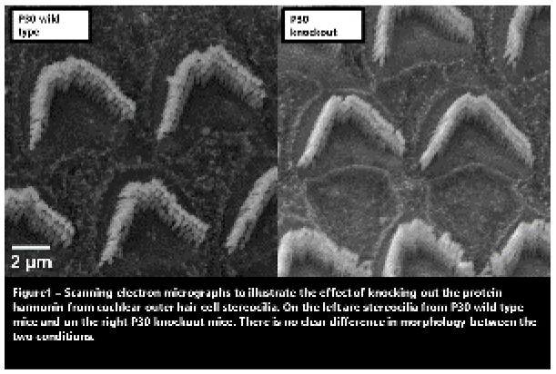
The role of protein arginine methyltransferase 8 in dendritic spine plasticity and excitatory/inhibitory balance during learning
Introduction. Protein arginine methylation is a major post-translational modification in synaptic proteins. Among the arginine methyltransferase (PRMT) family, PRMT8 is the only member with brain-specific expression. In vitro studies have shown the localization of PRMT8 in dendritic spines implicating its role in dendritic spine maturation. However, the role of PRMT8 in dendritic spine plasticity is poorly understood in vivo, which led to a significant gap in understanding the mechanism of dendritic spine pathology and potential therapeutic interventions in PRMT8- related neuropsychological diseases, such as autism. Methods. We utilized Prmt8 knockout-first mouse model (Prmt8KOF) in behavioural tests, in vivo transcranial two-photon imaging, electrophysiological recordings, and awake photometric recordings. Approach for statistical analysis. We used Welch’s t-test or one-way ANOVA followed by
Bonferroni’s multiple comparison test in experiments with 2 or more than 2 groups, respectively. Results and conclusions. Here we found that Prmt8KOF mice exhibited a male-specific auditory-cued fear learning deficit. While the dendritic spine plasticity was normal in Prmt8KOF during development, it exhibited deficit in fear learning- induced dendritic spine formation of layer 5 pyramidal neurons (PNs) in the auditory cortex. Besides, we found that Prmt8KOF showed a significantly higher percentage of c-fos positive parvalbumin interneurons (PVINs) 1 hour after fear learning. Further electrophysiological investigation revealed that Prmt8KOF had a significant reduction in membrane excitability of PVINs. Using enriched environment (EE) or selective chemogenetic activation of PVINs as an intervention to restore the excitatory-inhibitory (E/I) balance, both rescued the fear learning deficits of in Prmt8KOF in behaviour and spine plasticity. Collectively, our results suggest that PRMT8 plays critical roles in fear learning, PVIN excitability, and PV plasticity. The loss of PRMT8 may lead to E/I imbalance that could be rescued via selective activation of PVINs in fear learning.
Author Index
