
Abstract
Obsessive-compulsive disorder is a debilitating psychiatric disorder that is characterised by perseverative thoughts and behaviours. Cognitive and affective disturbances play a central role in this illness, and it is therefore not surprising that clinical neuroimaging studies have demonstrated widespread alterations in prefrontal cortex functioning in patients. Preclinical mouse experimental systems provide the opportunity to gain mechanistic insight into the neurobiological changes underlying prefrontal cortex dysfunction through new technologies that allow measurement and manipulation of activity in discrete neural populations in awake, behaving mice. However, recent preclinical research has focused on striatal dysfunction, and has therefore provided relatively little insight regarding the role of the prefrontal cortex in obsessive-compulsive disorder–relevant behaviours. Here, we will discuss a number of translational prefrontal cortex–dependent paradigms, including obsessive-compulsive disorder–relevant tasks that produce compulsive responding, and how they can be leveraged in this context. Drawing on recent examples that have led to mechanistic insight about specific genes, cell types and circuits that mediate prefrontal cortex contributions to distinct aspects of cognition, we will provide a framework for applying similar strategies to identify neural mechanisms underlying obsessive-compulsive disorder–relevant behavioural domains. We propose that research using clinically relevant paradigms will accelerate translation of findings from preclinical mouse models, thus supporting the development of novel therapeutics targeted to specific pathophysiological mechanisms.
Overview: prefrontal cortex dysfunction in obsessive-compulsive disorder
Obsessive-compulsive disorder (OCD) is a debilitating neuropsychiatric disorder that affects 2–3% of the population worldwide and is associated with significant disability (Lochner et al., 2014; Meier et al., 2016; Murray et al., 1996; Perez-Vigil et al., 2018). The core symptoms of OCD are persistent intrusive thoughts, or obsessions, and uncontrollable repetitive rituals, or compulsions. Convergent neuroimaging and neurocognitive findings support a model whereby abnormalities in cortico-striato-thalamo-cortical (CSTC) loops play a critical role in these symptoms (Ahmari and Dougherty, 2015; Ting and Feng, 2011). Prefrontal cortex (PFC) dysfunction is central in this model, with the most well-replicated finding being orbitofrontal cortex (OFC) hyperactivity at baseline and following symptom provocation (Baxter et al., 1987; Breiter et al., 1996; Rauch et al., 1994); this activity is normalised following successful treatment (Benkelfat et al., 1990; Nakao et al., 2005; Saxena et al., 1999; van der Straten et al., 2017). In contrast, neuroimaging studies conducted during cognitive testing (e.g. reversal learning) consistently reveal impaired recruitment of OFC in OCD patients (Chamberlain et al., 2008; Gillan et al., 2015; Gu et al., 2008; Remijnse et al., 2006, 2009, 2013).
In addition to these findings, there is substantial evidence for abnormalities in other PFC subregions in OCD patients either at baseline or associated with symptom provocation/cognitive impairment. Similar to observations in OFC, these abnormalities tend to follow the pattern of increased activity at baseline and impaired modulation during task performance. For example, the anterior cingulate cortex (ACC) shows hyperactivity at baseline (Perani et al., 1995), following symptom provocation (Breiter et al., 1996; Rauch et al., 1994), and during error processing (Fitzgerald et al., 2005), whereas successful treatment is associated with normalisation of baseline hyperactivity (Perani et al., 1995). In contrast, reduced ACC activation has been reported in OCD patients during tasks measuring executive functioning, including working memory and cognitive flexibility (Gu et al., 2008; Koch et al., 2012). Ventromedial PFC (VMPFC) hyperactivity has also been observed during symptom provocation in patients with prominent hoarding (An et al., 2009) and in response to threatening stimuli in a Pavlovian fear task; this VMPFC hyperactivity in turn predicted failure to adapt fear-related responses following reversal of the neutral and shock-paired cues (Apergis-Schoute et al., 2017). Again, opposite patterns of VMPFC dysfunction are observed during cognitive testing, with impaired recruitment observed during fear extinction training and recall in OCD patients with extinction recall deficits (Milad et al., 2013), and during task-switching paradigms (Gu et al., 2008). Dorsolateral PFC (DLPFC) also shows robust hypoactivity in OCD patients during tasks assessing executive functioning, including the Tower of London task, reversal learning and task switching (Gu et al., 2008; Remijnse et al., 2006, 2013; Vaghi et al., 2017a; van den Heuvel et al., 2005), and reduced resting-state DLPFC-putamen functional connectivity was recently associated with impaired set-shifting (Vaghi et al., 2017b). Finally, whereas OCD patients with high sensitivity to disgust demonstrate ventrolateral PFC (VLPFC) hyperactivity in response to facial expressions of disgust (Lawrence et al., 2007), reduced resting-state functional connectivity in the VLPFC-dorsal caudate has been associated with impaired goal-directed behaviour (Vaghi et al., 2017b). These recent functional connectivity studies from Vaghi et al. (2017b) may account for discrepancies between earlier studies reporting increased (Harrison et al., 2009, 2013) and decreased (Posner et al., 2014) resting-state functional connectivity in PFC-striatal circuits, because the directionality of functional connectivity changes may depend on the degree or type of cognitive impairment observed in patients in each particular study.
Synthesising these data, a common pattern emerges across PFC subregions: dissociation between the abnormal activity patterns that contribute to OCD symptoms and those that contribute to cognitive impairments (Table 1). Typically, hyperactivity in PFC (and striatal) regions is correlated with severity of obsessions, compulsions and symptom-associated affective states, including fear and disgust, whereas the same PFC regions often show hypoactivity across a variety of cognitive tasks. This is a striking observation that has not yet been reconciled within the clinical neuroimaging field, perhaps because the precise mechanisms underlying this double dissociation may be difficult to address given the limited spatial and temporal resolution of clinical neuroimaging methodologies. Preclinical models provide a number of unique advantages for gaining mechanistic insight into the contribution of different PFC subregions to specific OCD-relevant behaviours, but thus far they have not been fully utilised for this purpose. The goal of this Special Issue article is to (1) summarise recent findings from preclinical mouse models in OCD research, including attempts to probe PFC dysfunction; (2) describe OCD-relevant behavioural paradigms that are dependent on PFC functioning which can be applied in experimental mouse systems; (3) highlight advanced neuroscience approaches for investigating genetic, neuronal sub-type and circuit-specific contributions to behaviour and (4) use recent examples from the literature to help develop a framework for integrating these approaches with translational PFC-dependent behavioural paradigms to gain new mechanistic insight about PFC dysfunction in OCD.
Summary of functional imaging studies demonstrating PFC dysfunction in OCD patients.

PFC: prefrontal cortex; OCD: obsessive-compulsive disorder; OFC: orbitofrontal cortex; ACC: anterior cingulate cortex; VMPFC: ventromedial PFC; DLPFC: dorsolateral PFC; VLPFC: ventrolateral PFC.
Preclinical OCD research
Overview of OCD-relevant mouse experimental systems
A rapidly expanding literature is using preclinical mouse models to study pathophysiological mechanisms underlying OCD (reviewed also in Monteiro and Feng, 2016; Szechtman et al., 2017; Zike et al., 2017a). While it is important to recognise that no animal model can recapitulate all aspects of OCD, these experimental systems serve as valuable tools for examining component processes and pathophysiologic/therapeutic mechanisms with a level of precision that cannot be achieved in clinical research. These preclinical models, which aim to mimic disturbances hypothesised to contribute to the aetiology of OCD, have been generated through a variety of methods, including genetic manipulations (Greer and Capecchi, 2002; Shmelkov et al., 2010; Ullrich et al., 2018; Welch et al., 2007; Xu et al., 2017), pharmacological manipulations (Shanahan et al., 2009, 2011) and, more recently, neural circuit perturbations (Ahmari et al., 2013; Burguiere et al., 2013; Rapanelli et al., 2017a). A number of different behaviours relevant to OCD have been measured in these models, including compulsive self-grooming (Kalueff et al., 2016), other repetitive actions (Pogorelov et al., 2015; Xu et al., 2015; Zike et al., 2017b), prepulse inhibition (PPI; Baldan Ramsey et al., 2011; Shanahan et al., 2009) and anxiety-like behaviour (Ade et al., 2016; Shmelkov et al., 2010; Welch et al., 2007). Although construct and predictive validity have been traditionally used to determine the relevance of particular models for preclinical research (i.e. whether the model is generated via a pathophysiological change similar to that which generates the disease, and whether it is responsive to therapeutic interventions that are effective in patients), a large proportion of OCD patients do not show improvements with current treatment options (Eisen et al., 2013), and the precise mechanisms that cause OCD are still poorly understood. For this reason, it is important to note that face validity has often been emphasised in these models, despite the clear challenges that accompany translation of species-typical behaviours.
Compulsive grooming and striatal dysfunction in mouse models
A key advantage of using preclinical models is the opportunity for detailed characterisation of CSTC loop dysfunction implicated in OCD pathophysiology with a level of precision that is not possible in clinical imaging studies. A large number of studies have now demonstrated that compulsive grooming in mice resulting from both genetic and circuit manipulations is often associated with striatal dysfunction. These studies have identified changes in striatal synaptic density and structure via Golgi staining and electron microscopy (Nagarajan et al., 2017; Shmelkov et al., 2010; Welch et al., 2007), changes in excitability of spiny projection neurons (SPNs) using ex vivo slice electrophysiology (Aida et al., 2015; Krabbe et al., 2017; Nagarajan et al., 2017; Shmelkov et al., 2010; Ullrich et al., 2018; Welch et al., 2007) and calcium imaging (Ade et al., 2016) and altered SPN activity in vivo which has been linked to specific behavioural disturbances (Ahmari et al., 2013; Burguiere et al., 2013). In several different models, striatal manipulations were found to be necessary or sufficient for the induction of compulsive grooming. For example, viral-mediated striatal re-expression of a candidate risk gene for OCD (SAPAP3) normalised compulsive grooming in both SAPAP3 knockout (KO) mice and transcription factor MeCP2 mutant mice (Mahgoub et al., 2016; Welch et al., 2007). Similarly, striatal-specific deletion of histone deacetylase genes 1 and 2 in a conditional mutant model was sufficient to recapitulate the compulsive grooming phenotype that resulted from whole forebrain gene deletion (Mahgoub et al., 2016). Further supporting a role for striatal disturbances in pathologic grooming, others have recently demonstrated that artificially increasing activity in a population of striatal neurons targeted by hypothalamic histaminergic neurons is sufficient to produce compulsive grooming (Rapanelli et al., 2017a). Together, these studies have provided valuable insights regarding the relationship between striatal dysfunction and abnormal OCD-relevant behaviours. However, the questions of whether/how upstream cortical structures are involved in generating the striatal abnormalities leading to these pathologic behaviours remain unanswered.
Studying PFC dysfunction in preclinical mouse models
Compared to the literature detailing striatal abnormalities in preclinical OCD mouse models, investigation of PFC disturbances is limited. One recent example used electron microscopy to identify synaptic disturbances, including increased length and density of spines and thinning of the post-synaptic density, in the frontal cortex of the Hoxb8 KO mouse model, which displays compulsive grooming (Greer and Capecchi, 2002) linked specifically to Hoxb8 deletion in microglia (Chen et al., 2010). However, it is unclear how these changes in PFC contribute to the development of abnormal behaviours (Nagarajan et al., 2017). In another study, genetic deletion of Slitrk5, a member of a synaptic transmembrane protein family that has been associated with OCD risk (Ozomaro et al., 2013; Song et al., 2017; Wendland et al., 2006), resulted in compulsive grooming and anxiety. In addition, medial OFC (mOFC) hypermetabolism was inferred via elevation of the neural activity marker FosB. However, far more extensive anatomical, molecular and electrophysiological characterisation was completed in the striatum (Shmelkov et al., 2010). Finally, studies in mice have demonstrated that RU24969-mediated 5-HT1B receptor activation in ventrolateral OFC (vlOFC) induces disruption of PPI and perseverative locomotion (Shanahan et al., 2011) that are potentially relevant to OCD (Gross-Isseroff et al., 2004; Pittenger et al., 2016; Stein et al., 1999); however, the role of OFC 5-HT1B receptors has not yet been assessed in other preclinical models.
Nuanced interpretation of these findings requires consideration of cortico-striatal projection patterns. In particular, mOFC and lateral OFC (lOFC) differ markedly in their striatal outputs (ventromedial striatum (VMS) and dorsal striatum, respectively; Berendse et al., 1992; Mailly et al., 2013; Schilman et al., 2008). This gradient of OFC-striatal connectivity is well conserved in primates (Heilbronner et al., 2016) and may contribute to functional heterogeneity across the medial–lateral axis of the OFC (Izquierdo, 2017). This functional gradient is also highlighted by two studies that used optogenetics to determine how specific OFC-striatal projections contribute to OCD-relevant compulsive grooming. One study demonstrated that repeated optogenetic stimulation of the mOFC-VMS circuit can induce both excessive grooming and accompanying striatal plasticity in normal mice (Ahmari et al., 2013). Another study used optogenetics to probe cortico-striatal circuit involvement in OCD-relevant compulsive grooming in mice carrying a deletion of SAPAP3 (Welch et al., 2007), a post-synaptic scaffolding protein that has been implicated in OCD risk (Bienvenu et al., 2009; Mattheisen et al., 2015; Stewart et al., 2013; Zuchner et al., 2009). In this model, acute stimulation of the lOFC-centromedial striatum (CMS) circuit normalised compulsive grooming, potentially by boosting activity in striatal parvalbumin positive (PV+) interneurons (Burguiere et al., 2013). Together, these findings support an important role for the PFC in the manifestation of OCD-like behaviour that is consistent with findings from clinical imaging studies, and suggest areas that warrant further mechanistic investigation in preclinical rodent models.
Consideration of PFC homology between rodent and primate
In preclinical psychiatry research, it is important to consider the degree of similarity between the human brain and that of the model species being used. This is particularly significant for studies of PFC, as this area has undergone rapid development within the primate order relative to other mammals (Smaers et al., 2017). One of the main concerns about the use of rodents to study PFC functioning is that they do not have six-layered granular cortex in the relevant frontal regions (Wise, 2008). However, the functional significance of the layer IV granule cells that are missing in rodent PFC is not clear, and other features known to be important for PFC functioning are similar between rodents and primates, including thalamic and dopaminergic inputs (Carlen, 2017; Uylings et al., 2003). Continued investigation to more thoroughly understand the shared and distinct aspects of rodent and primate PFC is therefore critical for appropriate interpretation and translation of rodent studies. Recent examples of such studies include the first thorough histological characterisation of mouse PFC (Van De Werd et al., 2010), which was used to help refine parcellation of subregions (Van De Werd and Uylings, 2014), and a detailed comparison of anatomical and electrical properties of PFC inhibitory interneurons between rat and primate (Povysheva et al., 2008). Functional homology between rodent and primate species has also been addressed using PFC-dependent behavioural paradigms, but it can be challenging to draw satisfying conclusions because identical paradigms are often not used across species. This issue was highlighted in a recent review on cross-species investigation of OFC in decision-making (Wallis, 2011). It is therefore critical to directly compare behaviour in rodents and primates, as exemplified by studies by Arnsten and colleagues demonstrating functional homology between monkey DLPFC and rodent prelimbic PFC (PrL) during a working memory task (Gamo et al., 2015; Wang et al., 2007).
In the context of OCD research, careful consideration of the patterns of PFC projections to striatal targets in rodents versus primates is particularly critical, since PFC-striatal circuit disturbances are theorised to be central to the disorder (Ahmari and Dougherty, 2015). Cortico-striatal circuits are thought to be topographically organised in parallel (Alexander et al., 1986) or spiralling (Haber, 2016) loops, whereby PFC projections to striatum generally follow a rough dorsal–ventral gradient (Haber, 2003, 2016). This organisation produces broadly separable functional loops; with primary and supplementary motor areas projecting dorsolaterally to caudate to form the ‘motor loop’; DLPFC and OFC projecting centrally to caudate/putamen to form the ‘cognitive/associative loop’ and VMPFC/ACC projecting ventrally to nucleus accumbens (NAc) to form the ‘motivational/limbic loop’ (converging with inputs from limbic areas including the amygdala). Recent studies by Heilbronner et al. (2016) have demonstrated substantial homology in striatal projection patterns of medial and orbital PFC subregions through head-to-head anatomical comparison between rats and non-human primates. However, some discrepancies were observed in ACC-striatal projections, with reduced ventral spread of ACC projections in rodents relative to primates leading to decreased overlap with NAc shell and amygdala-striatal projection fields. This suggests the possibility that rodent ACC is less involved in ‘limbic loop’ affective and motivational behaviours than primate ACC. These examples highlight that careful consideration of functional homology of PFC subregions in the context of their outputs and associated behavioural domains is essential to design preclinical studies that can be used as a foundation for developing targeted circuit-specific neuromodulatory treatments.
All rodents are not created equal
Mice and rats are the predominant rodent models in neuroscience research. For purposes of this review, although most of the extant literature examining neuroanatomical homology between rodents and primates has focused on rats, we will assume that mouse and rat PFC have similar levels of homology. However, it is clear that there are other differences between these rodent species that influence their suitability for mechanistic studies of PFC contributions to OCD-relevant behaviours. Most broadly, rats typically have more rapid acquisition of complex instrumental behaviours, whereas a wider range of sophisticated genetic models are available for mechanistic studies in mice. The current preclinical OCD literature can therefore be largely segregated into two subfields: (1) mechanistic studies in mouse models focusing on simple OCD-relevant behaviours and (2) studies of more complex cognitive behaviours in rats with less ability to explore mechanisms. We will focus here on approaches for studying PFC dysfunction in mouse models, as this literature has expanded rapidly in recent years. However, it is important to note that because of advances in genetic technologies that can be applied in rats (e.g. clustered regularly interspaced short palindromic repeat – CRISPR-associated protein (CRISPR-CAS); Heidenreich and Zhang, 2016; Wright et al., 2016), and home-cage based instrumental paradigms that can facilitate more complex behavioural testing in mice (Remmelink et al., 2016, 2017), integration of these subfields is now possible.
OCD-relevant behavioural paradigms for probing PFC functioning in preclinical models
Development of paradigms in rats for the assessment of OCD-relevant compulsive responding
Very little behavioural characterisation of the mouse models described above has been performed beyond assessment of compulsive grooming, repetitive stereotypies, anxiety-like behaviours and PPI. A number of paradigms developed in rats may therefore be valuable for more deeply probing OCD-relevant compulsive behaviours in preclinical mouse models. Training in these paradigms causes rats to perform task-specific compulsive responses, in contrast to the non-instrumental behaviours that are typically studied in mice (i.e. grooming, stereotypy). Three key examples are as follows: (1) the development of compulsive avoidance after failed extinction with response prevention (Ex-RP) training in an avoidance conditioning paradigm (Rodriguez-Romaguera et al., 2016), (2) the development of excessive lever pressing following extinction of the reward-paired cue in the signal attenuation task (Joel and Avisar, 2001) and (3) the development of compulsive checking responses to gain information about the location of the active lever in an observable response task (Eagle et al., 2014).
Early studies using these paradigms in rats have demonstrated their potential for further mechanistic research investigating PFC dysfunction relevant to OCD. For example, using the avoidance conditioning paradigm, persistent avoidance following Ex-RP was found to be differentially modulated by pharmacological inactivation of the lOFC depending on the success of Ex-RP. Rats that were resistant to Ex-RP (high freezing throughout extinction) typically showed persistent avoidance in a probe test, and avoidance was reduced in these rats following lOFC inactivation. Surprisingly, persistent avoidance was increased following lOFC inactivation in rats that showed successful Ex-RP training (Rodriguez-Romaguera et al., 2016). This differential contribution of the OFC to compulsive behaviour in pathological (high freezing/persistent avoiders) and normal (low freezing/low avoiders) conditions is somewhat consistent with the differential effects of optogenetic stimulation of OFC circuits in normal (Ahmari et al., 2013) versus mutant (Burguiere et al., 2013) mice, and supports the importance of dissection of these circuits in both healthy and pathological conditions in preclinical models. In addition, persistent avoidance was rescued following deep brain stimulation (DBS) in the dorsal portion of the ventral striatum during Ex-RP training (Rodriguez-Romaguera et al., 2016) comparable to the ventral capsule/ventral striatum (VC/VS) target used in OCD DBS (Dougherty et al., 2016; Greenberg et al., 2006; Rauch et al., 2006). DBS at this site was previously found to induce expression of the plasticity marker phospho-ERK across PFC subregions, including mOFC, lOFC, PrL and infralimbic (IL) PFC (Rodriguez-Romaguera et al., 2012, 2015), and lOFC in vivo recordings during VC/VS DBS in rats show activity consistent with antidromic activation of cortico-striatal projections (McCracken and Grace, 2007, 2009). These findings support clinical neuroimaging data suggesting that modulation of PFC activity is involved in mediating the therapeutic effects of VC/VS DBS (Dougherty et al., 2016; Figee et al., 2013). Further studies in preclinical models are necessary to demonstrate a causal role.
The OFC has also been implicated in the development of excessive lever pressing following signal attenuation training, with lOFC lesions increasing compulsive behaviour in this paradigm (Joel et al., 2005; Schilman et al., 2010), and blockade of lOFC 5-HT2C receptors exerting an anti-compulsive effect (Flaisher-Grinberg et al., 2008). Although PFC involvement in the observable checking task has not been directly assessed, compulsive checking was exacerbated by quinpirole sensitisation (Eagle et al., 2014), a well-established paradigm for the induction of compulsive exploration that is dependent on lOFC functioning (Dvorkin et al., 2010). Thus, the quinpirole data support a potential role of lOFC in expression of compulsive checking in this task. Together, these studies provide convergent evidence of a critical role for PFC in the development of different types of compulsive responses, supporting the utility of these tasks for further mechanistic studies probing OCD-relevant PFC dysfunction.
Translatable neurocognitive paradigms that are consistently abnormal in OCD
In addition to the OCD-relevant paradigms developed in rats described above, there are several established rodent tasks that can be used to measure neurocognitive domains that are consistently abnormal in OCD (Table 2). These domains include cognitive flexibility (Chamberlain et al., 2006, 2007; Gottwald et al., 2018; Gu et al., 2008; Remijnse et al., 2006; Vaghi et al., 2017b), goal-directed behaviour (Gillan et al., 2011, 2014a; Vaghi et al., 2017b), response inhibition (Chamberlain et al., 2006; Menzies et al., 2007), working memory (Chamberlain et al., 2007; Jaafari et al., 2013) and aversive learning processes (Apergis-Schoute et al., 2017; Gillan et al., 2014b; Milad et al., 2013). Functional neuroimaging in patients during many of these paradigms has identified abnormalities in task-related PFC activity (Table 1). Testing preclinical models relevant to OCD in these translational paradigms will be an important first step towards gaining new mechanistic insight into PFC dysfunction in OCD.
Summary of select preclinical studies that have provided new mechanistic insight into PFC contributions to cognitive functions that are altered in patients with OCD.
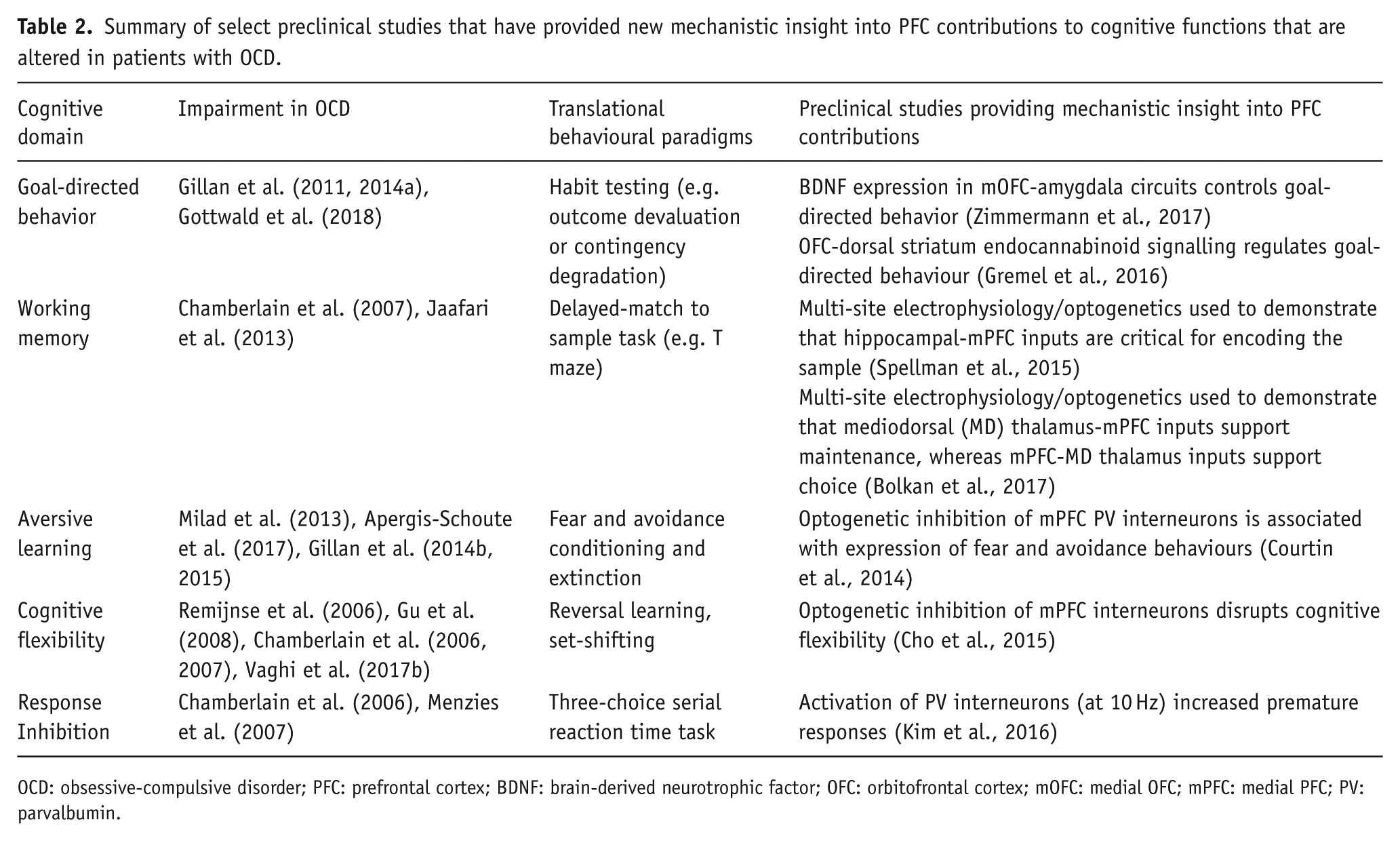
OCD: obsessive-compulsive disorder; PFC: prefrontal cortex; BDNF: brain-derived neurotrophic factor; OFC: orbitofrontal cortex; mOFC: medial OFC; mPFC: medial PFC; PV: parvalbumin.
Advanced neuroscience approaches for gaining mechanistic insight into PFC dysfunction in OCD
While findings from clinical neuroimaging provide strong evidence for disrupted PFC functioning in OCD, studies in patients are severely limited in their ability to identify neural mechanisms causing these changes in activity. The popularity of mouse models in neuroscience is largely due to compatibility with advanced approaches for examining contributions of specific genes, cell types and circuits to behaviour. In the following section, we will expand on the neuroscience approaches introduced in our summary of the preclinical literature, and highlight their utility for facilitating mechanistic investigations of PFC functioning.
Tools for precisely manipulating gene expression
OCD is a highly heritable disorder, with genetic variance accounting for ~40% of phenotypic variance as measured in familial studies (Taylor, 2011) and more recent genome-wide common variant analyses (Davis et al., 2013). Despite this, strong candidate risk genes are lacking. Genome-wide association studies (GWAS) conducted to date have been underpowered, with a recent meta-analysis of the two largest studies including less than 3000 patients (International Obsessive Compulsive Disorder Foundation Genetics Collaborative (IOCDF-GC) and OCD Collaborative Genetics Association Studies (OCGAS), 2018). Nevertheless, there has been strong convergent evidence that genes expressed at the glutamatergic synapse are involved in OCD risk (Wu et al., 2012), and a number of transgenic models have attempted to test this hypothesis (Aida et al., 2015; Shmelkov et al., 2010; Welch et al., 2007; Xu et al., 2017; Zike et al., 2017b).
A growing number of conditional and inducible expression systems are available to control cell type, brain region, timing and directionality of gene expression changes, providing new opportunities to go beyond constitutive KO models and dissect specific contributions of genes to behaviour (Branda and Dymecki, 2004; Tanaka et al., 2010; Wang et al., 2012; Zhou et al., 2018). Several preclinical OCD studies have used these approaches for cell-type-specific gene disruption (Aida et al., 2015; Chen et al., 2010; Nagarajan et al., 2017), temporal control over gene disruption (Aida et al., 2015) and selective rescue of gene expression (Mahgoub et al., 2016; Welch et al., 2007; Zike et al., 2017b). Cell-type specific expression systems will be particularly valuable for determining which PFC cell types (i.e. excitatory pyramidal cells, inhibitory interneurons or glia) are responsible for the development of OCD-relevant behaviours. Increased efficiency of genome editing using the CRISPR-CAS system will make generation of more sophisticated transgenic lines even more widely available in both rats and mice (Heidenreich and Zhang, 2016; Wright et al., 2016). Finally, genes can be deleted in specific circuits using retrograde viral approaches (Gremel et al., 2016), including herpes simplex virus (HSV), canine adenovirus (CAV) and recombinant adeno-associated viruses (rAAV-retro) (Kremer et al., 2000; Tervo et al., 2016; Ugolini et al., 1987). Recent advances have helped to limit toxicity and improve transfection efficiency (Reardon et al., 2016; Tervo et al., 2016), paving the way for using this approach to investigate the contribution of specific cortico-striatal circuits to OCD-relevant behaviours.
Tools for manipulating and recording neural activity
To date, the most consistent pathological hallmark of OCD is altered neural activity patterns in PFC and striatum (Baxter et al., 1987; Breiter et al., 1996; Del Casale et al., 2011; Rauch et al., 1994; van der Straten et al., 2017). Over the last 10 years, rapid development of new approaches to precisely control neural activity in vivo has facilitated the testing of causal relationships between specific changes in PFC and striatal neural activity and the development or resolution of OCD-relevant behaviours in preclinical models. Optogenetics (Boyden et al., 2005; Kim et al., 2017) and chemogenetics (Alexander et al., 2009; Smith et al., 2016) are the most widely used methods for specific manipulation of neural activity. Both approaches require viral-mediated expression of exogenous membrane proteins, such as light-activated ion channels and pumps (optogenetics), or G-protein-coupled receptors activated by otherwise inert ligands (chemogenetics). Genetically engineered variants enable either excitation or inhibition of target neural populations. In addition, a variety of promoter systems, including those described in the previous section, can be used to generate cell type and temporally specific expression of these tools, with Cre-lox being the most widely used conditional expression system. Recent developments also allow selective manipulation of neurons that are activated during a discrete temporal window; this approach was recently used to demonstrate that striatal neurons activated by hypothalamic histaminergic cells are necessary and sufficient for the induction of compulsive grooming (Rapanelli et al., 2017a). Spatial restriction of optogenetic light delivery can provide further circuit specificity by allowing selective activation or inhibition of neural projections to a specific target structure (see Ahmari et al., 2013; Burguiere et al., 2013). Furthermore, precise timing of light delivery during optogenetic stimulation is a powerful tool for determining when neural activity in a population of neurons is contributing to behaviour, that is, during specific task epochs of neurocognitive paradigms such as outcome presentation (Bolkan et al., 2017; Spellman et al., 2015). Unlike optogenetics, chemogenetic approaches have long-lasting effects that can be advantageous for chronic elevation or suppression of neural activity (though note that step-function opsins have also helped address this limitation of optogenetics; Yizhar et al., 2011). For example, a chronic approach may be more useful for suppressing neural activity throughout cognitive testing in a paradigm that requires long training sessions (e.g. 30 min or more) over several days or weeks (Parnaudeau et al., 2015) or investigating effects of chronic elevation or suppression of neural activity relevant to symptoms and treatment of psychiatric disorders (Urban et al., 2016). Integration of these advanced approaches for circuit manipulation with techniques for recording activity in specific neural populations while an animal engages in complex behaviours – for example, opto-tagging (Cohen et al., 2012; Kim et al., 2016; Kvitsiani et al., 2013; Lima et al., 2009), in vivo imaging with ultrasensitive calcium indicators (Cai et al., 2016; Chen et al., 2013; Ghosh et al., 2011) and projection-specific fibre photometry (Cui et al., 2013, 2014; Gunaydin et al., 2014) – will be essential for progress in preclinical OCD research.
Framework for integrating advanced neuroscience approaches to gain translationally relevant insight about the PFC in OCD
Sophisticated integration of the methods described above can be used to address the precise ways in which genetic and cellular disturbances in the PFC impact neural encoding, circuit function and resulting behaviour. Already, a number of groups have begun to use this approach in mice to understand cognitive domains that are impacted in OCD; however, these studies have generally focused on dissection of normal circuit functioning. This section will provide a summary of this work probing neural mechanisms controlling cognitive functioning in the normal brain, and discuss how these studies may serve as a guide for future investigations of similar behaviours in preclinical OCD experimental systems.
Studies linking neural activity in the PFC to specific aspects of cognitive functioning that are impacted in OCD
Several recent studies have used advanced neuroscience techniques to gain new mechanistic insight into cognitive functions that are impaired in OCD (Table 2). First, a recent study used conditional gene manipulation to investigate genetic regulation of the balance between goal-directed and habitual behaviour. Cannabinoid type 1 (CB1) receptors were selectively deleted in lOFC neurons projecting to striatum using retrograde viral approaches. Activation of presynaptic CB1 receptors in this circuit typically suppresses circuit function; conversely, CB1 receptor deletion is thought to disrupt this brake on lOFC-dorsal striatum signalling. Interestingly, this intervention disrupted the expression of habitual behaviour, while selective inhibition of this pathway using chemogenetic approaches disrupted the expression of goal-directed behaviour (Gremel et al., 2016). Expression of the neurotrophic factor BDNF (brain-derived neurotrophic factor) in the lOFC was also found to be critical for the expression of goal-directed behaviour, and ‘disconnection studies’ combining unilateral lOFC BDNF depletion with contralateral amygdala lesions have demonstrated that lOFC projections to the amygdala are responsible for this effect (Zimmermann et al., 2017). Together, these studies suggest that expression of specific synaptic genes in lOFC output circuits regulates the balance between goal-directed and habitual behaviour. Because OCD patients show a bias towards habitual behaviour (Gillan et al., 2011, 2014a; Vaghi et al., 2017b), it may be important to investigate this balance in OCD preclinical models.
Integration of in vivo electrophysiology and optogenetics has provided a powerful approach for demonstrating the precise temporal patterns of neural activity that are associated with specific aspects of cognition, and determining whether that activity is necessary for optimal behavioural performance. Working memory tasks have been an exemplar for this approach, as there are distinct phases of the task where information is presented, held online and retrieved in order to make a correct decision. Using multi-site electrophysiology followed by temporally specific optogenetic inhibition, it has been demonstrated that hippocampal inputs to medial PFC (mPFC) are critical for encoding during information presentation (Spellman et al., 2015). In contrast, thalamic inputs to mPFC support maintenance of information across a delay, and mPFC outputs back to thalamus are critical for using the information that was held online to select the correct choice (Bolkan et al., 2017). These two studies demonstrate the complexity of PFC input and output contributions to distinct components of a single cognitive function (i.e. working memory). Working memory impairments have been reported in OCD patients, with larger effects during more difficult (Chamberlain et al., 2007) and visuospatial (Snyder et al., 2015) tasks, which may account for some inconsistencies between studies (Ahmari et al., 2014). Similar approaches could therefore be used to examine the substrates of different components of working memory in preclinical OCD models.
Optogenetics has also been used with Cre-driver lines to dissect the role of specific PFC interneurons in different aspects of cognition. For example, optogenetic inhibition of PV+ interneurons in mPFC during presentation of a previously shock-paired stimulus can reinstate the expression of fear-related behaviours following extinction, whereas inhibition during exploration of a distinct context produces acute place avoidance (Courtin et al., 2014). Interestingly, optogenetic inhibition of all mPFC interneurons also disrupted cognitive flexibility in a task-switching paradigm (Cho et al., 2015), whereas optogenetic activation of PV+ interneurons at a much lower frequency than they typically fire (10 Hz) disrupted response inhibition in a three-choice serial reaction time task (3-CSRTT; Kim et al., 2016). Cognitive flexibility, response inhibition and fear extinction recall are all impaired in OCD patients (Chamberlain et al., 2006; Menzies et al., 2007; Milad et al., 2013; Remijnse et al., 2006; Vaghi et al., 2017b) and may contribute to impaired control over perseverative thoughts and actions (Gruner and Pittenger, 2017). These convergent findings of mPFC interneuron involvement in three different cognitive domains that are impaired in OCD suggest that they may be a good candidate for further examination in preclinical models, especially since striatal interneurons have also been implicated in the emergence of OCD-relevant behaviours (Burguiere et al., 2013; Rapanelli et al., 2017b; Xu et al., 2015).
Framework for applying this approach in preclinical OCD-relevant models
The studies summarised above provide examples of integration of advanced neuroscience approaches with OCD-relevant cognitive paradigms to uncover specific neural mechanisms underlying behaviour. The strategies used in these studies can provide a valuable framework for addressing experimental questions that are specifically related to OCD pathophysiology, using both existing and new preclinical models. Our proposed step-by-step framework is summarised below and in Figure 1.

Framework for new preclinical studies examining the PFC in OCD. New and existing mouse models can be characterised in PFC-dependent OCD-relevant behavioural paradigms including translational neurocognitive tasks (Table 2) and paradigms that induce task-specific compulsive behaviours that were originally developed in rats. Preclinical models that show changes in behaviour in these paradigms are good candidates for further characterisation of the PFC, using in vivo methods that assess neural activity underlying behaviour (e.g. electrophysiology and calcium imaging) and post-mortem methods to characterise neuropathology (e.g. gene expression analysis and immunohistochemistry and quantitative assessment of cell type and neural activity markers). Finally, neural changes identified in these studies can be further investigated to determine whether they are causally linked to the development of OCD-relevant behaviours, using various methods to control the functioning of genes, cell types and circuits in vivo.
1. Behavioural characterisation using PFC-dependent and OCD-relevant paradigms
A first step to accelerate discovery of neural changes in PFC that modify behaviours relevant to OCD is characterisation of existing (and new) preclinical models in OCD-relevant behavioural paradigms that are known to be PFC-dependent. Although there is some preclinical evidence that the PFC is involved in excessive grooming (Ahmari et al., 2013; Burguiere et al., 2013), this may not be the most effective behavioural paradigm for probing PFC contributions to behaviours in OCD. Here, we have highlighted translational neurocognitive paradigms similar to those that have been used in OCD clinical imaging studies, and behavioural paradigms first developed in rats that cause the development of task-specific compulsive responses. These paradigms can serve as an initial screening tool to identify promising preclinical models for further mechanistic studies.
2. Detailed investigation of the PFC in models that show relevant behavioural changes
Preclinical models that show OCD-relevant behavioural changes in PFC-dependent paradigms are good candidates for more detailed investigation of the PFC. There are a number of approaches that can be used at this step, which can be broadly categorised into methods assessing PFC functioning in vivo and those assessing the PFC in post-mortem brain tissue. In vivo measurements of neural activity are powerful tools for linking PFC functioning to behavioural deficits. Given the bidirectional changes observed in OCD patients in neuroimaging studies (Table 1), comparison of activity at baseline, during cognitive testing and during symptom-related behaviours (e.g. compulsive grooming) is highly warranted and may provide insight into mechanisms underlying these observations in patients. However, because simultaneous assessment of multiple subregions is not always feasible (e.g. using calcium imaging), post-mortem immunohistochemical analyses examining the distribution of specific cell types (e.g. interneuron subtypes or microglia) and neural activity indices (e.g. immediate early gene activation) can be valuable for selecting candidate PFC subregions for detailed in vivo characterisation.
3. Mechanistic studies to test causality
Guided by detailed characterisation of the PFC, mechanistic studies can establish causal links between gene expression changes, cell and circuit dysfunction, and behaviours relevant to OCD. We have provided a number of examples of how cell type and circuit-specific control of gene expression and neural activity can be used to understand specific neural mechanisms in the PFC that underlie distinct aspects of cognition (Table 2). Mechanistic insight provided by these studies may highlight candidates for the development of targeted treatment strategies, including pharmacotherapy aimed at specific molecular disturbances or stimulation approaches to target impaired circuits and subregions that are causally linked to behavioural dysfunction relevant to OCD.
Summary
Harnessing the strengths of preclinical models for mechanistic dissection can provide critical details that cannot be inferred from studies in human subjects. Building on the success of mechanistic studies in preclinical models focused on the role of the striatum, clinically relevant paradigms will help to guide studies of PFC contributions to behavioural dysfunction in OCD. Insight into how pathological changes in PFC functioning lead to abnormal behaviour is necessary to develop a foundation for targeting the PFC with novel therapeutic approaches, given the complexity of the changes that have been described in clinical neuroimaging studies in OCD patients.
Footnotes
Declaration of conflicting interests
The author(s) declared no potential conflicts of interest with respect to the research, authorship, and/or publication of this article.
Funding
BRAINS R01 MH104255, McKnight Neuroscience Scholar Award, MQ Fellows Award, Burroughs Wellcome Fund CAMS Award, and Klingenstein Fellowship in the Neurosciences.