
Abstract

BNA2017 POSTER ABSTRACTS
SESSION 1 – MONDAY 10TH APRIL
SESSION 2 – TUESDAY 11TH APRIL
SESSION 3 – TUESDAY 12TH APRIL
BNA2017 TALK ABSTRACTS
SESSION 1 – MONDAY 10TH APRIL
1.01. Neural mechanisms of post-traumatic stress disorder as seen through stress-enhanced fear learning
1.02. Prefrontal oscillatory mechanisms of fear behaviour
1.03. Neural mechanisms underlying recurrent fear memories in post-traumatic stress disorder
1.04. Cerebellar and periaqueductal grey contributions to fear behaviour
2.01. Descending control of bilateral circuits controlling limb movement
2.02. Bilateral organisation in the primate cervical spinal cord
2.03. Combinatorial approaches to promoting recovery of limb function in rats with chronic spinal cord injury
2.04. Plasticity in the Corticospinal Pathway after Human Spinal Cord Injury
3.01. Reciprocal interactions between pain and negative effect: Role of the endocannabinoid system
3.02. The microbiota gut brain axis as a key regulator of visceral pain
3.03. Treating chronic pain by inhibiting the stress regulator FKBP51
3.04. Dual basis for the anti-nociceptive action of SNARE proteases of botulinum neurotoxins: inhibition of the exocytosis of pain mediators and transducers
4.01. Context-dependent modulation by hypothalamic tanycytes of the arcuate neuronal network controlling appetite
4.02. Hypothalamic stem cells and neurogenesis
4.03. The role of tanycytes in energy homeostasis and stability
4.04. Modulation of adult hypothalamic neurogenesis by the photoperiod
5.01. Fractionating impulsivity: implications for brain disorders
5.02. Multidimensional apathy in neurodegeneration
5.03. Reward processing in psychiatric disorders
5.04. Reward and effort-based decision making in health and disease
6.01. The molecular basis of Rett syndrome
6.02. Epigenetic studies in Alzheimer’s disease
6.03. Stability of DNA modifications in Fragile X syndrome and Parkinson’s Disease
6.04. The role of genomic imprinting in neurological disorders
7.01. Primate retrosplenial cortex: defining its contribution to learning and memory
7.02. Retrosplenial cortex: on the outskirts of the spatial memory map
7.03. Navigating over complex terrain
7.04. Retrosplenial cortex and stimulus control: investigating non-spatial functions of the rodent retrosplenial cortex
8.01. Neuronal pathways and molecular targets for modulation of anxiety
8.02. Past, current and future drug treatments for anxiety
8.03. Targeting cognitive control to reduce anxiety vulnerability: implications for treatment efficacy
8.04. Deconstructing the molecular pathways to benzodiazepine tolerance - where do we stand and where do we go?
9.01. Using novel genetic approaches to probe the causes of neurodegenerative disease
9.02. Propagation of tauopathy: mechanisms and therapeutic opportunities
9.03. Alpha-synuclein trafficking as a rational mechanism for therapies in Parkinson’s Disease
9.04. Industry approaches to therapeutic development for Alzheimer’s Disease
SESSION 2 – TUESDAY 11TH APRIL
10.01. The functions of microglia and their diverse activation states
10.02. Therapeutic modulation of microglia – opportunities and challenges
10.03. Biomarkers of inflammation and treatment response in psychosis and depression
10.04. Genome-wide transcriptional profiling and structural magnetic resonance imaging in the maternal immune activation model of neurodevelopmental disorders
11.01. Neural orchestration of eating and locomotion
11.02. Sweet, light and beyond
11.03. Why did I eat that? Differences in striatal function and motivation that contribute to obesity
11.04. Mesolimbic response to energy and other nutrients
12.01. Nimble forgetfulness in healthy ageing
12.02. Finding the ageing brain’s natural capacity
12.03. Multi-scale integrative network dynamics (MIND) of the ageing brain: a new model of neurocognitive ageing and function
12.04. Constrained moment-to-moment brain signal variability as a principled marker of the ageing brain
13.01. Early adversity and psychotic experiences: bio-psycho-social pathways and resiliencies
13.02. Environmental risks and social behaviour - translational approaches
13.03. The hidden wounds of childhood trauma: psychoneuroimmunology of early stress and the impact on mental health
13.04. Genetic and environmental impact in psychosis
14.01. Control of cardiovascular responses to acute emotional stress by corticotropin-releasing factor in the bed nucleus of the stria terminalis: Involvement of local NMDA-NO-GMPc-PKG signaling mechanism
14.02. Microglia soothe the sympathoexcitatory response to seizure
14.03. Autonomic modifications induced by social defeat involve serotonin in the brainstem associated to activation of the dorsomedial nucleus of the hypothalamus
14.04. Cardiac autonomic and respiratory correlates of high-anxiety behaviour in rats: potential involvement of the endocannabinoid signaling
15.01. TNF-α dependent spine scaling after deprivation is localized in dendritic branches that have undergone recent spine loss
15.02. Optogenetic STDP: shaping hippocampal networks through temporal correlations
15.03. The formation of hippocampal cognitive maps during novel environment exposure
15.04. Neuromodulation of dendrites and synaptic plasticity
16.01. Fit to study
16.02. Reading, phonology and the brain
16.03. Inhibitory control and the learning of counter-intuitive concepts
16.04. Engaging the brain’s reward system
17.01. Genetic associations with variation in reading and language ability: present results and future directions
17.02. Using extreme traits to identify genetic contributions to speech and language disorders
17.03. Dyslexia and cilia biology: a new link between cognition and brain asymmetries?
17.04. Model systems to understand language disorders: FOXP2 and beyond
18.01. Ethologically relevant signals processed by the nematode nervous system
18.02. Socially induced phenotypic plasticity in the desert locust
18.03. Impact of neonicotinoid pesticides on bee behaviour
18.04. Challenges in Targeting the Neuromuscular System for Control of Agricultural Insect Pests
18.05. The challenges facing the UK food system – how can neuroscience help?
19.01. Overlapping mechanisms in CNS development and gliomagnenesis
19.02. A common pathway controlling cell migration in normal and neoplastic neural stem cells
19.03. Exploring the roots of paediatric brain cancers using epigenetic profiling
19.04. Epigenetic deregulation in brain cancer
20.01. Vulnerability to depression and emotional processing
20.02. Consequences of stress on emotional processing in humans and rodents
20.03. Stress, oxytocin and vasopressin regulation of emotion: insights from fMRI
20.04. Effects of early-life stress and brain derived neurotrophic factor (BDNF) on emotional processing
SESSION 3 – WEDNESDAY 12TH APRIL
21.01. Mechanisms of μ-opioid receptor desensitisation and tolerance
21.02. Ligand bias at the μ-opioid receptor
21.03. Biased ligand signalling for kappa opioid receptor agonists and antagonists
21.04. Circuit dynamics of in vivo dynorphin release in the nucleus accumbens shell
22.01. The pain matrix ‘reloaded’: a multimodal saliency-detection system for the body and the peripersonal space
22.02. Multiple stages of multisensory perception: evidence from local cortical oscillations and functional connectivity
22.03. Auditory-visual integration in auditory cortex facilitates auditory scene analysis
22.04. See what you hear - how the brain forms a representation across the senses
23.01. APOE4 from man to mouse
23.02. APOE4 across the ages: what changes when? MRI signatures of brain function in humans
23.03. Using APOE targeted replacement mice to probe APOE4 function
23.04. Structural and cellular studies to elucidate the mechanisms of APOE isoform action and provide targets for therapy
24.01. Epilepsy genetics: contributions to cause and management
24.02. Aberrant glutamatergic signalling in brain tumour related seizures: opportunities for precision medicine
24.03. Autoantibody-mediated forms of epilepsy
24.04. Autonomic modulation as a therapy for epilepsy: effective and non-invasive approach for future treatment
25.01. Slave to the rhythm - ultradian glucocorticoid rhythms regulate distinctive gene expression profiles in the brain and pituitary
25.02. Stress, glutamate receptor trafficking and synaptic plasticity
25.03. Dopamine-mediated regulation of expression of fear memory
25.04. Strategies for preventing in vivo hippocampal synaptic plasticity disruption by stressors
26.01. Neuroinformatics tools for sharing and analysing data
26.02. Modelling plasticity in networks
26.03. Statistical long-term excitatory and inhibitory synaptic plasticity
26.04. Linking network structure and function in the cerebellar cortex
27.01. Combining non-invasive brain stimulation with magnetic resonance imaging and spectroscopy to probe motor learning
27.02. Using non-invasive brain stimulation to study the role of primary motor cortex in motor learning
27.03. Non-invasive brain stimulation to dissociate the roles of the cerebellum and motor cortex in motor learning
27.04. The offline brain: understanding the regulation of memory consolidation using non-invasive brain stimulation
28.01. Prenatal glucocorticoids and the developing brain
28.02. Maternal protein restriction around conception increases foetal neuronal differentiation and is associated with adul t memory deficits
28.03. Sexually dimorphic programming of the developing dopamine system, with consequences for adult behaviour, by a low protein diet restricted to gestation
28.04. Prenatal maternal depression and aberrant placental imprinting
29.01. Inherited and acquired presynaptic channelopathies
29.02. What can we learn from tetanus toxin?
29.03. Ca2+ channels modulate dopamine-autoinhibition and vulnerability of dopaminergic neurons to Parkinson’s disease trigger- factors
29.04. Activity-dependent regulation of synaptic strength and cellular mechanisms of paroxysmal neurological disorders
30.01. CNS medicine discovery: starting and finishing with the patient in mind
30.02. Age dependent changes of synaptic composition in human cortical synapses
30.03. Investigating the correspondence between rodent models of epilepsy and human brain tissue from children with drug resistant epilepsy
30.04. Experimental models of cortical rhythms in live human brain tissue: translational biomarkers for CNS drug dev elopment
31.01. Epigenetic and behavioural outcomes associated with adverse caregiving
31.02. Is glucocorticoid programming by early-life stress adaptive or maladaptive? Insights from birds
31.03. Early life adversity and programming of the physiological stress response
31.04. Resilience to developmental stress exposure in serotonin-transporter deficient female mice
SESSION 4 – THURSDAY 13TH APRIL
SpE5.01. Microglial immune surveillance powered by potassium channels
SpE5.02. Is glutamate release required for synaptic plasticity?
SpE5.03. Sustained correction of associative learning deficits following brief, early treatment in a rat model of Fragile X Syndrome
SpE5.04. The psychological and neural basis of incentive habits: relevance for our understanding of addiction
32.01. Origin and fate of CNS macrophages
32.02. Multiple identities of microglia across the adult lifespan
32.03. Microglial self-renewal and proliferation in health and disease
32.04. Cellular and molecular mechanisms underpinning microglia-driven myelin regeneration
33.01. Sociality from primates to humans
33.02. Developmental perspective on ‘what is special about ‘social’?’
33.03. Toward a social psychophysics of face communication
33.04. Eye contact and social interaction
34.01. Somatosensory plasticity at 7T: fMRI, spectroscopy and behaviour
34.02. Uncovering the basis of sensory experience using 7T
34.03. High-resolution MRI of the human visual system - challenges and opportunities at ultra-high field
34.04. Applications of z-spectrum imaging at 7T
35.01. Disorders of visual imagery
35.02. Impulse control disorders in Parkinson’s disease
35.03. What amnesia tells us about memory functions
35.04. Brain control – scientific and clinical developments and ethical implications
36.01. The influence of prenatal stress, anxiety and depression on fetal and child neurodevelopment, and underlying biological mechanisms
36.02. Can the adverse effects of prenatal stress on the offspring’s brain and behaviour be prevented by targeting the placenta?
36.03. Transgenerational accumulation of impairments in maternal behaviour following postnatal social stress
36.04. Programming effects of peripubertal stress on brain and behaviour
Introduction: The iterated Prisoner’s Dilemma is used to investigate trust, cooperation and responses to violations of these concepts; one among a number of social decision-making tasks which are increasingly being used to study social cognition. The psychopharmacology of the processes underlying behaviour in these tasks is poorly understood. To address this, we carried out a functional neuroimaging study investigating the effect of the potent serotonergic compound, 3,4-methylenedioxymethamphetamine (MDMA), on cooperation and trust in an iterated Prisoner’s Dilemma (iPD).
Methods: Twenty, healthy, male participants were enrolled in to this double-blind, placebo-controlled study. 100mg MDMA or placebo was administered prior to playing an iPD during fMRI scanning. Participants played repeated rounds with ‘trustworthy’ (mostly cooperative) and ‘untrustworthy’ (mostly uncooperative) opponents, as well as a non-social control. On each round participants were asked to Compete or Cooperate, received feedback as to the other player’s decision, and were asked to rate their trust in the other player.
Results: MDMA increased cooperation when playing the trustworthy opponents (OR = 2.01 (1.46 – 2.96), p < 0.001), but not when playing untrustworthy opponents (OR = 1.25 (0.73 – 2.13)) or the non-social control (OR = 1.05 (0.72 – 1.54)). There was no effect of MDMA on trust ratings. When receiving feedback of the trustworthy players’ decisions, MDMA increased activity in regions involved with social cognition, including the mid-cingulate gyrus, supplementary motor area, superior temporal sulcus, and bilateral insula. Restricting the analysis to just cooperative feedback from trustworthy players did not appreciably alter the results but revealed increased bilateral putamen activation. No other contrasts showed statistically significant results.
Discussion: Increased engagement of social brain regions on MDMA underlies greater tolerance for untrustworthy behaviour of cooperative partners. Furthermore, higher activation of the putamen in response to cooperative behaviour suggests greater social reward processing on MDMA. These results provide evidence for some opponent and process dependent specificity in the role of serotonin in social interactions.
The cingulum bundle (CB) is a major white matter tract that supports communication between cortical regions within the so-called default-mode network (DMN). While the DMN is classically considered a “task negative network”, it has been increasing recognized that there is considerable overlap between components of the DMN and regions involved in social cognition, particularly mental state understanding. While microstructure of the CB has been shown to be related to the functional connectivity of the DMN network, no work has investigated whether these microstructural properties are related to individual differences in mental state understanding. We addressed this gap by investigating the relationship between microstructural properties of the CB and performance on a novel measure of mental state understanding, the Short Story Task (SST). Whole brain high angular resolution diffusion image (HARDI) and SST data were collected for 47 healthy participants. Constrained spherical deconvolution tractography was used to virtually dissect the CB and quantify, via tissue fractional anisotropy (FA), individual differences in the microstructure of the subgenual and retrosplenial segments of the CB in each hemisphere. We found that FA of the left sub-genual CB was significantly correlated with individual differences in mental state undertanding but not with a control measure of story comprehension, Mental state understanding was not correlated with FA in the retrosplenial CB of either hemisphere. These findings support the proposal that the sub-genual cingulum may support the functional integration of activity between anterior midline cortical regions implicated in mental state understanding and highlight the importance of white matter microstructure to inter-individual variability in social-emotional processing.
AIM: Visual imagery is a form of sensory imagination characterised by perception-like experiences in the absence of corresponding stimuli. Here, we report a co-ordinate-based meta-analysis of fMRI data that identifies the neural correlates of visual imagery. We will also share some initial results from the application of this method to the analysis motor imagery, and the protocol for a forthcoming study which will explore the neural basis of aphantasia: the absence of visual imagery.
METHOD: Search terms were optimised using the Web of Knowledge and TAPoRware; calculations were performed using the Activation Likelihood Estimation algorithm (ALE, Turkeltaub 2012, implemented in GingerALE, v2.3.5), with a cluster-forming threshold of P=<0.001, and a cluster-level inference threshold of P=0.05 and 1000 repetitions.
RESULTS: Searches identified 1554 papers on the 16th June 2015; on the basis of predetermined inclusion criteria, we extracted data from 45 papers, encompassing 762 foci and 510 participants. An overall comparison based on these studies identified 13 clusters of activation characteristic of visual imagery, within which there were 24 discrete foci. The largest clusters spanned contiguous areas of the left parietal lobule (encompassing BA7, BA40; 11,040mm3) and bilateral frontal areas (BA6; 6,552mm3). Other activations in prominently visual areas included the bilateral lingual gyrus (BA18), the right cuneus (BA17) and precuneus (BA7), and the bilateral fusiform gyrus (BA37). Finally, we found activation in the left claustrum, and both insulae. Differing patterns of activation were observed if the task required a decision based on the image, or accessed different memory systems.
CONCLUSION: Visual imagery activates many of the same areas as visual perception, supporting a depictive interpretation for many of the underlying mental representations. Activity in other areas highlights the diversity of processes involved in the interpretation of these mental representations.
Theory of mind ToM is the phenomenon of imputing mental state, emotion, and intention to self and other, and hence, it intensely impacts social interaction competency. Though previous empirical data signify the occurrence of ToM impairment among brain-injured individuals, there is regionally great limitation, if none, in addressing its prevalence and its correlation with other cognitive mechanisms.
A total of 62 participants with a history of brain injury (31TBI & 31 Stroke) will be compared to a similar number of a matched, non-brain injured participants (31) on social cognitive tests, that inclusively measure cognitive and affective capacities of ToM and its correlation with brain injury outcome measure. It is anticipated that current data will reveal significant declining in both dimensions of ToM task for brain-injured sample, in compare to the matched control. It is therefore, anticipated that this effect will be mirrored by low outcome measure in socialization domain
Results demonstrated significant low score across all ToM measures for TBI & Stroke group compared to control. In addition, ToM scores were positively correlated with socialization outcome measure post brain injury which emphasize the impact on this domain. These preliminary data will assist in establishing a rehabilitation protocol limiting the vulnerability to encounter socially demanding events.
Autism spectrum disorder (ASD) is a heterogeneous psychiatric disorder characterised by deficits in communication and social interactions as well as restricted interests and repetitive behaviours. Despite research into the underlying genetics and neurobiology of ASD there are relatively few drug treatments for this disease. The aim of this project is to investigate the function of two novel ASD-candidate genes reelin and ywhaz using zebrafish as a model organism. reelin (reln) codes for a large secreted glycoprotein that is expressed in the brain and has an important role in controlling neural migration and synaptic signalling. We have observed impaired social behaviour in a reln mutant line, manifested as a reduced tendency of groups of mutant fish to shoal. To further assess the contribution of canonical reln signalling to the aetiology of ASD we will now investigate the behavioural phenotypes of vldlr and dab1a mutant lines. ywhaz is a member of the 14-3-3 family of scaffold proteins that are predominantly expressed in the adult brain. ywhaz expression is restricted to Purkinje cells in the cerebellum. Importantly, recent research has implicated the cerebellum in the pathology of ASD, with some autism patients exhibiting a reduction in number of Purkinje cells. We have generated a novel zebrafish mutant line lacking ywhaz function and will now examine its behavioural phenotype, including measurements of social behaviour and motor stereotypies. If successful, we will then use these mutants in a screen to identify novel drugs for ASD-linked behavioural alterations.
Both the central cholinergic system and the amygdala have long been known to be important for cognition, motivation and mnemonic processes. Different cholinergic populations innervate the amygdala but despite a strong anatomical relationship and overlap in function the precise synaptic and behavioural impact of cholinergic inputs on amygdala processes has not been thoroughly investigated. Using optogenetic-mapping strategies in transgenic ChAT-cre mice we demonstrate that amygdala-projecting basal forebrain (NBm) and brainstem cholinergic neurons can differentially impact amygdala circuits. The underlying synaptic impact of brainstem inputs to the central lateral division were excitatory, mediated solely via the synergistic glutamatergic activation of AMPA and NMDA receptors, while activating NBm to basal nucleus (BA) projections resulted in endogenous ACh release that generated a fast inhibition followed by excitation. Such a biphasic inhibitory-excitatory response profile is a physiological hallmark of neural oscillations and could thus form the basis of acetylcholine-mediated rhythmicity in BA networks. Indeed, in vivo NBm activation strengthened NBm and BA synchrony that continued for seconds after stimulation. When photo-activated in behaving animals these differential projections resulted in opposing appetitive and aversive learning-related behavioural changes. Since learning and memory is supported by both cellular and network-level processes in central cholinergic and amygdala networks, these results provide a route by which distinct cholinergic inputs to the amygdala can aid in establishing associative biophysical modifications that underlie amygdala-dependent memories.
A critical component of adaptive behaviour is learned prediction of future rewards. While relying on frontal-striatal-dopaminergic networks, little is known about the dynamic contribution of different parts of this circuit as reward predictions are first formed. To investigate this, electrochemistry was used to measure local tissue oxygen levels – a proxy for blood oxygen level-dependent signals in fMRI – in the nucleus accumbens core (NAcC) and orbitofrontal cortex (OFC) while rats performed a probabilistic Pavlovian reward learning task. In each session, rats were randomly presented with two auditory cues (10s clicker or 10s tone), one of which had 75% reward probability (high value cue, HV) and the other 25% reward probability (low value cue, LV). Reward anticipation was assessed behaviourally by the time spent in the food magazine during cue presentation. The particular sounds used for the LV and HV cues significantly influenced the ability to learn the discrimination. To account for this, a simple reinforcement learning model including parameters for cue salience and intrinsic cue value was developed. Cue-elicited oxygen signals in both NAcC and OFC tracked learning, although the NAcC signals emerged earlier. NAc responses reflected the classic signature of a reward prediction error (RPE) as observed in fMRI studies: increased activation following unanticipated reward (LV cue trials, relative to HV), and reduced activation when reward was unexpectedly withheld (HV cue trials, relative to LV). However, it was clear that the RPEs dynamically varied across sessions, such that by the end of training there was little evidence for negative oxygen responses on trials where reward was unexpectedly omitted. In contrast to cue-related responses, RPEs were not present in OFC; activation patterns here more closely tracked the salience of the outcome for learning. Together, these findings demonstrate that NAcC and OFC play complementary but distinct roles during probabilistic reward learning. Moreover, the similarity between the RPE signals recorded here with that observed in human fMRI studies opens up opportunities to translate between dysfunctional reward-guided behaviours in neuropsychiatric disorders and the underlying neural substrates in animal models.
Disruptions in motivated behaviours are associated with a number of neurodegenerative and neuropsychiatric disorders (Salamone et al., 2015). Motivation can be probed, across species by progressive ratio (PR) schedule of reinforcement paradigms. PR tests an organism’s ability to maintain responding for reward under a progressively increasing work requirement (Hodos, 1961). The maximum ratio completed, known as breakpoint, provides a measure of effort related motivation. Drug discovery may also benefit through the use of translational imaging during PR performance. Amperometric recording of brain tissue oxygen (O2) can be used as a surrogate of human BOLD-fMRI (Lowry et al., 2010), in awake, behaving animals. The current study therefore used O2 amperometry to probe the neural responses to reward during PR responding, both at baseline and following drug challenge.
Twelve male Wistar rats were implanted with carbon paste electrodes into the nucleus accumbens (NAc) as well as into the lateral and medial orbitofrontal cortices (mOFC/ lOFC). Changes in O2 signals following reward delivery were assessed. Under baseline conditions, there was a significantly greater NAc and mOFC O2 response to reward following trials with a higher work requirement. Additionally, animals with higher breakpoints overall showed significantly greater NAc O2 responses, than low-breakpoint animals. We then investigated the influence of clozapine administration; a drug reported to increase breakpoints (e.g. Mobini et al., 2000). Alongside increasing breakpoints, clozapine significantly increased NAc O2 responses to reward, mimicking individual differences in motivation. This study demonstrates that the use of O2 amperometry during PR performance can reveal motivationally relevant signals that may be of benefit for evaluating novel treatments .
References
Salamone, J. D., et al. (2015). European Neuropsychopharmacology, 25, 1225-1238
Hodos, W. (1961). Science, 134, 943-944
Lowry, J. P., et al., (2010). Neuroimage, 52, 549-555.
Mobini, S., et al., (2000). Psychopharmacology, 152, 47-54.
Objective: Abnormal hyper-phosphorylated and mis-folded tau in the brain are prominent pathological signs associated with the disruption of on-going network activity in Alzheimer’s disease (AD) that parallels cognitive deterioration. Electroencephalographic (EEG) alterations have been associated with cognitive decline in AD, including attentional processing. The present study used a transgenic tau seed injection model to investigate changes in neuronal connectivity associated with tau pathology, during attentional performance. The aim was to identify functional biomarkers of early disease progression.
Methods: 40 male P301L mice underwent surgery for electrode implantation and also a guide cannula for future injection. K18, a synthetic preformed tau fibril, or buffer control was administered into the hippocampal (HPC) CA1 region when mice were 12 weeks of age. Network oscillations in the left and right HPC CA1 regions were monitored for 20 weeks, post HPC CA1 injection, while the animals performed in the 5 Choice Serial Reaction Time Task (5CSRTT). Cross-Frequency Phase-Amplitude Coupling (CF-PAC) was used to analyse the interplay between theta and gamma oscillations.
Results: For buffer mice, pre and post injection a similar CF-PAC was visible both left and right sides of the HPC, that also correlates with a stable behavioural performance. 8 weeks post-injection, there was a decrease in CF-PAC at the injected side of the HPC and an increase in CF-PAC at the contralateral HPC. K18 injected mice show similar connectivity changes from week 4. Behavioural performance gradually decreased over time for both experimental groups.
Discussion: The mechanisms underlying CF-PAC compensation may prevent behavioural differences between K18 and buffer injected P301L mice during the 5CSRTT. The contralateral HPC may be compensating for a loss of brain activity, which may correlate to a lack of behavioural differences between the two groups. The functional changes seen within both experimental groups may be an explanation for the gradual decline in cognitive performance. The addition of the cannula may be causing inflammatory damage to the CA1 region in a time-dependent manner, also contributing to some of the functional changes seen within the EEG.
Loss aversion is the tendency to prefer avoiding losses over acquiring gains of the same nominal values. Previous studies showed that loss aversion is associated with greater autonomic and cerebral responses to monetary losses compared to wins. Feedback-related negativity (FRN) is an electrophysiological response to choice outcomes, manifesting as an increased neural signal for loss compared to win feedback. The present study investigated the neural and temporal underpinnings of loss aversion and its effects on FRN amplitudes.
A monetary gambling task was used to assess loss aversion in 27 healthy participants. This task involved choices between a sure outcome and an uncertain (50% probability) gain or loss of variable amounts. Loss aversion, risk aversion and choice sensitivity were evaluated using non-linear parametric fitting of choice data. Electroencephalographic (EEG) activity was recorded continuously using a 128-channel EGI (Electrical Geodesics, Inc., USA) system. FRN was evaluated as the difference in electrical potentials between loss and win outcomes.
The amplitude of FRN in the latency interval 364-438 ms in central-parietal midline electrodes correlated with individual loss aversion values. The FRN potential was modelled by an equivalent current source dipole located in the posterior cingulate cortex (PCC); the source activity in PCC also correlated with individual loss aversion values.
Results accord previous studies demonstrating presence of a source dipole mediating FRN in PCC. PCC has been shown to participate in automatic calculation of subjective value of prospects during risky decision making. Thus, loss aversion appears to modulate the automatic valuation of outcomes by increasing the sensitivities of PCC neurons towards financial losses.
The foundation of human social interactions lies in the ability to accurately decode social cues depicted on another person’s face. Facial expression is highly relevant to social interaction and most importantly traits including trustworthiness of a face convey crucial social information for social exchange (Getov et al., 2015). However, research examining how affective priming may impact on trustworthiness judgements remains scarce. The current study examined the neural underpinnings of subliminal affective words on trustworthiness judgements about subsequent neutral unfamiliar faces. Twenty healthy females took part in an event-related potential (ERP) study to measure the temporal characteristics of affective priming on trustworthiness judgements. Specifically the study examined whether socially word primes induce a different effect on trustworthiness judgements than non-social ones. The manipulation of affective priming on trustworthiness judgements was evident in both behavioural and ERP results. The amplitudes of P3 and late positive potential (LPP) were greater following non-social positive primes compared to social ones. The findings reveal that: 1) there are distinct neural activation patterns between threatening and positive stimuli at 350ms post-target presentation; 2) affective priming operates relatively late during target processing; 3) trustworthiness judgements are more sensitive to the influence of positive non-social primes compared to social ones.
An organism’s behaviour is a continuous stream of actions and reactions to the changing demands of a complex, unpredictable environment. We are able to approximate a more natural level of environment complexity with a back projection video setup that engages rats in complex visual motor tasks. Using a reactive data stream processing framework we can control, in closed-loop, most parameters of the environment in response to the animals behaviour, thus generating a rich-yet-controlled dataset for quantitative behavioural analysis. We are now characterizing the role of cortex in playing different types of “Videogames”, focusing on the dorsal portion of the frontal, motor, somatosensory, parietal and visual areas (FMSPV cortex). We trained Long Evans rats in a foraging task that required them to collect projected spots of light at unpredictable times and positions. Rats quickly learned the task (less than one week) and we then performed bilateral FMSPV thermocoagulatory lesions. With this basic foraging task, lesioned rats do not show major impairments relative to shams, as they could learn and perform the task regardless of whether they had experienced it before or after lesion. This result strongly argues for increasing the complexity of the visual motor tasks in order to engage the fundamental role of FMSPV cortex, and we are now using dynamic visual stimuli that respond to the animals’ behaviour. In addition to these behaviour and causality studies, we are also now monitoring distributed cortical neural activity during our “Videogame” tasks. We thus designed a novel 11 shank, 128 channel silicon probe to simultaneously record from each area (and every layer) of FMSPV cortex. This unprecedented distribution of recording sites has provided a unique picture of the cortical dynamics ongoing during complex visual motor tasks. Preliminary results from these recordings will be presented along with new behavioural data from increasingly complex task paradigms.

Over the last decade, molecular tools have emerged as a valuable approach to asking questions in the field of systems neuroscience. Chemogenetic techniques, such as DREADDs (Designer Receptors Exclusively Activated by Designer Drugs), have been successfully used in rodents. There has been considerably less success in non-human primate (NHP) neuroscience. This is in part due to the impracticality of producing germ-line modifications in rhesus monkeys, and in part due to the cost and time required to develop effective transmission of genetic material via viral vectors. We present developments in injection technique and visualization that result in improved levels of receptor expression, allowing us to modify behaviour.
We induced high DREADD expression levels (up to 100% penetrance in a localized region) in both cortical and subcortical regions by injecting a lentivirus expressing an inhibitory DREADD, hM4Di, fused to a fluorescent reporter, CFP, expressed under a human synapsin promotor, at a titer 109 particles/L. Co-infusion with MnCl2.4H20 provided a localized MR detectable signal for ~12 hours after injection, a step that makes it straightforward to check that the viral construct was injected, and at the correct location. The developing expression can be followed by PET imaging with 11C-clozapine. By 6 weeks the expression stabilized and, using a blocking design, we were able to plot an occupancy curve for the DREADD activator, clozapine-N-oxide (CNO), showing ~70% occupancy at 10 mg/kg CNO.
Inhibitory DREADD was expressed in orbitofrontal cortex (OFC) of monkeys with a contralateral rhinal cortex (Rh) removal. When the monkey was treated with CNO (10 mg/kg i.m.), stimulus-reward association was disrupted. In a separate study, inhibitory DREADD was expressed in ventral striatum of a monkey unilaterally. Systemic CNO injection (10 mg/kg, i.m.) produced an increase in spontaneous early errors, consistent with a loss of response inhibition.
The studies performed here demonstrate that viral vector-based chemogenetic techniques can be applied to silence specific regions of NHP brain. We induced silencing of regions subserving reward valuation, thereby demonstrating the necessity of the interaction between these regions for stimulus-reward processing.
Motivational fatigue - a reduction of motivation following effortful exertion - is a highly prevalent and debilitating non-motor symptom of Parkinson’s disease (PD). Yet, little is known about its underlying mechanisms, with the majority of research using self-report approaches only. In contrast, behavioural and cognitive neuroscience frameworks characterise motivation as a series of cost-benefit decisions, where the rewards associated with acting are devalued by the effort that must be exerted. However, the willingness to exert effort is not static, it changes over-time and declines as we become increasingly fatigued due to effortful exertion.
Here, using a novel computational modeling approach on an effort-based decision-making task, we quantify the factors that influence the dynamics of motivation. Participants made choices about whether they would rather ‘work’ and exert a given level of effort (30-50% of their maximal grip strength) for high rewards (6-10 credits), or ‘rest’ and exert no effort for a low reward (1 credit). Comparing different computational models of people’s choices, we identify that the willingness to exert effort is influenced by a static factor of how motivated they are to exert effort for reward generally. However, we also identified and quantified three factors (2 short-term and 1 long-term) that dynamically influence people’s willingness to exert effort over time: (i) the recent exertion of effort leads to short-term reductions in motivation, (ii) choices to rest result in short-term increases in motivation and (iii) the total amount of effort exerted during the experiment results in long-term reductions in motivation. These factors influenced most people’s decisions but the extent to which they did was highly subjective. People were influenced by long-term and short-term, working and resting, to different degrees. Preliminary results of PD patients off medication, suggests that they show differences in how motivation is influenced by greater short-term effects of working and resting compared to controls. We propose that using this computational framework may provide a better understanding of the mechanisms underlying motivational impairments and the symptoms of fatigue in clinical disorders.
Animals are useful in pre-clinical research as they can be manipulated to model various neurological disorders including, for example stroke. However, such models can have a significant impact on the animal’s welfare. Environmental enrichment may improve animal wellbeing by allowing species-specific natural behaviours and better environmental control. Enrichment has been shown to benefit both healthy animals and disease models by increasing neurogenesis and improving performance in memory, motor and co-ordination tasks. However, enrichment protocols vary widely and are rarely validated in terms of animal welfare. Here we aim to show that this enrichment protocol benefits rodent’s wellbeing.
To assess whether healthy rodents prefer a standard or enriched housing environment, indicating improved welfare, preference tests were carried out with 10 male and 10 female C57BL6/j mice. The test consisted of a central cage with only bedding attached to two additional cages, a mouse cage with bedding, nesting material, shelter and a tube; and a larger cage with bedding, nesting material, shelter, running wheel, two tubes, tissues and lego structures. Groups of 5 mice were placed in the central cage and housed in the complex for 48 hours whilst movements were recorded. After the initial test, groups were housed in a standard or enriched environment for 39 days then completed another preference test.
The initial preference test showed both male and female mice prefer the standard environment. However, both groups spent significantly more time in the enriched cage during the dark phase compared to the light phase, so much that neither group had a cage preference during the dark phase. A preference for enrichment was observed in male mice following exposure to this cage, whilst exposure did not alter the preference of the female mice.
Exposure is required for male mice to prefer an enriched environment, suggesting that preference is impacted by familiarity and very short-term enrichment is not beneficial. Findings from females suggest that cage preference is related to nesting behaviour and not familiarity. Thus, this data identifies that wellbeing of male and female rodents are affected by different things, something which should be accounted for when housing.
Micronutrients are required for a number of vital functions including energy metabolism and neurotransmitter synthesis within the brain (Chi & Sauve, 2013; Harrison & May, 2009). As a consequence intake insufficiency may detrimentally affect cognition. Previous research has demonstrated cognitive improvements following micronutrient supplementation in normative populations and participants with neurodegenerative disorders (e.g. dementia, multiple sclerosis) (James et al., 2013; Oudshoorn, Mattace-Raso, Van der Velde, Colin, & Van der Cammen, 2008; Polidori & Schulz, 2014). Findings from the current research might inform future rehabilitative interventions across a range of neuropathological conditions. Participants (21-59 yrs, mean = 39.07 yrs, SD = 11.46; 75% female) were randomly assigned to three groups (multivitamin, vitamin D, vitamin C [used as placebo]; N = 60). Exclusion criteria included micronutrient supplementation over the previous month, prior head injury or neurodegenerative disease. Participants completed memory, executive function, social cognition and tacit learning measures and were randomly allocated to supplement group for an eight-week period, also completing a food diary to provide a metric of standard nutritional status. Follow up tests were administered in counterbalanced order at the end of the intervention phase. In contrast to previous research, analyses of variance found no significant differences between groups following supplementation for all measures. Diagrammatic representations comparing group performance on tasks however indicated differing changes over the study period. Therefore linear regression models were conducted to investigate if supplement levels explained these differences. These indicated that some micronutrients (particularly B vitamins) were significant predictors of score, particularly on executive function and tacit learning tasks. The identification a number of micronutrients acting as significant predictors of task performance in a normative population suggests that this model could show positive results in a head injured population, where the potential for insufficiency due to hypermetabolism and increased demand on micronutrient stores due to reparative mechanisms is higher.
In most animal species including humans, the post-natal acquisition of social behaviour critically depends on interactions with peers. Here we explore the possibility that animals carrying a mutation in a gene associated with autism spectrum disorders (ASD) impacts the development of their wild-type littermates. Genetic studies have linked NLGN3 with ASD and we found that socially deficient Nlgn3 knockout mice affect their wild type littermates’ behaviour. Re-expression of Nlgn3 in parvalbumin-expressing interneurons in mutant animals rescued the behaviour of the wild-type littermates, thus further indicating that the social behaviour of mutant animals measurably impacts wild-type animals behaviour. Given the extensive use of animal models to study mutations affecting behaviour, these findings have important implications and suggest that social deficiency affecting animal behaviour may be contagious.
In humans, several genes encoding for regulators of protein translation (e.g. FMR1, CYFIP1, TSC2 and eIF4E) have been associated with autism spectrum disorders (ASD). In addition, patients with Fragile X syndrome, associated to ASD, show and increased protein translation. Genetic mouse models of ASD which have contributed significantly to the molecular understanding of ASD also show a defective protein translation regulation. These results suggests the regulation of protein translation can be an important aspect of the ASD pathophysiology but, so far, little is known about the role of protein translation in specific phenotypes. To address this question we use mice heterozygous for the cytoplasmic FMR1-interacting protein 1 (Cyfip1+/-). We are testing the hypothesis that the heterozygous loss of the protein translation regulator CYFIP1 causes a dysregulation of basal protein translation which in turn gives rise to specific phenotypes. Biochemical analysis revealed a 50% decrease of CYFIP1 expression in some brain regions whereas other brain region showed expression levels similar to wild type levels. This suggests that a post-transcriptional mechanism could lead to a brain-region specific compensation of CYFIP1. We are testing this by measuring basal protein translation in vivo. Assessing the Cyfip1+/- behaviour we found a robust hypoactivity phenotype and an impaired motor learning in a Rotarod paradigm compared to wild type littermates. Motor learning requires synaptic plasticity and induces structural plasticity. Protein translation is linked to long-term potentiation and structural plasticity relies on the synthesis of proteins as building blocks. Therefore the motor learning deficiency in Cyfip1+/- could be a consequence of a dysregulated protein translation. To better understand this relationship we aim to couple behaviour paradigms with the monitoring of protein translation in vivo.
Recent work has revealed that the cortical hyperexcitation (CH) is not only found among clinical subjects such as migraineurs, but also in normal population who have either elementary or hallucinatory experience (e.g. out of body experience (OBE)). This finding has stimulated the idea that CH is correlated, or even leads, to the formation of numerous kinds of elementary and even more complex visual hallucinatory experiences. The present study attempted to test this hypothesis with a computerized behavioural task, namely Pattern glare (PG) task, and the questionnaire Cortical Hyperexcitability index – II (CHi-II). The former measures the visual discomfort on striped-patterns and the latter measures the presence of visual hallucinations and distortions, both indicating ones’ CH level. Three hundred and forty-three subjects completed both tasks, and a between-subject analysis (migraineurs vs. participants who had OBE vs. control) was conducted on their responses. Results showed that subjects with migraine and OBE both had a higher score in CHi-II and a stronger PG effect in PG task than the control group. The finding is consistent with the hypothesis, and may indicate that a stronger background CH is associated with aberrant perceptual experience, which outlines a possible mechanism for the formation of visual hallucinatory experience.
Tinnitus chronically affects an estimated 10-15% of adults and is characterised by the perception of sound independent of external stimuli. Nitric oxide synthase (NOS) expression has been studied in guinea pig ventral cochlear nucleus (VCN) where it is located in a sub-population of each cell type. Following unilateral acoustic over-exposure, a within-animal asymmetry of NOS expression was found exclusively in the 75% of animals that developed tinnitus (Coomber et al., 2015). The decrease in NOS expression in the contralateral VCN was observed as soon as 1 day after acoustic-over exposure, and the asymmetry in NOS expression was strongest at eight weeks after noise exposure. This provided evidence for a role of nitric oxide (NO) in tinnitus, and not simply as a biomarker for hearing loss. Here, we describe the use of iontophoresis to apply the NOS inhibitor L-NG-Nitroarginine methyl ester (L-NAME) to units within the VCN of the anaesthetised guinea pig. Upon identification and characterisation of a single unit, hour-long, pure tone pulse-trains were presented at the characteristic frequency (200 ms tone pip, 800 ms silence, 3600 repeats). The number of spikes per one second sweep were counted, allowing analysis of the changes in auditory-driven or spontaneous activity. An 80nA ejection current was applied through an iontophoresis barrel containing 50mM L-NAME during a 20 min. period starting 15 min. after the start of the pulse-train; allowing assessment of the impact of blocking NO production on identified neuronal types. Reducing NO production through NOS inhibition caused a significant increase in spontaneous and auditory-driven firing rate in 20% (2/10) of our VCN unit sample. This effect was found in both chopper and primary-like units. These results indicate that NO has a role within the VCN of reducing neuronal excitability. This effect of NO on excitability may be reversed in tinnitus animals, producing an increase in transmission with potential to contribute to the ‘increased central gain’ thought to be present in tinnitus animals. The next stage will involve application of L-NAME to VCN neurons in guinea pigs following noise exposure and behavioural confirmation of tinnitus, therefore allowing us to determine the functional role of NO in tinnitus.
Sensorimotor temporal recalibration (TR) refers to the subjective temporal realignment of action and delayed feedback. Adaptation to delayed sensory feedback following an action produces a temporal compression between the action and the feedback. TR is important to maintain a relationship between causally related events by compensating for the delay. Neural mechanism underlying TR has not been fully understood. In 3 experiments employing a sensorimotor synchronization task, we investigated whether TR is a sensory modality-specific phenomenon using cathodal transcranial direct-current stimulation (tDCS). We found that cathodal tDCS over the visual cortex, and to a lesser extent over the auditory cortex, decreased visual TR. However, we did not find any measurable effects of auditory and visual cortex tDCS on auditory TR. Our study revealed different nature of TR in auditory and visual modalities. Motor-visual TR is a sensory modality-specific phenomenon, modulated by the auditory cortex. The robustness of motor-auditory TR against auditory and visual cortex stimulation suggests the dominance of the auditory modality in temporal processing. We suggest auditory modality is providing a frame of reference in the realignment of sensorimotor timing signals.
To form a solid representation of our world, the brain needs to merge signals from different senses weighted by the relative reliabilities. The extent to which these integration processes are automatic or susceptible to top-down attentional control is unclear (Tang et al., 2016). Initial evidence suggests that attention can modulate the sensory weights applied during the integration process (Odegaard et al., 2016). To evaluate the role of endogenous modality-specific attention in audio-visual (AV) integration we presented participants with synchronous auditory and visual signals that were independently sampled from four different locations in a spatial ventriloquism paradigm. In a 2 x 2 factorial design we pre-cued participants to attend to the auditory or visual modality and post-cued them to report the auditory or visual location. Our results demonstrate that the pre-cued attentional focus increased the weight of the attended sensory modality in AV integration as quantified by a stronger AV spatial bias. Additional Bayesian Causal Inference modelling (Körding et al., 2007) revealed that auditory in comparison to visual attention decreased the reliability (i.e. inverse of variance) of the visual input and increased the reliability of the auditory input. Our results suggest that modality-specific attention influences multisensory integration by enhancing the reliability of the attended sensory signal. Ongoing studies aim to determine the hierarchical level and the neural mechanisms by which attention modulates the sensory weights in the multisensory integration process (Rohe and Noppeney, 2015).
Körding. K.P., Beierholm, U, Ma, W.J., Quartz, S., Tenenbaum, J.B. et al (2007) Causal Inference in Multisensory Perception. PLoS ONE, 2(9): e943.
Odegaard, B., Wozny, D. R., & Shams, L. (2016). The effects of selective and divided attention on sensory precision and integration. Neuroscience Letters, 614, 24-28.
Rohe, T., & Noppeney, U. (2015). Cortical Hierarchies Perform Bayesian Causal Inference in Multisensory Perception. PLOS Biology, 13(2): e1002073.
Tang, X., Wu, J., & Shen, Y. (2016). The interactions of multisensory integration with endogenous and exogenous attention. Neuroscience & Biobehavioral Reviews, 61, 208-224.
Purkinje cells are the only neuronal type to project out from the cerebellar cortex and influence downstream processing. They must therefore represent all computations performed within the cerebellar cortex. Purkinje cells fire two distinct types of action potential: simple spikes and complex spikes. Simple spikes occur at high, but variable, rates (~40Hz) and have a stereotypical waveform. In contrast, complex spikes occur relatively infrequently (~1Hz) with a variable waveform. Simple spikes and complex spikes interact within the same Purkinje cell, but it remains unknown whether variations in complex spike waveform influence simple spike activity, or vice versa. Activity from spontaneous and peripherally evoked Purkinje cells recorded in anaesthetised rats reveals that the number of spikelets generated in a complex spike positively correlates with simple spike rates before the complex spike, but after the complex spike the simple spike rate is depressed in a manner graded with spikelet number. In this way complex spikes may serve a homeostatic role, maintaining Purkinje cell simple spike activity within an operational range. Using optogenetics, in vivo complex spike waveforms were also found to be modulated by noradrenaline. When noradrenaline afferents are activated, complex spikes have narrower, faster and occasionally more spikelets. Despite the critical position of Purkinje cells in cerebellar pathways, a Purkinje cell model of complex spike waveform is lacking. A simple mathematical model of the Purkinje cell is therefore described that captures complex spike waveform dynamics and interactions with simple spiking in silico. Overall, this work suggests that differences in complex spike waveform are critical in shaping cerebellar cortical output.
Previous research has identified several dopamine-related genetic polymorphisms (i.e., COMT Val158Met, BNDF Val66Met, DRD2) that modulate the availability of dopamine in prefrontal and striatal regions, and are associated with varying levels of motor learning and performance. However, there is no evidence of the effects of these genetic differences on individual performance across different motor learning tasks. In the present study, 109 young healthy participants (mean age 19.8; 87 females; all from Caucasian/White British ancestry) learnt to adapt to a velocity-dependent force-field (Smith et al., 2006) and to a visuomotor displacement (Galea et al., 2011) in separate sessions. We quantified each participant’s motor learning and retention using early and late mean performance for the visuomotor task, or a two-state space model for the force field task. We found that carriers of the low plasticity-related BDNF Met- (N=80) allele exhibited significantly lower force field learning [F(2)= 3.48, p=.034], and greater retention [F(2)= 3.37, p=.038], for the fast learning component, compared to Val/Val carriers (N=29). However, BDNF genotype did not predict performance in the visuomotor adaptation task. For visuomotor adaptation, DRD2 A1 homozygote (A1/A1) or heterozygote (A1/A2) carriers exhibited lower rates of learning compared to the A2 homozygote (A2/A2) [F(1)= 4.79, p= .031]. However, DRD2 genotype did not predict performance for the force field task. None of the tested polymorphisms explained behaviour across both tasks. Together, these results suggest that different plastic mechanisms may contribute to these two forms of motor adaptation and retention.
The claustrum is a thin, paired subcortical sheet of grey matter, surrounded in its central and caudal levels by the putamen and the caudate nuclei of the striatum medially and the insular cortex laterally. The claustrum has extensive reciprocal cortical and subcortical connectivity. Perhaps understandably, given the claustrum’s elaborate connectome, the unanswered question of the its function has received considerable attention with an array of hypotheses posed.
Building upon one particular functional hypothesis, i.e. the ‘oscillation synchrony’ model of claustral function (Smythies et al., 2012), single units and LFP were recorded simultaneously from the anterior claustrum, i.e. rostral to the striatum, in unanaesthetised rats during both normal exploration and reduced wakefulness/immobilisation, i.e. putative sleep. In findings that are remarkably similar to those reported in the striatum (Berke et al., 2004), we report the presence of high-voltage spindle oscillations (HVS; 5-14 Hz), i.e. spike-and-wave discharges (SWD), in the anterior claustrum. Episodes of HVS oscillations during wakefulness occurred only during periods of immobilization, typically when the animal had fully explored it’s environment. During episodes of prolonged immobilization, typically 3-5 second episodes of HVS oscillations were observed every 15-60 seconds.
During HVS episodes, a high proportion of recorded tonically active fast-firing neurons became highly phase-locked to the spike of SWD but in some units, firing was confined to the refractory wave of the SWD. In addition a high proportion of lower firing rate units, with increased latency refractory periods also become entrained to HVS oscillations, albeit with a reduced number of spikes/oscillation and these were often found to skip one or more cycles. During HVS, theta entrained units were found to exhibit either a highly reduced rate of firing during HVS or, in some cases, maintain their firing rate but change their phase. Other units that were almost silent during wakefulness became highly active during HVS oscillations while the opposite was true for others.
We propose a role for the claustrum in the regulation of sleep cycles through the selective potentiation of cortical activity.
Introduction: The role of the contralesional motor cortex in stroke recovery is highly debated, but difficult to study due to heterogeneity of the clinical population. Virtual lesions created in healthy subjects using transcranial magnetic stimulation (TMS) have been suggested as an experimental model to study the contralateral cortex in the case of a disrupted primary motor cortex. However, virtual lesions created using TMS are flawed in the context of motor learning tasks involving use of the hand due to production of supra-threshold motor evoked potentials (MEPs). We wished to study the potential of cathodal transcranial direct current stimulation (tDCS) as an alternative method of inducing a down regulation of primary motor cortex to test the hypothesis that compensatory activity may occur in the contralateral M1.
Methods: We performed cathodal tDCS before and during motor task performance concurrent with functional magnetic resonance imaging (fMRI) at 3 Tesla in order to study both the neural and behavioural effects of stimulation on motor learning. 17 subjects participated in three experimental sessions (cathodal stimulation delivered prior to, and during learning, as well as a sham condition).
Results: We observed a timing specific difference in both behavioural performance and learning-related fMRI activity. Cathodal tDCS delivered prior to learning of the motor task resulted in significant slowing of response time, and an associated increase in learning-related fMRI activity in the contralateral M1.
Discussion: These results support the feasibility of using cathodal tDCS as a virtual lesion method, and also suggest that the activity seen in the contralateral M1 is associated with the change in behavioural performance.
Despite frequent suggestions that the proprioceptive system regulates spinal alignment, there is no published evidence to support this claim. Although there is a lot of physiological and anatomical information on the development of proprioceptive mechanosensors, much less is known about the genetic and molecular basis of development and function of muscle spindles. This lack of information hinders the investigation of the molecular mechanism by which the proprioceptive system may regulate spinal alignment. To overcome this obstacle, we have performed the first mapping of the transcriptome of muscle spindles. We isolated ~50 spindles from each deep masseter muscle of 3 rats, using a region of the muscle that contained few, if any, spindles as control. Utilizing the MARS-Seq method recently developed by the Amit lab (1), we successfully mapped the muscle spindle transcriptome for the first time. Preliminary analysis of the 1300 identified genes revealed many genes that are known to be highly expressed in muscle spindles, including Egr3, and Myh3. Interestingly, we also identified genes whose mutations are associated with scoliosis in humans. Our finding that genes linked to scoliosis are expressed in muscle spindles indicates this approach provides an exciting opportunity to uncover the mechanistic explanation for this proposed association.
1. Jaitin, D.A. et al. (2014) Science 343:776-779; doi:10.1126/science.1247651.
Recent studies of trans-cranial Direct Current Stimulation have raised the possibility that this is a relatively simple and well tolerated method that can be used as an effective therapeutic tool to treat neurological and neuropsychiatric disorders (Grimaldi et al. 2016). In particular, there is evidence that stimulation of the cerebellum (ctDCS) in humans modulates a wide range of functions, including motor learning and working memory (Grimaldi et al. 2014). Despite the increasing use of this method, it is still unclear what the underlying neurobiological basis of any effect(s) are. We have set out to measure the effects of ctDCS on the extracellular neural activity in a cat model of visuomotor (prism) adaptation, and in a standard human visuomotor paradigm. Described here are results of preliminary analysis.
Frequency-domain analysis of Local Field Potential data, simultaneously recorded in cerebellar cortex and primary motor cortex during (20 minute) ctDCS or sham stimulation of a cat, show polarity specific changes at both sites, but within different frequency bands from (0.5-250Hz). This may indicate motor network activity modulation in response to cerebellar electrical stimulation.
Experiments are underway to explore the effects of ctDCS in human participants to determine if similar changes in motor network activity can be detected using EEG.
Grimaldi G. et al. (2016) Neuroscientist.
Grimaldi G. et al. (2014) Cerebellum.
Fragile X-associated disorders including Fragile X syndrome (FXS), the most common cause of inherited intellectual disability and autism, result from the partial or complete loss of Fragile X Mental Retardation Protein (FMRP). FMRP is an RNA-binding protein involved in the control of local translation, which has pleiotropic effects, in particular on synaptic function. We have recently described a direct interaction of FMRP with voltage-gated calcium channels (CaV2.2) that reduces cell surface expression of the channels and reduces synaptic release (Ferron et al. 2014).
Dynamic regulation of CaV2.2 channel trafficking and turnover is key to the functions of these channels in neurons. Using a CaV2.2 channel with an α-bungarotoxin binding site in an extracellular loop of the membrane protein (Cassidy et al. 2014), we are investigating the trafficking (forward trafficking and endocytosis) of CaV2.2 channels expressed in a neuronal cell line Neuro2A. Our initial data indicate that forward trafficking of CaV2.2 channels is reduced when the channel is co-expressed with FMRP.
CaV2.2 channels are critical for neurotransmission both in central neurons and in the autonomic and sensory nervous system. To test whether FMRP affects the function of CaV2.2 channel in presynaptic terminals, we monitor Ca2+ transients at synaptic boutons in response to stimulation using a genetically encoded Ca2+ indicator GCaMP6f tagged to the presynaptic protein synaptophysin (syn-GCaMP6f). Dorsal root ganglion neurons are transfected with syn-GCaMP6f together with shRNA targeting FMRP and co-cultured with dorsal horn neurons and we follow the variation of GCaMP6 fluorescence in response to electrical stimulation. Preliminary results show that Ca2+ influx in presynaptic terminals is increased in neurons lacking FMRP.
Our data indicate that FMRP via CaV2.2 channels is a potent regulator of presynaptic activity, and its loss is likely to contribute to synaptic dysfunction in FXS.
The vestibular organs constantly sense linear acceleration by Earth gravity, signalling to the brain head posture with respect to gravitational acceleration. One aspect of this experience is the gravity vertical, which indicates what is up and what is down with respect to the gravitational field. Humans can accurately estimate the direction of the gravity vertical while on Earth: computing the direction of gravity is crucial for almost all successful interactions with the environment. However, little is known about whether vestibular-gravitational signals also influence aesthetic preferences.
Here we investigated whether people were more aesthetically attracted by visual vertical stimuli and whether these preferences were influenced by online vestibular-gravitational signals. Participants used a scale to rate the attractiveness of tilted (±45° to ±5° in 5° increments) and vertical (0°) lines. Lines were displayed in front of participants in an occluded visual field.
Participants were seated with their head fixed upright in a chin-rest (Experiment 1). This upright head posture was used to naturally stimulate the vestibular system in a gravity-congruent direction. Results revealed a strong aesthetic preference for vertical lines, which were rated as significantly more attractive than any of the tilted lines. Critically, roll-tilting the head 90°, and therefore leading to gravity-incongruent signals, cancelled this preference with no difference between vertical and tilted lines (Experiment 2).
Our results demonstrate a clear aesthetic preference for visual vertical stimuli. Importantly, this preference emerges only when the vestibular organs are aligned with the direction of the physical gravity vertical. Vestibular-gravitational signals may therefore play a role in aesthetic preferences, as well as basic judgements of orientation relative to gravity.
Mossy fibre input to the cerebellum is received by glutamatergic granule cells whose axons (parallel fibres) are a major feature of the cerebellar circuitry, activating GABAergic Golgi cells along their course. Golgi cells in turn inhibit the mossy fibre-granule cell relay.
David Marr (1969) proposed that Golgi cells might adjust the number of inputs needed to make a granule cell fire. Under strong inhibition more inputs are necessary, with weak inhibition fewer inputs are needed. By regulating inhibition, and because not all granule cells fire that receive input, parallel fibre activity in Marr’s model was a thinned out but still faithfully input specific. This was a good fit with his recoding model, which needed activity to be sparse so that pattern memories stored by Purkinje cells did not overlap and interfere with each other (Marr 1969).
It is proposed instead that granule cells fire when they receive the combination of mossy fibre input and baseline (and not elevated) Golgi cell input to what is probably either at least 2 dendrites or at least 3. Making few assumptions, this enables predictions to be tested of the effect of Golgi cell regulation of the amount of granule cell activity, using a mathematical model run in Matlab.
Among the predictions are (i) the amount of granule cell activity – the density of active parallel fibres – tends to reach equilibrium in a stable and relatively narrow range; (ii) because Golgi cell control is ubiquitous and automatic (as opposed to needing any higher or overarching logistical control) this causes parallel fibre traffic to be evenly distributed; and (iii) an effect of the action of granule cell and Golgi cell activity on each other is to confine themselves to an activity range where they have a mutual influence. Unlike Marr’s hypothesis, this model predicts homeostatic regulation of parallel fibre activity, and not simply inhibitory reduction.
This has implications for recoding by the granular layer and the way the cerebellum handles information generally, and answers the question: how sparse is sparse?
Understanding the mechanisms underlying perception of bodily sensations such as breathlessness is important for both health and disease. Endurance athletes regularly experience breathlessness, and we have shown they have closer matching between breathlessness and changes in ventilation compared with sedentary controls (Faull 2016). We have now investigated corresponding differences in brain activity when anticipating and perceiving breathlessness. We hypothesized improved efficiency in athletes (i.e. less functional activity for the same stimulus), with increases in cortical connectivity between key ventilatory control areas and attentional networks.
Forty subjects (20 athletes, 20 age/sex-matched sedentary subjects) were scanned using a 7T Siemens Magnetom (Nova Medical 32 channel Rx, single channel birdcage Tx). Anticipation and breathlessness were induced with a conditioned cue and an inspiratory resistance. Cue conditioning was conducted 15-30 hrs prior to fMRI. A resting-state scan was also acquired. T2*-weighted, gradient echo EPI (TE 24ms; TR 3s; flip angle 90; 2x2x2mm; grappa 3; 550 task volumes and 190 rest volumes) was used. Images were analysed using FEAT (FSL V.5.0). A mixed-effects analysis of group differences was performed for task fMRI. Independent component analysis (ICA) with dual regression was performed with non-parametric group comparisons on the resting state scan.
During breathlessness, athletes demonstrated less functional activity in primary sensory and motor areas. During anticipation, athletes had smaller BOLD decreases in the anterior cingulate cortex and dorsomedial prefrontal cortex; key areas of the default mode. These results imply an improved efficiency of cortical processing during breathlessness, and possibly reduced cognitive load during anticipation. Furthermore, at rest athletes demonstrated greater connectivity of a cingulo-opercular attention network to a key area of primary motor and sensory cortices that is active during ventilatory tasks. This difference in connectivity between ventilatory and attention areas may reflect brain mechanisms underlying closer matching between ventilation and breathlessness perception in athletes.
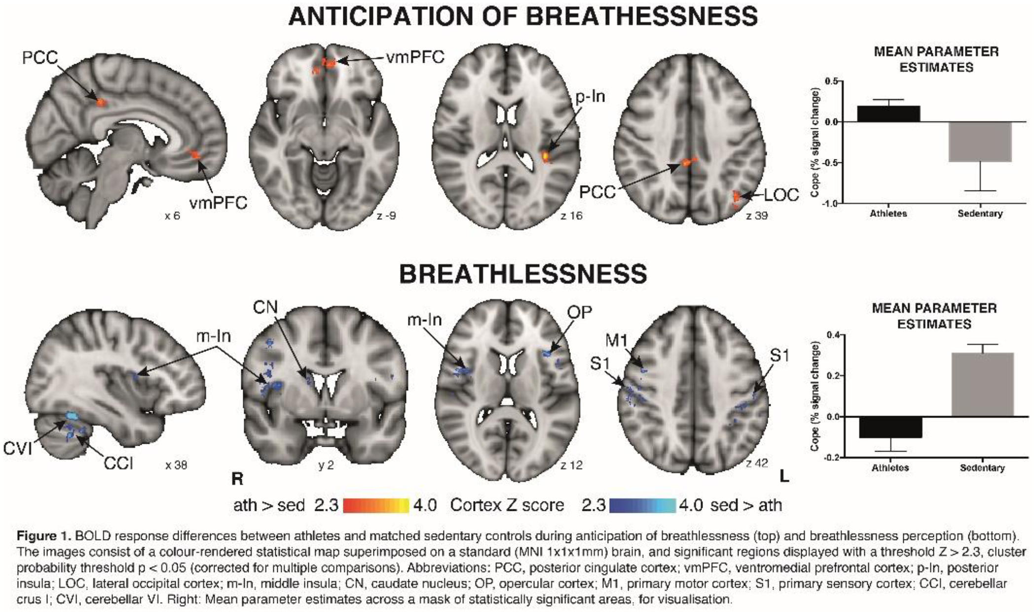
Faull OK, Cox PJ & Pattinson KT, 2016. Frontiers in Physiology, 7
The master biological clock, i.e. the suprachiasmatic nucleus (SCN) of the anterior hypothalamus provides individuals with the ability to predict the timing of circadian events and to adjust physiological processes accordingly. The evolutionary advantage of a biological clock rests on its predictive power, but adaptability to environmental changes is also important. Light is the principal zeitgeber of the mammalian circadian system and SCN neurons react to changes in the light/dark cycle by re-entraining their circadian oscillation. Accordingly, the SCN receives direct photic information from the retina via the retino-hypothalamic tract. The intergeniculate leaflet (IGL) of the thalamus seems to be, besides the SCN, another important neural structure in the mammalian circadian time-keeping system. The IGL is also densely innervated by the retina, and projects to the SCN. However, its role in mediating circadian entrainment remains somewhat elusive.
By using a new genetic mouse line (Sox14Cre) we selectively manipulate thalamic neurons that project to the SCN to investigate their ability to modulate circadian behaviour. We optogenetically stimulated IGL neurons in vivo over multiple days at different circadian times, and we showed that this specific subset of neurons was sufficient to phase-shift daily activity rhythms. Subsequently, we mapped the inputs to the IGL from the retina and other brain regions using mono-synaptic restricted ?G-rabies virus strategy. We demonstrated that different subtypes of photosensitive retinal ganglion cells (pRGCs) innervated the SCN and the IGL and that several neuromodulatory systems converged onto the IGL.
Overall, our data suggest that the IGL is sufficient to regulate circadian entrainment of the SCN. Its function may thus consist in integrating light information with other relevant cues to adjust the phase of daily activity and to adapt it to environmental parameters.
Information integration across the senses is fundamental for effective interactions with our environment. A controversial question is whether signals from different senses can interact in the absence of awareness. Models of global workspace would predict that unaware signals are confined to processing in low level sensory areas and thereby prevented from interacting with signals from other senses in higher order association areas. Yet, accumulating evidence suggests that multisensory interactions can emerge –at least to some extent- already at the primary cortical level [1]. These low level interactions may thus potentially mediate interactions between sensory signals in the absence of awareness.
Combining the spatial ventriloquist illusion and dynamic continuous flash suppression (dCSF) [2] we investigated whether visual signals that observers did not consciously perceive can influence spatial perception of sounds. Importantly, dCFS obliterated visual awareness only on a fraction of trials allowing us to compare spatial ventriloquism for physically identical flashes that were judged visible or invisible.
Our results show a stronger ventriloquist effect for visible than invisible flashes. Yet, a robust ventriloquist effect also emerged for flashes judged invisible. This ventriloquist effect for invisible flashes was even preserved in participants that were not better than chance when locating flashes they judged ‘invisible’.
Collectively, our findings demonstrate that physically identical visual signals influence the perceived location of concurrent sounds depending on their subjective visibility. Even visual signals that participants are not aware of can alter sound perception. These results suggest that audiovisual signals are integrated into spatial representations to some extent in the absence of perceptual awareness.
1. Rohe, T. & Noppeney, U. Distinct computational principles govern multisensory integration in primary sensory and association cortices. Curr. Biol. 26, 509–514 (2016).
2. Maruya, K., Watanabe, H. & Watanabe, M. Adaptation to invisible motion results in low-level but not high-level aftereffects. J. Vis. 8, 1–11 (2008).
About half of all patients with Parkinson’s disease suffer from intermittent freezing of gait (FOG), which can cause falls and thus poses a major risk to the well-being of patients (1). In many cases these motor blocks are unresponsive to medication or deep brain stimulation therapy. We set out to answer if activities in the left and right subthalamic nucleus (STN) are modulated by gait, and if so, whether in unison or in an opposing manner. As rhythmic auditory cues can improve gait rhythmicity as well as FOG (2), we also tested how any modulation changes when auditory cues are provided.
We recorded local field potentials from the STN in 9 Parkinson’s disease patients during stepping in place on a foot pedal. Patients sat on a chair to avoid falls and movement artefacts. The constant step interval of 1s was set by the timing of heel strikes displayed by a looped video of a walking man. In 7 of the 9 patients, we also provided a metronome sound synchronised with each heel strike in half of the stepping sequences.
We found that high beta oscillations (20-30 Hz) were most likely to occur after the contralateral step, when the contralateral foot rested on the pedal. After the ipsilateral step, when the contralateral foot had to be raised, beta oscillations were least likely. Power in the left and right STN, particularly in the 20-30 Hz beta band, was thus modulated separately in opposite patterns.
The metronome improved patients‘ synchrony with the heelstrikes displayed in the video and also increased beta modulation. Our results raise the possibility that alternating DBS patterns may provide better control of gait than constant stimulation of both STN.
1. Macht M, Kaussner Y, Möller JC, Stiasny-Kolster K, Eggert KM, Krüger HP, et al. Predictors of freezing in Parkinson’s disease: A survey of 6,620 patients. Mov Disord. 2007;22(7):953–6.
2. Arias P, Cudeiro J. Effect of rhythmic auditory stimulation on gait in parkinsonian patients with and without freezing of Gait. PLoS One. 2010;5(3).
Animal studies have demonstrated somatotopically organised sensory and motor maps within the cerebellum, but human experimental and clinical data suggest additional involvement in cognitive functions. This study mapped sensorimotor, verbal working memory, speech motor and language functions in the cerebellum. Twenty healthy adults underwent cerebellar optimised fMRI in a 3T scanner with 4 paradigms: 1) motor: moving right fingers or toes at visually paced irregular rhythm; 2) sensory: vibrotactile stimuli delivered to the right index finger, first toe or both; 3) language and speech motor: in response to aurally presented stimuli (nouns) subjects generated associated verbs (aloud or subvocally), repeated non-words aloud, or listened to nouns and non-words; 4) verbal working memory: using the Sternberg task. Following field-map unwarping and physiological noise correction, individual responses were estimated. Group activity was assessed with a mixed effects model in FSL software, with cluster forming threshold Z>3.09 and corrected significance level of P<0.05 for the motor, language and Sternberg paradigms. Dual representation was observed in the cerebellum for the motor paradigm, with finger and toe movements producing activity in the right hemisphere ipsilateral to the task, in lobules V and VIII (fingers) and lobules I-IV and VIIb-VIIIa (toes). Finger and toe areas for the sensory paradigm overlapped with the corresponding motor map in the anterior lobe (uncorrected, p<0.005). Speech motor results showed bilateral activation in both superior and inferior cerebellum with the activation in the anterior lobe positioned adjacent and caudal to the finger sensorimotor area. The language paradigm showed a right lateralised activity in lobule VI, Crus I and II superiorly and in lobule VIIb inferiorly. During the encoding phase of memory paradigm, load dependent BOLD activity was observed in right lobule VI, Crus II and VIIb and vermis VI. When the presented letters¬ were held in memory, the largest area of BOLD activation was observed in lobule VI, extending into Crus I bilaterally as memory load increased.The range of tasks probing cerebellar activity associated with sensorimotor and higher order tasks demonstrated a clear spatial compartmentalization of function.
Active whisking in mice and rats is one of the fastest behaviours known to mammals and is used to guide complex behaviours such as exploration and navigation. During object contact, whisker movements are actively controlled and undergo robust changes in timing, speed and position. This study focuses on characterising whisker movements in male and female 5XFAD mice, a model of Alzheimer’s disease, and their WT controls, in a number of different tasks, including object exploration, tunnel running and novel object exploration. As a result of genes from the background strains, some mice had retinal degeneration (RD). Forty-nine 6-7 month old mice were filmed behaving freely in an open field containing an object, under infrared light using a high-speed video camera at 500fps. Whiskers were tracked and variables, such as position, amplitude, speed and asymmetry, were extracted and compared pre-contact and during contact. Measuring whisker movements in these animals presents a quantitative way to capture exploratory behaviours. The transgenic mice had significantly altered whisker angular positions, amplitude and asymmetry during contact; and female 5XFAD mice with RD had lower mean angular positions during contact. Differences due to gender and RD were found in the data, with female mice making larger and faster whisker movements overall, and mice with RD making larger and faster whisker movements during contact. The data shows that measuring whisker movements can quantify the effects of the Alzheimer’s transgenes, sex differences and the problem of retinal degeneration on exploratory behaviour in these mice.
Sensory stimulation evokes negative BOLD responses (NBRs: signal decrease relative to baseline), both intra-modally (IM, in stimulated sensory cortex) and cross-modally (CM, in other sensory cortices). However, despite regular observation, these NBRs remain poorly understood. Here we used multimodal neuroimaging to investigate whether: 1) IM and CM NBRs exhibit similar underlying changes in neuronal activity and metabolism; 2) IM and CM NBRs are modulated similarly by stimulus intensity.
EEG, BOLD and CBF responses were simultaneously recorded in 17 subjects at 3T. 24 trials of four tasks were performed (14/20s on/off): viewing 100% or 10% contrast left-hemifield visual reversing checkerboards (3Hz), complex finger-tapping or simple handgrip motor task (right-hand).
Beamformer analysis localised changes in alpha (α) and beta (β) EEG oscillatory power between stimulation and rest. IM and CM EEG responses were extracted from virtual electrodes (VE in visual (V1) and motor (M1)) cortex and single-trial responses measured. GLM analyses localised: 1) main effect BOLD and CBF responses and also trial-by-trial fMRI correlations with 2) α- and 3) β-response variability. Metabolic demand (CMRO2) was calculated at peak regions.
IM and CM NBRs were evoked by both visual and motor tasks. NBRs and negative CBF responses were spatially coincident (Fig A&B). Increased visual contrast and motor task difficulty led to increased magnitude and extent of both IM and CM negative fMRI responses (Fig A&B). CMRO2/CBF ratio was significantly higher for IM NBRs than PBRs or CM NBRs (Fig C&D).
Close links between α&β responses and both IM and CM NBRs shown by EEG-fMRI correlations: positive during visual trials, contralateral M1 with the IM VE; negative during motor trials, contralateral V1 with the CM VE.
IM NBR regions coincide with α&β desynchronization whereas CM NBR regions show little group EEG response. Both IM and CM NBR show decreases in CMRO2, are modulated by stimulus intensity, correlate with α&β responses. These results support a neuronal origin of IM and CM NBR, possibly reflecting regional suppression during the task.
Future work: examine between-subject variability between α&β-fMRI responses in NBR regions.
Among numerous studies that have attempted to manipulate cerebellar excitability using anodal transcranial direct current stimulation (tDCS), a large number have demonstrated a facilitatory effect of tDCS on both motor and cognitive behavioural tasks [1-4] such as visuomotor adaptation [1]. However, a growing number of groups have also found cerebellar tDCS to have no effect on behaviour [5, 6]. Although these findings suggest that tDCS could be an effective technique to facilitate cerebellar function in clinical disorders, the latter results cast doubt over its reproducibility across varying task parameters. In this study, we aimed to assess the stability of the cerebellar tDCS effect during a visuomotor adaptation task in which we systematically modified a wide range of task parameters. To do so, 192 healthy young adults were recruited and placed in either an anodal or sham cerebellar tDCS group. Across 7 experiments, we manipulated a four task parameters, altering one for each experiment: position of screen, use of a device/tool, tDCS timing and the perturbation schedule. In each, the participants were measured at baseline, during adaptation to a 30-degree rotation, and in a retention phase.
We initially replicated the previously reported positive effects of anodal cerebellar tDCS on visuomotor adaptation [1]; however, this finding was not maintained during a second replication experiment with larger sample size, or across the other five experiments testing a range of varying task parameters. Thus, these findings cast doubt on the ability of cerebellar tDCS to have a consistent and robust effect on behaviour across participants and tasks parameters, calling into question the efficacy of using it within a clinical setting.
References
1. Galea, J.M., et al. Cerebral Cortex, 2011. 21(8): p. 1761-1770.
2. Cantarero, G., et al. Journal of Neuroscience, 2015. 35(7): p. 3285-3290.
3. Hardwick, R.M. and P.A. Celnik., Neurobiol Aging, 2014. 35(10): p. 2217-21.
4. Block, H. and P. Celnik. Cerebellum, 2013. 12(6): p. 781-93.
5. Conley, A.C., et al., Fron Hum Neurosci, 2016. 10.
6. Minarik, T., et al., Front Hum Neurosci, 2016. 10: p. 453.
Acknowledgments
FfWG main grant, the Wellcome Trust, ERC MotMotLearn (637488), EPSRC (EP/F50053X/1).
* Correction: Original document did not contain the correct abstract or contact information
Women are twice as likely as men to develop stress related disorders such as depression, being the population that receives antidepressant treatment more frequently. This sexual disparity is also observable in effectiveness of treatments. Despite this fact, most of studies that have used animal models for determinate the physiological mechanisms implicated in depression and to develop specific drugs for their treatment have been performed in males. The aim of this study was to analyze the effects of chronic social stress on the anhedonic behavior, immunity parameters and central monoaminergic activity in female mice. We also studied, if the treatment with venlafaxine, an ISNSR, reverses these effects. For this purpose, CD-1 female mice were subjected to social chronic instability stress for 7 weeks, and they were administered venlafaxine (20 mg/kg, ip) during the last 3 weeks of stress period. The behavioral results indicate that stressed mice consumed less sucrose solution than control mice, which is associated with depressive-like behavior. Furthermore, different changes were observed in the monoaminergic activity depending of brain structure analyzed. Thus, stress produced an increase in serotonergic activity in PFC, but not in HC, where stressed mice showed lower levels of 5-HIAA and 5-HT. In PFC, stressed mice showed lower levels of 3-HK and a lower ratio of KYN/5-HT in HC. Stressed mice showed lower levels of MHPG and NE in PCF and HC, respectively, suggesting a decreased noradrenergic activity in both structures. Likewise, the greater weight of the spleen in stressed mice suggests an increase of the immune activity in this group. Venlafaxine treatment did not produce strong changes, but it reversed the effects of stress on 3-HK levels in PFC and increased 5-HIAA and DOPAC levels in this structure. Results indicate that in female mice this stress model produce behavioral and immunitary disturbances, as well as changes in several monoaminergic metabolic pathways, which are partially reversed by venlafaxine. In sum, these results suggested that further studies would be necessary for greater knowledge of biomarkers implicated in stress related disorders in females that contribute to the development of more specific pharmacological treatments.
The cross-stressor adaptation hypothesis proposes that aerobic fitness leads to a decreased physiological response to exercise and psychological stress (Sothmann et al., 1996). However, others argue that exaggerated physiological responses to psychological stressors are not metabolically coupled (i.e. evoke blood pressure (BP) and heart rate (HR) increases without a concomitant increase in oxygen consumption) (Turner and Carroll, 1987). Thus fitness may not necessarily translate to adaptations to psychological stress even if it reduces responses to exercise stress. PURPOSE: The aim of this cross sectional study was to investigate cardiovascular responses in young and older, fit and unfit individuals at rest and during acute psychological stress. METHODS: Thirty healthy volunteers in two age groups: young (20 – 40 yrs; mean age 25 ± 7 yrs; 9 fit, 9 unfit; VO2max >45 mL·kg·min-1 vs. <40 mL·kg·min-1) and older (60 – 80 yrs; 68 ± 3 yrs; 6 fit, 6 unfit; VO2max >30 mL·kg·min-1 vs. <20 mL·kg·min-1) participated. During separate visits they completed an aerobic fitness test (VO2 max) and a paced auditory serial addition task (PASAT). RESULTS: Between group ANOVAs revealed a significant interaction between time (baseline and stress task) and age group on HR (p=.028), such that older individuals showed a greater change from baseline to stress. However, there were no differences in the response to stress between fit and unfit individuals (BP: p= .292; HR: p= .609). Nonetheless, the number of correct mental arithmetic responses was significantly higher in the older fit group than the older unfit group (p=.004). CONCLUSION: Fitness had no effect on BP or HR changes in response to psychological stress in either age group, but older fit individuals performed better at the mental arithmetic stress despite having a similar physiological response to the unfit group. These findings indicate that fitness does not result in adapted physiological responses to psychological stress, but may impact on behavioural engagement with psychological challenges and thus cognitive performance.

Differences in cerebellar structure and function are consistently reported in individuals exposed to early-life stress and individuals with diagnosed stress-related psychopathology. Saccadic adaptation is a cerebellar-dependent mechanism that restores the accuracy of saccadic eye movements, following repeated errors. It is currently unknown whether and how saccadic adaptation could be affected by stress. Recent studies have demonstrated that transcranial direct current stimulation (tDCS) over the cerebellum can either increase or decrease sensorimotor adaptation, probably via excitation or inhibition of the cerebellum. Consequently, the aim of this study was to investigate the effects of experimentally-induced acute stress on saccadic adaptation and to demonstrate how this would relate to either anodal or cathodal tDCS. Saccadic adaptation was elicited using the double-step target paradigm in young healthy men and women. In this paradigm, target position is manipulated to artificially induce a saccadic error, which subsequently aims to restore accuracy by progressively increasing saccade size. Saliva for cortisol determination and subjective measures of stress were collected repeatedly. In the first experiment, 49 participants were exposed to either a stress or a control condition using the offline version of the Montreal Imaging Stress Task (MIST), shown to generate significant physiological responses. Adaptation was assessed 10 minutes after stress induction, when cortisol levels peaked. Participants in the stress group reported significantly more stress symptoms than controls and did not demonstrate a significant increase of saccade size compared to the control group. In the second experiment, 46 participants underwent 15 minutes of anodal, cathodal, or sham tDCS whilst performing the same adaptation task. Preliminary results showed that anodal stimulation tended to increase the extent of saccadic adaptation. Conversely, participants exposed to cathodal stimulation did not show a significant increase of saccade size. Taken together, these results suggest that acute stress reduces the ability to acquire saccadic adaptation potentially via a decrease in cerebellar excitability.
Levels of adult hippocampal neurogenesis (AHN) integrate experiences in a valence-specific manner and may present an objective marker of welfare. In rodents, AHN is suppressed by cumulative chronic stress, whilst being increased by experiences associated with improved mood, such as exercise, environmental enrichment and antidepressant treatment. These responses are largely restricted to the ventral hippocampus, which coordinates emotional behaviours and provides negative feedback to the HPA stress-axis, whilst the dorsal region is involved in spatial memory and cognition. For anatomical reasons, we hypothesised that the caudal pole of the avian hippocampus is homologous to the stress-responsive ventral region in mammals and therefore our primary aim was to test whether AHN in the caudal hippocampus in poultry is sensitive to cumulative chronic stress.
Tissue was obtained from 64 HyLine Brown hens (aged 18-26 weeks during study) which were exposed to randomized and unpredictable stressors over an 8 week period. As expression of the protein doublecortin (DCX) provides a marker of immature neurons arising from AHN, we used immunohistochemistry to stain DCX-positive cell bodies, which were quantified via stereological cell counts.
Whilst the density of DCX-expressing multipolar neurons did not differ between hens exposed to chronic stress and control birds in the rostral hippocampus (χ21 = .173, p = .677), stressed-birds exhibited significantly fewer DCX+ multipolar neurons at the caudal pole (χ21 = 4.25, p = .039); indicating a suppression of AHN under stress specific to this subregion. In order to validate this finding and assess its amenability to measurement via the quicker method of real-time PCR, we are currently using this technique to compare the expression of DCX mRNA in hens which were housed in either a preferred (high welfare) or non-preferred (low welfare) environment. Comparison between DCX mRNA levels in the rostral and caudal hippocampi of birds with these two classes of cumulative experience will be presented at the meeting.
We conclude that the caudal end of the avian hippocampus is sensitive to chronic stress. Thus, measuring AHN in the caudal hippocampus post-mortem may provide an objective marker of the cumulative welfare state of poultry.
Introduction: Modelling chronic disease in mice is essential to further our understanding of disease aetiology and for developing treatments. However, mice models of chronic disease may suffer discomfort or pain due to the nature of disease or experimental procedures. To understand the impact that chronic disease has on the welfare of mice, it is necessary to quantify an animal’s affective state. Physiological markers of stress can provide a useful proxy measure for affective state. In particular, the birth of new neurons within the dentate gyrus of the hippocampus, adult hippocampal neurogenesis (AHN), is a process that is responsive to an animal’s cumulative experience. Increases in AHN are correlated with positive experiences (e.g. environmental enrichment), whereas decreases in AHN are correlated with negative experiences (e.g. unpredictable chronic mild stress). Changes in AHN also show functional specificity within the dorsal and ventral regions of the hippocampus. Whereas the dorsal region primarily serves cognitive-related behaviour and memory, the ventral region is primarily associated with mood-related behaviours. Measuring AHN within the ventral hippocampus may therefore be a suitable tool to assess the impact of chronic disease on the welfare of mice. AHN can be quantified by measuring the expression of doublecortin, a microtubule binding protein that is a marker for immature neurons within the granule cell layer of the dentate gyrus.
Aims: This project aims to determine the impact of a model of liver fibrosis on affective state in mice.
Methods: Male cRel fl/fl mice were injected bi-weekly with CCl4 for 8 weeks to induce liver fibrosis . Mice in the control condition were injected with olive oil bi-weekly for 8 weeks. Open-field tests were conducted to assess depressive-like behaviour. From all mice, the dorsal and ventral hippocampus from one hemisphere was processed to quantify doublecortin mRNA expression using real-time PCR. The other hemisphere was processed to quantify doublecortin protein expression using immunohistochemistry.
Results: This poster will present the current findings of the project.
Financial support: CNPq and PADC-FCF/UNESP.

The central amygdala (CeA) is a critical anatomical substrate for emotional regulation in response to stress, pain, and alcohol-related behaviors. While many cell-types have been identified in the CeA, much less is understood about the unique properties of these molecularly-defined neurons. We focus on a subset of neurons in the CeA expressing the neuropeptide dynorphin (Dyn+), the endogenous ligand of the kappa opioid receptor. To genetically identify dynorphinergic (Dyn) neurons, we crossed a Cre-dependent tdTomato reporter mouse to a mouse expressing Cre recombinase under the same promoter as preprodynorphin. In this model, only dynorphinergic cells express tdTomato, allowing complete visualization of dynorphinergic circuitry throughout the brain and visually-guided, targeted whole-cell recordings in amygdala slices. We report distinct patterns of c-fos expression in these neurons following stress, pain, and alcohol exposure. We also document the intrinsic electrophysiological properties of these neurons and the strengths of inputs they receive from the parabrachial nucleus and the basolateral amygdala. Furthermore, the morphology of CeA Dyn+ neurons is defined by filling the cells with Neurobiotin. To determine the long-range connectivity of Dyn+ CeA neurons, we utilized cell-type selective expression of reporter viruses to identify these molecularly-defined projections throughout the brain. Together these data provide a base knowledge for further cell-type selective manipulation and observation in vivo. Understanding the mechanisms by which the dynorphin/kappa opioid system regulates emotional processing in the context of stress, chronic pain, and alcohol abuse will provide valuable insight into potential therapeutic targets for these neurological and neuropsychiatric disorders.
INTRODUCTION: Stress is proposed to be involved in etiology of several diseases. Chronic stressors cause morphological and neurochemical changes in the bed nucleus of stria terminalis (BNST). Corticotropin-releasing factor (CRF) in the BNST has been implicated in control of stress-related behavioral and physiological responses. However, neuroplasticity in this neurochemical mechanism following exposure to chronic stressors has never been evaluated. Therefore, we investigated the expression of both CRF1 and CRF2 receptors in the BNST following exposure to a protocol of chronic variable stress (CVS) in rats. METHODS: The CVS protocol consisted of exposure to different stressors in variable schedules for 10 consecutive days. Twenty-four hours after the last session of stress, the animals were sacrificed and the brain were removed and stocked into a freezer in -80°C. Afterward, the BNST of all groups were collected by microdissection and protein levels of both CRF1 and CRF2 receptors were analyzed through Western-Blotting technique. Data were expressed as percentage of the control group values. RESULTS: The rats chronically stressed exhibited reduction in expression of both CRF1 (59±9 vs 100±13, t=2.4; P<0.03) and CRF2 (46±6 vs 100±15, t=2.9; P<0.01) receptors within the BNST. CONCLUSION: Chronic stress reduces expression of CRF receptors in the BNST, which can be related with stress-evoked diseases.
The periaqueductal gray matter (dPAG) is involved in coordinating aspects of the fear responses. The dPAG-electrical stimulation (ES) produces a defensive reaction characterized by freezing, escape and autonomic reactions. The main interest in studying these responses are based on clinical findings which suggest that these fear responses are related to panic disorder. There are two types of freezing associated with dPAG: the freezing directly produced by dPAG-ES and the dPAG post-ES. While the former is a preparatory response to the escape, the second reflects a transfer process of information to forebrain structures, which allows the animal to assess the consequences of the aversive situation and to recognize the threatening stimulus. Some findings pointed to the role of Substance P by NK1 receptors in the amygdala in the evaluation of these aversive consequences generated by dPAG-ES. However, the time course of these consequences is still unknown. The exploratory behavior of independent groups of rats treated with Spantide (SPA, NK1 antagonist) in the central nucleus of the amygdala in the dPAG-ES day was valued in the elevated plus maze (EPM) at 1, 7 and 14 days later. The results showed that the control rats reduced the frequency of entries and time spent in the open arms of the EPM in all intervals analyzed, while SPA treatment minimized these consequences only in the 1 day interval group. Together, these findings show that the effects of dPAG-ES are long-lasting and modulated by NK1 receptors. It suggests that the aversive information generated in dPAG is sent to rostral brain structures involved in the evaluation and recollection of the stressor, and do not result only in a motor output of defensive reaction as they have been currently thought.
Depression is associated with both, cancer disease and social stress, being of special relevance the way in which people deal with stress. Although mechanisms underlying this relation are still inconclusive, it is known that stress and tumor are related to increased inflammatory markers which activate tryptophan metabolic pathway towards kynurenine production. This activation can result in a rise of 3- hidroxikynurenine (3-HK) and neurotoxic products that affect cerebral activity, and hence contribute to depressive-like behaviour. Therefore, this study aims to analyse the effects of stress coping strategies on hippocampal tryptophan metabolic pathway activity in tumor inoculated mice.
For this purpose, OF1 male mice were inoculated with B16F10 melanoma tumor cells. A subgroup was exposed to social stress, using sensorial contact model, 6 days after inoculation. Interactions carried out in the social stress were recorded in order to analyse their behaviour and to classify subjects in active or passive groups. Seventeen days after inoculation mice were subjected to sucrose preference test and on day 21, they were sacrificed and hippocampus was dissected in order to establish tryptophan (TRYP), serotonin (5-HT), 5-hydroxyindoleacetic acid (5-HIAA), kynurenine (KYN) and 3-HK levels.
Results indicate that tumor inoculated mice show lower 5-HT/TRYP ratio, lower KYN levels and a rise in the 3-HK/KYN ratio in the hippocampus. Tumor inoculated mice also show lower preference of sucrose than non-tumor mice. Stress do not result in tryptophan pathway changes but higher levels of 5-HIAA are observed in stressed mice in relation to non-stressed, independently of coping strategy. Furthermore, increased 5-HT levels are observed in active mice, but not in passive subjects when compared with non-stressed mice.
This data indicate that the neurotoxic pathway of the kynurenine may be activated by tumor which could contribute to the appearance of depressive-like behaviour. Furthermore, stress gives rise to changes that suggest an increase of serotoninergic hippocampal activity, regardless of tumor and coping strategy. However, the high levels of 5-HT observed only in active mice might suggest differences in neurotransmission depending on the strategy used when dealing with stress.
When exposed to an unfamiliar open space environment, mice and rats experience fear and attempt to find an escape route. The presence of a challenging obstacle can prevent fear motivated escape in high anxiety mice. We exploited this fear motivated escape in a 3D open-field (OF) in order to assess anxiety in different mouse strains. The 3D OF consists of a platform with downward or upward inclined steep slopes (80cm x 25cm) attached on two opposite sides. In the downward slope configuration (DS), the platform is elevated 75cm (DS75) or 100cm (DS100) above ground. In the upward slope configuration (US), each slope leads to a stand (80cm x 25cm) and the platform is elevated 75 cm above ground (US75).
In DS75, DS100 and US75, mice spent more time in the areas adjacent to slopes than in the areas adjacent to void; only C57BL/6J and CD-1 crossed onto the slopes in DS75, and crossed onto the stands in US75. BALB/c explored the slopes in US75 only. The crossings onto the slopes in DS100 were significantly reduced in C57BL/6J and CD-1, and there were no differences between BALB/c and C57BL/6J. When tested in DS75 configuration, BALB/c mice demonstrated no difference in anxiety between early morning or late afternoon; they avoided the steep slopes and crossed onto shallow one. Administration of different doses of diazepam, but not amphetamine, facilitated crossings onto the steep slopes (DS75) in BALB/c mice.
It is widely recognized that the early postnatal environment, specifically within the context of the caregiving relationship, moderates the development of behavior and disease. Early, adverse experiences confer vulnerability to aberrant brain development, dysregulated immune function, anxiety and mood disorders, deficits in learning and memory, and a myriad of other consequences that persist throughout the life of the organism. Less understood are the mechanisms by which this disruption occurs, though epigenetic alterations have recently come to light as promising candidates. Our lab uses a model termed the scarcity-adversity model of low nesting resources wherein pup caregiver exposures occur outside the home cage with dams given very few nesting materials. This results in disrupted maternal care such that adverse behavior directed toward pups (i.e. dragging, dropping, stepping on, roughly handling, or actively avoiding pups) is significantly increased when compared to pups in the home cage, where nesting resources are plentiful. Using this model, our lab has previously reported altered methylation patterns of the brain-derived neurotrophic factor (bdnf) gene, a critical player in development and plasticity that is sensitive to stress and quality of caregiving. In the current experiment we have uncovered behavioral deficits in our adversity-exposed animals that parallel their altered epigenetic patterns. In order to investigate the link between these epigenetic and behavioral outcomes, we have administered epigenome-modifying drugs concurrent with caregiver manipulations. Preliminary data suggest that these agents can indeed prevent aberrant methylation patterns induced in adversity-exposed animals. Determining our ability to change (or prevent changes to) the epigenome is a critical first step in determining whether the relationship between those changes and later behavioral outcomes is a causal one.
The radical transformation of the animal behaviour toward human is a fundamental and major change occurred under domestication that accompanied by morphophysiological changes (e.g. appearance of floppy ears, curled tail, reduction of stress response). In the present work, we used a unique experimental model of the animal domestication, namely, “tame" silver foxes bred in Russia by long-term selection on emotionally positive reactions to humans. These foxes have the same behaviour, morphological and physiological changes as domesticated animals. The goal of this work is to identify differentially expressed genes and pathways for dorsal hippocampus between tame and aggressive foxes. For this, we used the RNA-Seq approach.
About half a thousand of differentially expressed genes were detected. The analysis of this data identified, among other, several pathways associated with the nervous system: “calcium signaling pathway” and “long-term potentiation” (e.g. genes of calcium voltage-gated channel subunit alpha1), “glutamatergic synapse” (e.g. NMDA-receptor genes), “GABAergic synapse” (e.g. the gene of a component of the GABA type A receptor). These genes and pathways are associated with the regulation of stress response, neuronal plasticity and a number of forms of the behaviour, such as anxiety, fear, social recognition, learning and memory changed under historical domestication. In addition, the functional “axon guidance” group was allocated. The allocation of this group is probably associated with increased neurogenesis in adult hippocampus in less aggressive foxes as it was previously demonstrated.
Thus, changes in the expression of the genes of key neurohumoral brain systems that probably play a leading role in the change of the fox behavior during the selection on domestication was found.
This study was supported by RSF (grant #16-14-10216).

Excessive activation of microglia during neuroinflammation is now known to exacerbate neuronal damage [1] in neurodegenerative conditions. Consequently, regulating the degree of microglia activation may be considered as an important strategy for treating neurodegenerative disorders. Formononetin (FMN) is a phytoestrogen present in food supplements like red clover. Earlier; we showed that FMN suppressed the release of pro-inflammatory cytokines in lipopolysaccharide (LPS)-activated BV2 microglia [2]. We also showed that the compound blocked neuroinflammation through mechanisms involving NF- κB signalling pathway [3]. In this study, we elucidated the neuroprotective effect of FMN in BV2 microglia/HT22 hippocampal neuron co-culture. BV2 microglia cells were pre-treated with FMN (2.5 10 μM) and then stimulated with LPS (1 μg/ml) for 24 h. HT22 cells were then exposed to BV2 microglia conditioned medium for 24 h. At the end of the experiment, neurotoxicity was determined using the MTT assay for cell viability. Levels of microtubule-associated protein-2 (MAP2) were detected by immunofluorescence and western blotting. MTT results showed that FMN significantly (p<0.01) prevented microglia conditioned media induced toxicity to HT22 neurons. Furthermore, western blotting reveals that pre-treatment with FMN produced a significant and concentration-dependent reversal of decreased neuronal MAP2 protein induced by microglia conditioned media. These results were confirmed with immunofluorescence imaging for MAP2. These results suggest that FMN prevents neuroinflammation-mediated HT22 neuronal death by inhibiting microglial activation.
1. Jin RG, Yang, and G. Li, Inflammatory mechanisms in ischemic stroke: role of inflammatory cells. Journal of leukocyte biology, 2010. 87(5): p. 779-789.
2. Elbakoush A, Olajide O (2014) Formononetin, a phytoestrogen in red clover inhibits neuroinflammation in LPS-activated microglia. Proceedings of the British Pharmacological Society at http://www.pA2online.org/abstracts/Vol12Issue3abst084P.pdf
3. Elbakoush A, Olajide O (2015) NF-kB-mediated inhibition of neuroinflammation by formononetin: role of ER?. Proceedings of the British Pharmacological Society at http://www.pA2online.org/abstracts/Vol13Issue3abst235P.pdf
The endogenous cannabinoid system, composed of neuromodulatory endogenous lipid ligands and their cannabinoid receptors, has crucial physiological and regulatory roles throughout the body. It is known that cannabinoids produce their biological effects via activation of CB1 and CB2 receptor subtypes (Battista et al., 2012), however in the CNS, the predominant cannabinoid receptor is CB1. Numerous studies have examined the modulatory effects of cannabinoids on excitatory synaptic transmission at hippocampal schaffer collateral (SC)-CA1 synapses. Indeed, evidence suggests that hippocampal cannabinoid receptors not only play a role in learning and memory formation, but they are also linked to neurodegeneration in Alzheimer’s disease (AD) (Hajos and Freund 2002). However the effects of cannabinoids on excitatory synaptic function at the anatomically-distinct temporoammonic (TA) input to CA1 neurons is not clear. Here, standard extracellular recordings were used to examine the effects of different selective agonists for CB1 receptors on excitatory synaptic transmission at the juvenile TA-CA1 synapses. Transverse hippocampal slices (350μM) were prepared from 12-18 rats and perfused with oxygenated aCSF. Application of (R) - (+) - methandamide (50nM; 15min) resulted in a transient increase (to 137 ± 7.1% of baseline; n=4; p<0.001) in excitatory synaptic transmission that returned to baseline on washout (104 ± 0.7% of baseline; n=4; p>0.05). On the other hand, application of ACEA (10nM; 15min) had no effect on synaptic transmission at TA-CA1 synapses (102 ± 0.7% of baseline; n=4; p>0.05). These data indicate that CB1 receptor activation modulates excitatory synaptic transmission at hippocampal TA-CA1 synapses. These findings may be important as the TA pathway plays a role in episodic memory (Remondes & Schuman 2004) and impairments in episodic memory is an early event in AD (Hodges 2000)
Heightened distractibility refers to a reduced ability to discriminate relevant from irrelevant information. It is found in normal ageing and a number of psychiatric conditions, including attention deficit hyperactivity disorder (ADHD), dyslexia, schizophrenia, and depression. The most effective form of treatment for heightened distractibility are psychostimulant drugs, such as amphetamine. These are administered orally and often prescribed over prolonged periods, despite unclear mechanisms of action.
Converging evidence suggests that a potential neural correlate for distractibility is the superior colliculus (SC). Alterations to this structure and its connections to the prefrontal cortex have been found to increase distractibility and collicular abnormalities are implicated in ADHD. Furthermore, we have recently shown changes in collicular-dependent behaviour and visual responsiveness in this region after amphetamine treatment, indicating that the therapeutic mechanism of action to reduce heightened distractibility may be, at least in part, within the SC. To date, however, there has been no systematic characterisation of the morphological features of the SC following chronic administration of amphetamine, despite such features being altered in other brain areas following similar treatment.
We chronically treated male Hooded Lister rats for a four week period with orally administered amphetamine (2 mg/kg, 5 mg/kg and 10 mg/kg) and compared the brains of these animals to untreated control animals and animals treated with a vehicle solution (distilled water). Following treatment, animals were perfused and the brains fixed. Brains were sectioned into 50 μm slices and Nissl stained for measures of collicular volume (employing the Cavalieri principle), neuron and glial cell counts and densities. Additional sections were used for immunohistochemistry to detect synaptophysin. Finally, we perfused a subset of animals separately to conduct a Golgi stain on 100 μm slices through the colliculus in order to examine the microstructure of the region. Specifically, we measured dendritic branching, spine density and spine type in the region. Volumes, cell counts and densities were unaffected, although sub-cellular changes were apparent.
Schizophrenia is a debilitating mental disorder affecting ~0.5% of the population, yet the neurochemical basis is poorly understood. Phencyclidine (PCP), an antagonist at NMDA-type glutamate receptors causes psychotic symptoms in normal people, and exacerbates symptoms in schizophrenia sufferers. Thus, short-term chronic pre-treatment with PCP (termed sub-chronic) in experimental animals has been proposed as a model for schizophrenia. Despite this central role of glutamate, dopaminergic signalling is also known to be involved in symptom expression, and PCP may have downstream effects on dopamine systems, particularly in the mesolimbic pathway, perhaps mediated through changes in dopamine receptor expression.
However, the changes in expression of dopamine receptors in dopaminergic brain regions following sub-chronic PCP pre-treatment are unclear. In this study, we investigated the expression of dopamine receptor mRNA in nucleus accumbens, (NAc), frontal cortex (FCx) and ventral tegmental area (VTA), brain regions associated with the mesolimbic dopamine pathway, in rats, 21 days after sub-chronic pretreated with PCP for 5 days.
Female Lister-hooded rats (c250g at start) were pretreated sub-chronically with PCP (2mg/kg, I.P) or saline (1ml/kg, I.P) twice/day for 5 days. They then remained drug-free for 21 days before being humanely killed, the brains removed and the areas of interest dissected out. Expression of the dopamine receptors (D1, D2, D3, D4, and D5) was measured by real-time quantitative polymerase chain reaction (RT-qPCR).
Looking at the terminal fields of the mesolimbic pathway, in NAc, we found increased expression of D1, D2 and D3, while in FCx we found increased expression of D2 and D3 receptors, and decreased expression of D4 receptors. In the cell body region in VTA, we found a decrease in D2 and D5 receptor expression.
In conclusion, there were differences in expression of dopamine receptors between saline pre-treated and PCP pre-treated in both cell body and terminal regions of the mesolimbic pathway. These changes may underlie some of the behavioural deficits seen after PCP pretreatment, and may be important in our understanding of the mechanisms underlying schizophrenia.
OGR1 is a proton-sensitive GPCR widely expressed throughout the mammalian body, including neurons[1, 2]. OGR1 is thought to play a role in pH homeostasis [3], and has been implicated in reducing tumour metastasis[4]. We transiently transfected HEK293 cells with a HA-tagged OGR1 construct to evaluate effects of receptor activation on intracellular calcium homeostasis (calcium levels measured using Fura-2-based microfluorimetry). Treatment of OGR1-transfected cells with acidic buffer (pH 6.8) for 15 min led to an increase in calcium levels. These calcium responses were variable, with single spike or oscillatory responses detected. Responses were reproducible on repeated challenge with acidic buffer, supporting previous claims that OGR1 does not desensitise[2, 3]. No effect of acidic buffer on calcium levels was observed in untransfected, control cells. However, these cells showed marked responses to carbachol. Treatment with the Gq inhibitor, YM-254890 (0.5 μM, 20 min) abolished all calcium responses in proton-sensitive HEK HA-OGR1 cells and responses to carbachol in controls. As OGR1 signals through Gq[3] this effect supports a role for HA-OGR1 in mediating calcium responses to acidic buffer treatment. Much of the published work on OGR1 has focused on cAMP and IP3 formation [3, 5]. This study provides insight into the effects of OGR1 activation on intracellular calcium levels and the effects of acidic buffer treatment on OGR1 mediated downstream signalling. Next we aim to explore the effects of native OGR1 activation on neuronal intracellular calcium homeostasis.
1. Xu, Y. and G. Casey, Identification of human OGR1, a novel G protein-coupled receptor that maps to chromosome 14. Genomics, 1996. 35(2): p.397-402
2. Huang, C.W., et al., Nociceptors of dorsal root ganglion express proton-sensing G-protein-coupled receptors. Mol Cell Neurosci, 2007. 36(2): p.195-210
3. Ludwig, M.G., et al., Proton-sensing G-protein-coupled receptors. Nature, 2003. 425(6953): p.93-8
4. Singh, L.S., et al., Ovarian cancer G protein-coupled receptor 1, a new metastasis suppressor gene in prostate cancer. J Natl Cancer Inst, 2007. 99(17): p.1313-27
5. Huang, X.P., et al., Allosteric ligands for the pharmacologically dark receptors GPR68 and GPR65. Nature, 2015. 527(7579):p. 477-83
Immune activation within the CNS is a classical feature of neurodegenerative diseases. It is now increasingly evident that diseases such as Alzheimer’s and Huntington’s trigger local inflammation and activate innate immune responses, which are primarily driven by microglia. Though recent years have seen a growing appreciation of the importance of research into the role of microglia-mediated inflammation, there is currently an unmet need for human in-vitro microglial models that enable in-depth mechanistic studies and bridge the gap between clinical and animal models. We have developed a protocol that enables differentiation of microglia-like cells from human iPS cells. Firstly, stem cells were differentiated to monocytes expressing the myeloid-specific marker CD14 as well as the haematopoietic markers CD45 and CD11b. Monocytes were further directed to a ramified microglial phenotype by treatment with the growth factors GM-CSF and IL-34 and by interaction with astrocytes and neurons. The iPSC-derived microglia were discriminated from macrophages and other monocyte lineage cells by the defining marker expression profile TMEM119+/IBA1+/GLUT5+/CD45low/CSF1Rhigh/TREM2high. The iPSC-derived microglia have been functionally validated against anticipated phenotypes including cytokine production, phagocytosis and intracellular signalling, and comparisons made with unstimulated cultures. Moreover, gene expression profiling has identified genes and pathways involved in the regulation of microglial response to amyloid beta. The development of protocols for the generation of in-vitro microglial models holds great significance in Alzheimer’s disease research as it will allow to investigate the contribution to the disease process of microglia and microglia-enriched genes identified as risk factors for Alzheimer’s disease.
Olfactory bulb dopaminergic neurons are inhibitory interneurons that co-release dopamine and GABA to regulate the early processing of odour information in the glomerular layer. These neurons can be readily identified by the expression of tyrosine hydroxylase (TH), the rate-limiting enzyme of catecholamine synthesis. We use a Cre-driver mouse line under the control of the dopamine transporter (DAT) crossed with a tdTomato reporter mouse to label dopaminergic neurons allowing for visual targeting in live preparations. Initial experiments have been to characterise the coverage and specificity of the labelling in these transgenic mice. Most DAT-tdTomato neurons were positive for TH (approximately 75%). TH expression is known to be activity-dependent in the olfactory bulb, therefore, the TH-negative neurons could still be dopaminergic. Staining with another dopaminergic marker, dopa decarboxylase (DDC), reveals co-localisation with TH-positive neurons and absence from DAT-tdTomato neurons that are TH-negative. Investigating markers of other known glomerular layer neurons demonstrated that this subpopulation is part of the calretinin population of neurons.
Olfactory dopaminergic neurons are known to be particularly plastic, altering their structure, function and gene expression in an activity-dependent manner. We use slice electrophysiology and immunohistochemistry to examine experience-dependent changes in DAT-tdTomato neurons after one and three days of unilateral naris occlusion. Using this data, we will investigate how functional plasticity is regulated by activity-dependent changes in gene expression and epigenetic modifications in these neurons.
NMDA receptors (NMDARs) are critically involved in the induction of short-tern potentiation (STP), long-term potentiation (LTP) and long term-depression (LTD) in the CA1 area of rat hippocampus. In this regard, we have shown that different di- and tri-heteromeric NMDARs are involved in the induction of the three forms of synaptic plasticity (Volianskis et al 2013, 2015; France et al 2016). Thus, GluN2D containing NMDARs were found to be involved in STP but not in LTP or LTD. Induction of LTP was mediated by tri-heteromeric GluN2A/2B containing NMDARs whereas GluN2B containing di-heteromers were found to be involved in LTD.
Here we studied for the first time the functional implications that knocking out the GluN2D subunit exerts on the induction of STP and LTP. We found that both STP and LTP can be readily induced in hippocampal slices from the GluN2D knockout mice (KO). Notably, whilst GluN2D preferring antagonist UBP145 had no effect on the induction of STP and LTP in the GluN2D KO, it partially blocked LTP in slices from the wild-type littermate controls. These data confirm the specificity of UBP145 and highlight the involvement of GluN2D subunits in the induction of LTP in mouse, in stark contrast to the rat. Moreover, both STP and LTP were partially inhibited by either 0.1 μM NVP-AAM077 or 10 μM Ro 25-6981 in the GluN2D KO and a combination of both antagonists completely blocked the induction of potentiation. This suggests that GluN2A/2A di-heteromers and GluN2A/2B tri-heteromers mediate the induction of STP and LTP in the GluN2D KO.
In summary, our results indicate that the pharmacological profile for inhibition of STP and LTP is altered in the GluN2D KO, suggesting a complex combination of compensatory effects. Furthermore, and in contrast to the rat, GluN2D-containing NMDARs appear to be important in the induction of LTP in mouse, suggesting species differences in the role of NMDAR subunits mediating the induction of synaptic plasticity.
This work was funded by the BBSRC grant number BB/L001977/1 and the National Institute of Mental Health of the National Institutes of Health under Award Number R01MH060252. All procedures involving the use of animals were done accorded with current UK legislation.
Introduction: Mannose-6-phosphate (M6P), a potential scar reducing agent, has previously been shown to enhance nerve regeneration, but its effects on the development of neuropathic pain following nerve injury remain unknown. Spinal glial activation has been reported as a key regulator of neuropathic pain and differences in activation levels may reveal further benefits of M6P treatment. There are many potential ways to treat the site of nerve repair with M6P; its addition to fibrin glue used in the repair could provide a simple method of therapeutic dosing.
Aims: The aim of the study was to investigate whether the application of M6P in fibrin glue results in differences in spinal glial activation following peripheral nerve repair.
Methods: 10 thy-1-YFP-H mice and 10 wild type mice were used. The common fibular nerve of thy-1-YFP-H mice was transected and a 3 mm gap was made. A nerve graft of 3 mm was obtained from wild type mouse and placed within the gap. The nerve ends and graft were then aligned and secured by fibrin glue with/without M6P (600mM). After 2 weeks, spinal cords were harvested, fixed and prepared for immunohistochemistry to label microglia and astrocytes.
Results: Glial activation was increased on the injured side for both repair groups. While no significant differences were observed between repair groups, activation was generally higher in the ventral horn for the non-M6P group, with astrocyte activation at 134.9% and microglia activation at 127.2% of the uninjured side values compared to 128.7% and 123% respectively in the M6P group. Differences in the dorsal horn were mixed, with astrocyte activation lower at 125.9% and microglia activation higher at 136.8% of the uninjured side values in the M6P group, compared to 128.5% and 129% respectively in the non-M6P group.
Conclusion: No significant differences in glial activation were observed between the use of fibrin glue implanted with M6P and fibrin glue alone. However, the differences observed in microglia activation (higher in dorsal horn, lower in ventral horn) appear to suggest that M6P may be more beneficial towards motor axons than sensory. Further investigation may be warranted in order to confirm the validity of this effect and, if confirmed, elucidate a potential mechanism of action.
The dopamine (DA) theory of schizophrenia posits a key role for DA dysfunction: drugs used in treatment share a DA antagonist action. However, glutamate systems are also critical: phencyclidine (PCP: NMDA glutamate receptor antagonist) causes psychotic symptoms in normal people and exacerbates symptoms in schizophrenia sufferers. In rats, short term chronic PCP treatment causes behavioural deficits mimicking schizophrenia symptoms, which endure long after the end of drug treatment, providing an animal model for studying processes underlying the disease.
Core deficits in glutamate function may impinge on DA systems, particularly in nucleus accumbens (NAc). Release of DA in NAc is under modulatory control of many transmitter systems, including GABA, and abnormalities in this modulation may underlie changes seen after PCP pretreatment. This study aimed to characterise GABA mechanisms modulating DA release in NAc, and to ascertain whether these were changed after PCP pretreatment, giving potential insights into mechanisms involved in schizophrenia.
Coronal slices (400μm) containing NAc were cut from juvenile female Wistar rat (c24 days) brains and placed in a 1ml tissue chamber, superfused continuously with oxygenated artificial cerebrospinal fluid (aCSF: 2 ml/min; 33°C). Fast cyclic voltammetry (FCV) scans (-0.4V to +1.3V to -0.4V; 400V/sec) were applied at 10Hz and DA was measured as the background subtracted current occurring at 600mV.
Twelve trains of electrical stimulations (30 pulses; 60Hz; 300uA) were applied at 3 min intervals and evoked DA release was measured. After 4 stimulus trains, drugs were applied in the superfusate for 12 min (i.e. until after stimulus 8), and a further 4 stimulations were applied during washout. Control slices underwent the same 12 stimulus trains, but were superfused with aCSF throughout.
Both muscimol (GABA-A agonist) and baclofen (GABA-B agonist) caused dose dependent attenuation of electrically stimulated DA release at 1, 10 and 100 μM. Therefore GABA systems exert inhibitory control over DA release in NAc, via both GABA-A and GABA-B receptors, which may be abnormal after PCP pretreatment.
Microglia, the brain’s resident immune cells, have many functions including the regulation of inflammation in brain disease and monitoring synaptic activity. In the healthy murine brain, microglial cell density remains constant throughout life, maintained by a fine balance of proliferation and apoptosis1. It remains unclear how altering microglial population dynamics may affect microglial function and brain physiology. In order to study this, we utilised the Vav-Bcl2 transgenic mouse2 which has a block of intrinsic apoptosis in cells of the myeloid lineage, including microglia, due to overexpression of human Bcl2 under the Vav promoter. Vav-Bcl2 mice have significantly increased microglial cell density throughout the brain, which peaks at postnatal day (P)44 and is maintained throughout adulthood, with no gross differences in neuronal or astrocyte populations. In particular, elevated numbers of the CD11b+CD45hi subset of microglia contribute to the increase in cell density, suggestive of an altered functional state in the brain compared to wild-type controls. Transcriptomic analysis of isolated microglia revealed differential expression of genes involved in metabolic processes, macromolecule biosynthesis and immune response, indicating that deregulation of microglial apoptosis changes their phenotype. This altered phenotype is associated with an exacerbated pro-inflammatory response in the brains of Vav-Bcl2 mice after systemic challenge with LPS. Our data shows that long-term deregulation of apoptosis in microglia results in significant phenotypic changes and alters their functional response to an inflammatory stimulus. Deregulation of microglia population dynamics throughout the life-course may have implications for the onset and progression of age-related neurological diseases.
1. Askew K, Li K, Olmos-Alonso A, et al. Coupled proliferation and apoptosis maintain the rapid turnover of microglia in the adult brain. Cell Rep 2016;In press.
2. Egle A, Harris AW, Bath ML, et al. VavP-Bcl2 transgenic mice develop follicular lymphoma preceded by germinal center hyperplasia. Blood 2004;103(6):2276-83.
Regeneration of the adult CNS is an ongoing challenge with many obstacles. Repair of mature neurons is limited largely down to two factors: the growth-inhibiting environment created after injury and the innate inability of CNS neurons to repair. In contrast to mature, aged neurons, during embryonic cortical development, neurons have a high capacity for plasticity, regeneration and repair. This may be due to the fact these cells express growth-promoting proteins called integrins. Integrins are transmembrane receptors involved in mediating cell-cell and cell-matrix interactions. Within the adult CNS, integrin expression is downregulated resulting in reduced plasticity and growth. Literature indicates increasing integrin expression in adult CNS axons can promote regeneration; however recent research suggests exogenous integrin expression in vivo results in the inability of integrins to travel down adult CNS axons which instead remain localised to the cell body.
In light of this, here stem cells are used as a vehicle to increase integrin expression within the CNS. With a regenerative approach in mind, we have grafted iPSC-derived NPCs into cerebral cortex of rodents. Using a combination of western blotting and immunofluorescence techniques, we determined both the endogenous expression level of integrin within iPSC-derived NPCs and the expression level following viral-mediated transduction. To assess transplant survival, neonatal cortical grafting of hNPCs was carried out. Using specific coordinates, wild type and integrin-expressing hNPCs were injected into layer V of the sensorimotor cortex of P0-aged rats. Following grafting, animals were perfused at 2, 4, 6, 7 and 8 weeks of age. In Sprague Dawley rats, wild type hNPCs are able to put out axonal projections up to 8 weeks in vivo. Further IHC analysis shows, grafted cells express deep-layer cortical neuron markers, and, as expected, are inducing a host immune response. Analysis of neurite length in vitro indicates the exogenous integrin is functional, promoting significant neurite outgrowth compared to controls. In analysis of integrin-expressing hNPC transplants we have shown the grafted hNPCs retain their exogenous integrin expression within the axonal compartment up to 8 weeks in vivo.
Background
Notch receptors and their ligands form a fundamental intra-cellular signalling pathway that controls maturation, proliferation and apoptotic events during development.[1] Mutations in the NOTCH3 gene cause the early-onset stroke disorder cerebral autosomal dominant arteriopathy with subcortical infarcts and leukoencephalopathy (CADASIL).[2] CADASIL is characterised by pathological changes within arteries and arterioles, resulting in disability and early-onset dementia.[3] Genetic variation in NOTCH3 has also been associated with increased risk of vascular changes in older people.[4, 5] Cerebral blood vessels are maintained by cells within the neurovascular unit (NVU).[6] It is not known whether CADASIL-causing NOTCH3 mutations interrupt Notch signalling, or how hypoxia influences the Notch pathway components within the NVU. We aimed to characterise the expression of the Notch pathway in the cell types of the NVU before and after hypoxia.
Methods
Rat C6 glioma and human umbilical vein endothelial cells (HUVECs) were cultured under normal cell culture conditions until 75% confluence, at which time cells were exposed to hypoxia (2% O2) for 45 minutes, 2, 4, 12, 24 and 48 hours. Total RNA was extracted using phenol/chloroform phase separation before cDNA synthesis for quantitative PCR assessment of changes in expression of NOTCH and hypoxia signalling pathways.
Results
Expression profiles of the NOTCH pathway components were defined for both cell types under normoxia and following hypoxia of increasing time periods.
Conclusions
Herein we report a full assessment of the expression pathway of NOTCH pathway components in cellular models of components of the NVU and the effects of hypoxia on their expression. The findings from these experiments will allow us to further develop cellular models of the NVU which can be utilised in the identification of novel drugs for the treatment of stroke and CADASIL.
References
1. Louvi, A. & S. Artavanis-Tsakonas. Semin Cell Dev Biol 2012;23:473-80.
2. Fortini, M.E. Dev Cell 2009;16:633-47.
3. Joutel, A., et al. J Clin Invest 2000;105:597-605.
4. Schmidt, H., et al. Brain 2011;134:3384-97.
5. Li, Y., et al.. Exp Ther Med 2016;11:28-32.
6. Sweeney M., S. Ayyadurai, & B.V. Zlokovic. Nat Neuro 2016;19:771-83.
Dynamic changes in the rate and pattern of action potential generation in midbrain dopaminergic neurons are thought to encode behaviourally-relevant information about salient and/or rewarding stimuli. These neurons project extensively branched axons to the striatum, where the release of dopamine (DA) does not necessarily represent a faithful read-out of presynaptic firing activity. DA signalling will be shaped by presynaptic mechanisms that determine the probability of DA release (Pr) and its short-term plasticity (STP), but these mechanisms are poorly understood. We measured evoked DA release using fast-scan cyclic voltammetry in slices of mouse striatum to explore key candidate mechanisms. We show that STP of DA release is characterised by facilitation at short inter-pulse intervals, and depression at longer intervals, as previously described. Large changes to release probability driven by changes to extracellular Ca2+ had only weak effect on STP, suggesting a limited dependence of STP on release probability. Instead, STP was primarily governed by release-independent mechanisms; changes in extracellular K+ likely to modify membrane potential and repolarisation did not change initial release probability but strongly modified STP. Since the dopamine uptake transporter (DAT) can influence dopamine release and membrane potential of DAergic neurons, we also investigated a potential role in STP. Pharmacological inhibition of the DAT reduced both facilitation and depression, and precluded modulation of STP by changes in extracellular K+. These findings reveal the DAT has dual roles on dopamine release, both limiting initial release probability and permitting STF at highest frequencies, and promoting release-independent depression through an interaction with K+-dependent processes. These mechanisms may give rise to the preferential reporting of high-frequency presynaptic activity by DA release, in conditions where the DAT is active.
The spinal cord contains the neural circuitry necessary for the generation of movements such as locomotion, as well as sensation and pain detection. Anatomically different neural circuits may show structurally and molecularly distinct synaptic features and that could also vary over development and ageing. In addition, astrocytes in the central nervous system, which associate with synapses via perisynaptic astrocyte processes (PAPs), may contribute to this synaptic diversity. Large scale mapping of both synapses and astrocytes together could therefore provide a more detailed understanding of the factors affecting synaptic physiology within distinct neural circuits.
In this study, we have used a genetically engineered mouse expressing fluorescently labelled PSD-95 and quantitative microscopy methods to map excitatory synapses and PAPs in the spinal cord. From large scale image analysis of synapses, we show inter-regional and age dependent diversity in excitatory synapse structure and molecular composition. We further used super-resolution microscopy to interrogate the structural differences between synapses of different spinal cord circuits, namely dorsal and ventral horn synapses.
Combining synapse analysis with immunohistochemistry staining of astrocytes, we show the degree of synapse association with astrocytes varies between ages. Furthermore, synapses found with a PAP are found to be structurally larger and molecularly more enriched with PSD-95 than synapses without a PAP. Furthermore, we find that the nature of this interaction between astrocytes and synapses was different between sub-regions of the spinal cord, and between different age groups.
Our data provides a thorough baseline understanding of excitatory synapse diversity in the spinal cord, providing insights into the functional differences between sensory, integrational, and motor circuits. Furthermore, we have applied a highly quantitative microscopy approach to study the relationship between synapses and astrocytes. We show that synapses are structurally and molecular enriched when contacted by astrocytes, and the nature of this interaction changes between ages and between different neural circuitry.
Stroke is a leading cause of death and disability worldwide. Blockage, or occlusion, of cerebral arteries causes irreversible neuronal damage as disrupted blood flow starves neurones of oxygen and glucose. The hypoxia inducible factors (HIFs) are master regulators of oxygen homeostasis and critical for adaptation to hypoxic insult. Although HIF-1 and HIF-2 share some common gene targets, they also promote specific adaptations to hypoxia. Differentiated PC12 and NT2 cells have been extensively used as a model to study the molecular changes associated with neurological pathologies, such as stroke. In this study, differentiated PC12 and NT2 cells were exposed to hypoxia for 4-24 hours in a hypoxic modular chamber before gene and protein expression was analysed by qPCR and immunoblotting. In order to validate the model, we characterised the in vitro changes associated with differentiation into neuronal-like cells, observing morphological, transcript and protein changes that revealed a neuronal-like phenotype. Following hypoxia, induction of the HIF-1 transcript or protein expression was not detected. Curiously, preferential activation of HIF-2 transcription and protein expression was detected. Increased expression of the neural progenitor stem cell-like markers, thought to be transcriptionally regulated by HIF-2, were also observed. Furthermore, hypoxia caused loss of neuronal characteristics in the differentiated cells, as seen by a decrease in the expression of the neuronal markers, and loss of neurite number and extension. Our data shows the HIF-2 pathway predominates over the HIF-1 pathway in neuronal-like cell adaptation to hypoxia, and suggests such adaption could promote regression to neural progenitor stem-cells and thus, potentially proliferative states. This is highly significant as it shows neuronal cells possess molecular mechanisms which could trigger recovery following ischaemic insult. By completely understanding such adaptive mechanism and translating these results to in vivo models, it could represent a novel therapeutic approach to stimulate recovery after stroke.
Prefrontal cortex (PFC) receives a direct glutamatergic projection from the hippocampal formation (HPC) and this connection is important in working and recognition memory. The pathway terminates on both excitatory pyramidal neurons and inhibitory interneurons within the PFC (1). Little is known about how HPC modulates the activity of interneurons in the PFC.
Using a transgenic mouse line to target parvalbumin (PV) interneurons (PVCre/tdTomato) and in vitro slice electrophysiology techniques we examined the role of NMDA and AMPA receptors (NMDAR, AMPAR) in controlling synaptic transmission between HPC and PV interneurons within the prelimbic PFC.
Whole-cell voltage clamp recordings were obtained while electrically stimulating the hippocampal fibre tract. The current-voltage (I-V) relationship was examined for both AMPAR and NMDAR mediated currents. The AMPAR I-V curve was inwardly rectifying, indicating the presence of GluR2-lacking AMPARs. The NMDA I-V curve showed a typical relationship for these channels, with the initial negative slope and the maximum peak amplitudes occurring at depolarised holding potentials. We examined the relative contribution to synaptic transmission of AMPAR and NMDAR-mediated currents and found a low NMDA/AMPA ratio (0.25±0.03, n=10). This indicates that synaptic transmission is largely mediated by GluR2-lacking AMPA receptors. We have recently shown that NMDARs contribute to HPC-PFC pyramidal cell transmission in a frequency dependent manner (2). Our preliminary data suggests that NMDARs play a role in summation at HPC-PV interneuron synapses during high frequency transmission (50 and 100 Hz). To compliment electrophysiological recordings, we built a neuronal model in Python to simulate the glutamatergic synapse onto the interneurons. We optimised the parameters for AMPA and NMDA receptor-mediated currents using a Nelder-Mead method and we are currently using the model to investigate synaptic summation at the HPC-PV synapse.
Determining the role of PV interneurons in controlling HPC-PFC circuit functions will be important in understanding the role of these neurons in working and recognition memory.
1. Godsil et al. (2012) Europ Neuropsychopharm, 1165-1181.
2. Banks et al. (2015) PNAS, 11096–11101.
Lipids play a central role in the function of the brain as structural components. An important additional role of lipids though is to act as signalling molecules to control function, both during development and within the mature brain. Two major lipid-signalling routes are the endocannabinoid (1) and retinoic acid signalling systems (2) acting respectively through the cannabinoid CB1 or CB2 G-protein coupled receptors or the RARα, β or γ ligand gated transcription factors. Both have essential roles to control neuroplasticity and cross-talk between the two pathways may have a profound effect on the brain. We have looked for interactions between the retinoid and endocannabinoid signalling pathways by determining the ability of a CB1/CB2 receptor agonist to target retinoid genes and of a RAR agonist to target genes involved in endocannabinoid signalling. The pathways were studied in the stem-cell-like embryonal carcinoma (P19) cell-line and rodent primary cultured neurons. Retinoic acid was found to regulate CB1 receptor gene (Cnr1) expression as well as the metabolic enzyme diacylglycerol lipase (DAGLα). Conversely, the cannabinoid CB1 and CB2 agonist (CP55, 940) was found to influence the retinoid signalling system by altering expression of the receptor genes Rara and Rarb, as well as expression of the gene encoding the metabolic enzyme Raldh1. This work demonstrates that the endocannabinoid signalling system and retinoid system may have primary or secondary influences on one another in the central nervous system.
1. Pertwee RG Endocannabinoids and Their Pharmacological Actions. Handb Exp Pharmacol. 2015; 231: 1-37.
2. Shearer KD, Stoney PN, Morgan PJ, McCaffery PJ.A vitamin for the brain. Trends Neurosci. 2012; 35: 733-41.
Demyelination disrupts neuronal signalling leading to impairment in neurological control. In multiple sclerosis (MS) as well as other demyelinating diseases, oligodendrocyte precursor cells (OPC), which are present in large numbers in the central nervous system, survive but do not mature and produce myelin. Leucine rich repeat (LRR) molecules such as LINGO1 have recently been shown to play a role in inhibiting the maturation and myelin production of OPC. We have identified a novel LRR Amphoterin Induced and Open Reading Frame 3 (AMIGO3), with similar properties and interacting partners as LINGO1, which appears to be important within the murine central nervous system. As such, AMIGO3 is predicted to be a novel inhibitor of OPC maturation. We observed the expression profile of AMIGO3 in the cerebral cortex and corpus callosum during postnatal development in BALB/c mice through semi-quantitative immunohistochemistry and western blot analysis. AMIGO3 protein expression is greatly upregulated at postnatal day 7 (P7) in the cerebral cortex, followed by a steady decline to P28. This demonstrated an inverse correlation with the extent of myelination. Interestingly there is no obvious correlation between signs of OPC maturation and AMIGO3 expression suggesting that AMIGO3 does not affect maturation but purely the production of myelin. We further analysed the expression of AMIGO3 in in vitro and in vivo models of trauma. This was in line with previous observations of raised AMIGO3 protein expression in the acute and chronic stages following spinal cord trauma. AMIGO3 was observed to increase between 1.5-2x (P<0.05) following excitotoxic treatment with AMPA-CTZ in Oli-neu cells. AMIGO3 was also observed in early stages of experimental autoimmune encephalomyelitis (EAE). These data suggest that AMIGO3 is playing a role within oligodendrocytes following trauma and thus is likely to be a promising therapeutic target. We further intend to examine AMIGO3 protein expression, as well as common signs of inflammation and demyelination, in later stages of acute EAE and human MS to gain an insight into the roles of the protein during the progression of demyelinating diseases.
In response to changes in whisker experience of adolescent mice, cortical neurons in layers 2/3 of the murine barrel cortex can exhibit two general forms of experience-dependent plasticity (EDP): coding plasticity (CP) and homeostatic plasticity (HP). CP refers to changes in neuronal transmission and connectivity of individual synapses and is thought to enable information storage within a neuronal network. HP is most often a global phenomenon that acts as a negative feedback mechanism to keep the activity of a neuronal network within a set operating range. To determine possible astrocyte roles in both CP and HP we utilised IP3-R2 knock out (KO) mice, in which astrocytes but not neurones exhibit diminished [Ca2+] responses. To evoke CP, all but one whisker (single-whisker experience – SWE) were removed unilaterally for 18 days, followed by regrowth for 5-9 days. To evoke HP, all whiskers were trimmed unilaterally for 1, 3, 7, 14, 25 and 32 days and re-attached on the day of recording. We found no significant differences in the magnitude of principal and surround responses in undeprived WT and KO mice and in the amount of plasticity induced in SWE animals (all pairs p>0.05, U-test, N=20). In all-whisker-deprived WT animals the principal whisker responses showed rapid depression 1 day after deprivation (t-ratio 7.3, p<0.0001, N=15), started to recover at 3 days, were above control levels at 7-14 days (t-ratio 3.7, p<0.003, N=15), indicating HP, and back to control levels at 25-32 days. However, IP3-R2 KO mice exhibited a linear decay in magnitude of responses with deprivation time which was significant after 14 days (p<0.05, N=6) with an impaired HP rebound at 25 and 32 days which was not significant (p>0.05, N=10). In both acute slice experiments and in vivo, the LTD induction protocol induced LTP in the KO mice. This scenario was also mimicked in WT slice recordings by patch electrode filling of astrocytes with the calcium chelator BAPTA. LTP induction protocol induced LTP in KO slices, but not in WT slices after filling of astrocytes with BAPTA. This data implicate astrocytes as potent regulators of experience-dependent coding depression and homeostatic up-regulation of whisker-evoked responses.
Following traumatic brain injury (TBI) or spinal cord injury (SCI), complex pathological processes evolve in which cerebral energy perturbations play a major role. In some circumstances mitochondrial dysfunction, where the brain is unable to utilise metabolic fuels and oxygen despite adequate provision, is thought to be responsible for these energy perturbations. Succinate is a tricarboxylic acid (TCA) cycle intermediate which interacts directly with the mitochondrial electron transport chain (ETC). The results of a recent study by our research group suggest that focal administration of succinate, via a microdialysis catheter inside the brain, could improve brain metabolism in TBI patients by fuelling and enhancing the TCA cycle even in the context of mitochondrial dysfunction. The aim of this study was to determine whether succinate supplementation can improve cellular energy state under induced conditions of metabolic stress in a tissue culture model, to confirm and provide better understanding of the results obtained in succinate-treated patients. An overview of the findings of this study is shown in Fig 1. Primary mixed glial cell cultures comprising astrocytes, oligodendrocytes and microglia, were exposed to rotenone (an inhibitor of Complex I of the mitochondrial ETC) to induce metabolic stress, in the presence or absence of succinate. Cellular response was determined by the measurement of intracellular ATP, extracellular metabolic markers (glucose, lactate, pyruvate, glutamate and glycerol), and oxygen consumption rate (OCR). The cultures showed metabolic flexibility, maintaining ATP levels in the presence of rotenone, possibly through glycogen stores in astrocytes and/or high glycolytic capacity. However, a metabolic deficit was observed in in the hours following rotenone administration, increasing the lactate/pyruvate ratio. The presence of succinate induced recovery from this metabolic deficit. The OCR of rotenone-treated cells also showed significant, immediate metabolic improvement in response to succinate. The results indicate that succinate can improve oxidative metabolism in mixed glia, consistent with our other observations in succinate-treated focal areas of injured human brain.

Primary motor cortex (M1) has been implicated in the pathogenesis of Parkinson’s disease (PD) which is characterised by loss of dopamine neurons in the substantia nigra leading to loss of dopaminergic input to many brain regions. Recent studies have reported that dopamine (DA) controls tonic currents in medium spiny neurons, ventrobasal thalamus and nucleus accumbens by modulation of GABARs or cation channels. This study investigates the DA modulation of tonic currents in inhibitory interneurons in primary motor cortex (M1) in rats. Whole-cell voltage clamp recordings were made in deep layers (V) of M1 in sagittal brain slices (350 μm) obtained from male Wistar rats (50 g). FS interneurons and pyramidal neurons were identified by their characteriastic shape and location. Application of DA (30 μM) induced a tonic inward current (-32 ± 8 pA, n=40). To investigate whether DA induced current was via GABAR, bicuculline (Bic; 20 μM) or picrotoxin (PTX; 50 μM) was bath applied. Itonic was partially (56%) reversed by both drugs (DA mean Itonic of -34 ± 7 pA was reduced to -21 ± 3 pA and -18 2 ± pA (when Bic (n=5, p<0.01) and PTX (n=5, p<0.05). All subsequent experiments were performed in the presence of Bic. The Bic-insensitive Itonic was found to be independent of Ih as DA increased Itonic in the presence of the HCN channel blocker, ZD-7288 (10 μM). Bath application of TRP channel antagonists, SKF96365 (100 μM) or 2-APB (100 μM) revealed a significant blockade of Bic-insensitive Itonic (mean amplitude of -26.4 ± 2 pA and -27.2 ± 7 pA reduced to -0.65 ± 1.8 pA and 1 ± 0.3 pA in the presence of 2-ABP and SKF96365 respectively, n= 5, p<0.01) suggesting a role of TRPc channels. These results suggest that there are two different types of tonic currents in FS cells in M1, one mediated via GABAR and another via non selective cationic conductance mediated by TRP channels.
The adipose hormone leptin has been revealed to play a neuroprotective role in cellular and animal models of Alzheimer’s disease (AD). Recent studies indicate that the neuroprotective effects of leptin in AD models may be associated with improvement in mitochondrial functions. Mitochondrial dysfunction has a recognized role in the pathophysiology of AD. However, the mechanism of leptin’s protective effect on mitochondria in AD remains largely unknown. Mitochondrial fission and fusion are beneficial processes that promote mitochondrial distribution across axons into synapses, and separate damaged mitochondrial constituents, to meet high neuronal energy demand and facilitate protective effects. Our aim is to explore effects of leptin on mitochondrial dynamics in a serum-free cellular model and an amyloid-beta-induced AD model. In this study, we examined changes in mitochondrial morphology, oxidative stress, protein expression and cell viability following serum withdraw or amyloid-beta toxicity with or without leptin treatment. We documented the roles of mitochondrial fission and fusion proteins on mitochondrial biogenesis and morphology. In conclusion, leptin may facilitate neuroprotective effects through regulating mitochondrial fission/fusion dynamics in in vitro models of AD, which provides a novel insight into the mechanisms underpinning the neuroprotective effects of leptin.
Background:
Changes in brain glucose metabolism are found in many neurological diseases, including Alzheimer’s disease, multiple sclerosis, epilepsy, and Down’s syndrome. Phosphofructokinase (PFK) activity is a key regulator of glycolytic flux, with three isozymes (M, L, & P); each subunit is 85kDa. Studying PFK structure and function may help our understanding of how energy metabolism is disturbed in neurological illnesses. This study aimed to investigate how PFK activity relates to quaternary structure for each pure isozyme.
Methods:
PFK-deficient S. cerevisiae cells were transformed with plasmid pJJH71 containing cDNA for N-terminal His6 tagged PFK isozyme. Cells were lysed with liquid homogenisation and purified with immobilised metal ion affinity chromatography. Quaternary structure was assessed with size exclusion chromatography. PFK activity was quantified using a linked-enzyme assay.
Results:
Size exclusion chromatography showed material divided into three distinct size distribution peaks. PFK activity was near zero in peak 1, high in peak 2, and low in peak 3. Calculating specific activities showed the majority of active PFK was in peak 2.
The size distribution and shape of the active peak differed greatly between each isoform (figure 1). PFK-M was most active around 522kDa (hexamer); PFK-L at 870kDa (decamer); and PFK-P was much smaller, at 174kDa (dimer). SDS-PAGE gels showed PFK extended over a wide size distribution, especially PFK-L.
Interpretation:
These results indicate that PFK activity is dependent on oligomeric state with a different relationship for each isozyme. Additionally, each PFK has varying oligomeric stability, as shown by the breadth of the respective chromatography peaks. A rapidly shifting dynamic equilibrium between different oligomeric states may complicate analysis of oligomeric status; further biophysical studies will help answer this question. The suggestion that PFK-P has a different profile than the other isozymes is particularly intriguing as expression of this isoform is thought to be more brain-specific than the others. Understanding the differences between PFK isozymes and how brain expression of each isozyme differs in health and disease will help unlock some of the mysteries of brain metabolism.
Neural interfaces allow the establishment of connections between the nervous system and external electronics, and hold great potential in both the clinic and basic research. The peripheral nervous system is a particularly attractive site to position an interface. Nerves are easy to access surgically and action potentials carried by them correlate well with activity at their target. Although many designs for peripheral nerve interfaces have been developed, they all face a major challenge upon chronic implantation.
Materials used in implants are orders of magnitude stiffer than most tissues, which tags them as foreign. As a result, the body responds to interface implantation with inflammation and fibrosis – a foreign body reaction – damaging the nearby fragile nervous tissue. This process is particularly detrimental in invasive implants which require penetration of the nerve’s epineurial sheath, such as Utah arrays or microchannel-based implants [1].
In order to avoid this foreign body reaction, we have tested low-stiffness materials as potential components for neural interface manufacture. Polyacrylamide hydrogels with mechanical stiffnesses imitating those of peripheral nerves reduce fibrotic behaviour in nerve fibroblasts, compared to stiffer materials. These results suggest that low stiffness materials can be used to manufacture peripheral nerve interfaces which avoid the host foreign body reaction.
1. Chew, et al. Sci Transl Med 5, 210ra155–210ra155 (2013).
Electronic cigarettes (e-cigarettes) have proved very popular with smokers and a meteoric rise in their usage is currently being experienced. They are purchased as an aid to giving up smoking or to reduce cigarette consumption. Although the safety and impact on health of e-cigarettes has not been evaluated, they are generally considered to be far safer alternatives to traditional tobacco cigarettes. However, e-cigarettes also contain nicotine which is highly addictive. Consequently, it is imperative to ascertain the neurobehavioral effects of the transition from smoking to e-cigarette use as well as the toxicity risk (if any) of consuming nicotine through e-cigarettes. Our pilot study monitors levels of nicotine, cotinine and carcinogenic tobacco-specific nitrosamines in the urine, cortisol and DNA adducts in saliva and carbon monoxide in expired breath of heavy smokers who give up smoking and transition to e-cigarette use for a period of 4 weeks. Moreover, cigarette craving, mood, anxiety, social anxiety, sleep quality, blood pressure and heart rate are measured throughout to assess the psychological and cardiovascular effect of this transition. Finally, resting state brain electrical activity are measured by electroencephalography (EEG) and regional changes in activity monitored before and after the transition to e-cigarettes. Study compliance is monitored by expired carbon monoxide. Our tomographic analysis of the first EEG data from the subject has identified highly significant changes in regional activations throughout the brain between pre- and post- transition to e-cigarette use, but of course which of them, if any could be related to the change in smoking can only be ascertained after many more subjects are studied. Our preliminary behavioural data have revealed modest changes in nicotine craving, nicotine withdrawal symptoms, social anxiety and sleep quality following the transition to e-cigarettes. Slight changes in blood pressure were also observed. Overall, the results from this study will provide important information on the safety and effectiveness of e-cigarettes for smoking cessation which we anticipate will drive policy decisions with respect to e-cigarettes and their use.
Epilepsy is a chronic disorder of the brain that affects an estimated 65 million people worldwide, and 30% of these cases are refractory to anti-epileptic medication. Refractory epilepsy patients can potentially be treated surgically but only a tiny minority of these become seizure free following surgery. The recent development of optogenetic technology provides an alternative strategy for epilepsy treatment with precise spatiotemporal control of targeting selective cells and neural circuits.
Experiments were performed using coronal brain slice preparations of neocortex from EMX-ChR2 and PV-ChR2 mice, selectively expressing channelrhodopsin-2 in either glutamatergic cells or parvalbumin-expressing cells (PV) respectively. Epileptiform activities were induced in slices by bath application of 4-aminopyridine 4-AP (200μM). Opsins were activated by using to a blue LED (473nm) with the light intensity being modulated using a custom-made close-loop (CL) controlling device. We studied the effects of a range of different CL filtering and phase-shifted algorithms applied to the LFP. Data were acquired using Spike2 and analysed with custom scripts written in Matlab.
We assessed whether continuous CL optical stimulation of either pyramidal cells or PV cells can modulate the ongoing pathological activity in the neocortical brain slices. Our results suggest optical stimulation of excitatory cells produces a phase-dependent modulation of seizure-like events (SLEs), where distinct phases reduce the duration of SLEs and reduce power in high gamma band range. In contrast, optical activation of PV interneurons failed to produce any modulation of epileptiform activity across any phases. Control experiments using amber light (565nm) failed to produce any epileptiform activity modulation in EMX-ChR2 brain slices.
Several studies have suggested the efficacy of on-demand optogenetic control of seizure activity. We propose that continuous closed-loop optical activation of excitatory cells in certain phase/frequency combination may be useful to modulate ongoing pathological oscillations and ameliorate seizure activity.
Epilepsy is a serious neurological condition that affects 1% of the population. In recent years there have been advancements made in the development of therapeutic targets to treat epilepsy, however these drugs only treat symptoms and roughly 30% of patients remain drug refractive. Recent studies have shown that neuroinflammation plays an important role in epileptogenesis due to the induction of pro-inflammatory cytokines such as interleukin 1β during seizures. It has been postulated that inflammation is driven by the P2X7 receptor (P2X7R), an ATP-gated ionotropic receptor. P2X7R is mainly expressed on microglia, however a neuronal localisation has also been suggested.
Our lab has shown that antogonism of P2X7R results in a reduction in seizure severity during prolonged seizures and reduces the epileptic phenotype in mice, and is neuroprotective. In order to fully understand the role played by P2X7R during seizure generation and epilepsy, we have developed a mouse model which overexpresses the P2X7R with a tagged green fluorescent protein. The main component of this project is the characterisation of this mouse.
To date, we have shown that there is a significant increase in cell death in the hippocampus of these mice. As mentioned previously, P2X7R plays a key role in inflammation and this has been supported by results to date. Increased mRNA levels of inflammatory markers are also seen in the overexpressing mice in comparison to wildtypes. Data also suggest that there are also increased levels of glutamate, the excitatory neurotransmitter, perhaps contributing to an increase in seizure severity which will later be analysed. Surprisingly, we found a decrease in P2X4R in the transgenic mouse which may represent a compensatory mechanism taking place for the overexpression of P2X7R. P2X4R is also involved in inflammation, thus potentially contributing to the inflammatory state. Further work needs to be completed on other purinergic receptor subtypes.
Future work will analyse differences between wildtype and transgenic mice after status epilepticus and the overall epilepsy phenotype over the course of 14 days.
There is a clinical need for neuropathic pain (NP) drug development in modern medicine due to significant heterogeneity in current disease diagnosis in patients and a lack of effective treatments. Most current drug development methods to elucidate mechanisms in NP biology are established through rodent models with a high attrition rate of compounds. The progression in technology since the initial discovery by Yamanaka in 2006 in generation of induced pluripotent stem cells (iPSCs) from readily available somatic cells through exogenous expression of Oct4, Sox2, Klf4 and cMyc gives a platform to develop iPSC-derived in-vitro models for human diseases. A NP patient-derived iPSC model offers a potentially unlimited source of disease relevant cells to elucidate the aetiology of NP, as well as the development of NP biomarkers for the discovery of novel analgesics.
For our preliminary analysis, skin biopsies have been collected and iPSCs generated through reprogramming using the Cytotune Sendai 2.0 virus from fibroblasts isolated and cultured feeder-free in defined xeno-free Essential 8 medium. Neuronal lines are generated from iPSC lines using commercially available defined neuronal differentiation medias and novel small molecule differentiation inducers. The role of hypoxia in neuronal differentiation is being assessed from iPSC colonies in comparison to normoxia, and monitored through culture using a range of hypoxic (1-5% O2) conditions.
iPSC cell lines have been reprogrammed from fibroblast samples, and immunostaining with TRA-1-60 and Oct-4 has confirmed iPSC marker expression within colonies (Fig 1). Pluripotency potential of the iPSC colonies has been determined through trilineage marker identification of βIII-tubulin, α -fetoprotein and smooth muscle actin expression within generated embryoid bodies, and genomic stability of colonies has been assessed through karyotyping.
In conclusion, progress has been made in the aim to generate NP patient iPSC-derived neuronal cell lineages. Once neuronal lineages are established, we will assess the potential of novel NP biomarkers as a tool for the purpose of high throughput drug development, allowing a more rapid turnover of potential lead compounds into a more focused preclinical stage of drug development.

There are several approaches on how to improve brain function for brain disorders, ranging from drug application or behavioural therapies to surgery and brain stimulation. There are also several options within each approach, e.g. what drug to give or which part of the brain to surgically remove. Computational models can be used to inform the decision on the right approach and target in individual patients. Our hypothesis is that testing only a subset of all possible approaches is sufficient to discover treatment solutions which are near-optimal by maximising treatment effects and minimising side effects.
Using a computational model of brain activity, we attempt to predict the outcome of surgical resection in the human cortex. The aim is to identify the areas of the cortex which, when resected, adequately reduce seizure occurrence and minimise side effects for patients with epilepsy. Efforts have already been made to model surgery in the brain in the context of epilepsy, but our focus is on testing many possible interventions in a reduced amount of time.
Working towards this goal, we present a novel computational model of brain activity based on Hopfield networks. By using this simple model and applying techniques from game theory, we are able to quickly predict the outcome of many different resections. In contrast to other more complex models, the simplicity of the model also allows us to predict the resection outcome for networks consisting of many hundreds of regions of interest, providing increased granularity in resection planning.
In recent years, the JmjC histone demethylases have come into the spotlight as major modulators of histone methylation and gene expression. In particular, JmjC family members KDM6A/UTX and KDM6B/JMJD3, which demethylate lysine 27 on histone H3 (H3K27), have been shown to play a major role in neural differentiation (Jepsen et al., Nature, 2007). H3K27 methylation is critical in transcriptional gene repression and is implicated in many biological processes such as X-inactivation, genomic imprinting, stem cell maintenance, body patterning and cancer (Conway et al., Curr Opin Cell Biol, 2015).
Aberrant methylation of H3K27 can be found in around 25% of all medulloblastoma cases, and KDM6A/B are mutated in >50% of group 4 medulloblastomas, indicating aberrant methylation of H3K27 is contributory to a major fraction of this form of cancer (Dubuc et al., Acta Neuropathol, 2013). However, the functional roles of KDM6A and KDM6B in medulloblastoma cells remain unclear.
5-carboxy-8-hydroxyquinoline (IOX-1) is a recently discovered broad-spectrum 2-oxoglutarate oxygenase inhibitor with activity against the JmjC histone demethylases (Schiller et al., ChemMedChem, 2014). By examining the effect of IOX-1 on proliferation and cell death in the Daoy cell line, we hope to gain a better understanding of the cellular functional roles of KDM6A/B in medulloblastoma cells. At the same time, the potential value of IOX-1 as a drug candidate can be assessed.
In this study, Daoy cells were treated with IOX-1 at a range of concentrations (1, 5, 10, 50uM) and treatment durations (24h and 48h). Cell death and proliferation after treatment were investigated by trypan blue and EdU assays respectively.
IOX-1 showed a dramatic inhibition of cell proliferation at 50uM concentration, at both 24h and 48h treatment durations. Increased cell death was seen only at the highest concentration and duration tested (50uM, 48h). This points to a role for KDM6A/B in proliferative and possibly anti-apoptotic pathways in medulloblastoma progression. Further, as the effective concentration of IOX-1 found is 50-fold higher than that required to inhibit KDM6A in vitro, these results also indicate low cell permeability of IOX-1.
Chronic subdural haematoma (CSDH) is an encapsulated collection of fluid and blood that forms over the surface of the brain. It develops over weeks to months and is primarily seen in elderly patients. It is hypothesised to occur following trauma (overt or covert) to the head which disrupts the dural border cells layered between the arachnoid and dura mater. Inflammatory cells are then recruited to this region to attempt to repair this cell layer, but instead promote formation of fibrocellular connective tissue which forms a new membrane. Many of the inflammatory cells also have pro-angiogenic actions and therefore this membrane develops a complex network of fragile vessels. Through repeated micro-haemorrhage and fluid exudation from these vessels, a progressive CSDH collection develops.
CSDH is normally treated with surgical drainage, however the collection can recur and require multiple operations. An adjunctive therapy, dexamethasone, is being investigated as an anti-inflammatory agent to promote resolution of CSDHs following drainage. We are currently collecting peripheral blood and intra-operative CSDH samples from patients who have been randomised to dexamethasone or placebo treatment prior to surgical drainage. A Luminex assay is being performed to measure a range of inflammatory markers in the samples. This will consolidate current knowledge on the abundance of inflammatory markers seen in CSDH (e.g. IL-6, IL-8, VEGF) and include investigation of novel markers (e.g. MMP-9, MIP-1). Further to this a method has been developed using high performance liquid chromatography (HPLC) to analyse samples for presence and concentration of dexamethasone. This will enable us to understand whether dexamethasone penetrates directly into the subdural collection and its influence on the inflammatory markers being assessed. This study allows the first insight into the mechanistic actions of dexamethasone in relation to inflammation in CSDH.

The histamine H3 receptor (H3R) is expressed in nociceptive pathways and our earlier studies identified H3R in A delta fibres. There is also growing evidence supporting a role for H3R in the modulation of mechanical pathological pain however many of the findings reporting the functional implication of H3R in chronic pain have been somewhat contradictory. Recent development of a selective and peripherally acting/centrally-sparing H3R ligand ZPL-8680872 has provided an interesting tool for further investigation of the role of H3R in nociception and validation of peripheral H3R as a potential target for therapeutic intervention in chronic pain. We therefore evaluated the analgesic efficacy of ZPL-868087 in peripheral neuropathic pain induced by chronic constriction injury (CCI) of the sciatic nerve in adult male C57BL/6J (B6) mice (n=6/group). The effect of ZPL-868087 (1, 3 and 10 mg/kg i.p.) on mechanical and heat hypersensitivity at the plantar surface of the hind paw was assessed using von Frey filaments and Hargreaves test and was determined 0.5, 1 and 24 h after each ZPL-868087 administration (4 injections every 24 h for 4 days). Treatment with 3 and 10 mg/kg resulted in alleviation of mechanical, but not heat hypersensitivity while 1 mg/kg was ineffective compared to controls. In a separate experiment, adult male B6 mice (n=5-8/group) were subjected to severe hyperglycaemia-induced hypersensitivity developed after a single streptozotocin (STZ, 200 mg/kg i.p.) injection. Here, the effect of ZPL-868087 (10, 30 and 100 mg/kg p.o.) on mechanical and cold hypersensitivity was assessed using von Frey filaments and the acetone test and was determined 1 and 24 h after each ZPL-868087 administrations (6 treatments every 24 h for 6 days). While all tested doses inhibited cold hypersensitivity, only 100 mg/kg inhibited mechanical hypersensitivity. None of the ZPL-868087 doses influenced nociceptive thresholds in control STZ animals. Our immunoblotting and immunohistochemistry studies indicated that ZPL-868087 analgesic effects were mediated through the extracellular signal-regulated kinases pathway. Together, this study emphasizes the importance of the H3R in the modulation of chronic pain and in alleviation of, in particular, peripheral neuropathies.
MicroRNAs (miRs) are ~22 nt non-coding RNA sequences, which typically suppress gene expression through specific binding to target mRNAs. MiR-134 is upregulated in multiple models of epilepsy and influences the density and volume of dendritic spines. MiR-134 knockdown protects against seizures, though it is unclear how. We explored the effects of miR-134 knockdown in naïve ex vivo brain slices.
Adult male Sprague Dawley rats were given intracerebroventricular injections of an ‘antagomir’ against miR-134 (ant-134). Rats completed a novel object location test at least 24 hours after injection and brain slices were prepared 2-4 days post-surgery. Anti-epileptic effects were tested by seizure challenge with 9 mM K+. Intrinsic biophysical neuronal properties were tested with whole cell voltage and current clamp. Recorded neurons were filled with biocytin for posthoc anatomical reconstruction.
Ant-134 significantly delayed the onset of epileptiform activity in 9 mM K+ by an average of 182 s relative to control (n = 9 control slices; 11 ant-134 slices; Mann Whitney U test p = 0.002). There was a tendency towards a faster action potential rising slope in pyramidal neurons though this did not pass significance after correction for multiple comparisons (control: 179 ± 82 mV/ms, n = 6 neurons; ant-134: 251 ± 18 mV/ms, n=7 neurons; independent samples t test p = 0.043, α=0.025). Preliminary analysis also suggested a counterintuitive increase in mEPSC frequency in response to ant-134. These results will be substantiated and correlated with effects on neuronal morphology and rats’ performance in the spatial memory test.
We have replicated the anti-seizure effects of ant-134 in an acute model of epileptiform activity, showing that the anti-epileptic effect can be produced in healthy brain tissue. We saw little or no effect of ant-134 on many biophysical parameters, suggesting it mediates seizure resistance through relatively specific mechanisms, which could be valuable for future translation to the clinic.
This work was supported by the EpimiRNA consortium and a fellowship from the Royal Society.
The epilepsy-aphasia spectrum (EAS) represents a continuum of genetic epilepsy syndromes with the EEG signature of focal sharp waves, concurrent with various speech and language disorders. They range from the frequent focal epilepsy Rolandic epilepsy, to the severe epileptic encephalopathies Landau-Kleffner syndrome (LKS) and continuous spikes and waves during slow-wave sleep (CSWS). Around 20% of cases in this spectrum are caused by mutations in the NMDA receptor GRIN2A. Using bioinformatic and patient data we shortlisted 10 diverse missense mutations for characterisation and investigate strategies for restoration of functional deficits. Human GRIN2A mutation constructs were transiently transfected into HEK-293 cells along with GRIN1 to form heterotetrameric receptors. Confocal imaging of immunolabelled cells showed normal membrane expression for wild type (WT), however protein from two GluN2A mutants were not trafficked to the cell membrane, and another three mutants had significantly reduced levels of membrane expression. Western blotting revealed mutations before the C-terminal domain of the NMDAR had vastly reduced total expression compared to WT, with those that disrupt the disulphide-bond of cysteine residues expressing less than 50% of WT protein levels. Single cell calcium imaging and patch clamp recordings showed that mutations both at the interface of GluN1 and GluN2A and close to the glutamate binding site caused a significant reduction in glutamate and glycine potency, with two mutants producing non-functional proteins. Mutations located after the glutamate binding domain responded as wild-type to agonists. High-throughput calcium flux assays showed that all studied mutations do not appear to alter NMDA receptor antagonist pharmacology. We were able to rescue the phenotype of the mutations with reduced glutamate affinity after treatment with an GluN2A-selective positive allosteric modulator. We show that mutations across GRIN2A affect the expression and function of the receptor in different ways, with the end result of altered NMDA receptor currents and neuronal excitability. Careful molecular profiling of these patients is essential for effective personalised treatment options.
In 2014, it was reported that neuropathic pain (NP) had a prevalence in the population of 7%-10%. With analgesics having an efficacy of ~30%, there is a distinct lack of effective medication available to patients. This is further compounded with a growing and ageing population and thus NP is fast becoming a significant clinical issue that requires urgent attention. Due to the complex nature of NP and ill-defined pathophysiology of the condition, identification of robust biomarkers is perhaps more challenging than for other diseases. Our aims are to identify and validate molecular and genetic biomarkers of NP. We also look to determine drug targets of NP for development of more effective analgesics.
Peripheral blood samples were collected from healthy volunteers and patients with chronic neuropathic pain (>6 months). Patient samples were selected based on an S-LANSS score of >12. A pilot sample set (n=20) and validation sample set (n=100) was selected and RNA extracted from PAX RNA tubes. Samples were analysed using the Human Transcriptome Array 2.0 system to determine expression levels of RNA species. Expression values with a significant differential expression (p≤0.05) vs control were analysed in the ingenuity pathway analysis (IPA) software.
Analyses identified several differentially regulated gene candidates involved in the immune system in patients with NP. These included the downregulation of the cytokine receptor CX3CR1 (Log ratio -0.480) and the killer cell receptor KLRB1 (Log ratio -0.633). We further explored whether there were distinct expression patterns through an IPA analysis of our array data. This identified that the observed differential regulation in NP is indicative of the depletion of immune cells.
These results suggest that the loss of immune cells in blood may contribute to the persistence of NP over an extended period of time through genetic pathways not yet identified. As NP does not occur in all patients following trauma or lesion of the CNS, it could be hypothesised that a lack of immune cells in a subset may lead to the presence of NP long after the aetiological cause has disappeared. In summary, this study has laid the groundwork towards the discovery of biomarkers relating to NP, which will be further explored in future research.
Epilepsy is a chronic neurological disease characterized by recurrent seizures. Despite the existence of numerous AEDs, 30-40% of patients do not respond to treatment, showing the urgent need for novel therapeutic strategies. ATP, an important signaling molecule in the CNS, has emerged as a potential contributor to seizures. Purinergic P2Rs, comprised of ionotropic P2XRs and metabotropic P2YRs, are expressed in the brain and activated by ATP. The majority of studies in epilepsy have been focus on the P2XR subtype; however P2YRs are now emerging as potential new targets. Among the P2Y1R has been shown to be strongly expressed in astrocytes, where they contribute to the propagation of calcium waves. However, the functional role of P2Y1R during seizures is poorly understood.
The expression levels of P2Y1R after SE and the effect of P2Y1R on seizures, neuronal death and inflammation were studied using two different mouse model of epilepsy. Seizures were induced by intra-amygdala kainic acid or intraperitoneal pilocarpine injections in mice. P2Y1R expression was analyzed in whole hippocampus at different time points and in the hippocampal subfields after SE in mice. In addition, specific P2Y1R ligands were administrated into the ventricle after seizure induction and electroencephalography was recorded to assess seizure severity. Procedures were approved by the relevant Research Ethics Committees of the RCSI.
Protein levels of P2Y1R were up-regulated in the hippocampus after SE, mainly in DG and CA1. In contrast expression of P2Y1R is reduced in CA3 and is showing reduced reactivity in the mossy fibers of CA3 region in the KA model after SE. Our results in the KA model revealed that mice post-treated with P2Y1R specific agonist MRS2365 showed an increase in seizure severity, neuronal death and inflammation, while post-treatment with P2Y1R specific antagonist MRS2500 decreased seizure severity, neuronal death and inflammation. The same results were observed in the pilocarpine model, selling out model-specific effects. In conclusion, SE induces an increase in the P2Y1R in the whole hippocampus. P2Y1R inhibition might be a good approach for the treatment of SE and prevention of seizure-induced brain damage.
The ubiquitin-proteasome-system (UPS) is the major intracellular pathway leading to the degradation of unwanted and/or misfolded soluble proteins. This includes proteins regulating cellular survival, synaptic plasticity and neurotransmitter signaling; processes controlling excitability thresholds that are altered by epileptogenic insults. Dysfunction of the UPS has been reported to occur in a brain region- and cell-specific manner and contribute to disease progression in acute and chronic brain diseases.
Prolonged seizures, status epilepticus, may alter UPS function but there has been no systematic attempt to map when and where this occurs in vivo or to determine the consequences of proteasome inhibition on seizure-induced brain injury.
To determine whether seizures lead to an impairment of the UPS, we used a mouse model of status epilepticus whereby seizures are triggered by an intraamygdala injection of kainic acid. To monitor seizure-induced dysfunction of the UPS we used a UPS inhibition reporter mouse expressing the ubiquitin fusion degradation substrate ubiquitinG76V-green fluorescent protein. Treatment with the specific proteasome inhibitor epoxomicin was used to establish the impact of proteasome inhibition on seizure-induced pathology.
Our studies show that status epilepticus induced by intra-amygdala kainic acid causes select spatio-temporal UPS inhibition after seizures, which is, surprisingly, most evident in damage-resistant regions of the hippocampus, including CA1 pyramidal and dentate granule neurons then appears later in astrocytes. In support of this exerting a beneficial effect, injection of mice with the proteasome inhibitor epoxomicin protected the normally vulnerable hippocampal CA3 subfield from seizure-induced neuronal death in the model.
These studies reveal brain region- and cell-specific UPS impairment occurs after seizures and suggest UPS inhibition can protect against seizure-induced brain damage. Identifying networks or pathways regulated through the proteasome after seizures may yield novel target genes for the treatment of seizure-induced cell death and possibly epilepsy.
Traumatic brain injury (TBI) is the leading cause of death and disability under the age of 45 years in Western countries. Despite many studies, no reliable biomarkers have been found to assess its severity and predict the recovery. MicroRNA (miRNA) profiling has become widely used to identify biomarkers and therapeutic targets.
The expression of 754 miRNAs was analysed in serum of 5 mild TBI (mTBI) with extra-cranial injury (EC) patients, 5 severe TBI (sTBI) with EC patients and 5 healthy volunteers (HV) at 1 day and 15 days post injury, by using the TaqMan® Array Human MicroRNA A+B Cards. The aim was to find candidate biomarkers able to discriminate between mild and severe TBI and assess the recovery from mTBI. Following this, it was possible to select 10 miRNAs for further study in an enlarged validation cohort of 120 patients by using single TaqMan assays at the following time points: T0-1h, T4-12h, T48-72h and 15 days from the injury.
Analysis revealed 2 miRNAs (miR-425-5p, miR-502) significantly downregulated (p<0.05) in mTBI at early time points and ideal candidates for diagnosis and monitoring the recovery; two miRNAs (miR-21 and miR-335) significantly upregulated (p<0.01) and valid biomarkers for the diagnosis of sTBI. In addition, miR425-5p and miR21 at time 0-1h were strongly predictive of 6-month outcome.
The panel of selected miRNAs shows promise as biomarkers to discriminate mild from severe TBI and to monitor the recovery from TBI. In addition, the selected miRNAs represent new potential therapeutic targets.
The aging process is associated with cognitive decline in several species, and is a primary risk factor for neurodegenerative disease. Greater understanding of the comparative effects of aging is important for determining how to successfully interpret such results in a translational neuroscience context.
In this study, the watermaze was used to assess the spatial working memory performance of young male Sprague Dawley rats (11 mo, n=12) and C57Bl/6J mice (3 mo, n=12) with that of old animals (rats: 24 mo, n=12; mice; 26 mo, n=12). All animals were tested in the same pool under the same conditions. Initially, subjects received spatial cue training to test acquisition of hidden platform location. Platform location remained fixed while start location was randomised. A trial was successfully completed when an animal dwelt on the platform for 5 s, otherwise the trial stopped after 90 s. All animals received 4 trials per day. Following this, one probe trial was administered to assess memory retention. Finally, 4 trials of visual cue training utilising a visual platform was administered.
Visual task performance was normal in all animals. Young rats and mice successfully learned the spatial task in an equivalent manner, and demonstrated memory retention during the probe trial. While aged rats took longer to complete the spatial task than young animals, they maintained a significant but decreased target quadrant preference during probe. In contrast, aging had no effect on escape latencies in mice during the spatial task; yet old mice demonstrated no significant target quadrant preference during probe. A time dependent decrease in thigmotaxis was seen in young rats and all mice, irrespective of age. Aged rats did not exhibit thigmotaxis to any degree.
This work showed that, under identical training conditions, watermaze performance declined in a qualitatively different manner as a function of age in rats and mice. Based on thigmotactic patterns, a stress/anxiety component may differentially contribute to deficits. As watermaze testing is often used as a “gold standard” spatial memory task in preclinical neurodegenerative research, some caution may therefore be required in the interpretation of datasets measured over longer lifespans in each species.
Despite much research, it remains unclear whether long term exposure from the radiofrequency (RF) fields associated with mobile phone use has an effect on brain function. Previous studies have shown an effect of RF fields on learning and memory in some animal models. We investigated the impact of repeated exposure on spatial learning and memory in the SAMP8 mouse which is a Senescence Accelerated Mouse model. Histologically, this model shows signs of Alzheimer’s disease with neuron atrophy and loss, an increase in oxidative damage, astrogliosis and an activation of microglia. This model also shows an age-related increase in senile β-amyloid plaques. In this study, male mice aged 8 weeks old were exposed to pulsed 1800 MHz fields for 30 minutes per day, 5 days per week for 2 months. The mice were randomly divided in 3 groups, and exposed at whole-body average specific energy absorption rates of 3 W.kg-1(High), 0.3 W.kg-1 (Low) or 0 W.kg-1 (Sham). At 8 weeks of age, a probe trial in the Morris water maze task showed no significant difference in memory retention between the exposure groups. Animals were retested at 30 weeks of age, and while there were no significance differences between the different groups in swim velocity or time spent in the platform zone, there was a significant increase in the number of visits to the platform zone between the exposure groups (ANOVA, p<0,02). Post hoc testing revealed that an exposure to 0.3W/kg (low) resulted in an increased number of visits to the platform zone when compared to sham exposed mice (p<0.02). Although exposure to 3W/kg (high) also resulted in an increased number of visits this was not significant when compared to sham exposed animals. Our results suggest that exposure to a low RF field can improve spatial memory in an aged mouse model. Immunohistochemistry for β-amyloid and neuronal loss is currently underway to investigate the potential underlying mechanism for the change in spatial memory.
Alzheimer’s disease (AD) is characterised by the presence of amyloid-beta (A-beta) plaques and tangles comprising hyperphosphorylated tau.However it is in fact synaptic degeneration that best correlates with the memory impairment and precedes neuronal loss.Therefore early changes in the AD brain may involve alterations at synaptic sites.These localised changes require rapid access to specific macromolecules and an attractive hypothesis is that several proteins required for synaptic function are locally synthesised within dendrites or spines,and are regulated by RNA-binding proteins and related molecules.A likely candidate for such local protein synthesis is the Cytoplasmic FMRP-Interacting Protein 2 (CYFIP2),a highly conserved protein that is abundant in synapses.While not much is known about the precise physiological role of CYFIP2 in the brain,it has been proposed to have functions in regulating protein synthesis of FMRP-regulated mRNAs,as well as in modulating cytoskeletal dynamics via a Rac-dependent pathway.We have previously found that CYFIP2 is reduced by about 50% in severe AD post mortem hippocampus when normalised for the number of synapses,suggesting it is an early event that precedes synaptic loss.Adult CYFIP2 heterozygous knockout mice have been used to model the condition.At the biochemical level,CYFIP2+/- mice have increased expression of FMRP-regulated proteins such as Amyloid Precursor Protein (APP) and the alpha subunit of the calcium/calmodulin-dependent kinase II (aCaMKII) at hippocampal synapses.CYFIP2+/- mice also have increased levels of the APP-cleaving enzyme Beta-secretase 1 (BACE1) in hippocampal synapses,and elevated Abeta 1-42 in whole hippocampi.Additionally there is increased tau phosphorylation in hippocampal synapses at Ser214,a site that is phosphorylated by aCaMKII in the AD brain and known to result in dissociation of tau from microtubules in vitro.These mice also have altered spine morphology in hippocampal CA1 neurons and impaired retention of spatial memory in the Morris water maze.Taken together,reducing CYFIP2 in the mouse brain is sufficient to recapitulate key aspects of the disease.Further studies will be done to study the impact of CYFIP2 reduction on different brain regions.
Both humans and flies have dopaminergic neurons in their visual system. Humans with Parkinson’s Disease (PD) lose the tyrosine hydoxylase staining in amacrine cells, and show visual deficits, while flies that express the PD-related gene Lrrk2-G2019S in dopaminergic neurons lose visual transduction and show degeneration of the photoreceptors. Since fly photoreceptors use histamine, rather than dopamine, as their transmitter, this implies cell/cell signalling.
dLRRK loss of function mutants show a deficit in signalling in the fly retina, though photoreception is unaffected. This deficit is recused by expression of dLRRK, hLRRK2, LRRK2-G2019S or even mLRRK1 in the dopaminergic neurons. Curiously, expressing these transgenes in the glial neurons is nearly as effective as neuronal expression. Again, this suggests cell/cell signalling.
Although dLRRK mutants and white-eyed mutants are both viable, when the dLRRK mutation is transferred into a white-eyed background no flies are found: the double mutant is lethal. Conversely, when the gain of function mutation LRRK2-G2019S is expressed in a white-eyed fly, neurodegeneration is prevented. These observations suggest a strong reciprocal interaction between LRRK2 and eye pigment pathways.
Since flies have no α-synuclein and mammalian kidneys excrete LRRK2 in exosomes, our data may be interpreted by an exosome mediated transfer of LRRK2 between neurons, photoreceptors and glia. Lysosomes are linked to production of the red/brown pigment granules in the fly eye, as well as to exosomes. As fly pigment granules, like melanosomes, are lysosomal-related organelles, our data provide an explanation for the high sensitivity of dopaminergic neurons to PD.
Autophagy is a cellular process involved in the turnover of materials and subcellular structures. WDR45 is a protein with a putative role in autophagy regulation, and mutations in WDR45 lead to disease. The function of the WDR45 protein has not been fully elucidated, and it is the focus of our investigations. It may act as a regulator of autophagy, possibly behaving as the scaffold for proteins involved in early autophagy to assemble. Patients with de novo WDR45 mutations are diagnosed with BPAN: beta-propeller protein associated neurodegeneration.
MRI scans of BPAN-patient brains exhibit iron accumulation in the substantia nigra and globus pallidus and generalised atrophy of cerebrum and cerebellum. Post-mortem brain sectioning reveals neurofibrillary tau tangles throughout the cortex, with mixed 3R-4R pathology similar to Alzheimer’s disease. Certain mutations in WDR45 have also been implicated in Rett-like syndrome and epilepsy, further highlighting the implications of our project to neurodegenerative research at large.
Methods: BPAN-patient skin fibroblasts will be reprogrammed into induced pluripotent stem (iPS) cells, and differentiated into disease-relevant, region-specific cortical and dopaminergic neurons. Normal control and BPAN-patient fibroblasts, iPS cells, and neurons will be examined for changes in autophagy. Protein and RNA investigations will be undertaken in frozen post-mortem brain and in cultured cells using RT-qPCR and western blotting. Other autophagy-related mechanisms such as mitochondrial dysfunction and iron metabolism will also be examined.
Results: Investigations into WDR45 expression by western blot and RT-qPCR suggest reduced protein expression in BPAN-patient skin compared to normal controls, as well as reduced autophagic flux as assayed by comparing LC3 protein levels. This pattern extends into iPS cells and neural stem cells early in cortical differentiation, and is corroborated by immunocytochemical data. We plan to further investigate proteins involved in early autophagy, elucidate binding partners for WDR45, and describe the effects of WDR45 mutation on other proteins involved in autophagy.
Parkinson’s disease (PD) is the second most prevalent neurodegenerative disorder in humans. It is characterised by the progressive loss of the A9 (Girk2+) subpopulation of dopamine (DA) neurones in the Substantia Nigra Pars Compacta (SNpc). The majority of PD cases are idiopathic. However, environmental toxins that inhibit the mitochondrial electron transport chain cause PD-like symptoms and recent studies of rare familial PD implicate metabolic dysfunction as a possible cause of DA nerve cell loss. We propose that the homeostatic hormone, acyl-ghrelin, may prevent DA neurone loss by preserving nerve cell metabolism during bioenergetics stress.
Methods;
In the in-vivo MTPT and 6-OHDA-toxin model of PD acyl-ghrelin prevents SNpc DA neurone loss in an acyl-ghrelin receptor (GHSR)-dependent manner. Here, using the eGFP-GHSR reporter mouse we demonstrate co-localised expression of the GHSR with TH+ and Girk2+ SNpc neurones. This suggests that acyl-ghrelin may exert a direct protective effect on A9 DA neurones via GHSR+ signalling. Using a mouse-midbrain-derived neuronal cell line (SN4741), immunopositive for TH+/ Girk2+/GHSR+, we assess the neuroprotective potential of acyl-ghrelin in an in-vitro rotenone-based PD model. High-Content Screening and Super Resolution microscopy (SIM) approaches were utilised to investigate mitochondrial health and morphology in dopamine neurones in-vitro.
Results;
• Acyl-ghrelin promotes phosphorylation of the cellular energy sensor AMPK and ACC, suggesting a switch to fatty acid oxidation as an energy source in neurones during energetic stress
• Rotenone-induced TH+-neurone loss is greater when cultured in nutrient-restricted medium
• Acyl-ghrelin pre-treatment significantly attenuates rotenone-induced TH+-neurone loss
• Acyl-ghrelin prevents mitochondrial membrane potential and fragmentation loss during rotenone-induced mitochondrial stress
• Rotenone increases mitochondrial fragmentation (fission) marker Phospho-DRP1 in dopamine neurones in-vitro
Conclusions;
Acyl-ghrelin activates cellular pathways associated with protecting neurones against energetic stress and promoting healthy aging. These data suggest that ghrelin may be a potential therapy for protecting against PD progression.
Background. The entorhinal cortex (EC) is the first region to show neurodegeneration in Alzheimer’s disease (AD). As such, detection of EC dysfunction will aid diagnosis of AD in its pre-dementia stages and stratification of individuals for future interventional therapies aimed at slowing the progression of disease.
The demonstration that EC cells have spatially related firing patterns (head direction cells and grid cells2) underpins the role of this region in spatial navigation.
To test the hypothesis that navigation is impaired in pre-dementia AD, this study used a novel immersive virtual reality (iVR) platform to test navigation within a simulated environment. The vestibular and locomotor feedback associated with the real world movement required for this iVR task delivers a more naturalistic paradigm than traditional “desktop” VR tasks. Prior to patient testing, normative data from older control participants have been collected and are detailed below.
Methods. Thirty control participants (aged 49-75, mean age 61, 17 female) were recruited from Join Dementia Research. All underwent multimodal MRI including whole brain volume, multiband resting state fMRI, diffusion tensor imaging and high resolution T2 acquisitions through the hippocampus with 0.4x0.4x2mm voxel size. The iVR environments consist of differing 4x4 metre arenas with boundary cues projected to infinity. Navigation was tested using a path integration paradigm in which participants sequentially walk up to, and “collect”, three objects before being asked to return to the location of object 1 (figure 1). 9 trials were undertaken in each of three different environments, with three different conditions for the return path (boundary cues present, boundary cues absent, removal of ground details to disrupt optic flow). Performance is measured in terms of the distance between the estimated and actual location of Object 1.
Mean displacement errors for the three return conditions were as follows. No environmental change, 30cm (S.D: 32cm); no boundary cues 26cm (S.D: 27cm); no ground details 34cm (S.D: 34cm). ANOVA did not reveal a significant effect of return condition on performance [F (2, 376) = 1.71, p=0.18].
These data will be used as normative data for future

Introduction: People with Parkinson’s disease (PD) commonly report visual problems, such as impaired eye movements [1,2]. Visual dysfunction can impact safe walking capability, particularly if task-relevant visual information is not gathered when walking. Limited research exists that has explored the location of gaze fixations when walking [3], which are important for appropriate visual input during locomotion.
Aim: This study aimed to examine the task-relevance of fixation locations during various walking tasks in PD.
Method: 40 control (68.8[8.8]y, 20m) and 38 PD participants (69.6[8.2]y, 23m) ; one with no additional stimuli and another with additional stimuli (either with visual cues or a high contrast obstacle to transverse) whilst wearing a mobile eye-tracking device. All walks were repeated under dual task (Wechsler digit span) conditions. The location of fixations was manually classified, coded as relevant/irrelevant to the task, and analysed using negative binomial regression.
Results: During single task walking, people with PD made significantly more fixations (p=.032) with the difference resulting from more irrelevant fixations (p=.014). Both groups had similar number and relevance of fixations with visual cues (p=.359). However, people with PD required more task-relevant fixations (i.e. looked at the obstacle/floor more) to complete both single task (p=.007) and dual task (p=.007) obstacle crossing trials.
Conclusion: People with PD make more irrelevant fixations than controls when walking, which may contribute to impaired mobility and falls. High contrast obstacles and visual cues attract visual attention to relevant areas when walking, which may reduce falls risk. An increased frequency of task relevant fixations during both single and dual task obstacle negotiation indicated that home based modifications such as improving the salience of trip hazards may redirect visual exploration even when attentional demands are high. Further work is required to examine fixations locations when walking in real-world environments which contain more visual distractors.
[1] Chan et al., (2005). Neuropsychologia, 43(5),p.784-796
[2] Amador et al., (2006). Neuropsychologia, 44(8),p.1475-1482
[3] Stuart et al., (2016). Neurosci Biobehav Rev, 62,p.76-88
Hereditary spastic paraplegias (HSPs) are a group of inherited conditions which primarily affect the longest motor neurons in the corticospinal tract. As a result, patients exhibit lower limb spasticity and muscle weakness. Though extremely heterogenous, the most prevalent cause of HSP arises from pathogenic variants in genes that encode endoplasmic reticulum (ER)-shaping proteins. Our lab has shown that loss of the ER-shaping proteins, Arl6IP1 and Reticulon-like 1, in Drosophila melanogaster disrupts ER organisation (Fowler & O’Sullivan, 2016). Additionally, loss of these proteins alters mitochondrial organisation and a loss of mitochondria from the distal ends of long but not short motor neurons. The ER is known to regulate mitochondrial division and our lab has found that upregulation of a mitochondrial fission protein, Drp1, restores mitochondrial organisation and ameliorates locomotor deficits associated with ER-shaping protein loss. However, the mechanism by which genetic variants cause this disruption remains unknown.
I am generating the first precise in vivo model of HSP using CRISPR/Cas9 gene editing in Drosophila melanogaster. Such models, expressing disease-causing genetic variants at endogenous levels, will provide a tool to investigate the effect of their respective proteins on ER-mitochondrial interactions in neurons. For example, I will use an existing live mitochondrial trafficking assay to examine mitochondrial transport in terms of anterograde and retrograde speed as well as total mitochondrial movement to determine if these rare models exhibit mitochondrial transport defects similar to other HSP models.
Inflammation in Parkinson’s disease (PD) is a new concept that has gained ground due to the potential of mitigating dopaminergic neuron death by decreasing inflammation. The solution to this question is likely to be complex. We propose here that the significance of inflammation in PD may go beyond the nigral cell death. The pathological process that underlies PD requires years to reach its full extent. A growing body of evidence has been accumulated on the presence of multiple inflammatory signs in the brain of PD patients even in very late stages of the disease. Astrocytes contribute to virtually all neuropathological conditions. Astrogliosis is an important component of cellular pathophysiology and its suppression generally aggravates neuropathology. The actual role of astrocytes in PD remains uncertain because these cells can both facilitate and prevent neuronal damage. Our recent results of L-DOPA-induced dyskinesia in rodents correlates to significant findings regarding astrocytes and neuroinflammation. We also showed that in the rat model of PD/L-DOPA-induced dyskinesia there was an increased expression of inflammatory markers, such as the enzymes COX2 in neurons and iNOS in glial cells, in the dopamine-denervated striatum. Striatal COX2 co-localised with cholineacetyltransferase, calbindin and DARPP-32 (dopamine-cAMP-regulated phosphoprotein-32), neuronal markers of GABAergic neurons. NOS inhibition prevented L-DOPA-induced dyskinesia iNOS, GFAP, OX42 and COX2 increased expression in the dorsal striatum. The gliosis commonly seem in PD was associated with modifications in astrocytes that occured after chronic treatment with L-DOPA. The described inflammatory reactions were almost absent in rats with 6-OHDA-lesion, without L-DOPA treatment. Either as a cause, consequence, or promoter of progression of neuronal degeneration, astrocytes and inflammation played a role in PD. PD research ought to be to elucidate (i) the time sequence in which the astrocytes act in PD patient brain and (ii) the mechanisms by which astrocyte response contributes to the collateral effects of L-DOPA treatment.
Neuroinflammation, a chronic or acute inflammatory process within the central nervous system is a ubiquitous characteristic of many neurodegenerative diseases including Alzheimer’s disease. The precise relationship between inflammatory processes and disease progression is complex and far from understood, but evidence from animal and human studies suggests that inflammation plays an important role in the developing neuropathology. Inflammatory processes are mediated by the cells of the extended neurovascular unit, which is also the substrate for the precise regulation of brain blood flow in accordance with local tissue requirements. An important question therefore is whether and how the effects of inflammation on neurovascular unit function impact upon neurovascular coupling. Furthermore, it is important to determine how changes that occur at the cellular level in inflammation impact upon blood flow regulation in-vivo.
To investigate this, we induced systemic inflammation in an acute rodent model in which cerebral blood flow (CBF), neuronal activity and haemoglobin oxygenation and concentration were measured to quantify the effects of inflammation upon haemodynamic responses and neurovascular coupling. In anaesthetised animals, a thin cranial window was prepared over the left somatosensory barrel cortex to enable recording of CBF using laser speckle contrast imaging as well as haemoglobin oxygenation and concentration with optical imaging spectroscopy. A surface electrode was placed adjacent to the window for neuronal activity recording. Data were acquired at two time intervals after lipopolysaccharide (LPS, 2mg/kg i.p) or vehicle (saline) administration. The brains were subsequently extracted for immunohistochemistry (IHC) to discern microglia, astrocytes, ICAM-1 and AQP4 expression.
In LPS treated animals. IHC findings indicate increased expression of astrocyte (GFAP) and microglial (IBA-1) markers, alongside increased ICAM-1 and AQP4 expression. In-vivo measurements reveal a rapid alteration of haemodynamic function as early as 6 hours post treatment. These findings have implications for fMRI experiments involving subjects/patients with an underlining systemic inflammation and for understanding how neurovascular function changes in systemic disease.
Amyotrophic lateral sclerosis (ALS) is an untreatable orphan disease that causes degeneration of motor neurons, usually culminating in death within 2-3 years of diagnosis. Although ALS has traditionally been considered an acute-onset disorder owing to the emergence of symptoms during mid to late stages of life, recent evidence suggests that neural defects can occur long before clinical presentation. However, the precise nature of these defects remains poorly understood. To address this problem, we are using a SOD1 G93R zebrafish model of ALS. This model is advantageous as it harbours a genetically encoded stress reporter (HSP70:DsRed), allowing the study of neuronal defects at presymptomatic stages of the disease.
Using in vivo patch clamping in combination with imaging approaches we have examined the electrical activity of stressed neurons in the spinal cord of presymptomatic zebrafish. In agreement with previous observations, we find that stress is first observed in the inhibitory interneuron population during early stages of life (from around 2 days post fertilisation). Analysis of the physiological properties of these neurons reveals an increase in input resistance, a decrease in capacitance and an uncontrolled firing phenotype. These effects are accompanied by a marked reduction in inhibitory postsynaptic currents within the spinal network. Subsequent morphological analysis of stressed inhibitory interneurons also reveals defects in axonal growth. Our findings point to an early and very specific defect in the growth and function of inhibitory neurons during early stages of the disease. We posit that, over time, these changes may exacerbate motor neuron stress and accelerate progression to symptomatic stages of the disease.
Trace conditioning (TC) procedures test the ability to associate events across a trace interval, thus providing a behavioural assay for working memory impairment. Localised actions at mPFC dopamine (Pezze et al., 2015, Psychopharmacology, 232:2669-2680) and muscarinic receptors (Pezze et al., under revision) were demonstrated in appetitive TC. It has yet to be established whether appetitive TC is impaired in older rats. We therefore compared appetitive TC in two matched cohorts of male Wistar Han rats (N=24/cohort) at 2 and 12 months of age. Rats were conditioned on two consecutive days at 2 versus 10s trace intervals, at the same 6 week timepoints, up to 8 and 18 months of age. Nosepoking during noise (CS) presentations was used to track age-related decline in the ability to condition with food (US) over a trace interval. We compared responding in the inter-trial-interval (ITI) and when the US was delivered to distinguish non-specific motor and motivational effects of ageing. Within the longer (10s) trace interval, we also examined age-related changes in the distribution of responding which may reflect changes in timing ability.
Older rats showed reduced improvement from one day to the next, at the early timepoint measures of CS responding at the 2s trace interval (Figure 1A). When the cohorts were tested longitudinally up to 8 and 18 months of age (late adulthood through middle age, prior to likely neuropathological changes), older rats conditioned over the 2s trace (shown as increased CS responding). In contrast, levels of ITI responding dropped and US responding was maintained in the older cohort. At later timepoints, responding within the 10s trace progressively distributed towards the end of the trace, in the younger but not the older rats (Figure 1B). This suggests that only younger rats learned to time their anticipatory responding. Novel object recognition (NOR) tests were used to provide some positive control and there was initially some NOR impairment at a 24hr retention interval (Figure 1C). As the rats aged, sample exploration declined but the NOR discriminative responding recovered. Finally, neurotransmitter and metabolite levels in striatum and mPFC were determined by HPLC-ED. 5HIAA/5-HT was reduced at 18.5 months in dorsal striatum.

In many chronic neurodegenerative diseases specific proteins misfold and aggregate. It is understood that over time these protein aggregates continue to accumulate and spread around the central nervous system and eventually overwhelm neurons which degenerate and die. The progressive neurodegeneration is restricted to specific neuronal populations which show clear accumulation of misfolded proteins, whilst neighbouring neurons remain unaffected. This is true of a range of neurodegenerative diseases, including Alzheimer’s disease, Parkinson’s disease and the prion diseases. Understanding the mechanism(s) of neurodegeneration is of obvious importance but an undervalued, and almost completely unstudied, question is how and why some neuronal populations are resilient to neurodegeneration. Discerning such enigmas might provide vital clues to develop therapeutic interventions which, rather than aiming to prevent mechanisms of neurodegeneration, could be targeted to upregulate pathways to enhance neuroprotection.
To examine this avenue of research, we have utilised a sensitive assay for detection of misfolded protein (RT-QuIC) in a murine model of prion disease. Misfolded prion protein was observed widespread throughout the brain, accumulating in all brain regions examined irrespective of neurodegeneration demonstrating active mechanisms of protein misfolding in all brain regions1. Neither time of exposure nor amount of misfolded protein determined regions of neurodegeneration. This shows that unaffected brain regions during disease in fact accumulate misfolded protein raising the question of what is facilitating neuroprotection in these regions. We examined the global gene expression differences between brain regions which are susceptible or resilient to neurodegeneration and demonstrate two distinct microglia responses in prion-infected brains: a novel homeostatic response in all regions and an innate immune response restricted to sites of neurodegeneration1. We conclude that protein misfolding events alone do not define targeting of neurodegeneration, which instead manifests only when misfolded prion protein accompanies a specific innate immune response.
1. Alibhai, J et al. PLoS Biol, 2016. 14(11): p.e1002579
The amyloid precursor protein (APP) and one of its terminal cleavage products, amyloid beta (Aβ) are proposed to play a central role in the pathogenesis of Alzheimer’s disease (AD). These proteins have also been shown to co-localise within mitochondria with Aβ directly inhibiting the electron transport chain. Whilst these interactions with mitochondria have been noted, the impact of manipulating APP processing via the distinct α- and β-secretase pathways on cellular metabolism and bioenergetics has yet to be elucidated. We utilised the human SH-SY5Y neuronal cell line stably overexpressing BACE1 and cortical slices from mice bearing inducible overexpression of APP harbouring the Swedish and Indiana mutations, favouring Aβ production. Protein expression and enzyme activity were determined via western blotting and enzyme-linked immunosorbance assays (ELISA). A Seahorse extracellular flux analyser and radio-labelled substrate assays were used to determine cellular bioenergetics in real-time. All data are expressed -test. Manipulation of APP cleavage resulted in alterations of 2-deoxyglucose uptake into cells. Chronic elevation in BACE1 resulted in impaired functioning of a key fuel-partitioning enzyme, pyruvate dehydrogenase (PDH, activity reduced to 69%). This reduced substrate delivery to the mitochondria and increased reliance upon aerobic glycolysis for ATP generation (oxygen consumption rate (OCR) reduced to 65% and extracellular acidification rate (ECAR) increased to 165%). Thus BACE1 overexpression impairs neuronal glucose oxidation resulting in an increased reliance upon aerobic glycolysis for ATP generation. Acute cortical overexpression of APP favouring amyloidogenic processing resulted in a significant reduction in PDH protein expression (reduced to 62%) and activity of key tricarboxylic acid cycle enzymes, isocitrate dehydrogenase and alpha-ketoglutarate dehydrogenase (reduced to 62% and 882% respectively). Therefore, manipulation of APP processing towards Aβ production in cells and tissue significantly impacts cellular metabolic pathways at the level of glucose uptake, substrate entry to the mitochondria and the TCA cycle.
Observation of biological movement influences the observer’s own movements. This motor resonance can be seen in visuomotor priming, whereby movement is influenced by the compatibility between the intended and observed actions. These effects are underpinned by the action observation network, which includes cortical and subcortical motor areas. Movement disorders such as Parkinson’s disease (PD) might be expected to impact on motor resonance; however, previous studies of visuomotor priming in PD have not differentiated imitative compatibility effects (specific to human movement) from general stimulus-response compatibility effects.
We tested visuomotor priming of hand movements in 23 participants with mild to moderate PD (63.5 ± 6.5 years; Hoehn & Yahr stage 2.0 ± .71) and 24 healthy older adults (68.3 ± 5.4 years), pitting imitative compatibility against general stimulus-response compatibility using a rotated image of a human hand compared with a non-biological shape (see Figure). Participants made a key press in response to a go-signal, following the presentation of a task-irrelevant compatible or incompatible moving finger or rectangle.
Imitative compatibility effects were found specifically for the human finger, and effects did not differ between groups, indicating intact motor resonance in the PD group. By controlling for general stimulus-response compatibility effects, we provide the first unambiguous evidence of imitative priming in both PD and healthy ageing. However, interference from observing incompatible movements correlated with disease severity (UPDRS motor examination), suggesting that imitative control may be affected in PD as the disease progresses.
These findings have implications for the development of therapies to facilitate movement based on action observation. Moreover, our results are relevant to the understanding of social cognitive deficits in PD, which have been linked to alterations in action representation.
Reference:
Gowen, E., Bolton, E., & Poliakoff, E. (2016). Believe it or not: Moving non-biological stimuli believed to have human origin can be represented as human movement. Cognition, 146, 431-438.

It has been suggested that S-nitrosylation is involved in the pathogenesis of various neurodegenerative disorders including Parkinson’s disease (PD), and Alzheimer’s disease (AD). The neuroinflammation that characterize these pathologies is largely associated with an elevated production of nitric oxide (NO) leading to abnormal protein S-nitrosylation. Here we use a mouse model of neurodegeneration to investigate the role of nitric oxide mediated pathways at the synapse and its contribution to neuronal decline. We show that in prion-infected mice synaptic activity is dramatically decreased. Indeed the amplitude and frequency of miniature EPSCs are lowered together with potassium currents. However chronic injection of a NOS inhibitor L-NAME totally rescues presynaptic release probability / the number of functional synaptic sites and the density/conductance of postsynaptic receptors at individual synapses. We further show that the expression of synaptic proteins synapsin and complexin1/2 diminishes in prion-infected mice as the disease progresses. Future work will evaluate the effect of chronic L-NAME treatment on synaptic protein expression and nitrosylation status.
Adult hippocampal neurogenesis (AHN) is the process of generating new, fully functional neurones from pools of neural stem cells in the sub-granular zone (SGZ) of the dentate gyrus (DG). AHN occurs throughout life and is important for pattern separation memory. It is regulated by a plethora of physiological and environmental factors. Recent studies suggest that epigenetics, which involves the heritable processes of regulating gene expression,without altering the DNA sequence, as an important modulator of AHN. Examples include DNA methylation, chromatin re-modelling and histone modifications. One epigenetic modulator of AHN is the immediate early gene, Gadd45b. Its transcription is sensitive to many environmental stimuli and its expression in mature DG neurones induces de-methylation of neurogenic gene-promoters, such as fibroblast growth factor (Fgf1b) and brain-derived neurotrophic factor (Bdnf).
In this study, we have studied whether the stomach hormone, acyl-ghrelin (AG), which is known to promote AHN, regulates epigenetic marks on neurogenic gene promoters.
First, AG treatment (i.v 48ug/day for 7-days) increased Gadd45b mRNA expression in the hippocampus of adult mice in a GHSR-dependent manner (P<0.05). GHSR-eGFP mice were used to confirm Gadd45b/GHSR co-expression in the DG. Next, to determine whether AG was having a direct effect, we treated SN4741 neurones in-vitro with AG (1uM) for 4h and 24h. Subsequently, extracted gDNA was used for methylated/hydroxy-methylated DNA immuno-precipitation assays (MeDIP/hMeDIP), to determine the methylation status of gene promoters for BDNF IX, BDNF IV, FGF-1b, FGF-1g, FGF-2 and Oct-4. AG-treatment significantly reduced methylation (5-mc) and hydroxymethylation (5-hmc) of the FGF-1b promoter after 4h and 24h (P<0.05). No significant changes were seen in the promoter regions of FGF-1g, FGF-2, BDNF IX, BDNF IV and Oct-4. These data are consistent with AG acting directly on neurones to de-methylate the promoter region of an essential neurogenic gene. Further experiments are now warranted to determine the role of AG, which is both neurogenic and neuroprotective, in wider epigenetic regulation of neurone function.
Hereditary spastic paraplegias (HSPs) are a group of inherited neurodegenerative disorders characterized by degeneration of the longest motor neurons in the corticospinal tract, leading to muscle weakness and spasticity of the lower limbs. Pathogenic variants in genes encoding proteins that shape the endoplasmic reticulum (ER) network within axons are a leading cause of HSP. Despite this, the mechanisms by which loss of ER-shaping proteins lead to motor neuron degeneration in HSP remain poorly understood.
To begin to address this, we have generated a novel in vivo model of HSP in Drosophila melanogaster caused by the targeted knockdown of the ER-shaping protein Arl6IP1, the homolog of which has recently been shown to cause HSP. Arl6IP1 RNAi flies display progressive locomotor deficits without a marked reduction in lifespan, recapitulating key features of HSP in human patients. Loss of Arl6IP1 leads to a striking disruption to the ER network within motor neurons that is accompanied by a decrease in contact points between the ER and mitochondria and a disruption to the mitochondrial network in a length-dependent manner. Moreover, we find that genetically increasing mitochondrial division, by overexpressing dynamin related protein 1, restores the mitochondrial network within the distal ends of the longest motor neurons and rescues locomotor defects in 2 independent models of HSP.
Taken together, these results propose a role for ER-shaping proteins in mitochondrial network organization and suggests that and suggest that impaired mitochondrial organization may be a common mechanism underpinning some forms of HSP.
Introduction: Brain ischaemia is the defining pathological process in vascular dementia (VaD); however, cerebral blood flow is also reduced in Alzheimer’s disease (AD) and there is evidence that the hypoperfusion contributes to tissue damage. Vascular endothelial growth factor-A (VEGF) is a pro-angiogenic factor expressed in response to tissue hypoxia. VEGF receptor 2 (VEGFR2) mediates the actions of VEGF on endothelial cells leading to the formation of blood vessels. VEGFR1 has limited kinase activity and its soluble form (sVEGFR1) acts as a negative regulator of VEGF. We previously reported that increased VEGF protein in AD was not associated with an increase in microvessel density1. Aβ42 was shown to bind to VEGFR2 and block signalling in vitro providing a possible mechanism for reduced angiogenesis2; however, there are no studies of VEGFR1 or VEGFR2 in AD brain. We have investigated the expression of these receptor proteins in parietal cortex in AD and controls, and related this to VEGF, microvessel density and Aβ levels.
Methods: Samples of medial parietal cortex were dissected from 49 AD, 19 VaD and 37 control brains from the South West Dementia Brain Bank. Total VEGFR1 was measured by dot blot. Total VEGFR2 and von Willebrand factor (VWF, a marker of microvessel density) were measured by ELISA. VEGF, Aβ40 and Aβ42 had previously been measured in parietal cortex from the same brains.
Results: VEGFR1 was lower in AD than controls but VEGFR2 was similar in the two groups, as was VWF level, in agreement with previous data1. VEGFR2 level positively correlated with both VEGF and VWF. It also correlated positively with soluble Aβ40 but not Aβ42.
Conclusion: Elevated VEGF fails to increase microvessel density in AD despite normal VEGFR2 level and a reduction in VEGFR1 binding sites. This suggests that VEGFR2 signalling is defective in AD. Further research is needed to elucidate the underlying mechanism.
References:
1. Thomas et al. Brain, 2015, 138(4):1059-69
2. Patel et al. J Neurochem., 2010, 112:66-76
Alzheimer’s disease (AD) is one of the main causes of dementia and its treatment is a real challenge. Phospholipase A2 (PLA2) signaling pathway was recently revealed to be involved in this pathology [1]; its inhibition has already been shown to protect neurons against apoptosis induced by amyloid beta (Aβ) [2]. Aiming to preclude the neurodegenerative effects of PLA2 by limiting its activation, we have identified a PLA2-targeted peptide (PL-P25) by phage display. PL-P25 is able to prevent the PLA2 binding to cell membrane phospholipids and restores its activity in the range of controls. In human astrocytes (1321N1) and mouse neurons (N18) induced by H2O2 and glutamate respectively, known as PLA2 activators, a lower release of arachidonic acid levels was observed following the incubation with PL-P25.
Furthermore, the treatment of brain diseases is complicated by the presence of the blood-brain barrier (BBB), protecting against xenobiotics and limiting the access of most molecules, including potential therapeutic agents. Non-invasive crossing strategies are thus indispensable to accede to the central nervous system (CNS) without BBB disruption. Because of its involvement in LDL transcytosis [3], LDL receptor (LDLR) turns out to be an attractive shuttling strategy for drug delivery. Following the LDLR-targeted phage display, the peptide LR-P2 was identified. This one colocalizes with LDLR on mouse brain slices and human brain endothelial cells (ACBRI376) and is endocytosed via a caveolae-mediated non-degradation pathway, whereas the lysosome degradation is bypassed. Recently, LR-P2 was covalently coupled to Ultrasmall Superparamagnetic Particles of Iron Oxide (USPIO-LRP2) and injected to healthy NMRI mice to evaluate the BBB crossing by Magnetic Resonance Imaging (MRI) and histology. USPIO-LRP2 was found within brain parenchyma, around the 3rd ventricle and brain capillaries, supporting the potential of LR-P2 to operate as a delivery agent of various pharmaceutical moieties, including our therapeutic peptide described above.
1. Schaeffer EL et al. Prog Neuropsychopharmacol Biol Psychiatry. 2010;34: 1381–1389.
2. Farooqui AA et al. Pharmacol Rev. 2006;58: 591–620.
3. Dehouck B et al. J Cell Biol. 1997;138: 877–889.
Multiple Sclerosis (MS) is a demyelinating disease of the CNS, whose pathophysiology involves both inflammatory and neurodegenerative components. CD4+ T cells are one of the key mediators of disease initiation and progression; however CD4 is also the receptor for the pro-inflammatory cytokine, interleukin-16 (IL-16). IL-16 has been proposed to play a role in several autoimmune diseases, but the exact role of IL-16 in the CNS during MS initiation and progression remains unclear. Therefore, the aim of this study was to examine the expression and distribution of IL-16 in CNS tissue and investigate whether expression levels correlate with neuroinflammation in experimental autoimmune encephalomyelitis (EAE), a murine model of MS.
EAE was induced in 6 week old C57BL/6J female mice by immunisation with MOG35-55 peptide and adjuvants. Tissue was harvested at onset (day 11), peak (day 16) and resolution (day 26), and immunofluorescence staining carried out to determine CD45, CD4 and IL-16 expression and localisation in the brain of both control and EAE mice. In addition, co-localisation of IL-16 with CNS and immune cell subtypes was performed using a Mesolens microscope (McConnell et al., 2016), which allows subcellular detail to be obtained from wide-field epi-fluorescence images.
Expression of IL-16 and CD4 was observed primarily within the lesions of cerebellum and hippocampus of the EAE brain, whereas little expression was observed in control brains. IL-16 expression was highest at onset with 76 ±2.8% of cells (n=3) within these lesions expressing IL-16. This was reduced to 48±2.4% (n=3) at peak and 16 ±1.3% at resolution (n=3). Co-localization studies revealed that IL-16 was expressed primarily by infiltrating immune cells but not by neurons or astrocytes. Co-localization of IL-16 with immune cells in brain lesions of EAE mice suggests that infiltrating immune cells are the primary source of IL-16. Further investigation is required if IL-16 is pro-inflammatory or anti-inflammatory in the CNS during EAE.
McConnell G et al., (2016). A novel optical microscope for imaging large embryos and tissue volumes with sub-cellular resolution throughout. Elife. http://dx.doi.org/10.7554/eLife.18659.001
Parkinson’s disease (PD) is the second most common neurodegenerative disease worldwide, affecting 3% of individuals >75 years of age. The disease is relentless and incurable, and has a heavy economic burden. Given the ageing populations of most high-income countries, the need to understand the causes of PD and develop new treatments is overwhelming.
PD genetics has been remarkably informative for early-onset forms of PD, leading to a widely accepted idea that it is a disease of mitochondrial dysfunction. By contrast, genetics has been less fruitful for more-common late-onset forms of PD. A number of potential pathological mechanisms have been proposed, including lysosomal perturbation, defects in protein and organelle clearance (i.e. impairments in the ubiquitin-proteasome system and/or autophagy), mitochondrial dysfunction, and deregulated cell signalling pathways (in particular, down-regulation of canonical Wnt signalling). However, there is little consensus; animal models have largely disappointed, and much of the in vitro data has relied on over-expression. The first events in PD aetiology – the deregulated processes that lead to α-synuclein accumulation and neurodegeneration – remain a mystery.
It is clear that alternative experimental models are required to unlock the potential of PD genetics. Thus, we are using Cas9/CRISPr-mediated genome editing to develop state-of-the-art human cell lines containing PD-causing mutations, starting with pathogenic LRRK2 mutations. The cell lines are isogenic, so they can be compared directly against each other, allowing common effects of mutation to be seen. Effects of mutation that are shared by most or all PD-causing mutations are likely to be central to the pathological process. As powerful controls we will also make cell lines containing protective mutations (e.g. LRRK2 R1398H). The cell lines will be studied using a combination of unbiased (e.g. proteomics, transcriptomics) and hypothesis-driven approaches (e.g. assays of lysosome function). We are confident that our study will yield new and important data about the aetiology of late-onset PD, and could lead to novel therapeutic strategies.
Hyperglycaemia contributes to both the risk of developing neurodegenerative disease and to alterations in the function and dynamics of mitochondria. It has been shown previously that elevated glucose decreased axonal mitochondrial motility (Pekkurnaz et al. 2014). Complex and overlapping mitochondrial morphologies make analysis of mitochondrial dynamics challenging however; particularly beyond linear neuronal processes. We have developed image analysis techniques that enable the discrimination of individual mitochondria within optically-crowded environments (Chalmers et al. 2015) and can track mitochondria to sub-pixel resolution (Chalmers et al. 2012). Primary hippocampal cell cultures at 7, 14 and 21 days in vitro (DIV) were co-loaded with the mitochondrial membrane potential (ΔΨm)-sensitive fluorophore tetramethyl-rhodamine ethyl-ester (TMRE), plus the ΔΨm-independent Mitotracker-Green for 1 hr in media containing either 5.5 or 25 mM glucose prior to epifluorescence imaging. For cells maintained to 21 DIV, mitochondrial motility was decreased in the high glucose media (4.76 ± 1.53% of total mitochondrial area moved min-1, n=9, compared to 8.04 ± 1.54% in 5.5 mM glucose, n=11; p=0.012), however there was no difference in the younger cells (7 DIV: 8.03 ± 3.13% total mitochondrial area moved min-1 at 5.5 mM glucose, n=14, c.f. 6.45 ± 1.89% min-1 at 25 mM glucose, n=15; 14 DIV: 6.37 ± 1.56% total mitochondrial area moved min-1 at 5.5 mM glucose, n=15, c.f. 6.78 ± 2.4% min-1 at 25 mM glucose, n=10). A wide range of mitochondrial morphologies were observed in all cell preparations and glucose concentrations, however there was a marked shift towards more small, punctuate mitochondria at 25 mM glucose, compared to a majority of elongated mitochondria at 5.5 mM glucose. In summary, hyperglycaemia causes a decrease in mitochondrial motility and size in mature hippocampal cells in culture, potentially contributing to cellular vulnerability by altering the involvement of mitochondria in calcium buffering or interactions with structures such as the endoplasmic reticulum.
Pekkurnaz G et al. (2014) Cell 158;54-68
Chalmers, S et al. (2015) Sci Rep 5;1-15
Chalmers, S et al. ( 2012) Arterioscler Thromb Vasc Biol 32;3000-11
The choice of disease models and efficacy endpoints is central to Alzheimer’s disease (AD) drug discovery. True alignment of these and clinical research remains challenging; illustrated by the inability of mouse models to recapitulate the human disease. Current models exhibit various degrees and patterns of pathological burden and diverse ranges of functional alterations. Given this, the pathological and functional endpoints used in AD drug discovery must be determined on a model-by-model basis, but following a similar experimental process. The present work sought to use the rTg4510 mouse model of tauopathy as a case study to define best practices for the selection and validation of cognitive and functional endpoints in preclinical AD drug discovery.
Male rTg4510 mice were first tested in a wide range of behavioural assays at an advanced pathological stage (12-15 months of age). In addition to extensive pathological tau burden and brain shrinkage, old rTg4510 mice displayed profound locomotor hyperactivity coupled with selective spatial reference and working memory deficits. Four behavioural assays were selected for further validation work, with the aim of investigating longitudinally (from 4 to 12 months) whether behavioural performance changed as a function of both the accumulation and suppression of tau burden. Progressive changes in behaviour and cognitive function were detected, where hyperactivity and rewarded T-maze alternation performance were found to most correlate with hippocampal and cortical tau burden and atrophy. Doxycycline administration from 4 months led to a 50% suppression of transgene expression; sufficient to arrest subsequent increases in tau burden and atrophy, and concomitantly prevented functional decline as measured by all 4 behavioural assays.
This work outlines a two-stage experimental process by which to characterize any mouse model of AD, and allow identification of model specific functional endpoints for future drug discovery efforts. It was vital to demonstrate robust relationships with the progression and experimental manipulation of tau pathology itself, increasing the likelihood that such functional endpoints have a construct validity that is likely to be of translational relevance.
Distributed brain regions communicate by temporally aligning their spike trains to form “neural ensembles” that encode and bind salient information. The network bases of ensemble formation and information processing are poorly understood. Here, we use tetrode recordings from rat dorsal CA1 and medial prefrontal cortex (mPFC) to reveal contributions of neural ensemble activation to information coding during an instrumental delayed non-match to position task.
Local and inter-area cell ensembles were detected from 50ms binned CA1 and mPFC unit firing rate co-fluctuations (293 CA1 / 319 mPFC units recorded from 6 adult Long-Evans rats) using a novel factor analysis method. Overall, 30% of CA1 and 11% of PFC units participated in ensembles. Ensemble activation patterns locked to task events (e.g. lever presses) offered superior information coding (discriminating left vs. right-lever trials, or sample vs. choice lever context) compared to individual neuron firing rates. Temporal profiles of neurons’ and ensembles’ spatial and contextual information content differed between brain areas: unsupervised pattern analysis revealed sequences of encoding that were either sharply tuned to lever presses (typically CA1 neurons), or evolved slowly to maintain information about elapsed or upcoming actions (typical in mPFC).
Compared to their constituent neurons, ensembles showed reduced orthogonality between spatial and contextual encoding, thereby acting to stabilise information content over time. Spatial (left/right) information was best encoded by hippocampal ensembles, whereas the most robust contextual (sample/choice) encoding was carried by PFC ensembles. During errors, sample encoding remained accurate, but degraded throughout the delay in mPFC, culminating in prediction of the wrong response. Ensembles incorporating both CA1 and mPFC neurons were additionally most informative during post-choice outcome.
These results shed light on mechanisms of information encoding by limbic-cortical networks, revealing the sequential contributions of inter-regional ensembles in spatial working memory. In particular, mPFC ensembles emerge as multiplexed encoding channels able to bind and stabilise specific task-relevant information carried by their individual constituent neurons.
Glucocorticoid hormones (corticosterone in rodents) are released in circadian cycles and acutely in response stress. Corticosterone activates mineralocorticoid and glucocorticoid receptors throughout circuits underpinning memory processing, including the hippocampus (HPC), basolateral amygdala (BLA) and prefrontal cortex (PFC). However, the net consequences of circadian and acute glucocorticoids for sleep-dependent memory consolidation remain unclear. We therefore quantified the effects of systemic corticosterone on network activity in HPC, BLA and PFC using in vivo local field potential (LFP) and multiple single neuron recordings in behaving/sleeping adult rats.
Injection of 3mgkg-1 i.p. corticosterone recapitulates systemic levels reached following stress. In an object-location test of rats’ ability to discriminate between objects in familiar and novel locations, corticosterone injected immediately after exploration of 2 identical objects impaired memory for object location 6h later: rats showed significant discrimination following saline injection (p<0.05, t=3.56 paired t-test), but not following corticosterone (p>0.85, t=0.19; n=6 Lister-Hooded). This indicates impairment of offline memory consolidation by corticosterone. However, in a parallel set of experiments recording network activity in CA1 and CA3 of HPC (n=4 Lister-Hooded), 3mgkg-1 i.p. corticosterone did not significantly impact mean firing rates of pyramidal cells (25 cells in CA3; 124 in CA1) or 120-250 Hz hippocampal ripple properties during rest/sleep, suggesting extra-HPC mechanisms. We next characterised corticosterone’s effects on interactions between CA1, BLA and PFC using simultaneous LFP recordings (n=6 Wistar). Here we found a marked enhancement in coupling between CA1 ripples and 60-100Hz gamma oscillations in BLA (p<0.05 Wilcoxon) following 3mgkg-1 corticosterone.
These data highlight a novel network mechanism through which stress may impact memory consolidation during rest/sleep by inducing aberrant coupling between the hippocampus and amygdala. We are currently quantifying how this may culminate in impaired spatial mapping via a decreased stability of hippocampal place cell codes.
The objective of our experiments was to investigate the causal role of neural synchrony between visual and auditory processing regions on associative memory formation for multisensory events.
Multisensory episodic memories rely on successfully binding elements that are processed in separate, specialised brain regions. The formation of episodic memories is thought to rely on the synchronization between distant brain regions in the theta frequency band. However, causal evidence for this idea from humans is missing. To provide such evidence we developed a novel multisensory memory paradigm where participants encode sound-movie associations.
Modulating the luminance and amplitude of the videos and sounds independently allowed us to control the degree of phase synchrony between the auditory and visual cortex. We then show in two experiments that memory for the sound-movie associations differs drastically depending on the degree of inter-sensory phase synchrony.
In the first experiment, in the encoding phase, all participants were shown short (3-second) videos that were luminance modified with a 4 Hz sine wave, with an accompanying audio clip that had been amplitude modulated with a 4 Hz sine wave. The phase offset (i.e., synchrony) between the audio clip and the video was 0, 90, 180, or 270 degrees. In a second experiment, the videos and sounds were modulated at 4 Hz, 1.7 Hz (delta), and 10.5 Hz (alpha). On each trial, participants rated how well the audio clip suited the contents of the video clip. Each of six blocks contained 16 audio-video pairings (four at each phase angle), and was followed by a brief distractor task and an associative recognition test.
Associations were better remembered in the synchronous compared to the asynchronous condition. This effect was specific to theta (i.e. 4Hz) and did not occur in a faster (10.5 Hz) or slower frequency (1.7 Hz). These findings suggest that episodic memory formation in humans relies on a theta specific synchronization mechanism.
The link between episodic memory formation and neural oscillatory activity is well-documented. However, the literature is conflicting with both oscillatory synchronisation and desynchronisation purported to facilitate encoding. In a step to resolve this contradiction, a recent opinion paper proposed that both are necessary for memory formation (Hanslmayr, Staresina & Bowman, Trends in Neurosciences, 2016). Specifically, desynchronised neocortical activity facilitates information processing of an ongoing event while synchronised theta and gamma oscillations within the hippocampus serve to bind this information into a coherent episode. Here, we tested this proposed interaction between the hippocampus and neocortex during memory formation. Epileptic patients with hippocampal depth electrodes learnt video-word pairs then were later cued with the word and asked to recall the associated video. Preliminary analysis revealed neocortical low frequency power decreases (3-20Hz), hippocampal theta and gamma power increases and greater hippocampal theta-gamma phase-amplitude coupling for later remembered items relative to later forgotten items. Critically, there was a significant negative correlation between hippocampal theta/gamma power and neocortical low-frequency power such that as hippocampal synchronisation increases, neocortical desynchronisation increases. This finding supports the theory that hippocampal synchrony and neocortical desynchrony co-occur during episodic memory formation.
The link between episodic memory formation and neural oscillatory activity is well-documented. However, the literature is conflicting with both oscillatory synchronisation and desynchronisation purported to facilitate encoding. In a step to resolve this contradiction, a recent opinion paper proposed that both are necessary for memory formation (Hanslmayr, Staresina & Bowman, Trends in Neurosciences, 2016). Specifically, desynchronised neocortical activity facilitates information processing of an ongoing event while synchronised theta and gamma oscillations within the hippocampus serve to bind this information into a coherent episode. Here, we tested this proposed interaction between the hippocampus and neocortex during memory formation. Epileptic patients with hippocampal depth electrodes learnt video-word pairs then were later cued with the word and asked to recall the associated video. Preliminary analysis revealed neocortical low frequency power decreases (3-20Hz), hippocampal theta and gamma power increases and greater hippocampal theta-gamma phase-amplitude coupling for later remembered items relative to later forgotten items. Critically, there was a significant negative correlation between hippocampal theta/gamma power and neocortical low-frequency power such that as hippocampal synchronisation increases, neocortical desynchronisation increases. This finding supports the theory that hippocampal synchrony and neocortical desynchrony co-occur during episodic memory formation.
The fact that retrieval can act as a powerful memory enhancer has been well established in the literature. However, the neurocognitive mechanisms underlying such enhancement are still unknown. One possibility is that retrieval solidifies memories through online reactivation mechanisms similar to those involved in offline memory consolidation (e.g. during sleep). If retrieval does indeed depend on neural memory reactivation, one could expect that new episodic memories, initially rich in contextual detail, to become gradually decontextualized as they undergo retrieval, and to transform into gist-like semantic representations. To test these decontextualization and semanticization hypotheses, we conducted a pattern fMRI study. Participants encoded scene-object pairs, with objects belonging to a number of different semantic categories. They then either retrieved or restudied the objects over two sessions, 48 hours apart. Using Representational Similarity Analysis, we traced the dynamic changes in item-specific and categorical activation patterns representing each memory in high order visual areas. Results show that across sessions, contextual information encoded at study (such as background colour) becomes lost across retrieval repetitions to a greater extent than across restudy ones. Moreover, retrieved (as opposed to restudied) objects become, neurally, less individualised, and more semanticized. Taken together, these findings support the hypothesis that retrieval can act as a fast route to memory consolidation.
Long-term Memory can integrate new information through a mechanism called reconsolidation. During reconsolidation, memory destabilises into a labile state, integrates new information and reconsolidates again. Instrumental memories, which associate a particular action with an outcome, have recently been shown also to undergo reconsolidation. Importantly, there appeared to be a requirement for memory updating in order to destabilise the instrumental memory. This was achieved originally by using a change in instrumental contingency from FR1 during training to VR20 at memory reactivation. Here, we studied further the capacity of instrumental memories to destabilise.
First, we tested the hypothesis that the different memories conditioned during training compete at reactivation, thereby influencing destabilisation. In particular, instrumental training also results in context-reward learning. Therefore, the contextual memory and instrumental memory might compete for destabilisation. If the strength of contextual memory is weakened, the instrumental memory may easier to destabilise. Adult male rats were trained for 10 days under an FR1 schedule of reinforcement. A post-training context extinction session rendered a subsequent VR5 reactivation session sufficient to destabilise the instrumental memory, as evidenced by an amnesic effect of pre-reactivation injection of MK-801. MK-801 prior to VR5 reactivation (i.e. without the context extinction) had no impact on instrumental performance. However, context extinction itself may not be necessary, as a simple day off with no behavioural session was also sufficient to render a VR5 reactivation session effective. In order to test whether a day off induces forgetting of the contextual memory, we injected memantine on the day off to prevent forgetting. However, this facilitated instrumental memory destabilisation, suggesting that the day off and context extinction facilitate memory destabilisation via different mechanisms. In summary, there are emerging behavioural strategies to optimise the destabilisation of instrumental memories.
Adaptive goal-directed behaviour relies on detecting the causal association between actions and their outcomes. This process of contingency learning is of great interest in neuropsychiatric conditions where goal-directed behaviour is impaired, but the specific neural correlates and psychological mechanisms are poorly understood. Previous work in animals and correlational fMRI studies in healthy people has implicated distinct fronto-striatal circuitry underpinning aspects of contingency learning, in particular dissociating the contribution of medial prefrontal and more superior cortical regions. In the current study, we aimed to explore the causal contributions of ventromedial prefrontal cortex (VMPFC) vs. superior motor regions to contingency learning in patients with stable lesions in those areas (N = 7 VMPFC and 7 superior lesion patients). We employ a novel instrumental contingency learning task that allowed us to measure responses to varying levels of outcome contingency relationships (probability that the action is followed by a reward: P(O|A)) and also under conditions of degraded contingency (probability the reward occurs in the absence of an action: P(O|-A)). Additionally, we asked participants to rate the action-outcome contingencies. Therefore, we measured both behavioural adaptation to contingency changes, and also the perceived causal relationship. We found a dissociation in performance in the lesion groups: VMPFC patients were impaired in their response rates and contingency judgements under varying values of P(O|A) and performance in the superior lesion was relatively intact ; whereas under the degraded conditions P(O|-A), the superior lesion patients showed impaired contingency judgements, and the VMPFC group was unimpaired. Our results suggest that the VMPFC is critical for encoding the probability that an action is followed by an outcome, whereas encoding non-contingent information involves more superior/dorsal circuitry. Our results are consistent with previous fMRI investigations and animal literature, and extend these to a human lesion model. Extensions to the current results are to incorporate additional patients and perform voxel-wise neuroimaging analyses to relate performance to patients’ lesion size and site.
Endogenous glucocorticoids (GCs) including cortisol and corticosterone exhibit a characteristic pulsatile secretory pattern. This pulsatile GC pattern induces a functional output in individual target cells, acting via the intracellular glucocorticoid receptor (GR), which is activated in distinct pulses causing a ‘gene pulsing’ effect. However, the effects of the more potent, long acting, synthetic GCs (sGCs) on GR dynamics is less well understood. We have therefore assessed the duration of GR activation in the mouse pituitary corticotroph cell line AtT20, and in the pituitary and brain of rats after treatment with the sGCs dexamethasone (Dex) and methyl-prednisolone (MPL) in comparison to the endogenous ligand corticosterone. We report the expected transient ‘pulsatile’ GR activation when a pulse of corticosterone was applied to AtT20 cells, as well as in pituitary, prefrontal cortex, hippocampus, perirhinal cortex and amygdala of rats given a single subcutaneous corticosterone injection. In contrast to this, we found a prolonged GR activation profile of over 6 hours with MPL and over 12 hours with Dex in the AtT20 cell line and in rat pituitary. In the brain, prolonged GR activation was also seen, although this was of a shorter duration than in the pituitary. GR activation continued for over 3 hours with MPL, and over 6 hours with Dex. The prolonged duration of GR activation with the sGCs compared to corticosterone is most likely the combined result of their higher potencies, longer GR binding durations, and longer half lives in the circulation. The shorter duration of sGC induced GR activation in the brain compared to the peripherally has previously been described to occur as a result of the MDR p-glycoprotein efflux transporter, which actively shunts the sGCs from the brain. Despite this protective mechanism, higher doses of sGCs can access the brain and cause prolonged GR activation when compared to the endogenous ligand. As the ultradian GR activity rhythm in the brain has been shown to regulate gene expression, and maintain neuronal function, the prolonged GR activation associated with sGCs may induce the adverse behavioural, cognitive and affective state side-effects reported in patients treated with sGCs.
Human memory performance is predicted by individual differences in hippocampal size - perhaps due to its involvement in processes involved in storing and organising information. Intracranial recordings in animals and humans suggest that synaptic plasticity within the cornu ammonis (CA)1 region of the hippocampus provides an anatomical basis for initial stages of consolidation. From there, the subiculum is proposed to play a role in generating firing patterns that support transfer of information from the hippocampus to neocortical and limbic regions, supporting later storage. Due to their functional involvement, we hypothesised that CA1 and subiculum volumes, measured using magnetic resonance imaging (MRI), predict episodic memory consolidation. 3T MRI scans were acquired using a CPMG-like in-house developed sequence (in-plane resolution = 0.34 x 0.34mm2) from 74 older adults (49-88 years). Volumes of hippocampal subfields CA1, CA2, CA3, dentate gyrus (DG) and subiculum were segmented manually in FSL, and normalised to total brain volume. Memory was assessed using the Hopkins Verbal Learning Task (HVLT-R) in which free recall is tested immediately and following a 20 minute delay. Consolidation was measured as the percentage of words retained at 20 minutes. In a stepwise linear regression CA1 (R2=.191, p<.001), but no other subfields or age, predicted consolidation. To overcome multicollinearity, bivariate correlations were also calculated revealing associations with CA1 (r=.437, p<.001), DG (r=.335, p=.002) and subiculum (r=.349, p=.002). For immediate recall, a non-significant trend was observed with the DG (r=.220, p=.059), but no associations were found with other subfields. These results demonstrate that larger CA1 is the best predictor of consolidation success in a verbal memory task. CA1 and subiculum volumes may support consolidation selectively while DG may play a more global role in memory processes.
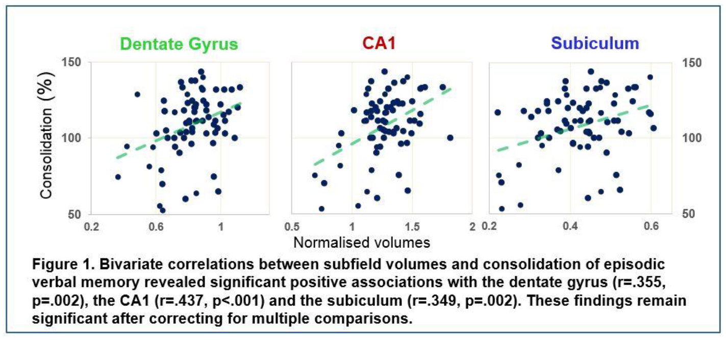
Post-traumatic stress disorder (PTSD) is more prevalent in women than men. PTSD is characterised by overgeneralisation of fear to innocuous stimuli and involves impaired inhibition of learned fear by cues that predict safety. While evidence indicates that learned fear inhibition through extinction differs in males and females, less is known about sex differences in fear discrimination and safety learning. Here we examined auditory fear discrimination in male and female rats. In Experiment 1A, rats underwent 1–3 days of discrimination training consisting of one tone predicting threat (CS+; presented with footshock) and another tone predicting safety (CS-; presented alone). Females, but not males, discriminated between the CS+ and CS- after one day of training. After 2–3 days of training, however, males discriminated whereas females generalised between the CS+ and CS-. In Experiment 1B, females showed enhanced anxiety-like behaviour and locomotor activity in the open field, although these results were unlikely to explain the sex differences in fear discrimination. In Experiment 2, we found no differences in shock sensitivity between males and females. In Experiment 3, males and females again discriminated and generalised, respectively, after three days of training. Moreover, fear generalisation in females resulted from impaired safety learning, as shown by a retardation test. Whereas subsequent fear conditioning to the previous CS- retarded learning in males, females showed no such retardation. These results suggest that, while females show fear discrimination with limited training, they show fear generalisation with extended training due to impaired safety learning.
Background: Aging is one of many factors associated with an increased susceptibility to neurodegenerative disorders which can be related to early life inflammation. Early life immune stimulation, however, can be characterized by the increase in cytokines, oxidative stress as well as changing in microglia phenotypes from activated to priming during the age. Indeed the early neuroinflammation can profoundly affect brain function which can elicit behavioral impairments and cognitive deficits.
Objective: The aim of our study is to explore the sex differences and the possibilities to accelerate the cognitive decline associated with aging after a neonatal immune stimulation.
Methods: Male and females Wistar rats were treated on a postnatal day 14 with PBS or LPS, and then tested for learning & memory at 3 or 10 months of age, using novel object, Y-maze, and a spatial water maze task.
Results: Neonatally-infected rats exhibited memory impairments in the water maze, but only at 10 months. And no significant differences in novel object and Y-maze. Neonatally-infected rats also exhibited greater aging-induced increases in a number of microglia-activating in DG, CA1, and CA3, as well as an increase in Nitrite Oxide and lipid peroxidation but not TNFα within the hippocampus, but not in prefrontal cortex compared to controls.
Conclusion: Taken together, these data suggest that early-life infection leads to less successful cognitive aging, which may be linked to changes in microglial reactivity.
Chronic cerebral hypoperfusion due to cerebrovascular disease is usually associated with loss of cognitive ability, including memory, language, attention and problem-solving, making it a serious medical, social and economic burden to society. The indoleamine melatonin, beside its critical role in the regulation of circadian rhythm, has antioxidant, anti-inflammatory and antiapoptotic properties. The aim of the present work was to investigate the effect of melatonin on oxidative stress, the anti-aging senescence marker protein-30 (SMP30) and the anti-apoptotic osteopontin (OPN) in a rat model of vascular dementia (VD).
Forty-eight male Sprague-Dawley rats (170-200 g BW) were randomly divided into four groups (n=12; each): 1. Control, 2. Rats exposed to VD by permanent bilateral occlusion of the common carotid arteries (BCCAO) leading to chronic cerebral hypoperfusion, 3. VD rats treated with melatonin (190 μg/100g BW; oral) for 28 consecutive days, starting the day after BCCAO, 4. VD rats treated with donepezil (3 mg/kg BW/day; i.p.). At the end of experiments, all rats were humanely killed, under terminal anaesthesia, by cervical dislocation. Expression of OPN was determined by immunohistochemistry, and SMP30 expression determined by western blot in the hippocampus. Hippocampal thiobarbituric acid reactive substances (TBARS) and total anti-oxidant capacity (TAC) were evaluated. Central levels of acetylcholine, norepinephrine and dopamine in the hippocampus were also measured. Significance (P < 0.05) was tested with ANOVA.
Rats exposed to BCCAO had significantly lower TAC and higher TBARs, compared to control. Additionally, BCCAO caused significantly lower expression of both OPN and SMP30. The central levels of acetylcholine, noradrenaline and dopamine were lower in VD rats as compared to control. Treatment of VD rats with melatonin significantly increased the expression of OPN and SMP30 as well as the central levels of acetylcholine in the hippocampus, as compared to untreated VD rats. Moreover, melatonin produced significant increase in TAC and decrease in TBARS.
It could, therefore, be concluded that, in a rat model of vascular dementia, melatonin ameliorates the brain oxidative stress, and produces a neuroprotective effects via upregulating SMP30 and OPN.
Neurons extend axons and dendrites to cover large areas. These extensions allow them to connect with other cells forming complex networks. Synaptic connections at distal axonal processes are far from the cell soma. A key question is how information transmitted from the distal axon is relayed to the soma? How this information is subsequently integrated and interpreted at the soma to modulate neuronal function may inform our understanding of neuronal network development, refinement and modulation.
Structural changes need to be closely coupled to activity-dependent events in order to strengthen active synapses and abolish or dampen unused connections. The neurotrophin brain-derived neurotrophic factor (BDNF) provides an example of a highly regulated growth factor that triggers intracellular processes to initiate protein synthesis dependent and independent changes in cell function. BDNF signals through the tyrosine kinase receptor TrkB. TrkB activation by BDNF has been shown to modulate neuronal growth, arborisation, axonal branching and synaptic transmission. However, the precise intracellular mechanisms underlying the effects of BDNF on morphology are not well characterised. Specifically what are the long-range axonal signaling pathways activated, and what is their outcome on cellular function?
Using microfluidic compartmentalization, we have shown that distal axonal TrkB activation initiates immediate early gene expression and protein production in the somatodendritic compartment of hippocampal neurons. Our data suggest that this activation is independent of long-distance trafficking of the BDNF-TrkB complex itself. Current work addresses the underlying mechanisms of information relay along hippocampal axons, and their functional consequences.
Dopamine is thought to play a role in learning from rewards and punishments, but its role in other stages of memory (i.e. consolidation and retrieval) have been hard to differentiate. We used a within-subject, double-blinded, placebo-controlled paradigm to examine the effect of levodopa on retrieval during a reward and punishment task. 33 healthy older participants (18 male, mean age=71.8 years, SEM=1.31) learned on day 1 and were given drug/placebo 24 hours later, 1 hour prior to testing of retrieval. The reward/learning task (based on Pessiglione et al. (2006) presented i) a gain card pair where card A had an 80% chance of winning 20p, and 20% chance of nothing, and vice versa for card B, ii) a look pair where card C had an 80% chance of showing a 20p (but not winning or losing) and 20% chance of nothing, vice versa for card D; and iii) a loss pair, where card E had 20% chance of losing 20p and 80% chance of nothing, and vice versa for card F. Memory retrieval was tested immediately, 30 minutes and 24 hours after learning, the last on drug or placebo. For each memory test, cards were presented in all combinations without feedback. At 24 hours, participants avoided the 80% losing card significantly better on levodopa than on placebo. Medication state did not change behaviour on other cards. Overall accuracy was also unaffected. In contrast to the putative effects of dopamine during learning, higher levels of dopamine during retrieval results in avoidance of memories learned through punishment. Computational modelling will be used to provide possible mechanisms by which this might occur.

Estrogens, a class of steroid hormones, are primarily produced within the ovaries and influence reproductive function. Additionally, estrogen production is also found in the CNS and estrogen receptors (ERs) are located within many brain regions including the hippocampus (Mitra et al. 2003; Lephart et al. 2001). This non-traditional source of estrogen production highlights estrogen’s potential to modulate cognitive functions. The ability of estrogens to modulate excitatory synaptic transmission have been investigated at hippocampal Schaffer Collateral (SC) –CA1 synapses (Warren et al, 1995). However, at the anatomically distinct Temporoammonic (TA) input to CA1 synapses, the effects of estrogens remain unclear (Smith et al. 2016). Here we have used standard extracellular recordings of field excitatory post-synaptic potentials (fEPSPs) to examine the effects of ER agonists on excitatory synaptic transmission at TA-CA1 synapses. Hippocampal slices (350μM) were prepared from juvenile male rats (P11-24) and perfused with oxygenated aCSF. In juvenile slices (P15), 17β-Estradiol (E2; 1μM; 15min) resulted in a biphasic response. Application of E2 caused initial depression of synaptic transmission (to 81 ± 12.7% of baseline; n=5; p>0.05) that was followed by a significant and persistent increase in synaptic transmission on washout (to 124 ± 3.6% of baseline; n=5; p<0.001). The non-selective nature of E2 and its ability to activate different ERs may provide an explanation for this biphasic response. These data indicate that E2 has the ability to bi-directionally modulate excitatory synaptic transmission at TA-CA1 synapses. Understanding the role of estrogen, and its receptors, at TA-CA1 synapses may be important as the TA pathway is believed to play a role in episodic memory (Remondes & Schuman 2004) and impairments in episodic memory is an early symptom of Alzheimer’s Disease (AD) (Hodges 2000). Therefore, these initial findings are important for understanding estrogenic regulation of hippocampal excitatory synaptic function in CNS health and disease
Retrieval of an associative memory can lead to two different phenomena depending on several factors, such as the duration of stimulus re-exposure. Brief stimulus re-exposure tends to trigger memory reconsolidation, whereas more extended re-exposure leads to memory extinction. Impairment of reconsolidation reduces memory expression, while disruption of extinction maintains expression. Interestingly, it has being observed that during the transition from reconsolidation to extinction of appetitive or aversive pavlovian explicit CS-US memory, there is period where amnesic agents have neither effect. These observations indicated the existence of a “null point” where neither reconsolidation nor extinction is being engaged. Here we investigated whether thisphenomenon extends to contextual fear memory. Adult male lister hooded rats were subjected to a Contextual Fear Conditioning (CFC) paradigm. During training, rats were placed in the chamber for 3 min, received 2 foot shocks (0.7 mA 1.5 sec), and after 1 min, returned to their home cages. Two days later, animals were re-exposed to the same context for 3, 5, 10, 20 or 30min. Immediately after re-exposure, the amnesic agent MK-801 was injected intraperitoneally (0.1 mg/kg). On the next day, animals were exposed again to the context, for 3 min, in order to test memory expression. The aversive response (freezing) was recorded during all sessions and used as memory index. We observed that MK-801 had a significant effect when administered after 3min or 30min reactivation sessions. However, it did not have any significant effect when injected after either 5, 10 or 20min sessions. Further analysis of larger cohorts of animals indicated that the lack of effect of MK-801 when injected after 5 or 10-min sessions are not likely a result of inter-individual differences (e.g. Low vs High freezing animals or reconsolidating vs extinguishing animals).In conclusion, the results show that in contextual fear memories there is a “null point” of 5-20-min re-exposure between the parameters that induce reconsolidation (3 min) and extinction (30 min), at which the memory is insensitive to any effect of i.p. MK-801. Therefore, extinction per se does not exert a boundary condition on memory reconsolidation.

A key obstacle in the treatment of addictions is the high propensity for relapse. Recent research has suggested the propensity for relapse could be reduced by weakening the memories which underpin maladaptive drug-seeking. We recently demonstrated that weakly-trained instrumental lever pressing memory for cocaine could undergo reconsolidation, and here we aimed to replicate this finding in a well-trained setting using cocaine and nicotine reinforcement. Rats were trained for 10 d to self-administer intravenous infusions of drug by lever pressing; each drug delivery was paired with a 20-sec conditioned stimulus (CS) presentation. Following training rats were injected with the NMDAR antagonist MK-801 (or vehicle) 30 minutes prior to a short reactivation session in which the reward contingency was shifted to a variable ratio (VR5). Treatment with MK-801 reduced subsequent lever pressing the next day in cocaine-trained rats, when performance was tested in the absence of the CS, indicative of an impairment in the instrumental component of drug-seeking. Interestingly, this intervention also reduced rates of responding during a drug-primed test of cocaine seeking. However, performance was rescued when lever presses resulted in a 1sec CS presentation, suggesting memory for Pavlovian conditioned reinforcement remained intact. Notably MK-801 was without effect in the nicotine reinforced paradigm, possibly due to the generally weaker performance in nicotine-trained rats; however this may also reflect differences in neural mechanisms of cocaine and nicotine seeking. Implications for disrupting the reconsolidation of drug-reinforced appetitive memories are discussed.
Learning is known to facilitate our ability to make perceptual judgements about our visual environment; that is extracting targets from cluttered scenes and discriminating similar objects. Previous functional MRI (fMRI) studies have shown differential activation patterns for these tasks; however fMRI does not allow us to differentiate between excitatory and inhibitory contributions to learning.
Here we use Electroencephalography (EEG) to measure inhibitory alpha range (8-12Hz) oscillations in the human brain during visual learning. We train participants to discriminate radial vs. concentric Glass patterns that a) are embedded in a noisy background (Signal-in-Noise task) or b) are highly similar (Fine Discrimination task). Our findings show reduced occipital alpha desynchronisation after training for low –rather than high– stimulus noise levels that show improved performance after training. Interestingly, occipital alpha desynchronisation is increased for noise levels that show stronger improvement with training. Further, for the Fine Discrimination task, we find that discrimination of highly similar features relates to increased occipital alpha desynchronisation.
Our results suggest that inhibitory alpha oscillations in the occipital cortex are modulated by visual learning. Occipital cortex involvement is reduced when processing trained features, as indicated by reduced alpha desynchronisation after training. In contrast, processing highly uncertain stimuli (high noise, high similarity) engages occipital cortex, as indicated by increased alpha desynchronisation. Our findings provide evidence that decreased inhibition mediates visual plasticity.
Synaptic plasticity is often studied in the CA1 area of dorsal hippocampal slices. Here, in response to high-frequency theta-burst stimulation (TBS), the Schaffer collaterals display two phases of potentiation readily induced through activation of NMDA receptors (NMDARs). The initial phase of potentiation, termed short-term potentiation (STP), declines in response to low frequency synaptic activation, leading to a permanently enhanced level of synaptic transmission, long-term potentiation (LTP). It has been recently shown that STP consists of two forms, termed STP1 and STP2 (Volianskis et al.,2013;2015). Induction of STP1 depends on activation of GluN2A/2B containing NMDARs, similar to LTP, which in the adult dorsal hippocampal slices is induced through GluN2A/2B containing triheteromeric NMDARs. In contrast, STP2 in the dorsal hippocampus is induced through GluN2B/2D containing NMDARs.
STP and LTP can also be induced in ventral hippocampal slices. However, the potentiation induced by TBS is smaller in the ventral hippocampus than in the dorsal. We report here that the NMDAR antagonist AP5 (3 μM and 30 μM) inhibited both STP and LTP induced by TBS in the CA1 region of ventral hippocampal slices from adult male Wistar rats. 0.1 μM NVP-AAM077 (NVP, a GluN2A selective antagonist), 1 μM Ro 25-6981 (Ro, a GluN2B selective antagonist) and 10 μM UBP145 (UBP, a GluN2D selective antagonist) were used to determine the identity of GluN2 subunits involved in synaptic potentiation. We show that NVP blocked both STP and LTP, whilst Ro had no effect on STP or LTP. UBP blocked STP and did not reduce LTP. Furthermore, we show that the GluN2A/2B preferring positive allosteric modulator UBP714 potentiates LTP in the ventral hippocampus. We conclude that in the ventral hippocampus GluN2A containing NMDARs are involved in the induction of LTP whereas induction of STP involves activation of GluN2D subunits.
Biogenic Amines (BAs) are a group of molecules that act as neurotransmitters in the brain to modulate complex behaviors, including learning and memory formation. Drosophila melanogaster, an animal model with important genetic tools that shows similar mechanisms of neurotransmitter storage, release and recycling as compared to mammalian systems, has been extensively used in assays of aversive learning. In these protocols, a Pavlovian conditioning approach is used. For instance, an electric shock is applied to flies (unconditioned stimulus, US) while they are exposed to an odorant (conditioning stimulus, CS). One of the open questions regarding this approach is what are the dynamic changes in fly brain neurotransmission during this conditioning. To answer this question, we generated a new system (fast scan cyclic voltammetry, FSCV) to measure the in vivo release of BAs in fly brain while the conditioning paradigm is carried out.
A single male fly was fixed to a recording chamber and the brain is exposed surgically. Using a carbon fiber electrode, we measured BAs release in the fly brain scanning from −0.4 to 1.2 V and back (vs Ag/AgCl reference electrode) at a scan rate of 400 V/s at 10 Hz. As positive control of identification and quantification of BAs, ATP-activated ion channels (P2X2 receptors) are expressed in specific aminergic neuronal populations and activated by ATP. To perform conditioning experiments, flies were exposed to a single electric shock every 5 seconds via an electric grid. In addition, we used a vacuum pump to deliver odorants.
We detected the release of BAs in the fly brain when ATP is applied. Release of octopamine, dopamine and 5-HT is detected within discrete structures of the brain in flies exposed to electric shock and mainly 5-HT is measured upon odorant stimulation. As negative control, no significant release of BAs is detected using a nominal zero calcium solution.
Our results show that we are able to measure the differential release of endogenous BAs in brains of flies exposed to stimuli relevant to olfactory learning and memory conditioning. Our data is consistent with the proposed role for the different amines in this process.
Supported by Fondecyt 1141233. SH is supported by CONICYT-PCHA/Doctorado Nacional/2016-21161611
Memory suppression and motor inhibition share the cognitive process of active stopping. Previous research associated cortical activations with these forms of stopping. However, despite having a well-established role in motor control, the basal ganglia’s involvement in memory suppression remains unexplored. Here we first tested the consistent activation of the basal ganglia in memory and motor stopping using a series of meta-analyses, and then investigated the role of the basal ganglia in these processes through dynamic causal modelling (DCM).
The meta-analyses included fMRI results from tasks requiring active suppression of prepotent thoughts or actions (e.g. Think/No-Think and Stop-Signal tasks), and revealed highly overlapping cortical and subcortical activations between memory and motor inhibition, including the right prefrontal cortex (DLPFC; VLPFC) and the basal ganglia. We then conducted the DCM effective connectivity analysis using a separate fMRI dataset that had participants performing the Think/No-Think and the Stop-signal tasks in one session. In the DCM models, we included both the putative domain-general regions (prefrontal cortex and basal ganglia), and the domain-specific regions (hippocampus for memory stopping, and primary motor cortex for motor stopping). We found that the basal ganglia were significantly involved in the network dynamics supporting the stopping processes in both tasks. Critically, it was the ‘stopping’ conditions that modulated the effective connectivity between the basal ganglia and the domain-specific regions instead of the retrieve/go conditions. These results provide strong evidence for a supramodal inhibition network in the brain, especially the novel indication that the basal ganglia play important roles in memory suppression in a similar fashion as in motor inhibition.
Magnocellular mediodorsal thalamus (MDmc) contributes to adaptive decision-making and learning of object-reward discriminations, with thalamo-cortical interactions potentially mediating these cognitive processes. Prefrontal cortex dysfunctioning has been attributed to problems with response inhibition, as assessed by reversal learning paradigms. The current study investigated whether bilateral neurotoxic (NMDA/ibotenic acid) lesions to MDmc in macaque monkeys impact cognitive abilities due to deficits in response inhibition, using two different reversal learning paradigms. One paradigm involved learning 10 visual discriminations presented concurrently and repeated 20 times within the session. Once a 90% learning criterion was achieved, monkeys were exposed to reversals of the reward contingencies on subsequent sessions. The other paradigm involved learning a single visual discrimination presented serially for 100 trials in one session. All monkeys achieved 85% learning criterion within a single session, and on 12 subsequent test sessions, they were exposed to reversals of the reward contingency.
Monkeys with MDmc lesions demonstrated dissociable performance on the two reversal learning paradigms. Damage to MDmc caused impaired learning of the reversal in reward contingencies during concurrent presentation of the 10 visual discriminations compared to unoperated control animals. In contrast, during the serial reversal learning paradigm, overall reversal learning performance of monkeys with MDmc damage was not affected, although the monkeys were slower to adapt their choices after the first reversal of the reward contingency only. Our results suggest that deficits in cognitive performance after damage to the MDmc cannot be readily attributed to problems in response inhibition. Instead, our results suggest that interactions between the MDmc and interconnected cortex are critical to support the rapid integration of task relevant object-reward associative information, while response inhibition as measured in serial reversal learning paradigms is associated with interdependent neural networks of the brain.
This work was supported by a UK Medical Research Council Career Development Award - G0800329 to Dr Anna S Mitchell.
The preterm period coincides with rapid neurological development, during which neural migration and synaptogenesis result in the maturation of the thalamocortical pathways. As such it is postulated that prematurity results in disruptions in this development, affecting the connectivity of the infant brain, contributing to the neurological deficits seen clinically. This study uses a mouse model of prematurity to investigate the development of the sensorimotor pathways, at a synaptic, cellular and behavioural level.
Premature birth was induced in C57BL/6 female mice with a subcutaneous injection of the progesterone receptor antagonist RU486. Experiments were carried out on offspring during the first 3 postnatal weeks. Whole-cell patch clamp recordings of stellate neurons in acute barrel cortex slices were used to measure neuron membrane properties, excitability and miniature excitatory post-synaptic currents (mEPSCs). Anatomical development of the barrel cortex was assessed histologically using cytochrome oxidase (CO) staining. The development of reflexive sensorimotor behaviours were assessed with a battery of behavioural tests.
RU486 successfully induced preterm birth (mean gestation lengths: preterm = 18days±1.4 hours (n=29); control = 19days12±17hrs (n=21). P < 0.0001; two-tailed T-test)). Membrane properties and excitability of stellate cells showed developmental changes and the frequency of mEPSCs in the barrel cortex increased with age. However we found no differences between the development of these features in preterm and term pups. Assessment of thalamocortical neuron migration using CO staining showed that preterm and term pups followed the same developmental time course, with distinct barrels being visible by the end of the first postnatal week. Pups ability to complete tasks requiring sensorimotor coordination, such as righting reflex, and reflexive sensory behaviours such as whisking, improved with age, with preterm and term pups developing at the same rate. At weaning open-field testing showed no differences in exploratory behaviours in premature pups compared to controls.
Premature birth in mice raised in a normal environment does not alter the developmental trajectory of the sensorimotor pathway, on a synaptic, cellular or behavioural level
GABAergic interneurons (INs) are thought to have an important role in normal cortical circuit development. Recently we have shown that transient circuits involving somatostatin positive (SST+) cells assist in wiring the local columnar connectivity of the neonatal mouse somatosensory cortex (Marques-Smith et al., 2016). How this process co-ordinates with the formation of cross-modal cortico-cortical connections is however still unknown. A possible candidate for mediating such long range, early interactions could be vasointestinal peptide-positive (VIP+) INs. This population of IN are primary located in layers (L)2/3 (Prönneke et al., 2015), and known mediators of long-range cortico-cortical input in the mature cortex by preferentially targeting SST+ cells (e.g. Lee et al., 2013). Intriguingly unpublished data from the lab have identified L2/3 GABAergic synaptic input onto SST+ INs in whisker barrel cortex (S1BF) at early postnatal ages. We hypothesised that VIP+ cells in L2/3 are responsible for this GABAergic input and that they could have a role in coordinating long-range and local connectivity during early circuit development. We have employed optogenetics in combination with laser scanning photostimulation (LSPS) to confirm VIP+ IN input onto the SST+ cells within the first postnatal week. In parallel we have characterised early VIP+ cells using immunofluorescence, whole cell patch clamp and LSPS to dissect their cortical distribution, electrophysiological profiles and local glutamatergic inputs at these early ages. These data identify two populations of L2/3 VIP+ cell based on local synaptic connectivity. We hypothesis that these two populations differ in their long-range cortico-cortical inputs and regulation of SST+ IN signalling thereby playing distinct roles in circuit development.
Marques-Smith et al. (2016) A Transient Translaminar GABAergic Interneuron Circuit Connects Thalamocortical Recipient Layers in Neonatal Somatosensory Cortex. Neuron 89:536-49.
Lee et al. (2013) A disinhibitory circuit mediates motor integration in the somatosensory cortex. Nature Neuroscience 16:1662-70.
Prönneke et al. (2015) Characterizing VIP Neurons in the Barrel Cortex of VIPcre/tdTomato Mice Reveals Layer-Specific Differences. Cereb Cortex. 25:4854-68
Dyslexia is one of the most common neurodevelopmental disorders characterised by difficulties with accurate and fluent word recognition. Previous research has made significant progress into studying the behavioural, neuropsychological and neurobiological causes of the disorder. More recently, important understanding of the neural circuits and the aetiology of dyslexia came from respectively, brain imaging and genetics findings.
For example, PCSK6 is a gene that has been linked to handedness, brain asymmetry and developmental disorders including dyslexia (Brandler & Paracchini, Trends in Molc. Science, 2014). This study explores how the neurophysiological correlates of the reading impairment are related to dyslexia genetic risk, using resting-state magnetoencephalography (MEG). We compared resting-state functional connectivity and network topology of two groups (N=7) of dyslexic children, with (risk carriers) and without (risk-free group) risk allele at PCSK6.
By applying an atlas-based (AAL) MEG beamformer approach (Hillebrand et al., NeuroImage, 2012) we obtained a detailed anatomical mapping of neurophysiological patterns for different cortical rhythms. We used Phase Lag Index (PLI) to measure the resting-state functional connectivity. Subsequently, we reconstructed the functional network where each AAL based region (ROI) formed a node and each PLI value an edge.
Based on the functional network, the network topology for both dyslexia groups was estimated using weighted clustering coefficient (Cw) and path length (Lw) and compared via permutation testing.
We found that the two groups differ in connectivity strength at the PLI level and in cortical topology of individual edges (Lw) in salient regions of the reading network (left Superior Frontal, Inferior Parietal and Fusiform cortex), in delta (1-4Hz) and theta (4-8H) frequency bands.
Introduction: Prenatal maternal immune activation (mIA) has widely been associated with the susceptibility of offspring to develop psychiatric disorders such as schizophrenia. This has been well characterised in terms of the maternal cytokine response elicited, the behavioural and cognitive changes in offspring and postnatal brain pathology in rodent models of mIA. This project specifically focuses on in utero changes in response to mIA, particularly in the placenta and yolk sac in a rodent model using poly(I:C) administration to induce mIA. We hypothesise that the placenta may be a critical mediator of developmental programming in schizophrenia, predominantly through alterations in its amino acid transporter activity.
Methods: A single intra-peritoneal injection of poly(I:C) or vehicle (10 mg/kg) was administered to pregnant female Wistar rats at GD15. Dams were sacrificed at two time points: GD16 and GD21 and two female and two male pups from each litter were randomly chosen for quantitative real time PCR analysis. Total RNA was extracted from the associated placentas and cDNA generated that was subsequently amplified using specific primers for Mapk1 and Stat3, system L transporter genes: Slc7a5, Slc7a8, Slc43a2, Slc3a2, placental cell-specific genes Gcm1 and Tpbpa and housekeeping gene Ywhaz. Placental expression changes of cytokines IL-6, TNF- α and IL-1β were also determined using the same method and protein level changes using ELISA.
Results: We show that poly(I:C) administration of GD15 had no significant effect on expression of system L transporter genes in GD21 placentas. We report a significant sex-specific (female) decrease in expression of Tpbpa, a marker of the placental junctional zone, at GD21 which suggests placental morphological changes in response to mIA, a finding also reported in models of fetal growth restriction. Ongoing work to be presented will further characterise cytokine changes and compare results between GD16 and GD21 time-points.

Activation of the maternal immune system during pregnancy is a known risk factor for schizophrenia, thought to be caused by transient increases in maternal cytokines. Injection of the viral mimetic polyinosinic:polycytidylic acid (polyI:C) into pregnant rats during mid/late pregnancy causes a transient increase in maternal serum interleukin-6 (IL-6) levels and induces behavioural deficits in the adult offspring. Impaired placental transport of amino acids causes growth restriction and may underlie later life neurological deficits. For example, the solute carrier family 38a (Slc38a) transporters allow uptake of amino acids to the developing fetus. Methionine is an essential amino acid transported by Slc38a1, 2, and 4, and is used as a methyl donor for establishing and maintaining DNA methylation patterns. Emerging evidence from human and rodent studies supports a role for attenuated gene promoter methylation in schizophrenia. Maternal care in early postnatal life stably affects the epigenetic status of gene promoters in the offspring brain. We hence aim to investigate the effect of prenatal poly I:C treatment on the expression of the Slc38a family, DNA-methyltransferase-1 (Dnmt1), and immune genes in the rat placenta, as well as on the quality of mother-pup interactions in early postnatal life.
Female Wistar rats received a single intraperitoneal injection of 10 mg/kg poly I:C or vehicle at GD15 of pregnancy. For real-time quantitative PCR the dam and pups were sacrificed at GD21. Two male and two female pups were selected at random and total RNA was extracted from the corresponding placentas. 1μg of retrotranscribed cDNA was amplified using specific primers for the genes Slc38a1, Slc38a2, Slc38a4, Dnmt1, the Toll-like receptor-3 (Tlr3), Tlr5, and the housekeeping gene Ywhaz. For behavioural analyses, mother-pup interactions were scored live at postnatal days 6, 10, and 14.
We show that poly I:C treatment at GD15 has no effect on the expression of Slc38a transporter genes in the rat placenta. Poly I:C treatment induced a significant downregulation of Dnmt1 in female, but not male, placentas, suggesting a sex-specific change in DNA methylation. Upcoming work to be presented will assess the effect of poly I:C on maternal care and pup ultrasonic vocalisations.
To understand how neurons grow and become integrated in neural networks, it is essential to investigate how the local environment influences these processes. We use the retina to study how coherent functional mature neural tissues emerge under the guidance of complex and dynamic developmental mechanisms including chemical signalling as well as physical interactions between cells (e.g. mechanical forces and electrical communication).
Computational modelling has been used to formalise and better understand the mechanisms involved in neuron growth (Graham and van Ooyen, 2007; Roberts et al., 2016). However, these studies are usually designed in a simplified context, without taking physical interactions into consideration. Here, we use the simulation framework Cx3D (Zubler and Douglas 2009) to computationally model the development of retinal ganglion cells (RGCs) in 3D physical space. RGCs play a key role in visual function, as they provide the only information channels between the eye and the brain, encoding all information about our visual world into trains of spikes sent to the brain via the optic nerve. A large number of RGC morphologies have been reconstructed and are publicly available.
In this study, we investigated the spatial growth of RGCs, and compared simulated morphologies with real RGCs available in the neuromorpho database (http://neuromorpho.org, Ascoli et al., 2007). We show how genetically encoded processes can yield complex and biologically plausible RGC morphologies by taking into account information available at the growth cone only, so without relying on a global supervisor. In particular, we demonstrate that a simple growth rule, named “2-thresholds growth rule”, can explain a number of measures that we inferred from real morphologies (branching number, tip and branching distance, isometry and tree size).
-Ascoli GA, Donohue DE, Halavi M. 2007 NeuroMorpho.Org: a central resource for neuronal morphologies.J Neurosci., 27(35):9247-51
-Graham P.B, van Ooyen A. 2006. Mathematical modelling and numerical simulation of the morphological development of neurons. BMC Neurosci;7(Suppl):1.
- Roberts PA, Gaffney EA, Luthert PJ, Foss AJ, Byrne HM. 2016. Mathematical and computational models of the retina in health, development and disease. Prog Retin Eye Res., 53:48-69.
-Zubler F, Douglas R. 2009. A framework for modeling the growth and development of neurons and networks. Front Comput Neurosci. 3:25.
The neuronal ceroid lipofuscinoses (NCLs) are a group of inherited, childhood-onset, neurodegenerative diseases caused by mutations in CLN genes. Several late-onset neurodegenerative diseases show lysosomal dysfunction, and inherited lysosomal disorders often present with neurodegeneration, which, taken together, argues that maintaining lysosomal function is vital for neuronal health. However, neurodegeneration in the NCLs presents so early in life, it suggests that lysosomal function might also be required for normal neural development. We have been investigating this possibility in Drosophila models and demonstrate reduced neural development in mutants of CLN7, a gene mutated in late infant-onset NCL. CLN7 encodes a transmembrane protein thought to reside in the lysosomal membrane, and lysosomal function is essential for autophagy, a known regulator for neural development in Drosophila. However, the autophagy pathway is dependent on the inactivation of mTORC1 in the absence of growth stimuli and here we show that the developmental changes in CLN7 mutant synapses are not due to defective autophagy, but more likely due to hyperactivation of mTORC1 signalling. We demonstrate that the CLN7 protein is present in a complex with Rheb, an activator of mTORC1, and that loss of CLN7 function results in increased growth and reduced autophagy. We are now using Drosophila genetics to identify which signalling pathways require or impinge upon CLN7 function combined with a proximity-labelling proteomics approach to identify components of the CLN7 interactome. Together these will clarify the involvement of CLN7 in mTORC1 signalling and as a regulator of growth, autophagy and neural development.
In contrast to mammals, zebrafish are capable of regenerating neurons in the central nervous system after an injury (1). For regeneration to occur, specific programmes of gene expression must be activated and modification of histone acetylation is one of the fundamental epigenetic mechanisms of gene expression changes. In particular, HDAC1 is essential for the regulation of two of the most important neurogenic pathway, Hedgehog and Notch, both of which are involved in motor neuron regeneration (2, 3). On this basis we decided to investigate the role of HDAC1 in motor neuron regeneration after spinal cord injury in zebrafish larvae.
We find that HDAC1 mRNA is upregulated in ependymo-radial glial cells (ERGs), the spinal progenitor cells, after an injury. We then used both pan and selective HDAC1 inhibitors to treat larvae after spinal cord transection, showing that inhibitor-treated larvae displayed a lower number of regenerated Hb9+ motor neuron after the lesion.
Recent findings in our group indicate that fish treated with immunosuppressant drugs (5) also fail to regenerate motor neurons. We demonstrate that in fish larvae lacking microglia and macrophages, HDAC1 is not upregulated after spinal injury. Conversely, triggering the immune system with LPS (in the absence of a lesion) is sufficient to induce HDAC1 expression in ERGs. This suggests that HDAC1 upregulation is controlled by the immune response after injury. In conclusion, we present evidence that HDAC1 plays an important role in the neuroregenerative process observed in zebrafish larvae after a spinal cord injury and that HDAC1 expression might be regulated by immune system activation.
1. Becker, C. G. and Becker, T. 2015. Dev.
2. Cunliffe VT. Development. 2004. Jun.
3. Canettieri G, Di Marcotullio L et al. Nat Cell Biol. 2010.
4. Kyritsis, N., Kizil, C. et al. Science 2012.
5. Ohnmacht J, Yang Y et al. Development 2016.
Autism spectrum disorders (ASD) are a group of complex, genetically heterogeneous neurodevelopmental disorders characterised by impairments in communication and social behaviour as well as a propensity to engage in repetitive behaviours. ASD ranges in severity from having a mild impact on day-to-day life to the need for constant care, often presenting with multiple neurological and neuropsychiatric comorbidities such as intellectual disability, developmental delay and epilepsy. Despite the fact that global prevalence is estimated at 1-2%, both their aetiology and pathophysiology are poorly understood and no medicines addressing the core symptoms currently exist.
Research into cellular and developmental mechanisms responsible for ASD has been hindered by a lack of reproducible cellular models that have construct validity. Induced pluripotent stem cell (iPSC) technology allows neural precursor cells (NPCs) and neurons sharing the same genetic background as living human patients to be grown in a dish, providing unique insight into cellular and network neurodevelopment.
Caused by maternal origin duplications or triplications of the notoriously unstable chromosomal region 15q11.2-13.1, 15q11-13 duplication syndrome or ‘dup15q’ is the most common known cytogenetic cause of ASD (Hogart et al., 2010). The link between genotype and phenotype is currently unknown, although the 15q11-13 region notably contains a cluster of GABAA subunit genes (alpha 5, beta 3 and gamma 3) as well as the ubiquitin E3 ligase UBE3A, whose function is lost in Angelman Syndrome.
In the present study, NPCs and neurons were produced from iPSCs reprogrammed from a patient with dup15q and a neurotypical first-degree relative. They were characterised with extracellular microelectrode array recordings, whole cell patch-clamp and quantitative immunohistochemistry. These results are indicative of pronounced network GABAergic dysfunction as well as altered subunit composition of the GABAA receptor, consistent with reports of lack of benzodiazepine efficacy in treating seizures in dup15q patients. We also report a two-fold increase in NPC numbers, which may be a product of disruptions in proliferation and differentiation relating to the early developmental role of GABA transmission.
The postsynaptic density (PSD) of excitatory synapses in the vertebrate central nervous system contains a conserved set of ~1,000 proteins and mutations in these cause over 130 brain diseases (1). PSD95 is a scaffold protein that organizes a family of multiprotein supercomplexes, some of which contain NMDA receptors. SAP102 is a paralog of PSD95 and organises distinct multiprotein complexes (2). The differential distribution of complexes into different synapses provides structural and functional diversity to synapses.
To study the distribution and diversity of complexes and we have used a genetic labelling technique that permits us to visualize PSD95, SAP102 and the NMDA receptor GluN1 subunit. The endogenous genes for these proteins were modified by gene targeting to create fusion proteins: PSD95-eGFP, SAP-mKO2 and GluN-FLAG, which can be visualized at the individual synapse level using fluorescence microscopy. The hippocampus, a hub of synaptic diversity, was examined using a spinning disk confocal microscope on fixed brain sections from 36 mice across 7 age groups (postnatal day 1 to 95). Advanced image analysis allowed the unsupervised classification of over 40 synapse subtypes based on the morphological features of these three protein markers.
Researchers are becoming increasingly aware of the importance of the synaptic diversity that arises from the combinatorial assembly of synaptic proteins (3). Our work primarily establishes a framework for the quantitative analysis of synaptic diversity and warrants caution when interpreting results of bulk measurements like western blotting, mass spectrometry and many electrophysiological techniques. Beyond this, correlating the trajectories of individual synapse subtypes with known developmental processes could provide insight into their functional roles. Studying the effects of diseases (in disease models) on specific subtypes could also reveal the function of synaptic subtypes, in addition to broadening our understanding of disease mechanisms and aiding drug targeting.
References:
1) Bayes et al. Nat Neurosci. 2011 Jan; 14(1): 19-21
2) Frank et al. Nat Commun. 2016 Apr 27; 7:11264
3) O’Rourke et al. Nat Rev Neurosci. 2012 May 10; 13(6): 365-79
The interaction of the neurotransmitter serotonin (5-HT) with its receptors (5-HTR) plays a diverse regulatory role on neural plasticity, including during early development, which has been implicated in the aetiology of various behavioural disorders. 5-HT1A and 5-HT7 receptors are both activated by the serotonergic agonist 8-OH-DPAT and exert their modulatory roles via interaction with a range of signalling cascades, notably through opposing effects on cAMP production. Contrasting reports of 8-OH-DPAT effects in different cell types could be due to complex interactions of these two 5-HTRs on neural development. We suggest that the interplay of 5-HT1AR/5-HT7R activity provides a mechanism that contributes to the overall modulation of cortical neurite growth.
This has been investigated using primary rat cortical cultures as a model system to study the role of 5-HT1AR/5-HT7R expression and the differential roles of their signalling cascades on neurite growth. As a first part of this study, we show that chronic application of 8-OH-DPAT (5 μM) causes a significant increase in the average dendrite length per neuron by 38±14% compared to control cultures (1-way ANOVA followed by Tukey post-hoc test: p<0.05; 18-19 coverslips from 2 independent cultures per condition). The growth promoting effect of 8-OH-DPAT is completely abolished by the co-application of the 5HT7R antagonist SB-269970 (1 μM; 8-OH-DPAT vs 8-OH-DPAT + SB-269970: p=0.05; control vs 8-OH-DPAT + SB-269970: p=1.0). In contrast, SB-269970 on its own has no significant effect on average neurite growth (p=0.99) indicating that its effect is due to inhibition of 8-OH-DPAT-activated 5-HT7R mediated signalling. This data strongly suggests that 5-HT7R mediated signalling, rather than 5-HT1AR signalling, plays a significant role in modulating cortical neuronal growth. However, it does not preclude the possibility that 5-HT7R mediated effects on cortical development can be modulated by direct interactions between 5-HT7R and 5-HT1AR.
Anhedonia, one of the two main clinical symptoms in major depression disorder (MDD), has been experimentally related to deficits in reinforcement learning. The reward deficit may point towards a dopaminergic imbalance, e.g, blunted neural activation in ventral striatum, caudate and less consistently in orbitofrontal cortex (OFC). In a behavioural probabilistic learning task based on a differential reinforcement schedule using virtual money, MDD patients showed reduced reward learning compared to healthy controls (HC). The aim of this study was first to adjust the reinforcement learning behavioural task to the scanner environment; and secondly to test the effects of different reinforcement ratios of rewarding taste stimuli at neural level in MDD vs HC.
Methods: 59 participants (N=26 MDD, N=33 HC) took part in a probabilistic learning task inside the scanner. In a three-block event-related design, participants had to distinguish between two highly similar stimuli, while trying to maximize the intake of chocolate reward. Unknown to the participants, chocolate reward was delivered four times more for one stimulus (target) compared to the other one (non-target). Reward bias refers to the participants’ tendency to define an ambiguous stimulus as target.
Results: Preliminary analyses showed less BOLD activation in MDD vs HC in the left caudate (p<.05, FWE for multiple comparisons) in response to the target vs non-target contrasts, and in the anterior cingulate cortex (p<.05, FWE for multiple comparisons) in response to the target vs missing the target contrasts. However, MDD vs HC showed increased BOLD activation in the OFC/insula (p<.05, FWE for multiple comparisons) in response to the target vs the bias contrasts.
Conclusions: In line with previous research, MDD participants with anhedonia symptoms showed decreased activation to rewarding stimuli in reward areas of the brain. Moreover, MDD compared to HC showed increased OFC activation when spotting the difference between the target and the ambiguous stimulus. Results of this task show that at the brain level, MDD compared to HC are better at differentiating between a rewarding and an ambiguous stimulus, while showing a conservative response in defining other stimuli as rewards.
Discs large homologue 2 (DLG2) is a membrane associated guanylate kinase (MAGUK) protein located in the post synaptic density (PSD) of neuronal synapses, here it acts as a scaffold to regulate receptor clustering and intracellular trafficking through associations both with other proteins and the actin cytoskeleton. Disruptions to the DLG2 gene such as in the 11q14.1 copy number variant (CNV) have be associated with neuropsychiatric disease, specifically an increased risk of schizophrenia, although the genotype to phenotype relationship is incompletely understood. Preliminary data showing DLG2 expression in neural precursor cells (NPCs), a much earlier stage of neural development than previously reported, indicates that the role of DLG2 extends beyond synaptic function. Here the results of ongoing experiments to verify these data by analysing the endogenous pattern of DLG2 expression throughout neural development are reported, using human embryonic stem (hES) cells differentiated to cortical projection neurons as a model system. As available anti-DLG2 antibodies exhibit both inconsistent binding to DLG2 isoforms and unspecific binding to other proteins CRISPR/Cas9 technology was used to generate tagged DLG2, enabling the spatial and temporal pattern of DLG2 expression to be determined through immunocytochemistry. Additionally DLG2 deficient hES cells along with wild-type (WT) controls were differentiated to cortical projection neurons and the phenotype of these cells characterised at various stages of neural development through staining for neural markers, using both immunocytochemistry and western blotting. Although these data are not fully analysed they suggest DLG2 is required for normal cortical projection neuron development. It is expressed in NPCs and likely has a role in regulating early neural development. DLG2 deficient neurons show a disruption to cortical layer markers providing further evidence for a key neurodevelopmental role for DLG2 and a potential link to neuropsychiatric disease.
Introduction
People with Tourette Syndrome (TS) experience ‘unwilled’ hyperkinetic movements known as tics. Despite the compulsive nature of tics, adults with TS can often exercise volitional control over whether to release or suppress a tic. Evidence from neurotypical populations suggests that control of actions proceeds via prefrontal modulation of cortico-subcortical interactions with the basal ganglia (Rae et al, 2015, J Neurosci). We tested how prefrontal regions influence the basal ganglia and motor cortex in TS, and how this shapes voluntary action decisions.
Methods
23 adult participants with TS (13 male; age 18-51, mean 34) and 22 matched controls (12 male; age 19-55, mean 34) underwent fMRI during an intentional inhibition task. 750 T2*-weighted echo planar images were acquired on a 1.5T Siemens Avanto (34 slices, 3x3x3.6mm resolution, TR = 2520ms, TE = 43ms). fMRI pre-processing and general linear modelling was run in SPM12 (www.fil.ion.ucl.ac.uk/spm). The intentional inhibition task required participants to press a button in response to a green cue (go, 50% trials), withhold their response to a red cue (nogo, 16%) and choose whether to press or not in response to a yellow cue (choice, 34%).
Results
Interaction contrasts suggested that compared to controls, participants with TS showed reduced activity in preSMA when choosing to move (choose-go) compared to instructed to move (go); greater activity in inferior frontal gyrus when withholding actions (choose-nogo and nogo); and greater activity in M1 when choosing to withhold an action (choose-nogo) (Figure 1). Dynamic Causal Modelling was applied to determine the influences of preSMA and inferior frontal gyrus on subcortical nuclei and M1.
Conclusions
When choosing to make or withhold voluntary movements, people with TS require less neural activity in motor preparation areas to produce actions; require greater neural activity in motor control areas to withhold actions; and even when withholding actions, suppress primary motor cortex activity to a lesser extent. These results suggest dysfunctional outflow of motor signals from basal ganglia can be modulated by prefrontal influences to determine action decisions. This may underpin the tic suppression strategy often employed by people with TS.
Aims:
Bipolar disorder is a highly heritable condition in which there is dysfunction of corticostriatal circuitry. This study aimed to elicit whether functional changes to corticostriatal circuitry represent a risk endophenotype for the condition. We aimed to achieve this via the medium of resting-state functional magnetic resonance imaging (rs-fMRI).
Methods:
38 bipolar disorder patients were recruited, 32 unaffected first-degree relatives and 23 healthy controls. Groups were demographically well matched. Initially, rs-fMRI data was appropriately cleaned using typical methods. A seed-based analysis was then run using left and right nucleus accumbens regions-of-interest.
Results:
A ventral-dorsal gradient was observed in corticostriatal correlation whereby ventral prefrontal cortex showed increased correlation with the nucleus accumbens and dorsal prefrontal cortex showed increased negative correlation with the nucleus accumbens (p<0.05). This finding was present in both patients compared to controls and relatives compared to controls and as such signifies a risk endophenotype.
Conclusions:
This study identified a risk endophenotype for Bipolar Disorder. This profile of changes to corticostriatal function predispose an individual to developing the disorder, but they’re not directly correlated with symptoms or caused by them. Future work could utilise this endophenotype in a clinical setting to improve patient outcomes.
Antidepressant prescribing in children and adolescents increased steadily in the United States and parts of Europe between 2005 and 2012 despite regulatory safety warnings. Little is known about the characteristics of those being prescribed antidepressants for the first time and their treatment course.
A longitudinal study of 3-17 year olds prescribed antidepressant for the first time in primary care was carried out using routinely collected anonymised primary care data from the UK Clinical Practice Research Datalink (CPRD) between 2000 and 2015. Changes in the incidence of first ever antidepressant prescriptions and the characteristics of those being prescribed them was examined. As prescriptions are not directly linked to the indication they were prescribed for in the CPRD linkage was inferred by temporal proximity.
Incidence of first ever prescriptions nearly doubled between 2006 and 2015 rising from 1.60 (95%CI: 1.51, 1.69) to 3.12 (3.00, 3.25) per 1000 person years, with females more than twice as likely as males to be a recipient of one of these prescriptions. Only 21% of the 1721 patients with incident prescriptions in 2015 could be linked to a depression diagnosis, with an additional 22% of prescriptions linked to alternative indications. The incidence of prescriptions linked to a depression diagnosis increased between 2012 and 2015, with an adjusted incidence rate ratio of 1.46 (1.26, 1.70). Overall antidepressant prescribing increased most rapidly in 15-17 year old females.
Antidepressant prescribing in children increased between 2006 and 2015. This is, at least in part, due to a rise in alternative uses of antidepressants, including the treatment of anxiety, chronic pain and migraines.
Cyfip1 gene dosage is reduced in 15q11.2 deletion syndromes, which are associated with a range of psychiatric symptoms. The neural bases of these symptoms are unknown, but abnormal functional connectivity between the frontal and temporal lobes features in other psychiatric patients and genetic risk factor carriers, in other rodent models, and is increasingly thought to contribute to cognitive deficits. We are using a novel line of Cyfip1 heterozygous knockout rats (Cyfip1±) to define the consequences of Cyfip1 knockdown for brain function and behaviour.
We trained 7 wildtype (WT) and 4 Cyfip1± littermates on a rewarded alternation T-maze task which invokes working memory, an aspect of cognition consistently impaired in psychiatric disorders. Chronically implanted microelectrode arrays were used to simultaneously record local field potentials (LFP) from dorsal CA1 of the hippocampus (CA1) and medial prefrontal cortex (PFC).
Cyfip1± rats tended to run fewer trials per 40min session (WT 27±2 trials, Cyfip1± 20±3; p=0.08), typically making more hesitant runs down the central arm of the maze, rather than continuous runs to the reward point (% of hesitant runs: WT 16±4, Cyfip1± 55±17; p=0.08). Choice accuracy, however, was normal (WT 79±4%, Cyfip1± 88±5%, p>0.05).
Considering only trials with continuous runs, LFP spectral profiles appeared normal in Cyfip1± rats, which showed CA1 and PFC 5-10Hz theta amplitudes similar to WT. This suggests that local networks remain broadly intact in Cyfip1± rats.
However, using spectral coherence to infer functional interactions between CA1 and PFC suggested that theta coherence in Cyfip1± rats was more sensitive to cognitive context compared to in WT. The difference in coherence between guided and choice turns on the T-maze was significantly higher in Cyfip1± than WT (p<0.05). This result is reminiscent of an fMRI study in which schizophrenia risk allele carriers showed increased coupling between PFC and HPC, with no impact on task performance (1).
These findings indicate that impaired neural network activity during a working memory task is a consequence of reduced Cyfip1 gene dosage, and may be an important component of the pathophysiology underlying psychiatric conditions.
1. Esslinger et al, Science 2009
The expression of psychotic phenomena such as hallucinations and delusions has been proposed to lie along a continuum and subclinical manifestations are more prevalent in the general population. These psychotic experiences (PEs) are typically transient in nature, but are associated with an elevated risk of transition to psychosis that further increases with persistence of PEs. Onset of PEs typically occurs during adolescence and shares many aetiological factors with schizophrenia. Here, we sought to assess the utility of PEs in psychosis research by examining cortical morphometry in relation to transient and persistent PEs; assuming comparable alterations in morphometry represent a vulnerability to psychosis and should be observable. Additionally, we sought to differentiate the effects of high genetic risk by including a polygenic risk score for schizophrenia.
Imaging data were acquired on 247 young adults who were assessed for PEs at ages 18 and 20. Surface maps of gyrification (lGI) and cortical thickness (CT) were computed using Freesurfer. Individuals with PEs at both assessments (persistent PEs) showed reduced lGI in the left middle temporal gyrus as well as reductions in lGI with increasing brain volume (TBV) in left lateral occipital and right middle frontal gyri. No main effect of polygenic risk for schizophrenia (PGRs) was found. Including both PEs and the PGRs did not change our findings and identified reductions in lGI with increasing PGRs in the left medial orbitofrontal gyrus for persistent PEs and in left inferior parietal and postcentral gyri for transient PEs.
The location of disturbances in lGI was similar to schizophrenia but effects were limited to persistent PEs. No effect was found for the PGRs but there were conflicting effects with PEs. There was no evidence of deterioration in thickness or volume. Gyrification is considered a marker of early neurodevelopment and the atypical associations between lGI and TBV could reflect early disturbances in cortical expansion that reflect a premature plateauing in those with persistent PEs. However, we cannot exclude the possibility of progressive changes towards a psychotic disorder.

Objective: We report preliminary findings of a study intended to improve the biopsychosocial understanding of Non-epileptic Attack Disorder (NEAD) and help destigmatise the condition by exploring its neurobiological basis. We compared structural magnetic resonance imaging (sMRI) of patients with NEAD and healthy controls (HCs). Two previous sMRI studies of patients with NEAD have yielded conflicting results with one reporting predominantly right hemispheric changes in NEAD (NEAD group n = 20, HC group n = 40) and the other bilateral changes (NEAD group n = 37, HC group n = 37) .
Method: T1 weighted 3T sMRI brain scans of patients with NEAD (n = 30, 23 female, mean age = 40.13, range = 18 to 75) and age and gender matched healthy controls sMRI brain scans (n = 30, 23 female, mean age = 39.37, range 19 to 65) acquired between 2009 and 2016 were retrieved retrospectively and automatically segmented using FreeSurfer (v. 5.3.0). Group differences for cortical thickness (CT), volume (V), cortical surface area (CSA), cortical folding (CF), and sulcal depth (SD) were examined using the built in GLM FreeSurfer utility (QDEC, v. 1.4), controlling for age, gender and intracranial volume. Results were corrected for multiple comparisons using FDR at p < 0.01.
Preliminary Results: Compared to HCs, patients with NEAD showed distinct bilateral structural abnormalities with decreases in CT in both the left and right superior temporal brain regions as well as left inferior frontal (pars opecularis) and the right superior frontal gyri. Greater CT was observed in the left and right paracentral lobules, left and right parietal cerebrum, as well as left and right occipital regions. In addition, patients with NEAD showed decreases in volume in the left pars triangularis. No significant differences between the groups were found for CSA, CF or SD.
Conclusion: We identified significant differences in cortical structure of bilateral frontal, parietal, temporal and occipital brain regions between individuals with NEAD and healthy matched controls. These areas are involved in higher cognitive functions, emotion processing, and motor function. While some of the observed changes are consistent with previous research, others differ, perhaps reflecting the heterogeneity of NEAD.
Kainate receptors (KARs) are ionotropic glutamate receptors involved in presynaptic and postsynaptic neurotransmission mechanisms. They form functional ion channels by tetrameric combinations of five different subunits (GRIK1-GRIK5, GluK1-GluK5) modulated by auxiliary proteins Neto1 and Neto2. We hypothesize that functional genetic variants within human KAR and Neto genes contribute to risk or protection for developing neuropsychiatric disorders such as mood disorders, psychosis, and autism spectrum disorder [1].
This study investigated how genetic risk factors and pharmacological compounds affect KAR ionic function and may contribute to disease. Using the UK10K cohort datasets, we performed bioinformatics analysis of next generation sequencing data of unaffected individuals and individuals with psychiatric disorders and learning disabilities. We also performed electrophysiological studies using Xenopus oocytes expressing cloned human KAR and Neto transcripts and treatments with pharmacological compounds. We report the identification of a number of rare, predicted damaging, missense mutations found exclusively in case populations. We provide further evidence of the protective role of a GRIK4 indel against cognitive decline. We also report GluK2 and GluK2/GluK4 subunits sensitivity to agonist and antagonist (ketamine, citalopram) compounds and their decay kinetics with and without co-expressing Neto1 and Neto2.
Our current findings support the hypothesis that genetic variation within KARs and Neto genes may contribute to neuropsychiatric phenotypes and that antidepressants/antipsychotics can alter the electrophysiological properties of KARs. This research will provide a better understanding of the link between genetic risk, biological processes and potential therapeutic avenues for brain disorders.
1. KNIGHT, H. M. et al., GRIK4/KA1 protein expression in human brain and correlation with bipolar disorder risk variant status. Am J Med Genet B Neuropsychiatr Genet, 159: 1: 21-9, 2012. ISSN 1552-485X.
Introduction The functional coupling between the ventral hippocampus (VH) and medial prefrontal cortex (mPFC) is essential for context dependent memory retrieval, working memory and goal directed behaviour. Disruption to VH-mPFC in schizophrenia (SZ) is thought to be responsible for deficits in these cognitive processes (Godsil et al., 2013, Eur Neuropsychopharm 23, 1165–1181). However, the underlying mechanisms for this remain relatively unexplored. In this study, we address this by assessing synaptic plasticity and functional connectivity between VH-mPFC in the sub-chronic phencyclidine (scPCP) model of neurocognitive deficits in SZ.
Methods 27 adult female Lister_Hooded rats were randomly assigned to receive intraperitoneal (i.p) injection of PCP-HCl (2 mg/kg, n=15) or vehicle (0.9% saline, n=12) twice daily for 7 days, followed by 7 days of washout. The novel object recognition (NOR) performance was assessed twice, once before the start of the electrophysiological work and once half way through in remaining rats to confirm persistence of the deficit. Electrophysiological recordings were obtained under urethane anaesthesia (30% w/v, 1.5 mg/kg, i.p). Spontaneous neural oscillations were recorded from electrodes in mPFC and VH to investigate phase-locking patterns between the two regions. Following replacement of the VH recording electrode with a stimulating electrode, VH-evoked mPFC responses were recorded to assess short/long-term synaptic plasticity and functional connectivity.
Results The vehicle-treated rats explored the novel object significantly more than the familiar object when tested at both time-points (P<0.05). This ability was lost in the scPCP treated rats when first tested (p=0.38) and at the second time-point (p=0.056). Electrophysiological results from VH-mPFC will be presented from these behaviourally characterised rats.
Conclusion Understanding the mechanisms underlying altered VH-mPFC communication in SZ will help our understanding of cognitive deficits associated with the disease. Here, a persistent cognitive deficit in scPCP rats suggests that the desired pathology is established. Hence this model is suitable for investigating this circuitry as a potential therapeutic target for cognitive dysfunction in SZ and similar disorders.
Background: Findings in functional connectivity in schizophrenia have so far been inconclusive with some studies reporting hyper-connectivity in the major resting state networks while others report hypo-connectivity. Of particular interest is the role of the Lingual Gyrus in schizophrenia which shows increased connectivity and is a reliable predictor for development of the disorder in at-risk individuals. In this study we used a seed based functional connectivity analysis to investigate how brain networks emerge in the brains of First Episode Psychosis (FEP) patients who are symptomatologically stable, and assess Lingual Gyrus connectivity.
Methods: Twenty FEP patients in a stable phase of their illness and 20 healthy controls were recruited. All the participants underwent resting-state functional Magnetic Resonance Imaging (rs-fMRI). The Data Processing Assistant for Resting-State fMRI Advanced Edition (DPARSFA) V3.1 (http://rfmri.org/DPARSF) (Yan & Zang, 2010) and the statistical parametric mapping software 8 (SPM8) (SPM, Friston, The Wellcome Department of Cognitive Neurology, London, Uk; http://www.fil.ion.ucl.ac.uk/spm) were used to preprocess and analyze the data.
Results: FEP patients exhibited deficient connectivity in the major resting-state networks (Default Mode Network, Executive Control Network, Salience Network) compared to healthy controls, albeit at a statistically not significant level. The Lingual Gyrus of FEP patients revealed increased connectivity in comparison to healthy controls with the Middle Frontal Gyrus, and the Cingulate Cortex.
Conclusions: Our findings suggest that deficient connectivity in resting-state networks is disorder generated and reversible. Lingual Gyrus connectivity appears to be a stable resilient trait neuroimaging marker for psychosis.
AIM
It has been widely reported that opiate and psychostimulant addiction in humans is associated with substantive cognitive impairment. However, it remains unclear which cognitive domains are most severely affected. This has fundamental implications for the theory and treatment of addiction. We therefore conducted a random-effects meta-analysis.
METHODS:
We systematically searched the Web of Knowledge suite and PubMed database, using the Taporware text analytics tool to optimise these searches. Searches were completed on 16th December 2015 and identified a total of 12,028 papers. Data that satisfied our a priori inclusion criteria were assigned to one of the following four cognitive domains: Language, Motor, Memory and Executive Function; each of these domains were further divided into sub-domains. Ultimately, we included 65 studies and data from 2752 users and 2356 healthy control participants. Following data extraction, random-effects meta-analyses were performed using Stata 14.
RESULTS:
Cognitive impairment was associated with opiate or psychostimulant abuse across all domains, though this did not reach statistical significance in some sub-domains: for opiate users, Verbal Comprehension, Verbal Declarative Memory and Auditory Declarative Memory; for psychostimulant users, Psychomotor Performance and Attention. The general trend across domains was for impairment to be more severe in opiate users than in psychostimulant users (Opiates, SMD = -0.68; P=<0.000; Psychostimulants, SMD = -0.43; P=<0.000), but there were notable differences between sub-domains. Specifically, the most substantial impairment shown in opiate users was in Visual Declarative Memory (SMD= -1.84; P=0.000). The most substantial impairment shown in psychostimulant users was in Verbal Comprehension (SMD = -1.17; P=0.000). Impairments in Impulse Control were modest in opiate (SMD = -0.48; P=0.000), and psychostimulant users (SMD = -0.35; P=0.000).
CONCLUSIONS:
There are substantive differences in the forms of cognitive deficit associated with psychostimulant and opiate use. This challenges some currently influential theories of drug addiction, and has immediate implications for treatment.
Rare, but highly penetrant, mutations increase the risk of developing schizophrenia. There is significant convergence of these mutations on synaptic signalling pathways. Scaffolding protein Discs, large homolog 2 (DLG2) is vital to post-synaptic density (PSD) function. The PSD mediates pre- and post-synaptic membrane apposition, post-synaptic receptor clustering and couples receptor activation to intracellular signalling cascades. De novo copy number variants spanning DLG2 are associated with increased risk of schizophrenia. DLG2-/- mice show impaired reversal learning, objection-location paired association, extinction, aversive learning and attention. The current study utilises DLG2+/- mice and rat models, which more closely mimic the heterozygous human deletion.
Initial work focused on gene dosage confirmation and basic behavioural assessment. DLG2+/- mice displayed no abnormalities of motor function, prepulse inhibition of startle, or anxiety, but had a significant motor learning deficit and a trend towards a habituation impairment in acoustic startle responses. We also explored adulthood neurogenesis, in the dentate gyrus, proposed as a dysregulated plasticity mechanism underlying the cognition symptoms in schizophrenia. Neurogenesis abnormalities have been described in several schizophrenia risk gene models. In contrast we found no differences in newborn neuron numbers in 8-week-old DLG2+/- mice. Both analysis of newborn neuronal survival from 8 weeks and the impact of age and behavioural tasks on proliferation in 8 month old mice will be presented. The project is now focusing on homeostatic plasticity mechanisms. DLG2 interacts with two key receptors in this process, NMDA and AMPA. Dark rearing (binocular visual deprivation) provides a reliable model for assessing homeostatic plasticity. Initial analysis of key synaptic protein expression in juvenile DLG2+/- and DLG2-/- mice will be presented. Future investigations of PSD associated protein expression after the critical period for visual cortex development, and hippocampal contextual fear conditioning, in the DLG2 models will elucidate the extent to which impairment of plasticity mechanisms, dependent on PSD function, contribute to the cognitive deficits seen in psychiatric disorders.
Heightened amygdala responses towards negative emotional stimuli have been observed in individuals with depression. Previous research has demonstrated that similar negative biases are present in high risk individuals even in the absence of a personal history of depression, thereby suggesting they may reflect a neural vulnerability marker. The current study aimed to investigate amygdala responses to fearful facial expressions in a novel at risk group – namely late chronotypes. It was hypothesised that late chronotypes would show elevated amygdala responses towards fearful facial expressions compared to early chronotypes. Seventeen participants underwent functional magnetic resonance imaging (fMRI) whilst completing an implicit facial expression task. Participant sleep quality and mood was also assessed. A significant negative correlation was observed between the amygdala blood oxygen level dependent (BOLD) signal and chronotype score (rMEQ). In conclusion, late chronotypes show altered responses to emotional stimuli which may, in part, mediate the vulnerability of these individuals to depression.
Since its inception, optogenetics has rapidly flourished as a method, for probing and modulating the function of neurons and other excitable tissue. So far, genes for optogenetic actuators (opsins) have been isolated and harnessed from several families of organisms including algae, bacteria and vertebrates, with many more mutants being continually engineered from their wild forms. The array of opsins now available gives experimentalists a wide range of characteristics to choose from (e.g. ion selectivity, kinetics, spectral sensitivity) which must be carefully matched to the target cell of interest. To facilitate their application, we are developing a database of models, parameterised by fitting them to patch-clamp recordings using the algorithms of PyRhO (Evans et al. 2016). Without the need for collecting data, these models can then be inserted into cellular or network models of interest to prototype experiments and assess the opsin’s suitability for the target system. We describe the minimal set of experimental data required to characterise these existing three-, four- and six-state models, for variations in flux and membrane voltage, along with the requirements for optional extensions including temperature, wavelength and pH. We present some fitting results for popular opsins such as Channelrhodopsin2 (ChR2) with additional pseudo-variables describing their kinetics for easy comparison, summarised by activation, deactivation and off time constants. Finally, plans are outlined for further developing the PyRhO web portal Prometheus (Evans & Nikolic, 2016) to allow data to be uploaded for fitting and parameter sets to be contributed. In this way, we hope that the database will be a community driven resource for computational and experimental neuroscientists, using and contributing to a wide range of prêt-à-porter models for in silico experiments.
References
Evans BD, Jarvis S, Schultz SR and Nikolic K (2016) PyRhO: A Multiscale Optogenetics Simulation Platform. Front. Neuroinform. 10:8. doi: 10.3389/fninf.2016.00008
Evans BD and Nikolic K. From bytes to insights with modelling as a service: A new paradigm for computational modelling illustrated with PyRhO. In Biomedical Circuits and Systems Conference (BioCAS), 2016 IEEE, New York, NY, USA, In Press.
Background: The human amygdala has long been a target for functional Magnetic Resonance Imaging (fMRI) studies because of its involvement in emotional processing. However, amygdala BOLD may be confounded by stimulus correlated signal from local draining veins (e.g. Basal Vein of Rosenthal). Here we used functional Magnetic Resonance Spectroscopy (FMRS) to assay right amygdala glutamate in combination with an amygdala activation task.
Methods: Seventeen healthy participants underwent whole brain fMRI and fMRS (voxel dimensions: 1.5 x 1.5 x 1.5 cm, placed over the right amygdala) in a single session. During scanning, participants completed a simple ABABA block design experiment. Baseline blocks (A) consisted of triplets of geometric shapes alternating with triplets of threatening scenes/negative facial expressions (B). Response latency and accuracy were recorded. Functional MRI data were pre-processed and analysed using FSL v5.0.1, fMRS data were pre-processed using the Matlab-based FID-A toolbox (Simpson, 2015) and subsequently analysed using LC Model (Provencher, 1993).
Results: Right amygdala BOLD response to threatening images/negative facial expressions was significantly increased as compared to baseline (z = 6.73, x 20, y -6, z -12, cluster size = 67 voxels, p = 1.34e-11). In addition, right amygdala glutamate was significantly increased relative to baseline (dependent samples t-test, t(16) = 3.76, p = 0.002).
Conclusions: Consistent with previous studies we observed significantly increased BOLD response to negative stimuli. We also observed a significant increase (~15%) in right amygdala glutamate levels during active vs. rest blocks. Amygdala metabolite concentration changes and BOLD signal are both strongly related to neuronal activity. The latter, however, may be confounded by stimulus correlated signal from local draining veins. Here, we demonstrate the utility of fMRS to measure metabolite changes in the human amygdala which may prove to be a useful additional metric when investigating the function of this structure.
BACKGROUND
The blood-oxygenation level dependent (BOLD)-fMRI response is one of the most widely used measures of human brain activity yet is not a direct measure of action potentials, or synaptic activity. 1H-MRS is a non-invasive measure of absolute concentrations of neurochemicals. 1H –MRS, particularly in the absence of any sensory stimulation, has been exploited to identify biomarkers of normal and pathological brain states. While several recent studies have measured functional 1H-MRS during specific tasks, no study to date has quantified simultaneous changes in neurochemicals and brain activity using BOLD-fMRI. Here, we reveal a specific relationship between changes in BOLD-fMRI and glutamate at time scales relevant to conventional fMRI block design experiments (64s).
RESULTS
We developed and implemented a novel functional MR-sequence that simultaneously recorded BOLD-fMRI and 1H-MRS (combined fMRI-MRS) in the human visual cortex. We acquired combined fMRI-MRS data in the same time volume at 7T. Participants viewed 64-sec stimulus blocks of a flickering checkerboard alternating with a blank black screen. Absolute glutamate concentrations increased by 0.15 ± 0.05 μmol/g (p = 0.011) during stimulation, equivalent to an increase of 1.92 ± 0.66% from the baseline concentration. We also found a significant correlation between glutamate and BOLD-fMRI time courses (r(31) = 0.381, p = 0.031) on the group level. Control measures show that these changes cannot be explained either by spectral line-narrowing during BOLD-changes or resting state variations in glutamate.
DISCUSSION
In summary, we tested the feasibility of a novel combined fMRI-MRS method by measuring responses to a flashing checkerboard in the human visual cortex. Our results demonstrate a strong link between BOLD-responses and glutamate: (i) average BOLD and glutamate changes increased during stimulation and (ii) glutamate and BOLD-fMRI signals correlated significantly over time. Importantly, we show that the relationship between the glutamate and BOLD-response is specific to the activated visual cortex, and absent in the resting visual cortex.

Anatomical and functional parcellations of the human brain are widely used, for example, ‘automated anatomical labelling’ into 90 cortical and subcortical regions (‘AAL90’, Tzourio-Mazoyer et al., 2002), spatially constrained clustering of functional correlations (‘C400’, Cradock et al., 2013), or multi-modal parcellation from the Human Connectome Project (‘HPC360’, Glasser et al., 2016). However, only a modest amount of correlational information can be retrieved at these comparatively coarse resolutions (and only about half of the pairwise functional correlations between resting-state signals are consistently significant).
We propose a finer parcellation (‘M758’) which increases the bivariate mutual information retrieved by functional correlations approximately 100-fold (and the multivariate mutual information approximately 10-fold). Subdividing each AAL area separately on the basis of local functional correlations, we define 758 highly inter-correlated and spatially largely contiguous volumes (‘functional clusters’). At this finer resolution, a large majority of pairwise functional correlations is consistently significant (86% with p<.01, cv<1.0).
Moreover, fibre tracking reveals consistent anatomical connectivity between these ‘functional clusters’, echoing the global pattern of functional correlations. In fact, even local patterns of cluster-to-cluster correlations often mirror cluster-to-cluster connectivity in detail and with high significance (p<.00001). The global and local correspondence of functional correlations and anatomical connectivity at the level of ‘functional clusters’ further validates the proposed parcellation.
We conclude that a finer parcellation, which combines both anatomical and functional criteria, unlocks a treasure trove of intricate correlational structure in resting-state BOLD signals.
References
Tzourio-Mazoyer, Landeau, Papthanassiou, Crivello, Etard, Delacroix, Mazoyer, Joliot (2002) Anatomical labeling of activations in SPM using a macroscopic anatomical parcellation of the MNI MRI single-subject brain. Neuroimage 15, 273-289.
Craddock, James, Holtzheimer, Hu, Mayberg (2012) A whole brain fMRI atlas generated via spatially constrained spectral clustering. Human Brain Mapping 33, 1-26.
Introduction
Multi-shell diffusion MRI allows exploration of white matter tracts using the Neurite Orientation Dispersion and Density Imaging (NODDI). White matter damage is commonly seen following traumatic brain injury (TBI) and results from diffuse axonal injury. Here we investigate the relationships between FA and NODDI metrics after moderate-severe brain injury, as well as exploring how diffusion measures relate to atrophy and cognitive impairment.
Methods
Thirty patients with moderate-severe TBI (26 male, mean age=38.5±10.1) and 21 age-matched controls (16 male, mean age=38±10.5) had high-resolution T1 and multi-shell diffusion imaging (b1=700, b2=2000 s/mm2). Analyses of DTI metrics FA, MD, RD were compared with neurite density (ND) and orientation dispersion (OD) and isotropic volume fraction (ISOVF; free water) from the NODDI model. These results were investigated in relation to atrophy and cognitive impairment.
Results
Widespread reductions of FA were seen in patients following TBI, as expected (Fig. 1). Extensive abnormalities in ND, OD and ISOVF were also observed, although these affected less numerous white matter tracts than FA (Fig.1). Distinct patterns of NODDI abnormalities were observed. For example, widespread FA, OD and ISOVF abnormalities were seen in the corpus callosum, but ND abnormality was only observed in the splenium. Decreased neurite density was significantly associated with decreased processing speed and worsening working memory retention. A similar relationship was also observed for FA, although the relationship was observed in a smaller number of voxels, principally within the corticospinal tract.
Conclusion
The results show that abnormalities in FA after TBI can be decomposed into non-spatially overlapping changes in neurite density and orientation dispersion. These measures provide an estimate of the impact of TBI on the density of axons in the white matter (ND) and their orientation (OD). Therefore, the measures potentially provide a more precise estimate of the underlying neuropathology seen after TBI. Importantly, the results are not explained by the presence of white matter atrophy. In addition, ND is strongly related to the degree of cognitive impairment, and this may be a more sensitive measure than FA.

The differences between men and women in relation to stroke risk and pathological outcome are well recognized and the mechanisms underlying any sex differences in injury mechanisms warrant investigation. Experimental models to study cell death include cell-specific cultures (e.g. neuronal), region-specific cultures (e.g. organotypic hippocampal sliced cultures) and ex vivo (intact) brain slices. In the current study we developed sex-specific ischemic models using brain slices and organotypic hippocampal sliced cultures (OHSCs) to determine the feasibility of such models for examining sex-specific differences in cell death. We used an ex vivo brain slice model whereby slices were exposed to oxygen and glucose deprivation (OGD) in order to mimic ischemia and stained with 2, 3, 5-triphenyltetrazolium chloride (TTC) to visualise (and measure) the ischemic area of cell death. In adult brain slices there appeared to be a sex difference in the amount of cell death with male-derived slices showing significantly more cell death than female-derived slices. We then developed a sex-specific model of OHSCs which were also exposed to OGD in order to mimic ischemia. We successfully prepared, and were able to maintain, OHSCs from pups at postnatal P6-9 days which were sexed prior to the OHSCs being prepared. We optimized the experimental parameters in order to maintain the OHSCs and produce an adequate amount of cell death to allow either increases or decreases to be detected. There was a significant (P < .0001) reduction in the amount of cell death in female-derived OHSCs compared to male-derived OHSCs following 4 hours exposure of OGD. Thus, we have developed two sex-specific models of in vitro ischemia which both show a female protection in terms of the amount of cell death produced. Such models will be useful in examining the mechanisms underlying these sex-specific differences in cell death following injury.
Transcranial alternating current stimulation (tACS) may shed light on the relationships between oscillatory activity and cognitive processes and so has been widely used to entrain or modulate brain oscillations in experimental settings. Brain oscillations show highly dynamic behaviour during cognitive tasks. For example, beta oscillations decrease in power within a couple of milliseconds during memory processing followed by a subsequent increase in amplitude. If tACS can be shown to influence brain oscillatory behaviour in a similar time range it would be useful for answering causal questions about these dynamics. In a series of experiments we addressed the question of whether event-related, transient tACS in the beta frequency range can be used to modulate 2 different processes: episodic memory formation and motor cortex excitability. Experiments 1 and 2 sought to replicate and extend findings from a recently published rTMS study. 72 healthy human participants engaged in an incidental encoding task of verbal and non-verbal stimuli while receiving tACS to the left and right inferior frontal gyrus (IFG) at 6.8Hz, 10.7Hz, 18.5Hz, 30Hz, 48Hz and sham stimulation for 2s during stimulus presentation. Experiment 3 addressed the question whether 10s of beta tACS can be used to entrain brain oscillations in the primary motor cortex (M1). By administering tACS to M1 at the individual motor beta frequency of 8 subjects, we investigated the relationship between the size of TMS induced MEPs and tACS phase. In experiments 1 and 2 beta tACS did not affect memory performance. Likewise, no entrainment effects were found in experiment 3. MEP size was not modulated by tACS phase, indicating that this stimulation protocol does not entrain beta oscillations in M1. Taken together, these findings suggest that event-related short duration tACS may not effectively modulate brain oscillations.
Foraging models of behaviour provide an ecological framework in which to understand cost benefit decision making, while disruption of these systems may underlie common neurobehavioural disorders such as apathy (loss of motivation). When to leave a location (patch) in which returns are diminishing over time to travel to a new one (at the expense of time and effort) is a crucial foraging decision. Numerous studies have found animal patch leaving behaviour conforms to the predictions of Marginal value theorem (MVT) – a framework that provides an optimal characterisation of factors that influence this decision. Humans must also make similar decisions, but what factors drive such behaviours, and the neurobiological substrates of these decisions are poorly understood. A separate literature suggests the neurotransmitter dopamine may play a crucial role in signalling background environmental reward rates, whilst it is also strongly linked to invigorating and maintaining behaviour towards goals.
We have developed a patch leaving task, and used it to test whether human decisions conform to MVT principles. Testing the same task in 40 patients with Parkinson’s disease – a common neurodegenerative condition in which apathy commonly occurs and a primary dopaminergic deficit is the defining feature– on and off their normal dopaminergic medications, we examined the role of dopamine in tracking background reward variables, and the effect of disrupted underlying motivational state.
Results demonstrate that patch leaving decisions in healthy subjects are influenced by factors incorporated within MVT, but that variability in such decision making relates to individual differences in self-report measures of motivation (apathy). Dopamine levels significantly altered the influence of the background reward environment on foraging decisions, an effect that was modulated by motivational state.
Overall we show that human patch leaving decisions conform to MVT principles and may be linked to the function of the dopaminergic system. Studying them more closely may provide a framework for understanding variability in motivation in health and disease.
Background: The Apolipoprotein E (APOE) e4 allele is associated with greater decline in cognition with age, yet effects of this gene are also observed earlier in the lifespan. This research selectively explores genotype differences in the allocation of visuospatial attention in mid-adulthood.
Method: 66 volunteers, aged 45-55 years, completed two complementary paradigms probing the active selection of information at the focus of attention (a dynamic scaling task) and the role of perceptual capacity differences. Performance differences were compared across APOE genotype groups (e2, e3, e4).
Results: Performance of the e4 carriers did not significantly differ from the homozygous e3 group on either the dynamic scaling task or perceptual load task. E2 carriers, however, were slower to detect targets on the dynamic scaling task, with this group showing poorer performance at larger cue size.
Conclusions: The lack of an e4 difference in visuospatial attention, despite previous suggestion in the literature of genotype effects, indicates that select attentional processes are maintained in e4 carriers in mid-adulthood. The association of e2 genotype with less efficient visual search complicates the premised protective effects of this allele on cognitive ageing.
Cuttlefish actively control their body color, texture, and shape (collectively called their “body pattern”). The Cuttle Shuttle examined predictive model-making behavior by asking cuttlefish (Sepia officinalis) to hunt prey that moved with ever-increasing complexity. Because cuttlefish use active camouflage (their body pattern is rapidly and directly controlled by their nervous system) this species presents the possibility of directly observing the state of a nervous system non-invasively. Tools and methods prototyped in rats by the Kampff Lab were translated into an assay appropriate for cuttlefish, the design of which was informed by both field and lab work done by the Marine Biology Lab at Woods Hole, USA. In this experiment, cuttlefish hunted for their food 4 days out of 7 while being video recorded; their prey was a piece of shrimp at the end of an arduino-controlled skewer. We observed body pattern sequences in five 15-month-old sexually immature cuttlefish (Sepia officinalis) which do not clearly categorize as camouflage or communication. Our quantitative analyses thus far measures mantle luminance and spatial frequency while the cuttlefish makes a tentacle shot. These initial measurements, along with qualitative syntheses of our video footage, suggest that cuttlefish body patterns can be treated as an observable correlate of the animal’s current model of the world, encompassing its expectations and the knowledge it has constructed thus far. We are therefore encouraged to translate infant cognition experiments into assays appropriate for cuttlefish, as treating the body pattern as a “read-out” of neural activity gives us more flexibility when studying complex behaviors that require environmental enrichment and freedom of movement in ways not allowed for by common head-fixing or neural recording mechanisms. In order to compare cuttlefish and human infants, we will next collaborate with the Brighton Sea Life Center and Sussex University to design learning games for cuttlefish, during which we will explore what a “surprised” cuttlefish looks like and how it behaves in the presence of novelty.

Midbrain dopamine neurons of the ventral tegmental area (VTA) and substantia nigra (SNC) are central to controlling voluntary movement, working memory, motivation and reward processing, as well as being implicated in multiple neuropsychiatric disorders. Dopamine neuron activity is strongly regulated by GABA neurons, including those found in the VTA and SNC, which make up around 30% of the neuronal population. Little is known about the functional roles of GABA neurons in this system and in particular whether there are functionally-distinct subgroups. Indeed, GABAergic neurons in other regions, including the hippocampus, cortex and spinal cord, exhibit considerable molecular, anatomical and functional diversity. We, therefore, hypothesized that GABA neurons in the VTA and SNC are also likely to exhibit similar levels of diversity. As a first step, we are seeking to uncover molecular markers that identify distinct GABAergic subpopulations.
To do this, we are taking two complimentary approaches. First, we are taking a biased approach by investigating the expression of molecular markers known to identify GABAergic subgroups in other regions (e.g., hippocampus) using immunostaining. For example, we find that nNOS is selectively expressed in a subset of GABA neurons in the VTA. Using NOS1Cre transgenic mice and stereotaxic injections of an AAV expressing ChR2-mcherry (cre-dependant) into the VTA, we have traced the projections of these nNOS expressing neurons. We found that nNOS+ neurons in the rostral linear nucleus sends long range projections, whereas nNOS+ neurons in the parabrachial pigmented area may be interneurons.
Second, we are taking an unbiased approach, using tagged ribosomal affinity purification to isolate RNA specifically from GABA neurons and dopamine neurons in the ventral midbrain for RNA sequencing. By directly comparing these two transcriptomes, we have identified a number of potential candidate molecular markers of subpopulations of GABA neurons. Identification of these molecular markers will allow for the future, selective targeting and manipulation of subpopulations of GABA neurons in the VTA and SNC and thus a clearer understanding of the role of these diverse GABA neuron groups within the midbrain dopamine system.
The motivation to establish and maintain social connections is essential for a social species to develop and thrive. This motivation may arise from the rewarding value of social interactions or the need to avoid situations of isolation or loneliness. We recently identified a functional role for dopamine neurons in the dorsal raphe nucleus (DRN) in representing the subjective experience of social isolation and providing the motivational drive to re-establish social contact. Using ex vivo electrophysiology in mice, we showed that 24 hours of social isolation induces robust synaptic potentiation at glutamatergic synapses onto these neurons. In vivo calcium imaging further revealed that DRN dopamine neurons show increased activity upon social contact following isolation. In freely-behaving animals, ChR2-mediated optogenetic activation of DRN dopamine neurons recapitulated a ‘loneliness-like’ state, in which mice displayed an increase in social preference, but (in the absence of social contact) avoided stimulation, suggesting a negative affective state. Conversely, in mice socially isolated for 24 hours, NpHR-mediated optical inhibition of DRN dopamine neurons prevented the rebound increase in sociability typically observed after a period of isolation. Interestingly, the magnitude of these effects was predicted by social rank, with dominant mice showing greater sensitivity to changes in DRN dopamine activity. We are continuing to tease apart the functional role of different downstream projections of these dopamine neurons, which densely innervate the bed nucleus of the stria terminals (BNST) and Central Amygdala (CeA). In addition, we are exploring the relationship between activity in the DRN dopamine neurons and social hierarchy, and how the naturally-occurring activity within this population is modulated by motivational state.
Evidence suggests a link between cannabis and schizophrenia. Cannabis exacerbates symptoms in patients and can precipitate psychosis in vulnerable individuals. Furthermore, cannabinoid agonists can induce psychotic symptoms in those without schizophrenia. A primary action of cannabis is via endogenous cannabinoid 1 (CB1) receptors. Cannabinoids can modulate dopamine (DA) release in the mesolimbic pathway and have actions in local circuits within nucleus accumbens (NAc).
Short-term chronic (subchronic) blockade of NMDA-type glutamate receptors using phencyclidine (PCP) causes robust and prolonged deficits in cognition, and has been proposed as a model for specific deficits seen in schizophrenia. The present study aimed to assess the modulatory effects of a CB1 receptor agonist on electrically evoked DA release in NAc of rat brain slices in vitro, and to investigate the effect of subchronic pretreatment with PCP, modelling changes seen in schizophrenia.
Using fast scan cyclic voltammetry (FSCV), DA was measured at carbon fibre electrodes placed in the NAc shell of coronal brain slices (400μm) cut from juvenile female Wistar rat brains. Slices were continuously superfused with oxygenated cerebrospinal fluid and a carbon fibre electrode was placed in the NAc shell. Voltammetric scans (-0.4V to +1.3V to -0.4V; 400V/sec) were applied and DA was measured as the background subtracted current at 600mV. Twelve trains of electrical stimulations (30 pulses; 60Hz; 300μA; 4ms) were applied at 3 min intervals. After baseline recording of 4 electrical stimulations drugs were applied in the next 4 stimulations. Finally 4 stimulations were delivered without the drug to allow for washout (total stimulations 12).
In control slices the CB1 agonist arachidonylcyclopropylamideon (ACPA) produced a sustained increase in stimulated DA release independent of dose (0.1, 1, 10μM). After PCP, the ACPA effect on DA was potentiated. Furthermore, in contrast to control slices, in slices from PCP treated animals, ACPA produced a dose-dependent increase in evoked DA release.
This modulation of DA release by CB1 agonists, and the enhancement after PCP pretreatment, may have implications for enhancing our understanding of the effects of cannabis in schizophrenia.
Social decision-making is the most complex cognitive function performed by the human brain (Seo & Lee, 2012) but there is little research on the temporal mechanisms of decision-making. The current study examined the temporal properties of preference choices in social and non-social domains using event-related potentials (ERP). Participants (N = 24) made attractiveness choices between pairs of faces or landscapes (each pair depicted one happy and one sad image). Results indicate that the amplitudes of the N1, N2 and early late positive potential (LPP) components were modulated by stimuli type. N1 and N2 were found to have enhanced activation for social stimuli (faces) compared to non-social (landscapes), indicating that early ERP components are sensitive to the properties of social stimuli. Whereas, the early LPP component (400-600 ms) was strongly sensitive to non-social stimuli than social, illustrating a distinctive allocation of attentional and motivational resources to non-social stimuli. Finally, during the later LPP (600-800 ms) findings suggest that there is a temporal overlap in the mechanism that processes social and non-social preference judgements. The results indicate that although initially there are temporal differences in the neural mechanisms supporting the “social-specific” hypothesis, during the later processing stages there is an overlap in temporal activity suggesting a common mechanism by which participants make choices, supporting the “common currency” hypothesis.
Ghrelin and orexin are two peptides implicated in the regulation of energy balance and modulation of food-related motivation at the level of the midbrain dopamine reward system. Their actions are mediated by G-protein coupled receptors (GPCRs): ghrelin 1a and 1b (GHS-R1a, GHS-R1b) for ghrelin and orexin 1 and 2 (OX1, OX2) for orexin. The ghrelin 1a (GHS-R1a) and orexin 1 (OX1) receptors are expressed broadly in the brain, particularly in the hypothalamus, an important feeding center. The relation between peptides in the hypothalamic arcuate nucleus and the ventral tegmental area (VTA), the major area in the mesolimbic dopaminergic system, has been described but the modulation at the level of receptors remains unclear.
Traditional approaches to know the mechanism of neurotransmission of dopaminergic neurons in the mesolimbic system have focused on targeting neuronal receptors as single entities. From the discovery that GPCRs for neuromodulators may form heteroreceptor complexes, our hypothesis is that ghrelin and orexin receptors may interact and form novel functional units that may specifically participate in the central regulation of food intake and energy balance. As a proof of concept we have investigated the potential of human GHS-R1a and OX1 receptors to form heterocomplexes.
Formation of GHS-R1a -OX1 receptor heteromers in transfected HEK293T cells was detected by Bioluminescence Resonance Energy Transfer (BRET) and Proximity Ligation (PLA) assays. Furthermore, a negative crosstalk was identified in cells co-expressing both receptors by assessing mitogen-activated protein kinase (MAPK) pathway, calcium signaling and by a label-free dynamic mass redistribution assay.
Ghrelin and orexin peptides stimulate food intake and modulate motivation of feed. GHS-R1a and OX1 receptors are expressed in feeding centers and constitute potential pharmacological targets for treating obesity and substance use disorders. Experiments in sources endogenously expressing GHS-R1a and OX1 receptors are needed to know the functional relevance of the heteromer. From the negative crosstalk here identified, it is tempting to speculate that GHS-R1a-OX1 receptor heteromers are important players in mediating the response to the combination of different orexigenic signals.
Introduction: Empathic accuracy (EA) describes how accurately individuals can infer other people’s emotions and as such is a crucial component of successful social interaction. Brain areas associated with mentalising have also been implicated in EA. In this study, we set out to further examine the neural bases of EA using a modified paradigm and novel stimuli to study the dynamic processes involved in tracking other’s emotional states and changes in emotional intensity.
Methods: In an fMRI study, 34 healthy participants (15 males) watched four newly acquired video clips of targets (one female and one male) talking about an emotional event from their past (two happy and two sad) and two neutral videos. Participants continuously rated the target’s emotional intensity using a button box. EA scores were obtained by correlating these ratings with the target’s own ratings throughout the clips. Participants were also asked to identify the main emotion depicted and rate their own emotional states for each clip (as a measure of affective empathy).
Results: Participants showed high EA scores across clips (mean r = .75, SD = .07). Furthermore, they showed high levels of affective empathy with affect sharing for 70% of the clips. Relative to neutral clips, the bilateral superior temporal cortex, temporal poles, inferior frontal gyri and supplementary motor area were more highly activated when watching emotional clips (p<.05, family-wise error correction). Moreover, during the video clips, activity of the same regions varied dynamically along with the targets’ self-rated emotional intensity as well as the participants’ ratings.
Discussion: To our knowledge, these data are the first to show that brain areas previously associated with mentalising and empathy are involved in tracking dynamic changes in other’s emotional intensity. Our novel neutral control condition enabled us to show that these brain changes are specifically related to emotion processing. Furthermore, the fact that we observed high levels of EA and affect sharing shows that this task successfully recruits both cognitive and affective components of empathy. This task provides a naturalistic way of assessing empathy on a moment-to-moment basis as well as the opportunity to study EA for distinct emotions.
Studies assessing the impact of sleep restriction on attention and cognitive performance in rodents often combine functional –behavioural measures with electroencephalographic (EEG) measures of vigilance. Such behavioural tasks commonly utilize food reward to motivate performance, yet it is unclear whether the differing biological drives of hunger and sleep interact to modulate outcome in such studies.
The effects of feeding status was compared (i.e., ad libitum vs. food restricted (>85% of free feeding weight)) on two appetitive behavioural tasks in sleep-restricted male Sprague-Dawley rats. One cohort was trained on a psychomotor vigilance task (PVT), responding to an imperative cue (i.e., magazine light) following a preparatory cue (i.e., house-light) to gain a food reward. Trial completion, number of omissions and response latencies were measured. Rats performing the PVT were also surgically implanted to record the EEG. A second cohort trained to press a lever for food reward under a progressive ratio (PR) schedule, with breakpoint as a primary measure (i.e., the press component at which a subject stops responding). Both cohorts underwent a previously validated 11-h sleep restriction protocol (McCarthy, Loomis et al. 2016) before performance was assessed.
Analyses of the EEG recordings confirmed that rats fed ad libitum and the food-controlled group underwent a similar amount of sleep loss during sleep restriction. PVT testing revealed significant impairments in ad libitum-fed rats, while food-controlled rats showed no deficits. Ad libitum-fed rats completed significantly less trials, made more omissions and had longer response latencies. EEG analyses showed that ad libitum-fed rats obtained more sleep during the task than food-controlled rats. In contrast, for the PR test breakpoint remained unchanged following sleep restriction for both feeding regimes, although it was significantly higher in the food-controlled group at baseline.
In conclusion, the present study cautions that while sleep restriction does not differentially alter motivation for food reward in rats, hunger drive will negate the effects of sleep restriction on appetitive task performance.
Introduction
Behavioural inhibition is subdivided into reactive (RI) and proactive inhibition (PI), the former becoming active using external cues and the latter an ingrained type of inhibition, serving to facilitate reactive stopping. Recent studies applying Transcranial magnetic stimulation (TMS) in different directions and with specific pulse widths have shown that different inputs into the motor cortex (M1) are differentially modulated depending on behavioural demands. The functional role of these different inputs into M1 with respect to mechanisms of behavioural stopping has not yet been elucidated.
Methods
We employed TMS during the conditional stop signal task to probe RI and PI in 15 healthy individuals. TMS was applied in two ways: a postero-anterior direction and antero-posterior with 120μs and 30μs pulse widths respectively (PA-120/AP-30). These parameters have been shown to selectively recruit the aforementioned inputs into M1. TMS was applied at different phases of the go (preparation to stop) and stop processes and MEPs recorded from the right FDI muscle, to reflect PI and RI respectively.
Results
During the go trials (PI), PA and AP MEPs were differentially modulated: AP inputs were reduced when PI was implemented, whereas PA inputs were enhanced. This difference reached statistical significance (p=0.039, t=2.404). For RI we found that stopping the left hand inhibited MEPs more than when the right hand was stopped, for both PA and AP inputs. However, this was statistically significant for PA MEPs only (p=0.007, t=3.319). Interestingly, PA MEPs during successful stopping were significantly higher than those at baseline (p<0.001, t=10.857, 95% CI = 1.01-1.50).
Discussion
AP and PA inputs into M1 are differentially modulated by behavioural inhibition: preparation to stop specifically inhibits AP inputs whereas stopping is mediated predominantly by PA inputs. We also have an insight into the nature of behavioral inhibition: stopping the left hand induces a global inhibition, which is more prominent than when the right hand is stopped (selective inhibition). Furthermore, we propose that the PA inputs post-stopping may reflect a remnant of the GO process, despite successful stopping.
The study aims to further explore the somatosensory mechanisms of (experimentally induced) itch, and scratching in humans; using microneurography, psychophysics and a case-study with a rare neuronopathy patient, IW, (absence of A-beta nerve fibres, intact C-fibre function). It is well established that itch itself is driven primarily by C-fibres, but the mechanisms driving scratching behaviour, and the associated reward, are unknown.
Two distinct itch induction pathways have previously been described; Histamine mediated itch, via H1 receptors on mechano-insensitve C-nociceptors, and cowhage (mucuna pruriens) mediated itch, via PAR2 receptors on mechanosensitive polymodal C-nociceptors. Spontaneous activity of mechano-insensitive C-nociceptors has previously been reported in microneurography experiments on patients with chronic pruritus.
In the current study we present single-unit microneurography recordings of the prolonged responses to the application of Cowhage spicules (mucuna pruriens) within the receptive fields of mechanosensitive C-nociceptors in healthy control subjects, paired with subjective measurements of itch quality.
In an attempt to identify the contribution of somatosensory afferents to the reward associated with scratching an itch, quantitative psychophysical testing was carried out in a control population in response to cowhage & histamine induced itch on the forearms, and compared to data obtained from IW. As expected, control subjects rated scratching an itch as pleasant (sig. mean increase: +3.0); A similar pleasant response to scratching an itch is seen in IW – despite the lack of A-beta afferents.
Results will be discussed in terms of the contribution of C-fibres, as opposed to A-fibres, driving the reward of scratching an itch, and therefore providing a potential mechanism to interrupt the itch/scratch cycle in chronic itch.
Skill levels on some motor tasks have been shown to spontaneously improve between practice sessions, a phenomenon known as consolidation. Consolidation on motor tasks which contain an explicit declarative component has been shown to be dependent on sleep; whereas similar tasks which rely only on implicit learning, without the declarative component, can show consolidation across wakefulness (Robertson et al., 2004). The serial reaction time task (SRTT), where participants learn a sequence of button presses distributed among random presses, shows this dissociation. However, in the many studies that use this paradigm, in order to achieve equivalent levels of skill across explicit and implicit learning, the amount of practice differs between the two conditions, with the implicit condition typically receiving around 40% more sequence repetitions than the explicit condition in both the initial training session and at later follow-up.
Here, we investigated whether wakeful consolidation could be demonstrated in the explicit SRTT if the amount of practice, rather than skill level, was matched with that used in the implicit condition. We tested 60 right-handed participants on one of 4 conditions of the SRTT: the original implicit and explicit conditions, as well as an explicit condition that was matched with the implicit condition for overall practice, and another that was matched for practice only in the evening session.
While we replicated the finding of a difference between consolidation on the original implicit and explicit SRTT conditions, we showed that controlling for amount of practice, specifically in the follow-up session, eliminates any difference. We also performed a curve fitting analysis to show that participants’ improvement in response time to the sequence in the evening session could be accounted for by a continuation of the same learning process which was active in the morning session, rather than by a an additional consolidation process.
Our results provide an alternative explanation for differences in wakeful consolidation between the implicit and explicit SRTT conditions, and also call into question the existence of the phenomenon of wakeful consolidation on the SRTT entirely.
During sexual maturation, the nervous system undergoes sexually dimorphic changes that couple behaviour to reproduction. Sex differences in behaviour include courtship, mating and cognitive-like processes such as learning that also enhance reproductive success. What are the precise mechanisms that generate sex-specific remodeling of behaviour and how universal are they?
Recently, we have identified a previously unknown sexual dimorphism in the C. elegans brain that is required for sexual conditioning, a form of associative learning by which a rewarding experience with mates overrides the effects of an aversive association with starvation. This behavioural flexibility allows males to prioritise mate over food location. We followed single cell development, from gene to circuit to behaviour, to define the neural basis of sex differences in learning. We found that sexual conditioning requires a pair of male-specific interneurons (termed MCMs) that are born during sexual maturation from differentiated, functional glial cells [1]. These glial cells are present in both sexes but re-enter the cell cycle to produce neurons only in males and this is regulated cell-autonomously by genetic sex. The MCM neurons remodel the integrative properties of pre-existing circuits present in both sexes to provide context-dependent behavioural plasticity.
Now we have found another pair of previously unknown male-specific neurons that also arise from glial cells, this time directly without a division. During sexual maturation, a class of differentiated, functional glial cells, undergo dramatic morphological and molecular changes becoming cholinergic sensory neurons that are incorporated into a sensory-motor mating circuit in the male. Functional imaging of neuronal activity combined with ablations and behavioural analysis suggest that these glia-derived neurons may act as proprioceptors to maintain sealed contact with the mate vulva during sperm transfer.
Our findings reveal an important role for glia developmental plasticity in the remodeling of circuits during sexual maturation that are essential for reproductive success.
[1] Sammut M, et al. Nature 2015;526:385–90.
Peripheral pathologies such as inflammation, nerve injury or cancer are associated with persistent pain. Effective control of persistent pain is an unmet medical need. However, the lack of satisfactory control of persistent pain has a detrimental effect on quality of life and it generates undue demand on health and social services and ultimately from the society. Thus, the development of new analgesics is of paramount importance.
The development of persistent pain depends on post-translational and transcriptional changes in neurons involved in nociceptive processing. Transcriptional changes are particularly important for the maintenance of persistent pain. Superficial spinal dorsal horn neurons (SSDHN) form neuronal circuits that are essential to present nociceptive information to supraspinal centres hence for the development of the pain experience.
Epigenetic mechanisms including post-translational modifications in histones are pivotal in regulating gene transcription. Here, we studied whether phosphorylation of serine 10 (S10) in histone 3 (H3) that is involved in transcriptional changes in hippocampal neurons occurs in a group of rat SSDHN in peripheral pathologies.
We induced burn injury in or induced tissue inflammation without injury by injecting capsaicin into one of the hind paws. Both of burn injury and capsaicin injection induced prolonged up-regulation of p-S10H3 expression in a group of SSDHN. In contrast, brief thermal or mechanical nociceptive stimuli, which fail to induce tissue injury or inflammation, did not produce the same effect. Blocking N-methyl-D-aspartate receptors or activation of extracellular signal-regulated kinases 1 and 2, or blocking or deleting the mitogen- and stress-activated kinases 1 and 2 (MSK1/2), which phosphorylate S10H3, inhibit up-regulation in p-S10H3 as well as fos transcription, a down-stream effect of p-S10H3. Deleting MSK1/2 also inhibited the development of carrageenan-induced inflammatory heat hyperalgesia in mice. We conclude that p-S10H3 is a novel marker for nociceptive processing in SSDHN and play a crucial role in the development of inflammatory heat hyperalgesia.
Transcranial magnetic stimulation (TMS) applied to one primary motor cortex (M1) exerts inhibitory effects on the contralateral hemisphere via transcallosal connections, giving rise to so-called inter-hemispheric inhibition (IHI). Previous studies have indicated that IHI effects are lateralised such that the dominant M1 (contralateral to dominant hand) exerts greater inhibition on the non-dominant M1. However, some contradictory results have been reported which may reflect differences in the TMS parameters that have been applied. Therefore, we investigated the role of the conditioning–stimulus (CS) intensity on the magnitude of the resting IHI elicited on dominant to non-dominant M1 and vice versa.
The level of IHI was measured in 17 right-handed participants by comparing motor-evoked potentials (MEPs) elicited by monophasic TMS when (i) a test stimulus (TS) was delivered over an individual M1, or (ii) the same TS was preceded (10ms) by a CS delivered to contralateral M1. CS and TS intensities were set to elicit MEPs of ~1mV in bilateral first dorsal interosseous muscles. In each subject, we recorded 30 trials of TS, and 30 trials of CS-TS, which were repeated for both hemispheres.
Repeated measure ANOVA showed MEPs decreased significantly when a CS preceded the TS, regardless of the hemisphere tested, confirming marked resting IHI (p<0.001). While the MEP reduction was greater when the CS was applied to the non-dominant hemisphere (TS to the dominant hemisphere), hemispheric effects remained statistically comparable (p=0.084). However, MEPs elicited by the CS varied across individuals and when CS difference across hemispheres was entered into the analysis as a covariate, a reliable interaction emerged (p=0.031). While MEPs were comparable following a single TS (p=0.6), MEPs were significantly smaller when the non-dominant hemisphere received the CS compared with the reverse situation (p=0.013).
These data suggest that, at rest, the dominant hemisphere shows susceptibility for receiving greater IHI than the non-dominant hemisphere when accounting for fluctuations in the intensity of the CS. We speculate that this relationship may reflect the greater flexibility of control over the dominant limb.
Upper limb hemiparesis is a common outcome of stroke, leaving many of the patients chronically impaired in performing basic daily life activities. In many cases the degree of hampered performance varies depending on movement parameters (e.g. target location and intended reach direction). Motor recovery can be improved by intensive practice and may further benefit from robot-assisted training. However, the degree of improvement may depend on the selection of practised movements. We hypothesized that motor learning and recovery would benefit from individually tailored training, by first mapping performance across a movement parameter workspace, and then practising movements that are located between sub-regions of better and worse performance.
We tested our hypothesis by comparing motor recovery of the upper limb in two groups of moderate-to severe chronic hemiparetic stroke patients (mean Fugl-Meyer upper-limb score: 21±12). Both groups received robot-assisted training, with 3 sessions per week for 5 weeks, and with weekly re-mapping of their performance across the whole workspace. Each movement was between a start location and a target 5cm distant, with levels of robotic assistance tuned to the patients needs, but held fixed throughout the protocol.
The test group (N=7) was trained with movements selected based on their performance maps, with the selection updated each week. The control group (N=9) received training with a standard protocol of “centre-out” reaching movements, from a fixed central location to radial target locations. Both groups improved average performance at the task, although individuals showed variable amounts of improvement in sub-regions of the map.
Following training, both groups also showed improvement in clinical scores of upper limb motor assessment, with an overall increase in Fugl-Meyer upper-limb sub-score of 3.1 (15%). However, training with performance-based selection of movements showed significantly superior recovery (23% compared to 10%, p=0.005, independent samples t-test), supporting our prediction.
In summary, rehabilitation based on performance-based criteria appears to be beneficial, and our novel method of mapping performance across the workspace may potentially serve as a diagnostic utility.
Supported by MRC.
Feeding in the pond snail (Lymnaea stagnalis) is controlled by a complex and distributed network of neuron types. This includes modulatory neurons that act on multiple functional groups, and can either promote or prevent feeding. The most thoroughly studied of these is the cerebral giant cell (CGC), which plays a permissive role in feeding, increasing excitatory output to the system when the animal senses food. Another modulatory cell, the pleural-buccal interneuron (PlB), has the opposite effect, by increasing inhibitory output when the animal receives an aversive touch, thus preventing feeding. A previously unidentified neuron, termed parietal dorsal 4 (PD4), has now been discovered, which can alter the tonic activity of CGC and PlB, thus giving strong influence over the entire feeding system.
Dye fills of PD4 reveal a bilaterally symmetrical cell with a soma on the dorsal surface of the parietal ganglion and an axon projecting through the pleural ganglion in to the cerebral ganglion. Electrophysiological recordings of PD4 in the isolated brain show a relatively negative resting potential with few spontaneous action potentials. When artificially depolarised, PD4 is capable of firing at high frequency (over 10Hz). Action potentials in PD4 result in short latency 1:1 EPSPs on the ipsilateral cerebral giant cell (CGC), suggesting a monosynaptic excitatory synapse. This causes a large increase in the firing rate of the CGC. In addition, a longer latency inhibition occurs on the ipsilateral pleural-buccal interneuron (PlB), sharply reducing the neuron’s tonic activity. Together both synaptic effects make feeding activity more likely.
Due to the powerful upstream effects of PD4 on the feeding system when active, and quiescence otherwise, it was hypothesised that this cell may be activated by chemical or tactile stimuli applied to the lips. When sucrose was perfused across the lips PD4 did not become active, despite an increase in the firing frequency of the CGC. A touch to the lips did reliably cause a small hyperpolarising input on PD4 with the cell remaining inactive. Further experiments will be needed to examine the functional role of PD4.
Food cues (particularly olfactory stimuli) are known to elicit hunger, indicating a strong link between the olfactory system and appetite, yet it is unclear at which level of olfactory processing this occurs. The olfactory pathway runs from the olfactory bulb to the piriform cortex and then to the orbitofrontal cortex. It is also closely connected to the nucleus accumbens, the amygdala, the hypothalamus, the anterior insula, and the hippocampus.
We conducted an fMRI study using food and non-food (flower) cues, presented in both visual and olfactory modalities. Data was acquired using a 3T Siemens scanner, a 32-channel head-coil, and a multiband EPI sequence with 1.5mm slice thickness designed to minimise susceptibility artefacts and provide good signal-to-noise in ventral olfactory-related brain regions. Twelve healthy subjects were presented with visual, olfactory or crossmodal (congruent olfactory and visual) stimuli of food or flowers in an event-related design with 5s stimuli and randomised inter-trial intervals. An MRI compatible olfactometer (ETT model 1) was used to present the olfactory stimuli. Participants also completed a hunger questionnaire, and provided pleasantness and intensity ratings of the olfactory stimuli. Analysis followed standard procedures for fMRI and included head motion correction, temporal filtering, registration to a standard template, and model-fitting with a general linear model. Group-level statistics used FSL’s FLAME-1 model, and a (cluster-corrected) threshold of Z < 2.3, p < 0.05.
Voxelwise analyses of the data using participant hunger as a regressor revealed a significant increase in brain activity for food vs. non-food olfactory cues in the ventromedial prefrontal cortex. ROI analyses using the anatomically-defined olfactory-pathway regions mentioned above generally showed approximately equal responses to food and non-food stimuli.
Hunger affected brain activity to the appetising food smell stimuli in the ventromedial prefrontal cortex, but processing in the olfactory system appears unrelated to the edible or inedible aspect of the stimuli. This suggests that olfaction and appetite interact in multimodal brain regions (e.g. prefrontal cortex) and this activity is somewhat independent of lower-level sensory systems.
Nematodes respond to a range of environmental cues to elicit behaviours. This includes signals pertaining to the abundance and quality of their food source. The interaction of the free-living nematode Caenorhabditis elegans with its bacterial food source has been studied with a view to understanding the neural mechanisms that regulate its foraging behaviour. It has previously been shown that C. elegans will increasingly leave a bacterial lawn as the abundance of the bacteria becomes depleted (Milward et al., 2011, PNAS). Here we report a phenomenon that underpins a drive to leave food, which would allow for foraging in the animals natural environment. We observed food-leaving behaviour of 7 adult worms placed on a defined bacterial lawn over a period of 24 hours. Surprisingly we found that the food-leaving rate increased over the 24 hour period even though there was no evidence for a depletion of the bacterial lawn. During this 24 hour period the adult worms had laid eggs which hatched into larvae therefore we tested whether or not the food-leaving response in the adult worms might be linked to the presence of larvae on the bacterial lawn. In support of this hypothesis we found that loading the bacterial lawn with larvae drove a food-leaving response in the adults. Furthermore, sterile adults did not exhibit this same enhancement in food-leaving after 24 hours. This suggests that food-leaving behaviour in adult C. elegans is triggered by their progeny. We have initiated an analysis of C. elegans mutants that show altered progeny enhanced food-leaving with a view to understanding the molecular mechanisms and inter-organismal signals that regulate this apparent social interaction. We have observed a role for neuropeptides in controlling this enhancement in food-leaving behaviour, with specific roles for orthologues of peptides involved in controlling human social interactions. We are also investigating the hypothesis that the C. elegans progeny involved in driving this enhancement in food-leaving produce an ascaroside signal, similar to dispersal signals that have previously been identified. This quantitative assay on interorganismal signalling will be used to better define the molecular, circuit and behavioural determinants of a simple model of social biology.
Sensory inputs are integrated extensively before decision making converging in a multisensory processing and a final integration. Dysfunctional sensory signalling is associated with a number of disorders including autism spectrum disorders, schizophrenia, and anxiety. The molecular and cellular mechanisms underlying decision-making behaviours are beginning to be elucidated. In the nematode C. elegans decision-making is mediated by interneurons, integrating an array of synergistic and antagonistic inputs from sensory neurons. Food is a potent modulator of nematodes behaviour, and context dependent feeding and locomotion are underpinned by a number of well understood microcircuits. We have investigated these behaviours in mutants deficient in nlg-1 gene, the homologue of the human neuroligin implicated in autism. We identify that deficiency of this gene directly impacts on food dependent behaviours. This adds to the understanding of the pharyngeal and extra pharyngeal circuits that integrate the decision to feed. The nature and cellular basis of this food dependent behaviour reinforces a top down route that imposes regulation of pharyngeal dependent regulation of feeding. These data highlight specific details of how neuroligin organized sub-circuits in C. elegans. Further it provides better resolution of the behavioural expression of the molecular determinants that neuroligin support in C. elegans and higher organisms. Further the ability to manipulate rescue of nlg-1 deficiencies with human homologues provides a platform to investigate autism related mechanisms.
The decision to perform one behaviour often comes at the expense of performing another incompatible behaviour. A proposed mechanisms by which such decisions are made is via reciprocal inhibition between the networks involved in each behaviour. We looked at such behavioural choices in the model system Lymnaea stagnalis. This system provides an excellent opportunity to study such decision making processes due to the extensive knowledge of the neural control of many of its behaviour at the level of motoneurons, interneurons and modulatory neurons.
We looked at the two incompatible behaviours which serve opposite functions, ingestion and egestion. Ingestion has been extensively studied in Lymnaea, however the neural mechanisms of egestion have not previously been characterised.
We first looked at differences in the two behaviours in vivo and in semi-intact preparations. Feeding movements in Lymnaea consist of protraction and retraction of the radula from the mouth. Ingestion was characterised by contraction of the supralateral-radula tensor muscle (SLRT) in the retraction phase whereas during egestion, SLRT contraction occurred mainly in the protraction phase.
We further characterised differences in movements using an in vitro preparation where we were able to co-record activity on the SLRT with activity in the retraction phase muscle – the anterior jugalis muscle (AJM). Ingestion and egestion were triggered via artificial activation of the command-like interneuron CV1a or by tactile stimulation of the oesophagus respectively.
A motoneuron (B11) which innervates the SLRT was identified which underwent a phase shift in its activity between the two behaviours. We showed that via differential recruitment of projection interneurons, B11 received varying levels of excitation and inhibition in the protraction and retraction phases, thus determining the phase in which it was active.
Finally, we found that interneurons of the ingestion network had inhibitory connections with those in the egestion network, whereas those in the egestion network recruited a phase switching, plateauing interneuron to prevent activity in the ingestion interneurons. Thus preventing conflicting inputs to B11 during each respective behaviour.
Warmth sensation is one of the basic sensory responses to maintain our body temperature and warn an injury from heating, however, the warmth perception has rarely been studied. Brain regions related to process the warmth perception are still controversial depending on warmth stimulators and neuroimaging methods. In this study, we developed a warm stimulator using a semiconductor diode laser. The cortical responses were recorded and analyzed by using magnetoencephalography (MEG).
The laser warm stimuli with 400 ms duration were irradiated on the index finger of thirty subjects. The subjects conducted three blocks of 50 trials. In each trial, the laser stimuli were delivered 50 times with 10 seconds inter-stimulus intervals. The subjects were asked to answer by pressing ‘yes’ or ‘no’ button.
The MEG signals segmented into epochs of 3 seconds before and 5 seconds after warm stimulation and a digital band-pass filter (0.02~40 Hz) was applied. Removal of eye and heart artifacts were processed by an independent component analysis method. Only the trials that subjects answered that they could feel warmth were selected for analysis. The cortical activations were investigated by analyzing event-related fields (ERFs) and time-frequency analysis.
In the ERFs, two components were found and their maximum peak appeared about 0.3 s and 1.3 s after the onset of stimuli. The cortical activation for the late component of ERFs were shown in the bi-lateral primary and secondary somatosensory cortex and ipsilateral primary motor and premotor cortex. In the time-frequency analysis, the amplitude of alpha and beta band started to decrease around 0.5 s after the onset of stimuli and its maximum suppression appeared at 1.1 s in the contralateral somatosensory sensors. In the cortical distribution of alpha oscillations, alpha event-related desynchonization (ERD) was found in the bilateral primary somatosensory cortex and inferior parietal lobule. The latencies of warmth-related ERFs and alpha ERD supports that the warm-specific unmyelinated C-fiber has slow conduction velocity. These MEG results provided additional information more than the previous EEG based reports.
Emodepside was launched as an anthelmintic for veterinary medicine in 2007. In 2008 we published the mode of action of emodepside showing that its ability to break resistance to all of the previously used anthelmintics was due to it exerting its action via an entirely novel mechanism 1,2: It is an activator of a class of voltage and calcium-gated potassium channels, SLO-1, that have a key role in regulating neuromuscular excitability 3. Thus activation of SLO-1 imparts neuromuscular paralysis on the nematode explaining the potent anthelminitic effect observed in vivo. The mode of action through SLO-1 was resolved using a forward genetic screen in the free-living nematode Caenorhabditis elegans and confirmed through heterologous expression of SLO-1 channels cloned from parasitic nematodes 4. Further studies investigating the selective toxicity of emodepside indicate it has a differential effect on nematode compared to human SLO-1 or Drosophila slo 5,6. To investigate whether this extends to other pest species of nematode we assayed the sensitivity of the plant parasitic nematode Globodera pallida to emodepside. The effects of emodepside on C. elegans feeding, reproduction, motility and viability was compared to its effects on G. pallida stylet thrusting, egg hatching, motility and viability. Emodepside had biological activity in all of the assays conducted for G. pallida and inhibited G. pallida motility and stylet thrusting with EC50 in the micromolar range and this was blocked by the SLO-1 antagonist verruculogen providing evidence for an action on SLO-1 in these effects. A bioinformatic search has identified an orthologue of C. elegans slo-1 in the G. pallida genome sequence. These data suggest emodepside impacts on G. pallida behaviours through a SLO-1 dependent mechanism.
Acknowledgement: Anna Crisford was supported on a Bayer funded graduate studentship. Caroline Rivers is supported by a Bayer BBSRC CASE studentship.
1. Guest, M. et al. Int J Parasitol 37, 1577-1588 (2007).
2. Harder, A., Bull, K., Guest, M., Holden-Dye, L. & Walker, R. in Antiparasitic and Antibacterial Drug Discovery: From Molecular Targets to Drug Candidates (ed Paul M. Selzer) 339-356 (Wiley-Blackwell, 2009).
3. Holden-Dye, L. et al. Inv Neurosci 7, 199-208 (2007).
4. Welz, C. et al. PLoS Pathog 7, e1001330 (2011).
5. Crisford, A. et al. Mol Pharmacol 79, 1031-1043 (2011).
6. Crisford, A. et al. PLoS Negl Trop Dis 9, e0004062, doi:10.1371/journal.pntd.0004062 (2015).
Many tasks in daily life involve a combination of perceptual decisions and navigation. Rodent parietal cortex has been implicated in both of these processes, with some studies focusing on its role in decisions and others on its role in navigation. Here we show that, when mice use vision to decide where to navigate, parietal cortex robustly encodes navigational, rather than perceptual or decision-related information. We trained mice in a two-alternative forced choice task, which required them to navigate in a virtual T-shaped corridor in which the correct choice was signaled by visual contrast on the corridor walls. 2-photon calcium imaging revealed that neurons in parietal cortex coded for combinations of the animal’s position and heading direction in the virtual room, and their responses were highly predictable based on these measures. Different neurons exhibited diverse heading-position tuning, so the population as a whole could be readily decoded to predict the mouse’s navigation paths in single trials. The neurons were also informative about the mouse’s choice, but the choice could be easily predicted from heading-position trajectories of the mouse through the room. Spatial coding in parietal cortex required active navigation, not just vision: during playback of previous navigation scenes to passive mice, activity in visual cortex matched that during active behavior, but activity in parietal cortex did not. We conclude that when mice use visual information to guide navigation, parietal cortex encodes spatial factors rather than visual information or abstract decisions.
Background
The aminoglycosides (AGs) are broad-spectrum antibiotics used for treating life-threatening Gram-negative bacterial infections. Whilst the AGs are highly efficacious, they have ototoxic side effects in a significant proportion of patients. The AGs enter sensory hair cells of the inner ear through their mechano-electrical transducer (MET) channels. Once inside the cell, they are thought to trigger mitochondrial dysfunction, thereby initiating apoptosis. To identify potential otoprotectants that might block AG entry by interacting with the MET channel we conducted a screen of the Tocris Ion Channel library.
Methods
Two assays were used, whereby gentamicin (a commonly-prescribed AG) was applied with or without the compounds for the duration of the experiment: 1) a 6-hour assay monitoring the death of zebrafish lateral line hair cells and 2) a 48-hour assay to assess the death of hair cells in mouse cochlear cultures. Electrophysiological recordings were carried out, examining ionic currents from mouse cochlear hair cells, to evaluate potential mechanisms of protection.
Results
Of the 160 compounds tested, 13 consistently protected mouse cochlear hair cells from 5 μM gentamicin when tested at a concentration of 50 μM. These 13 compounds were then re-screened at a higher concentration (100 μM) in the absence of gentamicin. Three of the 13 were without obvious toxic side effects and of these, all three protected at 10 μM, two at 500 nM, and only one (XE 991) at 10 nM. These three compounds were also protective at 100 μM against 10 μM gentamicin in the zebrafish assay.
Electrophysiological recordings revealed that only one of the compounds blocks the MET channels whereas all three block the basolateral potassium channels (IK,neo) to varying degrees . For XE 991, which does not block the MET channel at 50 μM, the modest level of IK,neo block (~40% at 30 μM) argues against depolarisation as the mechanism of protection.
Conclusion
Our screen of known ion channel blockers has identified compounds that protect hair cells from AG-induced death in vitro. One of these, XE 991, protects hair cells at nanomolar concentrations and may prove to be a suitable lead compound for the development of a clinically viable otoprotectant.
Supported by the MRC.
Dopamine (DA) is a highly conserved neurotransmitter that is involved in locomotion, emotion, learning and reward. Moreover, dysfunction of these neurons has been implicated in diseases including addiction, schizophrenia and Parkinson’s disease. Our current understanding of DAergic neurons is largely derived from physiological and functional studies of mammalian midbrain DAergic neurons. However, these models have ethical and technical limitations: the study of mammalian DAergic neurons necessitates the use of invasive techniques that can cause suffering, harm and sacrifice of animals protected by the Animals (Scientific Procedures) Act 1986 (ASPA). Moreover, limitations associated with the accessibility of mammalian nervous tissue mean that it is currently not possible to conduct detailed in vivo analysis of DAergic neuron physiology. To address this issue, I am using early stage (larval) zebrafish that are not protected under ASPA as an ethically-oriented in vivo model for physiological analysis of DAergic systems.
Previous anatomical and genetic studies have suggested that a DAergic interneurons within the subpallium are equivalent to mammalian midbrain DAergic neurons. However, the physiology and behavioural function of these cells has not been investigated. Using Tg(ETvmat2:GFP) zebrafish, in which aminergic neurons express GFP, I have targeted subpallial DAergic neurons for in vivo electrophysiological study. I have characterised the endogenous firing patterns, membrane properties and connectivity of these cells. My findings demonstrate that DAergic neurons of the zebrafish subpallium are intrinsically excitable, exhibit endogenous firing and respond to behaviourally-relevant stimuli at an age when these animals are not protected under ASPA. These findings suggest that zebrafish can be used as an ethically-oriented model for detailed functional analysis of vertebrate DAergic systems.
As we get older there is a decline in our ability to control simple and complex movements. Characterised by reduced speed and increased variability, these movement impairments can make it difficult to carry out activities of daily living and subsequently limit independence. Previous research has focused on a neuromuscular basis for these changes, and the contribution of somatosensory decline to this process is not well understood. Recently, it has been reported that proprioceptive acuity measured during passive wrist movement in the elderly is not predictive of their active motor performance. Here, we used a 2D robotic manipulandum device to investigate whether dynamic proprioceptive acuity could better predict reaching motor performance in older and younger adults.
Dynamic proprioception was measured using active movements of the unseen hand, guided through a constrained, smooth trajectory and ending at predetermined positions to either side of the visual target. Perceptual judgements regarding the hand’s lateral position relative to the target were used to estimate systematic errors in perception of hand location (bias) and the region of low response reliability (uncertainty range). This was measured at 3 separate locations in the 2D workspace. Motor performance was assessed from the error in rapid reaching movements to targets at the same spatial locations, without constraint on the trajectory. Mean lateral deviation and variability of the end-point were correlated with proprioceptive acuity.
We found significant age related change in proprioceptive bias for physically inactive older adults only. However, using a multiple linear regression model, we found that neither proprioceptive bias nor uncertainty range were able to predict endpoint lateral deviation or variability, for either older or younger adults. We conclude that age-related proprioceptive decline assessed in passive or active conditions is not predictive of performance in fast, ballistic-type movements. However, its association with controlled movements that rely more heavily on sensory feedback should be investigated further.
Acknowledgments - This work is funded by the MRC-Arthritis Research UK Centre for Musculoskeletal Ageing Research (CMAR).
Children with fetal alcohol spectrum disorder (FASD) are known to have impaired sensory processing skills as a result of neurodevelopmental anomalies. The somatosensory barrel field in the neocortex of rodent is known to be susceptible to alcohol effects. Each barrel processes sensory input from 1 – 3 facial vibrissae, and within the barrel field, the posterior medial barrel subfield (PMBSF) contains barrels where tactile inputs from the large vibrissae on the contralateral side of the face of a rodent is processed. In studies on rat FASD models, prenatal and postnatal alcohol exposures significantly reduced the total area of the PMBSF, the area of individual PMBSF barrels, and the area of the septal portion of the PMBSF. The present study aims to provide further experimental data on the modification of the cortical barrels by investigating the effect of prenatal alcohol exposure (PAE) on C57BL/6J mice at early adulthood (PND 56) using a chronic alcohol paradigm. Pregnant mice, and their in utero litters, were exposed to alcohol, through oral gavage (chronic alcohol, CA, group), on gestational days 7 – 16. Two control groups, an oral gavage sucrose control group (chronic alcohol control, CAc, group) and a non-treated control group (NTc group), were also examined. At PND 56, the left cerebral hemisphere of the pups from each group was stained for cytochrome c oxidase. The results showed reductions in the mean area of the PMBSF enclosure, total mean area of the PMBSF barrels, mean areas of the individual PMBSF barrels or mean area of the septal portion of the PMBSF in the CA group but was not significantly different from its controls. As the individual barrels are readily identified and have a specific nomenclature, specific individual barrels showed significant alterations in size with PAE. PMBSF barrels were smaller in the CA group compared to the CAc (74%) or the NTc group (56%). Although reductions in size were observed across barrel rows in the CA group compared to its controls, significant reductions in barrel sizes were observed in barrel rows D and E. This seems to indicate that PAE hinders PMBSF development which may explain the reason for the sensory-motor delays in children exposed to prenatal alcohol.
Mirror neuron system (MNS) is recognized as a network of premotor neurons that respond when one performs an action and observes that same action being performed by others. According to the simulation theory of the MNS, the observer ‘simulates’ the observed action in their own mind and predicts the actor’s mental states (Gallese and Goldman, 1998). On this account, the MNS must play a key role in understanding the vast range of emotional information provided by the most important social stimulus, the human face. Understanding and reacting appropriately and immediately to the emotional expressions of interaction partners are key skills to build and sustain mutually satisfactory social interactions. Individuals with autism spectrum disorder (ASD), a condition characterized by difficulties in social interaction and communication, frequently exhibit atypicalities in the processing of social-emotional information. A dysfunctional MNS theory of emotion perception could account for some of the social difficulties associated with ASD. In fact, contradictory findings from past research investigating the MNS functioning in ASD reveal the need for further research to establish or rule out an impaired mirroring mechanism as a biomarker of ASD. In this study, EEG data were collected from 17 ASD and 16 neurotypical participants while they were presented with black-and-white photos of isolated eye and mouth images that had either a sad or a neutral expression. The observed MNS functioning signalled by mu suppression over the sensorimotor cortex in response to the face stimuli will be further investigated to identify any mu rhythm differences between groups (ASD vs. neurotypical), expressions (sad vs. neutral) and facial regions (eye vs. mouth). Understanding the neurophysiological mechanisms underlying the behavioural patterns which interfere with the diagnosed individuals’ daily social functioning is vital to develop more effective intervention methods that directly address the causes of the ASD-related social difficulties.
References
Gallese, V., & Goldman, A. (1998). Mirror neurons and the simulation theory of mind-reading. Trends in cognitive sciences, 2(12), 493-501.
Anti-cholinesterases including the carbamates and organophosphates (OPs) have an associated neurotoxic risk linked with their use as pesticides. In addition, OPs are also used in chemical warfare and terrorism. Current antidotes to such poisoning have limitations therefore new approaches are required. The inhibition associated to cholinesterases (ChE) leads to continuous stimulation of both nicotinic and muscarinic acetylcholine receptors due to the increase of acetylcholine in the synaptic cleft and resultant widespread life-threatening effects on neuromuscular, autonomic and central neurotransmission. We hypothesize that plasticity and associated re-organization of the neuromuscular junction and other cholinergic synapses may provide distinct routes to understand and mitigate such toxicity. C. elegans is a model organism in which cholinesterase activity is pivotal to neuromuscular dependent behaviours and this can be readily observed in whole organism behavioural studies. This reinforces the wider value of this organism in studies of neurotoxication that readily translates to mammalian models1. Here we use quantitative assays of neuromuscular function in C. elegans to delineate the time-course and concentration dependence of the behavioural impact of ChE inhibition using the carbamate compound aldicarb. As previously observed1, we report that aldicarb impairs development and motility. In addition we quantify the inhibitory effect on pharyngeal pumping, a behaviour which underpins nematode feeding. The impact of the OP, DFP, is currently under investigation. Biochemical, transcriptional and genetic approaches will enable us to resolve organophosphate intoxicating pathways and adaptive mechanisms during prolonged exposure to ChE inhibitors.
1 Rajini, P. S., Melstrom, P. & Williams, P. L. A comparative study on the relationship between various toxicological endpoints in Caenorhabditis elegans exposed to organophosphorus insecticides. J Toxicol Environ Health A 71, 1043-1050, doi:10.1080/15287390801989002 (2008).
The cerebellum plays an important role in motor learning and coordination, and is also implicated in many aspects of cognitive control. Electrical brain stimulation can modulate behaviour during certain motor and cognitive tasks. But the circumstances that lead to changes in behaviour need to be better understood if brain stimulation is going to hold promise as a therapeutic tool for patients with motor or cognitive deficits. The present study employed cerebellar-transcranial Direct Current Stimulation (c-tDCS) to investigate task-specificity during a motor adaptation task. Thirty-three right-handed participants in three separate groups (anodal, cathodal or sham) performed two different versions (easy and hard) of a centre-out joystick tracking task during two separate sessions (5-7 days apart, pseudorandomised across participants). Electrical stimulation (2 mA) was applied to the right cerebellar hemisphere online for 20 mins during session one only. Motoric effects (angular end-point errors between hand and eye) between the three groups during the two tasks, and over the two sessions were investigated. ANOVA revealed an effect of Session such that task accuracy was improved during session two more than during session one, attributable to learning. However, of special interest, an interaction between Group and Task (F2,33 = 4.461, p = 0.042), revealed that task accuracy was better after cathodal stimulation during the hard task, but not the easy task. This result strengthens the view that the inhibitory effects of cathodal c-tDCS during motor adaptation are specific to the level of task difficulty. This finding should be taken into account when designing training paradigms that involve the use of tDCS for therapeutic purposes in clinical populations.
The present study focused on exploring the properties of spatially related thalamic neurons (perimeter units) in terms of firing parameters and assessing their stability over time, after selected environmental changes (i.e. the presence or absence of the walls in the recording arena).
Experiments were performed on male Lister-Hooded rats, implanted unilaterally into the rostral thalamic area with tetrodes, mounted onto drivable 32-channel Axona (Axona Ltd., UK) microdrives. During the course of each recording session rats performed a pellet-chasing task and rostral thalamic single units were recorded (anteromedial and paratenial nucleus). During the recording trials we used 4 different arena types: full vertical perimeter (four walls present), no walls, partial perimeter (two walls present) and full walled square with an object positioned on the arena’s floor. After completion of the experiment the position of the recording electrodes was verified.
Single units were isolated using the cluster cutting tool in TINT (Axona Ltd., UK). A custom written MATLAB suite (NeuroChaT) was used for spatial analysis. As units were recorded over multiple days, the stability and verification of the same unit from one day to another were assessed using the Bhattacharyya distance between the clusters. A measure of cluster similarity was also assessed using the chi-square distribution of the Mahalanobis distance of the each spike points to the clusters. To analyse the effect of rotation of the partial wall on firing pattern of particular unit, we split the firing intensity map (heat map) into 16 sections. Each section was associated with the position of partial wall. Mean firing intensity in each section separately allowed us to trace/quantify changes in firing pattern which follows the rotation of partial walls in the recording arena.
We found that the rostral nuclei of thalamus contain spatially-responsive cells exhibiting the characteristic phenotype of the perimeter units. We were also able to quantify their firing properties (i.e. firing intensity and waveform stability) determined by the presence or absence of arena walls and their position.
Supported by The Welcome Trust and Science Foundation Ireland.
Introduction/Rationale: “Anomalous” experiences (AEs) are common in several psychological and neurological disorders, but are also experienced with surprising frequency by many people in the absence of such disorders. The phenomenology and cognitive mechanisms of AEs do not appear to differ significantly between clinical and non-clinical groups. One mechanism by which AEs can arise is known as “cortical hyperexcitability” (CHE); heightened activation of cortical neurons. It is vital to explore how CHE influences conscious perception in non-clinical groups in order to compare data with clinical groups and produce findings relevant for the majority of the population.
Methods: A within-participants design was used. 62 participants (77% F, x̄ age=20yrs) completed two questionnaire measures exploring predisposition to AEs; “Cardiff Anomalous Perceptions Scale” and “Cortical Hyperexcitability index”. Participants underwent single-blind anodal (20 mins, 1.5mA) and sham (30s, 1.5mA) tDCS (anode at Pz, cathode at Cz), and completed the Pattern Glare (PG) task 20 mins after stimulation onset. PG task involves rating intensity of anomalous visual distortions (AVDs) experienced when viewing striped gratings (medium/ high frequency). Positive medium-high differences (M-HΔ) in intensity ratings indexes CHE.
Results: Mean CHi and CAPS scores were 51 (16%) and 49 (10%) respectively, and significantly correlated with one another (r= 0.69, p<0.01). AVD M-HΔ intensity rating correlated significantly with CHi “positive aberrations” factor scores under anodal stimulation only. A quartile split of overall CHi scores identified extreme high/low scores (Q1=0-25,n=15; Q4=75+,n=15) and these scores were used to split the sample. High CHi scorers rated AVD M-HΔ as more intense under anodal stimulation (and vice versa for low CHi scorers). AVD M-HΔ difference between quartiles only approached significance under anodal stimulation.
Conclusions: CHi and CAPS scores support the notion that psychologically-normal samples experience AEs. Interestingly, data trends suggest that Q1/Q4 may respond differently to tDCS. High latent CHE in Q4 may facilitate the effects of anodal stimulation (due to insufficient inhibition). This would support screening of tDCS participants for CHE in future research.
The ability to integrate multiple sensory signals into one unitary percept is crucial to have a coherent understanding of the world around us. Limited past research has investigated how cross-modal, emotion-laden sensory signals are integrated to influence perceptual processes. Such work has so far demonstrated that facial and vocal expressions are combined and can interact during emotion perception. These studies have observed enhanced multisensory integration, measured by judgment accuracy and speed. However, even fewer studies have investigated the precise time-course of emotion integration. Therefore, the current study (N = 25) used EEG to examine event-related potentials (N170 and P100) during emotional face processing, when the faces are preceded by congruent and incongruent emotional sounds (happy/sad). Analyses will focus on comparing differences in the mean amplitude and latency of the N170 and the P100, across emotionally congruent and incongruent conditions. We expect that the emotion-laden sounds, like crying, would adapt the visual perception of emotional faces, such that happy faces would appear less happy after listening to a sad sound, and vice versa. We also expect that this integration would result in differences in the neural correlates of emotion processing. Our results will have direct implications for understanding how emotion processing can influence multisensory integration.
The superior colliculus (SC) is both a sensory and motor structure. The superficial layers receive input from the retina and visual cortex, while the intermediate and deeper layers include neurons that are sensitive to multiple modalities, and are more strongly linked to motor output. However, we do not know how the relative influence of visual sensory signals and modulatory (such as those correlated with locomotion) signals depend on depth within SC. We made extracellular recordings from 404 single- and multi-unit clusters at up to 2.5mm below the surface of SC using chronically implanted tetrodes in 3 awake mice. Mice were head-fixed and allowed to run on a treadmill during presentation of visual stimuli (flashes of large white or black discs), or moved freely around a patterned square arena, in the light or in the dark. As expected, visual responsivity decreased with depth from the surface of SC, but robust visual responses could be observed at least 1 mm below the surface of SC. Visual latency was near 20 ms in superficial layers, and increased slightly in deeper layers. Pearson’s correlation between neural activity and movement speed was positive, on average 0.06 (s.d. 0.12) in freely moving animals and 0.02 (s.d. 0.11) in head-fixed animals, and increased slightly with depth. Correlations between neural activity and movement speed in freely moving animals were similar in presence and absence of light. We conclude that visual responses are faster and stronger in the superficial layers of SC, and that locomotion related activity may be stronger in deeper layers.
Topographic maps of sensory and motor neocortical regions are well established, however, connections between M1 and S1 are relatively underexplored at the neuronal network level in in vitro. The current study investigates the role of connections between M1 and S1 during pharmacologically-induced persistent oscillatory network activity. Local field potentials (LFP) were recorded in deep layers (V) of M1 and S1 in sagittal brain slices (450 μm) obtained from male Wistar rats (50-75 g). Intact and cut/regionally isolated slice preparations were used in the study. Theta (6-12 Hz) and gamma (30-40 Hz) oscillations were induced by bath application of kainic acid (KA; 150 nM) and carbachol (CCh; 2-10 μM). Low dopaminergic and low cholinergic states were required to induce delta oscillations. In both M1 and S1, initial theta (M1 6.5 ± 0.2 Hz; S1 7.1 ± 0.5 Hz) and gamma (M1 33.7 ± 0.7 Hz; S1 34.7 ± 1.8 Hz, n=10) activity was induced by application of CCh (2 μM) and KA (150 nM) and subsequent application of dopamine antagonists SCH23390 (10 μM) and haloperidol (10 μM) slowed the activity to 5-10 Hz and 20-30 Hz prior to emergence of large amplitude delta (M1 3.1 7.3 ± 0.2 Hz; S1 3.2 ± 0.7 Hz) oscillations. Pharmacological studies suggested the involvement of GABA, AMPA and NMDA receptors in generation of theta and gamma oscillations in both areas, whilst delta activity did not require NMDA receptors. Correlation studies and cut/isolated slice preparation studies revealed that local neuronal networks in deep layers of M1 are the likely source of delta activity in both M1 and S1, unlike theta and gamma oscillations which originated from deep layers of S1 in the intact slice preparation. Cuts placed between both regions suggest both M1 and S1 have local independent network theta and gamma generators. These data indicate that both M1 and S1 exhibit theta, gamma and delta oscillations in vitro, and that each region may lead or lag the other, depending on the oscillatory regime.
Attention deficit hyperactivity disorder (ADHD) is a persistent neurodevelopmental disorder which causes impairment across the lifespan. Current estimates show a prevalence rate of ADHD of 5-10% in children with psychostimulant medication used to treat up to two thirds of patients. Though the efficacy of psychostimulant medication (such as D-amphetamine) in relieving ADHD symptoms has been repeatedly demonstrated, the abuse potential of psychostimulants has emphasised the need to identify therapeutic interventions with safer profiles. A key step in this process is identifying drugs with similar neural targets. Recent evidence suggests that the midbrain superior colliculus a sensory structure associated distractibility – may be one such target. Electrophysiological observations in the rat have shown that D-amphetamine depresses visual activity in the superior colliculus (SC). This depression is reversed following the introduction of the broad spectrum 5-HT antagonist metergoline, suggesting that D-amphetamine’s action at the level of the SC is 5-HT mediated. To further explore the possibility of drugs acting on 5-HT-mediated transmission as therapeutic agents in ADHD, the present study investigated the effect of the 5-HT uptake inhibitor fluoxetine on SC visual responses in the anaesthetised rat. Fluoxetine was administered alone at a range of doses (i.v.), and was compared to vehicle and fluoxetine administered in the presence of NAD-299 (a highly specific 5-HT1a antagonist), to block 5-HT autoreceptors. Whilst fluoxetine alone had little effect, NAD-299 followed by fluoxetine resulted in a dose dependent depression of visual activity with a profile closely resembling the D-amphetamine effect. The results suggest that a focus on 5-HT drugs may be a useful route to safer therapies for ADHD.
Action potential timing is essential for accurate sound processing by the dorsal cochlear nucleus (DCN), an auditory brainstem structure involved in sound localization in the vertical plane. Previous studies have shown that exposure to loud sound leading to hearing loss and tinnitus increases neuron excitability and network synchrony in the DCN. We have previously shown that DCN fusiform neurons change from a regular to burst-like firing pattern in response to acoustic over-exposure, and this is associated with a down-regulation of Kv3-like K+ currents (Pilati et al., 2012). Here we test whether AUT1, a positive modulator of Kv3 K+ currents (Rosato-Siri et al., 2015) prevents fusiform cell bursting activity that can be observed following partial inhibition of K+ currents with tetra-ethyl-ammonium (0.5 mM TEA) or following acoustic over-exposure. Whole cell current clamp recordings were made from DCN fusiform cells of CBA mice (post-natal day 14 to 19), in vitro. Action potential regularity was measured within and across stimulus trains, using coefficient of variation and coincidence ratio methods, respectively. We show that partial block of K+ currents or exposure to loud sound similarly reduced the overall firing frequency, decreased the action potential afterhyperpolarisation and disrupted action potential regularity. AUT1 (30 μM) applied in the presence of TEA, or following acoustic over-exposure did not affect firing frequency. However, AUT1 rescued action potential afterhyperpolarisation and restored a regular action potential firing pattern, preventing the occurrence of bursts in both conditions. In conclusion, positive modulation of Kv3 K+ currents has the potential to rescue deficits in sound discrimination that occur following acoustic over-exposure.
Zinc pyrithione (ZP) is an antimicrobial and antifungal compound which is the active constituent of anti-dandruff shampoo. ZP interacts with mammalian voltage-gated potassium channels (Xiong et al., 2007) and we have shown it is an activator of KQT-1, a C. elegans orthologue of mammalian KCNQ potassium channel. kqt-1 is expressed in mechanosensory neurones and pharyngeal muscle therefore we tested the effect of ZP on feeding behaviour. ZP impairs C. elegans feeding and this is partly phenocopied in a kqt-1 mutant which is defective in dynamic regulation of pharyngeal pumping. Moreover, actions of ZP are occluded in the kqt-1 mutant suggesting a role for KQT-1. Using NeuroChip (Hu et al., 2013) we show ZP treated animals have a lower frequency of EPGs (ElectroPharyngeoGram), indicating that ZP inhibits pharyngeal activity. Additionally we have found that ZP causes larval arrest and we are investigating whether or not this is related to its inhibitory effect on the pharyngeal system.
Overall, these data suggest that the antimicrobial compound ZP additionally has efficacy against the free-living nematode C. elegans by impacting on the pharyngeal microcircuit and impairing its ability to feed. This has implications for the discovery of new nematicidal targets and may also have relevance to the ecotoxicity of ZP, a widely used antimicrobial agent.
Acknowledgements: We gratefully acknowledge the C. elegans Genetics Centre (CGC) for kqt-1.
Hu C, Dillon J, Kearn J, Murray C, O’Connor V, Holden-Dye L, et al. (2013) NeuroChip: A microfluidic electrophysiological device for genetic and chemical biology screening of Caenorhabditis elegans adult and larvae. PLoS ONE 8 e64297.
Xiong Q, Sun H, Li M (2007) Zinc pyrithione-mediated activation of voltage-gated KCNQ potassium channels rescues epileptogenic mutants. Nat Chem Biol 3: 287-296.
Large-scale genetic studies have identified variation in the gene calcium voltage-gated channel subunit alpha1C (CACNA1C) to substantially increase risk for psychiatric disorders. CACNA1C encodes an alpha-1 subunit of voltage-dependent calcium channels which mediate calcium influx into cells. This influx can be regulated by the HPA axis, suggesting that CACNA1C may be susceptible to environmental factors. Adult neurogenesis occurs in the dentate gyrus of the hippocampus. This neurogenesis is associated with normal cognition and hippocampal plasticity and may underlie impairments in cognitive behaviours in psychiatric disorders. The aim of this research is to assess how CACNA1C dysfunction may occur following an environmental insult and investigate any impact on neurogenesis.
Methods
Wild-type rats were subject to a series of stressors pre-puberty (PND26-28) and assessed for CACNA1C mRNA expression in adulthood. CACNA1C heterozygous knock out rats (CACNA1C +/-) were acquired from Sage Laboratories, USA and their dentate gyri examined for neurogenic markers.
Results
Prepubertal stress in wild-type rats resulted in decreased mRNA expression of CACNA1C in the Cornu Ammonis 1 (F = 14.8, p = 0.0017) and Cornu Ammonis 3 (F = 4.9, p = 0.04) subfields of the hippocampus. CACNA1C +/- rats show a 50% decrease in the neurogenic cell proliferation marker BrdU (F = 5.265, p = 0.039). However there was no difference seen in doublecortin (F = 0.04, p = 0.84). The impact of this defect on neurogenic dependent behaviours will also be presented.
Discussion
We demonstrate that prepubertal stress can result in significant decreases of CACNA1C hippocampal mRNA expression. Early life stress is associated with deficits in adult neurogenesis (Naninck et al., 2015). Dysfunction in the CACNA1C gene led to a decrease in cell proliferation in adult neurogenesis. However, there is no difference in the total number of immature neurons, suggesting that this decreased proliferation may be compensated by a lack of apoptosis. The mechanism surrounding this effect requires further analysis to determine if decreased CACNA1C following stress drives the neurogenesis deficit.
Reference
Naninck et al (2015) Hippocampus 25:309–328.
The development of the central nervous system strongly depends on carefully coordinated processes like proliferation, differentiation and migration (Götz & Huttner, 2005). It is a known issue that especially the process of neurogenesis is dependent on a variety of external and internal factors (T. Huang et al., 2012; Le Belle et al., 2011). In this context, a number of studies support the hypothesis that ionizing radiation (IR), persistently reduces the pool of neural stem cells (NSCs) in the subventricular zone and progenitor cells in the dentate gyrus of the hippocampus, which may explain mental deficits observed in patients treated with radiotherapy during neurogenesis (T.-T. Huang et al., 2012; Rodgers et al., 2013). Until today the precise mechanisms by which radiation results in a markedly increased level of mental disorders are poorly understood. Increasing evidence suggests that the activity of ion channels is intimately related to the control of proliferation and also the regulation of differentiation depends on the activity of ion channels (Giachino et al., 2014; Shimazu et al., 2005). In a previous study we showed that low-dose IR results in an immediate increase in the conductance of the human intermediate-conductance potassium channel hIK, which is a prominent regulator of cell motility and differentiation (Roth et al., 2014). The aim of the study is to understand if IR leads to similar effects in the biophysical properties during neurogenesis in embryonic and in adult NSCs. Therefore we characterized the profile of membrane currents during differentiation of a J1 derived neural stem cell line, using the whole-cell patch clamp technique. We indicated three different main conductances carried by K+-channels in J1-NSCs which show an increase during differentiation. The same increase in conductance can also be detected 24h after irradiation of these stem-cells at a dosage lower than 0.5 Gy or upon treatment with the nitric oxide (NO) donor NOC-18, especially by the voltage-gated potassium channel Kv3.1. Furthermore, the same treatment showed the occurrence of the neurogenesis marker doublecortin. These results lead to the assumption that clinical-relevant radiation doses induce differentiation via Kv3.1 channels mediated by NO signaling.
Glucocorticoids (GC) [expressed as CORTisol-man; CORTicosterone-rodent] are fundamental for the regulation of the body’s stress response, and act through endogenous receptors to modulate physiological functions such as learning and memory via experience-dependent synaptic plasticity. This well-established mechanism for learning and memory processing is regulated by the actions of molecules like glutamate receptors, and effects of stress on neuronal plasticity, particularly synaptic potentiation processes is via modulation of glutamate receptor actions. Athough actions of stress-level GCs on neuronal plasticity have been reported; typically in vitro and ex vivo, the role of GCs in mediating these receptor-dependent actions, as well as its effects on key components of an activated synapse has not been fully deciphered in vivo. Here, we assessed the influence of physiological stress on the regulation of well-characterized modulators of synaptic potentiation (GluA1 and PKA activity) at the gene and protein level. We further examined the effects of stress exposure on important synaptic plasticity processes i.e. GluA1 trafficking across the synapse, and stimulus-driven regulation of sub-unit specific receptor activity. Radioimmunoassay analysis of plasma cort revealed a significant induction (46.8±29.01 rising to 1358±260.8 ng/ml; p < 0.001) in cort levels post stress exposure. Data on expression dynamics of the investigated synaptic plasticity-associated molecules revealed adrenal-glucocorticoid mediated up-regulation of the studied genes in the hippocampal transcriptome [gria-1 peak at 180 min (p<0.001) and pkaca peak at 360 min (p<0.001)]; closely followed by expression of the corresponding translated/ activated protein in the same region. Kinetics of the studied genes and proteins were adrenal hormone-mediated as no observable changes in transcript or protein expression were noted in hippocampus of adrenalectomised animals. Sub-cellular fractionation experiments confirmed that stress-level GC exposure triggers the synaptic exocytosis of calcium permeable AMPARs, a feature that relates to synaptic potentiation processes. Thus we provide in-vivo evidence for adrenal-glucocortioid dependent potentiation of natural synaptic activity during periods of high arousal.
Introduction: Evidence of changes in brain anatomy and mental health following traumatic stress in patients with post-traumatic stress disorder, mood or personality disorders exists, but little is known about the association between everyday stress and brain structure in the general population. The secondary stress (allostatic load) markers, Allostatic Load (AL) index, Metabolic Syndrome (MetS) and Framingham Stroke Risk score FSRS) were selected as potential predictors of structural grey and white matter (GM, WM) integrity at follow-up, in Whitehall II (WHII) Imaging Sub-study participants (1,2).
Methods: T1 and DTI scans from 349 WHII participants (69.6 ± 5.2yrs, 305 males) were analysed (1-2). The sum of AL index, MetS and FSRS were calculated from two WHII phases, up to 10 years before the scan. The effect of each secondary stress marker was assessed using F-tests and linear associations between the markers and GM density (FSL-VBM) fractional anisotropy and mean diffusivity (FA, MD in FSL-TBSS) controlling for the other markers and socio-demographic variables (multiple comparison corrected, significance level TFCE p<0.05).
Results: AL index, the most closely linked measure to allostatic load, was the most important marker for predicting low GM integrity, and FSRS for predicting widespread, low WM integrity (Fig.1), most likely via risk, which is often associated with micro-vascular changes.
Conclusions: The three secondary stress markers are linked through the concept of allostatic load but with different measures of structural brain integrity, possibly by different physiological pathways.
References:
1. Filippini, N. et.al. 2014: BMC Psychiatry. 14:159-175.
2. Marmot, M. & Brunner, E. 2005: Int J Epidemiol. 34: 251-256.

Prenatal stress or inappropriate fetal glucocorticoid (GC) exposure increases susceptibility to neuropsychiatric disorders in later life. The developing brain is normally protected from GC by the enzyme 11β-Hydroxysteroid Dehydrogenase 2 (HSD2) which inactivates GC to inactive 11-dehydro forms. HSD2 is expressed throughout the fetus in early to mid-gestation, after which its expression declines. Removal of HSD2 specifically from the mouse brain (HSD2BKO) results in depressive-like behaviour and a mild memory deficit in adults1.
To investigate the molecular mechanisms underpinning the HSD2BKO phenotype, RNA-seq analysis was carried out on the hippocampi (a brain region important in mood and cognition) of HSD2BKO and littermate control mice. This analysis found 2359 genes differentially expressed (raw p<0.05) but only 37 remained after repeated sampling adjustment (adj P<0.05).
6 genes were taken forward for RT-qPCR validation of the RNA-seq result. Of these, only the result for Akt2 has been verified as it was found to be up-regulated in the hippocampus of HSD2BKO mice (P=0.022). Akt2 is a protein kinase involved in cell survival and metabolism. Akt2 deficient mice display anxiety and depressive-like behaviours as well as insulin resistance. Components of the PI3K/Akt pathway were analysed by RT-qPCR and GSK3β was found to be down-regulated in the cerebellum of HSD2BKO mice (P=0.0004). GSK3β is regulated by Akt and has been implicated in neuropsychiatric disorders. In a rodent model of juvenile stress, GSK3β was found to be down regulated in the hippocampus.
These data suggest a possible role for Akt2 signalling in mediating the neurological effects of inappropriate prenatal GC exposure.
Work supported by WT project grant (MCH), BSN project support grant (MCH) and a Centre for Cognitive Ageing and Cognitive Epidemiology MRC PhD studentship (FS).
1. Wyrwoll, C. et al. Fetal brain 11β-hydroxysteroid dehydrogenase type 2 selectively determines programming of adult depressive-like behaviors and cognitive function, but not anxiety behaviors in male mice. Psychoneuroendocrinology 59, 59–70 (2015).
The BNST is a nucleus in the limbic forebrain that plays a role in stress, fear and anxiety. The BNST is also sexually dimorphic and has been implicated in determination of sexual identity. As well as acting a major output pathway for the amydala, this nucleus is thought to act as a relay for cortical and limbic control of hypothalamic function, in particular the HPA axis. Even though the sexually dimorphic properties of this region are well known, to our knowledge, the cellular electrophysiological properties of BNST neurons has not been examined previously in females. Amongst the human population, females are far more susceptible to developing anxiety related disorders. Given the fact that the BNST plays a key role in anxiety and is also sexually dimorphic examining the differences in the electrophysiological profile could be of great benefit in examining this altered susceptibility.
This study examined the effect of gender on the intrinsic properties of BNST neurons. In vitro patch clamp recordings were made -5 months. At a set pre-stimulus membrane potential of -80 mV the action potential waveform properties were examined and a more depolarised threshold was observed in the female cohort (male -54 ± 1, female -52 ± 0.5)
To examine excitability, incremental depolarizing current stimuli (5-80 pA) lasting 500 ms were applied to BNST neurons at -80 mV. As the amplitude of the stimulus was increased, both the probability and the rate of AP production rose. A higher proportion of cells from the male animals fired in response to a number of the depolarising steps. This was most evident with the +20 pA step (male 18/40 cells, female 34/150 cells). It is quite likely the key underpinning factor in this gender-dependence difference in the spike output was the more depolarised threshold in the females BNST neurons. Consequently, it would be interesting to understand the cellular basis of this difference. Certainly these gender-associated differences in cellular neurophysiology are likely to contribute to sex –associated differences in limbic influences on HPA axis function.
Background: Stress is essential for survival as it enables increased alertness, focussed attention and heightened cognitive processing, in the presence of a threat from the external environment. The stress response is mediated by the HPA axis, which releases the glucocorticoid (GCC) stress hormone, cortisol (CORT; corticosterone in rats) into the circulation. The HPA axis is under both circadian (24 hour) and ultradian (~hourly) control, and this pattern of GCC release is important for good health. Patients who suffer from conditions where healthy HPA axis function is altered, such as Addison’s disease, experience extreme fatigue, severe weight loss and lack of motivation amongst other symptoms. GCCs have long been showed to act on the brain and their action is mediated by specific receptors, the glucocorticoid receptor (GR) and the mineralocorticoid receptor (MR). These receptors have varying distribution across the brain and different affinities for GCCs, properties to which we can attribute the fact that different brain regions respond to GCCs in different ways.
Aims: Here we aimed to use resting state functional magnetic resonance imaging (fMRI) to show that the pattern of GCC circulation affects large scale resting state networks.
Methods: We conducted a randomised, double-blinded, placebo-controlled, three-way crossover investigation with 15 healthy, male, right-handed volunteers. Each volunteer underwent three administration schemes of hydrocortisone replacement (total daily dose 20mg). These were organised into three arms: (1) oral administration, in line with current therapeutic protocol for patients who receive GCC replacement (2) continuous administration via a subcutaneous pump and (3) pulsatile administration via a subcutaneous pump. All participants underwent a resting state functional magnetic resonance imaging (fMRI) scan on the final day of each arm. The data presented here is a FEAT (FMRI Expert Analysis Tool) analysis (FSL software) at individual and group levels with a focus on the dorsal striatum, hippocampus and anterior cingulate cortex as regions of interest.
Results: The results of our study suggest that the pattern of GCCs influences the resting state activity in regions of the brain that are known to be glucocorticoid sensitive and involved in emotional processing and motivation.
Conclusions: thereby highlighting how stress hormone pulsatility plays a significant role in cognition.
Increases in hippocampal neuronal nitric oxide synthase (NOS) derived-nitric oxide (NO), a free radical with both physiological and pathological functions, has recently been shown to mediate chronic stress-induced depressive-like behaviour in rodents. However, we have previously demonstrated that a single acute stress decreases neuronal NOS expression in the hippocampus despite increased concentrations of NO metabolites (NOx) and nitrosative status. To identify if reductions in neuronal NOS are isolated to a single stress exposure, this present study utilised a model of repeated restraint stress to demonstrate the temporal changes in oxidative/nitrosative stress and antioxidant gene expression occurring in the hippocampus. Male Wistar rats were subject to control conditions or 6 hours of restraint stress applied for 1, 2, or 3 days (n=8 per group) after which the hippocampus was isolated for fluorescent and colorimetric assays of oxidative/nitrosative status, and relative gene expression. A single stress exposure produced highly significant increases in nitrosative status (p<0.001) and NOx (p<0.01), with subsequent episodes of restraint resulting in sustained increases in 3-nitrotyrosine (p<0.01), indicative of higher concentrations of NO. Despite these increases, expression of neuronal NOS decreased over all stress treatments (p<0.05). However, both inducible NOS (p<0.05), and the NOS-independent NO generator, xanthine dehydrogenase (p<0.05), increased significantly following stress exposure. In addition to the overall increase in nitrosative stress, exposure to restraint significantly decreased hippocampal concentrations of reduced glutathione (p<0.05) across all time points measured. This was accompanied by transient increases in expression of the major antioxidant regulatory pathway members, nuclear factor (erythroid-derived 2)-like 2 (p<0.01), NAD(P)H dehydrogenase [quinone] 1 (p<0.01), and haem oxygenase 1 (p<0.01). Together, these results demonstrate that decreases in hippocampal neuronal NOS are not restricted to a single stress exposure, with repeated restraint also causing increased NO production from inducible and alternative sources resulting in sustained protein nitrosylation, oxidative/nitrosative stress, and expression of antioxidant genes.
Aim: To evaluate the impact of two chronic stress regimens (homotypic and heterotypic) in cardiovascular function of Wistar (normotensive) and spontaneously hypertensive (SHR) rats.
Methods: Age-matched (60-days-old) female Wistar (n=21) and SHR (n=21) rats were divided into 3 groups (n=7 per group): (i) control, (ii) repeated restraint stress (RRS, homotypic stressor), and (iii) chronic variable stress (CVS, heterotypic stressor). The animals were subjected to daily sessions of stress for 10 consecutive days. The RRS animals were restrained daily for 60 minutes. The CVS consisted of twice daily exposures to alternating stressors. Cardiovascular recording was performed on the 11th, 24h after surgical cannulation of the femoral artery.
Results: SHR presented higher basal plasma corticosterone concentration than the normotensive rats (P<0.05). Moreover, RRS and CVS increased plasma corticosterone levels in both normotensive (RRS: P<0.05, CVS: P<0.05) and hypertensive (RRS: P<0.01, CVS: P<0.05) strains. As expected, SHR rats of all experimental groups showed higher arterial pressure than Wistar rats (P<0.01). However, neither RRS (P>0.05) nor CVS (P>0.05) significantly affected arterial pressure in either normotensive or SHR rats. However, although absence of strain differences in heart rate baseline, RRS increased values in both normotensive (P<0.001) and SHR (P<0.05) rats. The resting tachycardia induced by RRS in both normotensive (P<0.05) and SHR (P<0.001) rats was mediated by an increase in cardiac sympathetic activity.
Conclusion: Present findings did not indicate an influence of hypertension in cardiovascular and neuroendocrine changes evoked chronic stress. However, data suggest a stress type-specific influence in cardiovascular function of both hypertensive and normotensive rats.
Financial support: FAPESP and PADC-FCF/UNESP.
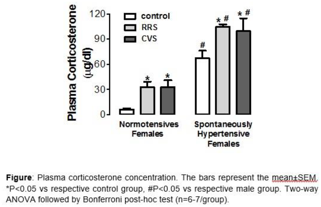
Glucocorticoid hormones (GCs), secreted after stress or during the circadian rise, act on the brain through binding to mineralocorticoid (MRs) and glucocorticoid receptors (GRs). After hormone binding and activation MRs and GRs bind to glucocorticoid response elements (GREs) within target genes (e.g. FK506-binding protein 5 (Fkbp5), serum/GC-regulated kinase 1 (Sgk1), Period 1 (Per1)) to evoke changes in gene transcription. Based on early hormone binding studies (Reul & de Kloet, Endocrinology 1985) it was thought that MRs and GRs exert a tonic and feedback/cognition-enhancing influence, respectively, on brain function. This concept has not, however, been investigated regarding MR and GR to GRE binding at the genomic level.
Male Wistar rats were exposed to stress and hippocampus tissue collected at various time points after stress or under baseline conditions for chromatin immuno-precipitation (ChIP) to assess MR and GR binding to GREs within target genes or for RNA analysis by qPCR.
Forced swimming, and the circadian rise in GCs, caused significant transient rises in Fkbp5, Sgk1 and Per1 hnRNA and mRNA levels. These changes were associated with substantially increased binding of both MRs and GRs to a specific GRE within intron 5 of Fkbp5 and to GREs within the Sgk1 and Per1 gene promoters. Despite generating distinct GC responses, novelty and restraint stress elicited similar MR and GR to GRE binding profiles as forced swimming. MR and GR Tandem ChIP provided strong evidence that these receptors bind as MR:GR heterodimers and GR:GR homodimers to Fkbp5 and Per1 GREs, whilst only bind as GR:GR homodimers to Sgk1 GREs after stress (Mifsud & Reul, PNAS 2016).
In summary, whilst the GR to GRE binding profile was expected based on earlier receptor binding studies, the low MR binding to GREs under baseline AM conditions was surprising given the high levels of MR occupancy shown previously. Different stressors resulted in similar GRE binding indicating that above a certain threshold responses are independent of GC levels. MR and GR to GRE binding as well as their binding as homo- versus heterodimers is very GRE- and gene-dependent. Thus, GC action at the genomic level is highly complex with multiple layers of regulatory control.
Performing complex music plays can cause stress and memory lapses with important impact on musician’s presentation and careers.
Cortisol is one of the glucocorticoids regulated by the HPA axis considered as an indicator of the activity of this system in stress response. In its biologically active form it is a component of saliva and the majority remains unbound to proteins allowing to a convenient method of estimating serum cortisol levels which rise in response to stress independently of the circadian rhythms that regulate the secretion of this hormone.
In a pilot study using a salivary enzyme-linked immunosorbent assay a panel of musicians were evaluated in terms of stress by monitoring salivary cortisol in “normal” day life and under recitals.
Neuropsychological measures were also taken from each voluntary.
The effect of a stress mitigating intervention (a memory improving technique) was considered by comparing with musician group control. Despite individual variations that were not unneglectable and the role of possible confounding factors as gender circadian patterns, all requiring an extended sampling, the pilot study detected evidences of hormonal variation in response to recital stressor as higher cortisol levels were obtained for the recital days .
Considering the effect of a learning technique over stress in performance ("superlearning") two groups of piano classes were compared in three repeated evaluations the control group have no special learning training techniques to improve the play or to reduce stress. The control group however showed similar results that are consistent with higher stress levels which maybe lead to consider the superlearnig technique as a stronger stressor itself than music performance.
During heat stress, sympathetic activity increases monotonically until the heat shock develops. Changes in parasympathetic activity are not described yet. To characterize the parasympathetic control of heart rate during heat stress, 4-months old male Wistar-Kyoto rats (n=8) were implanted with telemetric transmitters. Aortic blood pressure, ECG, core body temperature, and animal activity were monitored during exposure to hot (44°C) environment in a climatic chamber. Time-frequency analysis based on Wigner-Ville transform was used to estimate the high-frequency power of RR-interval variability (HFRRI) and low-frequency power of systolic pressure variability (LFSPV) in 2 s long intervals. Numerical values represent mean (standard deviation). After exposing rats to the heat stressor, core body temperature (Tc) remained stable for 2.9 (1) min and increased steadily thereafter. Heatstroke developed at Tc of 42.7 (0.2) °C. LFSPV, a surrogate measure of vascular sympathetic activity, rose from 4.2 (0.9) mmHg2 to a maximum of 14.3 (2.6) mmHg2 attained during heatstroke. Simultaneously, the pre-ejection time reduced from 42 (6) ms to 33 (3) ms indicating an increase in the cardiac sympathetic drive. HFRRI (a surrogate measure of the cardiac parasympathetic activity) augmented from baseline values of 3.4 (0.9) ms2 to 21.4 (6.8) ms2 at the beginning of the heat stress. The elevated parasympathetic activity was confirmed pharmacologically with heart rate and HFRRI response to the muscarinic blockade with atropine methyl nitrate. HFRRI remained elevated until Tc reached 41.3 (0.3) °C and then decreased to 0.9 (0.1) ms2. Heart rate initially rose slowly (2.8 (1.2) bpm/min), but the speed of heart rate increase accelerated and reached values of 10.2 (3.4) bpm/min after the reduction of HFRRI. In conclusion, exposure to high ambient temperature co-activates the sympathetic and the parasympathetic nervous systems. While cardiac and vascular sympathetic activity progressively increased until heatstroke developed, cardiac parasympathetic activity was strongly elevated from the beginning of the heat stress but virtually abolished at core body temperatures above 41 °C. The decline in the cardiac parasympathetic activity was associated with the accelerated rise of heart rate.
Stress-related psychiatric disorders are often characterized by dysregulated activity of both the hypothalamus-pituitary-adrenal axis (HPA axis) and the serotonergic system. Nonetheless, it’s still unclear if the irregularities of the HPA axis are the cause or effect of the anomalies in the serotonergic system. We hypothesize that the activity of the HPA axis has an important role in the regulation of the rate limiting enzyme in the biosynthesis of serotonin Tryptophan hydroxylase (Tph2), and therefore in the serotonergic system. Three models have been used; 1) The natural circadian rhythm of glucocorticoids (GC); 2) Alteration of the GC rhythm by the administration of a synthetic GC methylprednisolone (MPL); and 3) Modification of GC activity by constant light exposure for 5 weeks. To evaluate the effects of these manipulations we used; a) Radioimmunoassays (RIA) to assess GC concentrations in plasma b) In situ Hybridization Histochemistry (ISHH) to evaluate changes in mRNA expression of Tph2 in the Dorsal Raphe (DR) and the Median Raphe (MnR). The results have shown the expected circadian GC rhythmicity in the control rats, suppressed endogenous GCs in the MPL treated rats, and hyperactive GC secretion in the constant light exposed rats. The ISHH dataset analyses have shown that Tph2 expression is profoundly altered in the DRV and the MnR after MPL treatment compared to normal control rats. In the DRV, Tph2 expression is lowest at 9am and greatest at 3pm in control rats. In contrast, the circadian rhythm of Tph2 expression is far less pronounced after MPL treatment. Throughout the MnR, the lowest Tph2 expression is observed at 9am and highest expression at 3pm in control rats and there is a striking increase observed at 6pm across a region of the MnR as a result of MPL treatment. The overall amplitude of Tph2 mRNA expression in both the DRV and MnR is generally much lower in the MPL treatment groups than in the controls, potentially indicating an overall down regulation of Tph2 mRNA levels as a result of chronic sGC treatment and the associated prolonged GR activity. This decrease in Tph2 might have significant implications for serotonin biosynthesis in the DR and MnR, which in turn would impact upon the serotonergic system and affective state.
Neuroticism is a personality trait which refers to inclination of negative affectivity. It has been proposed that high level neuroticism leads decreased activations in prefrontal cortices during dual task processing by impoverishing the cognitive resources in prefrontal cortices. However, neuroanatomical correlates of these activations in terms of central executive functions (CES) are unknown. To investigate for this, 15 high and 15 low neurotics were performed a PRP dual task with short (higher CES demand) and long (lower CES demand) stimuli onset asynchrony (SOA), while I assessed brain activity by means of functional magnetic resonance imaging (fMRI). Behavioural results showed high neurotics had lower performance (response times and error rates) in short SOA dual tasks than in the long SOA tasks. Imaging data showed that high neurotics showed decreased activations mainly in lateral prefrontal lobe (inferior and middle frontal gyrus) as the demand increase on the central executive system. In conclusion, high neuroticism leads lower behavioural performance and at the same time decreased activations in lateral prefrontal cortices as the demand increase on the central executive system.
Serotonin (5-HT), via its various receptors, each of which is expressed in discrete brain regions, modulates aversion and reward processing and is implicated in various psychopathologies including eating disorders and depression. In mice, chronic social stress in adulthood leads to increased food intake in the absence of weight gain but reduces effortful motivation to obtain gustatory reward [1]. The aim of this study was to investigate for stress effects on three 5-HT receptors, namely 1A, 2A and 2C, by quantifying their specific binding in some brain regions underlying reward processing: medial prefrontal cortex (mPFC), hippocampus (HIPP), amygdala (Amy), nucleus accumbens (NAcc), ventral tegmental area (VTA), dorsal raphe nucleus (DRN) and locus coeruleus (LC).
Prenatal social stress (PSS) results in heightened anxiety behaviour in the adult male offspring, accompanied by increased gene expression for corticotrophin releasing hormone (CRH) in the central amygdala (CeA), and greater hypothalamo-pituitary-adrenal (HPA) axis responses to stress in the adult female offspring [1, 2]. This study aims to characterize if PSS has an effect on depressive-like behaviours, and whether maternal administration of an antioxidant can abrogate these abnormalities. The antioxidant, mitoquinone encased in nanoparticles (MitoQ-NP), does not cross the placental barrier and has previously been shown to prevent some of the effects of another paradigm of prenatal stress (hypoxic stress).
MitoQ-NP or saline was administered intravenously to pregnant dams on day 16 of gestation.
Dams were then left undisturbed or subjected to social stress (resident-intruder paradigm) on days 16-20. Male and female offspring were tested during adulthood on the light-dark box (LDB) and elevated plus maze (EPM) to assess anxiety-like behaviour, followed by the sucrose preference test (SPT) and forced swim test (FST) to assess depressive-like behaviour. After behaviour testing, brains were collected, frozen and sectioned for in situ hybridisation using radiolabelled probes for CRH mRNA. To a separate group of rats, blood samples were collected via an indwelling jugular vein cannula from adult female offspring to assess HPA axis activity before and after exposure to acute stress (30 min restraint).
PSS resulted in heightened anxiety-like behaviour in both the LDB and EPM in the adult male offspring. Maternal MitoQ-NP treatment prevented this anxious behaviour, and this was accompanied by the normalisation of CRH expression in the CeA. Neither the male or female PSS offspring exhibited a depressive-like phenotype in the FST or SPT compared with controls. Administration of maternal MitoQ-NP, however, decreased floating behaviour in male and female control and PSS offspring, indicating an anti-depressant effect. The corticosterone response to acute stress was prolonged in the PNS females compared with controls, and was not altered by MitoQ-NP treatment.
[1] Brunton PJ & Russell JA (2010) J Neuroendocrinol 22:258-71.
[2] Brunton PJ et al (2011) Stress 14:634-43.
Introduction: In adults, noxious stimulation evokes a cortical pain response and subjective pain report that are modulated by the level of physiological stress. The relationship between pain and stress is proposed to be a significant factor in individual variability of pain perception1. In newborn infants noxious stimulation evokes well-described pain behaviour, which is commonly used in place of pain report2 and nociceptive specific cortical activity (nociceptive Event Related Potential, nERP) as measured with EEG3. However, the effect of physiological stress levels upon infant cortical and behavioural pain measures is not known. Here, we investigate this by simultaneously measuring salivary cortisol, heart rate variability (HRV), nERP, and pain behaviour in neonates following a heel lance.
Method: 66 healthy neonates (mean GA 38.8 weeks; mean PNA 3.6 days) were studied during a clinically required heel lance. Ethical approval was given by the UK NRES and UCL/UCLH Joint Research Office.
Cortical activity, time locked to the heel lance, was recorded using EEG3. Salivary cortisol and ECG data for HRV calculation were collected before and after the procedure. Pain behaviour was scored using the Premature Infant Pain Profile (PIPP)2.
Results: The nERP amplitude was positively correlated with the PIPP following a heel lance (r=.36, p=.033). In addition, higher cortisol concentration and lower HRV, indicative of higher levels of physiological stress, were associated with larger nERP amplitude (r=.41, p=.029; r=-.42, p=.027). In contrast, PIPP was unrelated to the level of stress. Interestingly, the direct relationship between the nERP and PIPP was disrupted in babies that had a higher level of physiological stress (r=.27, n.s.).
Conclusion: The findings suggest that the cortical nociceptive response provides a more comprehensive measure of the pain experience of infants compared to pain behaviour as it also reflects their level of stress. Moreover, when infants are in a higher state of stress their behaviour in response to a lance is no longer an accurate reflection of the cortical pain processing.
References:
1. Vachon-Presseau et al. (2013). J Neurosci, 33: 6826–6833.
2. Stevens et al. (1996). Clin J Pain, 12: 13-22.
3. Fabrizi et al. (2011). Curr Biol, 21: 1552–1558.
Phytocannabinoid derivatives of Cannabis Sativa are an exciting new class of anticonvulsants and one, cannabidiol (CBD), has displayed potent anticonvulsant properties in recent clinical trials in patients with two forms of childhood onset epilepsy. Here, we investigated the anticonvulsant effect of 30μM CBD using whole-cell patch clamp recording in Layer II of the medial entorhinal cortex of adolescent male status epilepticus experienced (SE) and age-matched control (AMC) Wistar rats (50-100g) in vitro. The Reduced Intensity Status Epilepticus (RISE) model of acquired epilepsy was used to induce epileptogenesis. Data were also collected from ex vivo human tissue (HT), resected in pediatric neurosurgery from patients with drug-refractory epilepsy. The effects of CBD on amplitude (pA) and inter-event intervals (IEIs) of spontaneous inhibitory post-synaptic currents (sIPSCs) were compared between SE and AMC rats. In SE rats, CBD significantly increased sIPSC amplitude (39.5 ± 4.4 to 52.5 ± 6.5 pA, P<0.0001) and decreased IEI (74.6 ± 15.3 to 62.3 ± 12.2 ms, P=0.02, n=5) while in AMC we saw a reduced effect (43.6 ± 17.3 to 52.6 ± 19.1 pA, P=0.099; IEI: 122.2 ± 28.7 to 147.8 ± 38.8 ms, P=0.16; n=6). Elucidation of the mechanism of action focused on GABAAR and NMDAR involvement. Using SE rats, 500nM flumazenil and 5μM ß-carboline-3-carboxylic acid-N-methylamide (ß-Carb) were used to inhibit benzodiazepine binding sites of GABAARs. NMDARs were inhibited competitively using 5μM D-AP-5 and non-competitively using 100nM MK801. Flumazenil addition caused a significant decrease in amplitude upon CBD addition (32.4 ± 12.8 to 21.9 ± 5.9 pA, P=0.024) and increased IEIs (93.2 ± 20.9 to 125.3 ± 42.5 ms, P=0.36; n=5). ß-Carb reduced the effects of CBD (37.0 ± 5.8 to 34.1 ± 2.2 pA, P=0.69; IEI: 72.9 ± 16.5 to 63.1 ± 13.5 ms, P=0.36; n=5). Inhibition of NMDARs by both drugs was sufficient to block the effects of CBD (AP-5: 31.1 ± 6.5 to 27.5 ± 6.4 pA, P=0.18; IEI: 121.7 ± 39.2 to 142.6 ± 49.6 ms, p=0.37; n=5) (MK801: 28.9 ± 4.2 to 30.0 ± 6.6 pA, P=0.96; IEI: 75.5 ± 24.6 to 85.2 ± 27.3 ms, P>0.9; n=6). Experiments in ex vivo HT trended towards similar results as those above. These data indicate the possible involvement of GABAARs and NMDARs in the anti-epileptic mechanism of CBD.
Throughout adult life, new neurons are generated in the hippocampal subgranular zone (SGZ) and the lateral subventricular zone (SVZ) of rodent brain. More recent studies show that the adult hypothalamus also harbours neural stem/progenitor cells (NSPCs) called tanycytes. Tanycytes populate the floor of the third ventricle and give rise to neurons that get integrated into appetite regulating centres. Although little is known about genetic regulators of postnatal hypothalamic neurogenesis, the fibroblast growth factor (FGF) system is emerging as an important candidate. However, the endogenous FGF/FGFR signalling partners in adult hypothalamic neurogenesis remain unknown. To identify these, we surveyed the expression of FGF ligands, their receptors as well as signalling modulators in the adult hypothalamus niche by reverse-transcriptase PCR, in situ hybridization, immunolabelling, and through the use of reporter mice. We find that distinct FGF family members as well as FGF signalling modulators such as Sproutys are expressed by tanycytes. Beta-klotho, a co-receptor mediating the effects of endocrine FGF ligands was also detected. These differential patterns of expression within the ependymal lining, the hypothalamic neurogenic niche as well as the hypothalamic parenchyma are indicative of multiple roles for FGFs within and outside the hypothalamic neurogenic niche. Targeted gain- and loss of function studies are underway to decipher the postulated functions assigned to FGF signalling in the hypothalamic neurogenic niche.

Neuronal plasticity allows networks to learn and adapt to the environment and change behaviour accordingly. The highly plastic olfactory bulb network serves as a good model to study plasticity. Olfactory bulb plasticity is viewed as primarily an interneuron phenomenon as these cells replenish throughout life and undergo activity-dependent plasticity. External tufted cells (ETC) are excitatory interneurons found in the glomerular layer of the olfactory bulb that are major modulators of olfactory sensory processing. However, surprisingly little is known about activity-dependent alterations in these neurons, though their location and monosynaptic connection to olfactory sensory neurons (OSNs) would suggest a role in adapting the network to environmental changes.
To understand whether these cells are important for adapting the olfactory network, ETC functional and structural characteristics were compared in control and 24 hour naris occluded mice. Whole-cell patch clamp recordings in acute olfactory bulb slices reveal that intrinsic excitability, assessed by multiple spiking properties, does not change with this manipulation. Additionally, ETC-characteristic spontaneous burst firing does not change in terms of number of spikes fired, or burst properties. Furthermore, neither single spike properties nor sag potential amplitude show significant differences after occlusion. However, when assessing synaptic properties, ETCs in occluded conditions have larger spontaneous long lasting depolarising currents, with no change in the frequency of their occurrence. We further investigated whether occlusion results in a change in the release probability at OSN synapses by recording from ETCs while stimulating OSN axons. We found that after occlusion the paired-pulse ratio (PPR) of these inputs decreases, suggestive of an increase in release probability at OSN synapses.
These alterations are indicative of adaptive plasticity in excitatory signaling in the olfactory bulb glomerular network. They may act to control the gain of information flow through the circuit, maintaining sensory performance in the face of external perturbations.
The importance of myelin in cognitive functioning has increasingly been implicated, and its loss or damage is known to be associated with several neurological and mental illnesses1. Our understanding of myelination patterns and characteristics during development, despite the clear significance of myelin, is still incomplete. Across species, white matter volume increases have been observed in development in several brain regions2,3,4. Notwithstanding, the exact nature of developmental myelination in rodent models such as mice is unclear. Therefore, we investigated the myelination pattern and internodal length in the developing mouse brain. Differential patterns of myelin distribution have been found in distinct layers of the mouse cortex5 and more specific features of myelin (such as internodal length and amount of internodes along axons) might potentially affect conduction speed. In the peripheral nervous system, a functional relationship between internodal distance and conduction speed has been demonstrated and internodal length increases throughout development6. We were therefore interested whether the same phenomenon occurs in the central nervous system (CNS). In order to investigate the myelin distribution in the somatosensory cortex and to measure the internodal length in this brain region, we used immunohistochemistry and high-resolution confocal imaging in animals during development (p0-p118), over regular time intervals. We found that cortical myelination started later than in the white matter and gradually increased in over time. Our preliminary findings suggest that instead of an average increase of the internodal length, the variability of internodal lengths increases during development in the CNS. This variability may suggest that developing neural circuits may be accompanied by characteristic, staged changes in myelination.
References
1 Baumann, N. & Pham-Dinh, D. (2001) Physiol. Rev. 81:871-927
2 Barnea-Goraly et al. (2005) Cerebral Cortex 15:1848-1854
3 Imperati et al. (2011) PLoS ONE 6(8): e23437
4 Hamano, K., Iwasaki, N., Takeya, T. & Takita, H. (1996) Developmental Brain Research 93:18-22
5 Tomassy et al. (2014) Science 344:319-324
6 Wu, L.N., Williams, A., Delaney, A., Sherman, D.L. & Brophy, P.J. (2012) Current Biology 22:1957-1961.
Mitochondrial movement along microtubules using molecular motors, kinesin and dynein ensures adequate provision of ATP and Ca2+ buffering during neuronal activity. Previous work established that the kinesin adaptor proteins, TRAK1 and TRAK2, play an important role in mitochondrial transport in cultured neurons. They link mitochondria to kinesin via a TRAK acceptor protein in the mitochondrial outer membrane, the Rho GTPase, Miro. In addition, TRAKs associate with the post-translational modification enzyme, O-linked N-acetylglucosamine transferase (OGT), to form quaternary mitochondrial trafficking complexes. Miro acts as a Ca2+ sensor and OGT as a glucose sensor. Thus both aid localization of mitochondria to specific neuronal regions where Ca2+ buffering and energy are required.
Down-regulation of TRAK1 or TRAK2 utilising shRNAs as well as dissociation of TRAK proteins from the quaternary complex by a TRAK2 dominant negative (DN) impaired mitochondrial transport in cultured hippocampal neurons1. It is known that in these neurons, TRAK2 predominantly mediates mitochondrial transport in dendrites whereas TRAK1 mediates axonal transport2.
The effects of compromising TRAK function on the activity of mature hippocampal neurons are unknown. Here, we have investigated this by stereotaxically injecting an adeno-associated virus 2 (AAV2) vector containing a TRAK2 DN construct or an appropriate control into the CA1 hippocampal region of adult rats. Three weeks post-surgery, brain slices were prepared and electrophysiological recordings made from infected CA1 pyramidal neurons to determine the intrinsic membrane properties, action potential firing rates and evoked excitatory post-synaptic potential kinetics and integration. Neurons were also filled with neurobiotin during the recordings and post-hoc analysis was carried out to determine cell morphology.
These studies will aid our understanding of the contribution of TRAK function to normal neuronal activity in the adult brain.
Supported by BBSRC (UK).
1. Brickley, K and Stephenson, FA (2011) J. Biol. Chem. 286, 18079.
2. Loss, O and Stephenson, FA (2015), J. Neurosci. Res., 93, 1056.
Structural and functional plasticity at glutamatergic synapses are critical for learning and memory. Long-term potentiation (LTP), induces spine growth and synaptic localisation of major excitatory glutamate AMPA receptors (AMPARs), thus enhancing synaptic strength. Glutamate initiates these processes, but the contribution from extracellular modulators is not fully understood. Wnt secreted proteins are imperative for synapse formation but their impact on LTP mediated spine plasticity and AMPAR localisation is unknown. Using a multidisciplinary approach that combines cellular biology and electrophysiology techniques, we show that LTP induction rapidly increases Wnt7a/b at spines. Importantly, blockade of endogenous Wnts or loss of Frizzled-7 (Fz7) receptor function, a receptor for Wnt7a, impairs LTP-mediated spine growth and synaptic AMPAR localization. Wnt7a rapidly promotes the synaptic recruitment and trapping of AMPARs followed by an increase in spine growth. Wnt7a achieves this through CaMKII-dependent loss of Ras-GTPase SynGAP from spines and activation of the Ras-ERK pathway. Thus, our studies identify Wnts, through Fz7, as key initiators of LTP-mediated synaptic accumulation of AMPARs and spine plasticity.
Over the first two weeks of life, neurons in the neonatal mouse undergo enormous change, migrating, maturing, and adapting in electrical activity, morphology and synaptic connectivity. Action potentials, the primary indicator of neuronal activity, drive the patterns of synaptic transmission that underpin neuronal communication and control calcium influx that shapes patterns of gene expression. As such, changes to the waveform of the action potential over the course of development will have potentially a wide-ranging and profound influence on neuronal structure and function. We examine changes to the postnatal action potential waveform via whole cell current clamp electrophysiology in layer 4 stellate cells of the somatosensory cortex between postnatal days (P)3 and (P)11. We show that postnatal maturation is associated with large increases in the height and speed of individual action potentials. Using Hodgkin-Huxley style computational models, we attempt to characterise the changing ionic mechanisms in the neurons. We develop a computationally efficient analytical method of multiple-parameter optimisation of this model of active neuron dynamics, prior to fitting to data. The changing morphology and its impact on the intrinsic electrical properties of the developing neuron is also considered, with the capacitance of the cell membrane hypothesised to change with the increasing size and shape of the soma and dendritic arbour. Current clamp recordings of the passive voltage across the cell membrane are analysed using a two compartment model of exponential decay. Interestingly, a fast voltage decay component present in some cells may be attributed to the presence of gap junctions which could influence these passive currents, and have implications for the regulation of neural network development. Injection of dye into the patched cell during whole cell recording, followed by confocal imaging, is used to detect the presence of surrounding gap-junction-coupled neurons. We aim to compare this to the intrinsic electrical properties of the cell at different stages of development, to produce a complete picture of the changing biophysical nature of the neuronal action potential as the neuron approaches maturity.
The release of acetylcholine in the hippocampus during awake behaviour is important for encoding memory. Within the hippocampal network, acetylcholine has diverse effects: it increases neuronal excitability, controls synaptic strength and regulates the induction of synaptic plasticity. However, these effects are not ubiquitous and instead are exhibited at individual synapses within the network. The Temporoammonic (TA) pathway carries spatial information from grid cells in entorhinal cortex layer III to CA1 hippocampal place cells synapsing onto the distal dendrites. It is not currently known how acetylcholine regulates synaptic transmission in the temporoammonic pathway or which acetylcholine receptors mediate this regulation. To determine how acetylcholine regulates the TA pathway we made whole cell patch clamp recordings from CA1 pyramidal neurons or selected subset of interneurons in acute hippocampal sagittal slices from adult mice. Electrical stimulation in the Stratum Lacunosum Moleculare was used to isolate monosynaptic excitatory postsynaptic currents (EPSC) or disynaptic inhibitory postsynaptic currents (IPSC). The acetylcholine receptor agonist carbachol (CCh 10 μM) reduced both excitatory and inhibitory synaptic responses and increased paired-pulse ratio for excitatory responses, indicating a presynaptic locus of action. Specific pharmacological intervention showed that M3 receptor antagonist or genetic deletion of this receptors, blocked CCh induced reduction of synaptic probability of release. Furthermore, we revealed that PV+ Interneurons are feedforward upon TA pathway stimulation, whose excitatory inputs are inhibited by the activation of M3 receptors. Excitatory and inhibitory responses at pyramidal neurons were similarly reduced by CCh but the increase in paired pulse ratio for excitatory drive produced a facilitation of excitatory-inhibitory balance in response to repetitive stimulation. In addition, CCh produced an increase in the number of spikes in the CA1 pyramidal neurons when TA synapses were repeatedly stimulated over a range of frequencies. We conclude that acetylcholine modulates the temporoammonic pathway by presynaptically located M3 muscarinic receptors.

The expansion and activation of microglia, the brain’s resident population of myeloid cells, is a hallmark of many neurodegenerative diseases including Alzheimer’s disease and prion disease. Colony stimulating factor 1 receptor (CSF1R) is critically involved in regulating proliferation of microglia both in the healthy and diseased brain. CSF1R can be activated by two independent ligands, CSF1 and interleukin (IL-) 34. Ligand binding to the receptor leads to tyrosine kinase phosphorylation and activation of intracellular signalling pathways involved in survival, proliferation and differentiation of microglia.
In this project, we aim to obtain a clear understanding of the effect of CSF1 vs. IL-34 on CSF1R activation in microglia, with a specific focus on potential differences in the activation pattern between the two ligands. Using a murine microglial cell line (N13) and primary murine microglia, we observed a rapid and transient phosphorylation of CSF1R after stimulation with CSF1 or IL-34. Likewise, CSF1R downstream pathways ERK1/2, AKT, SAPK/JNK and p38 were activated upon receptor phosphorylation. Expression of genes implicated in proliferation and inflammation were modulated after CSF1 or IL-34 stimulation in a concentration-dependent manner. Finally, primary microglia demonstrated a pronounced increase in proliferation after CSF1R activation.
In consideration of the inflammatory response observed after treatment with CSF1R ligands, we aimed to determine whether “priming” with CSF1 or IL-34 affects the expression of inflammatory genes in microglia in response to an inflammatory stimulus (i.e. LPS). We observed a reduction in the expression of pro-inflammatory markers after short-term priming of CSF1R, which is reversed to baseline levels when cells were primed for a longer period of time.
These results provide insight into the kinetics of CSF1R activation by CSF1 vs. IL-34 in microglia cells and establish the basis to further study the differential role of both ligands in CSF1R function.
The ability of neural stem cells to self-renewal and to differentiate into neurons, oligodendrocytes and astrocytes is essential for a balanced neurogenesis in adult brains. Low-dose gamma irradiation endangers genomic stability in these cells by inducing DNA double-strand breaks (DSBs). Particularly, the immature neural stem cells are radiosensitive (Katsura et al. 2015, Saha et al. 2014). However, NSCs have two main repair ways to handle DSBs. On the one hand, there is homologous recombination which depends on a sister chromatid as repair template and is therefore only available in late S and G2 Phase. On the other hand, there is NHEJ (non-homologous end-joining) which includes the repair protein 53BP1. NHEJ is active throughout the cell cycle and consequently available in mature cells. It is little known about repair mechanisms in immature and mature neural cells and the consequences of unrepaired double strand breaks, so we irradiated murine stem cells (J1-NSCs) and focused on the NHEJ repair kinetics. Simultaneously, we differentiated the NSCs under specific culture conditions to their descendants (neurons, astrocytes and oligodendrocytes) and irradiated them as well. As expected, we see more 53BP1-Foci (indicator for DSBs) after irradiation in the radiosensitive neural stem cells compared to the descendants. Interestingly, the 53BP1-Foci quantity and the repair kinetics of astrocytes and neurons were different. We hypothesize that DSB repair in neurons is regulated by glutamate signaling through the N-Methyl-D-aspartate (NMDA) receptor (Yang et al. 2011). Our results indicate a connection between treatment with 30 μM glutamate and 53BP1-Foci formation in neurons. Moreover, this effect is inhibited by treatment with MK-801, an antagonist of NMDAR. Overnight treatment with glutamate affects the repair kinetics of neurons after low-dose irradiation, in a positive manner. Glutamate treatment neither induces 53BP1-Foci, nor has positive effects on the repair kinetics of astrocytes. We suppose that the excitatory neurotransmitter glutamate exclusively stimulates neurons via NMDAR activation. Our future experiments include the investigation of specific NMDAR subunit compositions in neurons and glial cells, to figure out the subunit influence on DSB repair.
Most evidence indicates that G protein-coupled receptors form heteromers between them and with other receptors. By allosteric mechanisms, them acquire a multiplicity of unique pharmacological and functional properties. Recently, we discovered that dopamine D1 receptors (D1R) and histamine H3 receptors (H3R) form heteromers through which H3R ligands can inhibit D1R function. D1Rs also physically interact and modulate ionotropic glutamate NMDA receptors (NMDAR). In the present work, we investigated if NMDAR, D1R and H3R form a heterotrimeric complex in brain.
The heteromer expression was studied in slices from both rat and mouse brain cortex by co-immunoprecipitation (Co-IP) and proximity ligation assays (PLA). The ability of D1R and H3R to interact with NMDAR in transfected HEK cells was analyzed by bioluminescence resonance energy transfer (BRET) with bimolecular fluorescence complementation (BiFC) experiments. Heteromer properties were studied by analyzing ERK1/2 phosphorylation and cell death in cortical slices.
Endogenous D1R-H3R heteromers were detected in rat and mouse cortical slices, where H3R ligands decreased D1R signaling (ERK1/2 pathway) and were also able to block the cell death induced by overstimulation of either D1R or NMDAR. By BRET experiments in transfected HEK cells, we demonstrated that both D1R and H3R form heteromers with NMDAR subunit 1A in the presence of subunit 2B. D1R-H3R-NMDAR heteromers were detected by BRET with BiFC. The expression of endogenous D1R-H3R-NMDAR heteromers were observed in rat and mouse cortex by PLA.
Many systems, including the glutamatergic and dopaminergic, are involved in neurodegeneration. Our innovative finding is that D1R, H3R and NMDAR form heteromers that may be a point of intervention for cognitive disorders in neurodegeneration.
Our results point out that D1R-H3R-NMDAR heteromers are expressed in brain cortex and a complex interaction exists between protomers in the heteromer, where H3R ligands act as a “molecular brake” for D1R and NMDAR signaling.
Introduction: Therapies to promote the differentiation of oligodendrocyte precursor cells (OPC) and boost myelin repair are lacking. In vitro culture systems are useful tools for the discovery of myelin repair treatments as they permit high-content screening of compounds in assays of OPC differentiation. Typically OPCs are derived from primary mixed glial cultures using expensive immunopanning methods, or complicated shaking procedures. Here we report a simplified and efficient protocol that generates early OPCs and later stage OPC from embryonic stem (ES) cell-derived neural stem (NS) cells. We also establish a simple protocol for OPC isolation from mixed glial cells derived from NS cells.
Methods: NS were cultured in serum-free media supplemented with growth factors including basic fibroblast growth factor (FGF2), Insulin-like growth factor 1 (IGF1) and platelet-derived growth factor (PDGF-AA). This culture condition converted NS cells into a population of early OPCs, which were easily distinguished by their different physical properties e.g. bi- and multi-process morphologies. Early OPC could then be used to generate mixed glial cultures, with OPCs adhered onto a layer of astrocytes. OPCs exhibited distinct diameters compared to astrocytes and so were easily isolated from the mixed cultures by cell straining. qPCR for marker genes was used to confirm the generation of early OPC (Nkx2.2), and OPC (NG2, PDGFRalpha), with results compared against primary OPC obtained from postnatal brain tissues. Also, cell diameters of the different cell types were measured to provide a simple way to predict the purity of each cell fraction.
Results and conclusion: Gene expression analyses showed that early OPC had a greater level of Nkx2.2 compared to OPC, while OPC expressed greater levels of the OPC marker genes NG2 and PDFRalpha. Cell diameter measurements further suggest that the simple protocol we established could isolate homogeneous cell population from mixed glial cells, demonstrating that we can generate early OPCs and OPCs from ES cell-derived NS cells, and that this simple protocol could isolate OPCs from mixed glial cells without complicated and costly steps such as overnight shaking or immunopanning.
Stroke is one of the major causes of death and disability worldwide. The harm caused by the interruption of blood flow to the brain unfolds in the subsequent hours and days, so it is critical to identify new therapeutic targets that could reduce neuronal death associated with the spread of the damage. The area that surrounds the infarcted core is the location of the continuing damage that takes place hours and days following an insult, an is referred to as the penumbra. The expression of the chloride co-transporters, NKCC1 and KCC2, mediators of the GABAergic response, was assessed following hypoxia in differentiated PC12 and NT2 neuronal-like cells and in a photothrombotic model of stroke in mice. Differentiated PC12 and NT2 cells were exposed to hypoxia (1% oxygen) for 8 hours in a hypoxic modular chamber before gene and protein expression was analysed by qPCR and immunoblotting. Following hypoxia, the expression of KCC2 was significantly decreased at both the transcript and protein level whereas NKCC1 expression remained unmodified. In the in vivo model, the development of the penumbra in the days following injury was assessed with the specific markers HSP70 and GFAP. Two distinct areas were identified, the penumbra up to 200 m from the ischaemic core and a glial migration zone up to 400 m. In the penumbra, a significant neuronal loss was observed up to 5 days following the insult. Our results show an increase in the number of neurons expressing NKCC1 in the penumbra up to 5 days following the insult when compared to the contralateral hemisphere. On the contrary, KCC2 positive cells were dramatically decreased in this area. Mice were treated with bumetanide and CLP257, an NKCC1 antagonist and a KCC2 agonist respectively. Neuronal loss was significantly reduced 3 and 5 days following the insult in the penumbra following bumetanide treatment. The reversal on NKCC1 and KCC2 might contribute to the excitotoxic damage that promotes the development of the penumbra in the days following an ischaemic event by interrupting or even reversing GABAergic mediated inhibition. Our results show how treatments targeting chloride co-transporters might represent a novel strategy to reduce the damage associated with stroke.
Located on both pre- and postsynaptic membranes, kainate receptors (KARs) perform various distinct roles in modulating neuronal and synaptic excitability. Further, KARs participate in the regulation of neuronal network activity and are involved in processes ranging from neuronal development and differentiation to neurodegeneration and neuronal cell death. An important property of pre- and postsynaptic KARs is that, in addition to direct ionotropic signalling, they can also signal through the activation of G proteins 1. By utilizing the metabotropic signalling pathway via PKC activation, KARs mediate both presynaptic (facilitation of glutamate release and down-regulation of GABA release 1,2 and postsynaptic actions (inhibition of afterhyperpolarisation current I(sAHP), 3,4.
Aims: To characterize the mechanism of KAR-mediated postsynaptic LTP in the hippocampus.
Methods: We used molecular biology tools, high resolution and live cell imaging, as well as electrophysiological (whole-cell patch-clamp, as well as field recording).
Results: We have characterized a novel, physiologically relevant NMDA receptor-independent mechanism that drives increased AMPA receptor recycling and results in LTP. The process is mediated by the metabotropic action of kainate receptors and requires activation of G-protein, protein kinase C and phospholipase C. In addition, the structural plasticity occurring as a result of KAR-dependent LTP shares the same properties like that of classical LTP and is manifested by: recruitment of recycling endosomes to spines, enhanced synaptic recycling, increased AMPAR surface expression and structural changes in spines, including their increased growth and maturation.
Conclusions: We have characterized a previously unsuspected role of postsynaptic kainate receptors in the induction of functional and structural plasticity in the hippocampus.
References:
1 Neuron (1998) 20:1211-18.
2 Neuron (2003) 39:543-53.
3 Neuron (2002) 34:107-14.
4 The Journal of Physiology (2005) 562:199-203.
Neurodegenerative conditions such as Alzheimer’s disease (AD) are associated with neuronal loss and cognitive decline. To date treatments have largely focused on symptomatic management and only in recent years has this focus shifted to therapeutically tackling disease progression. A therapeutic approach that can halt the actions of AD is therefore attractive as it will be a potential strategy to impede disease progression.
The orphan G-protein coupled receptor GPR55 is responsive to cannabinoids and is widely expressed in the neurons and glia of the brain. The suggested endogenous ligand for GPR55, L-α-lysophosphatidylinositol (LPI), exerts microglia-dependent neuroprotection after excitotoxic lesion (Kallendrusch et al., 2013), suggesting that GPR55 may have a regulatory role in neuroinflammation and neurodegeneration. This makes GPR55 an attractive therapeutic target for conditions such as AD. The present study aims to examine the role of GPR55 and its signalling pathways in the regulation of neuroinflammation and neuronal cell death using in vitro models.
Cultured primary rat cortical neurons were treated with LPI (1 μM & 10 μM). LPI-induced signalling effects were assessed using phospho-cAMP element binding protein (pCREB) immunocytochemical staining and confocal microscopy and imaging of intracellular calcium responses. LPI induced CREB phosphorylation in a concentration- and time-dependent manner. LPI (10 μM) induced calcium responses. Cortical neurons were treated with LPI (1 μM & 10 μM) in the presence or absence of the AD pathological hallmark, Aβ, for 72 hours. The conditioned medium was then applied to the BV2 microglial cell line and the subsequent migration of BV2 cells was assessed using a Boyden chamber assay. Microglial migration did not increase upon exposure to medium taken from neurons conditioned with LPI (10 μM), whereas LPI (1 μM) increased levels of migration compared to control cells. Neuronal apoptosis was assessed by active caspase-3 immunocytochemistry. LPI (10 μM) significantly downregulated neuronal apoptosis evoked by Aβ and glutamate. This study suggests that LPI can confer a neuroprotective effect and demonstrates a possible role for GPR55 in the regulation of gene expression, microglial migration and neuronal apoptosis.
Neuronal CaV channels are essential mediators of synaptic transmission, mediate the entry of extracellular Ca2+ at presynaptic terminals necessary for neurotransmitter release. N-type channels are implicated in a number of neuropathies including epilepsy and neuropathic pain; while the auxiliary α2δ subunit is the target of the antiepileptic drugs, Gabapentin and Pregabalin. The present study examines the interplay between N-type channels and the α2δ subunits which play an important role in CaVα1 trafficking and enhancement of macroscopic Ca¬V currents. Through confocal microscopy and patch-clamp recordings, we provide evidence that α2δ-1 enhances CaV2.2 plasma membrane expression through rab11a-dependent recycling. However, α2δ-3 has only a modest effect on CaV2.2 membrane expression and traffics in a rab11-independent manner. In addition, both α2δ-1 and α2δ-3 were seen to increase the expression of CaV2.2 in the processes of cultured hippocampal neurons; this effect was ablated by coexpression of the dominant negative Rab11a (S25N) mutant only when α2δ-1 was present. Interestingly, in neuro2A cells α2δ-1 and α2δ-3 both produce a comparable increase in total CaV2.2 expression, despite substantial differences in CaV2.2 surface expression. Together, our data suggests that α2δ-enhanced CaV current density may only partly relate to changes in CaV membrane expression. These findings could have significant implications for the development α2δ-targeted therapies for the treatment of CaV-associated neuropathies.
Long term potentiation (LTP) is a molecular substrate of memory consolidation. One key mechanism underlying LTP is phosphorylation of AMPA receptor subunits, a process that can affect both receptor function and location. For example, phosphorylation of GluA1 subunits at Ser845 leads to trafficking of AMPA receptors to the synaptic cleft to strengthen synaptic connections [1, 2].
We are interested in the plasticity changes involved in addiction. Risk of relapse back to drug-taking in abstinent addicts persists long after drug-taking has ceased. Changes in synaptic strength in the brain contribute to this, as emotional and environmental cues become associated with drug-taking and trigger cravings [3].
The overall aim of this study was to examine synaptic plasticity changes following expression of addiction-related behaviour in rodents. cFos-GFP transgenic mice [4] underwent morphine-primed conditioned place preference and reinstatement. Following reinstatement, animals’ brains were perfusion-fixed, sliced and GluA1 and phosphorylated GluA1 subunits at Ser845 in the hippocampus were examined by immunohistochemistry. The GluA1 subunits were largely expressed in the dendrites of neurons. Surprisingly, the majority of the Ser845-phosphorylated subunits were localised on the somas of pyramidal neurons, an observation which has not yet been reported. In LTP, the GluA1 subunits are thought to be phosphorylated in the post-synaptic terminal to trigger the trafficking of further AMPA receptors to the synapse, therefore their localisation in the cell bodies is unexpected. Further investigation into the relevance of this distribution is required for the better understanding of molecular mechanisms underlying motivational behaviour.
References:
1. Lee HK et al (2003) Cell 112:631-43
2. Henley JM & Wilkinson KA (2013) Dialogues Clin Neurosci 15:11-27
3. Korpi ER et al (2015) Pharmacol Rev 67:872-1004
4. Barth AL (2004) J Neurosci 24:6466-75.
Epilepsy is a chronic disorder of the brain characterised by recurrent seizures. 1-2 % of the world’s population are affected, with many sufferers developing resistance to drug therapy. Temporal lobe epilepsy (TLE) is the most common form of human epilepsy, in which the site of seizure initiation occurs in temporal lobe structures such as the entorhinal cortex (EC). In this study we examined the propagation of pharmacologically induced ictal-like epileptiform activity along the dorso-ventral axis of the medial EC (mEC). All procedures were in accordance with current UK Home Office regulations. 400 μm parasagittal slices were prepared from male C57BL/6 mice (3-6 month) and perfused (3-4 ml.min-1) with artificial cerebrospinal fluid. Recordings were made from layer ΙΙ of the mEC using a 16-channel silicon probe consisting of 16 individual shanks (55 μm wide, 100 μm apart), with a single electrode contact point at the end of each shank. Application of 100 μM 4-aminopyridine (4-AP) to the perfusing ACSF resulted in the initiation and propagation of ictal activity from the ventral to dorsal end of the mEC at a speed of 197 ± 24 μm.s-1 SEM (n=12 slices, R2=0.44). Addition of the GABAA receptor positive allosteric modulator diazepam (30 μM) significantly decreased the speed of ictal propagation from 148 ± 23 μm.s-1 to 65 ± 14 μm.s-1 (P<0.001, n=6 slices, paired Student’s t-test). Addition of the GABAA receptor inverse agonist Ro 19-4603 (10 nM) significantly increased the speed of ictal propagation from 170 ± 45 μm.s-1 to 1273 ± 117 μm.s-1 (P<0.001, n=6 slices, paired Student’s t- test). This demonstrates that the relatively slow propagation of ictal activity across the mEC is controlled by GABAergic synaptic transmission. The initiation of ictal activity at the ventral pole of the mEC likely occurs due to 1) the lower inhibitory tone and 2) the higher intrinsic excitability of individual stellate cells in this region cf. the dorsal mEC. This novel approach to understanding the underlying mechanisms involved in epileptiform activity in the mEC offers an in vitro model in which to probe antiepileptic drugs for the treatment of TLE.
In excitatory pathways through the retina – from photoreceptors to ganglion cells –glutamate acts as a fast synaptic transmitter, activating predominantly AMPA-type receptors (AMPARs) in primary retinal neurons. Within the retina AMPAR subunits are differentially expressed with respect to cell type and ontogenetic period, but the auxiliary subunits responsible for regulating AMPAR trafficking and function remain unknown.
Transmembrane AMPAR regulatory proteins (TARPs; γ-2, -3, -4, -5, -7 and -8) are the best understood of the known AMPAR auxiliary subunits. In most brain regions TARPs are differentially distributed within neuronal and glial populations and play distinct roles in shaping synaptic transmission. We sought to identify which TARPs are expressed in retinal neurons, with the aim of determining their precise synaptic location and cellular distribution. Using Western blot of wild-type retinal protein, we detected expression of TARPs γ-2, -3, and -5. Using antibody labelling and immunofluorescence, we analysed TARP expression and cell-type localization in retinal cryosections. We found γ-2 (stargazin) to have a punctate pattern of expression in both the outer plexiform layer (OPL), where photoreceptors contact bipolar and horizontal cells, and inner plexiform layers (IPL), where bipolar and amacrine cells contact ganglion cells. These puncta co-localized with postsynaptic markers and AMPAR subunits suggesting a synaptic location. Additionally, we found labelling for γ-3 in cell bodies and dendrites within the OPL and in large cell bodies of the ganglion cell layer. These locations indicate that γ-3 is likely expressed in Off bipolar cells and ganglion cells. Finally, we identified γ-5 expression in the IPL only. By performing double- and triple labelling of fixed whole-mounted retinas and retinal cultures, we identified horizontal cells, Off bipolar cells and several populations of amacrine and ganglion cells that express, either singly or together, TARPs γ-2, -3, and -5.
Our data provide the first detailed characterization of the localization of TARP proteins in the retina. The specific expression patterns are consistent with a role for these proteins in regulating AMPARs at retinal synapses.
Acknowledgements: Supported by the MRC (MR/J002976/1).
Novel sensory experiences are encoded by changes in cortical neuronal networks (Holtmaat and Svoboda 2009). This experience-dependent plasticity is driven in part by structural changes at the synaptic level. It is known that, in the adult mammalian brain, a small but significant number of synapses are constantly removed and replaced. However, little is known about the subcellular processes that control synaptic turnover. Indeed, within the same axon a transient synapse can appear and disappear alongside a highly stable one (De Paola et al. 2010). This raises the question of what molecular players inside the pre- or postsynapse contribute to the stability of that synapse. Mitochondria have extensively been shown to be vital to presynaptic neurotransmission (Vos et al. 2010), we have therefore studied mitochondrial localisation at presynaptic boutons and their influence on structural synaptic plasticity in vivo. We used an AAV2/1 expressing a mitochondrially-targeted tagRFP and cytosolic EGFP to label axons projecting to the mouse somatosensory cortex. We imaged the turnover of axonal boutons and mitochondrial localisation longitudinally by two-photon microscopy through a cranial window. We show that the pattern of mitochondrial localisation in these axons changed over a period of days, with mitochondria preferentially localising to presynaptic boutons. The stability of presynaptic boutons was much greater than that of mitochondrial localisation, however there were some sites where mitochondria were persistently found. Analysing the subcellular coordination of mitochondrial and synaptic turnover over time was used to establish the relationship between mitochondrial localisation and presynaptic bouton stability.
Holtmaat, A., Svoboda, K., (2009) Experience-dependent structural synaptic plasticity in the mammalian brain. Nat. Rev. Neurosci.
De Paola, V., Holtmaat, A., Knott, G., Song, S., Wilbrecht, L., Caroni, P., Svoboda, K., (2006) Cell Type-Specific Structural Plasticity of Axonal Branches and Boutons in the Adult Neocortex. Neuron.
Vos, M., Lauwers, E., Verstreken, P., (2010) Synaptic mitochondria in synaptic transmission and organization of vesicle pools in health and disease. Front. Syn. Neurosci.
Glia are the major cellular components of the central nervous system (CNS) that when activated play important roles in inflammation, neuropathic pain and opioid tolerance (Giaume et al., 2007). In this study we determine the expression and function of the non-classical Nociceptin/Orphanin FQ (NOP) opioid receptor, shown to have important roles in pain processing and opioid tolerance (Lambert, 2008), in a variety of glial cell types.
Methods
Using 1321N1 human astrocytes, MO3.13 human oligodendrocytes, HOG human oligodendrocytes and EOC-20 mouse microglia (De Vries and Boullerne, 2010), a series of in vitro assays including quantitative PCR (qPCR), [leucyl-3H] N/OFQ saturation binding and scratch wound healing/cell migration were performed. We investigated: (1) the presence of mRNA encoding for NOP opioid receptor (2) NOP receptor expression and (3) functional activity of the expressed receptor.
Results
Human glial cells differentially express mRNA encoding for the NOP receptor and where present, this was translated into NOP receptor protein as determined using [leucyl-3H] N/OFQ binding (Table 1). In functional assays, N/OFQ significantly inhibited wound healing/cell migration in cells expressing NOP receptors (Table 1).
Overall, these findings suggest that NOP may have a role to play in astrocyte and oligodendrocyte function.
This work is sponsored by the Higher Committee for Education Development in Iraq HCED/Office of the Prime Minister/Iraq.
References:
De Vries, G.H. and Boullerne, A.I. (2010). Glial cell lines: an overview. Neurochemical research, 35, 1978-2000.
Giaume, C., et al (2007). Glia: the fulcrum of brain diseases. Cell death and differentiation, 14, 1324-1335.
Lambert, D.G. (2008). The nociceptin/orphanin FQ receptor: a target with broad therapeutic potential. Nat Rev Drug Discov, 7, 694-710.

In order to maintain a healthy brain a constant supply of oxygenated blood is required. During hypoxia multiple signalling pathways are activated within neurons including the stabilisation of hypoxia-inducible factors. The activity of these proteins is regulated by O2, Fe2+, 2-OG & ascorbate-dependant hydroxylases which contain prolyl-4-hydroxylase domains (PHDs). Very little research has been carried out on the action of PHD inhibitors in the CNS and especially on synaptic transmission and plasticity. In this study we have investigated the acute effects of the PHD inhibitor and hypoxia mimetic, protocatechuic acid ethyl ester (EDHB) on cell viability and synaptic plasticity in isolated rat (Wistar) hippocampus and organotypic cell culture. Cell viability was assessed using propidium iodide in organotypic cultures. Excitatory post-synaptic potentials were elicited by stimulation of the medial perforant (mDG) or Schaffer collateral pathway. Long-term potentiation (LTP) was induced by high frequency stimulation. 2 or 24 h treatment with EDHB (100 μM) had no significant effect on hippocampal cell viability when compared to controls. Cultures treated with 24h hypoxia, 8h OGD or 24h excitotoxicity (Glutamic acid) showed a significantly higher percentage of cell death compared to control and EDHB treated cultures. EDHB (100μM) gave rise to an acute, inhibitory effect on synaptic transmission which was seen in the mDG but not in the CA1. There were no changes in the ratio of paired responses (50ms interval) after EDHB application suggesting a post-synaptic mechanism of action. EDHB at higher concentrations (100μM), was found to inhibit LTP in both the mDG and CA1 regions. Application of exogenous iron (100μM) and the HIF-inhibitor digoxin (100nM) did not reverse EDHBs inhibitory effect on baseline transmission or LTP, suggesting a HIF-independent mechanism of action. These results highlight a novel modulatory role for the PHD inhibitor EDHB in hippocampal synaptic transmission and plasticity. The effects are unlikely to be mediated pre-synaptically as is observed in hypoxia, where O2 levels are decreased in brain tissue and adenosine receptors are activated. A novel post-synaptic mechanism of action may be involved possibly involving NMDA & GABA receptor activation.
AMPA-type glutamate receptors (AMPARs) mediate fast excitatory neurotransmission in the mammalian brain and play key roles in synaptic plasticity and synapse development. Furthermore, AMPAR dysregulation contributes to neuron damage in a number of neurological conditions. The AMPAR pore-forming subunits, GluA1-4, are differentially expressed between cell types and ages, providing considerable functional diversity. This diversity is further increased by a variety of auxiliary transmembrane proteins that coassemble with AMPAR subunits to form macromolecular complexes. These associated proteins regulate receptor trafficking, gating and pharmacology. Proteomic studies identified the tetraspanin GSG1L as an AMPAR-interacting protein. Structurally similar to the transmembrane AMPA receptor regulatory proteins (TARPs), GSG1L has been validated as a bona fide AMPAR auxiliary protein capable of modulating channel gating and cell surface expression. To investigate further the possible roles of GSG1L we have explored its functional effects in various types of AMPAR assembly. Specifically, we recorded currents activated by fast application of glutamate onto outside-out membrane patches from cells expressing distinct combinations of AMPAR pore-forming subunits and auxiliary proteins. Furthermore, we investigated the importance of the C-tail of GSG1L in modulating AMPAR properties, by examining the effects of C-tail deletion (GSG1L-ΔCt). To determine the functional effects of GSG1L on native AMPAR complexes we manipulated its expression in different types of neuron and analyzed their synaptic currents. Using this approach we found that GSG1L exhibits subtype-specific effects on AMPARs. Our data show that while GSG1L slows desensitization in all AMPAR subtypes tested, the slowing of recovery from desensitization is limited to homomeric calcium-permeable AMPARs. Moreover, GSG1L mediates this recovery phenotype via an unusual carboxyl domain- and polyamine-dependent mechanism.
Supported by the Wellcome Trust (086185/Z/08/Z to SGC-C and MF) and the MRC (MR/J002976/1 to SGC-C and MF, MR/J012998/1 to MF and SGC-C).
Purpose: Recent patients studies have demonstrated an association between chemotherapy treatment and cognitive impairment. We have previously shown that 5-fluorouracil, a chemotherapy agent widely used for breast, prostate and bowel cancer, induces cognitive impairments and a reduction in hippocampal neurogenesis in a rodent model. In the present study we quantified the impact of chronic 5-FU treatment on cell proliferation (Ki67), differentiation (DCX) and stem cell subpopulations (GFP and GFAP) in the subgranular zone (SGZ) of SOX1-GFP transgenic mice. > 90% of SOX1+ cells are neural stem cells and are restricted to SGZ. In SOX1-GFP mice it is possible to distinguish between early radial and later horizontal orientated SOX1+ cells and to distinguish quiescent (GFAP+) and activated (GFAP-) SOX1+ cells.
Methods: Male SOX1-GFP mice were injected with 5-FU or saline twice a day, every second day for two weeks. Animals were killed 24h after the last injection and their brains were processed for immunohistochemistry. Confocal images were acquired and analysed using ImageJ software.
Results: Two weeks of 5FU treatment caused a significant reduction in the number of proliferating (Ki67+) cells but did not affect the number of differentiating (DCX+) cells. A different picture emerged from the examination of neural stem cell subpopulations where chemotherapy reduced the number of quiescent (SOX1+/GFAP+) and activated (SOX1+/GFAP-), radial neural stem cells but had no effect on the numbers of horizontally orientated SOX1+ cells present in later stages of neurogenesis.
Conclusions: Results presented here demonstrate that chronic 5FU has a severe effect on hippocampal neurogenesis by inducing depletion of early neural stem cells, an effect which explains the prolonged reduction in hippocampal neurogenesis and cognitive impairments found in patients and animal models. Further experiments will look at the potential protective effect of fluoxetine and indomethacin on neurogenesis.
During development, neurons are fully equipped with growth machinery to extend long axons, reach their target cells and form functional connections. Once these connections have been established, the elongation capacity of neurons declines dramatically. One reason why adult CNS axons have poor regenerative capabilities might be that a developmental change occurs where essential growth molecules such as integrins become excluded from axons. Our laboratory is particularly interested in elucidating the transport mechanisms and the machinery needed to transport integrins and other growth-associated molecules to the tip of injured axons in order to design strategies to promote regeneration. Protrudin, a newly discovered member of the ZFYVE family of zinc-binding proteins, is a peripheral membrane protein involved in neurite outgrowth and directional membrane trafficking in HeLa and PC12 cells (Shirane et al., 2006). Interestingly, phospho-protrudin binds to a small GTPase, Rab11 which is involved in selective trafficking of growth-associated cargo along axons and this interaction is necessary for neurite outgrowth. Here, we hypothesise that promoting the association of protrudin with Rab11 (by creating phosphomimetic forms of protrudin) will result in an increased anterograde axonal transport of growth molecules which will potentially lead to increasing the regenerative capacity of mature cortical neurons. Firstly, the localisation of endogenous protrudin in cortical neurons was studied with maturation – as neurons mature, protrudin seems to be downregulated in axons compared to dendrites, which is a phenomenon observed with integrins and Rab11 distribution as well. Interestingly, protrudin seems to be localised to the proximal part of the axon in mature cortical neurons. Furthermore, overexpression of the phosphomimetic forms of protrudin resulted in morphological changes in dendrites by creating a complex dendritic branching in the form of “hairy” structures. Currently, the effects of phospho-protrudin overexpression on Rab11-dependant integrin transport and on the regenerative capacity of mature cortical neurons are being studied.
Shirane M., Nakayama K. (2006) Protrudin induces neurite formation by directional membrane trafficking. Science, 314: 818-821.

With the development of new therapeutics for Alzheimer’s Disease (AD), practical diagnostic methods for early stages have increased importance. AD manifests itself as a amnesic syndrome and diagnosed by specific deficits in cognitive function but recent findings revealed that the pathogenesis of AD precedes the onset of these symptoms by up to 30 years with a gradual build-up of Amyloid Beta and Neurofibrillary tangles progressing going through 3 stages of the disease; the symptomless Preclinical stage, the Prodromal stage, manifesting as a Mild Cognitive impairment (MCI) which gradually progresses to dementia and full AD. The build-up of toxic misfolded proteins A-Beta/Phos-tau and their downstream effects is thought to be too advanced and irreversible at the time of conventional diagnosis and thus the discovery of new biomarkers to help diagnose earlier prodromal stages of Alzheimer’s disease is essential for the development of effective AD treatments. MicroRNAs are prime candidates for detecting early stages of AD with a distinct dysregulation in neuronal diseases, stable strucutre in extracellular environment and being able to readily pass the blood brain barrier. We focused on identifying miRNA biomarkers within minimally invasive biofluids, screening peripheral blood samples and Tear Fluid. Blood and tear fluid sample were collected from patients (n=30) clinically diagnosed with MCI and AD. Blood plasma and tear fluid microRNA were isolated and pooled for Open-array analysis followed by individual qPCR validations of miRNA candidates selected from OpenArray. Several miRNA biomarker candidates were identified in AD and MCI, validated within individual qPCRs, both novel and previously identified biomarkers within blood plasma as well as Tear Fluid. These results we potentially have identified biomarkers for AD and precursor stages which can be detected from a blood sample, d, tear fluid results are evidence that tear fluid is novel source of miRNA biomarkers for neurodegenerative diseases.
Introduction
Leptin regulates satiety and energy homeostasis via the hypothalamus but receptors are expressed throughout the brain including the hippocampus, indicating potential additional roles for leptin within the CNS. Leptin’s beneficial effects on memory are well established and it has been shown to increase amyloid-β and tau pathology clearance. Although this indicates leptin may be beneficial as a therapeutic in Alzheimer’s disease, leptin administration can induce a number of side effects. Additionally, it is expensive to manufacture and difficult to administer. As such we aim to identify a bioactive fragment of leptin which will produce the beneficial effects within the CNS with reduced peripheral side effects.
Materials and Methods
Differentiated SH-SY5Y cells demonstrating a neuronal phenotype were used to identify the effectiveness of leptin or leptin116-130 treatment in protecting against cell death, determined via LDH and crystal fast violet assays. Protein extracted from these cultures was used for ELISA to determine downstream signalling. Finally, C57 mice were used in an object-place-context memory task and treated with full-length leptin, leptin116-130 or saline to identify effects on episodic memory.
Results
Leptin and leptin116-130 were able to protect from cell death induced by copper chloride or amyloid-β. Furthermore, leptin116-130 activates both STAT3 and Akt pathways, confirming its status as a leptin mimetic. The OPC memory test showed mice treated with either leptin or leptin116-130 had significantly enhanced memory compared to saline-treated controls.
Conclusions
Leptin116-130 is sufficient to replicate known survival-promoting effects of leptin in neuronal cell cultures and activates key leptin-linked signalling cascades. Further it can enhance the performance in an OPC task, suggesting it is a valid therapeutic target in fight against AD.
A clinical need for novel approaches in the treatment of chronic neuropathic pain (NP) is essential to address an emerging issue in modern pain treatment, with a severe lack of effective pharmacological interventions comprising of antidepressants, opioids and anticonvulsants. Efforts to elucidate the de novo pathway in tetrahydrobiopterin (BH4) synthesis have shown promising analgesic drug targets in guanosine triphosphate cyclohydrolase 1 (GCH1), and sepiapterin reductase (SPR). The de novo enzymatic pathway is of interest in NP research as it is significantly increased in response to damaged or hyperactive peripheral nociceptors, inducing the synthesis of BH4 through GCH1 upregulation, the rate-limiting step in BH4 synthesis from guanosine triphosphate (GTP). Artificial stimulation of the BH4 pathway, highly active in a variety of immune cell lines by inflammatory cytokines such as interferon gamma (IFN-γ) in vitro cause a huge upregulation in GCH1 expression levels. This has been used as a pharmacological model for the identification of novel compounds for the treatment of NP, that inhibit the BH4 de novo synthesis pathway through GCH1 or SPR antagonism.
A small molecule library of 32 novel potential GCH1 inhibitors have been synthesised at the University of Huddersfield. Compounds were tested in comparison to the GCH1 inhibitor 2,4-Diamino-6-hydroxypyrimidine (DAHP) for their inhibitory properties of the GCH1 enzyme in a Neopterin ELISA assay. Neopterin formation in cell lysates of IFN-γ plus drug treated THP-1 monocytes was determined and used as a measure of GCH1 activity.
Addition of 1mM DAHP significantly reduced stimulated THP-1 neopterin production from 64.6 (±10.8) nMol/L to 4.5 (±4.2) nMol/L, close to basal levels of non-stimulated THP-1 cells of 2.74 (±2.18) nMol/L (n=9 for all). A number of compounds from the library caused significant reductions (p≤0.05, n=6) in neopterin concentration of IFN-γ stimulated THP-1 at 10 μM concentrations.
These results show potential in a select number of compounds from the library as inhibitors of GCH1 enzyme activity in the BH4 pathway. Further pharmacological analysis will be performed to build up compound profiles and assess their suitability as potential novel analgesics in the treatment of NP.
Ras-ERK pathway plays a central role in drug addiction. However, to date, no inhibitors of this cascade have been tested in experimental models of addiction. In order to develop new pharmacological tools acting on this signalling cascade two novel cell-penetrating peptides (CPPs), RB1 and RB3, were designed upon the docking domain of the phosphatase MKP-3 (RB1) and the Ras interacting domain of Ras-GRF1 (RB3), respectively.
RB1 and RB3 CPPs are able to inhibit the ERK pathway in a dose-dependent manner, within the low micromolar range, in an ex-vivo model of adult striatal slices. Importantly, in vivo, these two peptides not only prevent ERK signalling activation in response to cocaine but also block cocaine-induced place preference upon a single systemic administration.
In addition, we demonstrated that PD325901, a potent MEK inhibitor already in clinical trials for cancer, is able to penetrate the brain and efficiently inhibit Ras-ERK pathway within the nanomolar range. Furthermore, a single in vivo administration of PD325901 persistently blocks cocaine-induced place preference and significantly accelerates the extinction of cue-induced responding following cocaine self-administration.
Altogether, our results suggest that these drugs may represent a new valuable therapeutic approach to treat brain disorders characterised by an abnormal hyperactivation of Ras-ERK signalling, such as cocaine addiction.
Currently depressed (60-70%) and remitted patients (30-50%) suffer from cognitive impairments in functional domains of memory, executive function and attention. Cognitive impairments are often neglected when confronted with the burdensome core symptoms of depression: depressed mood, anhedonia and lack of energy. To date, procognitive treatment in depression is inadequate. Thus, a valid animal model is crucial for assessment of procognitive effects of novel and conventional antidepressants. Further, focus should be on translational tasks when addressing such a complex domain as human cognition. We investigated these impairments by using the validated chronic mild stress (CMS) paradigm for provoking a depressive-like phenotype in rodents. The CMS model includes good predictive, construct and face validity and elicits the core symptom anhedonia. We assessed cognitive performance of these rodents with a highly translational test apparatus –the touchscreen operant platform. It was developed based on the Cambridge Neuropsychological Test Automated Battery (CANTAB), a cognitive assessment tool for humans.
To evaluate learning and retention, we applied the rodent-version of the paired-associates learning (PAL) touchscreen task. It involves object-location association learning and we added a retention phase after a 10 day hiatus. Two phenotypes were induced by CMS exposure and included in the study: a stress susceptible (n = 10) and resilient (n = 9) group defined by their hedonic state.
We found a trend for the mean number of trials needed to acquire the PAL task (F(2, 26) = 3.30, p = .053). Non-stressed controls (n = 10, M = 1307, SD = 556) needed fewer trials than stress susceptible rats (M = 1824, SD = 485, p = .021). Stress resilient rats did not differ significantly (M = 1428, SD = 324, Fisher’s LSD).
We conclude that this finding suggests impaired cognition in stress susceptible, depressive-like rats as they need more training to learn the task. This was not found in resilient rats, suggesting that the depressive-like state, rather than the general exposure to stress, causes this impairment. Hence, we believe that further studies and data analysis will confirm this trend allowing highly translational testing in a well validated rodent model of depression.
Background
Prospective imaging studies of newly diagnosed epilepsy (NDE) may yield important information about the natural course of epilepsy and its treatment. However, patients have only rarely been studied from the time of diagnosis, despite this being a key point in time to understand the underlying biology of epilepsy and to identify potential interventions and biomarkers for seizure and cognitive outcomes. In the present study, we performed a quantitative MRI study of subcortical structures known to play a role in seizure modulation and propagation regardless of the epileptogenic focus in patients with focal NDE.
Methods
We studied 101 patients with focal NDE and 40 neurologically healthy controls. All participants received an MRI protocol that included high in-plane resolution T1-coronal images (0.4 mm x 0.4 mm x 3 mm) on a 3 T MRI system at the Walton Centre NHS Foundation Trust, Liverpool. MRI scans were obtained within one year of epilepsy diagnosis. Focal epilepsy was diagnosed by expert neurologists at the Walton Centre. Quantitative MRI measurements of the left and right thalamus, putamen and caudate nucleus was performed for all participants using stereology in conjunction with point counting. Volumetric comparisons were made between patients and controls.
Results
Relative to controls, the left (U=1294, p=0.001) and right (U=1488, p=0.015) thalamus were significantly smaller in patients (Figure 1). Eleven patients (11%) had thalamic volumes two standard deviations lower than the mean of control volumes, eight of these patients displayed this atrophy bilaterally (8%). There were no statistically significant differences between patients and controls in volume of the left (U=1936, p=0.701) or right (U=1888, p=0.546) putamen and left (U=1806, p=0.328) or right (U=1846, p=0.426) caudate nucleus.
Conclusion
Patients with focal epilepsy are neuroanatomically compromised at the point of diagnosis. To our knowledge, this work is the first to reveal thalamic atrophy in NDE, and suggests that previously reported thalamic abnormalities in patients with longstanding focal epilepsy are not necessarily a result of the chronicity of the disorder, and may potentially contribute to predisposing patients to seizures and cognitive dysfunction.
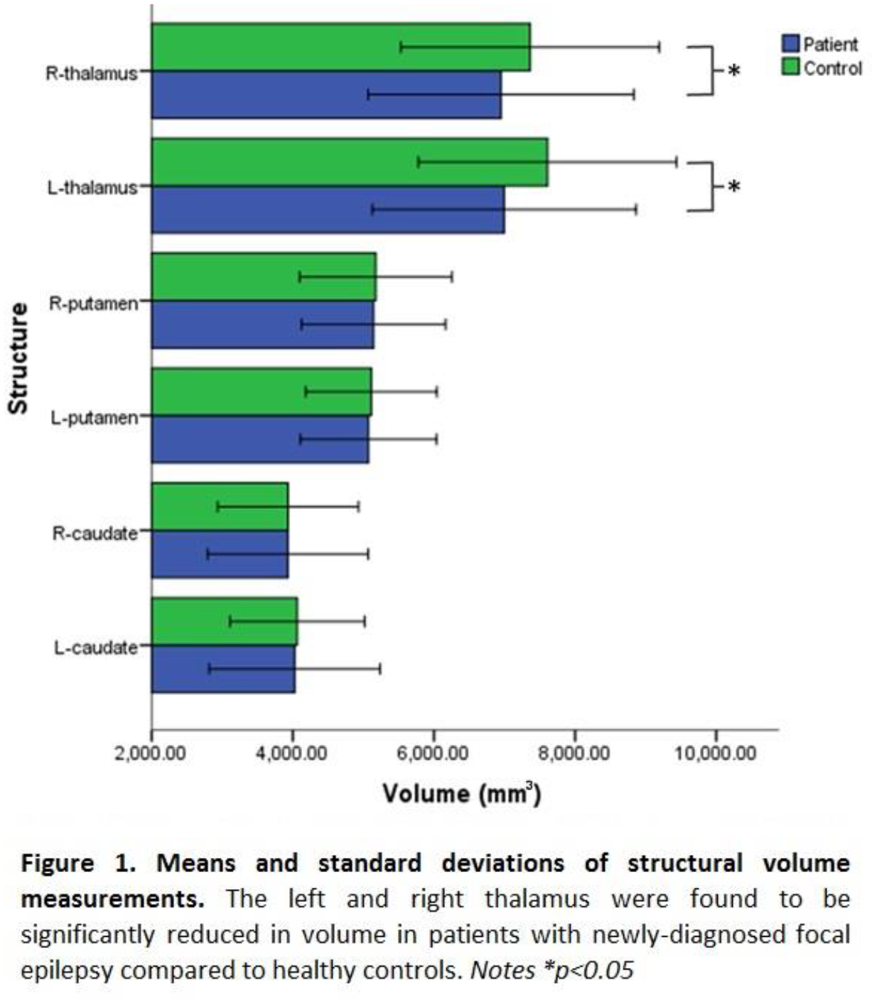
Anxiety-related disorders like phobias and post-traumatic stress are highly prevalent and their treatment using psychological therapies or medications can be ineffective. A promising area of study involves using drugs to enhance exposure therapy to treat these disorders. Cannabidiol, the major non-psychotropic phytocannabinoid of Cannabis sativa, shows broad therapeutic potential for treating anxiety. Evidence from translationally relevant animal models indicates that cannabidiol reduces innate fear and learned fear expression induced by contextual cues while also enhancing contextual fear extinction, which is the psychological process by which exposure therapy reduces fear memory expression. However, the effects of cannabidiol on learned fear expression and extinction related to discrete cues is poorly understood.
Here we investigated the effects of cannabidiol on learned fear expression and its extinction using an auditory fear conditioning paradigm in rats. On Day 0, rats were habituated to contexts A and B. On Day 1, rats underwent tone habituation followed by auditory fear conditioning (tone-shock pairings) in context A. On Day 2, rats were treated with cannabidiol (5, 10, or 20 mg/kg, i.p.) or vehicle before undergoing extinction (tones presented alone) in context B. On Day 3, rats underwent extinction recall testing (tones presented alone) drug-free in context B. Freezing during tone presentations was quantified as the measure of conditioned fear.
We found that 20 mg/kg of cannabidiol significantly decreased freezing during early extinction, compared to vehicle, but there were no drug effects on freezing later on during extinction. Moreover, there were no differences in freezing during extinction recall between the groups the following day. These results indicate that 20 mg/kg of cannabidiol dampened auditory fear expression at the start of extinction without affecting extinction learning or memory consolidation. Taken together, our results showing dampened learned fear expression combined with spared extinction suggest that cannabidiol is an interesting therapeutic candidate for fear management when used as a pharmacological adjunct to reduce anxiety during exposure therapy without interfering with its benefits or having adverse side effects.
Cognitive dysfunction is a hallmark symptom of schizophrenia. Studies in patients with schizophrenia and preclinical animal models have shown a disruption of synchronized high-frequency network activity and a dysfunction of parvalbumin-positive (PV+) GABAergic interneurons, both of which are critical for cognitive processing. Inhibitory fast-spiking PV+ interneurons orchestrate synchronized activity by firing at gamma (30-80 Hz) frequencies and entraining large populations of cortical pyramidal cells. The fast spiking properties and high temporal fidelity of PV+ interneurons are endowed by the selective expression of Kv3.1 potassium channels on these cells. Thus, targeting Kv3 channels, and enhancing the activity of PV+ interneurons, has potential as a pharmacological treatment for schizophrenia.
Using the sub-chronic phencyclidine (PCP) rodent model, we examined the effects of a range of concentrations of a novel Kv3 modulator (1, 3, 10, 20 uM AUT00206) in vitro. Prior to brain slice in vitro studies, animals were behaviourally tested (novel object recognition task) to confirm cognitive deficits in PCP treated rats versus vehicle treated rats. Kainate/carbachol induced gamma oscillations were recorded from prelimbic (PrL) and infralimbic (IL) regions of prefrontal cortical slices obtained from both groups of animals.
Results demonstrate that higher concentrations of AUT00206 (10 and 20 uM) significantly increased the area power of gamma oscillations in the PrL region in slices from PCP treated rats (10 uM: 250 ± 59 uV^2 v. 301.7 ± 88 uV^2, 21.4 ± 8.9%, p = 0.02, n = 10; 20 uM: 148.6 ± 71 uV^2 v. 157.6 ± 59 uV^2, 27.2 ± 10.2%, p = 0.037, n = 10). Slices from vehicle treated animals showed a significant reduction in gamma area power at 20 uM AUT00206 (209.1 ± 99 uV^2 v. 131.7 ± 57 uV^2, -29.5 ± 7.3%, p = 0.016, n = 8). A similar effect of AUT00206 was observed in the IL region, however only the 10 uM concentration produced a significant increase of gamma in the PCP group (305.7 ± 120 uV^2 v. 380 ± 180 uV^2, 14.6 ± 6.6%, p = 0.046, n = 14).
Our results suggest that modulation of Kv3 channels by AUT00206 may have the potential to correct aberrant neuronal oscillations in patients suffering from schizophrenia by augmenting gamma frequency oscillations.
Connexin43, a gap junction protein, is up-regulated following spinal cord injury (SCI) and contributes to secondary lesion spread. Peptide5, a mimetic peptide derived from the second extracellular loop of the Connexin43 protein, has been previously shown to reduce tissue damage and improve functional outcomes when delivered directly to SCI lesion site intrathecally. We have now investigated whether systemic delivery results in similar outcomes. Rats were subject to a 10g, 6.25mm weight drop injury at the vertebral level T10 using a MASCIS impactor. Peptide5 or control scrambled peptide was administrated intraperitoneally post-injury. Rats were then assessed for locomotor recovery and pain hypersensitivity and euthanised at 8 hours (n=8), 24 hours (n=32), 2 weeks (n=32) or 6 weeks (n=32) post-injury. Treatment with Peptide5 led to significant improvements in hindlimb function as assessed using the Basso-Beattie-Bresnahan scale and the error ladder test between 3 and 6 weeks following injury. In addition, there were reductions in at-level mechanical allodynia post-injury. Peptide5 caused a significant reduction in lesion size at all post-injury time points. Immunohistochemistry showed that Peptide5 treatment reduced Connexin43 protein and increased the phosphorylated Connexin43 protein levels at 8 hours after injury compared to the control treatment group. At 2 and 6 weeks following SCI, immunohistochemistry of tissue sections demonstrated reductions in astrocytic (GFAP) and activated macrophage and microglial (Iba1/ED1) responses, as well as an increase in neuronal survival (NeuN) at the dorsal and midline levels, compared to the controls. These results suggest that systemic administration of Peptide5 modulates the pathological opening of Connexin43 hemichannels at the lesion site to ameliorate the secondary damage resulting from SCI and has a significant effect on improving functional outcomes. This preclinical research has successfully demonstrated the therapeutic efficacy of Peptide5 in an animal model, and provides a strong basis for further translational studies.
Early onset neural pathology is characteristic of many inherited lysosomal storage disorders suggesting lysosomal function is essential for the development of the nervous system. However, exactly why lysosomal function is required for neural health is poorly understood. Mutations in the CLN7 gene which encodes a lysosomal transmembrane protein results in Neuronal Ceroid Lipofuscinoses (NCL), an inherited lysosomal storage disorder with late infant-onset neurodegeneration. The biology underpinning the disease is not known but the early onset of pathology suggests roles for CLN7 in neural development. We have taken advantage of the genetic tractability of Drosophila to examine CLN7 function in the development of the larval neuromuscular junction, a well-established model for studying synaptic development and function. We have generated mutations in CLN7 and used gene editing tools to create knock-in reporters to study its function and to identify where it is expressed in the developing fly. We find CLN7 mutants exhibit reduced synapse size, likely due to hyperactivation of mTORC signalling causing downregulation of autophagy. These developmental changes impact on the electrical properties of the synapses and on movement of the animal. Surprisingly, our knock-in reporters reveal CLN7 is largely absent from neurons but strongly expressed in subsets of glia and in the body-wall muscles which form the post-synaptic side of the neuromuscular junction. Knockdown of CLN7 function specifically in the muscle is sufficient to replicate the neurodevelopmental phenotypes of the mutant, suggesting CLN7 may regulate synaptic development via BMP-mediated retrograde signalling.
Neurodegenerative diseases are associated with the accumulation of misfolded proteins. In response to these insults cells generate different responses, including activation of the immediate early genes. However, it is still unclear if these transcriptional activations are protective or if they may exacerbate neurodegeneration.
In Alzheimer’s disease and other dementias aggregates of microtubule binding protein tau are observed. Malfunctioning of tau is one of the main cellular insults in these diseases.
Mutations in tau gene, such as P301L, are responsible for fronto-temporal dementia (FTD) and it is a well-characterized and often used model to study tau pathology.
We are using the transgenic rTg(tauP301L)4510 mouse model of FTD, which overexpresses this mutant tau. Using quantitative PCR and Western blot, we compared the expression of unfolded protein response (UPR) and other stress-related genes in the P301L mice and control mice. Cortical tissue from mice at 6 month of age and 12 month have been used to investigate the levels of stress markers at times points that initiate and progress neuropathology. We are mainly interested in the UPR components (ATF4, XBP1, BiP), activity-induced responses (Arc, c-Fos, c-Jun) and changes related to general insults (ATF3, GADD45) that are commonly found in other disease models.
We have detected no change in the level of expression of UPR components in mice at 6 month of age. In contrast we have observed changes in the expression of some immediate early genes and GADD45. We are repeating this analysis at the later time point to initiate an investigation of the time course of these changes. In particularly we want to probe if the UPR becomes induced by the progressed tau dysfunction.
After identification of the stress-related changes induced in rTg4510 model, we will modulate signalling mediated by stress responses to define if they are neuroprotective or cause neurodegeneration. Describing these intracellular pathways could help to understand the pathology and develop potential treatments for tauopathies.
Recent research has tried to delineate in how far the mechanisms of spatial and temporal orienting can be preserved with aging. Specifically, it has been claimed that older adults cannot use temporal information to improve performance, which was supported by neural evidence suggesting a lack of pre-target temporal orienting (Zanto 2011). In addition, while older adults were shown to exhibit most of the evoked markers of spatial orienting established in younger adults, cue-induced oscillatory lateralisation could not be found (Hong 2015).
We developed a multimodal attention study, where both spatial and temporal information were manipulated. Specifically, we combined behavioural testing with electroencephalographic recording (EEG) and functional magnetic resonance imaging (fMRI) and tested 24 healthy elderly participants.
We show that participants could benefit behaviourally from both spatial and temporal information. EEG analysis yielded significant effects on previously established cue (CNV, ADAN, LDAP) and target (N1 and P3 amplitude; P3 latency shift) evoked and induced (alpha lateralisation) modulations of attention. These results could be supported by regional evidence from fMRI, differentiating task positive and resting state networks.
We suggest that healthy elderly participants can engage in preparatory spatial orienting and extend recent behavioural findings on spared temporal orienting with aging (Chauvin 2016) to the neural domain.
The study serves as a baseline for a second branch of research, where we apply the paradigm to age-matched stroke survivors, assessing whether neural and behavioural signatures of spatial and temporal orienting are affected by focal subcortical and cortical lesions.
Chauvin et al. (2016). Temporal orienting of attention can be preserved in normal aging. Psychology and Aging, 31(5), 442–455.
Hong et al. (2015). Normal aging selectively diminishes alpha lateralization in visual spatial attention. NeuroImage, 106, 353–363.
Zanto et al. (2011). Age-related changes in orienting attention in time. The Journal of Neuroscience?: The Official Journal of the Society for Neuroscience, 31(35), 12461–12470.
The NCLs are a group of rare lysosomal storage disorders, which mainly occur in children and result in severe neurodegeneration and premature death. There are currently no treatments available for any form of NCL and for any therapy to be successful it is imperative to target them to all affected areas. This is especially important for the transmembrane-protein deficient forms of NCL, which cannot be treated via the principle of cross-correction. We have recently found that in CLN1 disease the spinal cord is profoundly affected at a surprisingly early disease stage. We are now extending this analysis of possible spinal pathology to other forms of NCL, including CLN3 and CLN7 disease.
We have undertaken an unbiased stereological assessment of neuron loss, astrocytosis and microglial activation in both Cln7KO and Cln3KI mouse models at different stages of disease progression. Analysing sections of cervical, thoracic and lumbar spinal cord sections has revealed a significant loss of neurons throughout all levels of the spinal cord in Cln7KO mice, as well as a significant loss of white and grey matter volumes. There also was a profound amount of astrocytosis and microglial activation throughout the whole Cln7KO spinal cord, as well as a loss of interneuron populations. Preliminary data from Cln3KI mice reveal less pronounced glial activation, and neuronal cell counts are underway.
Based on these findings, the nature and extent of spinal neuropathology appears to differ between forms of NCL. Defining the precise onset and progression of these changes and their relationship to events in the brain and peripheral nervous system will be important for devising and delivering more efficient therapeutic approaches in these profoundly disabling disorders.
Human post-mortem brain tissue remains one of the most important resources for neuroscience research and its collection and provision is essential if we are to develop new strategies and treatments for neurodegenerative disease. The scientific value of the tissue is greatly increased when accompanied by in depth clinical and neuropathological assessment.
The London Neurodegenerative Diseases Brain Bank has collected over 2000 cases since its establishment in 1989 – comprising formalin fixed and frozen brain and spinal cord samples and frozen CSF. We are constantly updating our procedures to ensure tissue is of the best quality for use in current research techniques, striving to reduce post-mortem delay, limit pH change and maintain DNA/RNA integrity.
We have collections of a wide variety of diseases comprising largely of neurodegenerative diseases, including Alzheimer’s disease, Dementia with Lewy Bodies, Motor Neurone Disease and Frontotemporal dementia. However, we also collect tissue from rarer diseases, such as psychosis, head injury and paediatric disorders. In order to provide comparative control tissue we also house a strong collection of healthy brain and spinal cord tissue. All donations undergo a comprehensive histological examination to provide detailed information on disease pathology.
The brain bank operates a transparent and open-door policy for provision of this tissue to researchers. Tissue requests are reviewed by a request committee and responded to in a timely fashion. In the last five years we have completed over 250 requests and have provided over 120,000 samples to national and international institutions.
We are a founding member of the ‘Brains for Dementia Research’ network – a cohort of over 3000 volunteers which combines longitudinal clinical assessments in life with subsequent brain donation. To maximise the availability and research potential of our tissue we are also part of the ‘MRC UK Brain Bank’ network which aims to encourage and facilitate both tissue donation and tissue accessibility for researchers.
Whilst cognitive decline is common in old age, some older adults retain intact cognitive abilities, but the neural basis of this is unclear. We aimed to provide direct evidence for individual differences in neural mechanisms of superior cognitive ageing. Using literature-based brain metrics, we classified older adults as maintainers (‘youthful’ brain structure & function), adapters (increased function & reduced structure) or decliners (reduced structure & function) relative to younger adults, and predicted superior cross-sectional (CS) cognition and less longitudinal (L) cognitive decline in maintainers and adapters, vs decliners. White matter (WM) integrity was also compared between groups, as preserved WM may underlie higher brain function in adapters.
Method: 343 healthy older adults with L cognitive data from 1997-2013 completed an MRI scan (T1, DTI, fMRI) and further CS cognitive tests. For each subject, grey matter (GM) and resting state (RS) metrics were extracted from regions affected by age in a separate sample of old vs young adults, and classified relative to the mean and standard deviation GM and RS in young adults (Figure 1), resulting in 51 adapters, 96 maintainers and 56 decliners. Cognitive scores (CS & L) and fractional anisotropy (FA) maps of WM integrity were compared between groups.
Results: Contrary to prediction, CS cognition was higher in maintainers than both decliners and adapters. L decline was observed across all subjects, but rate of decline was not significantly different between groups. Decliners and adapters performed worse across time points on short term memory, vs maintainers. FA was significantly higher in adapters than decliners, and was positively correlated with RS.
Conclusion: We identified different brain patterns implicated in superior cognitive ageing, but found only maintainers showed superior cognition, suggesting higher RS activity in adapters is not compensatory. No group difference in L decline suggests adapters and decliners’ poorer performance may reflect lifelong differences, rather than different trajectories of decline. Preserved WM may underlie higher RS in adapters, although whether this reflects a lifelong pattern or a change with age is unclear. Longitudinal imaging will expand on current findings.
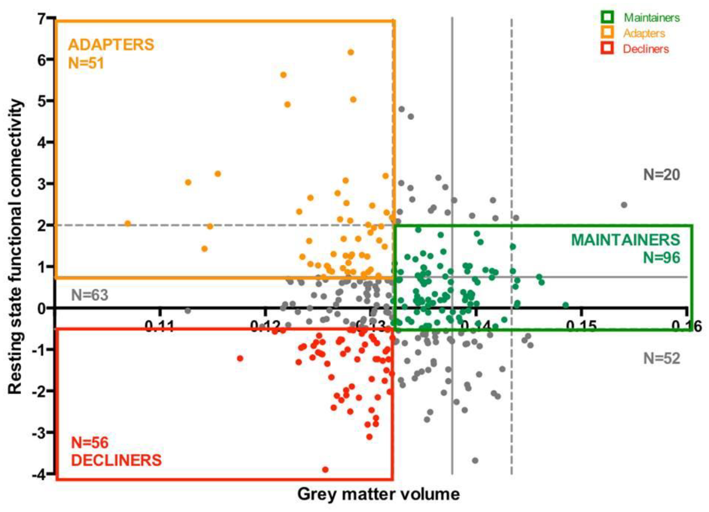
Inflammation is a major contributor to many neurodegenerative disease (Heneka et al. 2015). Microglia, as the resident immune cells of the brain and spinal cord, provide the first line of immunological defence, but can become deleterious when chronically activated, triggering extensive neuronal damage (Cunningham, 2013). Dampening or even reversing this activation may provide neuronal protection against chronic inflammatory damage.
The aim of this study was to determine whether lipopolysaccharide (LPS)-induced inflammation could be abrogated through activation of the receptor Fpr2, known to play an important role in peripheral inflammatory resolution. Immortalised murine microglia (BV2 cell line) were stimulated with LPS (50ng/ml) for 1 hour prior to the treatment with one of two Fpr2 ligands, either Cpd43 or Quin-C1 (both 100nM), and production of nitric oxide (NO), tumour necrosis factor alpha (TNFα) and interleukin-10 (IL-10) were monitored after 24h and 48h.
Treatment with either Fpr2 ligand significantly suppressed LPS-induced production of NO or TNFα after both 24h and 48h exposure, moreover Fpr2 ligand treatment significantly enhanced production of IL-10 48h post-LPS treatment. As we have previously shown Fpr2 to be coupled to a number of intracellular signaling pathways (Cooray et al. 2013), we investigated potential signaling responses. Western blot analysis revealed no activation of ERK1/2, but identified a rapid and potent activation of p38 MAP kinase in BV2 microglia following stimulation with Fpr2 ligands.
Together, these data indicate the possibility of exploiting immunomodulatory strategies for the treatment of neurological diseases, and highlight in particular the important potential of resolution mechanisms as novel therapeutic targets in neuroinflammation.
References
Cooray SN et al. (2013). Proc Natl Acad Sci U S A 110: 18232-7.
Cunningham C (2013). Glia 61: 71-90.
Heneka MT et al. (2015). Lancet Neurol 14: 388-405.
Microglia are the main resident immune cells in the central nervous system. The expansion and activation of microglia is a hallmark of many neurodegenerative diseases including Alzheimer’s disease or prion disease. Colony-stimulating factor 1 receptor (CSF1R) is involved in the control of the microglial proliferation and can be activated by two independent ligands, CSF1 and IL-34. These two ligands display differences in the signalling cascade suggesting a complementary role1. However, although CSF1 and IL-34 are expressed in many organs, IL-34 appears particularly expressed in the developing and adult brain, suggesting that maintenance of the populations of microglia is dependent on IL-34-CSF1R signaling2. Therefore, the aim of this project is the evaluation of novel IL-34 blocking strategies that can be used to modulate microglia proliferation in neurodegenerative diseases by using an in vivo model of neurodegeneration (prion disease; ME7).
Anti-IL-34 blocking antibodies were injected in ME7 mice 12 weeks post-induction of disease. Daily injection of 5-Bromo-2-Deoxyuridine (BrdU) for 1 week was performed, in order to follow proliferation of microglia. One week after IL-34 blockade, immunochemistry analysis of BrdU showed a significant decrease in microglia proliferation in prion mice treated with an anti-IL-34 antibody compared to prion mice treated with an isotype control or with an IL-34 antibody directed against the human protein. Other measures of target engagement were conducted in order to understand the dynamics of IL-34 blockade, including measures of downstream pathways or soluble mediators.
These results provide validation data to support the hypothesis the concept that control of the microglial response through IL-34 blockade could be a potential therapeutic approach in neurodegenerative diseases.
1. Wei S. et al., Functional overlap but differential expression of CSF-1 and IL-34 in their CSF-1 receptor-mediated regulation of myeloid cells. J Leukoc Biol. 2010, 88(3):495-505.
2. Wang Y. et al., IL-34 is a tissue-restricted ligand of CSF1R required for the development of Langerhans cells and microglia. Nat Immunol. 2012, 13(8):753-760.
Mounting evidence suggests the involvement of the immune system in neurodegenerative disorders including Parkinson’s disease (PD) (Benkler et al. 2012; Clin Rev All & Immun. 42:164). We recently reported increased levels of HMGB1 in PD patients as well as in the MPTP animal model of PD (Santoro et al. 2016; Nuerobiol Dis 91:59). In the present study we explored whether the release of HMGB1 in our mouse model of PD caused the activation of the NLRP3 (NOD-like Receptor Protein 3) positive inflammasome. NLRP3-inflammasome is a multiprotein complex, and part of the innate immune system that is activated in aseptic conditions such as tissue damage or metabolic impairment. Its activation leads ultimately to both formation and release of the proinflammatory cytokine IL-1β (Frank et al. 2016; Brain, Beh & Immunity 55:215).
C57BL6J mice were injected with the sub-acute regimen (30 mg/kg/day for five consecutive days i.p., control animals were injected with equivalent volume of saline solution) of the neurotoxin 1-methyl-4-phenyl-1,2,3,6-tetrahydropyridine (MPTP). Brain tissue was harvested 1-2 days post-injection. Tissue was then prepared for double immunofluorescent staining of three different cell types: dopaminergic neurons, astrocytes and microglia, performed on midbrain sections inclusive of substantia nigra, or for western blotting experiments conducted on protein lysate from ventral midbrain.
Our confocal microscopy analysis confirmed an increase in NLRP3 protein levels in the cytoplasm of microglia one day after MPTP injections. In parallel, heightened levels of the microglial MAC-1 protein were confirmed histologically at the level of the substantia nigra and by western blotting. This up-regulation of MAC-l, a surface receptor for HMGB1, may therefore constitute a critical link in the activation of cytoplasmic pathways leading to activation of the NLRP3-inflammasome in Parkinsonism.
Alzheimer’s disease is characterised by the presence of β-Amyloid plaques and neurofibrillary tangles. Despite the increased incidence of this disease due to an ageing population, the exact cause of the disease is still unknown and is of great interest to the scientific and medical communities. There is inherent difficulty in studying the disease in vivo as it does not naturally occur in other species and working with animal models can be too complex to investigate cell biology and intracellular mechanisms. Therefore, we have used human induced pluripotent stem cell (HiPSC) derived cortical neurons cultured in microfluidic devices to investigate the mechanisms underlying the disease.
The microfluidic system used creates an in vitro model relevant to the spread of tau isoforms associated with Alzheimer’s disease. The compartmentalised system involves a series of cell culture chambers connected via an array of microfluidic channels. This system allows two or more neuronal networks to form functional connections with adjacent networks whilst maintaining environmental isolation from each other.
We cultured HiPSC-derived neurons in microfluidic systems for 3-10 weeks prior to functional testing. Cells were derived from both control patients and those with 10+16 MAPT mutation that can lead to the formation of tau aggregates associated with frontotemporal dementia. Cells were cultured both in isolation and co-cultured with astrocytes to determine whether synaptic maturity was accelerated in these co-cultures.
Calcium imaging was used to demonstrate functional connectivity between networks in the microfluidic devices, where a primary network was chemically stimulated in isolation and the adjacent, secondary network exhibited a synchronous calcium response. To investigate tau spread, tau fibrils are seeded on one network in isolation and subsequently their synaptic spread to other networks can be investigated.
Overall, this model for investigating synaptic maturity and tau spread in vitro has the potential to provide new insight into the cellular mechanisms behind neurological disorders such as Alzheimer’s disease. It also provides a convenient on-chip platform for investigating novel therapeutics to help prevent the spread of disease.
The E4 allele of the Apolipoprotein E (APOE) gene is still the strongest genetic risk factor for sporadic Alzheimer’s Disease (AD), whereas the E2 allele of APOE is associated with reduced risk for AD and a later age of onset. The three major ApoE isoforms (ApoE2, ApoE3 and ApoE4) are distinguished by polymorphisms that alter the protein structure and thus function. Although a major role of ApoE in AD involves the modulation of Aß-homeostasis, other mechanisms underlying ApoE-mediated pathology in AD have begun to emerge. Under normal physiological conditions, ApoE is primarily expressed by astrocytes in the brain. However, neurons under stress conditions, which may occur during the early stages of AD, express ApoE. Although this is thought to be a protective response, evidence shows that ApoE undergoes isoform-specific fragmentation in neurons due to different intra- and inter-domain interactions, which generate bioactive fragments that may be toxic. This evidence supports an alternative model, which proposes that ApoE proteolysis in neurons is a key contributor to the development of the disease.
Surprisingly though, very little is known about ApoE processing and the factors that affect it, the identity of the fragments, or the function of ApoE in neurons. Therefore, we examined fragmentation of human ApoE2, ApoE3 and ApoE4 in transfected neuronal-like cells and primary rat neurons under various conditions. Here, we present evidence showing isoform-specific fragmentation for all three ApoE isoforms in vitro. We also show that the fragmentation depends on the model system used. For instance, a 15kDa fragment was unique for ApoE4-expressing primary neurons, but was common for ApoE2 and ApoE4 Neuro-2a cells. Moreover, the composition of ApoE fragments appeared different between neuroblastoma cells and primary rat neurons.
This is of importance because insights into ApoE processing for each isoform may partially explain the different effects these variants have in neurons, but could also provide new targets for preventing the generation of the toxic fragments. Additionally, differences in ApoE processing between cell models highlights the limitations of in vitro model systems in studying ApoE-mediated pathology.
The pathological accumulation of tau is associated with a number of diseases including Alzheimer’s Disease (AD). Tau is predominantly a microtubule stabilizing protein, enabling the replenishment and regulation of the key transport route for axonal cargoes. Tau can become hyperphosphorylated, becoming aggregative and toxic, spreading throughout the brain forming intracellular neurofibrillary tangles. Uncovering the functional elements that could underpin the synapse loss and cell death observed in tauopathies is crucial in slowing down or reversing these diseases. Mitochondria are crucial for neuronal health and maintenance of synaptic function, and have been linked to degenerative pathologies. Mitochondrial dysfunction can lead to changes in ATP production, Reactive Oxygen Species deregulation, disruption in calcium buffering and apoptosis control. These dysfunctional pathways can lead to synaptic damage and cell death. The changes and the time course of mitochondrial function and its relationship to synapse loss in tauopathies, AD patients and animal models is not well known. Here, longitudinal in vivo two-photon microscopy is performed in rTg4510 mice, which express a repressible form of human tau containing the potent P301L mutation. rTg4510 mice and control littermates were transduced with an AAV driving expression of cytosolic & mitochondrial-targeted fluorescent proteins in a subset of excitatory cortical neurons. Repeated imaging of the distribution and motility dynamics of axonal mitochondria was performed in head-fixed, anaesthetized mice. These results show the changes in mitochondria occurring with increasing tau pathology. Transgenic mice show decreased chances of motility and in the ratio of motile mitochondria vs total mitochondria. An initial increase in mitochondrial density along the axon is seen in the rTG4510 mice, followed by a decrease against control mice. An increase the pause ratio of the mobile mitochondria is seen along with an increase in the average pause time. Age related decreases mobile mitochondria speed are also seen in the rTg4510 mice. The data indicates mitochondria may have a key role in the tau related neurodegeneration from an early age.

Parkinson’s disease has been characterised as a synaptopathy that exhibits early synaptic deficits which occur prior to neurodegeneration. However, the mechanisms underlying synaptic dysfunction are largely unknown. Here we investigate mechanisms of α-synuclein induced synaptopathy in a Drosophila model of Parkinson’s disease. Using the Gal4/UAS system, we overexpressed wild type and mutant forms of human α-synuclein in a subset of 30 dopaminergic neurons in the protocerebral anterior medial (PAM) dopaminergic cluster in the adult central brain. Video-assisted motion tracking revealed that α-synuclein expression caused impaired motor behaviour characterised by decreased overall activity and speed, accompanied by increased action initiation and decreased maintenance of motor actions. These early behavioural deficits are neither caused by loss of synaptic arborisations nor degeneration of neurons despite accumulation of α-synuclein, thus suggesting synaptic dysfunction as underlying cause. Accordingly, further investigations are under way to determine whether proteins responsible for the maintenance of the active zone of synapses are altered due to the accumulation of α-synuclein. So far, we found decreased expression levels of nicotinamide mononucleotide adenylyltransferase (NMNAT) in adult flies expressing α-synuclein pan-neuronally. NMNAT has been shown to play an essential role in presynaptic terminals by stabilising Bruchpilot (BRP), which is required for structural integrity and function of synaptic active zones in Drosophila. Further analysis revealed that expression of mutant α-synuclein reduced the number of active zones in the presynaptic region of the Drosophila neuromuscular junction and altered Bruchpilot protein levels in larval brain. Taken together, our results suggest that accumulation of α-synuclein impairs the integrity and function of the presynaptic active zone, leading to synaptopathy and the progressive loss of neurons, thus mimicking early onset and progression of Parkinson’s disease.
Objectives: To examine attrition in a cohort of potential brain donors and identify predictors of withdrawal and lost donation.
Methods: Brains for Dementia Research cohort data (gender, age, diagnosis at registration, withdrawals, donations, lost donations) were analysed to determine attrition and to identify reasons for participant withdrawal and unrecovered brain donations. Logistic regression was used to identify participant characteristics that predicted withdrawal and lost donations.
Results: Of 3287 consented participants (Mean age 78.9 years, SD 8.8), to date 643 (19.6%) have died, including 194 (30.2%) healthy ‘control’ participants. Attrition was 5.8% comprising 105 (3.2%) participants withdrawing during life and 85 (2.6%; 13.2% of deceased) lost donations. Primary withdrawal reasons were ‘no reason provided’ (38, 36.2%), ‘family disagreement’ (20, 19.0%) and ‘participant changing mind’ (17, 16.2%). Reasons for lost donations were ‘brain bank not informed’ (34, 40.0%), ‘Coroner case’ (21, 24.7%) and post mortem delay/’No reason provided’ (7, 8.2%). Being older (OR=1.04, 95% CI: 1.02-1.07, p=<.001) and a dementia diagnosis (OR=2.15, 95% CI: 1.34-3.37, p=.001) increased risk of withdrawal. Being younger (OR=.97, 95% CI: .95-.99, p=.028) and being a control participant (OR=.36, 95% CI: .22-.59, p<.001) predicted lost donation.
Conclusions: Although attrition was relatively low, for cohorts collecting regular clinical data in life, withdrawal and failure to achieve brain donation represents a significant loss, especially from control participants from whom tissue is scarce and in greatest demand. Strategies to maximise participant retention and to minimise lost donations must reinforce the value of donation and enhance engagement with participants and families. However, this does leave a very large active cohort of participants with serial assessment data available to researchers. Furthermore, with more than 500 participants having already donated their brains, there is an extensive tissue bank of well-characterised samples available.
Amyotrophic lateral sclerosis (ALS) is a devastating, adult onset motor neuron disease (MND) that has no effective treatment to date. The current study investigates the possibility of targeting protein aggregation pathway for treatment. Modulation of this pathway is approached through targeting Arfaptin2 protein. A dominant negative form of Arfaptin2 (HC-ARFIP2), has been shown to have a neuroprotective property that maintains the proteasome activity and induces degradation of misfolded proteins. We thus proposed that the HC-ARFIP2 improves neuronal survival in ALS via maintaining the proteasomal pathway. Expression of HC-ARFIP2 in primary motor neuron cultures using LV-based vector, improved motor neuron survival significantly. The prosurvival effect was observable even in cells treated with H2O2 in both SOD1G93A transgenic and non-transgenic motor neurons. A further investigation on the pathway of which HC-ARFIP2 exerts its neuroprotective effect showed that HC-ARFIP2 induces Akt phosphorylation. In addition, protein degradation pathway-markers (p62, LC3II, ULK1) showed significant changes in response to HC-ARFIP2 expression. Furthermore, Arfaptin2 showed colocalisation with SOD1 and overexpression of FL-ARFIP2 caused aggregates formation in HEK293T cells compared to HC-ARFIP2 expression that maintained the cytoplasmic distribution of SOD1. In conclusion, the study presented here has provided a proof of concept that Arfaptin2 is involved in protein aggregation in ALS. In addition, HC-ARFIP2 expression can improve motor neuron survival in vitro through activation of Akt and proteasome activity.
Background: The complexity of the mammalian brain has made it difficult to understand neuronal ageing. As a result, simpler models can be utilised to investigate how neurones age. The invertebrate, Lymnaea stagnalis, is a suitable model due to its relatively simple CNS and ability to perform a top-down approach linking behavioural changes to properties of identified neurones. Importantly, Lymnaea neurones exhibit a number of age-related changes that are observed in mammalian neurons, including reduced excitability and an increase in the afterhyperpolarisation. In this study the mechanisms underlying age-related changes were investigated in the serotonergic cerebral giant cells (CGCs) in Lymnaea.
Methods: Current clamp experiments were performed on CGCs from young (3-4 month old) and old (8-9 month old) Lymnaea. To investigate the role of oxidative stress in neuronal ageing, a group of young Lymnaea CNSs were treated extracellularly with the pro-oxidant generator, 2′-azobis (2-amidinopropane) hydrochloride (AAPH). A lipid peroxidation assay was performed on CNSs to determine malondialdehyde (MDA) levels.
Results: Intracellular recordings revealed a decrease (approximately 50%) in spontaneous firing frequency and a significant increase in the amplitude and duration of the afterhyperpolarisation in both old and AAPH treated CNSs when compared to young controls (p<0.01). Spike frequency adaptation was observed in old CGCs but there was no alterations in SFA in AAPH-treated cells (p>0.05). Interestingly, experiments with AAPH also revealed that decreased firing caused by a low concentration of 3 mM was irreversible and due to lipid peroxidation. Conversely, the effects of 10 mM AAPH on firing was reversible and not associated with elevated MDA levels (p>0.05). The protective effect of 10 mM AAPH on MDA levels was blocked by perfusing CGCs with TEA, a potassium channel blocker (p<0.001).
Conclusion: Extracellular AAPH induces alterations in young CGCs that are largely consistent with age but at low concentrations does so by imparting a pro-oxidant effect and at higher concentrations may confer neuroprotection possibly via the modulation of potassium channels. Future experiments will involve studying potassium currents with age and AAPH in voltage clamp.
Synapses are an early pathological target in a wide range of neurodegenerative conditions including well-known adult onset Alzheimer’s, Parkinson’s and Huntington’s disease [1-3] and diseases of childhood such as the motor neurone disease - Spinal Muscular Atrophy and the neuronal ceroid lipofuscinoses (NCLs; A.K.A Batten disease) [4-6]. However, our understanding of the mechanisms regulating the stability of synapses and their exceptional vulnerability to neurodegenerative stimuli remains in its infancy.
To address this we are using the NCLs as a tool to identify novel regulators of synaptic stability, contributing to our understanding of a broad range of diseases and highlighting novel therapeutic targets. The NCLs, are the most frequent autosomal-recessive disease of childhood [7]. There are currently 14 individual genes which mutations are capable of affecting lysosomal function, all of which result in similar phenotype including blindness, cognitive/motor deficits, seizures and premature death. Mutations in CLN3 underlie a juvenile form of NCL (JNCL or CLN3 disease), the most prevalent variant worldwide [8]. Differential vulnerability of distinct synaptic populations across different brain regions has been described in other models of NCL variants [5, 6] but not yet in JNCL.
Here, we describe a similar pattern of synaptic loss in the Cln3 null mouse model of JNCL (Cln3-/-). Secondly, we use this differential pattern of synaptic loss to map molecular expression profiles across three brain regions. Thirdly, this region vulnerability expression mapping revealed conserved molecular alterations between JNCL and other neurodegenerative conditions [9]. Finally, we demonstrate that genetic and/or pharmacological manipulation of candidate expression in Drosophila is sufficient to modulate disease progression in-vivo.
1. DeKosky ST et al. Ann Neurol., 1990
2. Forno LS et al. J. Neuropathol. Exp. Neurol, 1996
3. Murphy KP et al. Neurosci, 2000
4. Murray LM et al. Hum Mol Genet, 2008.
5. Kielar C et al. Hum Mol Genet, 2009.
6. Partanen S et al. J. Neuropathol. Exp. Neurol., 2008
7. Goebel, HH, J. Child. Neurol, 1995
8. Palmer DN. et al. Biochim Biophys Acta, 2013
9. Wishart TM et al. PLoS Genet, 2012.
Double-strand DNA breaks (DSBs) are the most deleterious form of DNA damage. In young mouse brains they are generated and repaired quickly as part of normal learning but accumulate in the brains of Alzheimer’s disease model mice. DSBs also accumulate in the early stages of Alzheimer’s disease and correlate with reduced cognitive function. We are investigating whether the response of neurons to DNA damage contributes to pathology in neurodegeneration.
We have used genetic methods to reduce the activity of the MRN complex, the evolutionarily conserved tri-partite complex required for detection of DSBs in all cells. We applied the technique to Drosophila models of Alzheimer’s disease, fronto-temporal dementia and Huntington’s disease. We used a standard method of assessing general neural function in these fruit fly models based on quantifying the negative geotaxis of flies as they age and show that reducing MRN complex activity dramatically suppresses neuropathology in each model.
Given the MRN complex is so highly conserved and that reducing activity is neuroprotective in three different models of neurodegeneration, our data suggest small molecular inhibitors of MRN are a potential new therapeutic target to slow neuropathology. We have developed an in vivo screening platform based on our Drosophila assay to test a new library of second-generation targeted small molecule inhibitors of the MRN complex. Compounds emerging that suppress neuropathology will be candidates further testing in vertebrate models of neurodegeneration. We have already identified one protective compound in initial screening and will present updated findings.
Background: Parkinson’s Disease (PD) is a common, progressive neurodegenerative condition with an estimated prevalence of 160 people per 100 000. Approximately 15% of PD is familial or genetic. Although PD is primarily considered a motor disorder, visual changes include reduced visual acuity and reduction in colour vision discrimination. PD patients have reduced dopamine within retinal cells. The fruitfly, Drosophila melanogaster is a useful model for studying PD as both the human and Drosophila visual systems contain dopaminergic neurons. Electroretinograms (ERG) and steady state evoked potentials (SSVEP) have been used to examine the visual system in both humans and flies, with high fidelity readout.
Hypothesis: We aim to test for interactions between Parkinson’s disease related genes. If they are in the same genetic pathway, we would expect to find a more severe phenotype in double mutants, and be able to rescue the phenotype by increased gene expression.
Methods: Crosses were made between adult male flies and virgin female flies to create the required Drosophila genotypes. Flies aged 24 hours were aspirated into pipette tips, and restrained. Glass electrodes containing simple Drosophila saline solution were placed on the surface of the fly’s eye to record response and in the fly’s mouth as a control. ERGs and SSVEPs were carried out using flickering light and response recorded. N=270.
Results: The double mutant TH>Lrrk2-G2019S/parkinz3678 has a worse ERG phenotype than the single mutant (p=0.029), which the SSVEP analysis suggests is due to neural signalling deficits. Expression of PINK1 (TH>Lrrk2-G2019S>PINK1) rescued the visual deficit (p=0.024), but attempted rescue with parkin (TH>Lrrk2-G2019S>parkin) or DJ1 (TH>Lrrk2-G2019S>DJIA) appears to only partially rescue the phenotype, (p=0.46, p=0.12,) due to the small numbers of flies.
Conclusion: Our data suggests that DJ1, PINK1 and Lrrk2 genes are in the same cellular pathway phenotype and suggests that drugs developed for one genetic form of PD may also benefit other patients with genetic PD or even those with idiopathic PD.
Ageing is a complex process influenced by many factors. This project focuses on how stress affects brain ageing. It is well known that having a stressful environment during development induces permanent physiological changes in the organism to maximise survival (e.g. faster growth1 and altered stress hormone receptor expression in the brain2). However, it is not well known how developmental stress influences the ageing trajectory or which developmental period is most crucial for this effect.
Whilst ageing varies hugely between species, there appears to be many shared mechanisms, such as the accumulation of oxidative end products. In time I aim to test the similarities and differences in brain ageing across taxa and the consequences of developmental stress for different animal groups. The data presented here is derived from work on a novel in vitro model of aged human neurons, which has been developed in our lab group. The data demonstrates that with time in culture these neuronal cells accumulate biomarkers of ageing such as increased protein oxidation, lipid peroxidation and decreased antioxidant capacity. Furthermore these cells were manipulated by exposure to a variety of concentrations of cortisol to mimic chronic stress and the consequences of this for the ageing trajectory are presented and discussed. In conclusion, it is clear that stress influences how we age but that this differs across models of ageing.
1. Amugongo, S. K., & Hlusko, L. J. (2014). Impact of Maternal Prenatal Stress on Growth of the Offspring, 5(1), 1–16.
2. Zimmer, C., & Spencer, K. a. (2014). Modifications of Glucocorticoid Receptors mRNA Expression in the Hypothalamic-Pituitary-Adrenal Axis in Response to Early-life Stress in Female Japanese Quail. Journal of Neuroendocrinology, 26(12), 853–860.
Alzheimer’s disease (AD) is a progressive neurodegenerative disorder characterised by a wide range of cognitive and behavioural alterations including memory loss and apathy. Advancing age is one of the biggest risk factors for AD and as such, its prevalence is expected to increase as a consequence of the steady growth of the aging population. Normal aging and age-related diseases are usually accompanied by cognitive decline and structural alterations in brain areas such as the hippocampus and cortex. Converging evidence suggest that age-related GABAergic alterations in the hippocampus and prefrontal cortex may lead to deficits in spatial reference and working memory tasks respectively, while alterations to dopamine signalling pathways may be responsible for alterations in reward learning and motivation.
The present study aimed to investigate whether aged (15-20 months) male Sprague-Dawley rats exhibit any of the behavioural and neurochemical alterations previously associated with normal aging as compared with young (3-8 months) rats. Cognitive and behavioural functions were assessed using Y-maze spatial reference memory, T-maze delayed alternation, and progressive ratio schedule of reinforcement tasks. Neurochemical alterations were investigated at 22 months of age following behavioural testing in the dorsal hippocampus and the nucleus accumbens.
The aged rats performed significantly worse in both the Y-maze spatial reference memory and the progressive ratio tasks. No deficit was observed in the rewarded T-maze delayed alternation paradigm. Behavioural alterations were associated with age-related alterations in GABA neurotransmission in the dorsal hippocampus and DA neurotransmission in the nucleus accumbens as well as reduced DA, DOPAC and HVA basal levels in the aged rats. No other alterations in neurotransmitter function or basal levels were observed in either the dorsal hippocampus or the nucleus accumbens.
Such disruptions to key neuronal pathways in normal aging could be a reflection of the underlying dysfunctional mechanisms and circuits that contribute to symptom onset in AD. Future studies will extend neurochemical profiling to other brain regions to better understand age-related circuit and network dysfunction that may be beneficial to AD research.
Neurodegenerative diseases in which tau isoforms containing four microtubule-binding repeats (4R) are overrepresented, such as progressive supranuclear palsy, contain an N-terminally truncated form of tau (Tau35) that is absent from age-matched control brain. We have generated a new mouse model of tauopathy expressing Tau35, in the absence of any mutation and under the control of the human tau promoter (Bondulich et al., 2016). Unlike most existing tau transgenic mice, expression of Tau35 in these animals comprises less than 10% of endogenous mouse tau, which is comparable with tau expression in human neurodegenerative disease. Importantly, Tau35 mice demonstrate key features of human tauopathy, including aggregated and abnormally phosphorylated tau, progressive cognitive and motor deficits, altered protein kinase activity, loss of synaptic proteins, and reduced life-span. Western blots of mouse brain homogenates revealed a reduced amount of the mitochondrial marker, heat-shock protein 60 (HSP60) in Tau35 mice, compared to wild-type mice. To investigate the influence of Tau35 expression in neurons, we examined mitochondrial morphology and mobility in primary cortical neurons cultured from Tau35 and wild-type mice. In order to assess mitochondrial mobility, neurons were co-transfected with plasmids expressing (1) the mitochondrial targeting protein, cytochrome c oxidase, fused to DSRed2 and (2) enhanced green fluorescent protein. Live recording of mitochondria in cultured neurons identified a significant reduction in the total number of mitochondria present in neurites of neurons prepared from Tau35 mice, compared to those derived from wild-type mice. However, the percentage of moving mitochondria was similar in both Tau35 and wild-type neurons. The results suggest that defective mitochondrial function may be critically involved in the development and progression of tauopathy in Tau35 mice. This emulation of disease pathogenesis in neurons derived from a novel mouse model will aid identification of the molecular changes that cause neurodegeneration in the human tauopathies.
Alzheimer’s disease (AD) is the most common age-related dementia. Neuroinflammation plays a key role in the pathophysiology of neurodegenerative diseases, including AD. In this study we assessed the contribution of the neuroinflammatory response to the pathology associated to tau hyperphosphorylation and accumulation. We used transgenic mice expressing human P301S tau protein that exhibit many characteristics of the human tauopathies, including the formation of abundant hyperphosphorylated tau protein filaments and neurodegeneration. We found that P301S mice develop predominant spinal cord pathology, with altered locomotion. Tau accumulation is evident in spinal motoneurons at 6 weeks of age, leading to cell degeneration from 12 weeks of age. This is accompanied by a significant expansion of the microglial population and increased expression of pro-inflammatory cytokines, with no evidence of a contribution from infiltrating cells. Overall, our findings demonstrate that neuroinflammation significantly contributes to the pathology in P301S mice, and suggest that strategies aiming at controlling this process may represent a promising therapeutic perspective.
Background: Aggresomes are closely related to Lewy bodies (LBs), structures whose presence is a hallmark of Parkinson’s disease. LBs are related to aggresomes. A major constituent of these LBs is alpha synuclein, the gene for which is mutated in the inherited form of Parkinson’s. The cellular location of aggresomes coincides with that of the centrosome, the microtubule organising centre of the cell. Since the aggresome is positioned in close proximity to and shares components with the centrosome, it is possible that it hinders centrosomal function. If that is the case it could in turn affect intracellular transport and cell polarity, both of which are very important for neuronal function and survival.
Objectives: In this study we sought to test if any functions of the centrosome were impeded by the presence of aggresomes in their close vicinity.
Methods: Several cell lines including SH-SH5Y, HeLa cells and primary rat neurons were treated to induce aggresomes. This was achieved either by exposure to the proteasome inhibitor MG-132 or by transfection with alpha synuclein overexpression constructs. Centrosome function was assessed by microtubule regrowth, wound healing and ciliogenesis assay. Similarly zebrafish embryos were exposed to MG-132 were cilia at the olfactory neurons were stained for.
Results : We show here that aggresomes severely compromise centrosome function. Microtubule nucleation is severely reduced and the centrosome is unable to be repositioned during cell migration. We also show that aggresomes can prevent a cell from turning its centrosome into a cilium. Also in zebrafish embryos number of cilia in the olfactory epithelium is severely reduced.
Conclusion: Defects in the generation and organisation of the microtubule network would be predicted to affect intracellular transport. As well as the deleterious effects on core vesicular trafficking, the inability to move vesicles involved in neurotransmitter transport through the cell would be particularly problematic for neurons. An early symptom of Parkinson’s is loss of smell and anosmia. It maybe possible this symptom is a result of loss of cilia from olfactory neurons. If this is the case, then cilia density in the olfactory epithelium of Parkinson’s patients should be reduced.
Current knowledge of the structure of myelin has been mainly derived from the use of static electron micrographs, although these are useful to derive structural information, these do not provide the needed information to understand the behaviour of myelin during dynamic events such as the formation and repair. Understanding of these events relies upon more comprehensive direct observation of the microenvironments. Typically, these observations involve the use of conventional ensemble –averaged imaging of the molecular properties within the system. However, these ensemble-averaged observations can fall short when observing events which only occur in a sub-population of the system. In order to derive information on these events the full probability distribution of the different states of the molecules need to be observed, providing a need for single molecule imaging.
To aid in this observation a fluorescent construct was designed using a photoswitchable fluorescent protein bound to myelin basic protein, which was instilled in the semliki forest virus. This virus was used to infect Oligodendrocytes within both brain slices and within the Oli Neu cells, oligodendrocyte cell line. Images were obtained over a 5-minute interval on a TIRF microscope, and were analysed using single molecule analysis software. Analysis of particle tracks revealed that MBP diffuses around processes within Oli Neu cells, which do not have the complement of proteins a mature oligodendrocyte cells contain, however, results thus far suggest this is not the case when observing mature cells.
Presence of cytoplasmic protein aggregates called Lewy Bodies (LBs) in the brain of Parkinson’s disease (PD) patients represents one of the main pathological hallmarks. These aggregates are composed of α-synuclein, an abundant presynaptic protein critical for synaptic transmitter release. LBs have been linked with neuronal loss, microglia activation and neuroinflammation. In addition, mounting evidence suggests that the humoral immune response driven by T lymphocyte plays a role in disease progression in PD. Previously reported is the putative influence of alpha-synuclein on the adaptive immune response, which involves the infiltration of cytotoxic CD4 and CD8 positive T-cells in the substantia nigra, and leads to the subsequent activation of these cells and the production of pro-inflammatory cytokines. A long term corollary then is the accumulation of neurotoxins and eventually the neurodegeneration of neurones. In the present study we focused our attention on the presence of LBs and alpha synuclein in post-mortem brain tissue from PD patients; once these were recognised, we explored the presence and compartmentalisation of immune cells, in particular, microglia and T-lymphocytes, in the vicinity of aggregates.
Paraffin embedded brain human tissue from PD patients (striatum and substantia nigra) obtained from the MS and Parkinson’s Tissue Bank (Imperial College London) was analysed through immunohistochemistry. Immunostaining was performed on 5 μm-thick sections, incubated with different primary antibodies for 48h at 4°C. Biotinylated secondary antibodies were amplified and immunostaining confirmed using 3,3’-diaminobenzidine tetra-hydrochloride (DAB). These yielded clear synuclein-containing cell labelling in both the soma and processes of striatal neurones. These are most likely medium spiny neurones diffusely localised in the striatum. We currently explore the presence of immune cells through double labelling with T-cell specific antibodies. These preliminary qualitative data confirm the presence of pathological alpha synuclein and T-cells in the brain parenchyma of PD patients. Future work will seek to establish a correlation between α-synuclein and inflammatory response using the injection of patient-extracted LBs into the brain of mice as a model.
Multiple Sclerosis (MS) is a complex immune-mediate disease of the CNS, characterized by demyelination, axonal damage and scar formation. MS is one of the most widespread disabling neurological conditions around the world with more than 2 million people being affected. It includes several clinical subtypes, with the most common form being Relapsing Remitting MS (RRMS). Due to the heterogeneity of clinical presentation, MS is difficult to diagnose and therefore efforts have been directed towards the identification of biomarkers to facilitate diagnosis and improve treatment, but despite the large number proposed, only few are currently used for clinical purposes. The aim of this study is to identify molecules that can be measured in blood and provide a simple and cost-effective tool for improving MS diagnosis and treatment, as well as delineating between the MS subtypes.
For our initial investigation, we performed a gene expression analysis on a pilot set of blood samples obtained from RRMS patients and age- and gender-matched controls (n=20) using the Affymetrix GeneChip Human Transcriptomic Array 2.0. We identified 8721 significantly differentially expressed genes (p ≤ 0.05) that were then analysed through Ingenuity Pathway Analysis (IPA) to explore associated functions and pathways. We then employed a combination of qPCR and HPLC/Mass Spectrometry in a larger sample set to validate and further investigate our data.
We have identified a subset of molecules that are differentially expressed in RRMS and may present novel targets for MS diagnosis and treatment. These candidates include molecules involved in neurological and inflammatory processes, as well as novel targets such as microRNAs. For example, DEFA4, an antimicrobial peptide known to promote local inflammation, was significantly up-regulated, while NOTCH1, which mediates oligodendrocytes differentiation and remyelination, was significantly down-regulated.
The identification of MS biomarkers in the blood could facilitate an early diagnosis, allowing clinicians to apply treatment strategies more effectively. The MS-related molecules identified in this study, in fact, could be used to discriminate between MS and other inflammatory diseases and could potentially function as novel targets for drug development.
Nuclear receptors comprise a major group of signalling pathways in the brain. They have a well described mechanism of action to regulate gene expression. Additional “non-genomic” roles exist for many of these receptors such as the retinoic acid receptor alpha (RARα). Retinoic acid mediates a type of homeostatic plasticity by regulating the translation of the GluR1 subunit of the AMPA receptor through RARα (1). In the absence of retinoic acid, RARα binds with GluR1 mRNA directly preventing its translation. During synaptic scaling, blockade of synaptic activity triggers retinoic acid synthesis which in turn binds to RARα and releases the GluR1 mRNA to be translated which causes an increase in the postsynaptic AMPA receptor levels. Retinoic acid also regulates AMPA receptor trafficking (2).
The question was asked whether this non-genomic activity of RARα could be disassociated from its action as a transcription factor and whether different ligands for RARα may preferentially activate one pathway or the other. A bioluminescence reporter plasmid that expresses firefly luciferase under the control of the GluR1 5’ untranslated region was constructed and introduced into SH-SY5Y cells. The cell line is being used to screen synthetic retinoids for their ability to increase AMPA receptor translation versus activation of gene transcription. The results will indicate whether RARα ligands may be selected of high specificity for their capacity to increase AMPA receptor levels and which may provide therapeutics with fewer side-effects for disorders that result in cognitive loss, including Alzheimer’s disease (3).
1. Aoto J, Nam CI, Poon MM, Ting P, Chen L. Synaptic signaling by all-trans retinoic acid in homeostatic synaptic plasticity. Neuron. 2008 Oct 23;60(2):308-20
2. Arendt KL, Zhang Y, Jurado S, Malenka RC, Südhof TC, Chen L. Retinoic Acid and LTP Recruit Postsynaptic AMPA Receptors Using Distinct SNARE-Dependent Mechanisms. Neuron. 2015 Apr 22;86(2):442-56
3. Chakrabarti M, McDonald AJ, Will Reed J, Moss MA, Das BC, Ray SK. Molecular Signaling Mechanisms of Natural and Synthetic Retinoids for Inhibition of Pathogenesis in Alzheimer’s Disease. J Alzheimers Dis. 2016;50(2):335-52.
Prostaglandin-endoperoxide synthase (PTGS) catalyses the first step in the synthesis of prostanoids; consisting of prostaglandins, prostacyclin and thromboxanes. Both constitutive and inducible isoforms exist. PTGS2 is the inducible isoform, which is dramatically upregulated in response to pro-inflammatory molecules. Several lines of evidence point towards neuroinflammation as critical to the pathophysiology of Parkinson’s disease (PD). Epidemiological studies suggest risk reduction of PD with non-steroidal anti-inflammatory drug use. Inhibition of PTGS2 is neuroprotective in animal models of PD. Here, we quantified the expression of PTGS2 by Western blotting in mitochondria of post-mortem cortex tissue of individuals with PD and age and Braak stage matched controls. Further, we investigated the cerebellar cellular localisation of PTGS2 expression using immunohistochemistry. Our data suggest that PTGS2 levels are elevated in the cortex of individuals with Parkinson’s disease and distinct changes in the cerebellar cellular localisation of PTGS2 expression exist. We will present our latest work aiming to determine whether changes in PTGS2 are driving, or a consequence of Parkinson’s disease.
From a practical standpoint, biomarker data such as genetics and brain imaging are less economical or not commonly obtained in typical dementia care. Further, some assessments can take too long to evaluate within limited consultation time. This study aims to focus on more easily accessible data to obtain an optimal set of assessments for an individual by balancing classification accuracy and time spent.
Our previous work using the Australian Imaging Biomarkers and Lifestyle flagship study of ageing (AIBL) has shown that psychological/functional assessments provide higher classification accuracy than other data features. Hence, we emphasize on the clinical dementia rating (CDR), mini-mental state exam (MMSE), logical memory immediate/delayed recall (LMIR/LMDR) assessments, age, and 2 significant medical histories: neurologic history (NEURL) and renal history (RENAL). We first used Bayesian network (BN) modelling approach to identify probabilistic causalities among various data types. While applying 10-fold cross validation, a synthetic minority over-sampling technique was used to balance the unbalanced training data due to uneven proportion of diagnostic categories. The obtained BN showed a sensitivity of 0.79 and specificity of 0.97 for AD diagnosis, and the total accuracy of 0.89 for all classes of diagnosis. Diagnostic result was most strongly linked to CDR, followed by LMDR, MMSE, and LMIR.
The current experiments examined the role of the retrosplenial cortex in distinguishing the temporal order of events (i.e. recency memory). In Experiment 1, rats with lesions in the retrosplenial cortex (RSC) were tested on two types of recency memory task: Between-Block (i.e. objects were presented in two discrete blocks) and Within-Block (i.e. objects were presented in a continuous series). The RSC group were able to discriminate old from recent objects in the Between-Block condition but not in the Within-Block condition. In Experiment 2, the expression of the immediate-early gene c-fos in retrosplenial cortex was compared between groups of intact rats following either the completion of a between-block recency task or a control task. There were strong, positive correlations between discrimination performance and the levels of c-fos expression in both the granular and dysgranular retrosplenial cortex. Expression of c-fos in the granular retrosplenial cortex also correlated with expression in related areas, such as the ventral subiculum and prelimbic cortex. Taken together, the pattern of results supports a role for the retrosplenial cortex in both between-block and within-block recency problems. Furthermore, when viewed in the context of previous findings, these results suggest that the rat retrosplenial cortex is part of a group of anatomically and functionally connected regions, including the hippocampal formation, medial diencephalon, and medial frontal cortex, that work together to support recency memory.
Present-day medicine takes advantage of modern radiation diagnostics like computer tomography (CT) or curative and palliative radiotherapy, respectively. These treatments are also increasingly used in young children, although their developing brain is exposed to low dosages of radiation. Already after doses of about 50 mGy, as it is used in CT scans of the head, it is possible to detect DNA double-strand breaks in the brain (Saha et al. 2014). We are able to predict long-term effects of an in utero irradiation with higher dosages (> 1 Gy) relatively well from atomic bomb survivors of Hiroshima and Nagasaki (Otake and Schull 1998). Besides the carcinogenic risk, various sequelae like abortion, malformations or mental retardation dependent on the developing status of the embryo during irradiation can occur. Especially, impaired cognitive function could already emerge from dosages < 1 Gy, which is of great social relevance. Consequences of postnatal irradiation on cognitive abilities are less well known and there is still need of information and clarification existing. European radiation protection authorities have encouraged research activities for this issue (Averbeck 2013). Here, we demonstrate the long-term effects on spatial learning behaviour in C57BL/6 mice whole-body irradiated (x-rays) during postnatal brain development (postnatal day 10) and analyzed at 2 months of age. We show that a 500 mGy radiation dose led to longer swimming paths, increased escape latencies and decreased percentage of spatial searching strategies in the Morris Water Maze. Moreover, probe trial tests revealed diminished retention times in the target quadrant and increased distances to the former platform position. Differences to control group were not based on altered motor coordination or fear/exploration behaviour, as demonstrated in the Rotarod and Elevated Zero Maze. Besides further doses, current immunohistochemical analyses of brain sections are designed to reveal possible effects on the neurogenic niche in the dentate gyrus. We suppose that the vulnerability of postnatal brain development is caused by disturbance of the local proliferative microenvironment through low-dose radiation, resulting in the manifestation of hippocampus dependent cognitive impairment in later life.
Leading theories of learning and memory propose that the strengthening of specific synapses involves communication between pre- and post- synaptic terminals and local protein synthesis. Neuronal cadherin proteins (N-cadherin) have been implicated in synaptic plasticity due to their trans-synaptic localization, persistent expression at mature synapses, and ability to recruit AMPA receptors to the post-synaptic membrane. One of the properties of polyribosomes, the site of local protein synthesis in neuronal processes, is that they move into dendritic shafts and spines in response to plasticity events. Based on this information, we hypothesised that N-cadherin abundance at synapses could be used as an indicator of early plasticity protein synthesis. Using differentiated neuronal human cell lines and fixed human brain tissue, we performed immunofluorescent (IF) staining and confocal microscopy to visualise localization and measure the abundance of N-cadherin and ribosomal proteins at dendritic spines. Furthermore, we explored protein expression patterns in differentiated cells treated with protein translation inhibitors. Visualising these cells and quantifying N-cadherin at dendritic spines provided information about the sequence of events in post-synaptic plasticity. This study presents further support for a role for N-cadherin in synaptic plasticity and raises additional questions about the molecular mechanisms through which cellular adhesion proteins may impact upon learning and memory processes.
Lymnaea stagnalis is a model organism for investigating learning and memory formation. Its aerial respiratory behaviour can be operantly conditioned and the resulting memory can be traced to a well-characterized neuronal network. This memory is enhanced by various environmental stressors including detection of a predator and increased environmental temperature (i.e. thermal stress). Similar to conditioned behaviours in mammals, this memory can be both reconsolidated and extinguished. Additionally, there is evidence for the conservation of various cellular and molecular processes (e.g. protein phosphorylation, retinoid signaling) between vertebrates and invertebrates. Recently, epigenetic changes (e.g. DNA methylation) have been investigated as modulators of memory formation. Interestingly, DNA methylation is required for stress-induced memory enhancement in Lymnaea. Specifically, treatment with a methylation inhibitor (5-AZA) 1 hour before exposure to i) the scent of a predator or ii) a thermal stress, prevents memory enhancement. Here, we aimed to determine how long the memory-inhibiting effect of 5-AZA persists. Animals were treated with 5-AZA 24 hours before exposure to the scent of a predator or a thermal stressor and were then operantly conditioned. Neither group of animals displayed memory enhancement, indicating that the action of 5-AZA persists for at least 24 hours. We next aimed to further describe the involvement of DNA methylation in ‘normal’ (i.e. non-enhanced) memory. In order to examine whether DNA methylation is required for memory reconsolidation Lymnaea were treated with 5-AZA immediately before memory reactivation. All animals demonstrated memory reconsolidation, suggesting that DNA methylation is not necessary for the reconsolidation of ‘normal’ memory. Thus, DNA methylation appears to be necessary for memory enhancement, but not for the expression or maintenance of ‘normal’ memory. Together, these studies further elucidate the involvement of epigenetic changes in invertebrate memory and provide insight into the conservation of these mechanisms between vertebrate and invertebrate species.
Parkinson’s disease (PD) is classified as a movement disorder. However there is an increasing awareness of the significant non-motor burdens experienced by patients. For example PD patients experience cognitive difficulties, including memory deficits. As such there is a pressing need to better categorize and investigate the nature of these deficits, to guide the development of interventions that might ameliorate them. Electrical activity recorded from the brains of PD patients is excessively synchronised within the beta range (13–30Hz) compared to healthy controls. Evidence suggests that abnormal beta synchronisation is the cause of at least some of the motor symptoms of PD. However almost nothing is known about the relationship between the increase in beta activity and non-motor symptoms. Our research investigates whether there is a direct relationship between hyper-synchronised beta activity and the memory deficits experienced in PD. It has been shown in healthy adults that a greater amount of beta de-synchronisation occurs during deep-encoding for words that are subsequently better remembered than for words that are not. It is thought that this beta de-synchronisation is necessary for successful encoding to form a memory of the word. We hypothesised that hyper-synchronisation in the brains of people with PD prevents the necessary de-synchronisation of beta oscillations during encoding and therefore interferes with memory formation. Electroencephalography was recorded during an established memory-encoding paradigm to examine the brain activity of PD patients and healthy controls during memory formation. We will report on the ability of PD patients to recollect words placed in memory compared to healthy controls. We will also report on the association between the ability to remember words and the extent of beta de-synchronisation during deep-encoding. This will be the first presentation of preliminary findings from a novel, timely and important investigation into the relationship between hyper-synchronised beta activity and the memory deficits experienced in PD. If such a relationship exists, this evidence would lend further weight to the hypothesis that hyper-synchronised beta activity is causal to the symptoms of PD.
The use of large-scale data in neuroscience, for example functional neuroimaging studies with 1,000 participants, is important in enabling understanding of how brain systems operate in health and disease, by allowing voxel-level resolution (Cheng, Rolls et al 2016, Rolls 2016). Here we extend this approach to the analysis of the voxel-level functional connectivity of a brain structure with all other voxels in the brain in a large dataset of healthy humans. In this investigation we measured the resting state functional connectivity (the Pearson correlation) between every amygdala voxel (3x3x3 mm) with every other voxel in the brain in 488 healthy participants. Significant functional connectivities between every pair of voxels were corrected for multiple comparisons (Bonferroni or FDR). It was found that the amygdala has significant functional connectivity in humans not only with some expected regions including the medial and lateral orbitofrontal cortex, pregenual, subgenual and supracallosal anterior cingulate cortex; superior, middle and inferior temporal gyrus; medial temporal lobe regions including the perirhinal cortex; but also with the precuneus. The latter is of interest, for its functional connectivity of the precuneus with the lateral orbitofrontal cortex is increased in depression (Cheng, Rolls et al 2016), and we have now report that the functional connectivity of the amygdala with the precuneus is increased in depression. The method also shows which voxels in the amygdala have functional connectivity with different cortical areas, and some topological organization is evident, in that for example the medial orbitofrontal cortex and precuneus have significant functional connectivity with different but partly overlapping voxels in the amygdala. The approach described here provides a way of analyzing and understanding the connectivity of the healthy human brain at the voxel level.
Rolls,E.T. (2016) Cerebral Cortex: Principles of Operation. Oxford University Press: Oxford.
Cheng, W., Rolls,E.T., Qiu, J., Liu, W., Tang, Y., Chang, M., Wang, XF., Jie,Z., Lin, W., Zheng, L., Pu, JC., Tsai, S-J., Yang, A.C., Lin, C-P., Wang, F., Xie, P. and Feng,J. (2016) Orbitofrontal cortex circuits change in depression. Brain 139: 3296-3309.
Introduction
Memory deficits are a common cognitive consequence of traumatic brain injury (TBI), and are characterised by abnormal encoding (1). Successful encoding in healthy individuals is associated with recruitment of the dorsal attention network (DAN) and suppression of the default mode network (DMN) (2). TBI patients show abnormalities in the activity of these networks (3) and their interactions (4). We investigated the neural correlates of successful encoding in healthy controls and TBI patients, hypothesising that memory impairment following TBI would be associated with abnormal BOLD signal within the DAN and/or DMN
Methods
37 TBI patients (11 females, age= 42.84) and 16 healthy controls (6 females, age = 38.19) underwent fMRI while viewing abstract images that subjects were asked to remember. Encoding was tested outside the scanner. A median split of performance (d-prime) divided patients into High (HP) and Low Performance (LP) TBI groups. fMRI data were processed using FSL (5). General linear modelling was performed with correct and incorrectly encoded trials included as regressors. Higher level contrast included age as a nuisance variable.
Results
The LP TBI group (n=19; age=45.74) showed abnormally low performance (mean=57.30) compared to the healthy control group (mean=72.51; p<0.01) with no abnormality in the HP TBI (n=18;age=45.74) group (Fig. 1a). Successful encoding was associated with extensive activation within the DAN, as well as expected activation in the ventral visual stream and medial temporal lobes. Reduction of activation was seen in the DMN (Figure 1c). For successful encoding the contrast of HP TBI > LP TBI showed increased activation in the left DAN (parietal lobe) as well as left anterior insula and bilateral medial temporal lobes (Figure 1c).
Conclusions
Patients with memory encoding deficits after TBI show reduced activation within the DAN and medial temporal lobes, suggesting that chronic memory impairment after TBI may be associated with a failure of the attentional control of memory encoding.
References
1. Arenth et al., 2012 - J Clin Exp Neuropsychol
2. Kim 2015 - Hippocampus
3. Sharp DJ et al., 2014 - Nat Rev Neurol
4. Han K et al., 2016 - J Int Neuropsychol Soc
5. Smith et al., 2004 – Neuroimage.
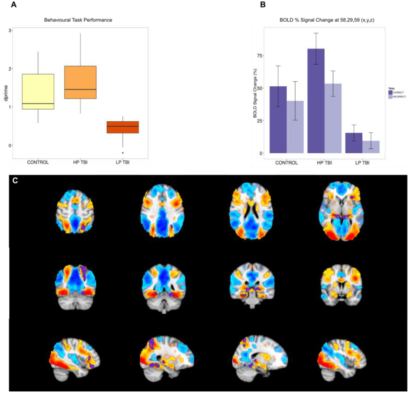
Serotonin is involved in the rapid (within hours) and reversible transformation of the Desert Locust (Schistocerca gregaria) between two different phenotypes (phases) - a process dependent on population density. At low density, locusts develop into their solitarious phase, which are slow moving, mainly night active and repelled by conspecifics. With increased density, however, locusts develop into their gregarious phase which are more active and are attracted to other locusts. Gregarisation is mediated through the repeated mechanical stimulation of the hind leg which has been shown to transiently increase serotonin in key neurons of the thoracic ganglia. However, the specific role of serotonin in regulating phase state is yet to be elucidated. We used in vivo fast scan cyclic voltammetry to measure the release of serotonin in the metathoracic ganglia following discrete mechanosensory stimulation. To mimic mechanosensory stimulation from crowded locusts, brush strokes were used to stimulate the hind leg and other non-gregarising sites. The recording electrode was positioned in the metathoracic ganglia, close to the serotonergic neurons. We used serotonin-specific ‘N’-voltage waveform to distinguish the serotonin signal from that of other neuromodulators. The amplitude of the oxidation peak of the voltammogram and its latency from stimulus onset were used as measures of serotonin release. In both solitarious and gregarious animals, hind leg stimulation caused a rapid large amplitude release of serotonin due to direct mechanosensory input. By comparison stimulation of the antennae elicited a delayed release of serotonin due to possible indirect descending inputs. Whilst a transient increase in metathoracic serotonin has previously been shown to initiate phase change, our data shows that serotonergic release may also remain involved in the transient responses of gregarious locusts to further gregarising stimuli. This methodology provides novel insight into the rapid dynamics of serotonin release thought to be key in transforming locusts into their swarming state.
The medial mammillary bodies and its projections to the anterior thalamic nuclei via the mammillothalamic tract (MTT) are needed for normal hippocampal and retrosplenial cortex function. MTT lesions produce ‘covert pathology’ in these distal regions as evidenced by a reduction in markers of neural activity (e.g. the immediate early gene, c-fos). However, it is not known whether these functional changes are accompanied by, or even the result of, structural changes at the dendritic level. To address this, the current study examined dendritic arbor complexity in CA1, dentate gyrus, and retrosplenial granular b cortex (Rgb) and spine density in CA1 and Rgb after bilateral MTT lesions. Rats with MTT lesions (n=9) and sham operated controls (n=11) were tested on a reinforced T-maze alternation task to confirm the efficacy of the lesions. Subsequently, the brains were removed, treated with Golgi-cox stain, and blinded Sholl analysis and spine density counts of three-dimensional image stacks were performed on dendritic arbors. Rats with MTT lesions were impaired on the T-maze task, thus confirming the success of the lesion. Spine density counts showed the MTT lesions significantly reduced CA1 spine density but did not influence the number of intersections in CA1, dentate gyrus, or Rgb after Sholl analysis. Work on Rgb spine density is ongoing and will be presented. Continuing work analysing protein levels of the immediate early gene Arc and brain derived neurotrophic factor (pro-BDNF and mature BDNF forms) in the hippocampus and retrosplenial cortex using Western blotting after MTT lesions will also be presented. Our findings suggest that damage to the MTT causes microstructural changes in the hippocampus as shown by reduced spine density in CA1. These findings provide novel evidence of the importance of ascending mammillary body projections for hippocampal integrity and our additional work will expand our understanding of the distal effects of MTT lesions.
The reactivation of a previously-learned memory, in addition to enabling expression of that memory, can lead its destabilisation. Memory destabilisation necessitates subsequent reconsolidation of the memory in order to restabilise it and integrate new information. Disruption of the reconsolidation process, therefore, results in experimental amnesia that might be harnessed translationally to diminish traumatic memories. However, the success with which memory reactivation leads to destabilisation is highly variable. Therefore, strategies to enhance memory destabilisation would be beneficial. Here, we explored potential pharmacological adjuncts to memory reactivation in order to stimulate tone fear memory destabilisation under conditions that do not normally lead to reconsolidation. First, we established that post-reactivation systemic administration of the glucocorticoid antagonist mifepristone (30 mg/kg, s.c.), but not the beta-adrenergic antagonist propranolol (10 mg/kg, i.p.) impaired the reconsolidation of a mildly-conditioned fear memory. Mifepristone also impaired the reconsolidation of a strongly-conditioned fear memory, but only when preceded by systemic injection of the nootropic nefiracetam (3 mg/kg, i.p.) 1 hr prior to memory reactivation. Administration of mifepristone or nefiracetam alone had no observable effect on subsequent fear expression. In contrast, systemic injection of the D1 receptor agonist SKF38393 (5 mg/kg, i.p.) immediately prior to memory reactivation had no impact on cued fear memory destabilisation or reconsolidation. It remains to be determined which target(s) of nefiracetam mediate the destabilisation enhancement observed. These targets include L-type calcium channels and cholinergic receptors, both of which have been implicated in memory destabilisation.
Desert locusts (Schistocerca gregaria) transform between two extreme phenotypes (‘phases’) depending on population density. Isolation from conspecifics results in a behaviourally cryptic solitarious phase, whereas crowding leads to an active gregarious phase. Behavioural gregarisation can be induced in the laboratory within 2h of crowding, so S. gregaria provides a useful model for analysing mechanisms underlying phenotypic plasticity. Previous studies have reported that the amount of serotonin (5-hydroxytryptamine; 5-HT) in the thoracic ganglia shows a pronounced increase in the first 4h of gregarisation, which correlates with the degree of behavioural gregarisation in this time window. Our attempts to replicate these effects have been unsuccessful, possibly due to using a different strain of locusts. To better determine the role of 5-HT and the importance of strain in phase change, we compared the behavioural and neurochemical characteristics of two locust strains: one reared on site for many generations (Leicester; ‘L’), and the other a recent wild-derived strain (Mauritanian; ‘M’). Juvenile solitarious locusts of both strains were either crowded with conspecifics for 4h to induce gregarisation or left uncrowded (controls). Each animal’s probability of belonging to the gregarious phase (p.greg) was assessed in an established behavioural assay. The locusts were then snap-frozen and 5-HT was quantified in their thoracic ganglia using HPLC. Solitarious M animals had a significantly higher p.greg than solitarious L animals and had 40% more ganglionic 5-HT. There was no correlation between p.greg and ganglionic 5-HT in either strain. Crowding for 4h gregarised both strains by a similar amount but 5-HT levels increased by less than 6%. These results indicate that the L and M strains differ in behaviour and baseline 5-HT levels, but have a comparable propensity to gregarise. Once again we find no link between 5-HT and behavioural phase transition. Our future work will establish efficacious methods for manipulating 5-HT action in the central nervous system to further clarify the role of 5-HT in gregarisation.
Episodic memory refers to our ability to store and to recall our personal experiences. These memories build our personal history, binding together specific details about our past: where, how and what happened in our life. Despite the importance of episodic memory, how the brain manages to bring back our memories is still unknown. Current studies in neuroscience focus on detecting the similarities between the brain pattern elicited during the encoding of an event and its subsequent retrieval, understanding episodic memory as a static “snapshot” of past episodes. Despite valuable results made following this point of view, this approach does not capture the reconstructive nature of our memories and the temporal dynamics of those elements that constitute them.
We here present electrophysiological work, in combination with multivariate pattern analysis techniques, to provide a novel perspective onto the temporal dynamics of memory reconstruction processes, decomposing memory’s architecture into relevant sub-components and tracking their re-emergence across the time course of retrieval. The paradigm involves electroencephalography (EEG) recording during the learning (encoding) of novel object-context associations, and the subsequent mental reconstruction of these object-context events. The critical aspect of this paradigm is that the episodes were configured on the basis of three predefined dimensions. The learned events shared a perceptual feature (pictures or drawings of objects), a conceptual relationship (the semantic category to which the objects belongs, e.g. fruits), or a contextual aspect (we used two main categories of context, displaying outdoor and indoor pictures). Using representational similarity analysis and machine learning algorithms, this configuration allowed us to detect at which specific moments across the EEG time course an episode’s sub-components are reactivated during retrieval, creating a temporal mapping of perceptual, semantic and contextual features during memory reconstruction. Together, our EEG results suggest that perceptual, semantic and contextual information are recovered at distinct time points after the presentation of a reminder.
Dopamine signaling in the hippocampus mediates aspects of attention and arousal. However, the site of dopamine release that drives the selective attention underlying spatial learning and memory has recently come into question. We attempt to address this problem by utilizing optogenetics to selectively stimulate dopamine release in the dorsal hippocampus (dHPC). Our results indicated that the locus coeruleus (LC) provides the main source of dopamine to the dHPC. We utilized HPLC with electrochemical detection and were able to directly measure co-release of norepinephrine and dopamine in the hippocampus following optogenetic LC axon stimulation. We therefore assayed the function of LC catecholamine release in the dHPC during a learning and memory task. Photostimulation of the LC-to-hippocampus catecholamine pathway enhanced performance in a spatial recognition task via the dopamine D1/D5 receptor, but not via the beta-adrenergic receptor. Optical stimulation of LC axons in the dHPC also increased the rate of learning in the Barnes Maze task. These findings indicate that dopamine is co-released from LC neurons and provide a framework for further exploring catecholamine anatomy and function in the hippocampus.
Papez circuit remains one of the most cited neuroanatomical concepts [1]. In recent decades its importance for memory and memory disorders has become apparent. Classically, it comprises a unidirectional, return loop from the hippocampus to the medial diencephalon and back, via cingulate (retrosplenial) cortices. While many subicular efferents are segregated by their columnar and laminar origin, the hippocampal projections to the mammillary bodies and retrosplenial cortex (areas 29, 30) appear to arise from overlapping subicular regions in both rats and macaque monkeys [2,3,4]. This overlap led us to inject pairs of retrograde tracers (Fast Blue and Cholera Toxin Subunit B) in these two locations in rats and examine the subiculum for neurons labelled by a single tracer or co-labelled by both tracers. We describe a substantial population of subiculum neurons in the rat hippocampus with collateral projections to both granular retrosplenial cortex (area 29) and the mammillary bodies. Additionally, we sought to describe the neurochemical properties of these projections. These findings challenge ideas of subiculum organisation and reverse information flow in Papez circuit.
1. Papez, J.W. Arch. Neurol. Psychiat. 38, 725-743 (1937)
2. Kobayashi, Y. & Amaral, D.G. J. Comp. Neurol. 502, 810-833 (2007)
3. Van Groen, T. & Wyss, J.M. J. Comp. Neurol. 463, 249-263 (2003)
4. Christiansen, K., Dillingham, C.M., Wright, N.F., Saunders, R.S., Vann, S.D. & Aggleton, J.P. Eur. J. Neurosci. 43, 1044-1061 (2016).
Dentate gyrus granule cells provide powerful feedforward excitatory drive onto a local circuit of CA3 pyramidal cells and inhibitory interneurons, and is believed to selectively activate subsets of pyramidal cells in the CA3 recurrent network for encoding and recall of memories. Cholinergic receptors provide a key means to modulate this circuit, increasing cellular excitability and altering synaptic release, but the combined action of these changes on information processing between the dentate gyrus and CA3 remains unknown. We recorded evoked monosynaptic EPSCs and disynaptic IPSCs in CA3 pyramidal cells in response to a range of frequencies and stimulation patterns and in the presence and absence of the cholinergic receptor agonist carbachol (5 μM). We found that carbachol strongly reduced IPSC amplitudes but only mildly reduced EPSC amplitudes. The short-term plasticity dynamics of these responses were used to constrain a computational model of mossy fibre driven transmission across a range of stimulation patterns. This model was then used to analyse how a single cell model of CA3 pyramidal cells driven by constant dendritic current is perturbed by excitatory and inhibitory synaptic input. We show how the timing, frequency, and excitatory-inhibitory balance of mossy fibre input influences the activation of CA3 pyramidal by granule cell bursts, and how the presence of acetylcholine is represented in this parameter space. We then used a spiking network model of CA3 to study encoding and recall of neuronal ensembles driven by mossy fibre input. We found that modification of mossy fibre short term plasticity by acetylcholine altered the balance between encoding and recall. This analysis provides insights into how the dynamics of mossy fibre driven activity affect the function of the CA3 network and how this is modulated by cholinergic input.
Although individually rare, together developmental disorders affect 2-3 % of live births and are a major cause of infant mortality and morbidity. Many developmental disorders have a genetic cause, yet only few affected children receive genetic diagnosis. Deciphering Developmental Disorders (DDD) and Windows of Hope (WOH) are large collaborative projects that aim to understand the underlying genetic causes of such uncharacterised developmental disabilities. Modelling these mutations in animal models will provide evidence supporting a causal link between the candidate genes and the previously uncharacterised developmental disorders, shed light on their underpinning neuronal circuitry, and potentially inform treatment. We have chosen to model mutations in six candidate genes, based on their relatively high causative probability and cover a dynamic mutation range found in developmental disorders, include consanguineously inherited recessive mutations (WOH dataset) and de novo dominant mutations (DDD dataset). The mouse models were either generated with CRISPR/Cas9 system or as standard ‘knock-out first’ alleles.
One of the recent findings from the WOH project are nine patients with a distinct developmental delay syndrome. All the patients have recessive mutations in the KPTN gene, and have macrocephaly and cognitive disability as their main endophenotypes. We have tested the mice in a series of behavioural assays and morphometric analysis, finding mutant mice accurately phenocopy the hyperactivity, cognitive impairment, and macrocephaly phenotypes observed in the human patients. We have then employed the robust cognitive array of tests, adapted from Kptn mouse model, on the five further mouse lines, each with mutations in a candidate DDD gene. Following learning and memory assays, we are currently carrying out morphometric brain analyses and RNA-sequencing of several associated brain regions in all the lines. Taken together, the results will not only aid biological validation of the candidate genes, but also provide a large scale platform for comparison between several previously uncharacterised developmental disorders.
The thalamic nucleus reuniens occupies a central position in the circuit that links frontal cortical regions with the hippocampal formation. In addition, nucleus reuniens contains head direction cells and is involved in memory related behaviour. As such, nucleus reuniens shares hodological and functional similarities with two groups of subcortical nuclei that are both involved in spatial memory, namely the anterior thalamic nuclei and the mammillary bodies. Accordingly, in two set of experiments we tested whether projections to a) the anteromedial thalamic nucleus and b) the mammillary bodies, originate from either separate or identical cell populations as the ones that project to nucleus reuniens. In a number of adult rats, we injected a retrograde tracer in nucleus reuniens in combination with another retrograde tracer in either the anteromedial nucleus or the mammillary bodies. Our initial data show that the dense hippocampal afferents to nucleus reuniens originate predominantly from the ventral subiculum whereas a more moderate projection originates from dorsal/intermediate subicular levels. This contrast with the projection to the anteromedial thalamic nucleus that originates almost exclusively from the dorsal and intermediate portions of the subiculum. Therefore, although a certain overlap between the two cell populations is present at the intermediate portions, the overall projection pattern indicates that there are two separate cell populations. While projections to the mammillary bodies originate densely from both dorsal and ventral subicular portions, a dorso-ventral distinction is present when comparing with the inputs to nucleus reuniens. Whereas the projections that originate from the ventral subiculum clearly co-localize with the cell population that projects to nucleus reuniens, at more dorsal levels a more discrete pattern appears to be present. A quantitative analysis of the proportion of double-labelled cells is presented in order to estimate the degree of collateralization in both ventral and dorsal subicular pathways.
Memory consolidation is generally conceived as a process whereby new information sequentially moves to successively longer-term stores. In invertebrates and vertebrates, including humans, there are short periods of memory lapses during consolidation. Formerly these have been regarded simply as moments of vulnerability in memory formation. Our recent work on the snail Lymnaea however suggests that they are adaptive, allowing consolidation to be regulated so that acquisition and storage are effectively modified by new information after initial learning.
Previously, we found that one-trial appetitive classical conditioning using sucrose as the unconditioned stimulus (US) and gamma-nonalactone (GNL) as the conditioned stimulus (CS), was accompanied by lapses in memory expression at 30 min and 2 hour after training. A second training paradigm involved the pairing of amyl acetate (AA) as the CS and sucrose as the US. We first trained the animals for GNL + sucrose (Primary training) and then trained with AA + sucrose (secondary training) at either lapse or non-lapse points of the primary memory and tested for the presence of either memory 24 h later (LTM in Lymnaea). We found that when secondary training was performed at a lapse point it replaced the primary memory. However if it was performed at a non-lapse point then the primary memory was retained and the secondary memory was not acquired.
We show that an inability to form two simultaneous memories was not the reason for the acquisition of only one memory. If the secondary training was performed once the primary memory had been allowed to fully consolidate into LTM (24 h) then the animal was successfully able to acquire both memories.
Using intracellular electrophysiology we were show that an in vitro correlate of the memory and the replacement of memories was present in a reduced preparation. This allowed for the interrogation of the activity of interneurons known to be involved in the maintenance of long-term memory in Lymnaea.
One of the fundamental questions of neuroscience is the identity of the memory engram. Some recent studies demonstrated the reliance of memory on the activation of distinct neuronal ensembles distributed across the brain. At least some ensembles which form engrams can be identified by the expression of immediate-early genes in a context relevant to the memory. The current study investigated the dynamic nature of cortical neuronal ensembles in mice expressing a short-lived form of EGFP under the control of the c-fos gene promoter. The mice were trained on a 4-arm version of the radial-arm maze (RAM) task and c-fos positive cells were imaged via a cranial window positioned above the retrosplenial cortex (RSC). The RSC is thought to be crucial for navigation and certain types of memory. Specifically, it has been proposed the RSC facilitates the translation between allocentric and egocentric viewpoints by utilising environmental landmarks. Moreover, the RSC also processes wider contextual cues and contributes to the formation of episodic memories. We therefore hypothesised that behavioural training would increase c-fos expression in the RSC and that continued training would lead to the formation of a distinct neuronal ensemble specific to the context of training. We have observed an increase in the fluorescent signal following behavioural training across the RSC as well as the emergence of distinct patterns of neuronal activation, showing varying degrees of overlap throughout the course of training.
Hippocampus is needed for normal episodic memory. Electrophysiological activity of the hippocampus is characterized by oscillatory phenomena such as theta that mostly takes place when the individual is actively attending to the external world. Hippocampal theta-band (3-8 Hz) responses to stimuli seem to predict learning about those stimuli. Specifically, if stimulation is targeted to a certain phase of the hippocampal theta cycle, hippocampal responses are compromised and learning is retarded (Nokia et al., 2015). Interestingly, it is suggested that heart beat correlates with the hippocampal theta rhythm (Komisaruk, 1970). Here, we tested whether the phase of the cardiac cycle affects hippocampal theta-band responses to external stimulation in adult female New Zealand White -rabbits. All procedures were carried out in accordance with the directive 2010/63/EU of the European Parliament. Under anesthesia, monopolar recording electrodes were implanted to the dorsal hippocampus aiming at the hippocampal fissure. Animals were let to recover and then subjected to a single recording session. During this session, an 8-kHz, 200-ms tone was played 300 times at 75 dB. The inter-trial interval varied randomly between 5 and 15 seconds. Oxygen saturation from the earlobe was measured using a pulse oximeter and heart beat derived from the signal offline using Matlab. Hippocampal local-field potentials were recorded and analyzed offline also using Matlab. Data analysis revealed that hippocampal theta-band responses to the tone were most uniform when the tone onset was aligned with a certain phase of the heart beat/cardiac cycle. Further studies are needed to clarify the behavioral relevance of the connection between the cardiac cycle and hippocampal processing of external stimuli.
References:
Komisaruk BR. (1970) Synchrony between limbic system theta activity and rhythmical behavior in rats. J Comp Physiol Psychol, 70(3):482-92.
Nokia MS, Waselius T, Mikkonen JE, Wikgren J, Penttonen M. (2015) Phase matters: responding to and learning about peripheral stimuli depends on hippocampal θ phase at stimulus onset. Learn Mem, 22(6):307-17.
Evidence indicates that the adipocyte-derived hormone leptin regulates excitatory synaptic transmission within the hippocampus as well as regulating satiety (Irving and Harvey, 2014). In the hippocampus, leptin modulates excitatory synaptic transmission in an age-dependent manner at schaffer collateral (SC)-CA1 synapses (Moult and Harvey, 2011). However, leptin also regulates the anatomically distinct temporoammonic (TA) input to CA1 synapses, as leptin induces a novel form of long term potentiation (LTP) at juvenile hippocampal TA-CA1 synapses (Luo et al. 2015). However the effects of leptin on excitatory synaptic transmission at adult hippocampal TA-CA1 synapses is unknown. Here, we used standard extracellular recordings to investigate the effects of leptin on excitatory synaptic transmission in adult male (12-24 week) rats. Addition of leptin (25 nM; 15 min) induced long term depression (LTD) at TA-CA1 synapses (to 76 ± 5% of baseline; n = 4; P < 0.001). This effect was NMDA receptor-dependent as 50 μM D-AP5 inhibited leptin-induced LTD (n = 5). Furthermore JAK2/STAT3 signalling was found to underlie this effect as inhibitors of JAK, (AG490; 95 ± 9% of baseline; n = 5; P > 0.05) and STAT3, (stattic; 104 ± 5% of baseline; n = 5; P > 0.05) blocked leptin-induced LTD. In immunocytochemical studies, under low Mg2+conditions, leptin resulted in a reduction in GluA1 surface expression in hippocampal cultures (to 79 ± 5% of control; n = 3; P < 0.01); an effect that was blocked by inhibitors of JAK/STAT signalling. Accumulating evidence suggests that the JAK/STAT pathway is involved in neuroprotection and AD (Chiba et al. 2009) and leptin prevents the detrimental actions of amyloid beta at hippocampal synapses (Doherty et al, 2013). Thus the ability of leptin to regulate excitatory synaptic strength at TA-CA1 synapses has important implications for leptin’s role in health and CNS-driven disease.
Desert Locusts switch reversibly between two strikingly different phenotypes: a shy and cryptic solitarious phase and a more brightly coloured gregarious phase. When population density is low, locusts exist as solitarious individuals. When food shortage forces them closer together however, they become gregarised through close contact with conspecifics: they become more active and are attracted to each other. Solitarious locusts are less willing to initiate walking, and walk more slowly and intermittently than gregarious locusts.
We investigated individual differences and the extent of behavioural plasticity within a phase. Does the ‘hesitant’ behaviour of solitarious locusts represent a response to unfamiliar environments that can be overcome by familiarity? Can these locusts become more active and behave like gregarious locusts without undergoing gregarisation?
Our arena contained a wooden beam with a holding tube on one end and a food source behind a screen on the opposite end. Air was drawn through the arena to carry the food odour towards the locust in the holding tube. Once a week locusts were tested 6 times in a row (10 min runs, 10 min intervals) for 9 weeks. We fitted mixed-effect Cox regression models to analyse the effect of repeated runs, age and phase state on the time taken to cross the beam.
Naïve young solitarious locusts were initially hesitant, taking longer to cross the beam than naïve young gregarious locusts. However, they became less hesitant over the 6 runs, which we interpret as a consequence of familiarisation with the arena. Over the following 8 weeks these solitarious locusts became progressively less hesitant, eventually matching the shorter crossing times of the naïve gregarious locusts. In a separate experiment, naïve old solitarious locusts had similar crossing times in their first exposure to the assay, to those of old familiarised solitarious locusts, and shorter crossing times than those of gregarious locusts.
Solitarious locusts display age-related behavioural plasticity in locomotion which can exceed the phase-related behavioural range. The hesitant behaviour of solitarious locusts can be overridden by age and experience to result in a locomotory phenotype that is no less hesitant than that found in gregarious locusts.
When we remember dynamic events from our past (e.g. driving to the beach last summer), we can vividly replay specific events in front of our mental eye in a temporally structured way. It remains however largely unexplored how the brain orchestrates the replay of dynamic memories, and in particular what the mental chronometry of such dynamic replay is. Recent evidence suggests that oscillatory activity in the alpha rhythm plays an important role in the temporal organisation of neural representations and that decreases in power relate to this phenomenon. We therefore set out to clarify the neural temporal dynamics of memory replay and their relation to the alpha frequency.
In one study we used Magnetencephalography (MEG) and participants were asked to associate a word to one of three scenes within a video clip. Later during memory retrieval subjects were asked to tell in which scene they saw the word. Importantly, to answer this question subjects had to mentally replay the video in order to know the temporal position of the word. In a parallel version of this experiment we recorded electrophysiological activity from patients suffering from intractable epilepsy. These patients were undergoing intracranial recordings for diagnostic purposes. Patients were instructed to associate a word with one of two scenes within a video. At retrieval they were also asked in which scene they remembered the word. To help memory performance the same associations were repeated three times.
Crucially in both experiments we presented (and subjects remembered) the same videos several times but associated with different words. This enabled us to use representational similarity analysis (RSA) in order to track the replay of individual scenes. In both experiments we found sustained power decreases in the alpha frequency range to be associated with successful memory. Studying the time course of replay for different scenes provided new indications on how dynamic memories are replayed, how their neural representations unfold over time.
The inorganic form of arsenic (iAs) is at the top of the list of toxic substances threatening human health. Mining and use of pesticides and herbicides are the greatest anthropogenic sources of iAs. Several studies revealed that consumption of As from drinking water in concentration that is higher than permissible limit causes mood disorders and behavioral disturbances in experimental animals. However, little is known how arsenic exposure during prenatal development effects brain and behavior in postnatal development. The aim of our study was to investigate the effects of chronic exposure to arsenic on learning and memory processes and brain morphology in rat’s offspring. Experimental animals (wistar rats) were divided into four groups (12 animals in each group): Group I and II – P21 rats at the initial day of experiments, got water containing As (NaAsO2) at concentration 35 ppm and 70ppm correspondingly for 3 month, Group III and IV - offspring of P21 rats got arsenic at concentration 35 ppm and 70 ppm correspondingly before pregnancy, during pregnancy, and three weeks after parturition. Our experiments, revealed that As exposure through drinking during pregnancy causes reduction in fecundity ( number of pups per litter) but doesn’t effect the body weight. The present results reveal that 35 ppm and 70 ppm Sodium arsenite does not influence locomotor activity and anxiety behavior in young adult rats, but it causes changes in their pups, specifically the locomotion activity was significantly reduced in pups, whose parents were exposed to As treatment. Also we observed that pups of As exposed parents have a tendency to depression and they perform learning and memory tasks more poorly than control ones. The most prominent changes in brain morphology was revealed in hippocampus CA1 area and motor cortex of offsprings’ whose parents got As (70 ppm) before and during pregnancy. The number of cells in these areas was reduced by 20% and the amount of vacuolated cell was increased significantly (p=0,03) compared to control. So we can hypothesize that As effect on animal behavior and brain morphology is more dramatic during prenatal and early postnatal development.
Introduction: Despite the growing interest to the WM in last decades all existing neuroimaging studies have at least one limitation for investigation of individual differences in WM performance. At first, most of WM studies had been using n-back paradigm but this task can’t distinguish manipulation and retention functions. Second, usually the studies included only tasks with moderate difficulty. And last, sample size in the studies didn’t exceed 14 subject in each group. In the current study we used varied complexity of the tasks from average to supercomplex and two types of tasks: with mental manipulations and just retention tasks. The main aim of this study was to reveal EEG correlates of individual differences in working memory performance.
Methods: The final sample included 65 women (mean age = 20,92, SD = 2,96). The random sequences of letters of the alphabet were used as stimuli for WM task. Participants were instructed to memorize sets of 5 and 7 letters either without any manipulations (retention task) or after mental recombination of letters in the alphabetic order (manipulation task). EEG data were collected from 19 sites according to standard 10-20 system. All participants were subdivided into two groups separated by the median of their mean performance across the tasks. The groups are referred to as high performance (HP; N = 32) and low performance (LP; N = 33) groups. Segments of raw EEG recorded during the delay period and the resting state EEG were analyzed.
Results and discussion: Our results suggest that the underlying individual differences can be explained by contribution of several factors including (i) a higher level of readiness to process relevant and to inhibit irrelevant information (higher resting alpha in HP group); (ii) stronger engagement of the left prefrontal cortex and the hippocampus; this factor can underlie efficient maintaining and manipulating information in WM due to a fast exchange of information between long term and working memory (higher theta power in the left hemisphere in HP group in the manipulation conditions) and (iii) an energy efficient strategy for distribution of frontal resources in order to maintain the necessary level of activity of the cingulate cortex (higher midline frontal theta power in HP group).
Adrenal ultradian glucocorticoid (GC) secretion is highly conserved within mammals. The GC corticosterone (CORT) is a ligand for the glucocorticoid receptor (GR), inducing GR recruitment to glucocorticoid responsive elements (GREs) to modulate target gene transcription. Ultradian CORT exposure has been shown to induce pulsatile GR recruitment and transcription of the Period1 gene in brain and liver of adrenalectomized (ADX) rats. In cell lines, constant GC treatment has been further found to induce prolonged GR activity and overexpression of target genes. However, the effect of disrupting the physiological GC pattern in vivo is less well understood.
The liver is a major GC target tissue, and metabolic dysregulation is commonly reported with conditions of GC excess (Cushing’s disease), synthetic GC treatment (Cushing’s syndrome), and a variety of GC rhythm altering conditions including chronic stress and shift work. Here, we have assessed the direct GR dependent effects on transcription of liver genes during physiological (pulsatile) and non-physiological (constant) CORT replacement in ADX rats. The principle methodology used is ChIP-Seq, next generation genome-wide sequencing of DNA fragments bound by GR or RNA Polymerase (Pol2). Liver samples were collected at 2h20m and 3h (corresponding to pulse peak and nadir respectively) from ADX male Sprague Dawley rats infused with pulsatile or matched dose constant CORT infusion.
GR binding at a large range of genomic sites were found to accurately track the pulsatile peak and trough, whereas constant CORT generally induced sustained GR recruitment. Interestingly, Pol2 activity was found to be highly dynamic and differentially regulated in a pattern-dependent and gene-specific manner throughout GC-regulated gene boundaries. From the dataset so far, we have identified a number of metabolic targets characterised as factors involved in metabolic syndrome pathology (i.e. Lpin1, Sds, Angplt4) that are distinctly and differentiated regulated with the different infusion patterns.
Therefore, we have demonstrated that disrupting the ultradian GC rhythm causes complex genome-wide dysregulation of metabolic targets, potentially playing a direct and causal role in the development of adverse metabolic phenotypes.
Nitric oxide (NO) is an important signalling molecule, produced by Nitric Oxide Synthase (NOS) and involved in many physiological functions, including memory formation (Wang, et al. 2016; Korneev et al., 2005) and regulation of neurogenesis (Gibbs, 2003). Through the process of S-nitrosylation, NO can perform post-translational modifications of proteins that are important for their functioning. However, when overexpressed, NO can become toxic to the cell and subsequently contribute to numerous pathologies, such as Alzheimer’s disease or cancer (Calabrese, 2007). Therefore, its production must be tightly regulated to maintain the balance between its positive and negative effects.
The production of NO within the brain can be controlled via the regulation of NOS gene expression, and particularly, by non-coding Natural Antisense Transcripts (NATs). In mammals, the NAT for Nos1 (Mm-antiNos1) had been discovered very recently at Sussex Neuroscience Centre. The study by Korneev et al. (2015) revealed that Mm-antiNos1 is dynamically regulated during development. Real time RT-PCR results had shown that the concentration of Mm-antiNos1 is high within the brain during embryogenesis and early postnatal period, but it drops dramatically after mouse reaching 4 months old (see Figure 1). The fact that the concentration of this transcript remains relatively high at site of adult neurogenesis (the olfactory bulb) throughout the adulthood strongly suggests that Mm-antiNos1 may be involved in positive regulation of NO-dependent neurogenesis in mammals.
In the current experiment, we are extending these studies by addressing the cellular localisation of both Nos1 and Mm-antiNos1 transcripts within the brain. By using in-situ hybridisation technique we investigate the cellular and regional distribution of these transcripts at different developmental stages. The experiment contributes to the better understanding of the Mm-antiNos1 RNA functional role in NO regulation and possible contribution to mammalian neurogenesis.

There is increasing evidence to suggest that variations in the claudin-5 gene may be linked to schizophrenia; in particular the prevalence of psychosis in 22q11 deletion syndrome where individuals are also haploinsufficient for claudin-5. However, understanding the role of claudin-5 in terms of behaviour has been difficult due to the lethality of knocking out the gene in mice and the lack of tools to suppress claudin-5 expression in vivo. Using RNA interference, we have generated two models that allow us to suppress claudin-5 expression in vivo: 1) An inducible knock-down mouse model to globally suppress claudin-5 expression across the mouse brain; 2) An adeno-associated virus (AAV) to suppress claudin-5 expression in specific brain regions. This has allowed us to investigate the effect of increasing blood-brain barrier permeability on behaviour in the mouse. For AAV injections, we targeted the dorsal hippocampus (Hipp) and the medial prefrontal cortex (mPFC). All mice were ran on a behavioural test battery covering learning and memory, affect, social behaviour, locomotor activity, and sensorimotor gating. We found that global suppression of claudin-5 was associated with significant impairments in recognition and spatial memory, significant increases in anxiety, and significantly impaired sensorimotor gating. In the mPFC, claudin-5 suppression significantly impaired recognition and spatial memory, and enhanced performance in the forced swim test. In the Hipp, claudin-5 suppression significantly impaired grooming behaviour, and performance in the social preference task. Global suppression of claudin-5 over a sustained period (3 weeks or more) resulted in spontaneous and marked shifts in behaviour. These animals showed seizure-like activity (behavioural arrest; hyperlocomotion; tail flicking) before becoming inactive and dying (approximately 48 hours following onset of symptoms). This is the first evidence to show that direct modulation of blood-brain barrier permeability (both across the brain and in specific regions) is associated with changes in mouse behaviour that are similar to those seen in human psychosis. Interestingly, long-term suppression of claudin-5 causes a profound change in cerebral physiology and behaviour that may be epileptic in nature.
Euchromatic Histone Methyltransferase 1 (EHMT1) encodes a protein involved in transcriptional repression through the addition of mono/dimethyl groups at lysine-9-histone 3 and complete loss (haploinsufficiency) or functional mutations in one of the copies of this gene have been implicated in neurodevelopmental disorders including Kleefstra Syndrome, autism and schizophrenia. In the mouse, Ehmt1 is highly expressed throughout the brain during embryonic development, with much lower levels and more restricted expression in the adult brain. Work in our lab showed that Ehmt1+/- mouse embryonic stem cells (ESCs) could be differentiated into biochemically normal neural progenitor cells but in significantly reduced numbers. These findings indicate an important role for Ehmt1 during brain development and suggesting a neurogenic component to its function. Here we aim to explore the neurogenic role of Ehmt1 in the brain further, using both a cellular and animal model approach.
We have established a novel conditional heterozygous knockout Ehmt1 mouse line by crossing a floxed Ehmt1 mouse line with a D6-Cre mouse line leading to forebrain specific deletion mouse model (Ehmt1D6cre/+). To discern whether haploinsufficiency of Ehmt1 leads to altered neurogenesis using BrdU on sectioned brain samples from adult Ehmt1D6cre/+ knockouts and Ehmt1flx/+ littermate controls. Initial data suggest no difference in proliferation between adult Ehmt1D6cre/+ and Ehmt1flx/+ littermate controls. We are now going on to assess survival and differentiation rates differences in these mice. Additionally, these ex vivo data will be complemented by analysis of primary cell cultures derived from Ehmt1D6cre/+ brain. Finally, the functional consequence of Ehmt1 haploinsufficiency in the brain has been assessed using behavioural tasks of relevance to the associated neurodevelopmental disorders and/or linked to neurogenesis. We demonstrate that Ehmt1D6cre/+ knockouts have deficits in sensorimotor gating using the acoustic startle task, and impaired learning and memory using the novel object recognition task and 1-choice serial reaction time task. Taken together, these data provide insight into the neural mechanism that underlie the neurodevelopmental disorders associated with EHMT1 mutation.
Adverse in utero factors, including an insufficient maternal diet, are associated with an increased risk of neuropsychiatric disorders in offspring. Imprinted genes, which are monoallelically expressed (i.e., from one parental allele) due to epigenetic regulation, are sensitive to the prenatal environment. Consequently, changes in imprinted gene expression may mediate the effects of maternal diet on postnatal neurobehavioural outcomes. Cdkn1c, a maternally expressed gene involved in midbrain dopaminergic neuron differentiation, has been implicated in the effects of a prenatal low-protein diet (LPD) on offspring brain and behaviour. Work from our lab suggests that this gestational LPD results in biallelic Cdkn1c expression and that overexpressing Cdkn1c elicits a similar phenotype to mice exposed to a prenatal LPD. Here, we investigated whether reducing Cdkn1c using a paternally-inherited knockout (KO) rescues the effects of a prenatal LPD on offspring behaviour. Adult offspring of mouse dams fed either a basal diet or LPD during gestation completed a series of behavioural tasks assessing locomotor activity (over four days), prepulse inhibition (PPI), anxiety, social behaviour, and reward-sensitivity. A prenatal LPD was associated with a slower rate of habituation to a novel environment, indicated by increased activity levels on day two. However, this effect was not normalised by reduced Ckdn1c dosage. There were no significant effects of prenatal diet or genotype on PPI or anxiety. Unexpectedly, in the basal diet condition, KO mice won more encounters than wild types in the tube dominance test (although this did not reach statistical significance) and made fewer ultrasonic vocalisations to a female in oestrus. In addition, KO mice consumed less of a palatable solution without a reduction in lick cluster size, suggesting altered satiety despite an intact hedonic response. Although the role of Cdkn1c in mediating the effects of maternal diet are not clear from these findings, they provide further evidence that Cdkn1c plays a role in regulating social behaviour and responses to rewarding stimuli. Furthermore, these data suggest that the paternal allele of Cdkn1c may normally be active at some stage of brain development and have a lasting influence on behaviour.
Epigenetic modifications and their mechanisms are under increasing scrutiny in research of health and disease states. Such processes are implicated as biological mechanisms of interaction between genetics and environmental influences such as dietary intake, physical exercise, and psychological stressors. One such modification, DNA methylation, has been associated with risk for familial forms of dementia, developmental delay syndromes, and disparate cognitive phenotypes. We hypothesise that genetic variation within methylation protein genes underlies change across multiple methylation states and consequently may influence cognitive function and disease status.
Using data from the OPTIMA study for individuals with mild cognitive impairment and the TwinsUK study of health in the general population, we investigated the effect of genetic variation within a DNA methyltransferase gene, DNMT3L, on cognitive performance. By analysing domains of cognition sensitive to dementia progression, we report a previously unseen relationship between B vitamins, homocysteine levels, and a functional variant within DNMT3L with cognitive decline and rates of brain atrophy.
To confirm the functional impact of this DNMT3L variant on normal DNA methylation behaviour, we applied in silico modelling analysis to investigate structural, thermodynamic, and electrostatic changes to the protein. The in silico analyses indicated that this variant causes disruption to the interaction sites between DNMT3A and DNMT3L, a complex necessary for normal methylation. By influencing this complex and the interaction with histone H3, the DNMT3L functional variant is likely to trigger genome-wide changes in methylation patterns. These findings provide a mechanistic understanding of genotype-epigenome-environment interactions which contribute to cognitive decline. Targeting key elements of these pathways at the early stages of cognitive disease could provide a viable treatment option for those at risk of dementia.
Medium-size spiny neurons (MSNs) are the major population of neurons in the striatum. All MSNs are born from neural progenitors in the lateral ganglionic eminence before migrating to the striatum. Normal striatal function depends on the accurate development of the electrical and morphological properties of the MSNs and their reciprocal connectivity. How these properties are established early in development and to what extent these emerge in parallel in the two main MSN types; the DrD1- and DrD2-expressing MSNs, is currently unknown. This is important to understand if we are to investigate the striatal circuitry in a range of basal ganglia developmental disorders.
We set out to characterize the development of the electrical and morphological properties as well as their reciprocal connectivity of DrD1 and DrD2 striatal MSNs at postnatal day 3-6, postnatal day 9-12 and at postnatal day 28 and older. We performed whole-cell patch-clamp recordings of MSNs in acute striatal slices of mice combined with posthoc immunocytochemistry to classify MSN type. We find that the electrical properties similarly develop for the DrD1 and DrD2 MSNs, including a gradual decrease in input resistance, an increase in firing rate and a more hyperpolarized resting membrane potential. DrD2 MSNs exhibit an increased excitability in all ages investigated. Conversely, the morphological properties of both types of MSN also develop in parallel, including an increase in dendritic length and complexity and a gradual change from a reticular to radial dendritic arborisation. Reciprocal synaptic connectivity seems to emerge at relatively late stages of development. In conclusion, our results suggest that the developmental time-course of both the electrical and morphological properties is similar for both the DrD1 and DrD2 population of MSNs.
Astrocytes provide structural and biochemical support for neurons and they are involved in synaptic plasticity. They are proposed to play a role in the changes to neuronal and synaptic structure and function during postnatal development. To investigate this, we monitored changes in astrocyte and PSD95 (a major postsynaptic protein) distribution during the neonatal development of the mouse cortex. Labelling astrocytes with SR101, we studied the developmental profiles of astrocyte distribution in the barrel cortex using 2-photon imaging of mouse pup thalamocortical slices. Astrocyte localisation patterns were measured in the first 3 postnatal weeks, when synaptogenesis is highest. Astrocyte density decreased during development and the cells preferentially distributed within the barrel structures during the major period of synaptogenesis, but afterwards they became more equally dispersed across the tissue. This strategic astrocyte positioning suggests an involvement in synaptic development in this area. Since whisker experience influences the synaptic and circuit development of the barrel cortex, we investigated a possible astrocyte effect during experience-dependent plasticity. Following daily one-sided whisker trimming of pups, we compared astrocyte densities with sham-trimmed, age-matched animals. Surprisingly, we did not find any difference between the two groups, suggesting that synaptic changes following sensory deprivation do not affect astrocyte distribution. PSD95 is the major postsynaptic density protein in glutamatergic synapses and its synaptic localisation correlates with synaptic plasticity. However, its developmental spatiotemporal distribution is unknown. We used a PSD95-eGFP knock-in mouse line and observed that most PSD95 was found in small fluorescent puncta. As synapses formed and matured, we measured an increase in the density of the puncta. The investigation of layer-specific distribution of PSD95 revealed an age-specific expression pattern. PSD95 clusters were first strongly detected within the barrels in layer 4, in layer 1 and layer 5A; this was followed by a later increase in layer 2/3. These changes in relative levels of fluorescence may mirror the spatiotemporal sequence of the formation of synapses during development in this area.
Neonicotinoids are currently the most widely used insecticides in the world. Although non-toxic to mammals, they have been found to harm other organisms. The list of impacted species includes both non-target insects such as bees as well as other invertebrates e.g., snails. Linked by the food chain, neonicotinoids are also harmful to vertebrates such as insectivorous birds.
Our study aims to determine the effects of neonicotinoids on the model invertebrate, Caenorhabditis elegans (C. elegans), which is not an intended target for the neonicotinoids. C. elegans was exposed acutely and chronically to clothianidin, nitenpyram and thiacloprid in the high μM to low mM concentration range. The results showed modest to no effect to C. elegans’ locomotion, feeding, egg-laying and egg-hatching. This is largely due to the worm’s cuticle that acts as a protective barrier. Repeating the experiment with a C. elegans’ mutant with a cuticle that is more drug permeable, revealed the efficacy of the test neonicotinoids. In addition, exposure of developing wild-type worms to the neonicotinoids found that while thiacloprid delayed their development, the same concentration of clothianidin or nitenpyram did not. Preliminary data suggest that this is accompanied by significant morphological changes, including alteration of the reproductive system. Similar results have been observed in neonicotinoid-exposed developing queen bees (Williams et al., 2015).
In this study we found that relatively high concentrations of neonicotinoids do not have an effect on C. elegans. Exploring this further, our results highlight the importance of the cuticle in forming a protective barrier for the nematode. However, there is a distinction between the neonicotinoids studied as thiacloprid delays neurodevelopment. Future research is aimed at determining the molecular targets responsible for these neonicotinoid-induced developmental defects. Using C. elegans as a model system, we also wish to establish if there is a common mechanism in bees. This will further our understanding of the molecular basis of neonicotinoid toxicity for non-target species.
Most neurogenesis in the mammalian brain is completed embryonically. In certain areas, however, the constitutive production of neurons continues throughout postnatal life, producing new cells that contribute distinct functions within existing circuits. These include dopaminergic (DA) cells in the olfactory bulb (OB), local interneurons that play a key role in the earliest stages of sensory processing. The functional properties of adult-generated OB DA neurons have been assumed to match those of their embryonically-produced counterparts. However, we show here that embryonic and adult neurogenesis produce separate DA populations with distinct structural and functional features. We identify two distinct subclasses of OB DA neuron, defined by the presence or absence of a key subcellular specialization: the axon initial segment (AIS). Morphologically, AIS-positive DA neurons have a large soma, an extended dendritic tree, and an axon that contacts multiple glomeruli. AIS-negative DA neurons, on the other hand, are small, anaxonic cells whose exclusively dendritic processes ramify across very few glomeruli. Ontologically, AIS-positive DA neurons are only produced during early embryonic stages and then persist throughout life, leaving AIS-negative cells as the only DA subtype to be continually generated via adult neurogenesis. Crucially, we find that these two modes of production also produce functionally distinct DA populations: large DA cells are more intrinsically excitable, and display stronger and more broadly-tuned responses to odorant stimuli in vivo. Embryonic and postnatal neurogenesis therefore generate DA cohorts that differ both morphologically and physiologically, placing important constraints on the potential functional roles of adult-born neurons in sensory processing.
Early life stress (ELS), such as abuse or neglect, is associated with increased rates of mental illness in adulthood. The mechanisms underlying this relationship are unclear, but one theory suggests that ELS gives rise to adaptations in neural processes which subsequently increase the individual’s vulnerability to mental illness. These adaptations, such as enhanced identification of anger in facial expressions, are beneficial in abusive childhood environments, but become maladaptive when applied to healthy relationships later in life. EEG studies have shown that children with high levels of ELS show alterations in face-sensitive event-related potentials (ERPs), including early visual waveforms such as childhood and infant precursors to the N170. However, it remains unclear whether this disrupted neural responding to emotional facial expressions is also present in adults who experienced ELS. We therefore used EEG to investigate neural responses to angry, happy and neutral male and female facial expressions in 61 women aged 18 to 25 (mean age 19.8 years), none of whom had been diagnosed with a mental illness, who reported high or low rates of ELS. This age range represents a crucial stage in development, when final brain maturation processes are still taking place. It is therefore vital to identify any ELS-related alterations in processing which could leave these individuals vulnerable to the development of mental illness. Preliminary analyses of the data show differences between the individuals with high and low ELS. These differences are localised to the right hemisphere and show altered N170 responses to angry female faces relative to other facial expressions. These data suggest that ELS-related disruptions in neural responses to facial emotions are a legacy phenotype of early life experiences, which could be a signature of latent vulnerability in individuals who have experienced ELS. Whether this altered neural signature represents a risk factor for future mental illness, or a sign of adaptive resilience in these individuals, warrants further investigation.
Maternal malnutrition during pregnancy is detrimental to foetal development and increases the risk of many chronic diseases in later life i.e. neurological consequences such as an increased risk of schizophrenia. Previous studies have shown maternal protein malnutrition during pregnancy and lactation compromises brain development in late gestation and after birth, affecting structural, biochemical and pathway dynamics with lasting consequences for cognitive function. However, the importance of nutrition during early pregnancy for brain development is unknown. We have previously shown maternal low protein diet confined to the preimplantation period (Emb-LPD) in mice is sufficient to induce cardiometabolic in adult offspring. We have also shown in the foetal brain that Emb-LPD and sustained LPD reduce neural stem cell & progenitor cell numbers through suppressed proliferation rates in both ganglionic eminences & cortex of the foetal brain at E12.5, E14.5 & E17.5 (p<0.01). Moreover, Emb-LPD causes remaining NSCs to upregulate the neuronal differentiation rate in compensation beyond control levels during gestation. Therefore, we investigated if there were changes in the adult offspring brain morphology & memory.
Using a diet model, female mice were fed different diets from conception to the end of pregnancy: normal protein diet (NPD), low protein diet (LPD) or embryonic LPD (Emb-LPD: LPD for 3.5 days, NPD thereafter). We carried out a number of behavioural tests at multiple age in the adult offspring, including the short-term memory novel object recognition and long-term memory test T- maze. We have also carried out western-blot for neuron (NeuN) and astrocyte markers (GFAP) on the cortex of the offspring brains.
The Emb-LPD adult offsprings show a highly significant deficit in the short-term memory test in both males and females (figure 1; p<0.001). These animals also have a long-term memory deficit, present in both genders & in LPD males (p<0.01). Moreover, we have seen an increase in astrocyte marker (p<0.05) but no change in neuron marker in the Emb-LPD group cortex.
These data are the first to demonstrate clearly that poor maternal nutrition around conception is associated with adult offspring memory deficits and possibly an increase in astrocytes.
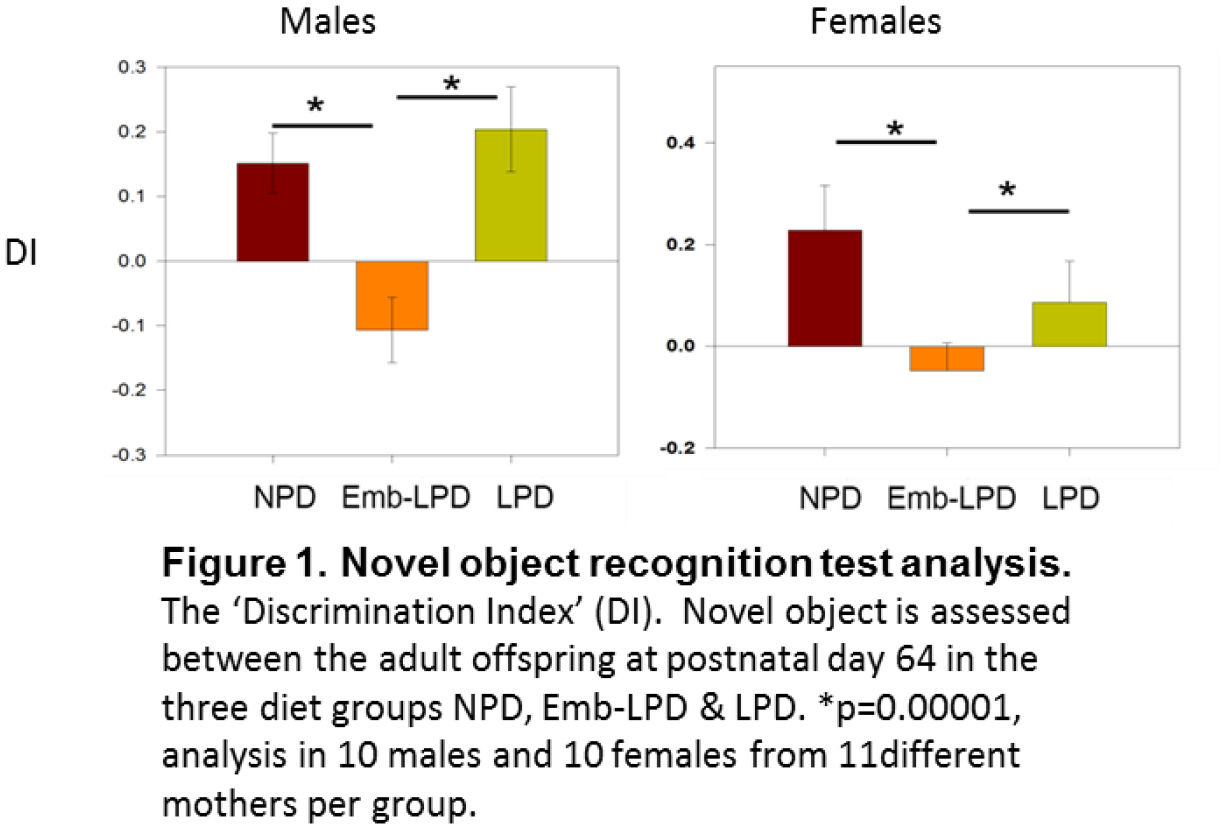
Social influence refers to the phenomenon by which an individual’s thoughts or behaviours are affected by those of other people. There are significant age effects on social influence, with previous research showing either a decline from childhood to adulthood, or a temporary increase in social influence during early adolescence. To date, most research has focussed on negative aspects of social influence, such as peer influence on risky behaviour. The current study investigated the impact of social influence on the reporting of prosocial behaviours, such as helping others. Participants (N=787) aged 8 to 79 completed a computerised task in which each trial was made up of three components. First, the participant rated how likely they would be to engage in a prosocial behaviour, e.g. ‘Looking after an ill friend’ (rating 1). Second, participants were shown the average rating (in fact fictitious) that other participants had answered to the same question (provided rating). Finally, participants were asked to rate their own answer again to the same question (rating 2). We found that age affected the extent to which participants were influenced by other people’s ratings (i.e. age affected how much participants changed their answer from rating 1 to rating 2). The study provides evidence that social influence is a significant factor in prosocial as well as antisocial behaviours, and that younger people’s increased susceptibility to social influence can have positive outcomes.
GABAergic interneurons are a fundamental structural and functional component of all complex neural circuits. Within the thalamus there are two main sources of inhibition, the thalamic reticular nucleus (RTN) and local interneurons, and little is known about the origin and diversity of the latter. In rodents, thalamic local interneurons are largely restricted to the dorsal lateral geniculate nucleus (dLGN), where they contribute to the processing of visual information. Here we examine the ontogeny and function of this local inhibitory drive.
The prosomeric models posits that all thalamic neurons are specified within the second (also known as dorsal thalamus) and third (also known as ventral thalamus or prethalamus) diencephalic prosomeres (p2 and p3). Inhibitory neurogenesis, including that of the RTN, takes place in p3 and in a GABAergic rostral p2 subdomain (pTh-R), which makes both p3 and pTh-R possible sources of origin for thalamic local interneurons. In fact, until now, the prevailing hypothesis states that dLGN interneurons have a prethalamic origin (p3).
In contrast, here we report that p3 or pTh-R are unlikely sources for thalamic interneurons. Using fate-mapping, time lapse imaging and transcription factor expression analysis, we show that Sox14/Gata2/Otx2-expressing precursor cells populate the dLGN with GABAergic interneurons, migrating from the dorsal midbrain in early postnatal development. This unexpected extra-thalamic origin differentiates them from the thalamic GABAergic neurons of the RTN.
The developmentally-defined genetic identity of the dLGN interneurons was then used to perform a combination of optogenetic and electrophysiological experiments. We were able to demonstrate that this cell type can generate tonic inhibition onto thalamic relay neurons, which becomes significant at high interneuron firing rates.
In conclusion, by revising the model of thalamic interneuron ontogeny, we demonstrate how a previously unappreciated mesencephalic inhibitory population controls thalamic relay neuron excitability.
We carried out intensive research on understanding of the underline neurobiological mechanism of stress induced depression on the brain hardware; however, it seems that the main picture located behind the cognition; where the software meets the hardware. As one line of our projects the present study aimed to evaluate the coherence of Personality pattern with depression.
Volunteer individual with diagnoses of depression were referred to our laboratory randomly (n=12). Beck Depression Inventory (BDI),Millon clinical Multiaxial Inventory-II(MCMI-II), were used at per-test, post-test, and follow up. The results were analyzed, using SPSS statistical package.
The results showed that there was a significant correlation between personality pattern and the intensity of depression. After the application of CBT procedures and the improvement of depression also reduced the personality patterns of schizoid by D=13.75 +/-3.42 (F=15.42), Avoidant by D=27.25+/-7.82 (F=24.66), Dependent by D=22.63 +/- 9.89 (F=5/23), Passive aggressive by D=14.5 +/- 5.57 (F=6.76), and self defeating by D=21 +/-7.8 (F=6.36)
These finding suggest coherence of personality pattern as axes II with depression as an axes I mental states; and correlation between the hardware and software.
Keywords: Hardware, Software, Personality Pattern, and Depression
Background: Previous research has shown that early in treatment, prior to an improvement in mood, serotonergic and/or noradrenergic antidepressants can remediate negative biases in information processing observed in major depressive disorder (MDD). However, it remains unclear whether dopaminergic antidepressants, such as bupropion, exert similar early actions on information processing. Here we investigate the early and longer-term effects of bupropion on behavioural measures of emotional and reward processing in MDD patients.
Method: Complete data sets were obtained for 41 MDD patients and 40 healthy controls (HC). In a repeated measures study design, open-label bupropion was administered to just the MDD patients over a 6 week period. All participants completed the Emotional Test Battery and a reward task at baseline, week 2 and week 6.
Results: Bupropion was found to reduce negative biases in emotional processing on the ETB early in treatment at 2 weeks. Specifically, only the bupropion-treated MDD group displayed a significant decrease in the percentage misclassification of faces as sad (F1, 80 = 4.09, p < 0.05; t41 = 2.72, p < 0.05) and the number of negative self-referent words falsely recalled (F1, 81 = 5.73, p < 0.05; t42 = 2.12, p < 0.05) between baseline and week 2. Conversely, bupropion was found to significantly worsen performance on the reward task between baseline and week 2 (t14 = 4.17, p < 0.01) prior to normalisation to HC levels after the full 6 week treatment (t14 = -10.5, p < 0.001; t28 = -0.25, p = 0.80).
AIM
It has been widely reported that opiate and psychostimulant addiction in humans is associated with substantive cognitive impairment. However, it remains unclear which cognitive domains are most severely affected. This has fundamental implications for the theory and treatment of addiction. We therefore conducted a random-effects meta-analysis.
METHODS:
We systematically searched the Web of Knowledge suite and PubMed database, using the Taporware text analytics tool to optimise these searches. Searches were completed on 16th December 2015 and identified a total of 12,028 papers. Data that satisfied our a priori inclusion criteria were assigned to one of the following four cognitive domains: Language, Motor, Memory and Executive Function; each of these domains were further divided into sub-domains. Ultimately, we included 65 studies and data from 2752 users and 2356 healthy control participants. Following data extraction, random-effects meta-analyses were performed using Stata 14.
RESULTS:
Cognitive impairment was associated with opiate or psychostimulant abuse across all domains, though this did not reach statistical significance in some sub-domains: for opiate users, Verbal Comprehension, Verbal Declarative Memory and Auditory Declarative Memory; for psychostimulant users, Psychomotor Performance and Attention. The general trend across domains was for impairment to be more severe in opiate users than in psychostimulant users (Opiates, SMD = -0.68; P=<0.000; Psychostimulants, SMD = -0.43; P=<0.000), but there were notable differences between sub-domains. Specifically, the most substantial impairment shown in opiate users was in Visual Declarative Memory (SMD= -1.84; P=0.000). The most substantial impairment shown in psychostimulant users was in Verbal Comprehension (SMD = -1.17; P=0.000). Impairments in Impulse Control were modest in opiate (SMD = -0.48; P=0.000), and psychostimulant users (SMD = -0.35; P=0.000).
CONCLUSIONS:
There are substantive differences in the forms of cognitive deficit associated with psychostimulant and opiate use. This challenges some currently influential theories of drug addiction, and has immediate implications for treatment.
Variation in the gene encoding zinc finger binding protein 804A (ZNF804A) is associated with schizophrenia and bipolar disorder. Evidence suggests that ZNF804A is a regulator of gene transcription and is present in nuclear and extranuclear compartments. However, a detailed examination of ZNF804A distribution and its neuronal functions has yet to be performed. Therefore, we examined the localization of ZNF804A in neurons derived from human neural progenitor cells, human induced pluripotent stem cells, or in primary rat cortical neurons. In addition, we used small interfering RNA-mediated knockdown of ZNF804A investigate the role of this protein in neurite formation and structural plasticity of excitatory synapses. We found that ZNF804A protein localized to somatodendritic compartments and colocalized with the putative synaptic markers in young neurons derived from human neural progenitor cells and human induced pluripotent stem cells. In mature rat neurons, Zfp804A, the homolog of ZNF804A, was present in a subset of dendritic spines and colocalized with synaptic proteins in specific nanodomains, as determined by super-resolution microscopy. Interestingly, knockdown of ZNF804A attenuated neurite outgrowth in young neurons, an effect potentially mediated by reduced neuroligin-4 expression. Furthermore, knockdown of ZNF804A in mature neurons resulted in the loss of dendritic spine density and impaired responses to activity-dependent stimulation.These data reveal a novel subcellular distribution for ZNF804A within somatodendritic compartments and a nanoscopic organization at excitatory synapses. Moreover, our results suggest that ZNF804A plays an active role in neurite formation, maintenance of dendritic spines, and activity-dependent structural plasticity.
Traumatic brain injury (TBI) is a major cause of morbidity and mortality worldwide. A clear understanding of the relation between acute clinical presentation and chronic neurocognitive and functional deficits is important in order to build better predictive models. The Glasgow Coma Scale (GCS) quantifies the neurological state of TBI patients by assessing verbal, motor and eye-opening responses. TBI patients are often stratified in terms of injury severity using GCS in research, but its prognostic accuracy is debated. We explored the predictive value of GCS in cognitive, physical, social and emotional outcome six months after TBI. TBI patients (n = 138; 40% female; GCS 3–15) aged 17 to 70 years completed a battery of cognitive and questionnaire measures six months after injury. Age and education matched orthopaedic trauma patients (n = 25) and healthy volunteers (n = 99) acted as controls. A clear dichotomy in the functional and cognitive measures that were predicted by GCS was observed, as reflected by the strength GCS correlation and the pattern of group differences. GCS predicted functional outcome in physical and social domains (disability, physical functioning, social functioning) but not in emotional domains (depression, well-being, subjective general health, subjective bodily pain). GCS also predicated outcome in the majority of cognitive domains (verbal fluency, episodic memory, learning, visual and spatial short-term recognition memory, sustained attention) but with the clear exception of working memory (WM; WM span, maintenance and manipulation of information in WM, attentional flexibility). We also observed a strong dissociation in six month outcome between mild and severe TBI cases (according to GCS) in the absence of dissociation between moderate TBI cases and other groups. These findings suggest that the stratification of TBI patients according to GCS for research purposes could be limited to mild and severe (and not moderate) cases. Although GCS may be useful for identifying TBI patients that could benefit from cognitive and occupational rehabilitation, the use of prognostic factors other than GCS are necessary to identify patients requiring early intervention for the prevention of emotional difficulties.
The complement system, a highly conserved branch of innate immunity, is emerging as an important contributor to normal and abnormal brain function. Using genetically modified mice deficient in either the central complement component C3, or the receptor for the C3 bioactive breakdown product C3a, known as C3aR, we investigated fear reactivity using the elevated plus maze and open field assays. Our data demonstrated a markedly heightened anxiety response in C3aR-/- subjects, as evidenced, respectively, by a profound reduction in open arm exploration (see figure) and central zone crossing in the open field (data not shown). This effect was absent from C3-/- subjects. Our data are consistent with a) an important role for C3aR in maintaining normal fear responses and b) the speculation that C3aR signaling is promiscuous and likely to be able to signal via ligands other than C3a. The mechanistic underpinnings of these effects are currently under investigation.
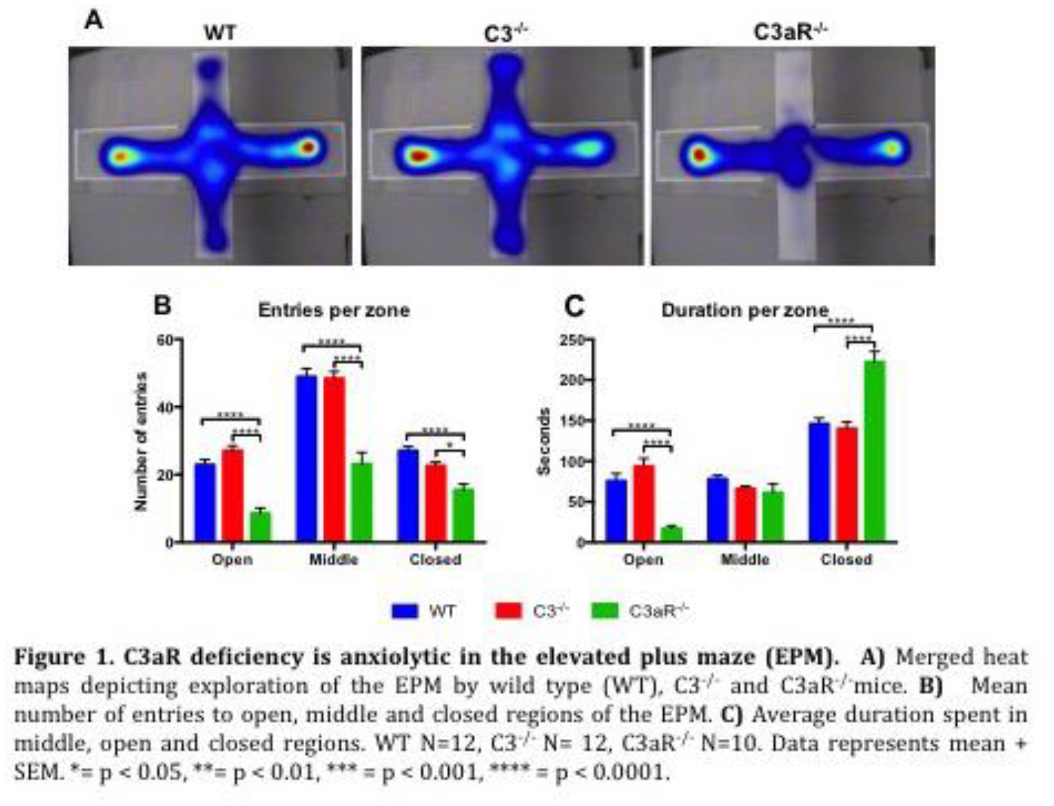
The purpose of this study was to adapt the English version of Fear of Happiness Scale into Turkish language. Exploratory (N = 171) and confirmatory factor analysis (N = 171) indicated that the Fear of Happiness Scale (FHS) is unidimensional. The results also showed that the Turkish version had good internal consistency (a = .86). In addition, the scale provided acceptable evidence of convergent validity by negatively correlating with measures of positive affect, life satisfaction and subjective happiness and positively correlating with measure of negative affect. These findings indicated that Turkish version of FHS can be used as a reliable and valid measure in Turkish culture.
Current thinking suggests that psychosis is a disorder of aberrant salience. This describes when a stimulus continues to grab inappropriately high levels of attention, and it is thought to be mediated via elevated dopamine (DA) levels, which have been robustly demonstrated in schizophrenia. However, the causes of this DA dysregulation are generally unspecified. Recent large scale GWAS meta-analyses have established genome-wide significant association to schizophrenia for the Gria1 locus which codes for the GluA1 subunit of the AMPA glutamate receptor. GluA1 KO mice have previously been studied in relation to schizophrenia but, notably, striatal whole tissue levels of dopamine and its metabolites appear normal in these animals. However, we might not expect to see changes in dopamine activity in anaesthetised animals, or in a home-cage environment. Indeed, changes in phasic DA responses are likely to be both behaviour-dependent and stimulus-specific.
To test this possibility we have recorded phasic DA signals with high temporal resolution, in freely moving, behaving wild-type and GluA1 KO mice, using fast-scan cyclic voltammetry (FSCV). This state of the art electrochemical recording technique involves chronically implanting carbon-fibre microelectrodes into the nucleus accumbens to allow sub-second, real time measurements of DA, allowing definitive assessment of extracellular DA changes in terminal regions. Here we demonstrate that phasic dopamine signals in response to neutral light stimuli fail to habituate in Gria1-/- mice, resulting in a behaviourally relevant, hyper-dopaminergic phenotype in these animals. This parallels previous behavioural data from these mice. In addition, phasic dopamine responses to unsignalled rewards were also significantly enhanced in the knockout mice. Thus, we provide evidence for behaviourally-relevant hyper-dopaminergic responses in a genetically modified mouse model of glutamatergic dysfunction relevant to schizophrenia. These data may have important implications for understanding the aetiology of aberrant salience in psychotic disorders including schizophrenia.
The corticothalamic loop has long been implicated in a range of neuropsychiatric diseases. The thalamic reticular nucleus (TRN), a part of the corticothalamic loop, plays a key role in selective attention and sleep spindles. Furthermore, sleep spindles are reduced in amplitude and duration in schizophrenia patients, implying clinical relevance of TRN functions. However, while the TRN is topographically organized, it remains unclear whether and how the TRN consists of functionally distinct sub-regions. Combining optogenetic and electrophysiological approaches in mice, we investigated changes in sleep spindles and EEG oscillations caused by optogenetic stimulations in different parts of the TRN. Archaerhodopsin (Arch), a light sensitive proton pump, was expressed specifically in either an anterior or posterior part of the TRN in parvalbumin (PV)-Cre mice using adeno-associated viral vectors. We found restricted expression patterns of Arch in PV-positive neurons of the TRN depending on injection sites. Effects of optical stimulation on cortical EEGs were assessed by delivering green light through chronically implanted optic fibers in up to 1 min periods in freely behaving animals. Tonic stimulations during awake states did not produce any significant change in EEGs whereas stimulations during sleep (mostly slow wave sleep) increased delta power and the number of sleep spindles . Together these data support the notion that activity in the TRN may have different impacts on the modulation of cortical states in a site-dependent manner.
Aims: Many in-vitro systems used during pre-clinical trials fail to recreate the biological complexity of the in-vivo neural microenvironment. Taking advantage of recent advances in microfluidic technology, we seek to develop a perfusion based drug discovery platform that is capable of high-throughput pharmacological profiling. This in turn will allow us to better understand how drugs influence the communication between functionally connected neural networks.
Methods: Mixed primary hippocampal networks were grown in microfluidic devices with environmentally separated chambers that allow synaptic connections to be formed with each other via an array of microchannels. The perfusion of multiple compounds in one chamber was achieved using computer controlled fluid actuation connected to the inlets/outlets of the microfluidic device. Responses to perfusates from directly stimulated neurons and those synaptically connected were recorded using calcium imaging.
Results: Following live/dead assays, a flow rate of 4μl min-1 showed the greatest cell viability and was used for subsequent experiments. Subsequently, a glutamate concentration response curve following direct stimulation was obtained which revealed an EC50 = 4μM. Pharmacological manipulation of neuronal activity was also achieved as the neuronal response to glutamate was reversibly reduced in the presence of ionotropic glutamatergic antagonists. Furthermore, repeated glutamate perfusions induced increasing levels of activity in the adjacent, naïve neural network.
Conclusion: The proposed microfluidic system is able to reliably produce pharmacological profiles for drugs in a neurological setting. The novelty of the presented drug discovery platform is its ability to not only determine the properties of a new drug, but how the drug influences communication between neural networks.
** Correction: Original document did not contain names or affiliations of co-authors.

Researchers have ethical and legal obligations to optimise the physical and emotional wellbeing of their animals. Furthermore, current European legislation places an emphasis on the animal’s lifetime experience. However, current methods for assessing the cumulative experience of animals are poorly validated and suffer from a lack of sensitivity and/or specificity. The general goal of this work was to develop and validate a new method to assess cumulative experience in non-human primates (NHPs).
Recent development in stress biology has shown that in rodents, NHPs and humans, the amount of grey matter in the hippocampus co-varies with the cumulative experience of individuals. These new findings open the possibility to use the amount of hippocampal grey matter as a biomarker of cumulative experience in laboratory animals. The hippocampus is not a homogenous region and its different functions seem to be spatially segregated. In this study, we tried to identify which part of the hippocampus is most sensitive to cumulative experience in NHPs.
As a proxy for cumulative experience, we used artificial weaning age (i.e. definitive separation from the mother forced by human caretakers). Early artificial weaning is a well-established early-life stressor in NHPs. It is also known to have long-lasting detrimental effects on emotionality, social, sexual and maternal behaviours, as well as growth, immune responses and in some cases survival, inducing a poorer life time experience in individuals weaned earlier. Eleven male adult macaques were scanned with a 4.7 T MRI scanner. In each subject, the amount of grey matter of each voxel comprised in the hippocampus was determined using voxel-based morphometry. After controlling for covariates including age and total brain size, a multiple regression analysis revealed a positive correlation between weaning age and the amount of grey matter in the right anterior hippocampus.
We argue that with appropriate strategies to control for potential confounding factors, the amount of grey matter in this specific part of the hippocampus can now be used to measure the cumulative experience of NHPs.
Dynamic clamp is a closed-loop electrophysiology method that allows experimenters to inject voltage-dependent currents into a live neuron. The method relies on a high-frequency control loop that consists of measuring the membrane potential, calculating the corresponding current, and injecting it back into the cell. Dynamic clamp is often implemented using highly specialised real-time software and/or hardware, which requires significant technical aptitude to set up. In contrast, the Windows-based StdpC software runs on any commercial computer and is straightforward to set up and use.
StdpC offers a wide range of dynamic clamp related features. It is designed to simulate ionic conductances, chemical synapses, gap junctions, and any combination thereof, up to and including entirely virtual neuron models with synapses leading to and from real neurons. For each of these basic building blocks, several mathematical formulations are built-in, ready for the user to parametrise. Simulated synapses support advanced features such as spike-time dependent plasticity and delayed transmission. StdpC can be used with separate voltage-measuring and current-injecting electrodes, but it also includes an active electrode compensation algorithm that allows accurate clamping with a single (patch or sharp) electrode. Experiments can be automated via a scripting interface, and the openness and modular structure of the source code give users with some programming facility the option to quickly extend the software to suit their needs.
As an example for the application of StdpC, we present an experiment where we use hybrid circuits to investigate bistability in the visual system. Two pyramidal neurons in the primary visual cortex are patch-clamped simultaneously. They are then coupled through mutual disynaptic connections using models of different inhibitory interneurons in an effort to learn more about the bottom-up processes that may underlie the phenomenon of bistable visual perception.
First author’s PhD funding provided by the University of Sussex School of Life Science.
Stroke is one of the most common causes of neurological disability in the Western World, yet little is understood about how motor deficits post-stroke can be predicted from lesions visible on acute clinical imaging. To address this question, we collected clinical computer tomography (CT) brain scans from 185 acute stroke patients (mean 1.93 days post stroke), along with measures of gross and fine motor skill within 5±4 days post stroke. We asked patients to perform a tap to their head and to pick up a pencil lying on a table with each hand separately. Results were scored on a scale of 1 to 4, designating a complete inability to a complete ability to perform the tasks.
Stroke lesions were manually delineated by trained technicians and were registered to a stereotaxic space using Clinical Toolbox (Rorden et al., 2012). To map the lesion affected-fiber tracts, disconnectome maps for each patient were generated using software from the BCBtoolkit (Thiebaut de Schotten et al., 2015). To further generate data on lesion-affected anatomy, patient lesion masks were overlaid with fiber tracks from a white matter atlas (natbrainlab.co.uk).
Of the 185 patients, 97 presented with minor to severe motor deficits, of whom 47 patients were unable to perform the task, 15 patients were only partly able to perform the task and 35 patients performed the task with minimal difficulty. The VLSM analysis using the task data and lesion masks localized the relationship between lesion site and motor deficit to a single cluster in the right posterior limb of the internal capsule. Using this identified cluster as a seed region, further analyses on intersecting tracks were performed allowing for groupings on the basis of affected tracks. Despite the laterality of the above mentioned VLSM results, patients with damage to the left (n=20) and right (n=22) corticospinal tracts were significantly worse at the motor tasks than those without lesions in these tracks (ps<0.001). When analyzing the motor data in a VLSM analysis with the disconnectome maps, significant relationships between motor deficit and lesion location was demonstrated in bilateral motor related regions. The results from this study demonstrate different approaches for comparing lesion affected anatomy and behavioral deficits.
Voltage sensitive dyes (VSDs) offer an alternative to Ca2+ sensitive dyes for the imaging of neuronal networks. The time-resolution of VSDs is faster compared to Ca2+ imaging, but their signal to noise ratio (SNR) is ~10 times less that of Ca2+ dyes. This work aims to improve the SNR of VSDs while maintaining their dynamics through the design of VSDs derived from Bodipy dyes (e.g. JULBD) that were shown as viable alternatives to standard VSDs (e.g. di4-ANEPPs)[1].
The stomatogastric ganglion (STG) of the brown crab (Cancer pagurus) is one of the most researched small biological nervous systems due to the relative large size and accessibility of the neurons located within the STG and its robust pyloric rhythm (PR) which controls the movement of muscles in the gastric system. Optical imaging of the STG using VSDs has been well studied (e.g. [2]), allowing the simultaneous recording of the electrical activity of many cells.
The toxicity (indicated by the increase in PR frequency) of 4 novel dyes (NDS3, NDS4, NDS8, SC114), di4-ANEPPs and JULBD (as baseline measures) was assessed. The STG was removed and desheathed, each dye bath applied for 20min followed by 20min washout. For each dye, the duration of light shone onto the STG ranged from 20s to 5mins in increments of 20s. di4-ANEPPs caused an increase in the PR (20-100s), before saturating (120-220s) and then returning to baseline (240s+) (N=5). JULBD increased by 60s light exposure, before disrupting the PR at 100s+. Initial results indicate that NDS3, 4 and 8 had no effect on the PR, while SC114 appeared to slow it (N=1 for last 5 VSDs, Figure 1).
Future work involves the further validation of these dyes, assessing toxicity and SNR, the results of which will feedback into design refinement of the structure of suitable dyes.
[1] Bai, D., Benniston, A.C., Clift, S., Baisch, U., Steyn, J., Everitt, N. and Andras, P., 2014. Low molecular weight Neutral Boron Dipyrromethene (Bodipy) dyads for fluorescence-based neural imaging. Journal of Molecular Structure, 1065, pp.10-15.
[2] Städele, C., Andras, P. and Stein, W., 2012. Simultaneous measurement of membrane potential changes in multiple pattern generating neurons using voltage sensitive dye imaging. Journal of neuroscience methods, 203(1), pp.78-8.

In vivo and in vitro extracellular neuronal recordings consist of local field potential and a mixture of action potentials, or spikes, from many neurons. It is necessary to isolate or sort the activity of individual neurons to study their cellular dynamics and response to external or internal stimuli. This process is known as spike-sorting. Spikes are detected, i.e. using a voltage threshold, and their features, e.g. waveform properties, are extracted. These features from individual neurons form dense regions or clusters in their distributions as they follow similar properties. The clusters are isolated either manually or by automated clustering algorithms. Most of the clustering algorithms for spike-sorting are based on the principle that cluster data points are Gaussian-distributed. But features like amplitude of the spikes often do not follow such distribution due to, for example, detection using amplitude threshold. Here, we used a density-based clustering algorithm to recursively isolate spiking activity of neurons of varying firing rates irrespective of the distribution patterns. It is based on the idea that cluster centres have high density and they are located far away from points of higher densities. The rest of the feature data points are assigned to the same cluster as of their nearest neighbour of higher density. Modality of the clusters were determined from the peaks in the kernel-based probability estimates, unimodal clusters were isolated, and the rest of the data points followed the recursion of the above algorithm. We show that the algorithm can effectively sort spikes of varying firing rates and can overcome the limitations of using centroid-based, e.g. k-means, or other density-based, e.g. DBSCAN, clustering algorithms. It may also be used to identify clusters in other biological data, i.e. clustering of DNA- or RNA-sequences, as the algorithm relaxes the assumption about the underlying data distribution function.
Neuroscience is one of the fastest growing interdisciplinary research fields that studies the central (CNS) and peripheral nervous system from the molecular and cellular levels to the systems level. Areas of research include neural development, structural and functional organization of the nervous system, cognitive and behavioral neurosciences, and clinical neurosciences including neurodegenerative diseases.
The RNAscope® assay provides a powerful method to detect gene expression within the spatial and morphological tissue context. The proprietary “double Z” probe design in combination with the advanced signal amplification enables a highly specific and sensitive detection of the target RNA with each dot visualizing a single RNA transcript. Therefore, this robust signal-to-noise technology allows for detection of gene transcripts at single molecule level with single-cell resolution analysis and can further expand our understanding of gene expression in cell lines and tissues samples. The multiplexing capabilities of both the chromogenic and fluorescent RNAscope® assays facilitate the simultaneous visualization of multiple targets in formalin-fixed paraffin-embedded (FFPE) and fresh frozen samples, enabling consistent characterization of cell populations within the nervous system. In summary, RNAscope® technology allows the visualization and quantification of virtually any gene from any genome in any tissue with unprecedented specificity and sensitivity.
Here we illustrate the utility of RNAscope® applications in neuroscience:
• Identification, characterization, and (co-) localization of both mRNAs and lncRNAs in the nervous system
• Identification, visualization and characterization of specific cell types in the nervous system
• Detection of mRNA in the nervous system when no (reliable) antibodies are available
• Visualization of neuronal network activity and plasticity
• Validation of target mRNA expression after high-throughput gene expression analysis
• Validation of (cell type-specific and conditional) genetic modifications.

When simulating models of neural networks in the brain, the size of the simulated networks matters. Modern technology such as Graphical processing Unit (GPU) accelerators can help Neuroscience researchers to simulate larger and more realistic brain models. Here we present the GPU enhanced neuronal networks (GeNN) framework [1,2] that we have created to gain the most from GPU accelerators. GeNN is using an approach of automatic code generation from straightforward model descriptions provided by the scientist to generate code that is optimized for the detected GPU accelerator and the defined model. The technical difficulties of using GPU accelerators are hence removed from users who can concentrate on interesting scientific questions instead. At the same time, GeNN remains flexible and expert users can manipulate virtually every aspect of the simulations. GeNN supports all typical computational neuroscience models by allowing users to define their own equations for the model elements, such as neuron dynamics, synapse updates and learning rules. GeNN has been used for networks of Hodgkin-Huxley neurons, with Hebbian learning, STDP and 3-factor learning rules, as well as for more simple models such as networks of Izhikevich, integrate-and-fire and Rulkov map neurons.
For less expert users, we have created additional interfaces to the SpineCreator graphical model definition interface [3] and to the popular Brian 2 simulator [4]. With the latter, it is as simple as issuing the command set_device(‘genn’) to take advantage of GPU acceleration.
Acceleration compared to traditional CPU-based solutions varies by model and GPU accelerator hardware and can be as high as 200 times but also as low as none. GeNN is available as open source software under GPL v2 [2].
1. E. Yavuz, J. Turner and T. Nowotny (2016). GeNN: a code generation framework for accelerated brain simulations. Scientific Reports 6:18854. doi: 10.1038/srep18854
2. GeNN, http://genn-team.github.io/genn/, accessed 2016-12-15
3. SpineCreator, http://bimpa.group.shef.ac.uk/SpineML/index.php/SpineCreator_-_A_Graphical_Tool, accessed 2016-12-15
4. Brian 2, https://github.com/brian-team/brian2, accessed 2016-12-15
This work was partially financed by the EPSRC, grant EP/P006094/1.
Previous fMRI and EEG studies have shown face-specific neural responses to faces compared to objects. Detection of face-specific brain activation in freely behaving and moving people has not been accomplished as of yet. The purpose of our research was to identify, using wireless multichannel EEG in freely moving participants, event-related potentials (ERPs) during viewing human faces.
Mobile EEG and eye tracking was recorded from 19 freely moving participants whilst they viewed a mock art gallery. Stimuli were presented on 20 panels (A0 poster size) displayed in the ground floor of the psychology department building of the University of Liverpool. Positive, negative and neutral valence images were viewed and later rated by the participants. EEG was recorded continuously using a 64-channel BrainProduct MOVE system. In absence of triggers indicating onsets of viewing of visual stimuli, a novel PupilLab head-mounted wearable eyetracker was used to capture real world video recordings and the calibrated XY locations of the gaze. After synchronising the time sources of EEG and eye-tracking recordings, BESA 6.1. program was used to process EEG data.
Wireless EEG recordings allowed identification of a face-related ERP component in the latency interval ranging from 165 to 210 ms (N170 potential); this component was not seen whilst participants were viewing non-living objects. The face ERP component was sensitive to the emotional face expression; in particular, the amplitude of N170 potential was stronger during viewing disgusted compared to neutral faces. Source dipole analysis revealed three equivalent current dipoles in the latency interval from 100 ms to 300 ms. Two source dipoles, located in the left extrastriate (BA19) and primary visual (BA17) cortex, modelled the visual P100 component, and one equivalent current dipole, fitted to the right fusiform gyrus (BA37), accounted for the face-related N170 potential.
This study is the first to demonstrate the face-related ERPs in freely moving individuals in natural settings. The study opens new possibilities in clinical, developmental, social or marketing research in which information about presence of face perception and the type of perceived facial expression is of importance.
The N-methyl-D-aspartate receptor (NMDA receptor) and its interacting proteins constitute large macro-molecular complexes (NMDAR complexes) at excitatory synapses1. Recent human genomic studies discovered several mutations in the genes encoding the components of NMDAR complexes in various neuropsychiatric disorders including intellectual disability (ID), autism and schizophrenia. However, little is known about how mutations in these genes alter molecular and cellular pathways leading to pathological phenotypes. To answer the question, our lab has been systematically mutating synaptic genes in mice using conventional gene targeting methods. To further accelerate generation of mutants we adapted CRISPR genome editing system in mouse embryonic stem (ES) cells.
SH3 and multiple ankyrin repeat domains 3 (Shank3) is a crucial scaffolding protein of NMDAR complexes and is indicated in autism2 and schizophrenia3. CRISPR-induced knockout mutation of Shank3 was successfully introduced in ES cells, and these cells were injected into blastocysts and 3 chimaeras were born. These chimaeras then were crossed with wild-type mice and germline transmission was confirmed. Having established a knockout mutation using CRISPR, we moved on to modify Synaptic GTPase-activating protein (SynGAP), another component of NMDAR complexes. De novo mutations of SynGAP have been found in ID4, autism4 and schizophrenia5. To introduce a defined mutation in SynGAP gene, we designed a CRISPR-mediated point mutation, and obtained ES cells with the precise mutation. Here we demonstrate CRISPR facilitates the disruption of Shank3, as well as the precise editing in SynGAP in mouse ES cells with high efficiency.
Reference:
1. Frank et al. Nat. Commun. 7, 11264 (2016).
2. Durand et al. Nat. Genet. 39, 25–27 (2007).
3. Gauthier et al. Proc. Natl. Acad. Sci. 107, 7863–7868 (2010).
4. Berryer et al. Hum. Mutat. 34, 385–394 (2013).
5. Purcell et al. Nature 506, 185–90 (2014).
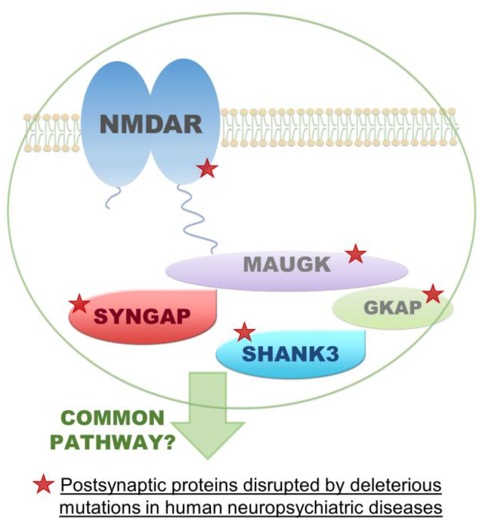
Attention (i.e. task relevance) and expectation (i.e. stimulus probability) are two critical determinants of perception. While attention is thought to increase the neural response to external stimuli, expectation is considered to attenuate it. Predictive coding models and recent neuroimaging research suggest that attention and expectation shape neural processing in an interactive fashion whereby attention reverses the attenuation for expected signals. Operationally, attention is often manipulated by asking participants to respond only to the ‘attended’ stimuli. Consequently, the synergistic effects of attention and expectation could only be evaluated at the neural level, but not at the behavioural level where ‘unattended’ stimuli are not responded to. This study developed a novel multisensory paradigm that allowed us to evaluate interactive effects of attention and expectation at the behavioural and neural level. In two experiments, we presented participants with auditory and visual signals in their left or right hemifields. We manipulated stimulus frequency or response requirements only to auditory signals, allowing us to measure the multisensory effects of spatial attention and expectation on behavioural responses to visual signals. Importantly, while experiment 1 manipulated expectation directly via the frequency of auditory stimuli as in (1), experiment 2 determined it indirectly via non-target stimuli that are not responded to as in (2). Our results demonstrate that the synergistic behavioural effects of attention and expectation differ across paradigms. While in experiment 1 attention and expectation influence response times interactively, in experiment 2 the two effects determine response times additively. We explain these discrepant results by a combination of overall response probability and response probabilities conditioned on the spatial hemifield where the stimulus was presented, that differ across the two paradigms. Response times reflect response probability determined by the specific manipulation employed. Concurrent fMRI experiments investigate the neural mechanisms underlying these multisensory effects of attention and expectation.
(1)Kok et al.(2012) doi.org/10.1093/cercor/bhr310
(2)Jiang et al.(2013) doi.org/10.1523/JNEUROSCI.3308-13.2013
Impulsivity is defined as a tendency for premature, unduly risky and poorly conceived actions and as a behavioural trait is associated with a number of psychiatric disorders including attention deficit/hyperactivity disorder (ADHD) and bipolar disorder, and is thought to involve dysfunction within cortico-striatal circuitries. Here we investigate the putative metabolic cortico-striatal correlates of impulsivity using in-vivo proton magnetic resonance spectroscopy (1H MRS) in rats selected on the basis of expression of innately high or low levels of premature responding on the five choice serial reaction time task (5CSRTT). High impulsive rats demonstrated significantly reduced prefrontal cortical levels of myoinositol, a metabolite associated with the inositol triphosphate/calcium (IP3/Ca2+) signalling cascade. No other differences in metabolite concentrations were observed between high and low impulsive animals in either the prefrontal cortex or striatum. Ex-vivo mass spectroscopy examining myoinositol levels in individual sub-regions of the prefrontal cortex in an independent group of animals confirmed a reduction in myoinositol levels in high impulsive rats, specifically within the infralimbic cortex. To further investigate the ontology of this metabolic dysfunction, we examined transcript levels of a number of key enzymes and proteins involved in the metabolism and cellular transport of myoinositol and its precursors within the infralimbic cortex. Significant reductions in transcript levels were observed for the enzyme inositol monophosphate synthanse 1 (IMPase1) and the sodium inositol co-transporter (SMIT1) in high impulsive rats compared with low impulsive rats. The main findings of this study suggest that trait impulsivity in rats is associated with reductions in the level of myoinositol in the infralimbic cortex, potentially driven by reductions in the capacity for intracellular transport and calcium signalling.
AIM
Imagination - the ability to call to mind things that are not present to the senses - allows us to explore the past, the future, and the potentially possible. For most people, visual imagery is a conspicuous element of imagination, but some people report its absence. We have called this absence of visual imagery aphantasia. Here, we present preliminary data from a large questionnaire survey of individuals whose imagery falls at the extremes of the vividness spectrum.
METHOD
2,012 members of our user group completed a Visual Imagery Questionnaire, (VVIQ, Marks 1973), a widely-accepted measure of mental imagery. Participants scoring between 16 and 24 on the VVIQ, were classified as aphantasic; those scoring >77 were classified as hyperphantasic. Their employment was categorised using Standard Occupational Classification (US Department of Labor, 2000).
RESULTS
We focus here on individuals with lifelong aphantasia or hyperphantasia who comprised the overwhelming majority of participants. 19% of people with aphantasia worked in computer and mathematical occupations; only 8% of people with hyperphantasia reported working in these fields. Amongst those with vivid imagery, 29% worked in the Arts, Design, Entertainment, Sports, and Media Occupations, compared to 13% of people with aphantasia. A family history in first degree relatives of aphantasia and hyperphantasia was obtained in 15-20% of participants. The majority of participants with aphantasia (70%) experience imagery in dreams. Roughly equal numbers of participants with aphantasia reported the presence and absence of imagery in other modalities. Face recognition difficulties were reported commonly by participants with aphantasia (35%). More individuals with aphantasia (34%) than hyperphantasia (5%) regarded their autobiographical memory as poor; conversely 23% of people with hyperphantasia compared to 8% of people with aphantasia regarded their autobiographical memory as good.
CONCLUSIONS
Preliminary data from this large sample of individuals falling at the extremes of the imagery spectrum suggests that imagery vividness is a lifelong trait. Low imagery vividness appears to be overrepresented among people working in IT related and mathematical domains, high vividness among those working in
The ability to consciously recognise visual objects is crucial for adaptive behaviour and survival. Conscious access to visual objects has been studied using the Attentional Blink (AB), where two targets (T1 and T2) are embedded with visual masks in a rapid serial visual presentation (RSVP). In the AB, the ability to detect T2 is reduced when presented 200-500ms post T1. Research using functional Magnetic Resonance Imaging (fMRI) has proven useful to identify the underlying brain mechanisms of conscious access. Given the challenges inherent to the limited temporal resolution of fMRI, researchers have designed AB-studies in which T1 and T2 targets are selected from image categories known to engage different regions in the visual stream. However, to integrate these findings into a consistent model of conscious access, the variability in detection thresholds across categories needs to be assessed. Specifically, we investigated the categorical differences in conscious and unconscious processing using a behavioural attentional blink task. Here, we presented participants with 48 pictures of objects from eight categories (fruits and vegetables, processed foods, objects, scenes, animal bodies, animal faces, human bodies, and human faces) in an AB-task (Fig. 1A). Each picture was presented as T1, and at two different T2-lags (200ms and 700ms post T1). To compare the performance at recalling target objects across categories, we used a factorial ANOVA with performance effect of T2-lag and object category as factors (Fig. 1B). We observed main effects of T2-lag (F(1,20)=51.47, p < 0.001) and category (F(7,140)=51.6, p < 0.001), along with an interaction between category and T2-lag (F(7, 140)=27.4, p < 0.001). Beyond the expected AB effect, this means that different object categories exhibit different detection thresholds. We further pooled the objects according to animate and inanimate categories, which are known to vary in their processing speeds. Here, a pairwise t-test revealed a markedly smaller AB-magnitude for animate objects (t=4.5199, df=37.297, p < 0.001). These findings indicate a behavioural advantage for animate objects in their representational readouts, advocating for careful consideration of stimulus materials in conscious access research.

BACKGROUND: SEP-363856-363856 is a novel compound effective in animal models of schizophrenia and depression, but without D2 or 5-HT2A receptor activity. It may act through 5 HT1A and TAAR1 receptors. This study evaluated the potential for antipsychotic/antidepressant-like effects on reward- and emotional-processing of SEP-363856.
METHODS: 96 healthy volunteers with high (HS) or low schizotypy (LS) scores were randomized to SEP-363856, amisulpride or placebo. Functional magnetic resonance imaging blood oxygen level dependent (BOLD) signals in brain regions of interest were measured during Monetary Incentive Delay (MID), N-back task and resting state connectivity (RSC). The antidepressant-like effects of SEP-363856 were assessed with P1vital Oxford Emotional Testing Battery (ETB).
RESULTS: MID. Reward anticipation activated ventral striatum and deactivated medial orbitofrontal cortex (mOFC). HS was associated with insula activations relative to deactivations in LS. Compared with placebo, SEP-363856 decreased striatal and induced mOFC activation. SEP-363856 prevented insula activation in HS in anticipation of loss (FWE p=0.02). The pattern of effects of SEP-363856 on activity in striatum and insula resembled amisulpride’s profile. In outcome phase SEP-363856 enhanced activation to win and loss compared to amisulpride in the left insula. RSC. HS participants had significantly reduced default mode (DMN), salience (SN) and right executive control network connectivity compared to LS. Both drugs reduced effect of schizotypy, amisulpride significantly in DMN and SEP-363856 in SN (anterior insula, FWE p=0.0026). N-back. Neither drug nor HS modified activations in the dorsolateral prefrontal cortex. ETB. Compared to placebo, SEP-363856 reduced performance independently of valence. The pattern of effects of SEP-363856 was similar to amisulpride, and different from reference antidepressants.
DISCUSSION: SEP-363856 effects in striatum, mOFC and insula in the MID suggest its novel effects on dopamine function influence hedonic processes with no effect on emotion processing in the ETB. These findings together with its reversal of the effect of HS in insula on the MID and RSC measures point to potential therapeutic benefits of SEP-363856-363856 in psychotic disorders.

The value of environmental cues and internal states are continuously evaluated by the human brain, either consciously or sub-consciously. Ultimately, it is this subjective value that guides the decision making process. The present study aimed to investigate the spatio-temporal aspects of brain economic valuation using electroencephalography.
Participants completed a stimulus rating task in which decisions where either value-relevant (desirability) or -irrelevant (material estimation). Willingness-to-pay (WTP) values were used as a measure of subjective economic value for the stimuli, obtained using the Becker-DeGroot-Marschak (BDM) auction. The stimulus set comprised everyday household items valued up to £4, split into high and low value based on subjective WTP values. A sequential strategy was used to examine value-induced modulation of event-related potential responses to stimulus presentation. Source dipole reconstruction highlighted the role of the right anterior insula cortex, left orbitofrontal cortex, right parahippocampal gyrus and the posterior cingulate cortex in these economic decisions relating to WTP.
Source activity was greater in the right anterior insular cortex and the right parahippocampal gyrus for the desirability rating condition than for the material estimation condition. Source activity was also greater for low value items than for high value items in the right anterior insula and the left orbitofrontal cortex. These effects were all observed within the latency of the P2 and N2 component at approximately 200ms.
Findings suggest a negativity bias towards low value items, possibly due to the low value items presenting a source of potential financial loss. The insula is well established as being the centre for risk and loss aversion and could potentially explain this finding. The importance of the right anterior insular cortex and the right parahippocampal gyrus in economic decisions is apparent with facilitated source activity in these regions when value was relevant.
Dopamine and serotonin neurotransmission are key, possibly antagonistic and interacting, modulators of reward-guided behaviour. There is evidence that dysfunction in either system can result in impulsive choice. However, their precise roles in action initiation and inhibition remain unclear, particularly in the context of switching between initiating or withholding goal-directed movement for reward.
To this aim, we employed a task where cues instructed rats either to make (‘Go’) or withhold (‘No-Go’) an action to gain a large or small reward. In a first experiment, we pharmacologically manipulated dopamine transmission using either a D1 antagonist (SCH 23390) or agonist (SKF 81297), or a D2 antagonist (eticlopride) or agonist (quinpirole), applied in a within-subjects counterbalanced design. Stimulation of D1 receptors caused a reward-dependent increase in inappropriate, impulsive actions on No-Go trials, particularly immediately after cue presentation. By contrast, both stimulation of D1 or D2 receptors reduced correct Go responses, though for different reasons: while D2 receptor stimulation increased the number of missed trials, D1 receptor stimulation increased incorrect selection of the large reward ‘Go’ option on trials when the cue instructed a small reward response.
In a second experiment, we investigated the role of 5-HT neurotransmission in the same task using a 5-HT2C receptor-selective ligand, SB242084, which is known to influence dopamine neuron activity and dopamine release. As with stimulation of D1 receptors, this manipulation also reduced rats’ ability to withhold movement for reward on No-Go trials. However, in contrast to dopamine manipulation, actions became more likely the longer it had been since cue presentation. Moreover, SB242084 increased accuracy and decreased response latencies on Go trials, thereby breaking the speed-accuracy trade-off.
In summary, while imbalance in either dopamine or serotonin transmission can cause an increase in impulsive actions, the underlying mechanisms may be different. Specifically, while D1 transmission influences how cues are used to promote and direct actions, 5-HT2C transmission shapes instrumental drive and response precision.
Complex human cognition, such as decision-making under ambiguity, is reflected in dynamic spatiotemporal activity in the brain. Here, we evaluated decision-making in a population of healthy adults (n=20), using EEG and computational modelling of task choices in the Iowa Gambling Task (IGT). In the IGT participants choose among four decks of cards which yield different average hypothetical monetary win and loss. The participants’ goal is to maximize profit. We combined a computational model of decision-making behaviour in the IGT with EEG to examine the brain-correlates of ostensibly subjective choice evaluation components. We used the Prospect Valence Learning Delta (PVL-Delta) model to generate measures of choice probability, which were applied as regressors in a general linear model of the EEG signal alongside objective trial outcomes (outcome magnitude and valence). The resulting three-dimensional spatiotemporal characterization of task-related neural dynamics demonstrated that outcome valence, outcome magnitude, and PVL-Delta choice probability were expressed in distinctly separate event related potentials, with surprisingly little overlap between the spatiotemporal characterizations of the regressors. We found that outcome magnitude and valence were both strongly correlated with two event-related potentials (ERPs) which are well established components of outcome processing: the Feedback related negativity (FRN), and the P300 potential. While past research has indicated that P300 shows a stronger association with magnitude than the FRN, and that valence shows the opposite pattern, our findings suggest that this distinction may not be as definitive as previously thought. Furthermore, our findings showed that P300 was associated with the ostensibly subjective and experience-based measure of outcome expectancy generated using the PVL-Delta model. This is in line with previous research that has found associations between P300 and subjective outcome expectation components. Our findings support a theory of P300 as reflecting decision formation, incorporating awareness of a mistake having been made. Future research could benefit from using a larger sample, and utilizing a money-earning variant of the IGT rather than the hypothetical rewards used in this study.

Psychedelic drugs such as lysergic acid diethylamide (LSD) activate the serotonin 2A receptor (Titeler et al., 1988) and produce marked reductions in functional coupling within high-level brain networks (Carhart-Harris et al., 2016), and simultaneous increased cross-talk between low-level areas (Tagliazucchi et al., 2016). This network “collapse” is argued to underlie psychedelics’ subjective effects (Carhart-Harris et al., 2014), that include intensified music-evoked emotion (Kaelen et al., 2015). The aim of this study was to investigate the acute effects of LSD on music-evoked brain-activity under naturalistic music listening conditions, and to relate these changes in brain function with changes in music-evoked emotion. 16 healthy participants were enrolled in magnetic resonance imaging (fMRI) while listening to a 7 minute music piece under eyes-closed conditions on two separate visits (LSD (75 mcg) and placebo). Music-evoked emotion was measured with the Geneva Emotional Music Scale. Inspired by recent work (Alluri et al., 2012; Burunat et al., 2016), 23 acoustic features were extracted from the two excerpts, and underwent principle component analysis (PCA) to reduce dimensionality. Timecourses of the first 8 principal components (PC’s, >90% of variance) were entered into subject-level fMRI analyses as regressors of interest. Resulting individual subject-level contrasts were entered into high-level analyses to obtain paired t-test contrasts of LSD>Placebo and Placebo<LSD. The study revealed altered brain activity and functional connectivity to acoustic features in music under LSD. Most pronounced changes were observed for the component timbral complexity, representing the complexity of the music’s spectral distribution. These occurred in brain networks previously identified for music-perception and music-evoked emotion (Trost et al., 2012), including inferior frontal gyrus, planum temporale and superior temporal gyrus, and showed an association with enhanced music-evoked feelings of wonder. The findings are the first to shed light on how the brain processes music’s acoustic features under LSD, and improve out understanding on brain processes underlying music perception and emotion under naturalistic listening conditions.
Less adequate social support can predict later depressiveness with perceived loneliness significantly contributing to depression. Specifically, social support can be categorised into different types and perceived loneliness appears to have three different dimensions, including emotional (intimate) and social (relational) loneliness. Therefore, this study investigates how different dimensions of loneliness can explain the relationship between social support and depressiveness.
Ninety-four healthy subjects ranging from 15 to 70 years of age are included in this study. Subjects’ objective support, subjective support and utilization of social support were captured. Their perceived loneliness were measured in three dimensions, namely the intimate loneliness, relational loneliness, and collective loneliness. Subjects’ depressiveness were also measured. Structural and diffusion MRI data were acquired. Voxel-based morphometry and tract-based spatial statistics were applied on grey matter volume and fractional anisotropy of white matter respectively to identify the neural correlates of social support, loneliness and depressiveness.
From apriori hypotheses and correlational findings, a path model is established with dimensions of loneliness explaining the prediction of social support on subjects’ depressiveness, of which intimate loneliness is crucial (Figure 1A). From the MRI analyses, loneliness did not correlate with the grey matter volume; with intimate and collective loneliness negatively associated with the fractional anisotropy of major white matter tracts (Figure 1B). Specifically, as per the subjective-support-intimate-loneliness-depressiveness path, fractional anisotropy of the overlapping white matter correlates of subjective social support and intimate loneliness were negatively related to depressiveness (Figure 1C).
It is concluded that intimate loneliness mainly explains the relationship between subjective social support and depressiveness with a neuobiological basis, providing insights in treating psychosocial disorders.

Functional imaging and psychometric assessments indicate that bright light can enhance mood, attention, and cognitive performance in humans. Indirect evidence links these events to light detection by intrinsically photosensitive melanopsin-expressing retinal ganglion cells (mRGCs). However, there is currently no direct demonstration that mRGCs can have such an immediate effect on mood or behavioural state in any species. We addressed this deficit by using chemogenetics to selectively activate mRGCs, simulating the excitatory effects of bright light on this cell type in dark-housed mice. This specific manipulation evoked circadian phase resetting and pupil constriction (known consequences of mRGC activation). It also induced c-Fos (a marker of neuronal activation) in multiple nuclei in the hypothalamus (paraventricular, dorsomedial, and lateral hypothalamus), thalamus (paraventricular and centromedian thalamus), and limbic system (amygdala and nucleus accumbens).
These regions influence numerous aspects of autonomic and neuroendocrine activity and are typically active during periods of wakefulness or arousal. By contrast, c-Fos was absent from the ventrolateral preoptic area (active during sleep). In standard behavioural tests (open field and elevated plus maze), mRGC activation induced behaviours commonly interpreted as anxiety like or as signs of increased alertness. Similar changes in behaviour could be induced by bright light in wild-type and rodless and coneless mice, but not melanopsin knockout mice. These data demonstrate that mRGCs drive a light-dependent switch in behavioural motivation toward a more alert, risk-averse state. They also highlight the ability of this small fraction of retinal ganglion cells to realign activity in brain regions defining widespread aspects of physiology and behaviour.
Milosavljevic N, Cehajic-Kapetanovic J, Procyk CA, Lucas RJ. Chemogenetic Activation of Melanopsin Retinal Ganglion Cells Induces Signatures of Arousal and/or Anxiety in Mice. Curr Biol. 2016; 26:2358-63.
A persistent problem in the field of laboratory animal welfare is the development of aggressive behaviour, particularly in mice. Not only can these behaviours lead to wounding, pain and suffering of the animal, but they can also induce altered physiology that may affect data variability and the scientific validity of the study. The most common solution is to house mice individually, but there is concern as to whether this creates new behavioural issues. In this study we investigated whether prolonged individual housing of mice causes behavioural changes that may be indicative of reduce welfare. 16 male CD1 mice (~16g) were housed either in individual cages (n=8), or in groups of 4 (n=8) for 4 weeks prior to behavioural testing. A behavioural screen was used to assess a number of active and static behaviours on a weekly basis for 6 weeks. Sampling was carried out under two conditions each week: control conditions vs a mild stressor (routine cage-cleaning). A novelty suppressed feeding test (NSFT) was carried out at the end of the study to assess anxiety-related behaviour. Our results showed that mice housed individually developed a more static behavioural profile under habituated conditions over the study period, resulting in a significant difference from group housed animals in the last two weeks of testing. Individually housed mice also demonstrated significantly more exploratory behaviours on cage-cleaning days than group-housed animals. Overall there was a greater tendency for individually housed animals to develop stereotypic behaviours during the study. In the NSFT, individually housed animals showed a faster approach latency but slower feeding latency than group housed animals. These data indicate that mice housed individually may develop general signs of negative affect, as well as anxiety-related behaviours in response to novel environments. This suggests that individual housing of laboratory animals presents a welfare concern and there is a need to find alternative measures to reduce aggressive behaviours in group housed animals.
Rationale: Impairments in behavioural flexibility lie at the core of anxiety and obsessive-compulsive disorders. Few studies, however, have investigated the neural substrates of natural variation in behavioural flexibility and whether inflexible behaviour is linked to anxiety and peripheral markers of stress and monoamine function.
Objectives: To investigate peripheral and central markers associated with perseverative behaviour on a spatial-discrimination serial reversal-learning task. Methods: Rats were trained on a reversal-learning task prior to blood sampling, anxiety assessment, and the behavioural evaluation of selective monoamine oxidase-A (MAO-A) and MAO-B inhibitors, which block the degradation of serotonin (5-HT), dopamine (DA) and noradrenaline (NA).
Results: Perseveration correlated positively with 5-HT levels in blood plasma, and inversely with trait anxiety, as measured on the elevated plus maze. No significant relationships were found between perseveration and the stress hormone corticosterone or the 5-HT precursor tryptophan. Reversal learning was significantly improved by systemic administration of the MAO-A inhibitor moclobemide but not by the MAO-B inhibitor lazabemide. Moclobemide also increased latencies to initiate a new trial following an incorrect response suggesting a possible role in modulating behavioural inhibition to negative feedback. MAO-A but not MAO-B inhibition resulted in pronounced increases in 5-HT and NA content in the orbitofrontal cortex and dorsal raphé nuclei, and increased 5-HT and DA content in the basolateral amygdala and dorsomedial striatum.
Conclusions: These findings indicate that central and peripheral monoaminergic mechanisms underlie inter-individual variation in behavioural flexibility, which overlap with trait anxiety and depend on functional MAO-A activity.
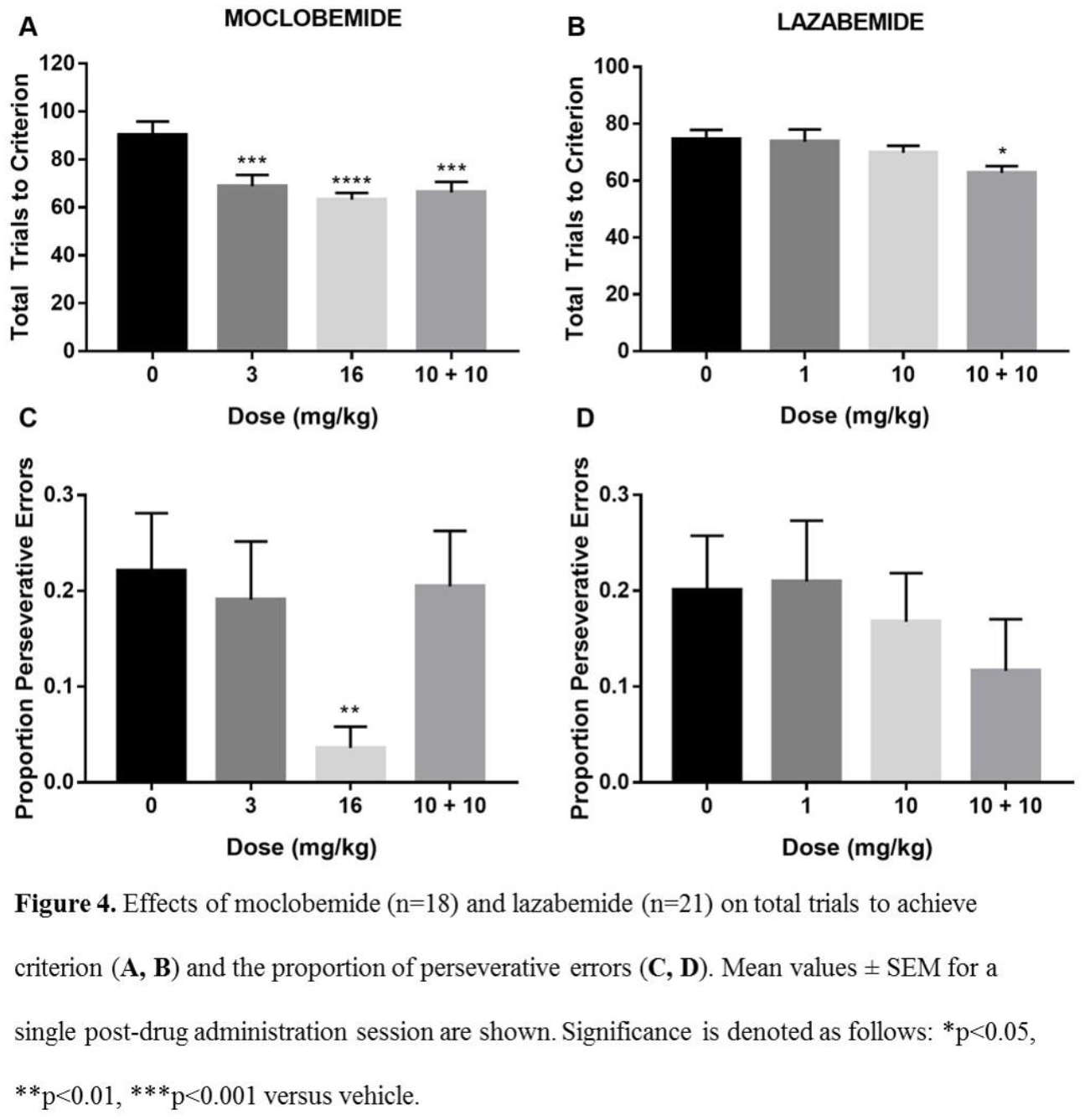
Safety signals are cues that predict the non-occurrence of aversive outcomes. They represent a specific variant of conditioned inhibition, insofar they reduce fear responses (e.g., freezing) upon presentation and are detected using summation and retardation tests. Thus, Pavlovian conditioned inhibition of fear (or learned safety) can be conceptualized as a learning process protecting against chronic stress, a predisposing factor for various psychopathologies. Conversely, serotonin release is triggered in response to stress, especially in key brain regions driving fear conditioning and expression. Whilst selective serotonin reuptake inhibitors (SSRIs) can effectively treat affective and anxiety disorders, they also produce anxiogenic effects prior to their clinical action (i.e., activation syndrome). Here, we report that an acute dose (i.p.) of the SSRI, escitalopram, leads to enhanced fear extinction and impaired safety signalling expression in naïve rats. We also show that the selective 5HT2C receptor antagonist, SB242084, neither consistently improves nor impairs acquisition and expression of safety in naïve rats but does exert baseline dependent effects on freezing behaviour. We conclude that enhanced global serotonin efflux during initial treatment with SSRIs may impair newly acquired inhibitory associations while inducing perseveration of excitatory ones.
Background:
Task switching paradigms are widely used to index cognitive flexibility. In clinical research, task performance can be used to monitor disease progression or to evaluate the effectiveness of interventions. Good test-retest reliability is required to determine the sensitivity of such studies and contribute to the interpretation of changes over time, or following interventions. Here, we sought to determine the stability of performance and brain activations using an fMRI task.
Methods:
Fourteen older healthy subjects were scanned in three sessions while performing a switching task (age: 60.5±7.8 yo; time intervals from the 1st scan: 5.8±2.0, 28.1±5.2 days for the 2nd and the 3rd scan respectively). A multivariate pattern classification was adopted for imaging data analysis given that this approach may be more appropriate for our task switching paradigm (Kehagia et al., unpublished). Imaging data were pre-processed and modelled in SPM12. Beta images for both repeat and switch conditions were subsequently submitted in pattern analyses. Classifiers for the first two sessions were evaluated and used for categorical predictions on the following ones using customised Matlab scripts and functions from PRoNTo. Medians of the third intraclass-correlation coefficient (ICC) for both behavioural and image data were calculated in a locally developed toolbox for the reliability assessments (Caceres et al., 2009).
Results:
Prominent switch costs were observed for all sessions and this behavioural index has good overall reliability (ICC=0.69) with the highest ICC for sessions 2 and 3 (ICC=0.80). The highest reliability of classification probability was also found between the last two sessions for differentiating brain networks involving in repeat and switch conditions (ICC=0.60). Scans acquired from the 1st session showed poor ability for reliably classifying conditions at the 2nd (ICC=0.39) and the 3rd (ICC=0.33) time point.
Conclusion:
We concluded that behavioural performance and brain activation pattern were highly consistent at the time-point two and three. When using the task switching paradigm repeatedly, an initial full-length training session is recommended in order to achieve a stable level of performance.
We have previously reported that bilateral transection of the lingual and chorda tympani nerves (Nx) results in behavioral depression in rats. Anhedonia, a core symptom of depression, can be easily measured by decreased consumption of sweet solutions in rodent models. Sucrose consumption was significantly reduced in Nx rats compared to sham operated controls, revealing anhedonic feature of Nx-induced depression. This study was conducted to examine if Nx-induced depression is mainly due to the loss of chorda tympani (taste) nerves rather than the loss of lingual (somatosensory) nerves. After a week of post-operational recovery from the bilateral transection of chorda tympani nerves (CTx) or Nx surgery, rats were subjected to a three-bottle preference test (one sucrose and two water bottles) daily for 9 consecutive days, and then to forced swim test. Nx rats did not prefer to drink sucrose during the whole experimental period. However, CTx rats drank more sucrose than water during the whole test period, although sucrose intake was reduced in CTx rats compared to sham rats during the first two days of the test. Immobility during the swim test was increased in Nx, but not in CTx, compared to sham rats. Stress-induced corticosterone increases did not differ among the experimental groups. Neuronal activities in the nucleus accumbens are currently under investigation. Results suggest that disruption of oral somatosensory, but not taste sensory, relay from the anterior two thirds of the tongue may induce depression with anhedonic feature, and the stress-axis function may not be involved in its underlying mechanism.
Supported by a grant from National Research Foundation of Korea through the Oromaxillofacial Dysfunction Research Center for the Elderly (2015048003) at Seoul National University in Korea.
Introduction
Leda-1/Pianp is a type-I transmembrane protein initially identified in rat liver endothelium. The Leda-1/Pianp protein is highly conserved among mammals. Transcript analysis and western blotting revealed the highest expression levels of Leda-1/Pianp in the CNS of human, rats and mice. Expression is also found in human astrocytes, glioblastoma cell lines and BALB/c but not C57BL6/J mice lymphoid organs (lymph node, spleen and thymus). The protein is glycosylated and undergoes multiple steps of proteolytic processing. Its N-terminus is cleaved by pro-protein convertases like Furin, ADAMs, MMPs. Subsequently the y-secretase complex cleaves it intramembraneously. The only known interaction partner of Leda-1/Pianp is the immune inhibitory receptor PILRa. However the function of Leda-1/Pianp in the CNS is so far not characterized.
Aim
Characterization of Leda-1/Pianp expression in different brain regions and behavioral phenotyping of Leda-1/Pianp knock out mice.
Methods
Expression analysis of mouse brain subregions by quantitative western blotting. Behavioral tests to assess locomotor activity, stress resisting behavior, anxiety like behavior, delayed fear conditioning and sociability of Leda-1/Pianp knock out mice.
Results & conclusion
Leda-1/Pianp-/- mice were viable. Also body weight and plasma lab values were comparable to wild type mice. CT and MRI imaging did not reveal major organ malformation or vascular abnormalities. Isolation and western blotting of several mouse brain regions showed that Leda-1/Pianp was expressed in all brain regions at variable levels. General and emotional behavioral tests showed that Leda-1/Pianp-/- mice were hyperactive under novel caging conditions and highly mobile under stressed conditions. These mice were also highly hesitant to explore novel objects, open arm of an elevated plus maze, brightly lit area and another social partner. These mice also showed impaired contextual learning, higher self grooming time and less nest building from cotton-nest. Overall these findings indicate functional involvement of Leda-1/Pianp in several brain functions including locomotor activity, stress coping, anxiety, learning and sociability.
Injury to optic nerve (ON) axons plays a major role in glaucoma progression. ON crush is an established model of axonal injury which results in retrograde degeneration and death of retinal ganglion cells (RGCs). However it is unknown how signal transmission to higher visual structures such as primary visual cortex (V1) is affected after ON crush.
Unilateral ON crush was performed on left eyes of adult C57BL/6 mice. Binocular V1 function of the contralateral (right) hemisphere was assessed longitudinally by optical imaging (OI) and in vivo two-photon calcium imaging under anaesthesia before and at 2d, 7d and 14d after ON crush. RGC numbers were quantified by counting Hoechst labelled cells in flat-mounted retinas. We found a significant cell loss in the RGC layer compared to normal adults after 30 days ON crush. Cell loss occurred progressively with 15% of cells lost after 7 days and 43% of cells lost 14 days after ON crush. OI experiments demonstrated an immediate significant shift in ocular dominance index towards the ipsilateral, intact eye and an almost complete loss of response in V1 to contralateral eye stimulation in all ON crush animals. Additionally we found that response magnitude to ipsilateral eye stimulation significantly increased after long term ON crush. Two-photon experiments revealed that responses to ipsilateral eye stimulation were increased along with a significant increase in orientation selectivity index of neurons in layer 2/3 of binocular V1.
ON crush causes acute and permanent loss of signal transmission from the retina to V1. The observed increase of responsiveness in V1 to intact eye stimulation indicates that severe ON injury in adulthood may evoke cortical plasticity that is normally seen during the critical period.
The primary motor cortex (M1) plays a fundamental role in the execution of skilled, dexterous motor behaviours. Descending motor output from cortical and brainstem motor areas shape the activity of spinal cord circuits to execute different types of movement from simple locomotion to skilled motor behaviours. Over the past century, work on human and non-human primates has significantly advanced our understanding of how cortical and brainstem motor areas coordinate their activity to achieve high-level motor control. But, how population representations of movement are organised in M1 still remains largely unresolved. To address this, we used two-photon calcium imaging of neuronal populations in head-restrained mice that were trained to execute a cued lever push-pull task for reward. Mice were injected with an adeno-associated virus (AAV1.Syn.GCaMP6s.WPRE.SV40) to express the genetically encoded calcium sensor GCaMP6s in layer 2/3 (L2/3) and layer 5 (L5) neurons in M1, habituated to head restraint and then trained to execute alternating lever push-pull actions in response to a 6kHz auditory tone (average training time to achieve ‘expert’ level: 11±2 days, 1 session/day). We performed population calcium imaging in contralateral forelimb M1 at depths corresponding to L2/3 and L5B, extracted ΔF/F values for individual neurons, and aligned them to behaviourally relevant time points (e.g. cue onset, movement onset, etc.). In a subpopulation of L2/3 and L5B neurons we observed tone-evoked calcium responses in trials where mice failed to execute a lever push or pull (“missed tone trials”), suggesting sensory-evoked responses likely contribute to M1 population responses during task engagement. During lever push-pull sequences, 96% of L2/3 neurons and 90% of L5B neurons displayed task-related activity with significant calcium changes seen during both push and pull movements. However, approximately 40% of L2/3 and L5B neurons displayed asymmetric calcium responses during push vs pull sequences, consistent with neuron-specific direction sensitivity in M1. Currently we are investigating the mechanistic underpinnings of direction sensitivity in M1 and how complex spatiotemporal patterns of L2/3 and L5B neuronal activity map onto different aspects of skilled forelimb behaviour.
Perturbations introduced in visuomotor adaptation tasks produce prediction errors, which are used to update forward models to generate more accurate sensorimotor predictions. Prediction errors are precision-weighted meaning an estimate of the inverse variance of the afferent signal (sensory precision) and the motor prediction (prior precision) is used to determine how readily prediction errors update forward models. These parameters are modulated during adaptation however their neurophysiological correlates are not fully understood. It has been suggested that beta oscillatory activity over sensorimotor cortex encodes this Bayesian updating process (Tan et al, 2014, 2016; Torrecillos et al, 2015). Here we designed a paradigm in which prediction errors were modulated orthogonally to precision parameters to determine their specific neurophysiological correlates. EEG was recorded from 24 subjects whilst they completed a visuomotor adaptation task in which angular perturbations were introduced into the visual feedback of a cursor during a finger reaching task on a track pad. High and low visual noise was introduced into the position of the cursor to modulate precision-weighting in a blockwise manner. A Bayesian hierarchical generative model (the HGF; Matthys et al, 2014) was used to track subjects’ beliefs about motor predictions, prediction errors and precision estimates throughout the task and these were correlated with the EEG data.
A GLM including prediction error, prediction, sensory precision and prior precision was used to regress oscillatory activity in an ROI over sensorimotor cortex with each predictor. Analyses were carried out before a trial (aligned to the GO signal to move) to measure the role of preparatory beta activity, and after a trial (aligned to movement offset) to measure the role of the PMBS. The results showed that in both periods beta oscillatory activity significantly correlated with prior precision (p<0.05). The results suggest that sensorimotor beta activity encodes the uncertainty around updated Bayesian motor predictions necessary to adapt subsequent behaviour. Further analyses are required to compare different perceptual models and explore other neurophysiological correlates of these parameters outside of sensorimotor cortex.
Our brain receives a series of sensory snapshots of the external world, which it must integrate to provide a description of the scenes around us. Vestibular inputs monitor changes in the position of the body relative to the environment. Here we tested the hypothesis that the vestibular system contributes to bridging the gap between successive visual snapshots of the external world. Accordingly, if gravitational signals provided by the vestibular organs cannot be aligned with those from vision, altered perceptual experiences may occur. This might underlie perceptual errors reported by pilots and astronauts exposed to altered gravitational forces.
We investigated the contribution of vestibular-gravitational signals to the process of updating perception of a series of visual scenes. Ten participants were seated facing outwards on a short arm human centrifuge (SAHC) platform, which simulated 1 +Gz artificial gravity for ten minutes at head level. A visual judgement task was performed at normal gravity baseline and during 1 +Gz artificial gravity, in counterbalanced order. An environmental scene (Scene A) was followed after a short delay by a second scene (B) involving slight perspectival modification of Scene A. The scenes differed either in angular perspective (as if the participant had turned leftwards or rightwards during the delay) or in translational perspective (as if the participant had moved forward or backwards). Participants judged whether the implied viewpoint change between the first and second scene corresponded to a left/right-ward rotation (angular perspective), or to an approach/retreat (translational perspective).
Artificial gravity influenced the perceived relation between the visual images: participants judged the second scene as significantly closer during 1 +Gz artificial gravity compared to a normal gravity baseline (t(9)=2.568, p=0.030). No differences were found in judgements of angular perspective. This dissociation rules out non-specific effects of artificial gravity or centrifugation which cannot readily explain this specificity. Our results support a vestibular-driven updating process in which gravity signals are computed to provide a dynamic description of the spatial position of the body relative to the external environment.
The spinothalamic tract (STT) forms the primary ascending projection system for thermoceptive and nociceptive A-delta and C-fibre afferents. On neuroanatomical grounds input from low threshold mechanosensitive C-Tactile (CT) afferents, which are hypothesised to encode the pleasant/affective nature of touch, and pruriceptors are also likely to ascend via the STT. However, direct evidence is lacking.
We assessed for alterations in affective touch and cowhage induced itch in patients undergoing STT lesioning for unilateral cancer related pain. STT lesioning resulted in contralateral thermal sensation deficits. Contralateral cowhage induced itch and pain was abolished. Pleasantness ratings for CT optimal (3cm/s) and sub-optimal (0.3 and 30cm/s) stroking touch showed no significant difference before and after lesioning or between lesioned and non-lesioned sides. However a significant contralateral reduction in CT preference index ([rating for 3cm/s x 2 - ratings for 0.3cm/s + 30cm/s] / 2 ) (p<0.005) was observed following lesioning. Ipsilateral itch and pleasant touch were unaffected.
The findings support the hypothesis that information salient to affective touch and pruriception ascend in the STT. Unlike the dramatic changes in thermoception, nociception and itch the effects on affective touch are subtle. This may reflect incomplete STT ablation or integration of A-beta and CT afferent inputs.

Rationale. Chronic neuropathic pain is a common, intractable and frequently debilitating consequence of neuromyelitis optica spectrum disorder (NMOSD) and has received relatively little attention in the literature, despite a prevalence within NMOSD of 80% and high subjective pain ratings(1). NMOSD is an immune mediated disorder that frequently targets the spinal cord causing inflammation, secondary demyelination and subsequent axonal loss and grey matter damage. This study investigates whether chronic pain severity in NMOSD relates to the craniocaudal location of culprit spinal cord lesions.
Method. Initially a retrospective cohort of 76 NMOSD patients from Oxford and Liverpool’s national clinics were assessed for current pain (brief pain inventory, BPI) and craniocaudal location of cord lesion contemporary to pain onset (clinical MRI). A subsequent focused prospective MRI study of 26 NMOSD Oxford patients, a subset of the retrospective cohort, assessed current craniocaudal lesion location and current pain (again BPI).
Results. Patients with isolated thoracic cord myelitis at the time of pain onset were significantly more disabled and suffered more pain. Furthermore, cervical and thoracic lesions that persisted from pain onset to “out of relapse” prospective MRI had highly significant (p<0.01) opposing effects on pain scores (std.?= -0.46 and 0.48, respectively; see figure). Lesion length, total lesion burden and number of cord relapses did not correlate with pain.
Conclusions. Persistent, caudally located (i.e. thoracic) cord lesions in NMOSD patients associated with high chronic pain scores, irrespective of number of spinal cord attacks, lesion length, and lesion number. Although disability correlated with pain in isolation, it became an insignificant predictor of pain when regressed alongside craniocaudal location. A similar association of severe pain with thoracic cord lesions has been described in a related neuroinflammatory disorder and an autonomic hypothesis has been proposed(2).
1. Bradl, M. et al. Pain in neuromyelitis optica–prevalence, pathogenesis and therapy. Nat. Rev. Neurol. 10,529–536(2014)
2. Okuda, D. T. et al. Central neuropathic pain in MS is due to distinct thoracic spinal cord lesions. Ann. Clin. Transl. Neurol. 1,554-561(2014)

During perceptual decisions, converging findings from electrophysiological and neuroimaging studies suggest that, a decision variable integrates sensory information until reaching a decision threshold. This allows, as supported by recent evidence, rapid decisions to be committed long before the end of incoming sensory information. However, it is largely unknown whether the brain processes sensory information consistently during decision-making.
Here, we investigated the neural representations of sensory information throughout trials, combining electrophysiological recordings with high temporal resolution (magneto encephalography, MEG; and electroencephalography, EEG) and multivariate pattern recognition approaches. Neural dynamics were recorded using a 306-channel MEG system with concurrent 70-electrode EEG. Eighteen healthy participants were presented auditory click trains at 40 Hz. On each trial, the auditory stimulus was comprised of a 750 ms click train during which each click was binaural (uninformative segment), followed by a 1000 ms train during which each click was monaural (informative segment, i.e., each click presented to either to the left or the right ear). Participants were instructed to decide, at the end of each trial, whether the left or right ear received more clicks.
By parametrically changing the ratio of monaural clicks to left/right ears, decision accuracy varied from the chance level (50%) to 90%. On pre-processed MEG/EEG data, we used linear support vector machine with cross-validation procedure to classify from all sensors and electrodes, at each time point, trials presented with left vs. right clicks. Classification accuracy on transit information from click trains was significant (p<0.05, FDR corrected) throughout the informative segment, at around when the stimulus was delivered, with a peak delayed by 150~200 ms. Classification accuracy at the uninformative segment remained at the chance level.
Our findings suggest that electrophysiological responses are constantly sensitive to incoming sensory information, regardless whether a pending decision has been made. Therefore, the human brain may process sensory evidence independent of cognitive demands, with relevant information being encoded in the cortex.
Animal models of tinnitus have revealed long-term hyperexcitability and altered neural synchrony, thought to arise from pathology affecting the balance between excitation and inhibition in the auditory system. This balance is regulated by neuromodulators, such as endogenous cannabinoids (endocannabinoids). Cannabinoid drugs are potent anti-nociceptive agents in models of chronic neuropathic pain, a condition that shares substantial parallels with tinnitus, i.e. phantom sensory percept in the absence of sensory input, initiated peripherally through deafferentation and subsequently involving central mechanisms. We therefore sought to determine whether the highly-selective CB1 agonist arachidonyl-2′-chloroethylamide (ACEA) could abolish putative neural mechanisms of tinnitus.
Guinea pigs (GPs) were first implanted with electrocorticography (ECoG) multi-electrode assemblies. Following baseline data collection, GPs were given intraperitoneal injections of either (1) sodium salicylate in order to induce tinnitus (350 mg kg-1; n = 8), (2) salicylate co-administered with ACEA (1 mg kg-1; n = 5), or (3) ACEA alone (1 mg kg-1; n = 4). Resting-state and auditory-evoked neural activity recorded in awake GPs was compared between groups. Hearing status was assessed using the auditory brainstem response (ABR).
Cluster-based permutation analysis indicated that salicylate altered resting-state activity, specifically by reducing alpha band activity (6-10 Hz) in cortical oscillations. Auditory-evoked responses were also enhanced (between 79-145%), whilst wave I ABR amplitudes were significantly decreased at 20 kHz (p < 0.01 for both left and right ears). Co-administration of ACEA still resulted in slight reductions in ABR amplitudes, but these were no longer significant (p = 0.07 left ear; p = 0.2 right ear). Decreases in oscillatory activity at 6-10 Hz were no longer evident, although enhanced cortical potentials were still present (between 61-159%). Administration of ACEA alone did not significantly affect auditory system function.
These data indicate that manipulating endocannabinoid signalling in a tinnitus model can affect some of the underlying neural mechanisms. We are currently collecting data to determine whether ACEA can also abolish behavioural evidence of tinnitus.
The primary motor cortex (M1) is a key brain area for the generation and control of complex motor movements. Output from M1 can be driven by long-range inputs from a collection of thalamic nuclei termed the motor thalamus (MTh). However, the role of MTh and precisely how its activity shapes the membrane potential dynamics of M1 projection neurons during skilled motor behaviour remains largely unresolved. To address this issue we first defined the 3D anatomical coordinates of mouse forelimb motor thalamus (MThFL) by employing conventional retrograde and viral-based tracing methods targeted to mouse forelimb motor cortex (M1FL). These complementary approaches defined MThFL as a ~0.8 mm wide cluster of neurons with central coordinates 1.1 mm caudal, 0.9 mm lateral to bregma and 3.2mm below the pial surface. Thus, MThFL incorporates defined areas of the ventrolateral, ventral anterior and anteromedial thalamic nuclei. To investigate the role of M1FL and MThFL during skilled motor behaviour, we developed and optimised a quantitative behavioural paradigm in which head-restrained mice execute a cued lever push task for reward. Forelimb movement trajectories were mapped using high-speed digital imaging and multi-point kinematic analysis. Independently inactivating M1FL or MThFL using a pharmacological strategy resulted in altered forelimb kinematic trajectories and a significant reduction in task performance in skilled mice. By combining whole-cell patch-clamp electrophysiology and two-photon population imaging in M1FL, single unit recordings in MThFL, and optogenetic manipulation strategies in vivo, we have generated new insights into how motor thalamus shapes motor cortical output and skilled motor behaviour.
Voluntary movement is accompanied by changes in beta oscillations in the motor cortex. This study uses rhythmic TMS (rTMS) at the individual’s motor beta frequency to modulate beta oscillations during the movement preparation phase, allowing us to explore their causal role in encoding the uncertainty of future movements and features of the movement execution itself.
18 participants performed a delayed reaching task. During the movement preparation phase, directional uncertainty about the upcoming movement was manipulated by varying the number of spatial cues (1, 3 or 12 spatial cues indicated potential locations for the target). Afterwards, another cue was presented indicating the actual target, upon which participants were instructed to move a joystick-controlled cursor to the target in a ballistic movement as quickly and accurately as possible. Participants performed 4 blocks in counterbalanced order (72 trials each): 1 without stimulation, and 3 ‘stimulation condition’. In the latter, 10 TMS pulses were delivered over the contralateral motor hand area during the movement preparation phase of each trial. The trains of pulses were either regular at individual beta frequency, or irregular with random intervals as a control. EEG was continuously acquired from 23 sites, while measuring the reaction time, movement velocity, and accuracy of each movement.
We found that increased target uncertainty resulted in increased reaction time (p<.001) and less beta desynchronization during movement preparation (bilaterally, p=.007). However, target uncertainty did not change the level of beta following presentation of the definitive target (p=.347), and did not affect the maximal speed (p=.511) or accuracy of movement execution (p=.055). Beta frequency rTMS did not change the reaction time or its dependence on uncertainty; rather, it led to a decrease in maximal speed of the forthcoming movement (p<.001) independent of the uncertainty condition, compared to irregular rTMS.
These results suggest there may be two broad components to beta oscillations in motor cortex: the first is spread bilaterally over the motor cortex, related to overall motor readiness, and modulated by uncertainty. The second is more lateralised and related to the actual execution of the movement.
Simple button presses are the most common response modality used in cognitive testing. However, it is an oversimplification of continuous and dynamic decision-action cycles in our daily lives. Here, in two perceptual decision-making experiments, we first investigated the validity of using joystick as a way of collecting responses (experiment 1), then demonstrated the contextual effect of preceding actions on forthcoming decisions in continuous action paradigm (experiment 2).
In experiment 1, participants were instructed to detect the coherent motion direction of random dot kinematogram from four possible alternative directions. In two counterbalanced sessions, responses were collected either using button presses or joystick movements. Bayesian multivariate test showed strong evidence in favour of the hypothesis, that the decision-making is not affected by response modality (reaction time: logBF=2.25; response accuracy: logBF=10.05). Furthermore, responding using joystick enabled to extract measures, describing action components unobservable in key presses (e.g., latency of movement’s peak velocity, and precision of direction detection).
In experiment 2, participants were instructed to perform continuous circular movements using joystick (clockwise/anti-clockwise), in response to the coherent motion direction in random dot kinematogram. In the second half of each trial, there was 50% probability that motion coherence and motion direction would change, signalling the change of circular movement direction. Bayesian multivariate test showed strong evidence for latency of action change being affected by motion strength (logBF=12.78). Lowered motion strength affected the accuracy more than change of direction only. Moreover, in trials when both direction and motion strength changed, accuracy was the lowest (logBF=15.46). Therefore, contextual information affected the subsequent decision processes.
Our findings suggested that joystick is a viable way to acquire behavioural responses in rapid decisions, with response profiles comparable to traditional button presses. Detailed view of the action, with relation to the sensory input, can help investigate sensory and motor components of sensorimotor transformation.
Several psychological and neurophysiological studies have suggested that decision-making is linked to the accumulation of evidence over time. Decision-making is often accompanied with decision confidence, in which lower decision confidence more likely leads to change of mind. There is also significant neural evidence that the brain uses error monitoring mechanisms to monitor and correct potential errors. These phenomena provide evidence of metacognition in animals and humans. While current computational models especially those based on post-decisional evidence accumulation provide good quantitative fit to behavioural data, they are abstract and do not provide neurally plausible assumptions nor take into account error-correction mechanisms.
This work proposes a computational neural circuit model of perceptual decision-making that can account for decision confidence, change of mind, and multimodal action outputs. The model, building on previous biological models of perceptual decision-making, consists of a decision module, a metacognitive module, and motor output modules. The modules are modelled by nonlinear firing-rate type neural model that exhibit winner-take-all behaviour. Our model is then applied to account for data from a perceptual decision-making experiment that we have previously conducted. The experiment has explored the relationships among saccadic eye movement, hand movement, and choice behaviour in a well-known motion discrimination task (“random dots”) paradigm. Our model can account for the psychophysical data and eye-hand trajectories, including change of mind trials. Finally, our model has dynamic features that can allow it to potentially adapt to other task paradigms.
Overall, we have developed a neural circuit model that sheds light on the interactions among decision confidence, metacognition and multimodal actions.
Previous studies have reported that the planum temporale - a language-related structure that normally shows a leftward asymmetry – had reduced asymmetry in people who stutter (PWS) and reversed asymmetry in those with severe stuttering. These findings are consistent with the theory that altered language lateralization may be a cause or consequence of stuttering. Here, we re-examined these findings in a larger sample of PWS. We evaluated planum temporale asymmetry in structural MRI scans obtained from 67 PWS and 63 age-matched controls using: 1) manual measurements of the surface area; 2) voxel-based morphometry to automatically calculate grey matter density. We examined the influences of gender, age and stuttering severity on planum temporale asymmetry.
Results showed that the size of the planum temporale and its asymmetry were not different in PWS compared with Controls using either the manual or the automated method. Both groups showed a significant leftwards asymmetry on average (about one-third of PWS and Controls showed rightward asymmetry). Importantly, and contrary to previous reports, the degree of asymmetry was not related to stuttering severity. In the manual measurements, women who stutter had a tendency towards rightwards asymmetry but men who stutter showed the same degree of leftwards asymmetry as male Controls. In the automated measurements, Controls showed a significant increase in leftwards asymmetry with age but this relationship was not observed in PWS.
We conclude that reduced planum temporale asymmetry is not a prominent feature of the brain in PWS and that the asymmetry is unrelated to stuttering severity.
Mirror visual feedback is used to treat painful conditions associated with alterations of body image resulting from neuropathy, amputation and complex regional pain syndrome. In clinical practice, mirror feedback techniques tend to use normal-sized reflections of body parts. The findings of studies on pain patients and healthy participants exposed to noxious stimuli suggest that observing magnified and minified body parts using mirrors, lenses, or virtual reality may affect pain perception. However, the direction of effect varies between studies. The aim of this study using healthy participants was to compare the effect of observing a magnified and minified reflection of the hand in front of a mirror on the intensity of experimentally-induced contact heat pain of the hand hidden behind the mirror.
A within-subject repeated-measures design was used with three reflection conditions (normal sized, magnified and minified) and two view contexts (reflected hand and box where participants observed a reflection of their left hand covered by a cardboard box).
Participants rated the intensity (NRS) of noxious heat stimuli set at a temperature 2oC above pain threshold and delivered to the skin of the right hand using a 30x30 mm thermode attached to a TSA-II Neurosensory.
Eighteen pain-free healthy volunteers (10 females; mean ± SD age = 24.3 ± 3.1 years) participated in the study. There was a significant main effect of reflection (F(2,34) =6.48, p = 0.004, ƞp2 = 0.276, power = 0.88). Pairwise comparisons showed that pain intensity was less when looking at the minified reflection compared with the magnified reflection (mean ± SD = 3.96 ± 1.75, 4.88 ± 1.31, respectively; p=0.001) but there were no differences between normal-sized reflection and magnified nor minified reflection (p > 0.05). There was no significant main effect of view context or reflection x view context interaction (all F’s < 4.86, n.s.).
Participants reported less pain when looking at a minified reflection irrespective of the reflection being of the hand or box. We are unable to determine at this stage whether this is a true finding or a result of methodological shortcomings contributing to a false negative finding.

It is well known that different directions of transcranial magnetic stimulation (TMS) current activate the motor cortex in different ways. A posterior-anterior (PA) induced current recruits a different sets of inputs onto the corticospinal neurones than the opposite anterior-posterior (AP) current. We recently found that pre-conditioning these two sets of inputs with TMS produced distinct effects on different types of motor learning [Hamada et al (2014), J Neurosci, 34, 12837]. We hypothesised that these circuits would differ in their sensitivity to the orientation of transcranial direct current stimulation (TDCS) currents, and that this would in turn influence the effects of concurrent TDCS and motor practice on physiological and behavioural outcomes.
Human volunteers received TDCS via two electrodes placed 3cm in front and 3cm behind the motor hotspot for the hand with either an anterior anode (AAn), posterior anode (PAn) or sham (S) TDCS. The electrodes were aligned with the preferred orientation of PA TMS. Expt.1: 15 volunteers received TDCS-PAn for 10min at 1mA. MEPs evoked by PA and AP TMS pulses were measured before and at 10min intervals afterwards. Expt. 2: 30 volunteers received TDCS-PAn, TDCS-AAn or TDCS–S during practice of a ballistic thumb acceleration task, which was used to assess motor learning and retention (48h). PA- and AP-evoked MEPs were recorded before and after practice on day 1.
Expt. 1: TDCS with a posterior anode 22uppressed PA evoked MEPs to 75% of control values, but had no significant effect on AP evoked MEPs. Expt. 2: PA-evoked MEPs were suppressed after motor practice with TDCS-PAn compared to with TDCS-AAn and TDCS-S. TDCS-AAn tended to impair 48h consolidation of motor learning and overall learning on day 2 compared to TDCS-PAn and TDCS-S.
The primary motor cortex is highly sensitive to the direction of TDCS current flow. This is relevant physiologically in terms of effects on MEPs and appears to also be behaviourally relevant in terms effects on motor learning and retention. Given the similar organization across the neocortex, we expect that the anisotropy of stimulation applies to other areas of cortex, such as those more involved in cognition.

Background
Unilateral sensorimotor stimulation induce negative BOLD fMRI responses (NBR: decrease from baseline) in ipsilateral sensorimotor cortex (S1/M1). Cross-modal NBRs can also be evoked in unstimulated sensory cortex. Animal data suggest NBRs may represent GABAergic inhibition via decreased neuronal activity. In humans, functional significance of NBR and its relation to cortical excitability is unclear.
Here we used MRI and concurrent TMS-EEG during sensory tasks to link changes in motor-evoked potential (MEP) measures of cortical excitability to neural activity and individual NBR and GABA levels.
Methods
In 17 young-adult subjects four tasks were used to evoke S1/M1 NBR: right median nerve stimulation (MNS); sustained right-hand pinch-grip; visual full-field 7Hz reversing checkerboard; auditory beeps at 7Hz. We measured subjects NBR (8/16s on/off) to each task and right M1 GABA concentration with 3T MRI. In a second session we measured changes in EEG alpha and beta power to each stimulus (9/12s on/off), whilst single-pulse TMS to right M1 induced MEPs in the left thumb at: 1s (sT1), 7s (sT2), 11-12s (rebound, RB) and 18s (baseline, BL) post task onset. MEP differences between timings were examined by ANOVA.
Results
Significant MEP differences were found during: MNS, BL-sT1 (p=0.034) and BL-RB (p=0.023); motor, BL-sT2 (p=0.027). No visual/auditory MEP differences were found (Fig1 C). We observed bilateral S1/M1 de-synchronisation of EEG alpha and beta power during MNS and motor, strongest in left S1/M1 (Fig1 B). Visual or auditory stimuli evoked little S1/M1 response. NBRs were observed in right S1/M1, in order of magnitude: MNS, motor, auditory, visual (Fig1 A). GABA significantly correlated with MNS and motor NBR, with no correlations found for visual/auditory.
Conclusion
MNS results suggest corticospinal excitability decreases are coincident with NBR in that region. While motor NBR was coincident with significant sT2 MEP increase . The lack of a MEP-NBR correlation during visual/auditory stimuli suggests a different origin of cross-modal NBR, reflected in GABA/NBR correlations.
Further work to correlate: subject’s MEP changes to NBR; EEG response in S1/M1 to MEP changes and NBR.

A complex relationship exists between physical activity levels, psychological attitudes to pain and pain perception. In this study, quantitative sensory testing (QST) was utilized before and after acute exercise to determine whether this can modify an individual’s pain thresholds. In another study, psychological profiling was undertaken in subjects reporting variable levels of exercise engagement in their normal routine to assess a putative link between physical activity and attitudes to pain.
Thermal pain perception pre- or post-exercise was assessed using a QST protocol (Pathway System, MEDOC, Israel) for measurement of hot (HPT) or cold pain CPT) thresholds. Aerobic exercise was with a cycle ergometer (70-75% max heart rate) and isometric exercise used a hand-grip dynamometer (25% of maximum voluntary contraction, 5 repeats). For psychological traits, Fear of Pain (FoP) and Pain Catastrophizing (PC) questionnaires were used with subjects declaring their routine cardio activity levels as never/once per month (p/m); 1-3 times per week (p/w) or >3 times p/w. All protocols had local ethical approval.
After isometric exercise, the CPT was significantly lower (20.4oC in control versus 17.4oC with exercise, P<0.05, n=7) – indicative of ongoing hypoalgesia. A hypoalgesic trend was also observed for HPT i.e. a post-exercise increase in HPT although data were not significant (P>0.05). After aerobic exercise, the CPT was significantly lower, indicating induction of hypoalgesia (15.3oC in control versus 4.61oC with exercise, P<0.05, n=6) whereas the HPT was unaltered. With respect to exercise engagement, participants engaged in exercise >3 times p/w showed significantly lower PC scores compared to those with less engagement, for example once p/m (>3 times p/w, mean score = 3.3; once p/m = 17.4; P<0.05, n=39). Similarly for FoP scores, the lowest values were observed in those exercising >3 times p/w.
Overall, these preliminary data support the view that bursts of acute physical activity can modify an individual’s pain experience, as measured by QST. Furthermore, high levels of physical activity engagement are linked to psychological traits such as low PC and FoP but further studies are needed to establish the existence of a causal link.
Predicting the sensory consequences of one’s actions is critical to perception and action in dynamically changing environments. In many cases, there is a fixed, general relationship between a given action and its sensory consequences, such as visual flow during locomotion. Oftentimes, however, the sensory consequences of an action are highly context specific and must remain plastic. To cope with this, humans and animals are thought to build and store internal models (Wolpert et al., 1998), traditionally associated with cerebellum-like structures. More recently however, neurons have been reported in primary visual cortices, which signal mismatch between predicted and actual sensory feedback (Keller et al., 2012). It currently remains unclear whether cortical mismatch signals are specific to fixed sensorimotor contingencies or whether they may also arise with arbitrary, newly learned contingencies.
We developed and validated a novel task that requires head-fixed mice to learn novel, arbitrary motor-auditory associations. Animals were trained to freely “navigate” through a one-dimensional, abstract auditory landscape of equally spaced pure tones, by licking either of two lick-ports (see Figure). We show that mice are capable of using auditory feedback to adaptively guide behaviour, and are sensitive to unexpected feedback. Next, we used two-photon calcium imaging to investigate the functional properties of auditory cortical neurons in mice performing this task. Among the various acoustic tuning profiles encountered in auditory cortex, some otherwise sparsely active neurons were found to be particularly responsive following sounds that violated the learned sensorimotor contingency (see Figure). This activity was dependent on whether or not the animal was actively engaged in the task. Taken together, these results suggest that a network of neurons in auditory cortex is engaged in predicting the sensory consequences of motor actions, even when the relation between motor action and sensory feedback is arbitrary and newly learned.
Acknowledgements
Supported by the Wellcome Trust
References
Keller GB, Bonhoeffer T, Hübener M (2012), Neuron 74:809–815.
Wolpert DM, Miall RC, Kawato M (1998), Trends Cogn Sci 2:338-347.
Previous studies have suggested that people with Parkinson’s disease (PD) are not as efficient as age-matched controls in retaining certain kind of motor memories. We sought to investigate this phenomenon more thoroughly by testing for interference effects using force-field adaptation. Motor learning can be tested in the laboratory using motor tasks that perturb sensorimotor feedback (e.g. prism adaptation). Often, after learning such an adaptation a motor memory of the perturbation is formed, so that when exposed to the same perturbation for a second time performance is improved (also called savings). Motor memories can also contribute to an interference effect. This effect occurs when memory of a learned adaptation makes it harder to learn an opposite adaptation. We hypothesized that if motor memory is less robust in people with PD they should display smaller savings and diminished interference. Fourty-eight people with PD (PwPD) and 48 healthy age-matched controls performed a force-field adaptation paradigm with their upper-limb to test motor learning and motor memory. Participants held the handle of a robotic arm and made repetitive movements from a central location to a single-target. Initially all participants learned to adapt to a clockwise force-field (12 N/m/s) that was imposed on the handle during reaching. Recall (i.e. savings) of the learned force-field was tested after a 1-hour or 24-hour break, or – to measure the amount of interference - performance on adapting to an opposite (counter clockwise) force-field was tested instead. Therefore, both PwPD and Controls were split up into 4 subgroups, each of n=12. Results reveal that PwPD (1hour subgroup) show similar improvement compared to Controls (1h) when tested for savings, but display less interference when tested on the opposite force-field. These results are comparable to the 24-hour subgroups and were reflected both in the lateral deflection and force production. In line with our hypothesis we observed a reduction of interference in PwPD compared to Controls. In contrast, the amount of savings was similar for both groups. These results suggest that motor memory may still be preserved in PD and that the altered interference might be caused by problems with motor memory retrieval.
Noise induced hearing loss (NIHL) is classically divided into permanent or temporary forms. Individuals with temporary threshold shifts (TTS) will experience elevated hearing thresholds immediately following noise exposure, which resolves over several days or weeks. TTS was once thought to cause little lasting damage to the hair cell stereocilia or supporting structures in this form of auditory insult. However, recent evidence suggests TTS triggers “silent damage”: a type of neuropathic damage where there are reduced numbers of hair cells synapses onto spiral ganglion neuron (SGN) processes and secondly where a slowly developing neuronal death exacerbates presbyacusis (Kujawa & Liberman, J. Neurosci, 26: 2115-23. 2006; Jensen, et al., PLoS One 10. 2015). Previous studies of gene expression changes following noise insult have used whole cochlea preparations that do not differentiate between the changes in the different cochlear structures. Here we have used micro-dissection of the modiolus to focus on the SGNs and minimise the contribution from other cochlea structures.
CBA/Ca, female, P40 mice were exposed to 105 dB SPL (sound pressure level) broadband noise for 1.5hrs under anaesthesia. Age matched controls (sham) were also anaesthetised for 1.5 hours and exposed to silence. Auditory brainstem recordings (ABRs) were taken before noise exposure and at the time of tissue collection, showed the exposure protocol produced an immediate threshold shift of 36±3 dB SPL (Click) which recovers to 10±3 dB SPL by 28d. Mice were divided into three groups, and allowed to recover for 24hrs, 7d or 28d following exposure. After recovery, the modiolus was micro-dissected from both cochleae. RNA-Sequencing was performed on the Illumina NexSeq500 at the Deep Seq facility at the University of Nottingham.
Preliminary analysis of the RNA-Sequencing data has revealed 421 differentially expressed genes over this 28 day time period. In addition to changes in 57 neuron specific genes, Gene Ontology analysis shows that the acute phase inflammatory response persists over the 28d period, with chronic upregulation of apolipoproteins (A1, C1 & H), serum amyloids (A1/A2), serum albumin, and complement C3.
Cortical network activity during NREM sleep is dominated by slow waves, which are associated with reduced neuronal firing and are enhanced after sleep deprivation (SD). Although the network slow oscillation was originally described in the striatum, the effects of SD on the neuronal activity in this region has received little attention. In this study, we recorded local field potentials (LFPs) and neuronal spiking activity from the dorsal striatum (n=7) and the motor cortex (n=4) during NREM sleep in freely moving C57BL/6J mice. Mice were recorded during an undisturbed baseline day and after 6 h SD.
In both the cortex and striatum, the LFPs during NREM sleep were dominated by slow waves in the frequency range 0.5-4Hz (slow wave activity, SWA). As expected, positive peaks of LFP slow waves recorded in the cortex were associated with reduced neuronal activity. Slow waves recorded in the striatum were not merely volume conducted, but correlated with locally recorded neuronal spiking. However, in contrast to the cortex, positive LFP slow waves recorded in the striatum were associated with an increase in spiking, which was preceded and followed by a pronounced suppression of activity.
NREM sleep after SD was characterised by higher LFP SWA in both the striatum (first 2h: mean±SEM 178.2±8.9% change relative to baseline day) and the frontal cortex (mean±SEM first 2h: 166.9±6.2%), and was not significantly different between the two regions. Notably, in both regions, the surge of MUA after the period of reduced firing, occurred on average approximately 40 m earlier during recovery sleep after SD as compared to baseline.
This is the first report that sleep slow waves in the striatum are homeostatically regulated in mice. Our data suggest that increased sleep pressure affects both local neuronal activity in the striatum as well as the large-scale network activity within the corticostriatal circuit. Acknowledgement: BBSRC (BB/K011847/1), MRC (MR/L003635/1), FP7-PEOPLE-CIG (PCIG11-GA-2012-322050), Wellcome Trust (098461/Z/12/Z), The Uehara Memorial Foundation (TY), The Naito Foundation (TY).
Anatomical studies in non-human primates have shown that the ventral premotor cortex (PMv) has a number of indirect projections to the cervical spinal cord [e.g. via the primary motor cortex (M1) and the brainstem], but also provides direct projections terminating predominantly in the upper and less in lower cervical segments. Since PMv plays such a critical role in the control of hand movements it is somewhat paradoxical that direct projections to lower cervical segments are sparse. In humans, it is classically thought that PMv contributes to hand movements via dense corticocortical connections with M1, yet whether there are direct projections to the spinal cord remains unknown. Here we used transcranial magnetic stimulation (TMS) over PMv to condition H-reflexes elicited via peripheral nerve stimulation (PNS) whilst subjects sat at rest. TMS was delivered over the caudal part of the inferior frontal gyrus, while PNS was applied to the median nerve in order to elicit H-reflexes in the flexor carpi radialis muscle. Descending and ascending volleys arriving onto the spinal motoneurons were timed accurately by measuring the central conduction time (measured from M1) and peripheral nerve stimulation time for each volunteer. H-reflexes were either elicited alone (baseline) or conditioned by PMv TMS. We investigated 6 inter-stimulus intervals (ISI), namely -4, -2, 0, 2, 4 and 6 ms, and 3 TMS intensities (80%, 100% and 120% of resting motor threshold). Our results show that while threshold conditioning pulses over PMv had no effect on H-reflexes, subthreshold and suprathreshold PMv conditioning pulses significantly facilitated H-reflexes at a later ISIs (4 ms; 2 and 6 ms, respectively). Additional experiments revealed that these effects were not due to a spread of activation from PMv to M1 and that longer central conduction times could involve the propriospinal network. Our results allow us to speculate that recruiting sub- or suprathreshold PMv projections reveals a different time course of possible slower direct or indirect interactions. PMv projections have a net and diffused facilitatory effect on spinal excitability; with subthreshold and suprathreshold outputs interacting directly or indirectly with lower cervical segments.
Obesity is a low-grade inflammatory condition, associated with an increased risk of age-related cognitive decline, dementia and Alzheimer’s disease (AD). The factors predisposing the obese population to cognitive impairment remain poorly understood, but are believed to include insulin resistance, ER stress and neuroinflammation. AD is characterised by the deposition of the toxic peptide amyloid-β (Aβ) in the brain; the primary inflammatory stimulus. The role of toll-like receptors (TLRs) in mediating Aβ-induced inflammatory changes has been widely reported and we, among others, have highlighted the involvement of TLR2 in promoting Aβ-induced microglial activation and neuronal dysfunction. In addition, we have described the contribution of infiltrating peripheral macrophages to facilitating the neuroinflammatory environment and neuronal impairment associated with ageing.
While not all obese people will develop AD, subsets of the obese population are clearly more vulnerable. We hypothesise that obesity promotes the sensitivity of macrophages to inflammatory challenge, which access the brain and likely amplify Aβ-induced neuroinflammation. Here we have assessed TLR2- and TLR4-mediated inflammatory responses in bone marrow-derived macrophages (BMDMs) prepared from genetically obese animals, and from an animal model of diet-induced obesity (DIO). In addition, we have examined the impact of macrophage-derived inflammatory mediators on the activation of microglia.
The difficulty maintaining weight-loss in the obese population has recently received significant attention. This highlights the importance of developing strategies to alleviate the risk of co-morbidities, despite weight-loss. We have extended our analysis to examine whether weight-loss is essential to alleviate obesity-related inflammatory sensitivity. We have compared the responsiveness of BMDMs from obese rats following gastric bypass surgery, and a mouse model of DIO consisting of a diet rich in monounsaturated fats to promote metabolic health, despite obesity. We report that surgical-induced weight-loss consistently alleviates obesity-related sensitivity to TLR stimulation. However, a metabolically healthy obesogenic diet can also attenuate specific obesity-induced changes.
Ethanol consumption induces both acute and chronic behavioural changes. At the molecular level, ethanol interacts with GABA and Glutamate receptors and induces the release of both endorphins and dopamine in the brain. We have previously shown that chronic alcohol intake affects differential expression of specific G proteins in Drosophila melanogaster brain when measured by RT-PCR. We have since developed flies that express siRNA for Gi and Gq in a temperature sensitive manner and demonstrate that G protein down-regulation alters the normal alcohol tolerance behaviour. Flies exposed to ethanol for three consecutive days typically display an increase in the sedation time (measured as ST50: time at which half of a group of flies is sedated). The mutant flies carrying the siRNA genes showed normal tolerance behaviour when maintained at 18°C but activating siRNA expression at 30°C significantly increased their sedation time at day one with no further increase in additional exposures. This further demonstrates that G protein expression plays a key role in the alcohol induced sedation behaviour which is leading us to investigate whether the G proteins coupled to GABA-B receptors contribute to tolerance.
The ability to infer directionality between neural signals is an important aspect of quantifying neuronal interactions, typically using parametric approaches based on auto regressive models for the neural signals. Here we explore the use of a non-parametric approach, where directionality is decomposed into forward, reverse and zero-lag components. The study assesses directionality in left and right hippocampal CA1 and CA3 and local field potential (LFP) recordings before and after local unilateral kainic acid (KA)-induced epileptiform activity in a rat model of mesial temporal lobe epilepsy (mTLE). Isoflurane-anaesthetised Lister-hooded rats (300-400g; n=6) had microelectrode arrays positioned in the left and right hippocampus. A cannula was attached for local injection of kainic acid (KA) in the left hippocampus (HPC). After 30 min basal recording, KA (1 mM, 1 μL) was administered into the left HPC. LFP activity was recorded using a Plexon MAP system. All procedures were carried out in accordance with the Animals (Scientific Procedures) Act 1986, UK. The non-parametric directionality analysis was undertaken by splitting each 3.5 hr recording into 1 minute non overlapping segments and performing directionality analysis on each segment. The directionality analysis decomposes the coherence into three components, here we consider the forward and reverse components. KA injection interrupted the directionality between LFP signals. In the ipsilateral (left) HPC it led to a decrease in the forward component from CA3 to CA1, resulting in an increased CA1 to CA3 coherence. In the contralateral (right) HPC the effect was less clear, but there was a tendency to promote an increased CA3 to CA1 coherence. Bilateral interactions in the basal period were predominantly from left HPC to right HPC, these were reduced in magnitude following KA injection. The results demonstrate that non-parametric directionality analysis can detect externally induced changes in LFP interactions and can quantify the changes in coherence that result.
The neuronal extracellular matrix has a pivotal role in physiology and in pathophysiological conditions such as epilepsy. LGI1 (Leucine Rich Glioma Inactivated 1) is a secreted trans-synaptic protein which interacts presynaptically with Kv1.1 potassium channels and ADAM23. Postsynaptically, LGI1 influences AMPA and NMDA receptors through a direct link with the ADAM22 adhesion protein. Mutations of LGI1 lead to a familiar form of temporal lobe epilepsy with audiogenic features. However, the role of LGI1 in acquired epilepsy and how acute reduction of interactions between LGI1 and its pre- and post-synaptic partners affects temporal lobe circuits and synaptic transmission in epilepsy remains unclear.
Currently, we are testing a knock down approach to reduce LGI1 expression in vitro using shRNA delivery by viral vectors. First, we recorded neuronal network activity using a MEA (microelectrode array) system. We observed a two-fold increase in the MFR (mean firing rate) of the cultures transfected with shRNA compared to controls. We also used a complementary approach monitoring seizure-like activity in vitro with calcium imaging in high density cultures exposed to picrotoxin. Treatment with LV-shRNA-LGI1 increased the frequency of calcium bursts.
Parallel in vivo experiments on rats aim to understand if LGI1 concentrations in the brain are affected after generation of spontaneous seizures. In particular, we used the perforant path stimulation model of chronic temporal lobe epilepsy. Induction of limbic status epilepticus with stimulation of perforant path fibres, results in later spontaneous seizures in the hippocampus and long-lasting tonic clonic attacks which begin a few days after the treatment. Results show a sharp decrease of LGI1 concentrations three weeks after severe status epilepticus within the ipsilateral side of the hippocampus.
Taken together our results show that LGI1 concentrations directly influence spontaneous network activity and seizure-like activity in vitro and that LGI1 expression in the hippocampus is altered after 21 days of chronic epilepsy in freely behaving rats.
The non competitive NMDA receptor antagonist, phencyclidine (PCP) causes behavioural deficits in experimental animals, which model schizophrenia, reflecting glutamate hypoactivity in the disease. Dopamine (DA) signalling is also disrupted in schizophrenia raising the possibility of a dysregulation of glutamate/DA signalling in expression of symptoms. In addition, acetylcholine, which is known to modulate DA release in striatum, also shows changes after PCP pretreatment: thus altered DA function after PCP may be mediated through disruption of cholinergic signalling, either indirectly via glutamatergic control of cholinergic activity, or through a direct effect of PCP on cholinergic receptors. However, the extent to which PCP activation of the cholinergic system leads to psychotomimetic effects is not yet clear. We hypothesis that PCP treatment affects DAergic transmission through either direct or indirect effects on cholinergic systems.
We have investigated the effect of subchronic pretreatment with PCP or saline on dopaminergic and cholinergic activation of the brain measured by pharmacological magnetic resonance imaging (phMRI). Animals were scanned twice at weekly intervals, under isoflurane anaesthetic, using a 9.4T preclinical MRI scanner (Agilent Technologies), and the effects of amphetamine and nicotine injections on blood oxygenation dependent (BOLD) contrast were assessed. They were then treated with PCP (2mg/kg, twice daily for 5 days) and left drug free for 10 days, before repeating the drug challenges during phMRI.
Both amphetamine and nicotine increased BOLD signal in cortical and striatal areas of the brain. Preliminary first-level analysis of phMRI data shows enhancement of amphetamine-stimulated BOLD signal, and attenuation of nicotine-stimulated BOLD activation following PCP treatment. These experiments show the utility repeated phMRI in rats to measure changes in transmitter systems following PCP pretreatment, modelling schizophrenia, and that PCP pretreatment causes opposite changes in responsiveness to amphetamine and nicotine. Therefore, cholinergic systems may be important in the dysregulation of dopaminergic transmission seen after PCP pretreatment, and may be important for understanding dysfunctions underlying schizophrenia.
In vivo, theta (4-7 Hz) and gamma (30-80 Hz) neuronal network oscillations are known to coexist and display phase-amplitude coupling (PAC). However, in vitro, these oscillations have for many years been studied in isolation. Using an improved brain slice preparation technique, we have, using co-application of carbachol (10 μM) and kainic acid (150 nM), elicited simultaneous theta (6.6 ± 0.1 Hz) and gamma (36.6 ± 0.4 Hz) oscillations in rodent primary motor cortex (M1). Each oscillation showed greatest power in layer V. Using a variety of time series analyses (Modulation Index, wavelet ridge analysis, bi-coherence and bi-spectral analyses) we detected significant cross-frequency coupling in some, but not all, preparations.
Differences were observed in the pharmacological profile of each oscillation. Thus, gamma oscillations were reduced by the GABAA receptor antagonists, gabazine (250 nM and 2 μM), and picrotoxin (50 μM) and augmented by AMPA receptor antagonism with SYM2206 (20 μM). In contrast, theta oscillatory power was increased by gabazine, picrotoxin and SYM2206. GABAB receptor blockade with CGP55845 (5 μM) increased both theta and gamma power, and similar effects were seen with diazepam, zolpidem, MK801 and a series of metabotropic glutamate receptor antagonists. Oscillatory activity at both frequencies was reduced by the gap junction blocker carbenoxolone (200 μM) and by atropine (5 μM). These data show theta and gamma oscillations in layer V of rat M1 in vitro are cross-frequency coupled, and are mechanistically distinct, being generated by both synaptic and non-synaptic mechanisms.
One key epigenetic writer is de novo DNA methyltransferase DNMT3A1 however not much is known on its regulation at protein level in the context of neuronal plasticity contributing to learning and memory. In our study we showed that nuclear protein levels of DNMT3A1 are tightly controlled by the activation of synaptic GluN2A subunit-containing NMDA receptors. Signalling from NMDARs led to the proteasomal degradation of DNMT3A1. We revealed a novel neddylation-dependent molecular mechanism executing the degradation of DNMT3A1. Our findings showed that this mechanism operates via the neddylation of the E3-ubiquitin ligase Cullin-4B in an activity dependent manner and this in turn ubiquitylated DNMT3A1. Further we found that nuclear DNMT3A1 protein levels in CA1 neurons are reduced following the induction of NMDAR-dependent LTP and also in an intact in vivo system following object location learning in mice. Neddylation-dependent degradation of DNMT3A1 resulted in hypomethylation of the Bdnf promoter IV, increased Bdnf IV expression, and promoted late long-term potentiation (LTP). Occluding the NEDD8 pathway interrupted activity-dependent de-methylation of the Bdnf promoter, late LTP and novel object location memory.
RNA editing is a posttranscriptional modification mechanism that changes gene-specified codons. One of the main forms of RNA editing is the conversion of adenosine to inosine by the enzyme adenosine deaminase (ADAR) acting on the RNA of the GluA2 subunit of AMPA receptors. AMPA receptors (AMPARs) are a subset of ionotropic glutamate receptor composed of one or more of four subunits (GluA1-4). AMPARs play a central role in normal and abnormal synaptic function within the nervous system and incorporation of the GluA2 subunit is required to reduce calcium permeability. RNA editing is essential for reducing calcium permeability in AMPARs. Deficient RNA editing has been shown to trigger significant neuron cell death and evidence for this has been found in patients with motor neuron disease. Phosphorodiamidate morpholino oligonucleotides (PMOs) have previously been shown to manipulate RNA editing in HeLa cells, but we have little information on how PMOs can be utilised to manipulate RNA editing in cell lines that endogenously express the GluA2 subunit.
In this study, we sought to investigate the effects of specifically targeted PMOs that manipulate the alternative splicing of ADAR2 in SH-SY5Y cells. ADAR2 exists as multiple splice variants and some of these exhibit decreased RNA editing efficiency.
The effects of the PMOs on RNA editing were assessed by transfecting them into SH-SY5Y cells over 24-48 hour incubation periods. Editing levels were measured following RT-PCR of RNA extracted from transfected cells and densitometric analysis of a BbvI restriction digestion. Preliminary experiments have shown that the PMOs were able to induce alternative splicing events in SH-SY5Y cells, where a 20% increase in exon skipping of the AluJ cassette was observed. Furthermore, an increase in RNA editing levels by 10% was observed compared to baseline levels.
This data shows that the PMOs are able to manipulate RNA editing in cell lines endogenously expressing GluA2. This suggests that there is potential for use as a motor neuron disease treatment. However further research is currently underway to investigate other potential effects and to elucidate cellular mechanisms regulating RNA editing in AMPA receptors.
Early immune challenges induce long-lasting brain developmental and behavioral impairments and increase the risk of diseases later in adulthood (Stoll, Hansen et al. 2004). The activation of the immune system results in the release of proinflammatory cytokines, such as interleukin-1β (IL-1β), tumor necrosis factor α (TNF-α) and IL-6 (Aderem and Ulevitch 2000). High levels of these cytokines during development are associated with low resilience to diseases in adulthood (Shi, Fatemi et al. 2003, Boisse, Mouihate et al. 2004, Yu, Yuan et al. 2004). The recent study demonstrated that the glial cells has a sequential activation, for example, the height level of cytokines production during the inflammation activate the microglia fist which leads to activate the astroglia after (Norden, Trojanowski, Villanueva, Navarro, & Godbout, 2015).
To compare the behavioral responses of the glial cells after LPS peripheral administration in PND14 in (2, 4, 24 and 48h) time points in both of sexes. By measuring the cytokines, and oxidative stress level. Our preliminary results showed that the NO level increased significantly in hippocampus and prefrontal cortex male after 2h from the LPS injection, this level returned to the normal level after 24h, while the female NO levels increased significantly after 4h from the LPS administration and continued to increase until 48h from the LPS injection. These results suggest subtle differences between males and females in the neonatal response to LPS administration.
Reelin is an extracellular glycoprotein essential for the development of laminated cortical brain structures in vertebrates. Disruption on Reelin receptor ApoER2 results in cognitive dysfunction in rodents and detectable layering defects in the cortex and hippocampus. Although, the downstream signaling of ApoER2 has been widely described, little is known about its traffic in presence of ligand. Our laboratory has previously demonstrated that ApoER2 is internalized through a Clathrin-Dependent Endocytosis (CDE) under basal conditions and dependent on the NPxY cytoplasmic motif of the receptor and that the receptor has a long half life. Besides CDE there are Clathrin-Independent (CIE) pathways, including one regulated by the small GTPase Arf6 which regulates membrane trafficking and actin cytoskeleton at the plasma membrane. It is known that expression of an active form of Arf6 leads to generation of a vacuolar structures where cargos stay herein, blocking the convergence with early endosomes. Here, in HeLa cells co-expressing ApoER2 receptor and Arf6WT, we show for the first time that ApoER2 reached Arf6 positive endosomes 5 minutes after internalization and stays herein even after 40 minutes. More interesting, ApoER2 with its ligand Reelin reached Arf6WT compartments already at 5 minutes and then experience a drastic decline at 20 minutes suggesting that the presence of Reelin might alter the traffic of the receptor. We also follow ApoER2 arrival to Rab5 endosomal compartments in the absence and in the presence of reelin. Without ligand, around 30% of the internalized ApoER2 gets into Rab5 compartment at 5 minutes. In contrast, when the receptor bound reelin, only 5% were in Rab5 positive compartment at 5 minutes. Altogether, these results suggest that ApoER2 has at least two different routes of internalization. We also found that ApoER2 receptors life time is shorter in presence of Reelin and is more ubiquitinated. Additionaly, in the presence of Reelin, ApoER2 is able to arrive to a Rab7 positive endosome in cells with a block in CDE. Finally, here we have some approaches of this in hippocampal neurons supporting the hypothesis of the existence of CDE and CIE routes for ApoER2 endocytosis.
Support by Fondecyt Regular 1150444
The activity of the nucleus accumbens (NAc) is crucial for the integration of rewarding, motivational and emotional processes. Therefore, alterations in NAc activity can play a vital role in the pathophysiology of depression, particularly through mediating motivational processes and anhedonia. Stress is involved in the pathophysiology of depressive disorders, due to, among others, alternations in glucocorticoid receptor (GRs)-dependent functions. Increasing evidence points to astrocytes as key mediators of GR-dependent effects in the brain. Even though astrocytes are well-equipped to integrate neuronal information through ion channels and neurotransmitter receptors, they have often been neglected in neurophysiology research.
The aim of this study was to evaluate the effect of silencing the astrocyte GR on membrane properties and basal excitatory synaptic transmission in NAc medium spiny neurons. We have injected lentiviral vectors with Cre activated shRNA cassete silencing GRs into ventral striatum of transgenic mice expressing Cre recombinase under the aldehyde dehydrogenase 1 family promoter (Aldh1L1Cre), specific for astrocytes. C57BL/6 mice injected with lentiviral vector were used as a control group. Whole-cell patch clamp recordings were performed using acute brain slices (300μm) containing the NAc. For each cell the resting membrane potential, input resistance, excitability and spontaneous postsynaptic excitatory currents (sEPSC) were determined.
Our results indicate that medium spiny neurons surrounded by GR-ablated astrocytes did not differ in intrinsic membrane properties such as membrane resistance, resting membrane potential and excitability. However, deletion of GRs in astrocytes attenuated sEPSC amplitude while increasing the intrinsic excitability of recorded neurons. This suggests a multifaceted effect of astrocyte GR ablation, involving postsynaptic changes and possibly homeostatic plasticity mechanisms affecting neural output.
In conclusion, our data show that selective silencing of GR in NAc astrocytes has a profound effect on NAc synaptic activity, indicating that the astrocytic response to glucocorticoids can be an active modulator of neuronal activity in the reward circuitry which may have important scientific and therapeutic implications.
Our group has previously shown that L-Lactate released from astrocytes or applied locally activates neurones of the locus coeruleus (LC) and leads to release of noradrenaline. An involvement of the cAMP-PKA cascade was also demonstrated. It was hypothesised that the effect is mediated by a G-protein coupled receptor (GPCR). Among poorly characterised receptors revealed in the LC by transcriptomic analysis was GPR4, a proton-sensing GPCR. We therefore decided to investigate whether it could be involved in L-Lactate-mediated cAMP signalling. We found that L-Lactate appears to act as a negative allosteric modulator of GPR4, causing concentration and pH-dependent inhibition of cAMP accumulation. We also characterised the GPR4 antagonist NE 52-QQ57 developed by Novartis and found it to be a highly potent competitive antagonist of protons with an IC50 of 26.8nM. Based on the previously hypothesised roles of GPR4 in the brain, NE 52-QQ57 was then used in vivo in rats to investigate possible effects on hemodynamics, BOLD responses to sensory stimulation, and respiratory responses to hypercapnic stimulation. Our results suggest that the biological activity of GPR4 via the cAMP axis is very limited and probably not critical for the immediate, short term phenomena. Moreover, in vivo GPR4 is under the tonic inhibitory effect of L-lactate. We believe that cAMP-independent signalling pathways or GPR4-mediated changes in gene expression may be more important in chronic situations such as new vessel formation and cancer where a role for GPR4 has been documented.
R-Ras, a Ras-family small GTPase, has crucial roles in the regulation of axonal morphology, including axon guidance, outgrowth and branching formation. A member of Ras-family small GTPases serves as molecular switch between GDP-bound inactive state and GTP-bound active state. Activation of them requires GDP-GTP exchange catalyzed reaction by guanine nucleotide exchange factors (GEFs) and only activated form is able to bind to its downstream effectors. We have revealed that R-Ras controls axonal morphology through many cytoskeletal regulators such as afadin, integrin and CRMP-2. However, the upstream signal for R-Ras activation is poorly understood. In this study, we are trying to identify the key players for R-Ras activation and elucidate their precise regulatory mechanisms.
Here we report that Brain-derived neurotrophic factor (BDNF) treatment resulted in the activation of endogenous R-Ras activity and the promotion of axonal branching formation in primary cultured cortical neurons. In addition, BDNF-induced promotion of axonal branching was not observed when the expression of R-Ras was down-regulated using RNAi technology. BDNF belongs to the neurotrophin family of growth factors and known to be important for neuronal survival, growth and differentiation. Recently, it has been reported that the level of BDNF secretion in brain is correlated to the onset of psychiatric disorders and therefore BDNF has been highlighted as effective therapeutic target.
We also investigated the molecular events that connect extracellular BDNF stimulation to intracellular R-Ras activation. We focused on Ras-GRF1, a member of GEFs for R-Ras. Ras-GRF1 was reported to bind to the intracellular region of TrkB, the receptor tyrosine kinase harboring high affinity for BDNF. We observed that BDNF treatment dramatically increased serine phosphorylation of Ras-GRF1, the important residue for its GEF activity. Moreover, the knockdown of Ras-GRF1 suppressed BDNF-induced promotion of axonal branching formation. These results suggest that BDNF promotes axonal branching formation via phosphorylation of Ras-GRF1 and following R-Ras activation. This work exhibits the potential involvement of BDNF in R-Ras-mediated axonal morphological regulation and its underlying cellular mechanisms.
Activation of group I mGluRs (mGlu1 and mGlu5) by the agonist (RS)-DHPG is known to induce long-term depression of synaptic transmission (LTD) and to persistently modulate intrinsic neuronal excitability in rodent hippocampal slices. However, the precise mechanisms involved in DHPG-mediated plasticity remain elusive, and it is not clear if synaptic and intrinsic forms of plasticity induced by DHPG are entirely separate phenomena, or if they interact. In this study, we have used electrophysiological recordings to further characterise DHPG-LTD in the CA1 area of slices taken from the dorsal (DH) and ventral (VH) sectors of the adult rat hippocampus, and have considered whether changes in neuronal excitability might play a role in this form of synaptic plasticity.
In extracellular recordings, a brief DHPG application (100 μM, 10 min) resulted in a substantially larger magnitude of LTD in VH compared to DH slices. This enhanced DHPG-LTD in VH slices was associated with a larger increase in paired-pulse facilitation when compared to the LTD induced in DH slices. DHPG treatment also resulted in a small but sustained depression of the pharmacologically isolated fibre volley in slices from the VH. In whole-cell patch clamp experiments from VH CA1 pyramidal neurons, DHPG treatment resulted not only in LTD of evoked synaptic responses, but also in a large and persistent increase in the frequency of spontaneous synaptic events. This effect was driven by the spontaneous discharge of presynaptic afferent fibres, since it was blocked by TTX, suggesting a role for enhanced excitability in the modulation of synaptic transmission by DHPG. We tested this hypothesis by applying the Kv7 channel opener and anticonvulsant retigabine to VH slices 30 min after DHPG treatment, and found that it partially reversed LTD. In contrast, retigabine applied to naïve slices caused a depression of responses.
Our results show that VH slices have a greater ability to exhibit DHPG-LTD than their DH counterparts. We propose that an enhancement of excitability by DHPG contributes to the larger LTD in VH slices by increasing the spontaneous firing rate of the presynaptic afferent fibres and subsequently increasing the number of refractory synapses present during a given evoked event.
Cognitive brain function requires the establishment of neuronal networks, which rely on the formation and elongation of axonal projections and the differentiation of presynaptic terminals during development. The cellular events involved in these processes are dependent on protein synthesis localized to the axon, which enables dynamic changes of the axonal proteome in response to neurotrophic cues that guide axons towards their postsynaptic partners with spatiotemporal precision.
MicroRNAs are short non-coding RNAs known to regulate protein expression by controlling mRNA translation repression/degradation. Recently, these regulatory RNAs have emerged as key players in the modulation of several molecular pathways underlying neuronal differentiation in early stages of development. However, their role in the axon, in particular axonal outgrowth and presynaptic differentiation, is only beginning to be unravelled.
Microfluidic culture models allow the functional compartmentalization and fluidic isolation of axons from their cell bodies (Fig1.a). Using next generation sequencing we identified a set of microRNAs enriched in the axonal fraction of primary cortical cultures, which were further validated by RT-qPCR. Candidate microRNAs were quantified over different developmental stages in cortical cultures (day in vitro 2, 5 and 12) to pinpoint highly expressed axonal microRNAs potentially involved in axon and neuron network development. Following the characterization of axonal microRNA expression levels, we selected miR-99a for subsequent functional studies where its activity was specifically manipulated in cortical neurons using LNA oligonucleotide technology, showing an effect in the growth of the developing axon (Fig1.b). Moreover, pathway analysis tools indicate that miR-99a is predicted to target molecular networks relevant for cellular growth and proliferation.
We are currently using different experimental models to address the role miR-99a in the formation of neuronal networks, including morphological (axon growth, synaptogenesis) and functional (microelectrode arrays) studies. This work is expected to provide new insights on the microRNA regulatory network that regulate the molecular pathways underlying neuronal connectivity in the central nervous system.

Astrocytes show bidirectional communication with neurons, spontaneously releasing gliotransmitters (GTs) such as glutamate, ATP, and GABA that modulate synaptic and neuronal network activity. Various GT release mechanisms have been proposed, such as via calcium dependent vesicular exocytosis, excitatory amino acid (EAA) transporters, ion channels such as TREK1 and Bestrophin-1 (Best-1), and hemichannels. We have investigated the mechanisms of spontaneous NMDA receptor mediated slow inward current (SIC) generation in an enhanced EAA release model. Patch clamp recordings were conducted in layer 2/3 pyramidal neurons in rodent thalamocortical slices from animals at P10-P28 after pre-exposure to 100μM D-Aspartate. Slow-inward currents (SICs), defined as having an amplitude >20pA and rise time >20ms, were recorded. SICs were observed in the presence of TTX and pharmacological methods were used to characterise the mechanisms of EAA release. SICs still occurred in the presence of inhibitors of VGLUT1, hemichannels, temperature sensitive and volume regulated channels, or anion channels such as Best-1. Similarly, manipulation of internal calcium with the SERCA pump blocker cyclopiazonic acid, did not decrease SIC rate. Moreover, inositol 1,4,5-triphosphate (IP3) type-2 receptor knock-out mice which show decreased somatic astrocyte calcium transients, did not have a significantly different SIC rate compared to wild type animals. This indicates that SICs occur through a mechanism that is independent of IP3 mediated intracellular calcium release. Together, these results suggest that this enhanced astrocyte EAA release model is resistant to pharmacological blockade of individual efflux pathways.
Information can be encoded in the brain due to the plastic nature of synapses. One form of synaptic plasticity is the long term decrease in synaptic strength termed long term depression (LTD) and this may be triggered by either N-methyl D-aspartate receptors (NMDARs) or group I metabotropic glutamate receptors (mGluRs). Here we have focussed on the mechanisms that underlie mGluR-LTD, a type of plasticity that has been implicated in spatial reversal learning and novel object recognition.
This form of plasticity may be expressed by trafficking of α-amino-3-hydroxy-5-methyl-4-isoxazolepropionic acid receptors (AMPARs) and the AMPAR trafficking involved may occur in a specific pattern compared to that in NMDAR dependent LTD. For example, using a version of the GluA2 AMPAR subunit tagged with the pH sensitive fluorophore super ecliptic pHluorin (SEP-GluA2), we previously found that activation of group I mGluRs with dihydroxyphenylglycine (DHPG) resulted in enhanced variability of surface expression, whereas activation of NMDARs caused a decrease. Here we have further studied this phenomenon in organotypic hippocampal slices by investigating how AMPAR trafficking induced by DHPG varies as a function of probability of neurotransmitter release, P(r).
We biolistically transfected SEP-GluA2 in area CA1 and examined its fluorescence 3-5 days later. In untreated conditions SEP-GluA2 fluorescence was stable over a 30 min time period at 109 ± 16 % of baseline values. As previously reported NMDA application resulted in a significant decrease in SEP-GluA2 fluorescence to 63 ± 7 % and DHPG resulted in a more subtle decrease to 100 ± 16 %. We then measured the P(r) of synapses using the styryl dye FM 4-64 in combination with SEP-GluA2 expression. This approach revealed that DHPG induced trafficking of SEP-GluA2 occurred specifically at low P(r) synapses.
These results indicate that DHPG-LTD specifically targets less active synapses for postsynaptic downregulation. This may ensure that resources are not used unnecessarily to maintain underused synapses.
Fluorescence imaging is an important tool in examining Ca2+-dependent machinery of synaptic transmission. Classically, deriving the kinetics of free Ca2+ from the fluorescence recorded inside small cellular structures has relied on singe-compartment models of Ca2+ entry, buffering and removal. In many cases, steady-state approximation of Ca2+ binding reactions in such a model allows elegant analytical solutions for the Ca2+ kinetics in question. However, the fast rate of action potential (AP)-driven Ca2+ influx can be comparable with the rate of Ca2+ buffering inside the synaptic terminal. In this case, computations that reflect non-stationary changes in the system might be required for obtaining essential information about rapid transients of intracellular free Ca2+. Based on the experimental data we propose an improved procedure to evaluate the underlying presynaptic Ca2+ kinetics. We show that in most cases the non-stationary single compartment model provides accurate estimates of action-potential evoked presynaptic Ca2+ concentration transients, similar to that obtained with the full 3D diffusion model. Based on this we develop a computational tool aimed at stochastic optimisation and cross-validation of the kinetic parameters based on a single set of experimental conditions. The proposed methodology provides robust estimation of Ca2+ kinetics even when a priori information about endogenous Ca2+ buffering is limited.
Traumatic brain injury (TBI) can lead to long-lasting and life-changing neurological deficits. The deficit is a consequence of the secondary injury which develops in the aftermath of injury, and which expands the damaged area beyond the injury epicentre. The secondary injury can be targeted by neuroprotective agents. We have previously shown that the omega-3 fatty acid docosahexaenoic acid (DHA) has significant neuroprotective effects when acutely administered after traumatic spinal cord injury. The aim of our study was to explore the potential of this treatment in experimental TBI. Adult male mice were injured using a unilateral controlled cortical impact injury (CCI) and were monitored for sensorimotor impairment using the modified Neurological Severity Score (mNSS), up to 28 days post-injury. DHA (500 nmol/kg) or saline were injected intravenously in CCI animals at 30 min after injury. Control animals received a craniotomy only. After completion of the behavioural experiments, the tissue was analysed using neuronal and non-neuronal markers. The results showed that the fatty acid led to a significant reduction in neurological deficit, which was already apparent in the first 3 days after injury.
The analysis of the tissue at 7 days after CCI showed that the DHA group had a significantly reduced lesion size and a reduced microglia and astrocyte activation. The injury induced a cell proliferative response, revealed by bromodeoxyuridine labeling in the perilesional area, and this response was amplified by DHA treatment. The fatty acid also led to a reduced tissue oxidation, as reflected by a lower 8-hydroxyguanosine level. The DHA-injected group displayed a much reduced accumulation of beta-amyloid precursor protein in the perilesional zone, indicating a reduced axonal injury. The results suggest that the acute DHA administration after injury has the potential to reduce the negative impact of severe concussion TBI, and the improved neurological outcome seen after acute treatment with this fatty acid is linked to early improvement in several tissue parameters.
Chronic neuropathic pain (NP) is a common yet poorly understood condition, with 20% of patients receiving treatment still experiencing moderate to severe pain. This ineffective use of healthcare resources is largely due to inaccurate diagnosis and inappropriate symptom management programmes. The development of objective diagnostic strategies requires robust biomarkers of NP to be identified, this could lead to more effective medications for pain management and its underlying pathologies. To this end, we aim to identify biomarkers of chronic NP by assessing the gene expression profiles of an animal model of NP. We will then ascertain their functionality as biomarkers in NP patient cohorts.
Dorsal horn tissue extracted form a Sprague Dawley rat spinal nerve ligation (SNL) model (30 days post-surgery, n=8) was used to gain a gene expression profile vs sham operated controls (n=8) following analysis using the Affymetrix Rat Transcriptome Array 1.0 and quantitative PCR. Genes with significant expression changes (p<0.05) were analysed using Ingenuity Pathway Analysis® software.
Our gene expression analysis revealed a subset of significant genes, including a subset of microRNA changes. These differentially regulated genes include those that influence inflammatory processes and apoptosis. For example, pro-apoptotic caspase 1 and caspase 4, chemokine receptor C-C motif 5 (Ccr5), and cluster of differentiation 4 (Cd4) are upregulated in SNL and highlights a potential pathway that could have implications in NP.
Validation of these findings in the gene expression profile of human blood samples will assess their clinical relevance. Molecules which demonstrate an active role in human NP have the potential to be developed into a biological measure for objective diagnostic tests, or as novel drug targets for improved pain management. Such developments could help to relieve the social and economic burden of NP by restoring patient health-related quality of life.
Stroke, is the second leading cause of death in the world. There are two types of stroke which are ischemic stroke and hemorrhagic stroke. Ischemic stroke, which contributes to 87% of cases, is caused by the interruption or reduction of blood supply to the brain by blood clots and lead to lacking of regional blood flow. Damage of neuronal populations or reperfusion of blood flow after ischemic stroke may activate regional glial cells such as microglia and astrocytes which leads to the induction of inflammatory responses. Accumulating evidence by clinical or rodent studies has indicated that over-activated or dysregulated inflammatory events might be linked to poor prognosis after hypoxic challenge. Therefore, investigation of the regulatory mechanisms modulating the hypoxia induced inflammatory responses might provide valuable therapeutic directions for stroke.
Previous data from our laboratory indicated that Shank3 is differentially expressed in distinct species of mice and there is a significant positive correlation between the Shank3 expression levels with the severity of hypoxia induced phenotypes. Moreover, the expression of neuro-inflammatory molecules, such as iNOS, IL-6, IL-1β, and TNF-α were profoundly increased in the animals with higher levels of Shank3. So we postulated that if Shank3 might have some roles in regulating of hypoxia induced inflammatory responses. By using in vitro primary culture of three different cell types (neuron, astrocyte, and microglia), we provide evidence showing that isolated microglial cells are the major population of cells which responsible for the expression of inflammatory molecules after hypoxic challenge. Meanwhile, the production of inflammatory molecules by microglia can be profoundly enhanced by the conditioned medium obtained from hypoxic challenged neuron. However, such priming effects were significantly attenuated after we knockdown of Shank3 in neuron by lentiviruses. Our current study has provided strong evidence showing the critical role of Shank3 in communication between neuron and microglia that modulates downstream inflammatory events after hypoxic challenge. These findings may have the potential to provide a promising therapeutic strategy for ischemic stroke by targeting Shank3.
More than 95% of the drugs that could potentially treat neurological diseases do not cross the endothelial cells of the blood brain barrier (BBB), and one of the fastest growing areas of research to overcome this barrier is the use of nanocarriers.
Gold nanoparticles have a number of unique properties that make them useful as nanocarriers, including stability, and low cytotoxicity/immunogenicity. Results from our group have previously demonstrated that they can cross in vitro models of the human BBB and can cross endothelium and enter cells of the CNS, following intravascular infusion in rats.
One of the major factors affecting the ability of these nanoparticles to cross the BBB is their coating. We have examined the rate at which 2nm gold nanoparticles with a covalently linked glycan coat can cross human brain endothelium and have selected 3 nanoparticle coating options which will be used as base nanocarriers.
We aim to attach BDNF (brain derived neurotrophic factor) a cytokine which has shown positive results in the treatment of neurodegenerative disorders, but its inability to cross the BBB has limited its development.
We have altered a plasmid encoding BDNF to insert a C-terminal cysteine residue, allowing covalent attachment to the gold nanoparticle. The modified BDNF, synthesised in transfected cells, is detectable by immunoassay and is currently being tested for biological function. Methods for attaching BDNF to the nanoparticles and evaluating their biological function and their potential to carry cytokine into the CNS, will be discussed.
Human cell types differentiated from induced pluripotent stem cells (iPSC) offer a unique source of cellular material for toxicity screening. Several examples have been presented on the use of iPSC-derived cardiomyocytes and hepatocytes, for example, in safety toxicology studies. Equally important is comparative neurotoxicity assessment in neuronal cell types for safety toxicology and uncovering molecular mechanisms underlying excitotoxic cell death pathways. Advances in iPSC technology provide access to previously unattainable cell types from the human brain opening new opportunities to address the shortcomings and limitations of rodent primary cells and immortalized cell lines.
Here we present the neurotoxic effects of the excitatory neurotransmitter glutamate and related compounds across a panel of cell types, including iPSC-derived GABAergic and glutamatergic cortical neurons, as well as midbrain dopaminergic neurons. For comparison, the cytotoxicity of a broad spectrum kinase inhibitor, staurosporine (STS), was also evaluated. To achieve robust signals across these different iPSC-derived neuron types, we have optimized the cell culture protocols (i.e., media, time in culture, cell plating density, etc.). Under the various conditions tested, we observed differential responses for glutamatergic compounds (e.g. glutamate, NMDA, AMPA, and kainic acid) versus STS, suggesting the toxicity responses were due to excitotoxic effects of neuronal synaptic receptors and not other mechanisms. Importantly, toxicity induced by glutamate could be reversed with antagonists of the AMPA and NMDA receptors, DNQX and D-AP5, respectively. We also highlight examples using human iPSC-derived neurons on multi-electrode arrays (MEA) to assess the effects of both developmental and environmental neurotoxicants.
Overall, these iPSC-derived neurons exhibit functional glutamate pathways that respond appropriately to known agonists and antagonists, thus providing biologically relevant models for identifying emerging targets for excitotoxicity research. Together with the developmental and environmental toxicity studies, these data establish a clear utility for each of these cell types in neurotoxicology
Opioids, like morphine, remain the mainstay of clinical analgesia. However, the clinical usefulness of opioids is restricted by therapeutic desensitization (tolerance) resulting from prolonged opioid administration and low opioid effectiveness in neuropathic pain. These suggest a pressing need for the identification of new strategies to improve efficacy of opioid-based treatments. Recently, it has become clear that the mammalian target of rapamycin complex 1 (mTORC1), a kinase which controls protein synthesis, regulates nociceptor sensitivity and modulates opioid efficacy. However, direct mTORC1 inhibitors are only used in limited clinical indications due to adverse effects. Thus, this study explored for the first time the effect of the widely used anti-diabetic drug metformin that inhibits mTORC1 through activation of the 5′adenosine monophosphate-activated protein kinase (AMPK) on the development of morphine tolerance in naïve neuropathic mice. Specifically, administration of morphine to both naïve (20 mg/kg, i.p.) and neuropathic (40 mg/kg, i.p., spared nerve injury model - SNI) adult male C57BL/6J mice (n=5-6) mice resulted in tolerance to its analgesic effect after 6-8 days of morphine treatment. When metformin (200 mg/kg, i.p.) was administered 24 hours before each morning morphine injection tolerance did not develop as measured by the tail-flick test, von Frey and acetone tests. Also, a single metformin dose injected on day 8-9 in morphine tolerant naïve and SNI mice fully restored the analgesic effect of morphine. In addition, when metformin was injected in combination with morphine (3, 10, 20 mg/kg, i.p.) in SNI mice it potentiated dose-dependently analgesic effect of morphine in mechanical and cold hypersensitivity tests. Our parallel studies using the direct mTORC1 inhibitor CCI-779 (25 mg/kg, i.p.) showed that these effects were attributed to mTORC1 inhibition. This mechanism was confirmed by Western blotting showing inhibition of one of mTORC1 downstream targets P-p70 S6 kinase in the dorsal spinal cord after metformin treatment. Together, our results support the idea that targeting mTORC1 the widely used drug may offer a novel avenue for the improvement of opioid therapy in humans, particularly when prolonged opioid treatment is required.
Ischaemic pre- and post- conditioning induce neuroprotection in various models of stroke and neuronal injurious. It is proposed that preconditioning protection can also be achieved with drugs that cause hypoxia-inducible factors (HIF) to accumulate in cells.
Herein, the neuroprotective effects of two small molecule HIF prolyl hydroxylase (PHD) inhibitors (GSK1278863 and IOX3) were investigated in the neuronal PC12 cell line whilst implementing combined 24 hours oxygen-glucose deprivation (OGD). OGD insulted PC12 cells were administered drug doses of 10 and 100 μM: immediately prior to OGD treatment; or 24 hours prior to OGD (pre-conditioned). OGD experiments were carried out in the hypoxic chamber at 1% O2, 5% CO2 and 37°C for 24 hours, using glucose-free medium. Cell survival/death and viability were then measured by lactate dehydrogenase (LDH) release and MTT assays, while gene expression of HIF-1α and numerous HIF downstream genes were analysed with qRT-PCR.
LDH release from cells was significantly higher following 24 hour OGD than those in normoxic conditions (14.1+1.1% vs 60.3+4.3, p<0.05). Likewise, cell viability was significantly reduced when compared to controls (30.7%). Additionally, a 6 hour OGD pre-treatment significantly reduced LDH release compared to untreated cells (50.6±3.1% vs 58.6±2.6%, p<0.05). Similarly, pre-treatment, with 100 μM IOX3 or GSK1278863 significantly reduced LDH release compared to vehicle (1% DMSO) only treatment (IOX3: 58.3±5.2% vs 87.9±5%, p<0.05; GSK1278863: 62.4±6.4% vs 87.9±5%, p<0.05). Conversely, doses of 10 μM did not evoke significant protective capabilities. Moreover, LDH release did not significantly reduce in cells treated with IOX3 and GSK1278863 immediately before OGD compared to the vehicle. Finally, gene expression analysis illustrated significant upregulation of PHD2 and PDK in PC12 cells pre-treated with 100 μM IOX3, while 100 μM GSK1278863 also significantly upregulated PHD2 in addition to GLUT1 and EPO. HIF-1α gene expression however was not altered following either treatment.
In conclusion, IOX3 and GSK1278863 mediate dose-dependent neuroprotection when administrated in preconditioning, correlating with upregulation of a number of HIF downstream gene expressions.
Rationale: Cognitive issues are common in all ages of the population and are known to be related to health, personal habits and home/work environment. Cognitive impairment is often associated with major psychiatric disorders, medical conditions, specific learning and behaviour disorders, and neurodegenerative diseases. However poor cognitive health will also occur within the general population, without a neuropsychiatric diagnosis, due to chronic stress, unhealthy life habits, or early-stage diseases. To date it has proven difficult to detect these conditions early enough to investigate preventative or treatment interventions. The potential of these interventions could be based on brain plasticity, e.g., through adaptations of the environment.
Method: An on-line, self-administered tool has been developed. This is a neuropsychological assessment of processing speed, attention, working memory, episodic memory, and executive function. The test produces a domain-specific score plus a measure of overall cognitive health. The assessment scores are automatically linked to a personalised cognitive training program. The training is embedded in an engaging and challenging video game, which trains holistically, but targets the user’s weaker domains with greater intensely. It is recommended to use the training program 90 minutes per week at least for 8 to12 weeks, and to initially use the assessment tool to monitor progress every 4 weeks.
Results: The program has been successfully adopted in clinical trials, and in real-world studies in schools and businesses. The assessment tool could identify people with overall cognitive health problems or issues related to a specific domain. The training game has been shown to produce positive improvements in cognitive function in groups following the training program compared with control groups.
Conclusions: The software described is being used as a time- and cost-effective innovative solution to target cognitive issues alongside medical treatments or where clinical interventions are not required. Current studies are targeting a range of health conditions involving mainstream students and those with learning difficulties, working adults, troubled families, and the elderly.
White matter fibre bundles can be reconstructed using diffusion tensor imaging (DTI) and tractography. Temporal lobe white matter fibre bundles are frequently reported to be abnormal in DTI studies of patients with temporal lobe epilepsy (TLE) and associated hippocampal sclerosis (HS). In the present study, we sought to delineate and assess the diffusion characteristics of two important temporal lobe white matter tract bundles in patients with TLE recruited into this study as non-lesional cases.
3D T1-weighted and DTI MRI sequences on 3 Tesla GE system for 40 neurologically healthy controls and 24 patients with non-lesional TLE (16 left TLE, 8 right TLE). Whole brain tractography and delineation of the uncinate fasiculus (UF) and parahippocampal white matter bundle (PWMB) was carried out using Diffusion Toolkit and TrackVis (Figure. 1.). Mean fractional anisotropy (FA) and mean diffusivity (MD) values were obtained for each bundle in each hemisphere. FA and MD were compared between patient and control groups, and correlated with clinical data.
Kruskal-Wallis and post-hoc testing revealed significantly increased MD in the left UF (p=0.01), right UF (p=0.003) and right PWMB (p=0.02) in patients with left TLE relative to controls. An increased seizure burden (log(duration of epilepsy x seizure frequency per week)) correlated with a decrease in FA of the ipsilateral (r=-0.50, p=0.02) and contralateral (r=-0.50, p=0.05) UF. An earlier age of onset correlated with a decrease in FA in the contralateral UF (r=0.43, p=0.04) and contralateral PWMB (r=0.41, p=0.05). A longer duration, when corrected for age, correlated with a decrease in FA of the ipsilateral (r=-0.52, p=0.009) UF, contralateral (r=-0.56, p=0.005) UF, ipsilateral (r=-0.45, p=0.03) PWMB and contralateral (r=-0.43, p=0.03) PWMB; and an increase in MD of the contralateral UF (r=0.47, p=0.02). HS was diagnosed in eight patients who were previously deemed to be non-lesional.
The UF is particularly vulnerable to pathological alterations in patients with non-lesional TLE. This abnormality is compounded by an earlier age of onset, a longer duration of epilepsy, and increased epilepsy burden. This data indicate that the abnormality is present regardless of the presence of HS.

There has been a growing number of research-led cognitive tools to assist with rehabilitation following neurological injury. This preliminary investigation explores the use of an individualised application, Constant Therapy (Constant Therapy Inc., 2016), when used solely in a clinical setting, in comparison to previous studies which examine its use both in clinical and domestic setting. Significant correlations were found between accuracy and latency improvements when these were seen across both cognitive and language-based tasks. This is a promising initial exploration of the outcomes seen when using this software as part of a rehabilitation treatment programme following traumatic brain injury.
Purpose. In previous studies, difference was found in neurotrophic factor expression between Pulsed electromagnetic field (PEMF)-treated mesenchymal stem cells and non-treated cells. Therefore, this study aims to evaluate nerve regeneration when each cell was injected into SD rats with mental nerve crush injury.
Material & Method. MSCs were collected from Sprague-Dawley rats of 5-weeks-old. MSCs were confirmed by using CD29 and CD105. MSCs were divided into two groups of which one was exposed to PEMF in the condition of 50Hz, 10Gauss, 1hr/d for 5, 7, 10, 14 days, and not for the other group. S100, GFAP, NGF, BDNF expression level was compared through RT-PCR. SD rats were divided into 8 groups in vivo: Sham, Sham_PEMF, PBS, PBS_PEMF, MSC, MSC_PEMF, PMSCs, PMSC_PEMF. The left mental nerve was given crush injury and MSCs, PMSCs (1x10⁶/5㎕) were injected into the nerve. The rats were exposed to PEMF on the same condition as in vitro, one day post-surgery for 14 days. At one and two weeks post-surgery, nerve regeneration of each group was evaluated through sensory test using filaments, histomorphometry and dil-labeled neurons.
Result. The MSCs group that was exposed to PEMF for 10 days was higher in GFAP, NGF, BDNF expression level compared to the group that was not. Statistical significance was especially seen in NGF and BDNF. Nerve regeneration in all groups was enhanced with PEMF exposure in vivo. The group that was exposed to PEMF after PMSCs injection was the best in nerve regeneration in Gap and Different score of sensory test. The result of histomorphometric analysis and retrograde labelling was higher in all the PEMF-treated groups. Especially, the group that was exposed to PEMF after PMSC injection showed a highest result. This supports the hypothesis that PEMF improves nerve regeneration and PMSCs-PEMF group is the most effective in regeneration ability.
Conclusion. This study confirms that applying PEMF in vitro and in vivo is effective in enhancing regeneration of damaged nerves.
Subcallosal cingulate gyrus (SGC) deep brain stimulation (DBS) has antidepressant effects in humans. Our previous work in rats using a novel affective bias test (ABT) suggests that the attenuation of negative biases may contribute to antidepressant efficacy. The present study assessed whether electrical stimulation of the ventromedial prefrontal cortex (vmPFC) attenuates affective biases in rats.
16 male Lister hooded rats were implanted with left unilateral bipolar electrodes into the vmPFC prior to ABT training. The ABT uses a bowl-digging task where rats encounter two independent positive experiences (finding food reward in a specific digging substrate). Treatment or control is administered prior to the experience, and the absolute reward value is kept consistent across all sessions. Affective bias is quantified in a preference test where both rewarded substrates are presented together and the rats’ choices recorded over 30 randomly reinforced trials. All animals underwent pairing sessions where they received either corticosterone (cort:10mg/kg, s.c.) vs. vehicle to induce a negative affective bias, or the antidepressant venlafaxine (vfx:3mg/kg, i.p.) vs. vehicle to induce a positive bias. Electrical stimulation (200μA, 130Hz, 90μsec) was administered for 5 min before and throughout preference testing using a within-subject counterbalanced design. Sham treated animals received no stimulation. The effect of stimulation on reward valuation was tested in a 2 pellet vs 1 pellet study which has previously been shown to induce a bias towards the higher value substrate.
Stimulation of the vmPFC significantly attenuated cort-induced negative affective bias (t14=3.57, p=0.003) but only tended to reduce vfx-induced positive bias (t14 = 1.79, p=0.09) compared to sham treatment. Stimulation had no effect on positive bias induced by an increase in reward value.
These data suggest that DBS has a non-specific effect on affective biases as it attenuates both positive and negative bias through disruption of prefrontal function. However, the tendency for a greater influence on negative affective bias may contribute to the overall antidepressant efficacy of DBS. The data also suggest the effects of DBS on affective bias occur independently of a deficit in reward valuation.
The significant socioeconomic burden of neurodegenerative disorders is greatly attributed to an absence of cures and effective treatments, and is expected to exacerbate with the predicted growth of the global ageing population and consequential rise of age-associated neurodegenerative disorders. The antiepileptic drug (AED) ethosuximide was previously shown to ameliorate neurodegenerative phenotypes across several Caenorhabditis elegans and rodent neurodegeneration models, making it a promising candidate for repurposing as a general treatment for neurodegeneration. However, it has a range of associated side effects, unknown neuroprotective efficacy, and an unclear molecular mechanism of action despite being an established AED. With the aim of developing more potent neuroprotective compounds with reduced side effects using ethosuximide as a starting scaffold, chemistry approaches were employed to facilitate the selection and synthesis of compounds with structural similarity to ethosuximide. Selected compounds were screened in a C. elegans pentylenetetrazol (PTZ)-induced model of epilepsy, which identified the most potent one for subsequent assessment of its neuroprotective properties in a C. elegans neurodegeneration model of amyotrophic lateral sclerosis (ALS). Findings demonstrated a protective effect of the compound on neurodegenerative phenotypes by amelioration of locomotion defects and extension of the shortened lifespan of the model. Furthermore, the compound directly protected against neurodegeneration in the model by reducing the number of breaks and cell body losses in the GABAergic motor neurons. Most importantly, μM concentrations of the compound exhibited comparable neuroprotection with an optimal mM concentration of ethosuximide, strongly showing enhanced potency of the compound in comparison to ethosuximide.
Background: Transcranial direct current stimulation (tDCS) is a non-invasive brain stimulation technique which enables selective inhibition or excitation of neuronal structures, and has previously demonstrated potential in as a therapeutic intervention in the treatment of mood disorders. Prior studies have predominately focused on stimulation of the PFC, and have achieved varying degrees of success regarding elevation of positive mood in healthy individuals. However, the Cerebellum (Cb) has an increasingly recognized role in emotion, affective state and the presentation of some psychopathologies. As such, tDCS research into mood modulation needs to expand beyond conventional PFC focused paradigms.
Method: Using a novel stimulation montage (left dlPFC anode, right Cb = cathode), and a single blind, pre-test/post-test design, we assessed changes in mood within healthy participants in response to acute stimulation (n = 45) and 3 repeated stimulations delivered bi-daily (n = 23). In a second experiment we then investigated the influence of reversed polarity upon alterations mood in response to acute stimulation (n = 25) and repeated stimulation (n = 11).
Results: Within both active conditions elevated mood was observed following acute and repeated fronto-cerebellar stimulation, of which the latter displayed a progressive elevation of mood. No change was noted in either single or repeated stimulations for the sham condition.
Conclusion: Fronto-cerebellar tDCS stimulation advantageously influences mood in healthy participants, with an accumulative and potentiated effect following successive stimulations. Fronto-cerebellar stimulation may provide a novel therapeutic adjunctive or pre-emptive intervention in stress related disorders and certain psychopathologies.
Brain-Computer-Interfaces (BCIs) are a family of devices that process recorded brain activity to perform a desired output. Recent development of Bidirectional Brain-Computer Interface (BBCI), neural implants that not only record single-neuron activity at precise spike-time resolution, but also stimulates neuronal sites, open the door to direct interaction with the dynamics of neural circuits in the brain and in the nervous system at large. Specifically, Bidirectional-Brain-Computer-Spinal Cord Interfaces (BBCSIs) are implemented to record motor cortex (MC) activity and stimulate spinal cord (SC) sites to promote rehabilitation following spinal cord injury (SCI). The neurochip stimulation aims at triggering neural plasticity to restore disrupted pathways by exploiting Spike-Timing Dependent Plasticity (STDP) rules.
In a probabilistic model that we numerically simulate, MC and SC were represented by excitatory and inhibitory neurons, which were recurrently connected according to set connectivity probabilities schematising the corticospinal tract (CST). We investigated how spike-triggered stimulation protocols changed mean synaptic strength of existing excitatory synapses through a simple multiplicative STDP rule. We run different simulations stimulating either a group of neurons in MC or SC, or both, after set delays from the time of spiking of a recording neuron. Results were qualitatively consistent with previous computational and experimental findings. As we hypothesised, synapses strengthened between recording group and stimulated groups, as well as between stimulated groups. We also explored SCI by testing a double-site stimulation protocol, finding that mean synaptic strength may evolve in time depending on CST connectivity. These simulations highlight the potential of a double-site stimulation protocol in eliciting plasticity along descending pathways.
Beyond memory impairment, which is the cardinal sign of Alzheimer’s disease, patients often experience neuropsychiatric symptoms such as depression. Depression in Alzheimer’s disease has been associated with increased mortality, a worsening of neuropathology and a more rapid cognitive decline. The protein β-amyloid, thought to be a key driver of disease pathology and impaired cognition in Alzheimer’s disease, also has potential links with depression symptoms. Thus, animal models of Alzheimer’s disease which recapitulate amyloid dysfunction are useful for exploring the nature of the relationship between amyloid dysfunction and depression. In addition, lesion studies can help determine whether brain structures damaged in Alzheimer’s disease are likely to relate to depressive symptoms.
Aged (10 – 12 month old) Tg2576 mice (expressing a human APP genetic mutation found in familial Alzheimer’s disease) had their licking microstructure examined when consuming 4% and 16% sucrose solutions. Tg2576 mice displayed a lower mean lick cluster size than their wild type counterparts, indicating a blunting of the hedonic response of Tg2576 mice in response to palatable sucrose. Beyond this effect, Tg2576 mice did not display the usual lick cluster size increase between 4% and 16% sucrose. These indications of impaired hedonic processing reflect a phenotype analogous to the anhedonia seen in human cases of depression.
The hippocampus is both critical for memory function and a major site of pathology in Alzheimer’s disease. In addition, the amyloid dysfunction seen in aged Tg2576 mice is particularly pronounced in this brain region. To investigate whether hippocampal damage produces affective changes, a cohort of C57BL/6 mice were given either hippocampal or sham lesions. An examination of licking microstructure when consuming 4% and 16% sucrose solutions revealed no alteration in hedonic responding; both lesioned and sham-lesioned mice showed comparable lick cluster sizes at each concentration, and the typical increase in lick cluster size between concentrations. These experiments suggest that the biological processes leading to depression in Alzheimer’s disease are unlikely to directly involve the hippocampus.

Ever since Alois Alzheimer first characterised the presence of amyloid plaques (Aβ) and neurofibrillary tangles within a post mortem study of a dementia patient, vast amounts of work have been undertaken to ascertain the origin of these pathological markers, and also to try to develop a viable treatment for patients that have been diagnosed. In recent years, it has been hypothesised and later verified that a peptide, derived from acetylcholinesterase (AChE) has the potential to facilitate cellular mechanisms linked to AD, such as increased levels of phosphorylated glycogen-synthase-kinase-3, decreases amyloid precursor protein levels, and increases both Aβ and phosphorylated tau levels (Garcia-Ratés et al., 2016).
Using voltage sensitive dye imaging, this study investigates the effects of a synthetic version of this peptide on rat brain slices, containing the basal forebrain, a key site of degeneration in AD. Such a preparation allows the visualisation of sub-second neuronal networks, known as assemblies, which serve as an indicator for functional neuronal activity.
Here, this study shows that application of AChE-derived peptide is able to robustly modulate activity within the basal forebrain with these distinct directional effects being negatively correlated with baseline network activity. In order to investigate the potential for pharmacological intervention for this peptide, two blockers were used; NBP14, a cyclised variant of AChE-peptide, that has been previously shown to block the actions of its linear counterpart (Garcia-Ratés et al., 2016, Badin et al., 2016) and Tri02, a new linear peptidomimetic. Both blockers were able to reduce the effects of AChE-peptide, however upon comparison, Tri02 was shown to be less effective than NBP14 at blocking these modulatory effects.
These observations thus highlight: 1. the characterisation of the actions of AChE-peptide and its role in imparting dysfunctional activity within a key brain region linked to AD related cognitive decline and 2. give insights into the prospective pharmacological action, future studies might take to develop a successful treatment.
The dentate gyrus (DG) is a neurogenic niche in the adult mammalian brain where new neurones are formed from neural stem/progenitor cells (NSPCs) throughout life. These new adult born neurones play a key role in learning and memory. Calorie restriction (CR) has been shown to modulate the DG and improve cognitive function, albeit via unknown mechanisms. The gut hormone, acyl-ghrelin (AG), is elevated during CR and travels via the blood to the brain where it binds to its receptor, GHSR, in the hippocampus.
Initially, adult male GHSR-eGFP mice were used for phenotypic characterisation of GHSR in brain. Immunofluorescence and confocal microscopy was performed with anti-GFP antibody along with antibodies to; type I (Nestin+) and type II (Sox2+) NSPCs, proliferating cells (Ki67+) and mature granule cells (NeuN+). We show that GHSR was not expressed in Sox2+, Nestin+ or Ki67+ NSPCs in the DG. However, GHSR was highly expressed on mature DG NeuN+ cells.
Next, adult male rats were used to determine the effects of AG treatment on adult hippocampal neurogenesis (AHN). They were given daily i.p. injections of saline or AG (10μg/kg) for 2 weeks and on day’s 5-8 BrdU (50mg/kg) was given to label dividing cells. On day 28 brains were collected for analysis of newly generated (BrdU+/NeuN+) neurones, rates of stem cell renewal (BrdU+/Sox2+/S100β-) and new astrocytes (BrdU+/Sox2+/S100β+). AG treatment significantly increased AHN (BrdU+/NeuN+, P<0.001) in the rDG. There was no significant effect on the rate of stem cell self-renewal or new astrocyte formation in the DG.
Next, using a similar BrdU pulse-chase method, adult male and female GHSR KO mice and WT littermates were used to determine the effect of CR (70% calories of ad-lib fed group) on AHN. CR was shown to induce a 52% increase in rDG AHN (BrdU+/NeuN+, P<0.05) in a GHSR-dependant manner.
Lastly, we quantified circulating levels of AG in healthy humans (n=20) and in a cohort of patients diagnosed with Parkinson’s disease dementia (n=8). AG was significantly reduced (P<0.05) in plasma from PDD patients.
In summary, these data suggest that AG may be a biomarker of dementia and that elevating circulating AG represents a novel therapeutic approach for preventing cognitive decline in humans.
Inflammation is believed to contribute to Alzheimer’s Disease (AD), but mechanisms remain unclear. Microglia priming was first described in a prion disease model as an exacerbated response of microglia to subsequent central or systemic inflammatory challenges. The aim of this work was to define the primed signature in APP/PS1 mice based on our previous results and other more recent microarray studies in purified microglia; as well as to assess vulnerability to acute cognitive dysfunction upon acute inflammatory activation in this AD model. Initially, we administered intracerebral (i.c.) IL-1β or LPS to wild type (WT) or transgenic APP/PS1 (TG) animals (17±1 months) and examined the cytokine expression at 2h post-challenge by PCR and immunohistochemical techniques. Histologically, we found a robust Iba-1 positive microglial population surrounding amyloid plaques and these cells showed an exaggerated IL-1β production, in comparison with WT mice. We also assessed whether these exaggerated responses were apparent after i.p. LPS challenge. Quantitative PCR revealed an increase in C1q-α and CD68 in TG mice with respect to WT mice and both IL-1β and CD14 showed exaggerated expression upon systemic challenge with LPS. These data show that microglia are primed in APP/PS1 mice to produce exaggerated IL-1β responses to acute LPS challenge, whether centrally or systemically applied. Astrocytes also showed an increased reactivity at the histochemical and molecular level in TG mice in comparison with WT (GFAP expression); however, further analyses are currently being undertaken to establish if the astroglia are similarly primed. We then tested the acute behavioural and cognitive consequences of LPS (100 μg/kg i.p.). LPS induced a mild decrease in core-body temperature and in the number of squares in the open field without differences between both genotypes. However, in a Y-maze cognitive flexibility task, TG mice treated with LPS showed significantly increased incorrect trials per block; suggesting that, although both strains were equally sick, there is a selective vulnerability to cognitive dysfunction in TG animals upon systemic inflammatory challenge. The findings have implications for understanding the inflammatory vulnerability of the AD brain and delirium.
Previous work suggests that dysregulation of the mTOR pathway is associated with increased risk of Alzheimer’s disease (AD). It has been shown that genes involved in downstream signalling of mTOR are highly polymorphic in humans. Recently, we have identified specific genes and SNPs associated with mTOR dysregulation. The aim of this study was to examine whether the genetic variations associated with mTOR dysfunction are able to discriminate between Alzheimer patients and healthy controls or not.
Methods
Genetic data was collected from elderly cohorts of European descent, which included both Alzheimer patients and age matched healthy controls. Early onset Alzheimer’s patients were excluded from the study. Data analysis was performed using the Ingenuity Variant Analysis and Metaboanalyst software to determine the relationship between variation burden on mTOR regulated pathways and disease state. ROC curve analysis was used to determine the accuracy of the diagnostic prediction using the pathway burden indicators.
Results
Statistical analysis of SNP data achieved significant discrimination between AD patients and healthy controls based solely on the effects of SNPs in the pathways downstream of mTOR.
Conclusions
Genetic array analyses of the effect of SNPs in mTOR associated pathways provides an accurate prediction of Alzheimer’s disease risk in populations of European descent. These findings support previous research linking dysfunction of the mTOR regulated pathways to AD susceptibility.
Background: Recent high profile failures of late stage Alzheimer’s disease (AD) compounds have led many in the field to critically analyse the translatability of currently available AD mouse models, and focus on earlier disease interventions. We aimed to introduce a novel mouse model of Alzheimer’s disease, utilising tau seeding to initiate tau pathology in the locus coeruleus (LC) of tau transgenic P301L mice, to better model the early stages of Alzheimer’s disease. Using this novel model, we have investigated longitudinal changes brain function in key brain regions of these animals, as measured by EEG, to identify possible functional biomarkers that could be used to test the preclinical efficacy of tau-targeted compounds. Experimental Approach: Male P301L mice underwent unilateral stereotaxic injections of K18 aggregates or buffer into the LC and were fitted with 6 stainless steel recording electrodes in the frontal cortex and hippocampal CA1 and CA3 regions. Following recovery, these animals underwent weekly spontaneous EEG recording sessions for 20 weeks.
This data was analysed with power spectral, coherence and phase-amplitude coupling (PAC) analyses, for investigation into neural network function. Following this, these animal’s brains were used for immunohistochemistry (IHC) to investigate the extent of tau pathology spread. Key Results: Tau seeding resulted in numerous alterations within the ipsilateral and contralateral hippocampus, notably: reduced spectral power within a range of frequency bands at the ipsilateral CA1; impaired theta-gamma PAC at the ipsilateral CA1, and deteriorating theta-gamma PAC at the contralateral CA1; and reduced gamma frequency coherence between ipsilateral and contralateral CA1 regions. IHC revealed an absence of tau pathology within the hippocampi of all animals. Conclusions: Tau seeding in the locus coeruleus resulted in numerous alterations in functional hippocampal network function, in the absence of tau pathology spread to these regions. Some of these alterations show promise as functional biomarkers, and these results suggest that robust functional impairments in the hippocampus in the AD brain can be elicited by distant tau pathology relatively confined to the brainstem.
A significant subset of Parkinson’s disease (PD) patients develop visual hallucinations, however their exact causal mechanisms remain poorly understood and treatment options are limited. Previous work has suggested that deficits in attentional processing contribute to the emergence of hallucinations in PD. The current study aimed to characterise the cognitive mechanisms contributing to attentional deficits in PD hallucinators. In 50 patients with PD (24 hallucinators and 26 non-hallucinators) and 13 controls, we administered the Attentional Network Task (ANT) to compare processing of neutral, congruent and incongruent perceptual stimuli. We applied a Bayesian hierarchical drift diffusion model, which allowed us to determine the latent psychological processes underlying task performance, including: drift rate, decision boundary threshold, bias and non-decision time. Behavioural results revealed that both PD hallucinators and non-hallucinators had slower reaction times compared to controls, but response accuracies were equivalent. Model comparison and simulations were performed to identify a model with the best fit, which incorporated drift rate and decision boundary threshold for each condition, and non-decision time. Results revealed that PD hallucinators had significantly slower drift rates compared to non-hallucinators and controls, whereas decision thresholds and non-decision times were equivalent for hallucinators and non-hallucinators. Slow drift-rates in hallucinators are consistent with a slower, more error prone perceptual decision making process where evidence accumulation is more vulnerable to noisey information. These findings highlight a potential cognitive mechanism contributing to attentional deficits in PD hallucinations, suggesting that inefficient perceptual processing may contribute to the development of visual hallucinations.
Performance on cognitive tasks that rely on the prefrontal cortex (e.g., attention switching task (AST) and mental arithmetic) naturally decline with age and are impaired in neurodegenerative disease (e.g., dementia). More recently, the effects of physical exercise interventions on such brain outcome measures have been investigated but report conflicting findings. This is possibly due to a lack of clarity around the underlying mechanisms targeted or consistency in the outcome measures assessed. PURPOSE: The aim of this cross sectional study was to investigate whether differences in cognitive performance will be observed between young and older individuals, with high or low fitness. METHODS: Thirty-one healthy volunteers in two age groups: young (20 – 40 yrs; mean age 25 ± 7 yrs; 9 fit, 9 unfit; VO2max >45 mL·kg·min-1 vs. <40 mL·kg·min-1) and older (60 – 80 yrs; 69 ± 5 yrs; 6 fit, 7 unfit; VO2max >30 mL·kg·min-1 vs. <20 mL·kg·min-1) participated. During separate visits they completed an aerobic fitness test (VO2max), cognitive tasks (5 tasks; CANTAB software) and a paced auditory serial addition task (PASAT). RESULTS: Between group ANOVAs revealed significant differences between all mean AST measures between young and older groups (all p< .005), such that the performance in the older groups was much slower than the younger groups. When comparing between fitness groups, no differences were observed with fitness in the young group (ranging from p= .617 to p= .941); however, in the older group there was a trend for a difference between fit and unfit individuals for most tasks (ranging from p= .054 to p= .428), and the older fit group performed significantly better for the ASTLDM (latency direction mean) measure (p= .032) (where participants were required to respond to the direction of an arrow and ignore conflicting information given about the side). Further, in the PASAT, significant differences were observed in the older group between fit and unfit individuals on the number of correct responses given (p=.004). CONCLUSION: Performance in AST declines with age but there is some evidence that it is offset by physical fitness. Further, in older adults higher physical fitness is associated with higher correct responses in a mental arithmetic task.

Compelling and growing evidence continues to define the crucial role that inflammation plays in the progression of neurodegenerative diseases and ageing. Microglia, the brain’s primary resident macrophage population forms an integral part of our inflammatory response to systemic and central insults. However, under chronic activation, such as that imposed in neurodegenerative conditions, it has been shown that they adopt a “primed” phenotype characterised by an exaggerated inflammatory response to secondary insults. This phenotype has also been shown to be present in ageing and we hypothesized that microglial priming would increase with age leading to a progressive increase in vulnerability to cognitive dysfunction induced by systemic inflammation. To test this hypothesis, we conducted behavioural studies of mixed gender cohorts of normal C57BL6 mice at three ages (6, 17 & 24 months old) challenged intraperitoneally (i.p.) with LPS (100μg/kg) and Poly I:C (12mg/kg) to mimic bacterial and viral infections in vivo. Both LPS and poly I:C induced acute working memory dysfunction at 24m that was largely absent at 6 and 17m. With a focus on older animals (25m) we demonstrate exaggerated hypothermic and sickness behaviour responses to LPS (100 μg/kg i.p.) in 25m animals with respect to 8m. We predicted that an increased microglial activity was a key contributor to this exaggerated vulnerability in ageing. To begin to elucidate the molecular underpinnings of this we conducted quantitative PCR to assess markers of microglial activation and acute inflammatory mediator production. The data indicate a clear activation of the complement system (C1q-α, C3, cd11b) and type I interferon pathway as well as an exaggerated IL-1β response to acute LPS stimulation. Further analysis of regional differences of these microglial profiles is underway. These data suggest that microglial priming increases with age and contributes to a progressively increasing susceptibility to secondary inflammatory insults and have significant implications for both dementia and delirium.
Resting heart rate variability (HRV) indexes autonomic cardioregulation, is used as an individual trait marker, and has been related to physiological and psychological well-being (Thayer et al., Neurosci Biobehav Rev 2012). Studies have associated HRV with brain structure (e.g., Winkelmann et al., Brain Struct Func 2016) and function (e.g., Chang et al., NeuroImage 2013). Given that HRV is known to decrease with age, we hypothesized that neural correlates may also differ with aging. Thus, we investigated the relationship between resting HRV, brain structure and resting-state functional connectivity in adults of different age groups. We used two data sets: Sample 1 included 273 healthy subjects (young: 28±4y, middle: 47±6y, old: 67±5y) from a large epidemiological study (Löffler et al., BMC Pub Health 2015). Sample 2 was used for confirmatory analysis and consisted of 53 healthy young subjects (24±3y) from an independent dataset. To measure parasympathetic cardioregulation, we calculated the root mean square of successive differences (RMSSD in ms: see Malik et al., Ann Noninvasive Electrocardiol 1996) of interbeat intervals and related it to both structural (voxel-based morphometry; Ashburner & Friston, NeuroImage 2010) and functional (eigenvector centrality mapping; Lohmann et al., PLOS One 2010) 3T MRI data. We did not observe significant changes in grey matter volume associated with resting HRV. In sample 1, we found a progressive decline of HRV with age (r = -0.45, p < 0.001). Higher HRV was significantly associated with increased centrality in right posterior cingulate cortex in all age groups and in bilateral ventromedial prefrontal cortex (vmPFC) in young subjects only (significant age group by HRV interaction, see Figure). In sample 2, we confirmed the vmPFC finding in young subjects. These two regions are central components of the default-mode network (Beissner et al., J Neurosci 2013) and have previously been associated with fluctuations in HRV. Our finding that vmPFC is no longer related to HRV in the elderly supports the view that the well-known HRV decrease in aging may have a central neural component leading to attenuated parasympathetic cardioregulation.
The ability to understand other people’s mental states – beliefs, desires, knowledge – plays a key role in everyday life, allowing individuals to engage in successful interactions and to communicate successfully. It has previously been shown that social-cognitive abilities such as these can decline with age, even in healthy individuals. The research presented here assessed potential differences in the neural basis of social-cognition abilities across the lifespan, exploring whether differentiations in belief-processing continue across the lifespan, or whether differentiations are reduced as social-cognitive abilities decline with healthy ageing. EEG measures were taken whilst participants (aged 18 – 80+ years) listened to a series of short stories in which a character held a true or false belief about the location of an object. The character was then described as acting in a manner consistent or inconsistent with this belief-state (i.e. the location they looked in for an object). Analysis using event-related potentials demonstrated that for both younger and older adults, there was a significant difference in how true and false-belief states were processed, with a significant role of belief-consistent versus belief-inconsistent actions of the character. When the character was in possession of a false-belief, belief-consistent outcomes led to a more negative-going N400 component than belief-inconsistent outcomes. Whilst following similar patterns, these distinctions were more pronounced in the older adult group than the younger adult group. Results indicate potential differences in the processes underlying belief-state reasoning across the lifespan.
Crosstalk between neuroimmune networks and the brain endocannabinoid (eCB) system contribute to the maintenance of neurogenesis. Moreover, the eCB system directs cell fate specification of neural stem cells (NSC) in the central nervous system (CNS). We have previously shown that the activation of CB1 and CB2 cannabinoid receptors suppressed chronic inflammatory responses through the attenuation of pro-inflammatory mediators such as interleukin-1β (IL-1β) by increasing the expression of IL-1 receptor antagonist (IL-1RA), an endogenous antagonist for the actions of IL-1 in the CNS. Endogenous IL-1RA mediates the neuroprotective and anti-inflammatory actions of CBs in primary neurons and glia. These effects appear to be mediated by both CB1 and CB2 receptors. CB-induced IL-RA release may negatively regulate IL-1 actions in the brain, via IL-1RA blocking the IL-1 receptor (IL-1RI), after inflammatory or excitotoxic insults. Interestingly, receptors for cannabinoids (CB1 and CB2 receptors) and interleukin-1 are co-expressed in NSC. In order to further explore the effects of IL-1RA on endocannabinoid signalling in NSC the levels of the endocannabinoids 2-arachidonylglycerol (2-AG), 1-AG and anandamide (AEA) were detected using liquid chromatography-mass spectrometry (LC-MS) on a Waters Acquity H-Class UPLC coupled to TQSmicro triple quadrupole mass spectrometer following IL-1RA treatment. Treatment with IL-1RA caused marked increases in the levels of AEA (approximately three-fold) and 2-AG (approximately three-fold) in NSC nuclear extracts respectively, compared to the control group. Whereas, in supernatants the levels of 2-AG and 1-AG and AEA were similar to that obtained in the control group. In this study we show for the first time that acute administration with IL-1RA significantly increases levels of AEA and 2-AG in NSC. Thus it may be hypothesised that IL-1RA increases proliferation by increasing the levels of endocannabinoids, which acts via CB1 or CB2 receptors. These results provide crucial new insights into the effects of IL-1RA in regulating NSC proliferation and the pathways involved, and highlight the therapeutic potential of their interplay with eCB signalling in brain repair.
Ageing population studies assessing semantic and episodic memory ability has revealed a decline in cognitive performance over the aging process. We suspect cognitive impairment results from changes in neuronal connectivity, but this theory is not fully established. Furthermore, how age dependent changes impact brain function and its influence on the manifestation and progression of disease, is yet to be intimately explored. We are therefore developing and utilizing novel technologies to investigate the connectomics of the brain, by combining serial two-photon tomography with neuronal tracing techniques. By inspecting neuronal circuits, we can establish age dependent changes, their effect on brain function, and their role in disease such as dementia. With the population of those aged 60 years or over is projected to increase by 60% over the next 15 years, understanding the impact of age and disease on brain function can lay the bedrock for ensuring the aged population maintain healthy and functioning lives. In this study, we demonstrate the feasibility of this technique with an investigation of Sox14 driven GFP expressions in the mouse brain. Quantitative analysis of Sox14 interneuron density across different thalamic nuclei was performed in order to map a novel 3D tomography data-set with a mouse brain atlas via a registration process; an important step when assessing changes in the ageing brain.
Previous work suggests that reduced rapamycin response in peripheral blood mononuclear cells (PBMC) is associated with an increased risk of Alzheimer’s disease. We propose that this altered rapamycin response in PBMCs is due to genetic variations on the genes regulating mTOR dependent cellular functions.
Methods
Blood samples were taken from a cohort of age matched Alzheimer patients, healthy controls and patients with mild cognitive impairment. All subjects were of European descent. Lymphocytes were separated from blood, and the Rapamycin response was tested using established protocols (Yates et al 2013). Whole exome sequencing (WES) data was analysed using the Ingenuity Variant Analysis and Metaboanalyst software to determine the relationship between variation burden on mTOR regulated pathways and the actual Rapamycin response test result.
Results
The results indicate that the burden of genetic variations on the mTOR regulated pathways was a strong predictor of the Rapamycin response in PBMCs. The accuracy of the prediction was better when all SNPs (common and rare) were included in the analysis. Although the exclusion of rare (<1% MAF) variants reduced the AUC and increased the 95% confidence interval, the panel of common SNPs on mTOR regulated genes is sufficient to predict the Rapamycin response accurately.
Conclusions
Our study indicates that the Rapamycin response in PBMCs indeed is determined mostly by genetic variations of the genes downstream from mTOR. We also find that it is sufficient to genotype the common SNPs on these genes to predict the Rapamycin response accurately.
Genome wide association studies (GWAS) of Parkinson’s disease (PD) have highlighted a previously unappreciated association between tau gene (MAPT) and PD. However, prior to GWAS, several genetic association studies had already shown a robust association between the MAPT H1 haplotype and increased risk of PD. We hypothesize that the PD patients with risk MAPT H1 haplotype may have an increased distribution and burden of cortical tau pathology which could contribute to the development of cognitive impairment in PD. In addition, we want to examine the potential effects of MAPT haplotype variation on the tau mRNA levels (3R/4R ratio) in the brain regions affected by PD-related pathology.
Using virtual microscopy digital imaging (Aperio Scanscope) and associated software we have developed algorithms that allowed the high-throughput quantitative analysis of both cortical alpha-synuclein (ASN) and tau pathologies in a large cohort of cases with Lewy body disorders (i.e. 130 PD, 76 PD with dementia, 75 dementia with Lewy bodies and 61 controls). Tau pathology has been quantified in the entorhinal cortex (EntCx), middle temporal gyri (MTG) and occipital cortex (OccCx) according to Braak’s tau staging, whereas ASN pathology was quantified in EntCx/MTG as well as in parietal, frontal and cingulate cortices according to Braak’s synuclein staging. Quantification includes both counts of ASN-immunopositive Lewy bodies and tau-immunopositive neurofibrillary tangles as well as load of neuritic component of both proteins that may have separate pathogenetic significance. All these patients have been sequenced on a custom designed high-throughput sequencing panel (SOLiD 5500xl platform), capturing the exonic regions of candidate genes previously associated to neurodegenerative disease (including MAPT). Data analysis between genetic, clinical and quantitative pathologic data is ongoing. In addition, we are examining MAPT mRNA expression using commercial Taqman assays in EntCx, striatum and OccCx in a smaller sub-cohort (~50 cases) with differential MAPT status.
Understanding these regulatory effects and possible shifts in the equilibrium of aggregation/non-aggregation prone isoforms can be used as diagnostic or potentially predictive biomarkers of disease progression.
Cortical population activity during sleep is characterised by periods of generalised neuronal silence (OFF periods), which correspond to slow waves on the electroencephalogram (EEG) or the local field potential (LFP). Sleep undergoes changes with ageing, both with respect to sleep-wake architecture and cortical EEG. However, it is unknown whether local cortical network dynamics are also affected by age.
We performed chronic recordings of LFPs and multiunit activity (MUA) from the primary motor cortex of freely moving male C57BL/6J mice (early adulthood, EA: 5.06±0.07 months, n=6; old age, OA: 24.69±0.36 months, n=6-7), using 16-channel microwire arrays. We analysed undisturbed 12-hour baseline light periods followed by 6-hours sleep deprivation (SD) and recovery sleep the next day. The 16-channels of MUA were concatenated to obtain population activity. LFP power spectra were calculated for artifact free epochs of NREM sleep, and averaged between 0.5-4 Hz to obtain slow-wave activity (SWA, 0.5-4Hz). OFF periods were defined as total neuronal silence across all channels lasting >100ms.
After SD, initial levels of LFP SWA were significantly lower in OA mice as compared to EA (% baseline: EA: 190.51±11.37%; OA: 159.65±11.92%; p=0.005). This difference was attenuated when all LFP channels were averaged (% baseline: EA: 186.25±5.75%; OA: 165.54±15.50%; p=0.07). To investigate the relationship between LFP slow waves and underlying neuronal activity, all detected OFF periods were aligned to their onset and the corresponding LFP signals were averaged. OFF periods were associated with a positive LFP slow wave in both ages. However, the average amplitude of the slow-wave triggered by OFF-periods was substantially reduced in older animals as compared to young (EA: 164.19±43.94 μV; OA: 59.85±12.53 μV; p=0.02), despite similar average OFF period duration (EA: 135.28 ms; OA: 139.98 ms, ns).
Our preliminary results suggest that the spiking activity and silence of cortical neurons may be uncoupled from the slow LFP oscillation in older animals, as they had a reduced capacity to generate high amplitude slow waves in relation to population OFF periods. We therefore propose that ageing alters the balance between local and global cortical synchronisation during sleep.
The rate of living theory proposes smaller animals with higher metabolic rates have shorter lifespans. Mice have a maximum lifespan of 4 years, the recorded equivalent is 11 years in the Common Pipistrelle bat, which is exceptional. Mitochondrial dysfunction is associated with age-related decline in cognition and muscle strength. Proteins required for oxidative phosphorylation are differentially expressed in the bat and mouse mitochondria. We find high polyunsaturated fatty acids and N-acylethanolamines in the bat brain, these are considered neuroprotective, favouring mitochondrial functionality. In mouse skeletal muscle mitochondria, we found an increase in fatty acid binding protein 3 and a corresponding decrease in long chain fatty acids these are both associated with metabolic syndrome in ageing humans. Our comparison delineates metabolic profiles in the bat which are consistent with an intrinsic resistance to ageing processes.
Acknowledgements: This work was supported by Biogen IDEC and Invicro.
Convective influx/glymphatic drainage of cerebrospinal fluid (CSF) into the brain parenchyma where interstitial fluid (ISF) resides has received much attention and the exact pathways of communication between CSF and ISF are not known. Here we test the hypothesis that soluble fluorescent amyloid-β (Aβ) injected into the cisterna magna of young mice enters along the glial-pial basement membranes of arteries, shows variation in depth of penetration into the brain tissue related to time after injection and the region of the brain examined. Adult mice were injected with 2 μL of 100 μM fluorescent Aβ1-40 into the cisterna magna. Three mice were sacrificed at 5 minutes after intracisternal injection and three mice were sacrificed 30 minutes after injection. Brains were processed for double-labelled immunofluorescence using antibodies against vascular smooth muscle actin and collagen. Sections from the level of the olfactory bulbs, cortex, ventricles, midbrain and cerebellum were analysed using a confocal SP8 microscope. Five minutes after intracisternal injection, Aβ was observed colocalized with collagen IV in the walls of leptomeningeal arteries and cortical arteries but not associated with veins or capillaries. 30 minutes after intracisternal injection, Aβ was observed in the walls of leptomeningeal and cortical arteries and veins. The distance that Aβ was observed within the parenchyma at 30 min after intracisternal injection was significantly higher compared to 5 min. In all mice, Aβ was observed in the walls of blood vessels within the brain and the farthest from the surface of the CNS within the parenchyma was the midbrain. Our results suggest that soluble tracers enter the parenchyma of the brain as early as 5 minutes after intracisternal injection along the arterial pial-glial basement membranes. This pathway is different from the pathway of drainage of Aβ from the parenchyma (significant for Alzheimer’s disease), which occurs along the basement membranes surrounding smooth muscle cells. This study may have implications for intrathecal drug delivery.
Parkinson Disease (PD) is a progressive neurodegenerative movement disorder. The PD pathology is characterised by distinct types of cellular defects; abnormal protein aggregation, oxidative damage, mitochondria dysfunction and a selective loss of dopaminergic neurons in the substantia nigra pars compacta (SNpc). The PD brain SNpc has also been found to have higher levels of iron than age-matched controls, which has been associated with mitochondria dysfunction. The cellular toxicity of the exogenous heavy metal, vanadium, has been recently shown to be exacerbated in the presence of iron in oligodendrocytes Progenitor Cells (OPCs). As a heavy metal nutraceutical, vanadium has also been used at low doses in different health supplement formulations. Environmental exposures to low dose vanadium can have deleterious effects on the population over extended periods. This study explored the interplay between vanadium with iron in an in vitro (Catecholaminergic a-differentiated (CAD) cells) and an in vivo model (PINK1 mutant Drosophila melanogaster). Exposure of differentiating CAD cells (for 6 days) to vanadium (sodium vanadate) resulted in significant neurotoxicity (>200 μM) which was ameliorated with an iron chelator, deferoxamine (DFO). In the Drosophila melanogaster model, a progressive decrease in the locomotor activity of the flies exposed to vanadium over time, was observed across the three groups tested: saline, L-dopa and vanadium (1 μM in liquid feed) treatment groups in the WT (p<0.05) and the Phosphatase and Tensin-induced Putative Kinase 1 (PINK 1) mutant flies (p<0.05). Notably, while exposure of PINK1 mutant fly to vanadium significantly exacerbated existing locomotor deficits (P<0.05), a slight improvement was observed with WT. Taken together, these findings provide new evidence for the potential differential effects of heavy metals in the healthy population and PD sufferers, which highlights the need for caution in using vanadium health supplements in individuals with neurodegenerative disease. Furthermore, these findings also offer a new therapeutic option for combatting the neurological effects of chronic exposure to vanadium accumulation in heavily polluted regions of the world.
Cognitive Reserve (CR) is used to explain why some individuals maintain cognitive function despite declining brain health (BH) owing to aging and neurodegenerative disorders. Identification of modifiable contributors to CR would have major implications for public health strategies to prevent dementia. To test the hypothesis that activities in middle age contribute to CR, we analysed data from the Lifetime Experience Questionnaire (LEQ) from retired people aged > 55 in the Cambridge Centre of Ageing and Neuroscience (CamCAN; www.cam-can.org). The LEQ evaluates activities across three phases of adulthood (youth, middle age, old age), divided into activities “specific” to a phase (eg education vs occupation) and “nonspecific” activities (eg reading, sports and social activity). Cognition was assessed by the Cattell test of fluid intelligence. We found: 1) the degree of mental/physical activity in middle age made a unique contribution to Cognition in old age, and 2) this activity moderated the relationship between BH and Cognition.
In multiple regression with all 6 LEQ scores (N=162), plus sex and age, there was a significant, unique contribution to Cognition in old age from middle-age, non-specific activity (MNSA), T(153)=3.26, p=.0014 (R2=7%). The only other LEQ score making a significant contribution was youth specific activity (i.e, education), T(153)=3.27, p=.0013 (R2=7%). While the association of fluid intelligence with education is not surprising, the additional association with MNSA, over and above education, middle-age occupation and, most importantly, current mental and physical activities in old age, is noteworthy. Further evidence for MNSA contributing to CR would arise if the relationship between Cognition and BH were moderated by MNSA, such that Cognition in individuals with high CR were less dependent on BH. BH was measured by total gray matter (TGM), adjusted for total intracranial volume (TIV) (N=156). After a median split of MNSA, the correlation between BH and Cognition, after adjusting for age, sex and education, was significant in those with low CR (R=+.22, p=.05), but not in those with high CR (R=+.14, p=.23) (see Figure). This supports the hypothesis that middle-age activity protects cognition against brain atrophy in later years.
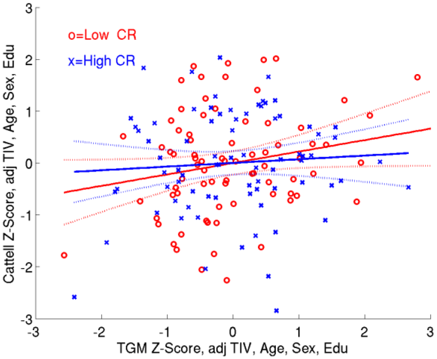
The human mirror neuron system may play an important role in social abilities, such as our ability to empathise and understand other people. The functioning of the human neuron system in healthy ageing and its relationship to social abilities has not been previously investigated. We therefore examined age-related differences in sensorimotor mu desynchronisation as an EEG marker of the human mirror neuron system across the pre-motor cortex, motor cortex and supplementary motor area during action observation. Participants aged 18 to 86-years-old completed a hand movement observation task during EEG recording. Firstly, participants completed a 2-minute resting-state EEG as a reference period and, secondly, watched different video clips that depicted either a static hand or various hand actions, such as locking a door or clicking fingers. Participants also completed the Autism Quotient and Empathy Quotient as self-report measures of social abilities. For younger adults, we replicated previous findings of greater alpha and low beta desynchronisation during hand movement observation compared to static hand observation. We also found greater sensorimotor mu desynchronisation with increasing age. In addition, we examined how sensorimotor mu desynchronisation was related to general social abilities, including autistic traits and empathy ability. Therefore, this study reports the functioning of the human mirror neuron system across adulthood and how it may be related to social abilities.
The human mirror neuron system may play an important role in social abilities, such as our ability to empathise and understand other people. The functioning of the human neuron system in healthy ageing and its relationship to social abilities has not been previously investigated. We therefore examined age-related differences in sensorimotor mu desynchronisation as an EEG marker of the human mirror neuron system across the pre-motor cortex, motor cortex and supplementary motor area during action observation. Participants aged 18 to 86-years-old completed a hand movement observation task during EEG recording. Firstly, participants completed a 2-minute resting-state EEG as a reference period and, secondly, watched different video clips that depicted either a static hand or various hand actions, such as locking a door or clicking fingers. Participants also completed the Autism Quotient and Empathy Quotient as self-report measures of social abilities. For younger adults, we replicated previous findings of greater alpha and low beta desynchronisation during hand movement observation compared to static hand observation. We also found greater sensorimotor mu desynchronisation with increasing age. In addition, we examined how sensorimotor mu desynchronisation was related to general social abilities, including autistic traits and empathy ability. Therefore, this study reports the functioning of the human mirror neuron system across adulthood and how it may be related to social abilities.
*** Correction: Original document contained incorrect author information.
Previously we reported that transgenic rats overexpressing mutant human APP (McGill-R-Thy1-APP) develop an Aß-dependent deficit in LTP induced by standard conditioning stimulation (200 Hz-HFS) as early as 3-4 months of age, whereas there was no deficit in LTP induced by a strong conditioning stimulation protocol (400 Hz-HFS) (Qi et al., Acta Neuropath. Comm., 2014). Here we studied the glutamate receptor-dependence of LTP induced by 400 Hz HFS and the ability of novelty exploration to reverse this LTP. Electrically evoked field EPSPs were recorded at CA3 to CA1 synapses in the dorsal hippocampus of chronically implanted adult freely behaving male rats. A cannula was also implanted in the lateral ventricle under recovery anaesthesia. In stratum radiatum LTP induced by 200 Hz-HFS, which is NMDA receptor-dependent, was completely inhibited in the transgenic rats. In contrast 400 Hz-HFS induced robust and large LTP both in wild type and transgenic littermates. Either CPP (an NMDA receptor antagonist) or mibefradil (a VDCC blocker) alone partly inhibited this 400 Hz-HFS-induced LTP. However, when both agents were given together, this robust LTP was totally blocked. We also examined the ability of novelty exploration to reverse previously established LTP, that had been induced by 400 Hz-HFS. Remarkably, behaviourally-induced LTP reversal (Qi et al., Cerebral Cortex, 2012) was strongly inhibited in the transgenic rats compared with wild type littermates. Different from LTP induced by 200 Hz-HFS at apical dendrites, LTP at basal dendrites in stratum oriens, induced by the same 200 Hz-HFS protocol, was not inhibited in transgenic rats at any age tested. In conclusion, although NMDA receptor-dependent LTP is inhibited in transgenic rats, an LTP that is both NMDA and VDCC-dependent is unaffected. Moreover, this robust form of LTP was resistant to reversal by novelty exploration in transgenic animals.
Most of our knowledge of how the human brain supports episodic memory (EM) is based on lesion studies and intracranial recordings in the hippocampus. Two important principles that have emerged from these data are that EM critically depends on the hippocampus, and that neuronal oscillations in the theta (3-7Hz) and gamma (30-100Hz) frequencies are prominent features of hippocampal population activity. Recent computational models suggest that these oscillations may support the formation of new memories by grouping distributed cell ensembles via regulating synaptic plasticity. The available evidence indicates that the relative timing of spiking activity is coordinated by the phase of oscillatory activity, thereby raising the question of whether this coordination plays a functional role in establishing so-called Hebbian cell assemblies during EM. Recent advances in micro-wire recording technology have permitted the simultaneous measurement of responses from multiple units, but also from local field potentials (LFP), thereby providing a crucial step towards evaluating the functional role of neuronal oscillations in EM.
Here we hypothesised that the learning of pair-wise associations between two concepts, such as the Taj Mahal and Jeniffer Aniston, is supported by the synchronization of neurons coding for these concepts. To examine this hypothesis we simultaneously recorded activity from multiple micro-electrodes in the hippocampus of human epilepsy patients (n =2) that were undergoing intra-cranial epilepsy monitoring. The analyses of the present data will examine the functional relationship between EM and the spike timing of concept neurons relative to the phase of oscillatory activity in the LFP.
**** Correction: Original document did not contain abstract.
The role of the hippocampus in important types of rapid everyday learning, such as place learning, is well established. However, the mechanisms via which rapidly-acquired place memory may be translated into behaviour are yet to be determined. The intermediate hippocampus, which has been shown to be critical for the hippocampal learning-behaviour translation, combines neural substrates of accurate place encoding with links to prefrontal and subcortical behavioural control sites. This supports that these sites, including the nucleus accumbens (NAc), may be critical for this translation (Bast et al., 2009, PLoS Biol; Bast, 2011, Curr Opin Neurobiol). The NAc is a main candidate due to strong hippocampo-NAc projections that have been implicated in behaviour based on place memory (van der Meer et al., 2014, In: Space, Time and Memory in the Hippocampal Formation, ed. Derdikman & Knierim).
To examine the role of the NAc in the hippocampal learning-behaviour translation, we combined functional inhibition of the NAc (via microinfusion of the GABA agonist muscimol) with measurements of behavioural performance based on hippocampus-dependent rapid place learning using the watermaze delayed-matching-to-place test (DMTP) (Bast et al., 2009). In preparation for these studies, we conducted in vivo electrophysiological and sensorimotor experiments (locomotor activity, LMA and startle response/prepulse inhibition, PPI) to establish suitable muscimol doses to reduce NAc neuronal activity without causing gross sensorimotor side effects. Muscimol infusions into the NAc (125-250 ng/0.5 μl/side) reduced neuronal firing in the infusion site vicinity by 40-50% and, if at all, only moderately reduced LMA and PPI; the latter is consistent with findings that these sensorimotor functions mainly depend on the NAc core (Pothuizen et al., 2005, Neuropsychopharmacology), whereas our infusions targeted the NAc shell, which is the main recipient of hippocampo-NAc projections (Humphreys & Preston, 2010, Prog Neurobiol). Preliminary findings from our studies combining functional inhibition of the NAc with DMTP testing support that the NAc is required for performance based on hippocampus-dependent rapid place learning. Additional experiments to confirm and extend these findings are on the way.
The impact of reward (REW) and punishment (PUN), referred to here as feedback, (FB) on skill learning is not well understood. 72 participants (HVs) underwent fMRI during serial reaction time task (SRTT) or force tracking task (FTT) learning and received monetary REW, PUN, or control FB (CONT) based on their performance. For both tasks, stimuli followed a fixed sequence (SEQ) and skill was indexed early and late in training by comparing performance in SEQ and random (RAND) blocks. HVs in the SRTT (n=36) pressed buttons according to visually presented cues. HVs in the FTT (n=36) modulated their grip to match a target. SEQ and RAND BOLD responses were compared across the FB groups using an ANOVA with Group (CONT/REW/PUN), Type (SEQ/RAND), Epoch (Early/Late) as factors. In the SRTT, both REW and PUN showed more early skill than CONT (Group x Probe x Type: F(2,33)=5.37, p<0.01; PUN v CONT: t(22)=3.46, p<0.005, REW v CONT: t(22)=2.55, p<0.02). FMRI from the SRTT revealed a Group x Block Type interaction in bilateral cerebellum (biCereb), left ventral medial prefrontal cortex (lvmPFC), and right dorsal premotor cortex (rPMd). In REW, biCereb and rPMd activity was greater during SEQ compared to RAND blocks. PUN showed the opposite, reflecting the amount of FB given. During RAND blocks compared with SEQ blocks, lvmPFC activity increased in PUN and decreased in REW, reflecting FB valence. CONT showed no effect of Block Type. All groups showed skill in the FTT, but PUN improved less than REW (Main effect of Probe: PUN v REW: t(35)=2.37, p<0.03). FMRI data showed a Group x Epoch interaction in right middle temporal gyrus (rMTG), right caudate (rCaud), left superior parietal lobule (lSPL), and left cerebellum (lCereb). In REW, activity increased over time in all regions. In contrast, in CONT activity decreased in all regions. PUN was more complex: in rMTG activity increased over time, but in lSPL activity decreased over time. Pun did not impact rCaud or lCereb activity. In sum, in the SRTT, activity elicited by REW and PUN reflects FB frequency, and thus information, quickening learning. During FTT, REW causing activity to increase over time, which might reflect continued processing. Therefore, FB modulates regions responsible for skill learning, which impacts learning.
The coordinated activity of subsets of neurons across multiple circuits is thought to support complex behaviours. These functionally coupled subsets are often referred to as cell assemblies. The detection of cell assembly patterns from single-unit recordings usually relies on finding significant co-firing within a particular time bin. Choosing a bin length based on synaptic integration times, e.g. 20 ms, makes these methods well-suited to detecting Hebbian-like cell assemblies within a single structure such as the hippocampus. However, for assemblies that span multiple circuits it may be that the assembly-forming neurons interact at longer latencies or over successive temporal windows. Here we apply independent component analysis to the cross-correlation between each neuron pair at multiple lags in order to incorporate these interactions. We show that this method is able to capture cross-structural assemblies, and contrast its performance to other methods, using both spike-train simulations and in vivo recordings from the rodent hippocampus and ventral tegmental area. Importantly we found that different assemblies detected in this manner show distinct neurophysiological correlates such as their coupling to different phases of hippocampal theta oscillations, responses during sharp-wave ripples, and speed modulation.
The posterior cingulate cortex (PCC)/precuneus is a core region of the default mode network (DMN) and may form part of an extended hippocampal-navigation system. This system is critical for performing complex scene discriminations, underpinning a broader role in episodic memory via re-experiencing spatial context. Functional magnetic resonance imaging (fMRI) studies have identified individual differences in the response of the PCC/precuneus specifically during scene processing, which may place people at increased risk of developing memory problems in later life. The neurochemical underpinnings of such scene processing activity differences are unknown.
Here, we combined 3T fMRI with proton magnetic resonance spectroscopy (1H-MRS) to explore how inter-individual variation in PCC BOLD-fMRI activity is related to the concentration of local metabolites. Participants (n=40, mean age 22 years) completed a perceptual odd-one-out fMRI task for scenes, objects, and faces. The metabolites N-acetyl-aspartate (tNAA), glutamate (Glx) and γ-amino-butyric acid (GABA+) were quantified via PRESS and MEGA-PRESS scans in PCC (2x2x3cm) and an occipital control voxel (3x3x3cm). TNAA is considered a marker of neuronal density and mitochondrial energy metabolism, and Glx and GABA+ indicate excitatory and inhibitory tone.
We found a category-sensitive PCC BOLD-tNAA relationship, such that the PCC BOLD response for scenes, but not faces and objects, was positively correlated with PCC tNAA. There was no significant correlation between PCC BOLD for scenes and occipital tNAA, suggesting regional selectivity of this relationship to the PCC. A complementary voxel-wise analysis within the PCC MRS voxel mask supported this finding, as this identified a significant cluster reflecting a positive association between scene-sensitive BOLD and PCC tNAA. There were no category sensitive relationships between PCC activity and PCC GABA+ or Glx.
These results demonstrate, for the first time, that variability in PCC BOLD during scene processing is related to PCC tNAA. This has implications for understanding individual differences in PCC/precuneus activity, and potential biochemical mechanisms that could underpin activity alterations in this region in disorders, for example Alzheimer’s disease.
Introduction: Cognitive deficits in schizophrenia remain an unmet clinical need and have a significant impact on outcome and quality of life for patients and carers (Harvey & Keefe, 2001). The sub-chronic phencyclidine (PCP) rat model and novel object recognition task (NOR) have been well validated for relevance to schizophrenia (Neill et al., 2010; Horiguchi et al., 2012; Grayson et al., 2015). Exercise increases hippocampal and plasma levels of brain-derived neurotrophic factor (BDNF), a protein that modulates synaptic change and long-term potentiation (Berchtold et al., 2005), providing a hypothesis for its beneficial effects in the illness. Our aim is to investigate the mechanisms of exercise-induced reversal of cognitive deficits in the scPCP model, with a focus on BDNF.
Methods: Four groups of adult female Lister Hooded rats (n=10 per group) were used: vehicle control, vehicle exercise, scPCP control, and scPCP exercise. Rats were treated with either saline or PCP (2mg/kg i.p.) twice a day for 7 days, followed by 7 days washout then given access to running wheels in individual cages for 1 hour a day, 5 times a week, for 6 weeks. Control groups had access to immobilised running wheels. Blood sampling and NOR (with a 1 minute inter-trial interval) were conducted pre- and post-exercise, and 2 weeks following exercise cessation. Plasma and hippocampal BDNF levels were quantified by ELISA. Data were analysed by ANOVA and post-hoc student’s t-test.
Results: Pre-exercise vehicle, but not scPCP groups, successfully discriminated the novel from familiar object (p<0.05). The exercise regime reversed this cognitive deficit (p<0.05), while the scPCP control group remained unable to complete the task and vehicle groups successfully discriminated the novel from familiar object (p<0.05). The cognitive deficit reversal was sustained 2 weeks post-exercise (p<0.05). Current work is evaluating plasma BDNF and subsequent studies will measure brain BDNF in hippocampus and prefrontal cortex.
Conclusions: This work demonstrates that aerobic exercise reverses a robust cognitive deficit in a rat model for cognitive deficits in schizophrenia. Our work to evaluate potential mechanisms of this effect through BDNF could inform future therapeutic strategies in patients.
Models of episodic memory in the medial temporal lobe often suggest that spatial and non-spatial information reaches the hippocampus via the medial entorhinal cortex (MEC) and lateral entorhinal cortex (LEC), respectively. However, there is evidence that the LEC binds these two types of information together prior to the hippocampus. Further, the LEC contains neurons which are spatially tuned to objects. Notably, object-related firing in the LEC strongly resembles object-modulation of place cells in the hippocampus; neurons in both structures encode object location, respond to object displacement, and fire at locations where an object was previously located. However, the origin of object-modulation in place cells is unknown. One possibility is that input from the LEC drives the spatial representation of objects in the hippocampus. To explore this hypothesis, we implanted microdrives in rats (n=5), with tetrodes targeting either the proximal or distal CA1. These regions receive differential input from the entorhinal cortex, this strategy permitted us to record from neuronal ensembles which primarily receive MEC or LEC input. Place cells were recorded during exploration in an open-field containing objects which underwent a series of spatial manipulations, including object dislocation and novel object-place recognition. Object-modulated and non-object modulated place cells were recorded in both regions. Object-modulation of place cells in CA1 conformed to patterns which have been described previously. A sub-set of cells displayed ‘trace’ firing at previous object locations in response to the movement of objects within the environment. Place cells which receive LEC input contained more spatial information than those in which receive MEC input. Further, place cells which receive LEC input had increased stability across standard sessions where the objects underwent no change. These findings influence our understanding of how entorhinal-hippocampal communication supports the integration of item and location information in episodic memory.
Induction of spike timing-dependent long-term potentiation (STD-LTP) at hippocampal Schaffer collateral (S/C)-CA1 synapses requires the sequential activation of postsynaptic NMDA receptors (NMDARs) and voltage-sensitive Ca2+ channels at dendritic spines (1, 2). Both NMDAR function and spine Ca2+ signals (EPSCaTs) during STD-LTP induction are facilitated by metabotropic glutamate receptor (mGluR1)-dependent inhibition of postsynaptic SK channels (2). In addition, muscarinic M1 receptors facilitate spine Ca2+ signals and induction of theta burst LTP at S/C-CA1 also via SK channel inhibition (3, 4). We now show in acute hippocampal slices from adult rats that the highly selective allosteric M1 agonist GSK-5 (5) (1μM) or the direct inhibition of SK channels by apamin (100 nM) rescued STD-LTP induction at S/C-CA1 synapses in the presence of mGluR1 selective antagonist YM 298198 (100 nM). Whole-cell current clamp recordings were performed at 36°C under GABAA receptor inhibition (50 μM picrotoxin). STD-LTP was induced with a theta frequency conditioning train of 300 spike pairings at 5 Hz for 1 min. Each pairing delivered one EPSP evoked in stratum radiatum followed by two post-synaptic action potentials elicited by somatic current injection, at 100 Hz. Two-photon Ca2+ fluorescence imaging in CA1 pyramidal cell dendritic spines showed that apamin enhanced synaptically evoked EPSPs and EPSCaTs. Furthermore, Ca2+ imaging during conditoning revealed that LTP induction requires a sustained sequence of spine EPSCaTs, which was facilitated by apamin. mGluR1 blockade inhibited the EPSCaTs during conditioning, and this effect was reversed in the presence of GSK-5. Our data indicate that SK channels are a common downstream target in a convergent signalling pathway for LTP facilitation by metabotropic glutamate and cholinergic neuromodulation.
1. Magee, J. C., and D. Johnston. 1997. Science 275:209-213
2. Tigaret, C. M., et al. 2016. Nat Commun 7:10289.
3. Giessel, A. J., & B. L. Sabatini. 2010. Neuron 68:936-947.
4. Buchanan, K. A., et al. 2010. Neuron 68:948-963.
5. Dennis S. H., , et al., 2016. Cereb Cortex 26:414-426.
Hippocampal theta is thought to be crucial for binding sensory information from multiple cortical regions into coherent memory episodes. Studies in rodents showed that cortical inputs arriving at the appropriate hippocampal theta phase induce long-term potentiation, which is a possible mechanism for how theta phase modulates memory formation. Using a multisensory entrainment paradigm, we recently showed in humans that episodic memory performance is enhanced by theta (4 Hz) phase synchrony between visual and auditory cortices. In this current EEG study, we investigate if such theta phase synchrony between visual and auditory brain regions varies as a function of successful episodic memory formation. Scalp EEG activity is recorded while 24 healthy adults perform associative memory encoding and recall tasks. A series of sound-movie pairs are presented at encoding. Luminance of the movies and amplitude of the sounds are modulated from zero to full at 4 Hz with either 0 degree, in-phase or 180 degrees, out-of-phase to create phase offset in the visual or auditory cortices, respectively. Participants are asked to judge how well a sound corresponds to a movie while memorising the association between them. During later recall task, participants recall the correct scene presented with a given sound clip. Our preliminary results (N = 10) showed that 7 out of 10 participants had higher accuracy for the associative memory task in the in-phase condition than the out-of-phase condition, replicating our previous finding. Phase differences of the theta activity in 4 Hz between the auditory source and the visual source is categorised depending on later associative memory performance. In the in-phase condition, subsequently remembered trials are predicted to be perfectly synchronised in phase with mean difference of 0. Phase differences of subsequently forgotten trials are predicted to have a more uniform distribution. In the out-of-phase condition, the pattern of remembered trials is predicted to resemble the pattern in the in-phase condition while the distribution of phase differences for the missed trials is predicted to be non-uniformed with a mean phase difference of 180. The findings will further our understanding of theta phase synchrony for episodic memory encoding.
Working memory relies on the prefrontal cortex (PFC) and is strongly modulated by dopaminergic signalling. However, the relationships between encoding of task-relevant information and responsivity of PFC neural subpopulations to dopaminergic input remain poorly defined. We therefore compared the behavioural correlates of rat medial PFC (mPFC) neurons during an instrumental delayed non-match to position (DNMTP) task with their responses to stimulation of dopaminergic projections from the ventral tegmental area (VTA).
Channel rhodopsin 2 was selectively expressed in tyrosine hydroxylase (TH)-positive neurons in the VTA of adult TH-Cre rats (n=3) simultaneously implanted with tetrode recording electrodes in hippocampal CA1 and mPFC. VTA stimulation (5ms pulses, 20Hz, 20mW) during wakefulness enhanced the power of low frequency oscillations in mPFC local field potential; 40% of 140 mPFC neurons responded to the same stimulation with significant increases or decreases in firing rate (p<0.05 by comparison with shuffled data). The responses of PFC neurons to VTA stimulation were not associated with their putative neuronal class (inferred from extracellular action potential waveforms and bursting characteristics), but were related to their behavioural correlates in the DNMTP task: neurons responding during the cue encoding phase of the task (sample lever press), or during reward consumption, were also significantly more likely to respond VTA photo-stimulation (p<0.05 vs. other classes of neuron).
These results indicate that mPFC principal neurons recruited to encode sample and reward information during a working memory task are more directly responsive than their peers to dopaminergic input from the VTA. These subpopulations may therefore preferentially contribute to dopamine’s stabilisation of information in working memory.
Pavlovian memories undergo reconsolidation, whereby the memory becomes unstable at reactivation and can be disrupted by amnestic agents. However, less is known about the reconsolidation of instrumental memories, whether these are goal directed (action-outcome) or habitual (stimulus-response). Our previous, preliminary data indicated that expression of a habit-like memory on a T-maze task correlated with an increase in Zif268, a protein critical for memory reconsolidation, in the posterior dorsolateral striatum. Here, the requirement for Zif268 for the restabilisation of a habit-like memory was tested by administering zif268 antisense oligodeoxynucleotides prior to memory reactivation; however, knockdown of Zif268 did not affect subsequent memory expression tested 24h, 7d and 28d later. Furthermore, when animals were more extensively trained, there was no correlation between the expression of the habit-like memory and Zif268 expression in the dorsal striatum, hippocampus, or nucleus accumbens. Zif268 expression increased in the basolateral amygdala after extended training, but this was not correlated with the use of a habit-like or goal-directed strategy during reactivation. We propose that Zif268 expression in the basolateral amygdala may be linked to prediction error, generated by the absence of reward at re-exposure. Altogether this work supports the role of Zif268 in the maintenance of instrumental memories.
Associative recognition memory, the ability to associate an object with a location or position in a sequence requires a neural network (Barker & Warburton 11). A recent study has identified a separation in the function of projections from the CA1 region of the hippocampus (HPC) to the medial prefrontal cortex (mPFC), thus deactivation of direct projections from the dorsal CA1 (dCA1) selectively impaired object-in-place memory whereas deactivation of direct projections from the intermediate CA1 (iCA1) selectively impaired temporal order memory (Barker et al 17). The nucleus reuniens of the thalamus (NRe) is one indirect route through which the mPFC can send information to the HPC and ablation of the nucleus reuniens impaired associative recognition memory formation (Barker & Warburton 15). Therefore this study aimed to test the hypothesis that projections from the NRe to the CA1 region of the HPC would show the same functional/anatomical dissociation between the dCA1 and iCA1 as was observed for the CA1 to mPFC projection.
Male Lister hooded rats were injected with a virus expressing an inhibitory DREADD (AAV5-hsyn-hM4Di) into the nucleus reuniens and cannula were implanted into the hippocampus targeting the dorsal (dCA1) and intermediate (iCA1) regions of the CA1. Different types of associative recognition memory were tested using spontaneous tests of preferential exploration. CNO (3μM) was infused either before the sample phase or before the test phase. A 4-way within subject design was used thus animals received either CNO infusion into either the dCA1, iCA1 or both or vehicle infusion into both dCA1 & iCA1.
In an object-in-place and an object temporal order task, infusion of CNO into the HPC before the sample phase resulted in impaired performance only when CNO was infused into both the dCA1 and iCA1, in contrast when CNO was infused before the test phase; in the object-in-place task performance was impaired after infusion into the iCA1 whereas in the object temporal order task performance was impaired after infusion into the dCA1. Thus the projection from the NRe to the HPC regulates associative recognition memory formation in distinct anatomical regions of the HPC dependent on the stage of memory processing and the type of association formed.
A variety of interneuron types has been identified in the rodent hippocampus based on differences in their post-synaptic targets, their expression of molecular markers and their spike timing relative to rhythmic fluctuations of the local field potential. Such interneuron types are thought to have distinct contributions to the temporal organization of principal cell firing. However, current progress in testing the role of each interneuron type has been hindered by the difficulty to assign interneurons to anatomically well-defined types when solely recorded with extracellular recordings (i.e., without further labelling) in behaving rodents.
Here we present results from a data set of 679 putative interneurons recorded using multichannel extracellular techniques from the dorsal CA1 region of the hippocampus of 38 mice. We employ an unsupervised clustering framework to attempt sorting interneurons into distinct types based on their (1) spike train dynamics, (2) spike waveform, (3) spatial tuning of their spike discharge, (4) spike coupling to well-known hippocampal oscillations, (5) coupling to the summed population activity of principal cells and (6) firing response to sharp wave-ripple oscillatory events.
Although we do not find clear support for the possibility to identify discrete types of interneurons solely based on their extracellular recordings, we do find structure in this dataset indicative of clusters of interneurons with overlapping firing properties. We suggest that our framework for an unsupervised interneuron clustering, although not absolute, nevertheless provides a useful way of classifying hippocampal interneurons that could contribute to further our understanding of their diverse roles in network dynamics and behaviour.
AMPA-type glutamate receptors are embedded at postsynaptic sites, aligned with the presynaptic neurotransmitter release machinery, mediating fast excitatory neurotransmission. As AMPARs diffuse rapidly in the plane of the membrane, a prerequisite for faithful signal transmission is their trapping and clustering at postsynaptic sites. Synapse strengthening, as occurs during learning, results from the recruitment to and enrichment of AMPARs at synapses. Therefore, the mechanisms underlying AMPAR positioning are fundamental to synaptic transmission and plasticity.
AMPAR synaptic anchoring has historically been explained by interactions of the receptor C-termini with components of the postsynaptic scaffold, yet this model has recently been challenged. Using mouse organotypic hippocampal slices, we show that the AMPAR N-terminal domain (NTD), which projects midway into the synaptic cleft, plays a fundamental role in this process. This highly sequence-diverse domain mediates synaptic anchoring in a subunit-selective manner. Using a combination of electrophysiological and imaging techniques we have revealed that receptors lacking the NTD exhibit increased mobility in synapses and are unable to maintain faithful synaptic transmission. Furthermore, despite being robustly expressed at extra-synaptic sites, AMPARs are unable to sustain long-term potentiation (LTP) without their NTD. Thus, synaptic transmission and plasticity are critically dependent upon an AMPAR anchoring mechanism that is driven by NTD interactions.
Sleep has now been linked to brain plasticity at many levels, with converging evidences from the molecular, cellular and behavioural fields. Studies in humans and animals support of specific role for spindle oscillations (9-16Hz) in this process but the underlying physiology remain elusive. It has been suggested that spindle bursts promote calcium increase specifically in dendrites, a condition that would favour dendritic plasticity processes (1, 2). Although this hypothesis is supported by computational modelling (1), to date, evidence that such a relation exists during natural sleep is missing.
To address this issue, we measured calcium activity from layer 5 (L5) dendrites in the somato-sensory cortex using one-photon (fibre-optic) and two-photon imaging in naturally sleeping rodents. Calcium imaging was combined with electroencephalographic (EEG) recordings to monitor behavioural states and underlying network oscillations.
Our results show that activity of population of dendrites during slow-wave-sleep was specifically correlated with spindle-beta (9-30 Hz) power changes. Two-photon imaging of single dendrites further suggests that this relationship was largely explained by an increase in synchronization of dendritic activity during spindles. Interestingly, this effect was specific to dendrites as L2/3 and L5 cell bodies did not show such correlation.
Our results support the current hypothesis of a direct link between spindles and dendritic activity regulation and further reveal an important, yet unexplored, functional coupling between spindle and beta oscillations (15-30Hz). Further (and ongoing) experiments probing the influences of experience on this relationship will reveal important information on the physiology of spindles and their role in learning and memory.
References:
1. Contreras, D., Destexhe, A. & Steriade, M. Intracellular and computational characterization of the intracortical inhibitory control of synchronized thalamic inputs in vivo. J. Neurophysiol. 78, 335-350 (1997). 2. Sejnowski, T. J. & Destexhe, A. Why do we sleep? Brain Res. 886, 208-223 (2000).
Memory reactivation can lead to two phenomena: memory updating / reconsolidation with possibility of having inaccurate memories and memory strengthening. In Study 1, we attempted to replicate previous findings of episodic-like memory reconsolidation that re-exposure to the initial learning context is sufficient to induce reconsolidation. In a visual list-learning paradigm, participants learned 2 lists in different ways on 2 days. The experimental group learned both lists in the same room and with the same experimenter. The control group learned the two lists in different rooms with different experimenters. At test, participants were returned to the original context and recalled images from the 1st day of learning. ANOVA unexpectedly showed no difference in intrusions of Day 2 items into Day 1 recall between Experimental and Control groups, thereby failing to replicate published findings. While the Control Group had poorer recall of Day 1 items compared to a no interference control, performance in the Experimental Group was preserved. This may reflect an effect of training context re-exposure to strengthen the memory and mitigate against the deleterious impact of interfering material. In Study 2, we tested directly the capacity of memory reactivation to facilitate memory strengthening. Participants learned visual object-scene paired associated and two days later were subjected to a retrieval test and/or further learning in the same room and with the same experimenter. When subsequently tested on the paired associate recall, participants that received retrieval followed by relearning, relearning followed by retrieval, or two relearning episodes all had greatly improved performance. Groups that received one or two retrieval episodes performed as poorly as a control group, with all three groups showing evidence of memory decay. Finally, participants that received a single relearning episode performed at an intermediate level, with mild improvement. The common effects of retrieval-relearning, relearning-retrieval and relearning-relearning to strengthen episodic memory may reflect different underlying processes, one or more of which might be related to memory reconsolidation.
Synaptic long-term depression (LTD) is believed to underlie critical mnemonic processes in the adult hippocampus. Considerable controversy exists over the roles of the metabotropic and ionotropic actions of glutamate in the induction of synaptic LTD by electrical low frequency stimulation (LFS), based largely on studies in hippocampal slices from very young animals. Here we examined the requirement for metabotropic (mGlu) and NMDA glutamate receptors in LTD induction by either electrical or optical LFS. Approximately 2-4months after transfection with AAV5-CaMKIIa-hChR2(H134R)-EYFP in the dorsal hippocampus, electrically or optically evoked synaptic transmission at CA3-to-CA1 synapses was recorded under urethane anaesthesia. Application of either electrical or optical 900 pulse 1Hz LFS, with the intensity increased to 95% maximum amplitude, induced robust and stable electrical (eLTD) and optical (oLTD) LTD, respectively. We found that: (i) On their own neither the competitive NMDAR antagonist CPP, nor the selective mGlu5R antagonist MTEP administrated via systemic injection had an effect on eLTD. However, the systemic co-administration of CPP and MTEP blocked eLTD. (ii) Consistent with the CPP results, whereas the negative allosteric modulator of GluN2B, Ro 25-6981, alone failed to significantly alter LTD the same dose of Ro 25-6981 given together with MTEP, greatly attenuated eLTD. (iii) Administration of relatively high doses of NMDAR antagonists D-AP5 and Ro 25-6981 locally near the hippocampus, via the i.c.v. route, the magnitude of eLTD was greatly attenuated. (iv) Standard doses of NMDAR antagonists CPP, D-AP5 and Ro 25-6981 that failed to prevent eLTD, strongly attenuated oLTD. (v) In the animals pretreated with the mGlu5R positive allosteric modulator VU 0360172, a peri-threshold electrical 300 pulse 1Hz LFS facilitated robust eLTD but a peri-threshold optical 300 pulse 1Hz LFS only induced a transient depression of synaptic transmission. The present data provide strong evidence in the living animal that the engagement of mGlu5 receptors during electrical, but not optical, low frequency conditioning stimulation increases ion flux via the recruitment of more GluN2B-containing NMDA receptors, thereby boosting LTD induction.
Rationale: Large animal models of neurocognitive dysfunction hold unique advantages over rodent models, e.g. functional heterogeneity of brain structures and species longevity. Recently, the use of the horse as a complementary animal model has been considered, with animals successfully achieving learning criterion of an adapted 3-choice serial reaction time task (3-CSRTT). To examine the usefulness of an equine model further, a cohort of high, medium and low dopamine horses not yet exhibiting symptoms of spontaneous neural dysfunction (e.g. hyperdopaminergic stereotypy or hypodopaminergic pituitary pars intermedia dysfunction) were studied to determine if phenotypic differences, if apparent, could be detected by our adapted 3-CSRTT.
Methods: High (n=10), medium (n=10), and low (n=10) dopamine horses were recruited utilising spontaneous blink rate (SBR; Low<440; Medium 440-622; High >622 blinks/30min). All horses were trained on the 3-CSRTT with a pre-established training regimen. Following learning criterion attainment, six test sessions were utilised to obtain data regarding omission and commission errors, as well as impulsive and compulsive responding.
Results: One high and two low animals did not reach learning criterion and were removed from subsequent analysis. Repeated measures ANOVA indicated performance parameters (accuracy, impulsive responding, compulsive responding, commission errors, omission errors) between the six test sessions were not significantly different for any group (p>0.05). However, one-way ANOVAs highlighted significant differences between the cohorts over the six test sessions (Table 1). No significant differences were apparent between cohorts for omission and commission errors.
Conclusions: The adapted 3-CSRTT for equine use is sensitive to differences in responding between dopamine phenotypes. Furthermore, the significant increase in compulsive and impulsive responding in the non-symptomatic high dopamine animals could indicate that such behaviour patterns may arise prior to the development of the hyperdopaminergic condition stereotypy. Subject to further investigation, the utilisation of impulsive and compulsive responding may prove fruitful in screening methods to identify those at risk of developing stereotypic behaviours.

The cerebellum and cortical motor areas are interconnected in a closed anatomical loop that contributes to the acquisition of motor skills. Other parts of the cerebellum, in lobule HVIIA, are connected with the prefrontal cortex and could sustain more complex forms of learning. During instrumental rule learning, conditioned stimuli (CS) can be paired arbitrarily with actions. Using 3T fMRI, we have previously shown that such CS evoke activity in lobule HVIIA, but limited signal-to-noise makes it difficult to understand the anatomical detail in single subjects. Here, we used 7T event-related fMRI to achieve greater sensitivity and anatomical detail than previously has been possible.
Fifteen adults (21–39 y; 6 F) underwent 18.5 min of 7T fMRI scanning (1.5-mm isotropic voxel). A high-resolution structural image was also acquired (0.7mm isotropic voxel). Subjects were required to form associations between a set of arbitrary visual CS and one of four finger movements using trial-and-error learning. CS were followed by a variable delay (0.12–3.85s), a Go! signal and visual feedback (correct/incorrect). In control trials, the visual cue directly specified the movement and so no arbitrary rules were learned.
The frequency of correct responses increased during learning. Activity time-locked to the onset of symbolic instruction cues was compared with that of control cues. Activations were present bilaterally in three main locations (Fig. 1) that included mostly lobule HVIIA. First, in an area that extended across the superior posterior fissure across lobules HVI and HVIIA, second, in the vermal parts of lobule HVII, and finally in lobule HVIIA at the border with lobule HVIIB. The anatomical consistency of these effects is visible in three cases presented in Fig. 1a.
Our results provide further support that areas of the cerebellum connected with the prefrontal cortex are sensitive to instruction cues that acquire associative properties during instrumental learning. This study is the first to use 7T event-related fMRI to understand cerebellar excitability changes related to instrumental learning. The findings verify previous experiments but go further by providing anatomical detail at high resolution in single subjects.

The recovery of information from episodic memory can be influenced by goal-directed control applied prior to the point of retrieval. In two event-related potential (ERP) studies we investigated the links between this preretrieval control and proactive control abilities assumed to operate across multiple cognitive domains. We assessed the relation between retrieval orientation – the degree to which retrieval cue processing varies according to retrieval goals – and established measures of proactive attentional control (the AX-Continuous Performance Task; AX-CPT). In Experiment 1, participants made either an Artist, a Function or a Pleasantness judgement on words at study, then at test had to accept items studied in just one of these tasks as “targets” (other studied items and new items were “non-targets”). In Experiment 2, they made either living/non-living or indoor/outdoor judgments at study for words presented to the left or right of the screen. At test, they had to accept items as targets either according to screen location or the judgement performed at study. To measure retrieval orientation, ERPs to correctly rejected new items were contrasted according to the type of information targeted at retrieval, in a priori time windows based on previous studies using these tasks. In Experiment 1, both proactive control measures from the AX-CPT and the magnitude of ERP retrieval orientation effects from 500-700 ms predicted ability to discriminate targets from non-targets in the exclusion task. Individuals with better target performance on the AX-CPT also showed larger ERP retrieval orientation effects. In Experiment 2, individual differences in the magnitude of ERP retrieval orientation effects from 400-800 ms were again associated with the engagement of proactive control on the AX-CPT. In both studies, ERP retrieval orientation effects were more pronounced in individuals who engaged proactive control to a greater degree, although there were differences between experiments in the AX-CPT measures showing these associations. The results of both experiments suggest a relationship between episodic memory control via retrieval orientation and more general proactive cognitive control processes.
Previous work [1] has implicated a network of brain regions that is highly innervated by the catecholamine system in experience-based, but not vicarious, learning (learning from an indirect source of information such as advice from a colleague). Such results raise the possibility that these two types of learning are underpinned by dissociable neurochemical mechanisms – a controversial suggestion consistent with the hypothesis that humans have evolved ‘special’ mechanisms for social learning. Here we investigate whether:
1) catecholamine system modulation has dissociable effects on experience-based and vicarious learning
2) dissociable effects seen in (1) are due to the social nature of vicarious learning
Experiment 1: Participants took methylphenidate (MPH) - which increases dopamine and noradrenaline in prefrontal and striatal [3] areas - and placebo (PLA) according to a cross-over, double-blind, within subject design. All participants completed a learning task in which they learned outcome probabilities in stable and volatile environments. Outcome probabilities could be learned via one’s own personal experience of reward outcomes, via vicarious learning from a social source, and/or by combining both personal experience and social information. Learning rates for personal-experience based and vicarious learning in volatile and stable environments were estimated. Relative to PLA, MPH improved experience-based learning, but not vicarious learning. More specifically, under MPH participants were better able to adjust to the environment, learning fast when the environment was fast-changing (volatile) and slow when the environment was stable.
Experiment 2: The procedure was identical to Experiment 1 except the source of vicarious learning was non-social in nature (i.e. advice originated from a system of rigged roulette wheels). As in Experiment 1, MPH improved experience-based, but not vicarious, learning. Thus, MPH has dissociable effects on experience-based and vicarious learning that can be observed irrespective of whether the source of vicarious learning is social in nature.
[1] Behrens ea. (2008) Nature,456,245–249.
[2] Volkow ea. (2001) J Neurosci,21,RC121
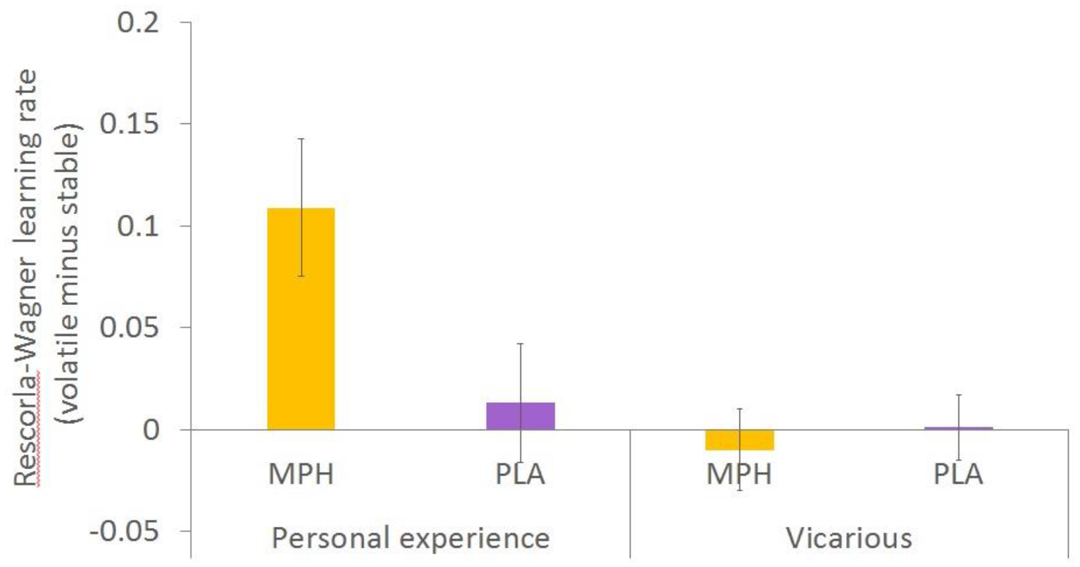
Truncating CHD8 mutations are amongst the highest confidence autism risk factors identified to date. To investigate how reduced Chd8 gene dosage may predispose to autism, we constructed a mouse Chd8 allelic series. Whereas the pan-neuronal, homozygous deletion of Chd8 results in brain hypoplasia, we find that Chd8 heterozygous mice display subtle brain hyperplasia and only minor gene expression changes. A small additional decrease of Chd8 expression in Chd8 hypomorphs causes robust changes in the expression of 168 autism-associated genes and hyperplasia of several autism-associated brain areas. Unexpectedly, neither Chd8 heterozygous nor hypomorphic mice display autism-like behaviours. Together, these data show that gene expression and brain growth respond in a non-monotonic fashion to changes in Chd8 expression. We propose that CHD8 haploinsufficiency represents a sensitised genetic background that is not necessarily sufficient to cause autism, but may strongly predispose to autism by reducing the threshold for additional autism risk factors.
Background: Stroke leads to long-term disability and cognitive dysfunction. Improved neurotrophic factor signaling is one hypothesis for the positive effects of physical activity on cognition. It is unknown if BDNF changes with exercise in stroke. We hypothesize that BDNF is involved in the skeletal muscle changes post-stroke and exercise exerts beneficial effects through epigenetic regulation of BDNF expression.
Purpose: 1) To compare paretic (P) vs. non-paretic (NP) skeletal muscle BDNF and the effects of resistive training (RT) on systemic and skeletal muscle BDNF mRNA expression in stroke; 2) To compare the DNA methylation profile for BDNF and BDNFAS (BDNF Antisense RNA) between P and NP muscle and the effects of aerobic training (AEX) on DNA methylation in stroke.
Methods: Chronic stroke survivors (50-76 years) had a fasting blood draw and 12-week (3x/week) RT (n=16). Bilateral vastus lateralis muscle tissue biopsies (n=10) were conducted and BDNF expression determined by RT-PCR. A separate group of five male older chronic stroke survivors completed 6-months of AEX (3x/week) and had bilateral muscle biopsies. DNA methylation status in gene BDNF and BDNFAS was assessed by Illumina 450k Methylation array.
Results: Paretic muscle had ~45% lower BDNF expression than NP muscle (6.79±1.30 vs. 10.52±2.06 AU, P<0.05) and exhibited differential methylation status in the DNA sequences of BDNF (3 CpG sites, P=0.016 to 0.044) and BDNFAS (1 CpG site, P=0.016) compared to NP. Bilateral leg strength and muscle area increased with RT (P<0.05). Plasma BDNF increased 25% with RT. Muscle BDNF mRNA expression did not significantly change after RT (P: 7.21±1.38 vs. 7.06±1.85 AU and NP:11.39±2.12 vs. 7.84±1.32 AU). DNA methylation in BDNFAS in P muscles relative to NP increased after AEX in P (P=0.017).
Conclusions: This is the first evidence that BDNF skeletal muscle expression is reduced by hemiparesis, which may be caused by methylation alterations on the DNA sequence of BDNF and BDNFAS gene. Preliminary findings indicate that AEX increases methylation in BDNFAS gene, which presumably could regulate the expression of BDNF. Future research could examine if changes in epigenetics with exercise training are associated with improved cognitive ability in stroke survivors.
Homozygous mutations of TRAPPC9 in humans cause a neurodevelopmental disorder characterised by microcephaly, intellectual disability, white matter hypoplasia, speech impairments and developmental delays. These symptoms are consistent with Trappc9 expression in neurons, although it is also found in several peripheral tissues. Trappc9 forms part of the trafficking protein particle II complex. It is implicated in vesicle transport at the ER / Golgi and also interacts with the dynactin/dynein motor complex that mediates retrograde transport and signalling along microtubuli.
The Trappc9 gene is part of a cluster of imprinted genes on mouse chromosome 15 and human chromosome 8. Imprinted genes are regulated by epigenetic marks, which are established differentially in male and female germ cells and maintained in the developing embryo and adult tissues. Differential DNA methylation on one of the parental alleles results in parent-of-origin dependent, monoallelic gene expression.
Here, we describe knock-out (KO) mice for Trappc9, which we found to be viable without major embryonic or postnatal deficiencies. However, 3-months old female KOs show a significant 20% increase in body weight. Brain weights in male and female KOs are significantly reduced by 10%. This was confirmed in measurements of total brain volume by high-resolution μMRI using a 9.4T magnet. Initial data indicate a 15% reduction in Sox2-positive neural progenitor cells in the hippocampal dentate gyrus.
To analyse imprinted, allele-specific expression of Trappc9 we quantified exonic SNPs in brain and kidney cDNA from C57BL/6J x JF1 strain intercrosses by pyrosequencing. 70% of Trappc9 RNA in brain was found to be derived from the maternal allele, confirming genomic imprinting of the gene. In kidney, equal biallelic expression was detected. Analysis of DNA methylation at CpG-islands at the 5’-end of Trappc9 by pyrosequencing of bisulphite-treated brain DNA showed very low levels (10%) of CpG methylation.
In conclusion, Trappc9 KO mice reproduce a microcephaly phenotype, similar to human patients. Trappc9 is imprinted in the mouse brain with preferential (70%) expression from the maternal allele. Its epigenetic regulation is mediated by other mechanisms than promoter CpG-island methylation.
Background: Major depressive disorder (MDD) affects around 15% of people worldwide, and MDD patients have a high risk of suicide. Genetic studies in twins indicate that genetic factors may be important in MDD and suicidal behaviour. Genetic variation in genes coding for subunits of the glutamate NMDA receptor (GRIN) has been associated with other psychiatric disorders such as schizophrenia and bipolar disorder, but have been little studied in MDD, despite increasing evidence for glutamatergic dysfunction in the disease. Therefore, this study aimed to evaluate the association of single nucleotide polymorphisms (SNPs) in GRIN1 and GRIN2B with MDD in a Thai sample.
Methods: Subjects included patients with MDD (n=100) (including non-suicide MDD (n=50) and suicide MDD (n=50)) and controls with no history of psychiatric disorder (n=100). DNA was extracted from FTA dried blood spots and genotyped with TaqMan™ SNP Genotyping Assays. Five SNPs were selected to study: rs4880213 in GRIN1 and rs1805502, rs890, rs3764030 and rs1019385 in GRIN2B, all of which have been reported in association with psychiatric disorders and/or are potentially functional.
Results: We found significant differences in allele and genotype frequencies of rs890 between the MDD and control groups (P=0.009 and P=0.022, respectively). Furthermore, the genotype frequency of rs3764030 was significantly associated with MDD (P=0.013). In addition, strong linkage disequilibrium (LD) was observed between rs1805502 and rs890 (D′=0.82), and rs3764030 and rs1019385 (D′=0.97). We found that the AA haplotype of rs1805502 and rs890 in the MDD group was significantly higher (P=0.005), while the frequency of AC haplotype was significantly lower than the control group (P=0.014). Interestingly, the genotype frequency of rs3764030 was also significantly associated with suicide attempt in MDD (P=0.043).
Conclusions: This preliminary study is limited by the relatively small sample size and needs replication in a substantially larger sample. Further studies will address this and explore the relationship of the genetic association with treatment response. However, these findings do provide initial evidence for variation in the gene for the NMDA receptor 2B subunit in risk for MDD.
Mammalian-wide interspersed repeats (MIRs) integrated into the mammalian genome during the Mesozoic period; their continued recognisability, in spite of the passage of time, suggests they may now contribute to their host’s genome. As a transposable element (TE), MIRs historically duplicated and were dispersed throughout the host’s genome, a feature that potentially ratifies and impacts upon internetwork regulation. Despite this, their potential impact with respect to epilepsy, a prevalent condition associated with neuronal dysregulation, has not been fully considered.
To further current knowledge, this study drew upon databases and repeat masking tools in order to identify epilepsy associated genes that contain MIRs. Given the ultimate aim was to identify the role of MIRs, attention was then afforded to those occupying coding regions. In the cohort examined, MIRs were found exclusively within the 3’ UTRs of qualifying genes.
The second element, as mentioned, sought to identify the impact MIRs may have, to this end several lines of inquiry were enacted upon.
• Firstly, co-occurrences between MIRs and mutated regions were sought, however none were found.
• The original MIRs were then compared to the MIRs identified in the human gene, these comparisons showed degradation typically beyond 50%, suggesting the original roles fulfilled by the MIR have likely been compromised.
• Using the bioinformatics tool ClustalW, MIRs were sought across divergent mammalian species, where found, the then aligned sequences were scanned for motifs, revealing several capable of impacting upon gene regulation.
The culmination of results suggests that some epilepsy associated genes have exapted MIRs into positions of regulatory influence. However, further research is recommended to discern the full extent of their impact upon their host genes and indeed upon the condition
Development of the cerebellum is under tight temporal control and perturbances at different developmental stages lead to discernible cerebellar phenotypes. Previous work in our group has demonstrated a key role for the ATP-dependent chromatin remodeller CHD7 (chromodomain-helicase-DNA-binding protein 7) in mouse cerebellar development. Deletion of Chd7 from mouse cerebellar granule cell precursors (GCps) results in cerebellar hypoplasia, developmental delay, motor deficits and down-regulation of Reln, a gene essential for cerebellar development. In this project we sought to establish the underlying cause of the cerebellar hypoplasia in these mice and the mechanisms by which CHD7 regulates gene expression in GCps and in particular Reln. We demonstrated that Chd7 deficiency in these cells results in reduced proliferation and increased apoptosis of GCps during early postnatal development. Through ATACseq analyses we found that CHD7 functions primarily to maintain an open “accessible” chromatin state at the Reln locus and at many other loci in GCps. Finally, we used promoter capture Hi-C in primary GCps to identify putative long-range regulatory elements. Preliminary findings on regulatory interactions of Reln and other genes involved in cerebellar development will be discussed.
Shared genetic factors between Schizophrenia and Major Depressive Disorder (MDD) have been documented through multiple studies, enabled by the increased use of genome-wide association studies (GWAS). Such studies are limited in their view of psychiatric disorders as sums of their total parts; focusing on categorical symptoms within these disorders can provide a more precise understanding of comorbidity and the influence of genetic abnormalities on mental health. This study aims to explore the polygenic risk score for Schizophrenia (PGRS-Sz) as a predictor of symptoms and treatment response in patients with MDD. Data from the Psychiatric Genomics Consortium’s GWAS on Schizophrenia was used to calculate PGRS-Sz as the base phenotype for analysis on data collected by the genome-based therapeutic drugs for depression (GENDEP) study (http://gendep.iop.kcl.ac.uk), which provided the following target phenotype data: depression scores on the Montgomery-Asberg Depression Rating Scale, the Hamilton Depression Rating Scale, and the Beck Depression Inventory, and cognitive, neurovegetative, core mood, observed mood, pessimism, anxiety, anhedonia, sleep, and appetite symptom scores. A series of multiple linear regressions was calculated to predict each target phenotype based on PGRS-Sz. No significant regressions were found. Next, a series of mixed effects logistic regression models was estimated to explore PGRS-Sz as a predictor of treatment response in patients treated with an SSRI, a tricyclic, and both, using each target phenotype in turn as a fixed effect. No significant model was found for patients treated with the SSRI, but significant models were found at various thresholds for core mood, anxiety, and appetite in patients treated with the tricyclic and both drugs, and observed mood and pessimism in patients treated with the tricyclic (p<0.05). The results suggest that PGRS-Sz may not be a reliable predictor of severity of MDD and its symptoms, nor for treatment response to SSRIs. However, PGRS-Sz may be a predictor of change in core mood, observed mood, pessimism, anxiety, and appetite in response to tricyclic treatment, and of change in core mood, anxiety, and appetite in response to a combination of both treatments within a short period of time.
For most of molecular biology’s era methylation of cytosine (5-methyldeoxycytidine/ 5mdC) was thought to be the only base modification in mammalian DNA formed by enzymatic reactions. The catalogue of known epigenetic DNA modifications has expanded and includes three cytosine methylation-derivatives (5hmdC, 5fdC, and 5cadC), as well as methylation of adenosines (6mdA). We are currently measuring and mapping 5hmdC as well as 6mdA in DNA isolated from the central nervous system. We find that nuclear DNA of the cerebellum from individuals with Parkinson’s Disease has significantly higher levels of 5hmdC when compared to age-matched DNA samples isolated from individuals who were not affected by this progressive neurological condition. We are in the process of exploring how different enzymes, cellular conditions and environmental stressors may influence the epigenome and thereby be associated, or even cause, neurological disorders.
The maternal caregiving environment has critical implications for development of the brain and behavioral trajectories in both humans and rodents. For example, exposure to maltreatment by the caregiver is associated with a variety of negative outcomes in adulthood, including deficits in cognitive function and increased prevalence rates of certain psychiatric illnesses. Epigenetic mechanisms, which involve changes in genetic expression without alterations to the underlying genomic sequence, are dynamic throughout the lifespan and offer one potential mechanism via which it is possible for these early life experiences to alter adult phenotype. DNA methylation is one type of epigenetic modification involving the addition of methyl groups to cytosines on DNA that is typically linked with gene repression. Using a rodent model of caregiver maltreatment, our lab has detected alterations in DNA methylation throughout the brain of maltreated animals. These epigenetic alterations coincide with behavioral changes in these maltreated animals, including deficits in novel object recognition memory and maternal behavior. However, the role of epigenetic alterations resulting from maltreatment exposure in mediating these aberrant behavioral outcomes is unknown. In the current study, we administered Zebularine, a drug that has been shown to rescue maltreatment-induced DNA methylation, to adult female animals. Preliminary data suggests that administration of Zebularine rescues deficits in both maternal behavior and novel object recognition in females with a history of maltreatment. Interestingly, drug treatment disrupts maternal behavior and novel object recognition in females with a history of normal maternal care during infancy. We likewise found differences in levels of DNA methylation throughout the brain in brain regions relevant for maternal behavior, including the medial preoptic area, in dams that had been maltreated by their caregiver in infancy that were normalized by Zebularine treatment. Data suggest that altering DNA methylation in adulthood is capable of rescuing brain and behavioral outcomes of experiencing caregiver maltreatment. Additionally, this drug has divergent effects in animals dependent upon infant caregiver experience.
Temporal lobe epilepsy (TLE) is a neurodegenerative disease characterised by recurrent, unprovoked focal seizures that originates in the temporal lobes. In recent years, gamma-aminobutyric acid receptor 1 (GABBR1) has been implicated as a novel target in the pathogenesis of TLE. Pharmacogenomics has revolutionised the approach to medicine, allowing single nucleotide polymorphisms (SNPs) to inform response to therapy, much needed for this drug resistant neurodegenerative disease. To assess the contribution of GABBR1 in TLE pathogenesis two SNPs (rs29218, rs29220) were selected and genomic DNA was extracted using Qiagen QIAmp Mini Kit (250) from samples from 15 patients whom had been diagnosed with TLE. Genomic DNA was genotyped and analysed using StepOnePlus RT-PCR. rs29218 is a single base change A7265G within the promoter region of GABBR1, with the G allele present in 27% of the TLE patients. Sequence analysis of the promoter region around rs29218 indicate that this SNP alters the binding of the transcription factor USF-1, previous data has shown that USF-1 deficient mice present with spontaneous epileptic seizures (Sirito et al., 1998). Similarly, rs29220 is a C10497G single base change in intron 9, the C allele is present in 20% of the TLE patients. GABBR1 gene expression, quantified using RT-PCR, in sclerotic hippocampal tissue is influenced by genotype with impaired expression associated with the risk allele of both SNPs.
Gene expression data, although non-significant, when coupled with the genotyping analysis shows a clear trend in that GABAB1 receptor expression is impaired in the presence of these SNPs, supporting the conclusion that they are involved in pathogenesis of TLE. In summary, we hypothesis that the promoter polymorphism rs29218 alters the binding of the transcription factor USF-1, which influences GABBR1 expression, implicating a role for GABBR1 in the pathogenesis of TLE.
Alzheimer’s disease (AD) is an incurable neurodegenerative disorder; in which the death of brain cells characteristically result in memory loss and cognitive decline. It is the most common form of dementia, affecting around 850,000 people in the UK. The sporadic late-onset form (LOAD) accounts for 95% of all cases and is genetically complex in nature. It is believed that a combination of genetic and environmental factors are at play. Recent genome-wide association studies (GWAS) have uncovered 20 new gene candidates for AD risk, however these generally exhibit small effect sizes. Following on from GWAS, which addressed common variation associated with disease, we are now utilising whole exome next generation sequencing (NGS) to explore the contribution made by rare variants (MAF<5%). Recently this approach has highlighted the role of the TREM2, CD33 and SORL1 genes in AD risk and emerging NeuroX chip and NGS data is set to generate more genes of interest.
DNA was extracted from post-mortem brain tissue obtained from the BDR brain banks for healthy and diseased individuals. Whole-exome sequencing was performed on 292 samples, including 128 AD cases and 50 controls. Using a combination of bioinformatics and statistical tools we investigate how sequence variation, including deleterious mutations, within genes differs between a population of healthy and diseased individuals.
Using bioinformatic approaches, identified risk variants will be tested for association with disease. Pathogenic variants in known risk genes will be prioritised and their potential functionality assessed. A polygenic risk score (PRS) will be calculated for the individuals.
Presently over 340,000 variants have been identified in the whole-exome dataset. Variants in genes of interest that are annotated as highly damaging or protein modifying will be investigated further. Initial analysis revealed no mutations in the familial genes APP, PSEN1 or PSEN2. The generation of PRS will be a useful metric to inform individual’s level of risk for disease.
Mutations in the gene encoding the ATP dependent chromatin-remodeling factor, CHD7 are the major cause of CHARGE (Coloboma, Heart defects, Atresia of the choanae, Retarded growth and development, Genital-urinary anomalies and Ear defects) syndrome. Neurodevelopmental defects in these patients lead to neurological dysfunction, including developmental delay, incoordination, intellectual disability and autistic traits. We previously demonstrated cerebellar vermis hypoplasia and abnormal cerebellar foliation in a proportion of patients with a CHD7 mutation and CHARGE syndrome. Abnormal foliation implicates CHD7 in perinatal cerebellar development, however its precise role is currently unclear.
We conditionally deleted Chd7 from granule cell precursors (GCps) in the mouse and identified cerebellar hypoplasia, purkinje cell disorganization, motor deficits and developmental delay in these mice. We found that Chd7 is critical in regulating granule cell proliferation and apoptosis in vivo. We report that Chd7 has a critical role in the regulation of Reln gene expression and identify a significant down regulation of Reln in GCps of the conditional Chd7 mutants by genome wide transcriptomic analysis and qPCR. We further provide functional evidence that Reln contributes to cerebellar hypoplasia in vivo, by demonstrating a partial rescue of central lobule hypoplasia in Chd7 mutants through the ectopic expression of Reln. Recessive mutations in RELN are associated with cerebellar hypoplasia and altered expression has been linked to a number of neuropsychiatric diseases. Through analysis of genome- wide chromatin accessibility we demonstrate that CHD7 is necessary to maintain an open, accessible chromatin state at the Reln locus and many other genomic regions. In conclusion, we show that CHD7 can be viewed as a previously unattributed upstream regulator of Reln, and that CHD7 dependent chromatin remodeling regulates Reln gene expression in the perinatal cerebellum. These data provide the first evidence of a direct in vivo role for a mammalian CHD protein in the regulation of cerebellar development through the modulation of chromatin accessibility in neuronal progenitors.
Neuronal network synchronisation within the gamma (30-80 Hz) frequency range is fundamental for a variety of cognitive and perceptual functions. In humans, gamma oscillations (GO) emerge during early childhood where they continue to mature until early adulthood, enabling coordination of spatially distributed neuronal activity. GO can be reliably replicated in vitro using kainic acid (KA) in the rat medial entorhinal cortex (MEC), an area associated with higher cognitive functions such as memory, spatial representation and navigation. However, the development of GO in the MEC has not been documented.
Here, we explored the developmental changes in kainate-induced oscillations (KA-O) in layers II (LII) and V (LV) MEC of Wistar rats at 5 ages (P8-11, P12-15, P16-19, P20-23 and P24-27) using local field potential recording in rat hippocampal-MEC slices. Measurements of peak amplitude and frequency were compared in the two layers.
KA-O were apparent at all ages and in both layers. However, an age dependent increase in amplitude was noted in L2 from 17.8±3.7 nV2/Hz in the youngest age group (P8-11; n=29) to a maximum average amplitude in group P20-23 (n=14) of 308.3±51.7 nV2/Hz (P<0.001). Unlike in L2, there was no age-dependent increase in amplitude in L5. Interestingly, in the youngest age group, the amplitude of L5 exceeded that of L2 (27/29 slices, P=0.01), a difference that was eliminated in the P12-15 group, and reversed in all slices thereafter (50/50 slices).
In both layers, there was a gradual increase in the peak frequency of KA-O from an initially low beta range of 18.1±0.4 Hz (L2) and 18.1±0.4 Hz (L5) in the youngest age group (n=29), attaining gamma frequency by P16-19 upwards, reaching a maximum of 31.3±0.7 Hz (L2) and 31.6±0.7 Hz (L5) in the oldest age group (P24-27; n=18, P<0.001). No differences in the average frequency were noted between the two layers.
These findings highlight clear laminar differences in the development of GO in the MEC. This has implications for understanding neuronal synchrony and normal development of cognitive function and spatial navigation behaviour and for neurodevelopmental disorders such as schizophrenia and epilepsy, which involve EC dysfunction.
INTRODUCTION: Left-right asymmetry is an important organising feature of the human brain and has been found altered in neurocognitive disorders such as language impairments, schizophrenia and autism. Nevertheless, the underlying molecular mechanisms of such alterations are still almost completely unknown. A genome-wide association study revealed the association of the genetic variant rs11855415 in PCSK6 with relative handedness in individuals with dyslexia (P < 10-7) (Brandler et al. 2013). PCSK6 plays a key role in Left/Right patterning early in development via NODAL signalling and ciliogenesis, but we do not know yet how PCSK6 impacts on the brain.
OBJECTIVES: The purpose of this neuroimaging genetics study is to investigate the correlation between the genetic variant rs11855415 in PCSK6 and grey matter’s total volume and distribution in dyslexic children, to find out whether this single-nucleotide polymorphism (SNP) is associated with altered hemispheric asymmetries in dyslexia.
METHODS: Dyslexic children between the ages of 7 and 16 were recruited for DNA collection and MRI scan. The subjects were stratified by genotype in two groups: (1) children without the minor rs11855415 allele (T/T); (2) children with the minor rs11855415 allele, in homozygosity (A/A) or heterozygosity (A/T). The subjects were matched by age, gender and handedness. The neuroimaging phenotypes of the two groups were compared by performing structural analysis on brain T1-weighted images. Tissue type segmentation, tissue volume quantification and voxel-based morphometry (VBM) analysis of grey matter were carried out by using the anatomical structure image analysis software FSL.
FINDINGS: The volumes of both grey matter and white matter have been found increased in the group carrying the SNP in comparison with the not carrier group. In the VBM analysis, the group without the SNP has shown a higher density of grey matter in the right inferior frontal gyrus (pars opercularis) than the carrier group.
CONCLUSIONS: These findings seem to be supportive of the hypothesis that PCSK6 might play a role in both driving altered hemispheric asymmetries and influencing traits such as reading ability.

Sensory activity plays an important role in the maturation of visual cortex (V1), in particular, ocular dominance plasticity. Similarly, in rodent somatosensory (S1BF) cortex, whisker input is required for barreloid development. These structural changes must be underpinned by changes on the circuit level. While the contribution of PV+ interneurons to cortical plasticity is well documented, less is known about the role of other interneuron (IN) subtypes.
Recently, we identified transient GABAergic input from L5b to L4 in the developing S1BF of the mouse. This connection consists of a reciprocal loop between L5b somatostatin-positive (SST+) INs to L4 spiny stellates that is only present prior to the end of L4 critical period (~P10). The presence and duration of this connection is modified by surgical perturbations to sensory input. Evidence suggests that this circuit is important for the timely acquisition of thalamic input onto L4 excitatory neurons. The extent to which sensory information influences remodelling of this circuit and whether this may be a general mechanism for sensory integration is unclear.
To address this we have mapped GABAergic connections onto L4 in a mouse devoid of any thalmo-cortical/cortico-thalamic connections. In the absence of thalamic input to the neocortex, the transient L5b onto L4 GABAergic connection is still present with the time course of this transient circuit un-altered. This suggests a possible genetic component to the formation and maintenance of this early IN-spiny stellate synapse. If this circuit is genetically hardwired to appear during development and is involved in correct integration of thalamic input, we might expect it to be present in other primary sensory areas. To investigate this we mapped GABAergic connections in the developing V1 to determine if similar connections exist between L5 INs and L4 pyramidal cells. Using laser scanning photostimulation we find no evidence of L5 GABAergic connections arising from SST+ INs onto L4 excitatory neurons in V1. This suggests that the connection between L5 SST+ INs and L4 excitatory neurons in S1BF is hardwired and area specific, highlighting a unique modality-specific role for SST+ INs in cortical development.
Background: Viral infection in pregnancy has been associated with an increased risk for the development of autistic spectrum disorder (ASD). Maternal immune activation (mIA), using the viral mimetic poly(I:C), produces phenotypes relevant to ASD in rodents. mIA at gestational day (GD) 12.5 has been widely investigated in mice. However, no studies have explored mIA at GD12.5 in rats. Our aim is to characterise effects of mIA at GD12.5 on offspring neuro and gut biology and behaviour in Wistar rats.
Methods: Pregnant female Wistar rats were injected (i.p.) with poly(I:C) (10mg/kg, n=12 dams) or saline (n=14 dams) at GD12.5. Male and female offspring were monitored for changes in morphometric parameters at GD21, (body weight (BW), brain weight (BrW) and placental weight (PW) n=6 dams/treatment). BW was measured postnatally, and on postnatal day (PD) 21 BrW was measured (n=6-8 dams/treatment). Gene expression in frontal cortex of GD21 offspring was measured using qPCR. For data analysis between treatment groups, a nested-ANOVA was used with litter as a random variable.
Results: At GD21 no significant effect of mIA was observed on BW or BrW in either sex. A significant reduction was found in the PW of female poly(I:C) offspring (p<0.01). Gene expression analysis revealed a significant increase in microglial marker Ofml3 in frontal cortex of male poly(I:C) offspring (p<0.01). Postnatally, a significant reduction in BW was found at PD1 in both sexes from poly(I:C) dams vs. saline (p<0.001). This reduction was maintained at PD12, 18 and 21 (p<0.001). When normalised to BW, BrW from offspring at PD21 was increased in offspring from poly(I:C) dams vs. saline (male p<0.01 female, p<0.001).
Conclusion: To our knowledge this is the first mIA study investigating the effects of 10mg/kg poly(I:C) in Wistar rats at GD12.5. We provide an in depth early developmental analysis of both male and female offspring in this model. mIA resulted in increased frontal cortex gene expression for microglia in male offspring at GD21. An increase in BrW at PD21 may indicate reduced synaptic pruning during development. Further validation of this model is underway to explore effects on gene expression related to synaptic pruning and other brain markers relevant to ASD at PD21.
The mammalian respiratory system is immature at birth. In mice, this immaturity is characterized by a fragile and highly variable breathing pattern during postnatal days 1-3 (P1-3). Around P3-P4, the respiratory system undergoes a step in maturity, after which breathing is less variable and has a higher frequency. The neural mechanisms underlying this maturation step are unknown. Evidence from in vitro studies suggests that two distinct medullary neuronal clusters, the opiate sensitive preBötC and the opiate-insensitive RTN/pFRG, play a critical role in generating respiratory rhythm but little is known of their interaction during development and early post-natal life when the respiratory system is fragile. To pharmacologically tease apart the function of these neuronal clusters during early postnatal maturation of breathing and to investigate the long term effects of opiates on breathing pattern, neonatal mice were exposed to the μ-opioid receptor agonist fentanyl (0.08mg/kg i.p daily), or saline as a control, from P1-5 (n=16) or P9-13 (n=16). Mice were continuously monitored post injection and breathing recorded by closed plethysmography at regular intervals from 5 minutes to 2 hours post injection.
Fentanyl had a modest effect on breathing at all postnatal days by increasing variability, decreasing frequency and increasing the number of apnoeas and hyperpnoeas, compared to saline-exposed mice. At 6 weeks of age, all saline and fentanyl exposed mice were exposed to a single dose of fentanyl (0.04 – 1.0mg/kg ip) and monitored as above. Post-fentanyl, respiratory frequency was significantly decreased (190±10 vs120±15 breaths per minute) in all mice previously exposed to saline as neonates (P1-P5 and P9-13); however, in mice previously exposed to fentanyl as neonates (P1-P5 and P9-13), exposure to a single injection of fentanyl in adulthood had no effect on respiratory frequency (180±8 vs 150±10 bpm). Tidal volume increased slightly in all mice post fentanyl regardless of previous exposure to fentanyl or saline. These data suggest that the respiratory system in younger animals is less susceptible to fentanyl and that pre-exposure to fentanyl during early post-natal maturation brings about a desensitization to fentanyl during wakefulness in early adulthood.
This study aimed to investigate how the cortical sources of spontaneous alpha during eyes-open and eyes-closed conditions change during the course of adolescence as a function of age and pubertal stage. In addition, with regards to spontaneous alpha there is a long-standing theory suggesting that relatively greater left frontal cortical activity is associated with reward orientated behaviours and relatively greater right frontal neural activity is associated with avoidance-orientated behaviours, as indexed by spontaneous EEG alpha activity (Davidson, 1984, 1994). While there is evidence for this theory in adults, research examining frontal asymmetry and its relationship to risk-taking in adolescents is limited. Hence, the aim of this study was to examine whether frontal asymmetry could account for the developmental differences in risk-taking behaviours in adolescents. To that end, preadolescents (9-12 years), mid-adolescents (13-17 years), and late adolescents (18-23 years) had their resting brain activity measured using EEG during eyes open and eyes-closed conditions. The findings revealed significant changes during the course of adolescence, with an age-dependent reduction in occipital alpha in the eyes closed condition, and shift in prefrontal cortical sources of alpha during the eyes-open condition. In addition, more advanced pubertal development was found to predict reduced alpha activity in male, but not female, adolescents. Unexpectedly, frontal asymmetry was found not to be a reliable marker of risk-taking behaviours. In conclusion, this study provides an important step towards understanding the development of spontaneous alpha in the typically developing brain. Nonetheless, a great deal more research needs to be conducted before we have a complete understanding of the development and functional significance of alpha in the maturing brain.
Dopamine (DA) is a key neuromodulator of the adult nervous system. In addition, recent evidence suggests that DA also has neurodevelopmental roles, although these have yet to be fully defined. To address this problem, we are using early stage zebrafish to investigate DAergic regulation of motor behaviour. To determine when spinally projecting DAergic neurons first innervate the spinal cord, we used anti-tyrosine hydroxylase immunohistochemistry. We find that by 22 hours post fertilisation (hpf), DAergic axons reach the caudal part of the hindbrain, reaching the rostral most aspect of the spinal cord shortly afterwards at 24 hpf. Thereafter, DAergic axons gradually extend to more posterior compartments of the spinal cord. To investigate the role of DA signalling on the maturation of zebrafish motor behaviour I used pharmacology and laser ablation methods to study the effects of DA disruption on coiling, a simple form of motor behaviour that is transiently expressed when DAergic axons first invade the spinal cord. Subsequent behavioural and patch clamp analysis revealed that blocking of DA signalling increases the frequency of coiling behaviour and the periodic depolarisations that underpin them while DA receptor agonists has the opposite effect. Therefore, during early stages of development DAergic signalling may promote the termination of transient forms of immature motor behaviour and permit the transition to more complex forms of motor output.
Gestational exposure to environmental toxicants can adversely affect postnatal and adult development. Neurotoxicology data suggests that certain environmental toxicants (such as the compound TCDD) have a potent effect on cerebral development, through pro-apoptotic effects on cerebellar granule cells (MA Williamson, 2005 ToxSci; LL Collins, 2008 ToxSci), defects in pituitary activity (Takeda T et al, 2011 Tox Sci), defects in brain vasculature (Teraoka H et al 2010) as well as alterations in hippocampal neurogenesis and function (Opanashuk et al, 2013 J Neurochem).The major pathway through which TCDD signals is the aryl hydrocarbon receptor (AhR) pathway, whereby the ligand-activated transcription factor AhR binds to associated nuclear translocator proteins (ARNT/ARNT2), is transported through the nuclear membrane and evokes its downstream effects through binding to XRE sequences in the genome. This signalling axis is regulated via negative feedback by AhRR (aryl hydrocarbon receptor repressor). In the context of certain complex disorders, such as cancers, the epigenetic mechanisms as well as the conclusive role of environmental factors contributing to disorder aetiology remain to be elucidated.
The focus of this research is whether aberrant AhR pathway signalling in the developing cerebellum contributes to medulloblastoma tumorigenesis in murine models. To achieve this, we have utilized double conditional Ptch1 (Patched1)/AhR and AhRR mutants, which have allowed us to model the Shh subclass medulloblastomas and pose the question of whether key AhR pathway components are able to influence the process of medulloblastoma development, and if so, through what mechanisms. Our data from non-neoplastic cerebellar granule precursors (GCPs) suggest a role for the AhR in maintenance of GCP cycling throughout the early postnatal time period of cerebellar neurogenesis via repression of the Tgf beta pathway, specifically through inhibiting phosphorylation of Smad3. The pathological medulloblastoma context, we hypothesize, may involve a role reversal for the AhR, with loss of function leading to enhanced tumorigenesis. Along with key data pertaining to AhRR function this study aims to provide a more thorough mechanistic understanding of the AhR pathway in medulloblastoma.
Background: In infants, reliance on surrogate pain-related measures is essential for pain assessment. As cortical activation is a pre-requisite for pain perception, inferences based on brain activity patterns may provide the most reliable surrogate measures. Acute mildly noxious stimulation generates a widespread pattern of brain activity in newborn infants, which is similar to that observed in adults [1]. We want to establish whether it is possible to refine and test a template of pain-related brain activity in infants.
Methods: 21 newborn infants had structural and functional magnetic resonance images (fMRI) collected at the Oxford Centre for Functional MRI of the Brain (FMRIB), John Radcliffe Hospital. All infants were between 1 and 11 days old at the time of the study (gestational age at study: 35 - 43 weeks).
T2-weighted turbo spin echo structural and functional echo planar imaging scans were acquired in all infants at 3T. During the functional scans, a 64 mN and 128 mN mildly noxious force was applied to the left foot.
All data were collected and pre-processed as described in Goksan et al. 2015 [1]. A template of pain-related brain activity, developed for use in adults [2], was registered to a group structural image of an infant’s brain at 40-weeks gestation [3]. The template was then applied to each individual subject’s functional data.
Results: The template was able to discriminate between pain and no-pain trials, and predict stimulus intensity with approximately 80 % sensitivity. 17 of the 21 infants had a response characterised by the template that was greater following the 128 mN force as compared with 64 mN force.
Conclusions: A template of pain-related brain activity, that was initially developed in adults, has been adapted for use in infants, and can discriminate between different intensities of noxious-evoked brain activity. A machine-learning approach can be applied to infant fMRI data to refine and test this template, which may provide a sensitive tool for identifying pain in individual infants and for assessing analgesic efficacy.
1. Goksan, S et al. eLife 2015.
2. Duff, E P et al. Sci Transl Med 2015.
3. Serag, A et al. NeuroImage 2012.
A fundamental question in neuroscience is how neurons establish their functional identity and what instructs them to make specific synaptic connections with other neurons. One hypothesis is that these properties are determined by the developmental origins of the neurons. Our work builds upon the observation that excitatory cortical neurons are generated from a heterogeneous pool of neural progenitors located within the ventricular zones of the embryonic brain. Short Neural Precursors, SNPs, have been shown to represent a distinct population of cortical progenitors, which can be distinguished from other progenitors based on their cell cycle kinetics, gene expression profile and morphology.
Using the somatosensory cortex of the mouse as a model system, we have investigated the extent to which SNPs may influence the functional identity and connectivity profiles of the excitatory pyramidal neurons that they generate. This was achieved by labelling SNP and non-SNP derived neurons using a differential fluorophore expression technique. Our data show that while SNP and non-SNP derived pyramidal neurons in layer II/III have similar intrinsic electrophysiological properties, they differ in their long-range input from the thalamus, and their translaminar output to layer V. Specifically, SNP-derived neurons preferentially receive input from the posterior medial (POm) nucleus of the thalamus, while non-SNP derived neurons preferentially receive input from the ventral posterior medial (VPM) nucleus. With respect to translaminar output, SNP-derived neurons preferentially drive activity in layer Va pyramidal neurons, whilst non-SNP derived neurons preferentially drive layer Vb pyramidal neurons. This work uncovers a novel influence of neuronal lineage upon mature neuronal connectivity within the cortex.
Spinal cord injury (SCI) is a devastating condition causing long lasting consequences. Among various therapeutic strategies employed for SCI, stem cell therapy is a potential treatment. By far variety of stem cells have been evaluated which epidermal neural crest stem cells (EPI-NCSCs) is one of the attractive types. Although these multipotent stem cells have been assessed in several SCI models, so many works remain to be done to clarify all aspects of its therapeutic effects. Here, EPI-NCSCs in combination with valproic acid (VPA), a well-known histone deacetylase inhibitor was evaluated in ex vivo model of injury. To do so, the contusion was stimulated in organotypic spinal cord slice cultures. Subsequently, 5 μM VPA was administered to the injured slices one hour after injury. Then, green fluorescent protein- expressing EPI-NCSCs were grafted following treatment with the VPA. The treated slices were assessed with immunohistochemistry and immunoblotting seven days after transplantation. Obtained data revealed that grafted stem cells can survive on the injured slices and express GFAP- traditional astrocyte marker- while did not express any detectable level of doublecortin- neural progenitor marker- which was common marker ahead of transplantation. Also immunoblotting revealed significant increased expression of GFAP, BDNF, NT-3 (neurotrophin-3) and Bcl2 in injured slices treated with stem cells alone or combination of stem cells and VPA. This study illustrated that EPI-NCSCs transplantation in the ex vivo model of injury can increase neurotrophic and neuroprotective factors which in turn may provide a hospitable context and contribute to promotion of axonal regeneration.

Non-invasive trigeminal nerve stimulation (TNS) is under investigation as an adjunctive neuromodulation therapy for treatment-resistant epilepsy and treatment-resistant major depressive disorder. The mechanism of action of TNS is unclear but the nucleus tractus solitarius (NTS), a key brainstem region associated with cardiovascular autonomic control, is known to receive trigeminal inputs. The present study investigated the potential acute effects of TNS on cardiovascular autonomic function in healthy human volunteers.
27 volunteers (16 female, 11 male; age range 21-59 years) attended two separate visits at least one week apart to receive either high frequency TNS (H-TNS; 120Hz, 250μs) or low frequency TNS (L-TNS; 30Hz, 200μs). Stimulation was applied for 15 minutes to the supraorbital region using transcutaneous electrical nerve stimulation (TENS). Ten volunteers returned for a further visit where a sham protocol (sham-TNS) was performed. Heart rate, blood pressure and respiration were recorded at baseline, during stimulation and after stimulation. Heart rate variability (HRV) was calculated from power spectral analysis of beat-to-beat oscillations in heart rate. The LF/HF ratio was calculated using low frequency power (LF power; 0.04-0.15Hz) and high frequency power (HF power; 0.15-0.4Hz).
No changes in LF/HF ratio were observed for any of the three stimulation parameters. Female participants had a reduction in HF power (p<0.05) during H-TNS but this reduction was not detected during L-TNS or sham-TNS. Male participants experienced a slight decrease in mean heart rate from baseline during L-TNS (p<0.05), but there was no concurrent change in HRV. Female participants experienced a slight decrease in heart rate after L-TNS and H-TNS had ceased (p<0.05) but did not experience a decrease during stimulation. No changes in blood pressure or respiration rate were observed as a result of TNS.
These preliminary results indicate that TNS may have no overall acute effect on cardiovascular autonomic function in healthy human volunteers. Further studies are needed to assess the cardiovascular and autonomic effects of chronic TNS in both healthy volunteers and clinical populations with neurological or psychiatric disease.
The pineal gland is an integral component of the circadian timing system due to its role in producing the nocturnal hormone melatonin. Previous studies have alluded to an important role for vitamin A (retinol) in this gland. Vitamin A deficiency has been shown to lead to a disappearance in the daily rhythm in MAPK activation, as a well as a reduction in the night-time peak in melatonin (1, 2). Retinol primarily acts through its active metabolites, retinal and retinoic acid (RA). Previous studies have shown that retinal is absent from the pineal gland, suggesting the effects of retinol are mediated by RA. RA is a potent regulator of gene transcription and has been shown to have non-genomic activities including activation of kinases. The overall aim of this study was to investigate whether the RA signaling system is present in the rat pineal gland and whether it exhibits a daily rhythm in activity. The study also investigated whether RA regulates kinase activity in the pineal gland, including ERK1/2 and p38 MAPK. The presence of RA signaling components in Sprague Dawley rat pineal glands was investigated by PCR and western blotting. Sprague Dawley rat pineal glands were then collected at six hour intervals and analysed by qPCR to determine the expression of RA signaling components throughout the light/dark cycle. Organotypic culture of rat pineal glands together with qPCR and western blotting were used to study the effect of RA on expression of RA-responsive genes and kinase activation, respectively. RA receptors and key synthetic enzymes were found to be present, some of which were shown to exhibit day/night changes in expression. One of these, the RA-responsive gene Cyp26a1, was found to be rapidly upregulated by RA in cultured pineal glands, suggesting it may be used as an indicator of RA activity. Together these results suggest that there are diurnal changes in RA activity in the rat pineal gland. This study identifies a new rhythmic signaling system in the mammalian pineal gland which may have a role in driving diurnal changes in kinase activation and gene expression.
1. Guillaumond F et al. (2005) Eur J Neurosci. 21, 798-802.
2. Herbert DC & Reiter RJ (1985) Life Sci. 37, 2515-22.
Synthetic glucocorticoids (sGC) are widely used in the clinic due to their potent anti-inflammatory properties, however some patients exhibit adverse side effects including behavioural, affective and cognitive dysfunction. The sGC dexamethasone (Dex) is a potent and selective glucocorticoid receptor (GR) agonist, with an extremely low affinity for the mineralocorticoid receptor (MR). In contrast, endogenous glucocorticoids including cortisol and corticosterone are mixed agonists, with a high affinity for MR and a low affinity for GR. In the hippocampus, both MR and GR are highly expressed and the balance between the two receptors is thought to play a crucial role in homeostatic mechanisms, stress responses, and memory and learning processes. Therefore, here we have assessed the effects of chronic Dex treatment on the GR/MR ratio in the hippocampus.
Male Lister-Hooded rats were treated for five days with 12 hourly subcutaneous Dex injections (1mg/kg) then killed 1 hour after the final injection at either 8PM or 8AM, corresponding to the circadian peak and nadir respectively. Results from in situ hybridization immunohistochemistry studies show suppressed CRH expression in the hypothalamic paraventricular nucleus (PVN), consistent with chronic central GR activation. Consistent with increased negative feedback throughout the hypothalamic-pituitary-adrenal (HPA) axis, adrenal corticosterone secretion was suppressed to undetectable levels. Hippocampal GR protein levels were also downregulated, due to an autoregulation mechanism. Despite lower total protein levels, there was a notable increase in GR protein in the active nuclear fraction in both the AM/PM, consistent with Dex-induced ‘hyperactive’ GR activation during both the circadian nadir and peak. In contrast, MR activation was significantly decreased, as would be expected with suppression of its endogenous ligand. Therefore, this combination of GR ‘hyper-activation’ and MR ‘hypo-activation’ may contribute to the development of adverse behavioural, cognitive and affective state symptoms in susceptible individuals treated with sGCs. Our results highlight the importance of considering both pharmacodynamics and chronobiology when treating patients with GCs in the clinic.
Hypothalamic tanycytes are glial-like cells that contact cerebrospinal fluid of the 3rd ventricle, and send long processes into the brain parenchyma of hypothalamic arcuate nucleus (ARC) and the ventromedial hypothalamic nuclei (VMH). The ARC and VMH are accessible to circulating hormones such as leptin or insulin and metabolites such as glucose, free fatty acids or amino acids. These nuclei integrate this information to regulate food intake, food preference and bodyweight.
We have recently shown that tanycytes respond to glucose and amino acids via a variety of receptors, which include members of the Tas1r gene family. The aim of this study was therefore to test whether members of the Tas2r gene family that encode a series of bitter taste receptors might also be functionally expressed in hypothalamic tanycytes.
We used acute hypothalamic slices prepared from C57/BL6 mice in which tanycytes had been loaded with Fura-2 to measure intracellular Ca2+. Selective stimulation of tanycyte cell bodies by bitter tasting compounds (such as L-Phe, L-Trp, strychnine emetine and quinine) evoked robust Ca2+ responses in tanycytes. The agonist profile of the evoked responses is consistent with the presence of Tas2r108 and Tas2r140 in tanycytes and excludes the presence of Tas2r105, Tas2r110, Tas2r119 and Tas2r144.
We propose that tanycytes use members of the Tas2r gene family to sense bitter tasting essential amino acids, such as phenylalanine and tryptophan in the cerebrospinal fluid.
In basal conditions ultradian rhythm of glucocorticoid hormones depends on pulsatile secretion of ACTH. We have recently shown that ACTH and cortisol dissociation occurs in patients undergoing cardiac surgery, with high levels of cortisol despite normal levels of ACTH. We have also shown the acute administration of lipopolysaccharide (LPS) results in a similar dissociation between ACTH and corticosterone in the rat, and this is associated with a increase of StAR mRNA and a decrease of DAX-1 mRNA expression in the adrenal gland. In this study we further investigated the dynamic activity of the adrenocortical steroidogenic pathway in response to LPS in the rat, and to better elucidate the specific role of inflammatory mediators, the effects of LPS were compared to these induced by a high dose of ACTH.
Male adult rats were injected with LPS (100 ng/i.v.) or ACTH DEPOT (2 ug/kg; s.c.).Trunk blood and adrenal glands were collected at specific time point prior to and following each treatment. Plasma levels of ACTH and corticosterone were measured using Radioimmunoassay; Transcriptional activity of steroidogenic genes in the adrenal was investigated by measuring hnRNA and mRNA by RTqPCR; Steroidogenic protein expression and activity was measured using Western blotting.
While administration of ACTH induced a rapid and transient effect on both ACTH and corticosterone, in LPS-treated rats corticosterone remained high after plasma ACTH returned to basal. These effects were associated with a more prolonged increase in pHSL-Ser660 -but not pHSLSer563, in LPS-treated rats, compared to ACTH-treated rats.
Furthermore, differences between ACTH and LPS treatment were also observed in the dynamics of the steroidogenic pathway regulating steroidogenic protein expression, including a more robust increase in newly synthetized StAR and a biphasic effect on DAX-1 expression in LPS-treated rats. Remarkably, these affects were not associated with any increase in glucocorticoid receptor phosphorylation. Our data show that inflammatory mediators can affect dynamic of the steroidogenic pathway in vivo in the adrenal of rat and suggests novel mechanisms through which ACTH and glucocorticoids dissociation occurs during inflammation. Importantly, our data are consistent with our recent
Hypothalamic tanycytes are glial cells that line the third ventricle of the hypothalamus and send processes into other areas of the hypothalamus involved in appetite control. They monitor the concentration of nutrients such as glucose and amino acids in the cerebrospinal fluid composition. Our aim is to identify the mechanisms by which tanycytes detect amino acids.
We used Ca2+ imaging in hypothalamic mouse and rat brain slices to show that amino acids activate tanycytes via both the Tas1r1/Tas1r3 heterodimer and mGluR4. These two receptors are known to be involved in rodent and human umami (savoury) taste detection in taste buds. Tests in Tas1r1-KO mice revealed sexual dimorphism: in the female KO group, tanycyte responses to L-arginine and L-lysine, but not L-alanine, were reduced (median ΔF340/F380 Arg WT 0.292 vs KO 0.047, Lys WT 0.125 vs KO 0.045, Ala WT 0.048 vs KO 0.038). No such effects were observed in males. However, blocking mGluR4 with a selective antagonist MAP4 in WT males partially reduced the responses to L-lysine and eliminated the responses to L-alanine (median ΔF340/F380 Lys control 0.157 vs MAP4 0.081, Ala control 0.041 vs MAP4 0.015). These data suggest that both mGluR4 and Tas1r1/Tas1r3 are necessary for tanycyte amino acid sensing. mGluR4 could act as a compensatory mechanism where Tas1r1/Tas1r3 is lost and this may be more effective in males than females.
This is, to date, the first known non-neuronal mechanism of direct amino acid sensing in the brain. As amino acids are a powerful signal of satiety, our discovery may facilitate new mechanistic approaches to the treatment and prevention of obesity and other metabolic disorders.
We have shown in urethane-anaesthetised rats that 1-3kHz stimulation of the pelvic nerve inhibits urinary voiding(1). This finding raised the possibility of developing pelvic nerve stimulation as a novel approach to manage urinary voiding dysfunction in humans.
Pelvic nerve stimulation in freely moving rats has not previously been reported. As the first step towards assessing its translational potential we investigated how rats tolerated electrodes chronically implanted on the pelvic nerve, and their responses to high frequency stimulation. In 5 female Wistar rats a bipolar stimulating cuff electrode (Pt/Ir wire with cobalt core embedded in a silicone cuff) was implanted onto the left preganglionic pelvic nerve under isofluorane anaesthesia. Leads were tunneled subcutaneously and exteriorized via a connector embedded in dental acrylic anchored to the skull via 4 stainless steel screws.
The rats showed a normal diurnal voiding pattern in a metabolic cage 5-7 days post-operatively, i.e. smaller, more frequent voiding in the dark period (16.00-04.00h). Compared to the previous hour (baseline), pelvic nerve stimulation (30-60min, 1-3kHz, sinusoidal 0.125-4mA during the dark period) evoked a change in pattern to smaller, more frequent voids but no change in voided volume (median 2.0 v. 1.3ml/h, p>0.05 Dunn’s post-hoc test). On cessation of stimulation, the volume voided in the next hour was reduced compared to baseline (median 0.7 v. 1.3ml/h p<0.05). This effect was not secondary to a change in time asleep. Comparable changes were seen when rats were re-tested 2-4 days later. In terminal experiments 19-21 days post implantation, high frequency stimulation (1-3kHz, 0.25-4mA) evoked only a brief (3.3-15.5s) rise in bladder pressure (an ‘on response’), whilst low frequency stimulation (10Hz, 0.1-5mA for 10s) evoked an intensity-related increase in pressure.
The results indicate that 1) chronic implantation of electrodes on the pelvic nerve for 3 weeks does not compromise the normal pattern of voiding 2) high frequency pelvic nerve stimulation is well tolerated, 3) high frequency pelvic nerve stimulation can modulate urinary voiding in a reversible and reproducible manner.
Supported by MRC, GSK & IMPRESS
1) Crook JJ, Lovick TA (2016) Proc Physiol Soc 36 184P
The hypothalamic-pituitary-adrenal (HPA) axis regulates the release of glucocorticoids (CORT). CORT secretion is characterised by both circadian and ultradian rhythms which are strongly affected by age, gender, and disease states in the rat. The FK506 binding protein 51 (FKBP51) regulates the effects of glucocorticoids by inhibiting nuclear translocation of the glucocorticoid receptor (GR) and affects the negative feedback of CORT release. In humans, polymorphism and overexpression of the FKBP51 gene is associated with elevated levels of CORT, linked to mental disorders including anxiety and depression. To further investigate the role of GR and FKBP51 in regulating ultradian rhythm of CORT, we used the recently developed FKBP51-specific antagonists SAFit2 (central and peripheral effects) and SAFit1 (peripheral effects only). Adult male Sprague-Dawley were given acute treatment of SAFit2 (20mg/kg, 09.00h and 17.00h, SC) or SAFit1 (20mg/kg, 09.00h, 14.00h and 19.00h, SC). The ultradian rhythmicity of CORT was assessed using an automated blood-sampling system, collecting blood every 10 minutes for 24 hours. A noise stress was used to investigate the effects of SAFit2 and SAFit1 on stress-induced CORT secretion. Plasma CORT was measured in the blood samples using radioimmunoassay. SAFit2 decreased both stress-induced and basal CORT secretion, suggesting that inhibition of central (and peripheral) FKBP51 increase GR-mediated negative feedback. This supports previous studies in our lab indicating that ultradian CORT rhythm is generated and maintained by a positive feedforward- negative feedback interaction between the anterior pituitary and the adrenal gland. In contrast, SAFit1 significantly increased basal CORT levels but had no effect on the stress-induced CORT response. This suggests a central regulation of the stress-induced negative feedback, whereas the mechanisms underlying the enhancing effect of SAFit1 on basal CORT are not yet clear. Ongoing sub-chronic and molecular studies will help elucidate the mechanisms behind these effects. Overall, our data provide insights into the regulation of ultradian rhythmicity, and show that inhibition of central FKBP51 may represent a novel therapeutic approach for disorders associated with increased HPA axis activity.
Kisspeptin is a crucial activator of reproductive function. It plays a role in the hypothalamus to activate GnRH neurons and downstream reproductive hormones, and its receptor is also expressed in limbic brain areas. Kisspeptin signalling in the amygdala modulates neural activity and reproductive hormone secretion in rodents and humans. Comninos et al. [1] demonstrated that kisspeptin modulates limbic brain activity in men in response to sexual and emotional stimuli. Here we explore the effects of kisspeptin administration by examining resting state networks (Default (DMN)/Executive, Salience, and amygdala networks), MRI data were acquired for 29 healthy men (mean age 25.4) on a Siemens 3T Trio scanner within a randomized blinded two-way placebo-controlled protocol. Participants received a 75 minute infusion of 1 nmol/kg/h kisspeptin-54 to provide steady-state levels of circulating kisspeptin. A resting state eyes-open scan was acquired 1 hour post infusion start. Imaging parameters included: EPI 36 slices, 3x3x3mm voxels, TE=31 ms, TR =2000ms. Data was processed using AFNI, FreeSurfer, ANTs, and FSL, following the method used in [2]. This included de-spiking, slice timing and motion correction, brain extraction, non-linear spatial normalisation, spatial smoothing (6mm FWHM), band-pass filtering (0.01 to 0.08Hz), linear and quadratic detrending, and regression of nuisance signals. Synchrony between three seed regions (posterior cingulate cortex, anterior insula, and amygdala) and the whole brain were examined. Higher level analyses compared placebo and kisspeptin conditions using FSL’s FEAT in a mixed effects cluster corrected (z>2.3, p<0.05) analysis. No effect was observed in the DMN/Executive network. However, in the salience network (anterior insula seed) kisspeptin decreased synchronous activity between the anterior insula and primary visual areas, compared to placebo. Conversely, for the amygdala seed kisspeptin increased synchrony in the middle cingulate gyrus, compared to placebo. Kisspeptin does not have a global effect on the resting brain, but a focal effect in specific networks. Kisspeptin modulates functional connectivity in key limbic and perception areas even at rest. This suggests kisspeptin acts as an emotional modulator in the human brain.

Chronic treatment with the synthetic glucocorticoid (GC) prednisolone has been reported in association with many detrimental health effects. In addition to well-documented adverse metabolic effects and deficits to memory, there is also evidence for sleep disturbances in these patients. Cell experiments have shown that synthetic GCs such as methylprednisolone (MPL) cause an alteration in timing of glucocorticoid receptor (GR) activation, inducing a prolonged GR activation profile in contrast to the rapid and transient ‘pulsatile’ GR activation associated with the natural GC hormones cortisol and corticosterone. However the effect of MPL treatment on GR activation in vivo, particularly in the brain, is less well understood. Here, we have treated 9-10 week old male Lister Hooded rats with 1mg/ml MPL in drinking water (provided ad libitum). We report that this dose supressed endogenous corticosterone secretion but induced significant hippocampal GR activation during the circadian peak as well as the nadir, consistent with prolonged MPL-induced central GR activation. Transcriptional profiling of total RNA from whole hippocampus revealed significant dysregulation in circadian rhythmicity of the clock gene expression network in MPL treated rats, compared to controls. Period1, which is important for the maintenance of circadian rhythm, increased to maximal mRNA expression levels at ZT10-18 in control rats as expected. Notably, MPL treated rats exhibited elevated and phase-shifted mRNA expression throughout. Further dysregulation of the circadian clock was evident in Per2 (ZT10), Cry1 (ZT22-2), Bmal1 (ZT10-14) and Rev-erbα (ZT6-10). At the functional level, we report that locomotor activity and core body temperature were significantly dysregulated with MPL treatment. The characteristic circadian (24hr) rhythm, evident in vehicle treated rats, reverted to a predominant ultradian (4hr) rhythm in MPL treated rats. Our data strongly supports the conclusion that MPL treatment acts centrally to disrupt the molecular circadian clock via a direct GR-dependent mechanism, to cause sleep disturbances in patients treated with prednisolone and other synthetic GCs.
Glucocorticoids act through two receptor systems namely, the mineralocorticoid receptor (MR) and the glucocorticoid receptor (GR). Both receptors are co-expressed in the hippocampus and understanding how gene regulation by MR and GR is disrupted when the balance of these activities is changed is vital. Previously, luciferase reporter assays have investigated how MR+GR cooperatively act utilising different cell types, promoter constructs, receptor cDNAs and hormone doses. Findings have been inconsistent regarding the cooperative role of MR+GR relative to either receptor alone.
We performed transient transfections into COS-1 cells (no endogenous receptors) to further investigate cooperative MR+GR function. A firefly luciferase driven by the GRE-containing MMTV LTR sequence provided expression data from Promega’s dual-luciferase reporter assay system while western blot determined receptor expression levels in similar transfections.
Transfecting rat MR or GR alone, or co-expressing MR+GR, we found the reported cooperative outcome for MR+GR was dependant on the approach used to adjust the total plasmid DNA to a constant amount. When equivalent picomolar amounts of each plasmid were used, and EGFP substituted for MR or GR in controls, co-expressed MR+GR appeared synergic. A second approach used equivalent nanogram amounts of plasmids with total DNA adjusted to 1 microgram with pcDNA3. This produced an additive response in MR+GR samples where the sum of the individual receptor contributions defined the output.
The last decade has witnessed a surge in research investigating the trial application of intranasal oxytocin as a method of enhancing social interaction in human, although all aspects of its effects are not well understood. Since anxiolytic effects of oxytocin are completely identified in animals that also reported in human, we hypothesized that, at least part of the described effects of oxytocin might be mediated by gonadotropin-releasing hormone (GnRH) as a key reproductive neuroendocrine pathway of social behavior. Accordingly, we evaluated effects of different levels of oxytocin following intranasal administration on GnRH expression in the male rat hypothalamus. In addition, we assessed expression of two excitatory (kisspeptin and neurokinin B) and two inhibitory (dynorphin and RFamide related peptide-3) neuropeptides upstream of GnRH neurons as a possible messengers. Here, twenty four adult male rats received 20, 40 or 80 μg oxytocin intranasally for 10 days and then posterior (PH) and anterior (AH) hypothalamus dissected for evaluation of target genes. In the PH, all three levels of oxytocin, elevated GnRH expression; although in the AH, only the highest dose of the treatment was effective. Also Kiss1 mRNA was decreased in the PH whereas no expression change was detected in the AH. Moreover, basal level of neurokinin B mRNA was increased by intranasally applied oxytocin, however these treatments did not affect the expression of dynorphin mRNA. Only the highest dose of 80 μg oxytocin produced a significant decline in RFRP mRNA expression. Our findings revealed that at least part of oxytocin effects might be mediated by GnRH system.

VERTEX, a MATLAB tool, provides an easy to use environment for the simulation of local field potentials (LFPs) generated by spatially organised spiking neural networks consisting of thousands to hundreds of thousands of neurons [1]. In its second release it will provide support for easily incorporating the effects of electric field stimulation and including synaptic plasticity (short term plasticity as well as spike time dependent plasticity). Here we present details of our implementation and two use cases for our software.
As VERTEX simulates neuronal networks positioned in three dimensions we calculate the electric field across the geometry of our model using the finite element method provided by MATLAB’s partial differential equation toolbox. The “Mirror Estimate” is used as an approximation of the membrane polarisation caused by the extracellular electric field. This has been shown to provide an accurate estimate of the effect of point stimulation and of a uniform electric field [2].
To demonstrate the use of our tool we will use a rat neocortical slice model based on experimentally derived anatomy and physiology taken from the the Neocortical Microcircuit Collaboration Portal. We will investigate the LFP response to paired pulse stimulation at a range of frequencies and compare this to equivalent experimental data. We will also present an example of the application of a direct current uniform field and an alternating current field to our neocortical slice model.
[1] Tomsett, R. J., et al (2015). Virtual Electrode Recording Tool for EXtracellular potentials (VERTEX): comparing multi-electrode recordings from simulated and biological mammalian cortical tissue. Brain Struct Funct, 220(4), 2333–2353.
[2] Joucla, S., & Yvert, B. (2009). The “mirror” estimate: An intuitive predictor of membrane polarization during extracellular stimulation. Biophysical Journal, 96(9), 3495–3508.
We currently lack high-resolution data from animal models that could identify differences in both synaptic number and synaptic strength. Our laboratories are concerned with establishing a strategy for mapping differences in the strength of synaptic connectivity in defined axonal projections in combination with changes in anatomical connectivity. In this study, we have utilised optogenetics to demonstrate clear differences in the strength of synaptic connectivity in the cortiocollicular projections from primary auditory cortex (AC) to contralateral and ipsilateral dorsal cortex of the inferior colliculus (cDCIC & iDCIC). As well as using this optogenetic approach to map the strength of connections, we have also taken advantage of a novel tool to selectively label synapses with GFP, the mammalian GFP reconstitution across synaptic partners (mGRASP). In accordance with the results from optogenetic mapping, we found that the AC projection to the iDCIC made a larger number of synaptic contacts than to the cDCIC. This study demonstrates the feasibility of combining optogenetic mapping and mGRASP to quantify changes in synaptic connectivity at both meso- and micro-scopic scale.
There is an increasing wealth of intricate model-based analyses of behavioural and neural data in complex decision-making tasks performed by diverse populations of young and old, healthy and patient subjects. Typically, entire experimental datasets are modelled as a whole, under a hierarchical generative model: this specifies population parameters that govern the distribution of parameters for individual subjects, which in turn determine the distribution of the responses for those subjects. Conventional such schemes capture within subject variability very competently, but suffer from an impoverished model of individual differences, shoehorning them into a simple distributional form. This is a significant problem, for instance, for the otherwise attractive suggestion that the statistical structure of the posterior distributions over the parameters can be a meaningful contributor to a new form of psychiatric nosology.
The next more sophistcated model involves a mixture at the level of the population parameters. One common way to treat these makes the severe approximation of fitting each element of the mixture separately to the entire dataset; this over-regularizes the mixture components. We consider the full problem of simultaneously inferring the responsibilities that each mixture component takes for each subject and the parameters of the components, paying particular attention to different noise models in the choice generating processes of different components, and also to models with different degrees of flexibility. The inversion of such hierarchical models is often intractable, and additional difficulties appear due to the different types of noise, therefore approximations and sampling techniques are used.
We illustrate our approach on simulated data, comparing different schemes, and also fit data from a previously-published decision-making task in which the categorical refusal of some, but not other, subjects to perform a particular class of (cruel) actions led to the need for multiple noise models.
Food or water restriction regimes are often used in the operant behavioural testing of rodents, to motivate learning. Food restriction has been shown to improve health, extend lifespan and modify behavioural results in wildtype and genetically modified mice. However, few studies have been conducted regarding the use of water restriction in mice and a limited number of studies have directly compared food or water restriction for behavioural testing in mice to determine which is most beneficial to the welfare of the animal.
14 male C57BL/6 mice (n = 7 in each group) were obtained from Harlan. Animals were food restricted to 90% of their free feeding body weight. Water restriction was introduced gradually and maintained at 3 hours given per day, after behavioural testing. All animals were either food or water restricted for 5 days prior to behavioural testing. Behavioural testing was conducted in 9-hole operant boxes. Animals were trained to respond on a simple fixed ratio schedule of one response for one reward. Initial testing was conducted for 28 days. Animals were given a 4 day rest period, then the restriction was then reversed for 15 days.
The results demonstrated no statistically significant differences in the overall number of nose poke responses observed between food or water restricted animals. However, upon the reversal of the restriction regime, food restricted animals lost significantly more weight in comparison to water restricted animals (F1,21 = 11.75, p<0.01).
In conclusion, our results demonstrate that water restriction produces consistent behavioural results and causes comparatively less weight loss than food restriction, in the operant behavioural testing of mice. These results have important implications for refining restriction regimes, producing consistent and reproducible behavioural results and in providing the least detrimental restriction regime for the welfare of animals.
Acknowledgement: This work was funded by an MRC PhD Studentship awarded to Emma Yhnell
We have developed a novel in vitro brain slice model for mitochondrial epilepsy based on neuronal respiratory chain inhibition using rotenone (complex I inhibitor) and cyanide (complex IV inhibitor) as well as astrocytic Krebs cycle inhibition using fluorocitrate (astrocyte-specific aconitase inhibitor). We aim to characterize the metabolic state of the tissue during the seizure state by labelling with [U-13C]-glucose. Quantification of the amount of relevant amino acids is performed using HPLC and tracing of the metabolic labelling of the 13C using GC-MS. There is significant increase in alanine and lactate pool in the epileptic slices, suggesting significant upregulation of glycolytic activity. 13C labelling indicates that in addition to upregulation of glycolysis, there is also significant increase in the pentose-phosphate-pathway suggesting the generation of NADPH, an important cellular reducing-agent against oxidative stress. Krebs cycle activity is significantly reduced, as demonstrated by reduced labelling in α-ketoglutarate, fumarate, malate, and succinate. Accumulation of labelled citrate confirmed a severe block in aconitase. Glutamate and GABA pool is increased, suggesting the lack of use of these metabolic substrates for energy production. Interestingly, glutamine pool is significantly depleted in epileptic slices, showing a preferential use of glutamine as metabolic fuel during a seizure state. Pool size of branched chain amino acids (leucine, valine, and isoleucine) is also increased, again suggesting the inability to utilize these amino acids as energy source. Our results indicate severe metabolic changes that occur during the seizure state in mitochondrial epilepsy. In particular, several pathways are implicated such as the astrocytic glutamate-glutamine cycle and anaerobic glycolysis. Analytical biochemistry techniques represent a novel approach towards interrogating changes in tissue metabolism during a seizure state.
Tissue dissociation and preparation of single-cell suspensions with high cell viability and a minimum of cell debris are prerequisites for reliable cellular analysis, cell culture, and cell separation. As dissociation of adult brain requires sophisticated mechanical and enzymatic treatment to successfully disaggregate the tightly connected neural cells, cell analysis is often restricted to embryonic or neonatal rodent tissue. We have set up technologies for dissociation of neonatal brain by combining automated mechanical dissociation using the gentleMACS™ Octo Dissociator with an optimized enzymatic treatment. To extend the analyses to adult neural cells we have further optimized the method by including a novel protocol for removal of debris and erythrocytes, which is crucial for effective cell isolation and culture.
The standardized process allows fast and reproducible dissociation of adult murine brain tissue and was optimized to increase the number of viable cells. Protocols for the magnetic isolation (MACS® Technology) of astrocytes, oligodendrocytes, neurons, microglia, and endothelial cells to high purities were also established and cultivation conditions were optimized to sucessfully cultivate adult neural cell populations. Furthermore, highly purified astrocytes were subjected to single-cell mRNA sequencing analysis in order to characterize neonatal and adult astrocyte diversity. In summary, we present a novel standardized technology to generate highly purified and viable adult neural cells that extends the analysis from neonatal to adult murine brain tissue and facilitates sophisticated cellular and molecular analyses.
The two-process model of sleep describes the interaction between a sleep/wake dependent ‘Process S’ and a circadian ‘Process C’ regulating the timing, duration and intensity of sleep episodes. Traditionally, the dynamics of Process S are inferred from the distribution of sleep and waking over 24h periods, and empirical values of electroencephalogram (EEG) slow-wave activity (SWA, 0.5-4 Hz) are used to estimate the time constants of Process S. The aims of the present study were to adapt an elaborated version of the two-process model to mouse EEG recordings and to investigate the influence of the brain region and the time of day on the dynamics of Process S.
To that end, the vigilance states from undisturbed 48-h EEG recordings performed in 9 adult male C57BL/6J mice were annotated. EEG electrodes were implanted in the occipital and frontal regions of the neocortex. All analyses were based on 4-s epochs.
The time course of SWA was successfully simulated on a time-scale of 24 h, but also, for the first time in mice, within individual episodes of non-rapid eye movement sleep. The applicability of the two-process model to EEG recordings obtained in mice was confirmed by the close fit obtained between empirical and simulated SWA levels. The discrepancy between simulated and empirical data was consistently smaller during the light phase than in the dark phase, in both occipital and frontal derivations (Frontal derivation: Light=7.7±1.4%, Dark=17.4±1.4%, p<0.001; Occipital derivation: Light=7.6±1.0%, Dark=12.7±1.3%, p<0.01, mean±SEM).
The decay rate of Process S was significantly different between derivations, attaining higher values in the frontal region (Frontal: 10±1x10⁻⁴ epoch-1, Occipital: 7±1x10⁻⁴ epoch-1, p<0.01, mean±SEM).
Overall, the results suggest regional inhomogeneity in the dissipation of sleep pressure across the brain, which supports the notion of local sleep regulation. Furthermore, the light/dark differences in the goodness of fit may reflect the impact of specific waking behaviours, lighting conditions or a direct influence of the circadian clock on the build-up or dissipation of sleep-pressure.
Acknowledgements: BBSRC, MRC, Wellcome Trust, Clarendon, SNSF.
Demand for an efficient delivery tool capable of delivering payloads for modulating gene expression in vitro and in vivo has been growing. Of the tools available, developments in the field of lipid nanoparticles (LNPs) have allowed for the reliable and efficient delivery of nucleic acids, both in research and clinical settings. Here, we bridge that gap by describing the development of an LNP delivery system for nucleic acids, robustly manufactured with clinical-grade materials using microfluidic technology at scales for screening applications, in vitro experiments and research in animals. We describe the use of lipid-based nanoparticles for highly efficient encapsulation and delivery of payloads, such as siRNA, mRNA and plasmid. In this proof of concept, we show that representative small RNAs, mRNAs and plasmids can be successfully delivered to primary neurons. LNPs manufactured to encapsulate various nucleic acids can do so with high efficiency, encapulating more than 95% of the payload, minimizing payload loss. Transfection efficiency of the LNPs is >95%, quantified using a flourescent dye. The biological endpoint assays used to determine the accessiblility of the payloads delivered varies for siRNA, mRNA and plasmid. Using doses of 1g per mL of media, we achieved >90% knockdown with siRNA delivery, >90% of the primary neurons are GFP+ with GFP mRNA delivery and >60% of the primary neurons are GFP+ with GFP plasmid delivery. The LNPs are well tolerated, such that 5x the required doses have no oberservable cytoxicity. We show that the LNPs can also be used to deliver payloads into various regions of the animal brain. The localized injections into the cortex and the striatum are well tolerated and have extensive distribution. These validation studies provide suitable insights in establishing strategies for efficiently delivering nucleic acid payloads into primary cultures and into the animal. The use of LNPs can be extrapolated to other applications such as delivery of CRISPR-Cas9 components with a simple change in payload.
Recording neurophysiological correlates of behaviour is essential for understanding modus operandi of behaviour specific neuronal circuits and functional connectivity between various brain areas. Achieving this goal requires significant technological advancement, as 24/7 recording of action potentials from large number of neurons in freely moving animals during the natural sleep-wake cycle is highly demanding. Chronically implantable high-density neural interfaces provide action potentials from many neurons, however as channel counts increase, the data bandwidth and power dissipation of implantable electronics present a major bottleneck. This work develops a miniature neural logging system with on-node spike sorting achieving a massive data reduction.
We have developed a 2-stage approach in which first; raw data is collected and spike templates identified off-line using established spike sorting software (WaveClus). Spike parameters are then uploaded to custom template matching hardware implemented in low power FPGA platform. The algorithm has been designed to provide an optimal balance between power consumption (i.e. complexity) and performance. The digitised raw data, detected action potentials snippets, and/or timestamped sorted events can then be streamed onto a high capacity microSD card. The headstage also provides a real-time (0.3ms latency) spike event output to a digital bus (SPI) for closed-loop applications, e.g, to trigger electrical or optogenetic stimulation.
The current hardware provides 32-channels of amplification, filtering, and real-time spike detection and sorting (4 templates/ch) with a total power of under 40mW. Data (raw data/spikes/events) can be streamed via USB to a PC running a custom GUI for display, or stored onto a microSD card logger. We have successfully tested the system to obtain 24-hour real-time spike recordings from monkey cortex.
Conclusions. The system will in future support wireless configuration and monitoring instead of USB connection. This will allow the user to setup the system and actively monitor the data without impeding on a freely-behaving animal. We anticipate this will provide a key component enabling future high-channel wireless recording systems and closed-loop neuroprostheses.
EPSRC-funded.
Neuronal spiking activity and local field potentials (LFPs) are usually separated from broadband recordings via linear bandpass filters, as they may carry different information about underlying neuronal dynamics. However, spike components can survive low-pass filtering(1), leading to inflated LFP power and artefactual spike-LFP correlations(2,3). We systematically tested whether sliding median filtering in the time domain after spike detection could provide a robust approach for separating LFP and spiking activity.
Numerical simulations were based on a previous study in which 100 datasets of “noise” (1/(f^α) pink noise, α=1.4) and “noise+spikes” (same noise plus spike waveforms extracted from recorded neuronal data) were created(1). FFT amplitudes across pairs of “noise” vs. “noise+spikes” were compared before and after spike removal, with p-values used to fit a risk zone curve for spike contamination in an LFP frequency vs. spike rate plane. The real dataset was comprised of recordings in cortical motor areas of two rhesus macaques during a reach-to-grasp movement task(4).
Spiking activity was always identified by high-pass filtering and thresholding(5). A sliding median filter (width 3ms) was applied to 1.5ms windows around detected spikes.
Spectral power <300Hz attributable to spike contamination was significantly dampened following median filtering. This shifted the risk zone for spike contamination towards higher LFP frequencies, and improved coherence estimates between artificial signals. In the real dataset, median filtering reduced gamma power (60-100Hz) caused by spiking activity.
This study demonstrates the potential use of median filtering as a better alternative to linear filters for separating spikes and LFP. This simple method easily removes spikes from the signal with minimal artefact introduction and does not require spike sorting. Further studies may investigate more advanced median filters to improve retention of desired signal components.
References
1. Waldert et al. J Physiol 591, 5291-303 (2013).
2. David et al. Comput. Intell. Neurosci. 2010, (2010).
3. Zanos et al. J Neurophysiol. 105, 474-86 (2011).
4. Waldert et al. J Neurosci. 35, 8451-61 (2015).
5. Quiroga et al. Neural Comput. 16, 1661-87 (2004).
The use of fluorescent calcium indicators, such as GCaMP6 to monitor neuronal activity is widespread. But relationship between GCaMP6 fluorescence and action potential firing is poorly understood. Furthermore, the effects of the indicator characteristics on this fluorescence signal are unknown. For example, it is known that the GCaMP indicator accumulates within neurons over weeks and months, which creates difficulties in comparing activity statistics across timepoints. As a result, whether or not spike train inference is always possible using GCaMP6 fluorescence remains unknown.
The aim of this project was to simulate the fluorescence traces produced by a fluorescent calcium indicator in a neuron soma, given parameters such as binding rate, dissociation rate, and molecular concentration from a specified spike train. The ultimate goal of the simulations were to allow benchmarking of the various spike inference algorithms that thave been developed (Theis et al, 2016), and to understand how indicator characteristics affect the quality of spike train inference.
The modelled cell contents consisted of free calcium, fluorescent indicator molecules, and mobile and immobile endogenous calcium buffers. The indicator molecules which were bound to a calcium molecule could be either excited, i.e. able to release a photon, or relaxed. In order to reproduce the noise in the system dynamics and the photon capturing process, the system was modelled as a piecewise-deterministic Markov process.
The fluorescence traces produced by the simulation were calibrated to reproduce the signal-to-noise ratio of observed in GCaMP6 data (Chen et al, 2013). The noise level was then varied to examine how this affect spike train inference. Then, the parameters of the model, i.e. GCaMP concentration, binding and dissociation rates, and endogenous buffer properties were varied, again to examine the effects on spike inference.
Background
Simultaneous EEG-fMRI is a valuable tool in the clinic as it provides excellent temporal and spatial information about normal and diseased brain function. In pre-clinical research with small rodents, obtaining simultaneous EEG-fMRI in longitudinal studies faces a number of challenges, including issues related to magnetic susceptibility artifacts. The aim of this study was to develop a method that would allow us to conduct long-term follow-up studies using video-EEG and EEG-fMRI in order to investigate dynamic cortical and subcortical network changes during brain injury
Methods
We used screw-free method for the chronic implantation of radiofrequency coil (RF-coil) and EEG electrodes in adult Wistar rats. First, the RF-coil/EEG electrode set-up was tested for several months for the coil (7T) and EEG function. Then, to examine changes before and after a brain injury, a group of rats were subjected to chemically induced epileptogenesis using systemic injection of kainic acid.
Results
Our findings showed that the screw-free implantation method is well suited for long-term follow-up studies in both freely moving video-EEG settings and fMRI without causing MRI susceptibility artifacts. Furthermore, the results demonstrated that a multimodal approach can be used to track the progression of structural and functional changes.
Conclusion
This new multimodal EEG-fMRI approach provides a novel tool for concomitant analysis and follow-up of anatomic and functional MRI, as well as electrographic changes in a preclinical research
The Kymata Atlas is a database of information processing in the human brain (Thwaites et al., 2015). The information in this database is generated from electro-magnetoencephalographic recordings, taken from healthy subjects. The participants are asked to watch a sequence of moving dots and listen to a podcast (without any further tasks asked of them) during this period. These raw recordings – and the estimates of dendritic current that gave rise to them – are being made available so that other researchers can use them in their own research.
15 right-handed participants were recruited. All gave informed consent and were paid for their participation. The study was approved by the Peterborough and Fenland Ethical Committee (UK). Audio and visual stimuli (both lasting 6 minute 40 second) were presented simultaneously. The auditory stimulus was a BBC Russia radio interview about Colombian coffee (presented at a sampling rate of 44.1 kHz with 16-bit conversion.) The visual stimulus was a pattern of randomly placed dots with a grey mask in the surrounds and centre. The colour and horizontal movement of these dots fluctuated pseudo-randomly. 10 seconds of stimulus were added to the beginning and end of the stimulus to avoid edge effects. The continuous 6 minute 40 second stimulus was presented four times. The continuous MEG data were recorded using a 306 channel VectorView system. Simultaneous EEG data was recorded from 70 Ag-AgCl electrodes. The locations of the cortical current sources were estimated using Minimum-Norm estimation. Source activations for each trial were averaged over participants.
The Kymata measurement datasets are made available under a Creative Commons Attribution 4.0 International License. They are available for download at https://kymata.org/datasets.
Thwaites, A., Nimmo-Smith, I., Wieser, E., Soltan, A., & Marslen-Wilson, W. D. (2016) Measurement datasets 1-3.01 for the “Kymata Atlas” [dataset]. doi: 10.17863/CAM.1660
Thwaites A, Wieser E, Soltan, A., Nimmo-Smith, I. and Marslen-Wilson, W. D. (2015) “The Kymata Atlas: visualising the information processing pathways of the human brain”, [Abstract] BIH, London.
Smart drugs are prescription drugs taken by individuals, either without a prescription or at a higher dose than prescribed, intending to improve their cognitive abilities, usually in the context of academic achievement. Empirical research has examined the prevalence of smart drug use among student populations, with consistent reports of around 16% using the drugs at some point.
However, what exactly is driving these individuals to engage in the activity has been relatively unexplored and remains unclear. This anonymous online investigation used a modified Perceived Stress Scale and Theory of Intelligence Scales to examine whether students experiencing certain levels of stress and holding specific intelligence beliefs were more likely to be aware of, hold more positive attitudes towards and use, smart drugs. To date, over 150 UK full-time undergraduates have participated. In line with previous research, 14% of participants reported having taken smart drugs, of which the majority reported using modafinil. The most common reason given for use was ‘to look smart’. An independent-samples t-test revealed that, compared to non-users, users were significantly more aware of other students using smart drugs and using them at intense study times, spoke more with students about them, thought they were less harmful, had a more positive attitude towards using them, and were more likely to use them in the next 12 months. There was a positive correlation between experienced study stress and awareness of others taking smart drugs at intense study periods but no direct relationship between stress and personal use. In terms of intelligence beliefs, although no relationship was found with use, students holding a fixed, as opposed to an incremental view of intelligence, were more likely to have spoken to other students about their use.
In summary, the results so far indicate that students using smart drugs generally have a higher awareness of their use by others, communicate more about the drugs and perceive them as safer than students not using them. There appear to be no direct relationships between stress and intelligence beliefs and smart drug measures, but it is possible that peer interactions may impact on this.
The information age has propelled scientific progression into the public eye, and the pursuit of advancing knowledge is no longer a closed topic. The academic world has been relatively slow to adapt, but recently we have begun to see institution focused interest in public engagement. As this change occurs, it is becoming apparent that effective science communication is gradually becoming another criteria of the modern scientist. Several grant funders and institutes now require researchers to engage in science communication, and actively seek this skill during recruitment. Yet for those of us at the beginning of our careers, communicating high level concepts to a lay audience in a non-academic way is an untrained skill. Furthermore, existing outlets for science communication to build these skills tend to be inflexible and highly time consuming.
The Brain Domain is a blog and article focused public engagement website, geared towards helping young neuroscientists improve their skills in science writing and communication. It is designed to take advantage of an active writing community to collectively edit and improve our science writing skills. We aim to achieve this in a way which does not interfere with work, and subsequently have no required publishing quotas, which means writers can submit articles when they have spare time or inspiration. Additionally, this enables the writer to determine how involved they want to be. Whilst there are general guidelines and article structures, we want to encourage writers to explore any topic of neuroscience they are interested in. This is enabled through communal editing, to improve both readability and scientific accuracy. The Brain Domain also benefits from an in-house lay editor, who can offer advice on structure and lay understanding. The Brain Domain is free, however we rely on social media to improve our impact and reach, and ask that writers occasionally promote articles other than their own.
Organisation & Website: www.thebraindomain.org

Questionnaire based assessment of one’s beliefs is a subjective measure and as such is prone to errors. This is especially true for religious beliefs because of their personal character and importance, which can affect subject’s capability of accurate self-evaluation and influence willingness to reveal own honest opinions [1].
In this study event-related potentials were considered as an alternative approach to religiosity measurement. N400 potential classically is interpreted as linguistic component, that appears in response to semantic incongruency [2], however it also has been shown to emerge in response to statements, which violated common knowledge [3].
During EEG registration participants were presented with religious and atheistic statements created based on popular religiosity questionnaires. Additionally, they completed two standard questionnaires: Individual Religiosity Scale and Supernatural Belief Scale, which were used to assign subjects to one of three groups: religious, atheistic and neutral.
In response to religious statements we observed significantly greater N400 amplitude in atheistic than religious group, but amplitude of the potential in response to atheistic statements did not differ between groups. For intergroup comparisons N400 amplitude was greater for atheistic than religious statements within religious group, but no difference was observed within atheistic group.
In atheistic group strong N400 emerged in response to both religious and atheistic statements, which suggests that they also disagreed with our atheistic statements. This is probably due to the character of atheistic statements used, which were constructed by analogy to religious ones i.e. “It is important to me for my children to be raised as Christians/atheists”, which is not necessarily as relevant for non-believers as for believers.
1. Hill, P. C. & Maltby, L. E. in 33–50 (2009).
2. Lau, E. F., Phillips, C. & Poeppel, D. A cortical network for semantics: (de)constructing the N400. Nat. Rev. Neurosci. 9, 920–933 (2008).
3. Lindeman, M. et al. Sentences with core knowledge violations increase the size of N400 among paranormal believers. Cortex 44, 1307–1315 (2008).
Europe has a rich history of neuroscience research and clinical neurology, but where can the history of European neuroscience be found? The historical artifacts, documents and discoveries of European neuroscience exist in many museums, but these are often forgotten or neglected within Europe and relatively unknown outside of Europe. The purpose of this project is to present a tour of the brain museums of Europe on a WEBSITE, showing the museums with materials relevant to the history of neuroscience in each country. The history of neuroscience relies of objects from the past and this website describes the collections related to brain research in European museums. Using this website will enable students and researchers to locate historical objects in museums and plan visits to these museums for teaching and research. The presentation will consist of a poster/oral presentation and a website which meeting participants can browse for information. The present poster/Website contains information on 31 brain museums in 18 countries, with more being added as we find them. The website is a work in progress and we hope that users will provide us with information about brain museums which we have not yet discovered. If you are planning a trip to one of the European cities with a brain museum, this website will guide you to the location and the exhibitions on view. Enjoy your tour of Brain Museums in Europe! This project is sponsored by the FENS History of Neuroscience Committee. If you know of brain museums not presented on this poster, please contact Richard Brown at
In 2006, a Wellcome Trust grant to L.L. Iversen and E. M. Tansey supported a project to record video interviews (conducted by R. Thomas) with 12 eminent neuroscientists in the fields of neuropharmacology, neuroimaging and neuropsychology [1]. More recently a Strategic Award from the same source has allowed for a further series of audio and video interviews to be conducted with neuroscientists [2], in addition to extending the already well known Witness Seminar series [3]. Our Group studies the history of recent biomedicine principally by employing oral history methodology (with ethical committee approval QMREC 0642) as we generate resources such as individual interviews, Witness Seminars, and other publications and outputs, by collecting, transcribing, editing and publishing oral testimonies from groups and individuals who have made significant contributions to the legacy of modern biomedicine. Neuroscience is a fundamental component of our work, and a significant part of our output is related to neuroscientific research and achievements, with a particular emphasis to meetings and interviews on the development of drugs affecting 5HT systems, the creation of novel treatments for disorders such as migraine, depression, or even seasonal affective disorder, as well as the establishment of brain banks in the UK. This evidence is accompanied by testimonies that shed light on the ways the scientific community has reacted to challenges; the nature and the complexity with which scientific collegiality has been shaped throughout the years; the bureaucratic and legal pitfalls; as well as the role of research funding and industrial relations. We herein present materials gathered through oral history methodologies which provide unique resources that help inform our understanding, contextualization, reconstruction and communication of important aspects of neuroscience as a discipline, and emphasise their significance in the framework of modern biomedicine.
[1]. http://www.histmodbiomed.org/article/todays-neuroscience-tomorrows-history
[2]. http://www.histmodbiomed.org/article/nervous-system-and-neuroscientists
[3]. http://www.histmodbiomed.org/article/wellcome-witnesses-volumes
We thank the Wellcome Trust for support.
It is widely accepted that more funding needs to be invested into scientific research for mental health (MH). However, when determining which areas of mental health research are most in need of funding and research development, we must assess the funding allocations across all areas of MH research, whilst identifying areas of greatest need for patients.
MQ: Transforming mental health (MQ) previously mapped out the funding landscape across all facets of MH funding in great detail across all disciplines of MH research, with a publication of findings in 20151. However, the methods involved manual classification of grant records, which is an arduous and highly variable process, thereby highlighting the need for automation and a universally accepted classification system for MH grants. Until now, there were no standard classification criteria for grants within the field of mental health, and classification was not reproducible across funding bodies. Therefore, MQ is developing a new method using the Uber Dimensions database as a tool for devising a standardised method for gathering data and grant categorisation, thereby generating reproducible results. This is crucial for the comparison of grant sets as we track the status of MH research both retrospectively and in the future.
We present this method for grant classification here, alongside the corresponding funding conclusions drawn from the resulting analyses, showing, for example, that the number of UK grants within the areas of depression, anxiety, and substance abuse and addiction were 42, 13 and 18 respectively between April 2014 – March 2016. We subsequently make a case for the need for this standardised classification system in order to coherently determine a funding strategy for long term improvements in MH research via educated investment strategies. It is only with these informed investments in MH research that we can begin to see improvements in clinical outcomes for patients.
1Kirtley, A. MQ: Transforming Mental Health Through Research. (April 2015). Mental Health Research Funding: MQ Landscape Analysis. http://b.3cdn.net/joinmq/1f731755e4183d5337_apm6b0gll.pdf
Professor Michael Fanselow – UCLA, USA
I will describe how exposure to stress causes a nonassociative sensitization of future fear learning. This stress-enhanced has some correspondence to the symptomatology of PTSD. I will go on to describe the necessary but not sufficient role of corticosterone in inducing this sensitization. I will conclude with a description of how changes in the nature of glutamate signaling provide the basis for expression of stress-enhanced fear learning.
Mr Nikolas Karalis - Ludwig-Maximilians University, Munich
The medial prefrontal cortex (mPFC) is believed to regulate fear behaviour via projections to the amygdala, a neuronal structure encoding associative fear memories. Recent converging evidence suggest that the expression of conditioned fear and extinction memories relies on the coordinated activity between the mPFC and the basolateral amygdala (BLA).
Precise spike timing through the coordination and synchronization of neuronal assemblies is an efficient and flexible coding mechanism that is widely employed in the brain for sensory and cognitive processing. Decades of research have identified neural oscillations as a mechanistic substrate for the formation of cell assemblies and the coordination of information transfer between remote brain regions.
However, to date, the mechanisms allowing the long-range network synchronization of neuronal activity between the mPFC and BLA during fear behaviour remain virtually unknown.
Using a combination of extracellular recordings and optogenetic manipulations, we investigated the oscillatory and temporal coding mechanisms mediating mPFC-BLA coupling during fear behaviour.
We found that freezing, a behavioural expression of fear, is tightly associated with an internally generated brain state that manifests in sustained 4 Hz oscillatory dynamics in prefrontal-amygdala circuits. 4 Hz oscillations accurately predict onset and termination of the freezing state. These oscillations synchronize prefrontal-amygdala circuits and entrain neuronal activity to dynamically regulate the development of neuronal ensembles. This enables the precise timing of information transfer between the two structures and the expression of fear responses. Optogenetic induction of prefrontal 4 Hz oscillations promotes freezing behaviour and the formation of long-lasting fear memory, while closed-loop phase specific manipulations bidirectionally modulate fear expression.
Our results unravel a physiological signature of fear memory and identify a novel internally generated brain state, characterized by 4 Hz oscillations. This oscillation enables the temporal coordination and information transfer in the prefrontal-amygdala circuit via a phase-specific coding mechanism, facilitating the encoding and expression of fear memory.
Dr Sarah Garfinkel - University of Sussex, UK
Posttraumatic Stress Disorder (PTSD) is associated with persistent and recurrent fear memories. It has been hypothesized that individuals with PTSD cannot effectively use contextual information to guide appropriate memory expression. Here we detail evidence of impaired contextual processing in veterans with PTSD relative to Combat Control veterans. Using a two-day fear conditioning paradigm with concurrent fMRI, both veteran groups were able to successfully acquire and extinguish conditioned fear responses (coloured lights initially paired with shock, occurring in a danger and safety context respectively). On day two, contextual information successfully modulated memory expression in the Combat Control individuals, with the safety (extinction) memory prevailing in the safety context, and the original fear memory prevailing in the danger context. In contrast, PTSD individuals were impaired in their capacity to use contextual information to guide appropriate memory expression. These findings suggest that excessive fear expression in PTSD may be maintained through altered vmPFC-hippocampal-amygdala circuits involved in emotional memory and contextual processing.
Dr Charlotte Lawrenson - University of Bristol, UK
Brain regions such as the amygdala, prefrontal cortex, hippocampus and periaqueductal grey (PAG) play an important role in fear behaviour. The talk will present evidence that the cerebellum should also be added to this network. In the rodent, electrophysiological mapping has shown that neuronal connections exist between the PAG and cerebellum (Koutsikou et al, 2014). The PAG is well known for its role in survival circuits and controls the expression of fear-induced freezing behaviour, cardio-respiratory responses and ultrasonic vocalisations. The amount of freezing is often used as a measure to quantify fear and in rats freezing can be initiated innately e.g. via the predator odour test, or can be learned e.g. via auditory cued fear conditioning. Lesioning studies targeting the pyramis of the cerebellum disrupted innate and fear-conditioned freezing behaviour in the rat showing the cerebellum also plays a role in the expression of fear (Koutsikou et al, 2014). Furthermore, inhibition of the PAG modulates transmission in spino-olivocerebellar projections, but also the excitability of spinal motor circuits, determined by changes in H reflexes (Koutsikou et al, 2015). To investigate PAG-cerebellar connectivity further our current experiments are recording from both brain regions simultaneously during fear behaviour. Tetrodes have been implanted into the medial cerebellar nucleus and contralateral PAG of rats to record local field potential (LFP), event related fields cued to tones and unit activity. For example, following auditory cued fear conditioning an increase in theta LFP activity (4-10Hz) in the cerebellar nuclei occurs during presentation of the conditioned tone, and there is also an increase in gamma LFP activity (40Hz) related to freezing. The gamma activity also occurred during freezing elicited by cat odour. These preliminary results suggest that significant changes in neural dynamics occur related to cerebellar processing during conditioned and innate fear behaviour. This research was funded by the Biotechnology and Biological Sciences Research Council UK and the Medical Research Council.
Koutsikou et al, (2014) J Physiol 592.10: 2197–213
Koutsikou et al, (2015) J Neurosci 35.42: 14132-47
Professor David Maxwell - University of Glasgow, UK
Descending systems have a crucial role in the selection of motor output patterns by influencing activity of interneuronal networks in the spinal cord. Premotor commissural interneurons (PCIs)
that project to the contralateral grey matter are essential components of such networks as they coordinate left-right motor activity of fore- and hind-limbs. Although there has been a focus on the role of PCIs in locomotion they also serve other functions including the coordination of reaching and grasping activity of forelimbs. PCIs form heterogeneous populations and may be classified according to a variety of criteria. In the cat midlumbar spinal cord we have identified 4 populations of PCIs1: 1) Inhibitory deep dorsal horn cells activated by GpII muscle and cutaneous afferents along with the corticospinal tract (CST) which project bilaterally; 2) Excitatory cells in the intermediate grey matter that are co-activated by GpI and II muscle afferents along with CST, reticulospinal (RST) and rubrospinal tracts which project either bilaterally or contralaterally; 3) Lamina VIII cells activated by GpII afferents that project contralaterally and are a mixed excitatory and inhibitory population; 4) Lamina VIII cells activated by the RST that project contralaterally and are a mixed excitatory and inhibitory population. Although we have considerable knowledge of the organisation of PCIs in lumbar spinal cord, information regarding their organisation in cervical regions by comparison is limited. Recently we performed a series of experiments in the rat cervical spinal cord2 to examine the organisation of PCIs and their relationship with descending systems. The results showed that PCIs receive very few contacts from CST terminals but large numbers of contacts are formed by RST terminals. Cervical PCIs received about 80% excitatory and 20% inhibitory RST contacts. Therefore, in the rat, the CST appears to have minimal direct influence on cervical PCIs but the RST is likely to have a powerful influence. Therefore, the RST may be the dominant descending pathway for bilateral coordination of forelimb activity in the rat. Supported by the MRC and NIH.
1. Bannatyne BA, et al., J Physiol. 2009; 587 :379-99.
2. Mitchell EJ, et al., PLoS One. 2016; 11 :e0152094.
Dr Demetris Soteropoulos - University of Newcastle, UK
Most everyday movements require the bilateral co-ordination of limbs on either side of the body. This applies to relatively ‘automated’ movements such as locomotion, but also to more object oriented actions, where an object is manipulated by both hands together. Although it is well established that the spinal cord has a critical role to play for locomotion, for manipulative movements the emphasis is on the importance of higher motor centres such as the primary motor cortex. While higher motor centres are indeed critical, there is increasing evidence that the spinal cord also makes contributions to voluntary movements [1, 2]. Some of our recent work has highlighted that like the lumbar cord, cervical spinal circuits also show substantial degree of bilateral organisation [3], but their role during bimanual movements is far from clear. We will discuss how spinal cord circuits in the cervical cord could contribute to bimanual actions. Sensory information from both hands can impact spinal circuits and we will discuss how this could shape neural activity during movement. During a bimanual task that requires either or both hands to carry out reaching and grasping movements, primate spinal cord interneurons are not only highly active but show activity patterns that take into account the bimanual context of the movement. When this activity is compared to that of cells in the primary motor cortex for the same task, the activity of spinal circuits is much less lateralised than that of cortical cells. How this fits with current models of bimanual control will be discussed.
Funded by the MRC and BBSRC.
References:
1) Prut Y, Fetz EE. Primate spinal interneurons show pre-movement instructed delay activity. Nature. 1999 Oct 7;401(6753):590-4.
2) Kinoshita M, Matsui R, Kato S, Hasegawa T, Kasahara H, Isa K, Watakabe A,Yamamori T, Nishimura Y, Alstermark B, Watanabe D, Kobayashi K, Isa T. Genetic dissection of the circuit for hand dexterity in primates. Nature. 2012 Jul 12;487(7406):235-8.
3) Soteropoulos DS, Edgley SA, Baker SN. Spinal commissural connections to motoneurons controlling the primate hand and wrist. J Neurosci. 2013 Jun 5;33(23):9614-25.
Dr Ronaldo Ichiyama - Leeds University, UK
Spinal cord injuries often result in compromised sensorimotor and autonomic functions below the level of lesion. For several years, different groups have demonstrated that the spinal cord below the level of the injury can generate functional movements when provided with appropriate physiological conditions such as with locomotor training and rehabilitation (Lovely et al., 1986; Barbeau and Rossignol, 1987). For example, we have previously demonstrated in a complete spinal transection model that epidural electrical stimulation of the lumbar cord enables intraspinal circuitry to produce coordinated weight bearing steps which is driven by afferent input (Ichiyama et al., 2005). With the advent and development of promising new neuroregenerative interventions a new environment of enhanced plasticity can now be induced to facilitate functional recovery. We have also previously demonstrated that activity-driven plasticity must be carefully combined and delivered because it can result in detrimental effects. We will discuss results from experiments demonstrating different degrees of functional motor recovery following different interventions (epidural stimulation, locomotor training, anti-Nogo-A antibody, chondroitinase ABC) in severe incomplete spinal cord injuries. The capacity of lumbar spinal circuits to control locomotor behaviours will be highlighted. Our results strongly suggest that when plasticity is enhanced after lesions to the spinal cord, activity must modulate formation and consolidation of synapses to result in functional movements. The emergence of a critical role for neurorehabilitation in this context will be discussed.
Barbeau H, Rossignol S (1987) Recovery of locomotion after chronic spinalization in the adult cat. Brain research 412:84-95.
Ichiyama RM, Gerasimenko YP, Zhong H, Roy RR, Edgerton VR (2005) Hindlimb stepping movements in complete spinal rats induced by epidural spinal cord stimulation. Neuroscience letters 383:339-344.
Lovely RG, Gregor RJ, Roy RR, Edgerton VR (1986) Effects of training on the recovery of full-weight-bearing stepping in the adult spinal cat. Experimental neurology 92:421-435.
Dr Monica Perez - The Miami Project to Cure Paralysis, USA
The corticospinal tract is an important target for motor recovery in humans with spinal cord injury (SCI). Here, I will discuss novel paired stimulation protocols aiming at enhancing corticospinal transmission and residual voluntary motor output in humans with partial paralysis due to cervical incomplete chronic SCI. In a first protocol, we used paired transcranial magnetic stimulation (TMS) pulses precisely timed to increase the amplitude of motor evoked potentials at interstimulus intervals compatible with the later I-waves (I3) recorded from the epidural space. In a second protocol, we precisely timed the arrival of descending and peripheral volleys at corticospinal-motoneuronal synapses of an intrinsic finger and a lower-limb muscle. Presynaptic volleys elicited by TMS were timed to arrive before or after depolarization of spinal motoneurons elicited by peripheral nerve stimulation. Both protocols resulted in distinct improvements in different aspects of corticospinal transmission and voluntary motor output. Thus, tailored stimulation of the corticospinal pathway may present a novel therapeutic tool for enhancing voluntary motor output following SCI.
Professor David Finn - National University of Ireland, Galway
Pain and affective state interact reciprocally, whereby the latter can both influence, and be influenced by, the pain experience. Wistar-Kyoto rats exhibit an anxiodepressive phenotype and also display hyperresponsivity to noxious stimuli. These effects are associated with alterations in levels of endocannabinoids and related N-acylethanolamines and altered expression of their receptor targets or metabolizing enzymes in brain regions regulating pain and affect. Pharmacological blockade of the CB1 receptor exacerbates hyperalgesia to persistent inflammatory pain in Wistar-Kyoto rats, while pharmacological blockade of endocannabinoid degradation attenuates hyperalgesia. Additional data suggest an important role for the endocannabinoid system in the periaqueductal grey and rostral ventromedial medulla in the Wistar-Kyoto model of hyperalgesia associated with negative affective state. Our results also suggest an important role for TRPV1 and PPAR? in the periaqueductal grey in the Wistar-Kyoto model. Further evidence that deficits in the functionality of the descending inhibitory pain pathway likely underlie the hyperalgesic phenotype of Wistar-Kyoto rats comes from our recent data suggesting that these rats exhibit impaired expression of fear-induced analgesia. Interestingly, induction of neuropathic pain in the Wistar-Kyoto rat (L5 spinal nerve ligation) is associated with significantly increased anxiety- and depressive-like behaviour compared with Sprague-Dawley counterparts, results which may be due, at least in part, to deficits in endocannabinoid signalling. This result maps onto clinical data that we and others have generated indicating increased anxiety and depression in neuropathic pain patients. Our work also points to a role for non-CB1 receptor targets of endocannabinoids and N-acylethanolamines in the affective dimension of pain, particularly in higher brain centres including the medial prefrontal cortex. These targets include peroxisome proliferator activated receptors (PPARs) and GPR55. Increased understanding of the neurochemical and receptor mechanisms underpinning pain-affect interactions may facilitate identification of novel therapeutic targets for the treatment of pain, affective disorders, and their co-morbidity.
Dr Siobhan O’Mahony - University College Cork, Ireland
Visceral pain is a significant and prevalent feature of several disorders including the functional gastrointestinal disorder, irritable bowel syndrome (IBS). Treatment strategies are limited and often unsatisfactory which has opened up new research avenues into the aetiology of visceral pain. This research has led to an increased appreciation of the role of the microbiota gut brain axis in modulating viscera pain responses. More recently, the interactions between the gut microbiota and the central nervous system have emerged indicating that visceral pain related disorders may be prospective candidates for symptom relief via microbial manipulation. There is now recent work to highlight the enormous and exciting potential the gut microbiota has for visceral pain in the context of IBS and disorders of early life such as infantile colic.
Funding: The APC Microbiome Institute is funded by Science Foundation Ireland (SFI), through the Irish Government’s National Development Plan.
Conflicts of interest: The author has no conflict of interest.
Dr Sandrine Geranton - University College London, UK
Stress can have dramatic effects on our pain experience and, while the neurobiological mechanisms that underlie these effects remain poorly understood, stress-regulated genes are likely to be involved.
We have recently identified a novel important target for the treatment of chronic pain: the stress regulator FKBP51 [1]. FKBP51 is up-regulated following activation of the glucocorticoid receptor (GR) by steroid hormones upon stress exposure. In a negative feedback loop, FKBP51 modulates GR sensitivity and therefore has a significant influence on the duration and intensity of the stress response. Supporting this mechanism, genetic variants in FKBP5 linked with higher levels of FKBP51 expression are associated with major depression and post-traumatic stress disorder in humans [2].
Here, I will discuss our recent findings that suggest that FKBP51 is a significant driver of chronic pain states. We have found that mice with global deletion of the gene FKBP5 do not develop mechanical hypersensitivity to the same extent as wild-type animals in a variety of models of long-term pain states. Crucially, local silencing at spinal level of FKBP51, using siRNA or pharmacological blockade with the state-of-the-art inhibitor SAFit2, can reduce the severity of established persistent pain states. This strongly suggests that FKBP51 can regulate chronic pain states independently from its role of mood regulator, presumably occurring at brain level. I will also present our preliminary findings suggesting that FKBP51 regulates long-lasting pain states by modulating glucocorticoid signalling. Finally, I will discuss the idea that modulation of FKBP51 by epigenetic mechanisms, in particular DNA methylation, might be responsible for the increased susceptibility to chronic pain seen in some individuals.
References:
[1] Maiarù M, Tochiki KK, Cox MB, Annan LV, Bell CG, Feng X, Hausch F, Géranton SM. The stress regulator FKBP51 drives chronic pain by modulating spinal glucocorticoid signaling. Sci. Transl. Med. 2016;8:325ra19.
[2] Zannas AS, Wiechmann T, Gassen NC, Binder EB. Gene–Stress–Epigenetic Regulation of FKBP5: Clinical and Translational Implications. Neuropsychopharmacology 2016;41:261–274.
This study was funded by the MRC grant G1100577 and the NIHR Fast Track Grant F195 to SMG.
We have no conflict of interest to declare.
Professor Oliver Dolly - Dublin City University, Ireland
The many forms of intractable chronic pain, a major societal and financial burden, require non-addictive and effective medicines with prolonged action. Botulinum neurotoxin type A (BoNT/A) has shown promise in easing some types of pain in certain patients. Although this accords with its inhibition of K+-evoked release of calcitonin gene-related peptide (CGRP) from rat sensory neurons(1), suppression of the inflammation normally associated with pain has not been established. Our recent findings(2) demonstrate that a pro-inflammatory cytokine, tumour necrosis factor alpha (TNFα), greatly elevates the appearance on sensory neurons of transient receptor potential (TRP) A1 and V1 channels, membrane proteins pivotal in transducing sensory signals. This involves their movement to the surface via CGRP-containing vesicles, a process found to be dependent on SNAREs and Munc 18-1. Knock-down of the latter or inactivation of SNAP-25, syntaxin 1 and vesicle-associated membrane protein 1 by requisite BoNT serotypes reduced the increases in surface content of the TRP channels, without affecting the basal pre-stimulation levels. Likewise, the TNFα-raised Ca2+-influx elicited by their agonists could be abolished by BoNT/A. These collective findings imply that BoNT/A exerts a dual action in normalising the pain-induced increased exocytotic delivery of TRP channels onto sensory neurons, and inhibiting the SNARE-dependent release of CGRP. This relates to mechanical and cold hyper-sensitivities being alleviated in a rat model of neuropathic pain after an intraplantar injection of BoNT/A.
(1)Meng, J., Wang. J., Lawrence, G. and Dolly, J. O. (2007) Synaptobrevin I mediates exocytosis of CGRP from sensory neurons and inhibition by botulinum toxins reflects their anti-nociceptive potential. J. Cell Sci. 120,2864-74
(2)Meng, J., Wang, J. Steinhoff, M. and Dolly, J.O. (2016) TNFα induces co-trafficking of TRPV1/TRPA1 in VAMP1-containing vesicles to the plasmalemma via Munc18-1/syntaxin1/SNAP-25 mediated fusion. Sci. Reports Nature 6,21226
Research funded by an IvP grant (to JOD) from Science Foundation Ireland.
Dr Matei Bolborea - University of Warwick, UK
Hypothalamic tanycytes are glial cells lining the third ventricle of mammalians’ brain. Recently, we have demonstrated that these cells play an important role into the regulation of body weight by sensing the cerebrospinal fluid (CSF) of nutrients such as glucose and amino acids, using influx of extracellular Ca2+ into the cell. This mechanism relays on tanycytic ATP release. These cell-sensors contact the CSF of the third ventricle, and send processes into the hypothalamic nuclei that control food intake and body weight.
How tanycytes pass on the metabolic message to the central nuclei of feeding is not yet described. We proposed thus to investigate the neural network that involves tanycytes nutrient sensing and neurones regulating the appetite.
We used optogenetic tools, to remotely activate tanycytes and mime responses to nutrients.
We observed that optostimulation of tanycytes in acute hypothalamic slices induces depolarisation of neurones of the arcuate nucleus. To a greater extend, tanycytes can also induce network changes by increasing the frequency of spontaneous synaptic potentials. Both phenomena were observed in neuropeptide Y-expressing (NPY) and proopiomelanocortin-expressing (POMC) neurons.
We also demonstrated that ATP release by tanycytes is also the transmitter that allows these neuronal responses.
Furthermore, according to the metabolic state tanycytes do not induce similar responses and the network effect appears to be altered only within POMC-expressing and unchanged in NPY-expressing neurons.
This is the first description of the hypothalamic neural network involving tanycyte-neuron communication.
Professor Marysia Placzek - University of Sheffield, UK
Recent evidence has shown that adult neurogenesis is sustained in the hypothalamus, a region of the brain that is the central regulator of homeostasis. A number of cell types appear to act as hypothalamic neural stem/progenitor cells, including tanycytes, radial-glial like cells that line the 3rd ventricle. We have investigated some of the molecular mechanisms governing tanycyte development, and will describe studies that suggest how developmental programmes can be sustained over the lifecourse. to provide stem/progenitor cells and so a plasticity that underlies allostasis. We will describe current studies in which we are analysing the response of alpha-tanycytes to glucocorticoids, and asking how glucocorticoids provoke long-term changes in the hypothalamus
Dr Jo Lewis - University of Nottingham, UK
Maintenance of energy homeostasis and seasonality requires the brain to monitor circulating concentrations of metabolites and hormones. These must cross the blood brain barrier to integrate with higher-order brain nuclei which then orchestrate an appropriate whole-body response. The resurgent interest in hypothalamic tanycytes is based on their potential to communicate with the cerebrospinal fluid, circulation and hypothalamic neurons, as their elaborate projections adjoin key nuclei implicated in energy homeostasis and seasonality. Indeed tanycytes have been implicated in the pathophysiology of leptin resistance and ghrelin uptake. Our studies provide evidence that tanycytes are an integral part of the mechanism which facilitates annual cycles of physiology and behaviour in seasonal mammals. It was previously shown that antibody-mediated targeting the FGFR1c, the primary receptor for FGF21, reduced body weight, adiposity and insulin resistance in mouse models of obesity and T2DM. We have demonstrated via in situ hybridisation studies a high level of expression of the FGFR1c in tanycytes in the Siberian hamster. Targeting of the FGFR1c with a monoclonal antibody in the long day (LD) obese Siberian hamster either peripherally or centrally via intracerebroventricular infusion reduced food intake and body weight. This was associated with a decrease in expression of type II iodothyronine-5’-deiodinase (DIO2) in the ependymal cell layer containing tanycytes. This enzyme governs the local generation of active thyroid hormone (T3) in tanycytes and the surrounding hypothalamus, a hormone known to be a major driver of seasonal cycles of energy balance. This supports the hypothesis that tanycytes are an important component of the mechanism by which the hypothalamus integrates central and peripheral signals to regulate energy homeostasis. Furthermore, we observed an attenuated response to targeting of FGFR1c in short day (SD) lean animals, further emphasising a role for tanycytes in seasonal cycles. Investigating tanycyte biology in the context of seasonal cycles of weight gain and loss should be beneficial to our understanding of their role in nutrition sensing and energy homeostasis.
Professor Martine Migaud - INRA-CNRS-Université François Rabelais de Tours, France
Adult neurogenesis is recognized as the process consisting in the production of new neurons from adult neural stem cells. The hypothalamus, a structure critically involved in the control of neuroendocrine functions, ranging from reproduction, to energy intake/expenditure balance, has recently been shown to host adult neural stem cells within a neurogenic niche. In the ependymal lining of the third ventricle, tanycytes act as the neural stem cells supporting this continuous neurogenesis process. In sheep, a large long living mammalian model, we have recently shown that the hypothalamic neurogenic niche harbours adult neural stem cells (NSCs), the tanycytes capable of generating new neurons and glial cells. In this seasonal species, the function of reproduction is characterized by the alternation of two periods, a period of sexual activity during the short days of autumn and winter followed by a period of sexual rest during the long days of spring and summer. We have shown a seasonal peak in hypothalamic cell proliferation rates occurring around 55 days after the onset of the sexual activity period, concomitant to an increase in the expression of doublecortin, a marker expressed in young migrating neurons, indicating a simultaneous enhancement of the rate of neurogenesis. We provide evidence that this peak of neurogenesis is pineal dependent, suggesting a regulatory role for melatonin in this process. Furthermore, the disruption of hypothalamic neurogenesis following the administration of the antimitotic cytosine-b-D-arabinofuranoside (Ara-C) leads to an alteration of the timing of reproduction. Our results suggest that the photoperiod-regulated hypothalamic neurogenesis plays a role in seasonal reproductive physiology.
Professor Trevor Robbins - University of Cambridge, UK
The construct of impulsivity is considered within a neuropsychological and neuroscientific theoretical framework that considers different aspects of cognitive control, as well as its possible hierarchical organisation. One neural system, including the medial prefrontal cortex and nucleus accumbens mediates ‘waiting’ impulsivity, including premature responding in the rodent 5-choice serial reaction time task and temporal reward discounting, with dopaminergic, serotoninergic and noradrenergic modulatory influences. A second system including the dorsal striatum and associated circuitry including the inferior frontal cortex mediates inhibitory control in such tasks as the stop-signal reaction time task and is also modulated by monoaminergic systems. Translational applications of these findings, through the use of tasks with common requirements in rodents and humans and similar effects on task performance of neuropharmacological agents, are identified with respect to human drug addiction, attention deficit/hyperactivity disorder and Parkinson’s disease.
Supported by a Wellcome Trust Senior Investigator Award (104631/Z/14/Z to TW Robbins)
Dr Ratko Radakovic - University of Edinburgh, UK
Apathy is a form of demotivation and is a symptom observed in neurodegenerative disease, frequently reported in dementia, Parkinson’s disease and motor neurone disease. It has been linked to cognitive decline, faster disease progression, shorter survival and impairment in daily living in these diseases. There are many issues in the assessment of apathy, examples being the potential overlap with depression and confound of detection due to motor disability. Furthermore, apathy is often measured as a one dimensional construct, summarised as a unitary score, but is in fact multidimensional. This has been observed at descriptive, diagnostic, psychometric and neurobiological levels although there is a lack of consensus on the type and quantity of the dimensions of apathy. Conceptualising apathy as a multidimensional construct has clear research and clinical advantages, allowing for exploration of specific apathy profiles in disease. In our work we firstly developed a new multidimensional apathy tool, suitable for use in people with neurodegenerative diseases. The Dimensional Apathy Scale (DAS) was designed to assess the triadic subtyping of apathy, independent of motor disability. Here we present profiles of apathy in different neurodegenerative diseases, both through patient-control and inter-patient group comparisons, as well as clinical and descriptive associations within these apathy profiles. Further to this we explore cognitive functioning, in the form of executive and emotional cognitive dysfunction, associations with apathy subtypes in motor neurone disease. The impact and possible application of these findings is discussed as a part of research and potential compensatory strategies or interventions. Overarching implications of characterising apathy profiles are presented, as well as the potential benefits of deep subtyping of demotivation.
Funding: Alzheimer Scotland Dementia Research Centre, Anne Rowling Regenerative Neurology Clinic and the University of Edinburgh.
Dr Ciara McCabe - University of Reading, UK
It’s been suggested that traditional diagnostic boundaries are not entirely useful for capturing the fundamental underlying mechanisms of psychiatric dysfunction (Insel et al., 2010). Rather, examining clinical symptoms as a continuum across symptom severity ranges may be more useful for identifying neurobiological signatures and risk markers. In our study we examine Anhedonia, this is the loss of interest and pleasure when depressed and is both a key symptom of depression and suggested as a possible biomarker of risk for depression (Argyropoulos and Nutt, 2013). We examined how anhedonia, as a continuous measure in adolescents with symptoms of depression, might be related to neural reward function.
Methods: We examined 84 adolescents with high and low depression scores on the Mood and Feeling Questionnaire and the Beck Depression Inventory. 43 adolescents had depressive symptoms and 27 of these clinical depression. Our functional MRI task examined an anticipatory phase (pleasant or unpleasant cue), an effort phase (button presses to achieve a pleasant taste or to avoid an unpleasant taste) and a consummatory phase (pleasant or unpleasant tastes).
Results: Adolescents with depression scores had significantly higher anhedonia scores than those with low depression scores. We also found a significant positive correlation between the scores on the Temporal Experience of Pleasure Scale (TEPS), anticipatory (r=.254, p=.02) and consummatory (r=.263, p=.016) subscales and the brain activation in the ventral striatum for chocolate cue in all subjects. Also, the ventral striatal activation during the chocolate cue correlated with the TEPS consummatory scale in subjects with depressive symptoms (r=.357, p=.019) and those with clinical depression (r=.583, p=.001) sub groups.
Conclusion: Taken together we show that as the experience of pleasure decreased so did the brain activation in the ventral striatum across all subjects. Furthermore we show that this decreased activity is more pronounced in those with depression symptoms and clinical depression. This data supports the utility of neural reward function as a continuous measure underlying the experience of pleasure, that can cut across diagnostic boundaries.
Professor Masud Husain - University of Oxford, UK
What makes us act? Why do we do the things we do? Motivation to pursue goals varies enormously across healthy people. It can be pathologically reduced in patients with brain disorders where it is recognized as a variety of syndromes, including apathy.
We have focused on how apathy relates to the way in which people evaluate rewards for the effort required to obtain them. In healthy people, behavioral apathy (reduced motivation to make act) is associated with increased effort sensitivity. Social apathy is correlated with how much people are willing to invest effort for others compared to themselves. Functional imaging reveals greater recruitment of medial frontal and basal ganglia regions in people who are more behaviourally apathetic, suggesting they might encounter greater brain costs in making effort-based decisions for reward.
Recent work has revealed that rewards can incentivise behaviour by simultaneously increasing movement velocity and improving response precision, thereby breaking the classical speed-accuracy trade-off. We have devised a model to explain these effects by considering the possibility that exerting control to improve response precision might itself come at a cost – a cost to attenuate intrinsic neural noise. Application of a noise-reduction cost to optimal motor control is able to predict empirical findings which show that reward can increase both velocity and accuracy, as well as reduce reaction times and errors.
In patents with Parkinson’s disease (PD), a condition associated with dopamine depletion and frequently behavioural apathy we observed reduced reward sensitivity in both speed and accuracy, consistent in our model with higher noise control costs. The pattern of reduced reward sensitivity in PD might be accounted for by a higher cost for controlling noise.
Sensitivity to upcoming rewards is blunted in PD patients who suffer from pathological apathy, but this can be improved on dopaminergic medication. Similarly, on reward for effort-based decision-making tasks or foraging paradigms, patients’ choices are modulated by dopamine. These findings suggest that effort-based decision making for rewards provides insight into normal and pathological motivation states in humans.
Professor Adrian Bird - University of Edinburgh, UK
Autism is genetically complex, but several conditions within the autistic spectrum have simple genetic causes. Because of their known origin, single gene disorders of this kind are more straightforward to understand and may hold lessons that apply broadly. An example is Rett syndrome, a profound neurological disorder that almost exclusively results from mutations in the MECP2 gene. Duplication of the MECP2 gene also leads to a distinct autism spectrum disorder. The MeCP2 protein binds to sites on DNA that are chemically altered by DNA methylation and appears to interpret this “epigenetic” mark to affect gene expression. Both the spectrum of mutations causing Rett syndrome and the biochemical and genetic analysis of MeCP2 function support the view that the primary function of this protein is to inhibit transcription in a DNA methylation-dependent manner. Why should loss of this function affect the brain? Are the resulting defects reversible? What are the prospects for therapy? Current research that aims to address these questions will be presented.
Dr Katie Lunnon, University of Exeter, UK
Increasing knowledge about the complexity of the genome has implicated an important role for epigenetic variation in human health and disease. Recent methodological advances mean that epigenome-wide association studies (EWAS) have now been undertaken in a number complex disease phenotypes including Alzheimer’s disease. Epigenetic epidemiology is a relatively new endeavor, however, and there are important considerations regarding study design, tissue-type, analysis strategy and data interpretation. Here we describe our recent systematic EWAS and subsequent meta-analyses of DNA methylation and DNA hydroxymethylation in AD. Our studies provide compelling evidence for an association between epigenomic dysfunction and AD-related neuropathology
Dr Reinhard Stöger - University of Nottingham, UK
Epigenetic inheritance and the stability of gene expression states often involves modification of DNA. A central question in epigenetics concerns the mechanisms by which a locus maintains, or changes, its epigenetic state. We developed ‘‘hairpin-bisulfite PCR’’ to analyse the symmetry – and stability - of cytosine modification events between the complementary strands of individual DNA molecules and used this method to study the FMR1 promoter. Dense methylation at the FMR1 promoter and long-term gene silencing are a common molecular epigenetic signature of Fragile-X syndrome (FXS), the most common form of X-linked intellectual and developmental disability. We used hairpin bisulfite data and applied a new metric, the Ratio of Concordance Preference (RCP) (1), to quantify and compare epigenetic flexibility and stability in a differentiated FXS cell line and induced pluripotent stem cells (iPS) derived from this FXS cell line. The implications for possible epigenetic reprogramming of the FMR1 locus will be discussed. In contrast to FXS, which is characterised by an altered epigenetic state at a single genomic locus, we find evidence that genome-wide epigenetic changes take place in the cerebellum of individuals with Parkinson’s Disease (PD). Levels of 5-hydroxymethylcytosine (5hmC), an oxidation product of 5-methylcytosine (5mC) are significantly higher (p < 0.001) in cerebellar DNA of both male and female PD individuals compared with age-matched controls (2). The distinct epigenetic profile identified in PD patients raises the question whether this reflects a compensatory role of the cerebellum, or if elevated 5hmC levels are associated with a primary pathophysiological change of PD. We are currently exploring factors that influence the levels of different DNA modifications in this chronic and progressive neurodegenerative disorder.
1. Choi M, et al. (2016) Evaluating the Stability and Flexibility of DNA Methylation Patterns from Stem to Differentiated Cells. bioRxiv 072488; doi: https://doi.org/10.1101/072488
2. Stöger R, Scaife PJ, Shephard F, & Chakrabarti L (2016) Elevated 5hmC levels characterize DNA of the cerebellum in Parkinson’s disease. npj Parkinson’s disease (in press).
Part of this work was supported by the Michael J Fox Foundation
Professor Rebecca Oakey - King’s College London, UK
Genomic imprinting refers to the parent-of-origin-specific transcription of a subset of genes controlled by epigenetic mechanisms. Imprinted genes are good models for studying epigenetic mechanisms of gene regulation because the active and silent alleles are present in the same cell. Imprinted genes are generally co-ordinately regulated in groups by specialised CpG islands termed germline differentially methylated regions which are further connected to imprinting networks across the genome (Patten et al 2015). In terms of function, genomic imprinting is essential for embryonic and foetal development and growth which include roles in placental function and nutrient transfer from mother to offspring. In addition to diverse roles in physiology, it is increasingly acknowledged that imprinted genes influence postnatal functions such as suckling, energy homoestasis and adult behaviour (Wilkinson 2007). It is not therefore surprising that imprinted genes are expressed in the brain and a large number of genome-wide studies have tried to estimate the numbers and pin-point the expression profiles of imprinted genes in the mammalian brain. These studies have been designed to identify parent-or-origin specific and tissue (brain) specific expression with a view to deciphering the contributions of this unusual category of genes to neurological function. Indeed, some brain regions have been identified as ‘‘hot spots’’ for genes with parent of-origin-specific expression bias (reviewed in Dent & Isles, 2014). Imprinted genes are associated with neurological disorders and psychiatric illnesses including the classic examples of the human imprinting disorders on 15q11-13, namely Prader-Willi syndrome and Angelman’s syndrome. Mouse models have also provided a way to identify the roles of many individual imprinted genes in specific behavioural phenotypes. It has been estimated that of the well-characterised imprinted genes, up to half are expressed in brain and a variety of detailed molecular studies have centred around this important tissue-specific expression. Here we focus on one family of imprinted genes, the imprinted retrogenes, which share the characteristics of exclusive paternal expression, location within the intron of a host gene and derivation from the X-chromosome.
Dr Anna Mitchell - University of Oxford, UK
Primate retrosplenial cortex remains a puzzling cortical brain region. It is one of the earliest brain areas to show hypometabolism in mild cognitive impairment and Alzheimer’s disease. While in healthy humans, a plethora of cognitive tasks performed during magnetic resonance imaging, particularly those with a spatial or episodic memory component, produce prominent activation of retrosplenial cortex. Yet still very little is known about its functioning. In my talk, I will present the effects on behavioural and cognitive performance in macaque monkeys after selective, bilateral lesions to the retrosplenial cortex. Retrosplenial cortex damage impaired the ability of monkeys to readily retain object-in-place reward associations learnt prior to brain injury. In contrast, learning new object-in-place reward associations remained relatively intact after retrosplenial cortex damage, although the monkeys showed impaired retention of this initial learning after a 24 hours delay only (Buckley and Mitchell, 2016. Retrosplenial Cortical Contributions to Anterograde and Retrograde Memory in the Monkey. Cerebral Cortex: 26(6): 2905-18). In addition, I will present the structural brain changes, recorded using anaesthetized magnetic resonance imaging, in these same monkeys with bilateral retrosplenial cortex damage. A deformation-based analysis (Sallet J et al. 2011. Social network size affects neural circuits in macaques. Science 334: 697-700) was performed to test for areas of reduced grey matter in the five lesioned monkeys compared to a group of matched control animals.
This work was supported by a UK Medical Research Council Career Development Award - G0800329 to Dr Anna S Mitchell. Dr Mitchell is currently supported by a Wellcome Trust Senior Research Fellowship in Basic Biomedical Science.
Dr Rafal Czajkowski - Nencki Institute of Experimental Biology, Poland
The ability to remember the external environment and to utilize this knowledge is one of the most fascinating adaptive features in the animal kingdom. The development of such capability is related to complexity of brain structure and function. In mammals the central structure involved in spatial memory is the hippocampus. It is responsible for indexing and retrieval of a coherent spatial representation (cognitive map). The entorhinal cortex is anatomically positioned as a gateway to the hippocampal formation. It gathers information from other brain areas and feeds it to hippocampus. It also receives the output of hippocampal processing. One of the less explored elements of this network is retrosplenial cortex (RSC). It harbors head direction cells and damage to this structure impairs spatial navigation based on environmental cues. It is unclear whether this structure encodes or stores the spatial information. We used a reporter mouse line, in which the expression of GFP was under the control of the c-Fos promoter, and time-lapse two-photon in vivo imaging to monitor neuronal activation triggered by spatial learning. We uncovered a repetitive pattern of Fos activation in RSC. Additionally, we showed that temporary RSC inactivation disrupts spatial memory. Also, overexpressing the transcription factor CREB in the RSC results in spatial memory enhancement. Importantly, silencing the CREB-expressing neurons occludes this effect. These results indicated that RSC engages in formation and storage of memory traces for spatial information. Since RSC projects to the deep layers of MEC, we next tested the functionality of this connection. We applied an optogenetic approach combined with whole cell intracellular recording. RSC fibers and their terminals in MEC were stimulated with brief laser pulses. This protocol revealed a number of hot spots where EPSPs were evoked in recorded layer V cells. Interestingly, these connections underwent spike timing-dependent plasticity. These results confirm that RSC input can directly affect the function of principal cells in MEC LV.
Czajkowski R et al. (2014) Proc. Natl. Acad. Sci. 111:8661-6.
Czajkowski R et al. (2013) J. Neurosci. 33:15779-92.
Supported by National Science Centre, grant 2014/14/E/NZ4/00172
Professor Kate Jeffery - UCL, UK
How the brain collects and organises spatial information is a critical and not-yet-answered question. Study of the neural encoding of space has revealed several classes of neurons that handle different kinds of spatial information including direction, distance and place. However, experiments to study the properties of these neurons have mostly been conducted in simple, flat environments, whereas the real world is complex and three-dimensional. This talk will introduce some of the complexities introduced by complex terrain, and present neuronal data that shed light on how such environmental complexity may be processed.
Dr Andrew Nelson - University of Cardiff, UK
Given its dense interconnections with the hippocampus and anterior thalamic nuclei, research into the functions of the rodent retrosplenial cortex (areas 29, 30) has understandably focused on its role in spatial learning and memory. However, the retrosplenial cortex also receives sensory inputs from both visual and parietal areas and shares dense connections with the frontal cortex. A consideration of these other connections points to an additional role in cognition beyond the spatial domain. To examine systematically an array of non-spatial functions that may reflect its multimodal inputs and interconnections with frontal cortex, we tested the impact of retrosplenial damage on a series of non-spatial tasks that have either been typically linked to frontal function or tax the integration of different classes of sensory information. Rats with excitotoxic lesions in the retrosplenial cortex were impaired on tests of object recency memory, a rodent analogue of the Stroop task, cross-modal recognition memory and sensory preconditioning. However, other tasks typically associated with frontal cortex including response inhibition, rule switching and attentional set shifting were all unaffected by retrosplenial damage. Taken together, these results extend the class of stimuli that depend on retrosplenial processing, thereby highlighting the parallel importance of the retrosplenial cortex for both non-spatial, as well as spatial, stimuli. To unify these apparently disparate cognitive functions, it is proposed that these sub-roles are linked by the overarching property of stimulus control: that this regions enables the translation of information between different frames of reference with comparable roles for both spatial and non-spatial problems.
This work was supported by the BBSRC.
Professor Esa Korpi - University of Helsinki, Finland
Feelings of anxiety and fear, present at varying intensities in anxiety disorders and depression, are mediated by a variety of brain pathways, often conditioned to signals from external environment and internal thoughts. The main anxiety mechanisms are associated with the extended amygdala, septo-hippocampal system and the prefrontal cortex. Autonomic responses are mediated via hypothalamic and brainstem pathways activating the sympathetic nervous system. There is a genetic basis for susceptibility to anxiety disorders in humans and animal models, but the mechanisms are poorly characterized. Furthermore, early life experiences may strongly affect adult anxiety-like behaviors. Traditionally, anxiety has been damped by alcohol and sedative drugs of abuse, sometimes leading to addiction. Allosteric benzodiazepine receptors at the GABA type A receptors show great heterogeneity that has been used in developing subtype-selective anxiolytics, but so far they have failed in early clinical trials. Another class of rapidly acting anxiolytics is the gabapentinoids, which bind to an auxiliary subunit of voltage-gated calcium channels and downregulate presynaptic channel trafficking leading to reduced glutamate release. Down-regulation of glutamate transmission is thus considered as a possible target for rapid anxiolysis. These treatments may lead to tolerance. Novel findings on mitochondrial functions have recently emerged, with the peripheral benzodiazepine receptor (known as the translocator protein, TSPO) that regulates neurosteroid synthesis, being actively pursued. The present first line treatment of anxiety starts with antidepressants, based on the idea of allowing relearning/neuroplasticity of synaptic contacts in critical points of the pathways mediating abnormal conditioned anxiety. These drugs, however, are not acutely efficient. Modulation of anxiety has been achieved in some models by activation/inhibition of specific brain pathways, which holds the promise of the better understanding of the molecular/cellular pathways for increased susceptibility and resilience, leading to ways to alleviate anxiety disorders.
Supported by the Academy of Finland and Sigrid Juselius foundation. Received consultation fees and honoraria from Orion Pharma and Lundbeck Finland.
Dr Gerry Dawson - P1Vital, Oxford, UK
In the past 15-20 years, new technologies and techniques to probe brain function have been developed and a wealth of new drug targets aimed at improving the treatment of anxiety and other psychiatric disorders have been proposed. Often, new compounds show great promise in animal studies, but then fail in early clinical trials for lack of efficacy. A case in point is the development of subtype selective GABA-A receptor agonists that had anxiolytic-like effects in animals and were also free of sedative and other side effects. However, in healthy human volunteers at doses that resulted in low receptor occupancy profoundly sedative effects were observed. As a result new treatments for anxiety and other psychiatric disorders have declined significantly as the effects of compounds in animals are not always predictive of effects in human clinical trials. Thus the classical route for drug development from animal to humans failed in this case. One mitigating strategy is to conduct human experimental and translational medicine studies that focus on detecting the efficacy of new compounds before large patient trials are initiated. Two types of study have evolved. The first employs healthy volunteers performing tasks that activate specific brain circuits that may be modulated by compounds of interest and observed by modern brain imaging techniques. The second has a surrogate patient population with mild symptoms or a subset of symptoms, present in patient populations that may respond to the compound of interest and where symptom reduction may be observed. Such studies provide valuable information on the location and mechanism of existing and new drug treatments. The resulting data demonstrate the value of relatively small studies with a high degree of replication and reproducibility. They facilitate early and even late stage, treatment development by elucidating the mechanisms of action of drugs or classes of drugs and the brain systems they modulate. In some cases the biomarkers emerging from such studies can also be deployed in primary care to aid, for example, the diagnosis and management of the treatment of anxiety and depression.
Professor Nazanin Derakhshan - Birkbeck, University of London, UK
The modulating role of cognitive control in anxiety treatment and prevention is becoming increasingly important with vital implications in clinical and educational sciences. Accumulative evidence from neurocognitive training interventions aiming to increase processing efficiency in vulnerable populations support theoretical predictions that the adaptive and systematic exercise of cognitive control processes should reduce anxiety and depressive vulnerability. In this talk I will present new evidence showing that adaptive cognitive training can reduce anxiety and depression in a variety of vulnerable populations suffering high anxiety in clinically related (e.g., survivors of breast cancer) and educational settings (e.g., anxious adolescents). I will also discuss evidence to show how such neurocognitive training protocols can aid in the longer term efficacy of traditional therapies such as mindfulness.
Dr Jasmina Jovanovic - UCL, UK
Benzodiazepines facilitate the inhibitory actions of GABA by binding to an allosteric binding site on the GABAA receptor. This activity underpins distinctly potent and rapid anxiolytic, anticonvulsant, myorelaxant and hypnotic effects of benzodiazepines in patients. However, the clinical use of benzodiazepines leads to a gradual development of tolerance to their pharmacological effects and physical dependence.
GABAA receptors represent a large and diverse family of GABA-gated chloride/bicarbonate channels, which mediate the majority of the fast inhibitory neurotransmission in the brain. To date, the cellular and molecular changes in this neuronal system caused by sustained exposure to benzodiazepines remain poorly characterised. Here we report that prolonged GABAA receptor activation by diazepam, the most widely used benzodiazepine in clinic, led to a gradual disruption of functional inhibitory GABAergic synapses, which was correlated with a pronounced decrease in the number and size of synaptic GABAA receptor clusters as well as in their association with the presynaptic GABA-releasing terminals. Moreover, a concomitant time- and dose-dependent decrease in the overall cell surface expression of GABAA receptors in response to diazepam was detected and shown to be mediated by the dynamin-dependent internalisation. In the presence of Ro 15-4513, a benzodiazepine site antagonist, bicuculline, a GABA site antagonist, or picrotoxin, a GABAA channel blocker, both the loss of synapses and endocytosis of GABAA receptors were abolished, indicating that the receptor activation is integral to the mechanisms triggering these processes. Further characterisation has revealed the critical role of calcium released from the intracellular calcium stores and the calcium/calmodulin-dependent phosphatase calcineurin in regulating the internalisation of GABAA receptors and disruption of GABAergic synapses. Thus, sustained activation of GABAA receptors by benzodiazepines has a paradoxically opposite effect on the stability of GABAergic inhibitory synapses. It remains to be established whether these synaptic changes represent the missing piece in the puzzle of mechanisms underlying benzodiazepine tolerance in patients.
Dr Rita Guerreiro - UCL, UK
The development of different whole genome genotyping and sequencing platforms has enabled an ever increasing number of genetic discoveries in neurodegenerative diseases. We are now able to determine variation and structure at a genome-wide level, with base-pair resolution and to assess its impact on phenotypes in an unprecedented manner. For example, the application of whole exome sequencing to the study of dementias has allowed the identification of novel causative genes in mendelian forms of frontotemporal dementia (e.g.: CHCHD10 p.S59L mutation as the cause of FTD-ALS (Bannwarth et al. 2014)), rare variants decreasing and increasing the risk for Alzheimer’s disease (e.g.: APP p.A673T and TREM2 p.R47H, respectively (Jonsson et al. 2012; Guerreiro et al. 2013)) and a series of pleiotropic events (e.g.: ATP13A2 mutations as the cause of Kufor-Rakeb syndrome and of Neuronal ceroid lipofuscinosis with juvenile onset (Ramirez et al. 2006)(Bras et al. 2012)).
In this talk I will review the most recent findings established by the application of these platforms to the study of familial and sporadic forms of different neurodegenerative conditions. I will describe the main approaches currently being used and discuss how genetic results can be used towards the development of therapeutic agents and the improvement of clinical trials.
References
Bannwarth, Sylvie, Samira Ait-El-Mkadem, Annabelle Chaussenot, Emmanuelle C. Genin, Sandra Lacas-Gervais, Konstantina Fragaki, Laetitia Berg-Alonso, et al. 2014. “A Mitochondrial Origin for Frontotemporal Dementia and Amyotrophic Lateral Sclerosis through CHCHD10 Involvement.” Brain: A Journal of Neurology 137 (Pt 8): 2329–45.
Bras, Jose, Alain Verloes, Susanne A. Schneider, Sara E. Mole, and Rita J. Guerreiro. 2012. “Mutation of the Parkinsonism Gene ATP13A2 Causes Neuronal Ceroid-Lipofuscinosis.” Human Molecular Genetics 21 (12): 2646–50.
Guerreiro, Rita, Aleksandra Wojtas, Jose Bras, Minerva Carrasquillo, Ekaterina Rogaeva, Elisa Majounie, Carlos Cruchaga, et al. 2013. “TREM2 Variants in Alzheimer’s Disease.” The New England Journal of Medicine 368 (2): 117–27.
Jonsson, Thorlakur, Jasvinder K. Atwal, Stacy Steinberg, Jon Snaedal, Palmi V. Jonsson, Sigurbjorn Bjornsson, Hreinn Stefansson, et al. 2012. “A Mutation in APP protects against Alzheimer’s disease and age-related cognitive decline.” Nature 488 (1): 96-98.
Professor Karen Duff - Columbia University, USA
The spread of tau pathology in Alzheimer’s disease is predictable and consistent, and the route taken suggests that the pathology follows neuroanatomical connections. We have modeled the spread of tauopathy from regions of initial vulnerability in transgenic mice (Liu et. al. PLoS One 7(2) 2012) and have demonstrated that the exacerbation and spread of pathology to secondary areas correlates with degeneration and cognitive impairment similar to that seen in human AD (Fu et. al. 2016). In vitro studies have shown that tau can pass from neuron to neuron, via the extracellular space (Wu et. al. 2016) suggesting that pathological forms of tau can be propagated between brain regions following the release of tau from donor neurons, and uptake and templating by recipient neuron. Enhanced neuronal activity can accelerate tau transfer between neurons in vitro, and can accelerate tauopathy progression in vivo (Wu et. al. 2016). Cellular clearance mechanisms are impacted by the accumulation of pathological tau in neurons (Myeku et. al. 2016), and deficits in these pathways may explain how pathology worsens and spreads. Therapeutics aiming to boost clearance pathways may delay or halt disease progression and be of clinical benefit.
Fu et. al. PLoS One 11(7) 2016
Wu et. al. Nat. Neuro. 19(8) 2016
Myeku et. al. Nat. Med. 22(1) 2016
Supported by grants from NIH (NINDS, NIA), Brightfocus
Professor George Tofaris - University of Oxford, UK
Parkinson’s disease is the second most common neurodegenerative disorder, with only partial symptomatic therapy and no mechanism-based therapies. The accumulation of α-synuclein is causatively linked to the sporadic form of the disease which accounts for 95% of cases. The pathology is due to a gain of toxic function of misfolded α-synuclein conformers, which can template the aggregation of soluble monomers and lead to cellular dysfunction as well as transcellular propagation. We have used a multifaceted approach including neuropathological studies, cellular and biochemical assessment of mechanisms and modeling of proteotoxicity in Drosophila to understand the molecular underpinnings of ubiquitin signalling in α-synuclein biology. Our work has unraveled a pathway by which the E3 ligase NEDD4 and deubiquitinase USP8 target α-synuclein to the lysosome and demonstrated the relevance of these enzymes in models of α-synuclein toxicity. These findings suggest a central role for endosomal/lysosomal trafficking in α-synucleinopathies, which will be discussed in the context of genetics, prion-like mechanisms and therapeutic potential.
Michael Hutton - Eli Lilly & Co. Ltd., UK
Alzheimer’s Disease is the most common cause of dementia that affects an estimated 10.5M patients in Europe alone. This represents a major and increasing challenge to the sustainability of national healthcare systems in addition to the impact on patients and their caregivers. However at present only symptomatic treatments that provide limited cognitive benefit are available and there are currently no disease modifying treatments that can slow or halt clinical progression by targeting the underlying mechanism of the disease. The predominant hypothesis proposed to explain the pathogenesis of the disease focuses on the early accumulation of toxic assemblies of Aβ that are proposed to drive the development of other downstream pathological changes including Neurofibrillary (tau) tangles, synaptic and neuronal loss. However the recent failure of multiple potential drugs (including bapineuzumab, solanezumab and verubecestat) designed to target the amyloid pathway has led many to question the role of Aβ in the disease and its potential as a route for therapeutic intervention.
This presentation will review the potential reasons for the failure of these amyloid-based therapies and examine the remaining molecules designed to target the amyloid pathway that are still in clinical development. In addition to amyloid-based approaches, a number of other targets are now being explored by Pharma, Biotech and Academic teams, as a source of potential disease modifying treatments with particular emphasis on tau and neuroinflammation. The current status of these other approaches will also be discussed.
Professor Hugh Perry - University of Southampton, UK
The microglia are the resident macrophages of the brain, they are derived from the yolk sac and populate the embryonic brain. They are maintained by local division with little replacement or recruitment from circulating monocytes. Microglia in the adult brain adopt a distinct morphology and phenotype that sets them apart from other tissue macrophages. A growing number of functions in the developing and adult brain have been attributed to microglia including the removal of apoptotic cells, surveillance and removal of supernumerary synapses – so called synaptic stripping- and many others. Apart from these homeostatic functions microglia rapidly respond to perturbations of their local environment and become activated. These activated microglia alter their morphology and phenotype and have often been described in terms of an M1 or M2 phenotype, which is unlikely to reflect the phenotype or potential of these cells in vivo. The microglia are highly plastic cells, and exist in diverse states with the potential to rapidly change in response to other stimuli arising both from within and outside the brain. The relevance of these findings for psychiatric disease will be discussed.
Dr Irene Knuesel - Roche, Switzerland
As the resident immune cells of the central nervous system, microglia play key roles in CNS maintenance, including refinement of synaptic networks, phagocytosis of cellular debris, and secretion of neurotrophic factors. They actively survey the environment for the presence of pathological elements such as neuronal death or protein aggregates (1). On the other hand, their activation is not only essential to brain recovery and repair; sustained activation or deregulated responses may exacerbate brain injury and play a major role in neuronal cell damage and death by releasing a variety of inflammatory and neurotoxic mediators (2-4). In addition, aged microglia undergo striking molecular changes which affect their neuroprotective functions, ultimately leading to a failure in proper damage response during aging and resulting in progressive neurodegeneration (5). The full range of microglial activities is still not completely understood, but there is strong genetic evidence supporting a crucial role for both innate and adaptive immunity dysfunction in aging-associated neurodegenerative disorders (6). The seminar will cover recent progress in our understanding of both deleterious and beneficial effects of microglia in the setting of chronic neurological insults, and the emerging concepts surrounding pharmacological therapeutic interventions.
Cited References:
1) Knuesel et al. Nat Rev Neurol. 2014; 10:643-60.
2) Ramanan et al. Brain. 2015; 138:3076-88.
3) Kreisl et al. Brain. 2013; 136:2228-38.
4) Hamelin et al. Brain. 2016; 139:1252-64.
5) Streit and Xue. Curr Opin Immunol. 2014; 29:93-6.
6) Gagliano et al. 2016, in press.
Professor Carmine Pariante - King’s College London, UK
The presence of increased inflammation in patients with mental disorders is one of the most important recent developments in mental health and clinical neuroscience. Most of the data derive from studies in depression, where there is consistent evidence that around one-third of patients presents with high levels of inflammation. We have shown that these patients are more likely to have a more enduring form of depression, with a genetic component or a neurodevelopment trajectory that starts with exposure to stress early in childhood or even in utero. Most importantly, we have also shown that these patients are less likely to respond to conventional antidepressants, and current clinical trials are testing whether they are more likely to respond to combinations of antidepressants with anti-inflammatories. In schizophrenia and psychotic disorders there is less research, but some evidence that increased inflammation is present in a subgroup of patients that do not respond to conventional antipsychotics is also emerging. Together, these studies demonstrate a new important role of increased inflammation in the pathogenesis and treatment of mental disorders.
Funding: Funded by the Medical Research Council, the Wellcome Trust and NIHR BRC at Maudsley and King’s College London, as well as from research grants from Johnson & Johnson and two MRC/Wellcome-led Consortiums that also include GSK, Johnson & Johnson, Pfizer and Lundbeck. In the last 12 months, Professor Pariante has received speakers fee from Lundbeck and consultancy fees from Eleusis Benefit Corporation.
References:
Baumeister D, et al. Childhood trauma and adulthood inflammation: a meta-analysis of peripheral C-reactive protein, interleukin-6 and tumour necrosis factor-?. Mol Psychiatry. 2016 May;21(5):642-9.
Cattaneo A, et al. Absolute Measurements of Macrophage Migration Inhibitory Factor and Interleukin-1-? mRNA Levels Accurately Predict Treatment Response in Depressed Patients. Int J Neuropsychopharmacol. 2016 Sep 30;19(10).
Barnes J, et al. Genetic Contributions of Inflammation to Depression.
Neuropsychopharmacology. 2017 Jan;42(1):81-98.
Dr Anthony Vernon - King’s College London, UK
Prenatal exposure to maternal infection increases the risk of schizophrenia and autism1, but the molecular processes underlying this association are only partially understood. Through a collaborative network, we sought to address this by exploring convergent molecular and neuroanatomical alterations in corticostriatal areas of a well-characterized developmental immune activation model with relevance to schizophrenia2.
Developmental immune activation was induced by treating pregnant C57BL6 mice with the viral mimic poly(I:C) on gestation day 173. The offspring of immune-challenged and control mothers were first assigned to behavioural testing in pubescence and adulthood, followed by unbiased genome-wide transcriptional profiling with follow-up epigenetic analyses3. Separate cohorts of offspring also underwent ex vivo structural magnetic resonance imaging3.
Immune challenged offspring displayed behavioural impairments relevant to schizophrenia, including deficits in prepulse inhibition and working memory3.
Genome-wide transcriptional profiling revealed that prenatal immune activation caused a differential expression of 116 and 251 genes in the medial prefrontal cortex and nucleus accumbens, respectively. Genes that were commonly affected in both brain areas were related to myelin functionality and stability3. Epigenetic analyses indicated that altered DNA methylation of promoter regions might contribute to the differential expression of these myelin-related genes3. MR imaging revealed increases in T1 relaxation times and consistent reductions in T2 relaxation times, but sparse anatomical changes3.
This powerful multi-systems approach demonstrates that prenatal viral-like immune activation causes myelin-related transcriptional and epigenetic changes in cortiostriatal areas. Whilst these abnormalities do not seem to be associated with overt white matter reduction, they may provide a molecular mechanism whereby prenatal infection can impair myelin functionality and stability.
Financial support from the Medical Research Council (MR/N025377/1) is gratefully acknowledged.
1Estes ML, McAllister AK. Science. 2016; 353(6301): 772-7.
2Meyer U. Biological Psychiatry. 2014; 75(4): 307-15.
3Richetto et al., Cerebral Cortex. 2016; DOI: 10.1093/cercor/bh
Dr Denis Burdakov - The Francis Crick Institute, London, UK
The talk will focus on deconstructing neural circuits and dynamics that co-ordinate physical activity and eating. Specific focus will be on deciphering energy-related hypothalamic signals and circuits (orexin, Agrp, GABA) in mice, using ontogenetic, chemogenetic, and in vivo cell-type-specific recording technologies.
Dr Ana Domingos - Gulbenkian Institute of Science, Portugal
Sugars that contain glucose, are generally preferred to artificial sweeteners owing to their post-ingestive rewarding effect, which elevates striatal dopamine (DA) release. The post-ingestive rewarding effect of sucrose, which artificial sweeteners do not have, occurs even in sweet-blind mutant mice and bias food preference. Melanin-concentrating hormone (MCH) neurons are located in the lateral hypothalamus projecting to reward-related areas, and are glucose sensitive. We showed that optogenetic activation of MCH neurons during intake of the artificial sweetener sucralose increases striatal dopamine levels and inverts the normal preference for sucrose vs sucralose. We also show that loss of MCH neurons suppresses sucrose to sucralose preference and lead to reduced striatal DA release upon sucrose ingestion. MCH neurons are required for the post-ingestive rewarding effect of sucrose in sweet-blind mutant mice. These studies delineate an essential component of the neural circuit linking nutrient sensing and sugar reward. More recently we have used optogenetics to discover that peripheral neurons directly controlling fat mass depletion. Leptin is a hormone produced by the adipose tissue that acts in the brain, stimulating white fat breakdown. We find that the lipolytic effect of leptin is mediated through the action of sympathetic nerve fibers that innervate the adipose tissue. Using intravital two-photon microscopy, we observe that sympathetic nerve fibers establish neuro-adipose junctions, directly “enveloping” adipocytes. Local optogenetic stimulation of sympathetic inputs induces a local lipolytic response and depletion of white adipose mass. Conversely, genetic ablation of sympathetic inputs onto fat pads blocks leptin-stimulated phosphorylation of hormone-sensitive lipase and consequent lipolysis, as do knockouts of dopamine β-hydroxylase, an enzyme required for catecholamine synthesis. Thus, neuro-adipose junctions are necessary and sufficient for the induction of lipolysis in white adipose tissue and are an efferent effector of leptin action. Direct activation of sympathetic inputs to adipose tissues may represent an alternative approach to induce fat loss, circumventing central leptin resistance.
http://www.cell.com/cell/abstract/S0092-8674(15)01107-1
Dr Carrie Ferrario - University of Michigan, USA
While urges to eat are regulated by hunger, satiety, and energy demand, they are also strongly influenced by stimuli in the environment that are associated with food (food cues). For example, in non-obese people, food cues increase food craving and consumption. Obese people are more sensitive to these motivational properties of food cues, reporting stronger cue-triggered food craving and consuming larger portions after food cue exposure. Additional human studies suggest that cue-triggered craving in obese individuals involves alterations in function of the nucleus accumbens (NAc), a region that mediates motivation for food and drug rewards, and that is increasingly implicated in obesity. For example, human fMRI studies show that activations in the NAc triggered by food cues are stronger in obese people. In addition, enhanced responsivity in the NAc to food cues predicts future weight gain and difficultly losing weight in humans. These data suggest that interactions between susceptibility and consumption of sugary, fatty foods may enhance NAc activity to enhance motivation and facilitate weight gain [1]. AMPA receptors (AMPARs) excitatory drive to the NAc, and cue-triggered food-seeking relies in part on activation of NAc AMPARs. Thus, we began a series of studies to examine how alterations in NAc AMPAR transmission contribute to cue-triggered food-seeking in obesity susceptible and resistant rat models. Using whole-cell patch clamping and biochemical approaches, we have found that consumption of sugary, fatty “junk-foods” increases NAc AMPAR-mediated expression and transmission in obesity-prone, but not obesity-resistant rats [2], even prior to the development of obesity. The unpublished data presented in this talk will expand upon this initial study by showing that increases in NAc AMPAR-mediated transmission mediate enhanced cue-triggered food-seeking in obesity-prone vs. obesity resistant rats. Implications for the development and persistence of obesity will be discussed [3].
Funding: NIH NIDDK: R01DK106188; 3R01DK106188-02-S1; 31DK111194; FDK112627A; NIDA: NIDA T32DA007268.
1.Stice, E, et al; Neurosci Biobehav Rev, 2012
2.Oginsky, MF, et al; Neuropsychopharmacology, 2016. 41(13): p.2977-2986
3.Ferrario, CR; Neuropsychopharmacology, 2017. 42(1): p.361
Dr James McCutcheon - University of Leicester, UK
Efficient foraging behaviour relies on linking the taste and nutritional value of food with environmental stimuli that predict the food’s availability. Mesolimbic circuitry, including dopamine projections from the midbrain, is an important substrate of these processes. As well as procuring enough energy to survive, it is also important to acquire sufficient macro- and micro-nutrients to fulfil metabolic demands. As such, the value of a food is based not only on its energetic content but will also be sensitive to an interaction between the food’s nutritional profile and an animal’s physiological state. For example, if animals are depleted of a specific nutrient then foods that counteract this depletion may be favoured. We have used multiple in vivo techniques to understand how nutritional value is encoded in the brain with a focus on mesolimbic circuitry. Previously, we used fast-scan cyclic voltammetry to show that phasic dopamine is modulated by the energetic value of food with sucrose evoking a greater dopamine response than saccharin (McCutcheon et al 2012). Currently, we are using intragastric infusions in combination with fibre photometry to understand how neural populations downstream of this dopamine signal in the nucleus accumbens are affected by energetic value. In addition, we are exploring appetite for specific nutrients, in particular dietary protein. When rats are maintained on a low protein diet, they develop an appetite for protein and we observe altered activity in mesolimbic circuits. Thus, similar to our previous work with sodium (Cone et al 2016), physiological state seems to gate both behaviour towards specific nutrients and associated neural responses. Thus, in summary, nutritional value, conceptualised as energetic content or nutrient profile, is relayed to and encoded by mesolimbic circuitry. It is therefore likely to be a crucial determinant in generating appetite and food-seeking behaviour and future work aims to tease out these mechanisms.
Funding: BBSRC, EC, and NIH.
Cone et al (2016) Physiological state gates acquisition and expression of mesolimbic reward prediction signals. PNAS 113:1943-8
McCutcheon et al (2012) Sucrose-predictive cues evoke greater phasic dopamine release than saccharin-predictive cues. Synapse 66:346-51
Professor Anna Christina Nobre - University of Oxford, UK
Our advancing years bring along frustrating deficits in long-term and short-term memory. Attention functions have been strongly linked to memory functions, with the two proposed to coexist in a mutually supportive relationship. The ability to focus on relevant items improves the likelihood of successful retrieval; in turn, short-term and long-term memory representations guide attention to improve selection of relevant item. Recently, deficits in using proactive and selective attention have been suggested as a hallmark of ageing, which has cascading effects on the quality of memory. We have addressed this possibility by testing healthy older participants in tasks specifically looking at the interaction between selective attention and memory – both short-term memory and long-term memory. Our findings consistently show preservation of flexible attention-related mechanisms in the context of memory-related deficits. Recordings of brain activity using magnetoencephalography in a large group of older individuals show that individual differences in short-term memory performance correlate with neural markers of efficient top-down attention control. So far the findings suggest that, rather than adding to cognitive deficits in healthy ageing, selective attention can be preserved and may acts as a means to bolster cognition.
Dr Karen Campbell - Brock University, Canada
Our understanding of how age affects the mind and brain is largely based either on tightly controlled, though largely artificial, experimental tasks or, on the completely uncontrolled resting state. Neither of these approaches is ideal, as the former introduces a number of task demands (e.g. decision making, responding) that are usually external to the cognitive process under investigation (e.g., language comprehension), while the latter offers no control over participants’ thoughts whatsoever. In this talk, we advocate for a naturalistic approach to neurocognitive ageing, by driving neural activation with stimuli that more closely approximate everyday life and measuring age differences (or lack thereof) in resulting network responsivity/connectivity.
Data are taken from the Cambridge Centre for Ageing and Neuroscience (Cam-CAN) cohort, a large population-derived sample of 700 people aged 18-88 years with a rich combination of cognitive, lifestyle, and neuroimaging data.
a) We show that during naturalistic viewing (i.e., movie-watching) neural synchrony declines with age, such that older adults respond to life-like scenarios in a much more idiosyncratic way. Decreased neural synchrony related to measures of attentional control, suggesting that it results from differential patterns of attention. Thus, age differences in attention are not limited to cognitive tasks, but likely affect our processing of events in everyday life, resulting in a more individualised experience of the world as we age.
b) In a second study, we show that age differences in attentional control may also underlie excess frontal activations commonly attributed to “compensation”. Using independent components analysis and a language comprehension paradigm, we show that while natural task-free language comprehension only activates the auditory and frontotemporal language networks, performing a simple task with the same sentences activates several additional networks, and it is these task-related networks that differ with age while sentence comprehension remains unchanged.
Taken together, these studies show that age differences in attention are pervasive and can interact with artificial task demands to affect our understanding of those processes which are otherwise preserved with age.
Dr Kamen Tsvetanov - University of Cambridge, UK
The preservation of cognitive function is critical for well-being across the lifespan and requires homeostatic and resilient systems of brain function. Previous studies of ageing provide a partial understanding of these systems, usually described in terms of neural signals from local brain activity or covariance among activations (network connectivity) and higher order interactions between such brain networks (e.g, “default mode network” interaction with the “salience network”).
We propose that the effects of ageing on brain function can best be identified in a set of high-dimensional spatio-temporal “fingerprints”, which can be characterized from brain imaging using activity and connectivity metrics derived from functional magnetic resonance connectivity (fMRI) and magnetoencephalography (MEG). The combination of spatio-temporal representations of neural activity and connectivity on multiple spatial and temporal scales provides a new approach which we call “multi-scale Integrative network dynamics” (MIND).
I will introduce MIND, building on methods that demonstrate the ability to represent more accurately neural signals (e.g. by separating neural from vascular contributions to fMRI BOLD signal) in a large population-based cohort (www.cam-can.com). I will show the behavioural relevance of joint connectivity and activity signals, to cognitive function with age, and then present initial studies in support of MIND in the context of cognitive control systems. The joint consideration of activity and connectivity within distributed networks provides a rich description of the repertoire of brain dynamics across the lifespan with implications for our understanding the normal process of individual differences, ageing and neurodegenerative disorders.
Dr Douglas Garrett - UCL and Max Planck Institute, Germany
Neuroscientists have long observed that brain activity is naturally variable from moment-to-moment, yet neuroimaging research rarely considers signal variability as a within-person measure of interest. Our work on younger and older adults suggests that within-person brain signal variability offers highly predictive, complementary, and even orthogonal views of brain function compared to traditional measures. In particular, we continue to find that older, poorer performing adult brains often exhibit less signal variability, within and across brain regions and tasks. Accordingly, I will discuss the idea that contrary to traditional theoretical expectations of adult-developmental increases in “neural noise,” brain aging could instead be re-conceived of as a generalized process of increasing system rigidity and loss.
Dr Helen Fisher - King’s College London, UK
Psychotic experiences are reported by approximately 1 in 10 children at 12 years of age and include paranoid thoughts, hearing or seeing things that others do not, and believing that others can read one’s mind. These experiences are often distressing and highly predictive of schizophrenia, other psychiatric disorders and suicide in adulthood, particularly if they persist during adolescence. Moreover, these sub-clinical phenomena are a major risk factor for self-harm and suicide attempts in adolescence. Therefore, the aetiology of these early psychotic experiences and the mechanisms underlying their persistence urgently require further investigation to facilitate early identification of children at increased risk in order to optimally target preventive interventions. To begin to address these important questions, data were utilised from the Environmental Risk (E-Risk) Longitudinal Twin Study, an epidemiological study of 2,232 children (1,116 twin pairs) and the Avon Longitudinal Study of Parents and Children (ALSPAC) birth cohort of 14775 children born in Bristol, both followed to age 18. Comprehensive assessments (including collection of biological samples) were repeatedly conducted with mothers and children throughout childhood and adolescence. This talk will present findings demonstrating that biological (DNA methylation patterns) and psychosocial (exposure to multiple forms of victimisation and threatening neighbourhoods) factors are associated with the onset and persistence of psychotic experiences in these children. Moreover, it will be demonstrated that children’s characteristics, family context, and the wider community they are brought up in can protect children from developing psychotic experiences, even when they have been victimized multiple times. The future integration of these findings and their implications will be discussed.
Funding:
This project was funded through an MQ Fellows Award to Dr Fisher (MQ14F40). The E-Risk Study is funded by the MRC (G1002190), NICHD (HD077482), Jacobs Foundation, MRC and ESRC PhD studentships, and the British Academy (SQ140024). ALSPAC is funded by the MRC and the Wellcome Trust (102215/2/13/1) and the University of Bristol. ARIES was funded by the BBSRC (BBI025751/1 and BB/I025263/1).
Dr Nichola Brydges - Cardiff University, UK
Adverse experiences early in life significantly increase the risk of developing neuropsychiatric disorders later in life. Abnormalities in social functioning are a key component of many of these disorders, however, the underlying mechanisms linking early life adversity with altered social function are not well understood. Our research uses translational approaches to investigate the relationship between early life adversity and social behaviour in adulthood.
Using an animal model, we assessed the impact of exposure to short term stressors during the pre-pubertal phase on social behaviour later in life. Rats experienced stress during postnatal days 25-27. In adulthood, these animals were introduced to unfamiliar rats from the same treatment group, and their interactions were recorded and scored. Animals exposed to stress were faster to initiate contact and demonstrated reduced contact duration. They also vocalised significantly less than control animals during social interactions. Arginine vasopressin (AVP) levels were significantly elevated in the plasma of stressed animals, whereas oxytocin levels were unchanged. Hypothalamic mRNA levels of oxytocin, AVP and their receptors (OXTR and AVPR1a) were similar between groups. AVP and oxytocin play central roles in stress responses and social interactions. Therefore, increases in AVP following pre-pubertal stress may underlie alterations in social interactions. Further work is needed to clarify this.
Deficits in social functioning are also correlated with exposure to childhood trauma in humans. We found that individuals with borderline personality disorder (BPD), a condition associated with childhood trauma, exhibited impairments in correct identification of emotional facial expressions and measures of social judgement, and these deficits correlated with a measure of childhood trauma1,2. We are currently extending this work by assessing AVP levels in this patient group to investigate its potential relationship to childhood trauma and social behaviour in BPD.
1. Nicol, K. et al. 2013. PLOS One 8, e73440
2. Nicol, K. et al. 2014. Psychiatry Research 218, 256-258.
Funding: The Waterloo Foundation, NMHRI Fellowship.
Dr Andrea Danese - King’s College London,UK
Childhood maltreatment is arguably the most common, modifiable risk factor for psychopathology. Yet, the mechanisms through which childhood maltreatment affects psychopathology remain unclear. We tested the effects of maltreatment on inflammation, a key pathway in the pathophysiology of several psychopathological conditions.
We tested this association in members of the New Zealand Dunedin Multidisciplinary Health and Development Study, which involves 1,000 children born in 1972–73. Childhood maltreatment was prospectively assessed during the first decade of life and inflammation levels were measured at age 32 years.
We found that maltreated children had high levels of multiple blood biomarkers of inflammation in adulthood compared to non-maltreated children [1]. These abnormalities were not explained by the influence of co-occurring early-life risks, stress in adulthood, and adult health and health behaviours. Inflammation levels were particularly elevated in maltreated children who had depression at the time of assessment in adult life. These findings were replicated in the U.K. Environmental Risk (E-Risk) Longitudinal Twin Study, which involves 2,000 children born in 1972–73. The findings have been replicated in more than two dozens other studies and back-translated to animal models.
We suggest that inflammation could contribute to risk for psychopathology in maltreated individual. These findings have important implications for future research and treatment [3].
REFERENCES
[1] Danese, A., Pariante, C.M., Caspi, A., Taylor, A., Poulton, R. Childhood maltreatment predicts adult inflammation in a life-course study. Proc Natl Acad Sci U S A. 2007; 104(4):1319–24. doi: 10.1073/pnas.0610362104
[2] Danese, A., Caspi, A., Williams, B., Ambler, A., Sugden, K., Mika, J., Werts, H., Freeman, J., Pariante, C.M., Moffitt, T.E., Arseneault, L. Biological embedding of stress through inflammation processes in childhood. Mol Psychiatry. 2011; 16(3):244–6. doi:10.1038/mp.2010.5
[3] Danese, A. & Lewis, S. Psychoneuroimmunology of early life stress: The hidden wounds of childhood trauma? Neuropsychopharmacology, 2017. doi:10.1038/npp.2016.198
FUNDING
U.K. MRC, U.S. NIH, NARSAD, ESRC
Dr Jim van Os - Maastricht University, The Netherlands
Bringing together genetic and environmental influences impacting on the liability to suffer psychotic disorder represents a major challenge. We used summary molecular measures of genetic risk (polygenic scores -G) and measures of early environmental adversity, cannabis use and urban environment (E) to examine G and E effects on psychosis-related phenotypes of depression, aberrant salience and neurocognitive impairment in a large national sample of patients, relatives and controls. Both G and E impact on psychosis-related phenotypes - but in different fashions across different phenotypes and with little evidence of synergistic interaction.
Dr Carlos Crestani - Universidade Estadual Paulista (UNESP), Brazil
Activation of corticotropin-releasing factor (CRF) receptors within the bed nucleus of the stria terminalis (BNST) facilitates the local release of glutamate. NMDA-glutamate receptor activation results in nitric oxide (NO) formation that in turn activate cyclic guanosine monophosphate (cGMP)-cGMP-dependent protein kinase (PKG) signaling pathway. Despite these pieces of evidence, a possible interaction between these neurochemical mechanisms in control of cardiovascular responses to stress has never been investigated. Thus, here we evaluated an involvement of local NMDA-NO-cGMP-PKG signaling mechanism in control of the cardiovascular responses to acute restraint stress by CRF within the BNST in rats. For this, male Wistar rats had cannula-guides bilaterally implanted into the BNST. A catheter was implanted into the femoral artery for mean arterial pressure (MAP) and heart rate (HR) recording. Tail skin temperature was recorded using a thermographic camera. Animals received bilateral microinjection into the BNST of the NMDA receptor antagonist LY235959 (0.5nmol/100nL), the selective neuronal NO synthase enzyme (nNOS) inhibitor N?-Propyl-L-arginine (NPLA) (0.2nmol/100nL), the soluble guanylate cyclase inhibitor ODQ (0.5nmol/100nL), the cGMP-dependent protein kinase (PKG) blocker KT5823 (0.1nmol/100nL), or saline (100nL). Five minutes later, CRF (0.07nmol/100nL) or saline (100nL) was microinjected into the BNST. Five minutes after BNST pharmacological treatment rats underwent a 30 min session of restraint. Bilateral microinjection of CRF into the BNST enhanced the MAP (P<0.0001) and HR (P<0.0001) increase caused by restraint stress, without affecting the drop in skin temperature (P>0.05). Pretreatment of the BNST with either LY235959, NPLA, ODQ, or KT5823 completely abolished the effects of CRF on restraint-evoked pressor (P>0.05) and tachycardiac (P>0.05) responses. These results provide evidence that the facilitatory influence of CRF neurotransmission into the BNST in cardiovascular responses to stress is mediated by activation of local NMDA-NO-cGMP-PKG signaling mechanism.
Financial support: The Physiological Society, FAPESP, CNPq, and PADC/FCF-UNESP.
Dr Paul Pilowsky - University of Sydney, Australia
Epileptic seizures exhibit specific clinical syndromes that include changes in motor function, and loss of consciousness. The causes of epilepsy are not always easy to determine, but whatever the cause a frequent complication of epilepsy is dysautonomia, although this is not well characterised. Dysautonomia manifests as a complex of symptoms including hypertension, arrhythmia, and hyperthermia and is most likely due to an overactivity of sympathetic neurons. However, neurons are not the only cell type present in the sympathetic nervous system. Microglia, the brain’s immune cells, are of particular importance. Work from our laboratory has revealed that microglia restrain the seizure-induced sympathoexcitation (1, 2). In the spinal cord, antagonism of PACAP receptors, or microglia, both enhance the sympathetic response to kainic acid induced seizure. In the rostral ventrolateral medulla (RVLM; a sympathoexcitatory cardiovascular nucleus), microinjection of the glutamate antagonist, kynurenic acid, abolishes the seizure induced sympathoexcitation. Antagonism of PACAP or microglia in the RVLM does not affect the sympathoexcitation, but prevents pro-arrhythmogenic changes. We conclude that the dysautonomia that occurs following seizure is related to multiple neurotransmitters acting on different cell types. Our work suggests new options for treatment of dysautonomia in epilepsy.
1. Bhandare AM, Kapoor K, Pilowsky PM, and Farnham MM. Seizure-induced sympathoexcitation is caused by activation of glutamatergic receptors in rvlm that also causes proarrhythmogenic changes mediated by pacap and microglia in rats. J Neurosci 36: 506-517, 2016.
2. Bhandare AM, Mohammed S, Pilowsky PM, and Farnham MM. Antagonism of PACAP or microglia function worsens the cardiovascular consequences of kainic-acid-induced seizures in rats. J Neurosci 35: 2191-2199, 2015.
Funding: National Health and Medical Research Council, HRI.
Dr Caroline Sévoz-Couche - Sorbonne Universités, France
Mood disorders are associated with the occurrence of ventricular arrhythmia. However the mechanisms involved in the dysautonomia at the origin of arrhythmia are still unknown.
The dorsal medial nucleus of the hypothalamus (DMH) contributes to acute arousal responses in stress situation, and its stimulation causes sympathetic activation coupled to baroreflex parasympathetic inhibition through secondary activation of 5-HT3 receptors in the nucleus tractus solitarius (NTS). We therefore evaluated the possible involvement of the DMH and NTS 5-HT3 receptors in the dysautonomia induced by anxiety.
We used an original model of chronic stress based on anticipatory social defeat, that induced an anxiety- like state in defeated rats. Using our paradigm, we were able to modelize long-term stress-evoked cardiac baroreflex reduction. The central pathway involved in these modifications involves DMH activation. Downstream to the DMH, a decrease in baroreflex gain and parasympathetic activity via NTS 5-HT3 receptor excitation, and an increase in sympathetic tone independently of the NTS, are triggered. Continuous ECG recordings by telemetry demonstrated that arrhythmias occur in parallel to baroreflex inhibition, suggesting a pivotal role of NTS 5-HT3 receptors in this harmful effect induced by anxiety.
Our data bring the possibility that systemic treatment with a specific 5-HT3 receptor antagonist like granisetron —a potent anti-emetic with a highly safe profile— could be used to improve parasympathetic activity and, thus, to reduce the likelihood of adverse cardiac events in patients with high anxiety scores and in patients with induced dysautonomia, as observed after ischemic stroke, for example.
Dr Luca Carnevali - University of Parma, Italy
Numerous studies suggest that high trait-anxious individuals are at greater risk for developing respiratory and cardiovascular disorders. Research with valid and reliable animal models can (i) offer important insights into the neurobiological bases of the comorbidity between anxiety and respiratory and cardiovascular disturbances, and (ii) guide the development of new anti-anxiety therapies which may also be useful for preventing and/or treating respiratory and cardiovascular symptoms. By using the high/low anxiety-related behavior (HAB/LAB) rodent model, in our studies we first aimed at characterizing respiratory (via plethysmographic recordings) and cardiac autonomic (via heart rate variability (HRV) analysis of ECG recordings) correlates of high-anxiety behaviour in rats. We found that adult male HAB rats exhibit (i) a higher resting respiratory rate, (ii) reduced sniffing in novel environment, (iii) increased incidence of sighs, and (iv) no habituation of the respiratory response to repetitive stressful stimuli compared to LAB animals. Moreover, HAB rats show signs of (i) impaired autonomic modulation of heart rate (low vagally-mediated HRV), (ii) poor adaptive heart rate responsiveness to stressful stimuli, (iii) increased vulnerability to isoproterenol-induced ventricular arrhythmias, and (iv) cardiac hypertrophy. Prompted by these findings, we then tested the hypothesis that pharmacological augmentation of endocannabinoid signaling, which has been recently implicated in the regulation of emotional states and cardiovascular function, would exert anxiolytic-like and cardioprotective effects. In HAB rats, acute pharmacological inhibition of the endocannabinoid anandamide-degrading enzyme, fatty acid amide hydrolase (FAAH), with URB694 (0.3 mg/kg), (i) decreased anxiety-like behavior, (ii) reduced isoproterenol-induced occurrence of ventricular arrhythmias, and (iii) corrected pro-arrhythmic alterations of ventricular refractoriness. Taken together, these findings highlight the utility of the HAB/LAB model for investigating the mechanistic bases of the link between anxiety and respiratory and cardiovascular disorders, and suggest that inhibition of FAAH might be a viable pharmacological strategy for the treatment of anxiety-related cardiac dysfunction.
Dr Tara Keck - UCL, UK
Homeostatic synaptic scaling is thought to occur cell-wide. We used repeated in vivo two-photon imaging in mouse visual cortex after sensory deprivation to measure TNF-? dependent increases in spine size as a proxy for synaptic scaling in vivo in both excitatory and inhibitory neurons to investigate the spatial extent of spine scaling. We found that after sensory deprivation, increases in spine size are restricted to a subset of dendritic branches, which we confirmed using immunohistochemistry. We found that the branches that had individual spines that increased in size following deprivation, also underwent a decrease in spine density. Within a given dendritic branch, the degree of spine size increases is proportional to recent spine loss within that branch. Using computational simulations, we show that this compartmentalized form of synaptic scaling better retained the previously established input-output relationship in the cell, while restoring activity levels.
Funding: This work was supported by the European Research Council, the Royal Society, the Medical Research Council and the Wellcome Trust. There is no conflict of interest.
Professor Thomas Oertner - Hamburg University, Germany
Long-term plasticity (LTP, LTD) not only changes the strength of synapses, but also affects the long-term structural stability of synaptic connections. Imaging individual spine synapses in hippocampal slice cultures, we find that in the days after induction of LTD, synapses with a low release probability often completely disappear. Synaptic lifetime can be rescued by LTP induction 24 h after LTD. LTP by itself stabilizes directly activated synapses, but destabilizes neighboring synapses on the same dendritic branch. To test the long-term effects of temporal correlations in pre- and postsynaptic spike trains, we used blue- and red-shifted channelrhodopsins to sequentially activate CA3 and CA1 pyramidal cells inside the incubator. Repeated sequential firing (pre- before postsynaptic neuron) led to significant and selective strengthening of the synaptic connection compared to neighboring non-transfected CA1 neurons. Even after 3 days in the incubator, Hebbian LTP was clearly detectable. Interestingly, distributing the same number of spike pairings over 1h induced much less long-term plasticity, suggesting that individual synapses perform a leaky integration of Hebbian events.
Dr Mark Sheffield - Northwestern University, USA
The hippocampus is critical for the formation and storage of spatial memories. Hippocampal place-cells fire when animals move through a particular location in space and can sequentially reactivate offline, suggesting that place-cell ensembles represent a cognitive map of space and a memory of places. Two general mechanisms have been proposed to explain how cognitive maps arise in novel environments: one involves the selection of pre-strengthened cellular ensembles and the other involves de novo formation through experience-dependent synaptic plasticity. Whether one or both of these mechanisms underlie cognitive map formation remains unknown. We used high-resolution functional imaging and virtual reality to measure place-cell dynamics when mice were exposed to novel environments. We observed immediately present maps (pre-strengthened), which were then enriched during experience with delayed-onset place-fields (de novo) through dendritic spike induced synaptic potentiation during a reduced dendritic inhibition time-window. This representation was then refined such that on the following day the skeleton map receded and the delayed-onset de novo place fields made up a greater fraction of the representation than the immediate place fields. This process led to a unique representation of a novel environment, and implicates interplay between pre-strengthened cellular ensembles and experience-dependent synaptic plasticity in the formation and storage of new cognitive maps.
Dr Jack Mellor - University of Bristol, UK
Synaptic plasticity is a fundamental process underpinning the encoding of spatial memory in the hippocampus. It is proposed that networks of synaptically coupled place cells within the hippocampus can form spatial maps of multiple environments providing a substrate for encoding spatial memory. We have shown that the activity of synaptically coupled place cells in the hippocampus can induce synaptic plasticity and that this process depends on the presence of acetylcholine suggesting a mechanism for the formation of place cell ensembles under the control of neuromodulation. Activation of muscarinic M1 receptors at postsynaptic sites relieves negative regulation of NMDA receptors by calcium activated potassium (SK) channels opening a window for the induction of synaptic plasticity. This is demonstrated by a combination of in vitro electrophysiology, 2-photon calcium imaging and 3-D biophysical modelling of spine dynamics. By measuring acetylcholine release in the hippocampus during a working memory task we found that acetylcholine is preferentially released at reward locations suggesting that the formation of ensembles of place cells by synaptic plasticity occurs preferentially at locations associated with reward. The formation of place cell ensembles is thought to be crucial for their reactivation during sharp wave ripple events which occur during rest or sleep. We show that these reactivation events can induce further synaptic plasticity providing a mechanism for the observed consolidation of memory during sleep.
This work was funded by Wellcome Trust, BBSRC, MRC, EPSRC and Eli Lilly & co.
Dr Heidi Johansen-Berg - University of Oxford, UK
There is growing neuroscientific evidence that physical activity has positive effects on brain and cognition. This offers a potential powerful route for impacting on educational attainment by targeting physical activity. Meanwhile, levels of physical activity among UK schoolchildren are shockingly low, with the vast majority of children failing to reach recommended activity targets.
‘Fit to Study’ is a research project, funded by the Education Endowment Foundation and the Wellcome Trust, which aims to investigate the effects of school-based physical activity on academic outcomes. Fit to Study is being jointly run by the University of Oxford and Oxford Brookes University. We are recruiting 100 secondary schools to participate in the trial.
This talk will discuss the aims and rationale for the Fit to Study project. We will also share insights gained through our pilot phase, which has provided useful experience of implementing large scale trials in a school setting.
Professor Usha Goswami - University of Cambridge, UK
Recent neural studies of speech processing provide a novel “oscillatory” perspective on the mechanisms that the brain uses to encode speech (Luo & Poeppel, 2007). Using these insights, I will develop and explain an oscillatory “temporal sampling” neural framework for linking auditory processing to phonological development in dyslexia (Goswami, 2011). Individual differences in children’s “phonological awareness” are the major factor in individual differences in reading, across languages. I will show that for English, sensitivity to rhythmic structure is core to developing good phonological skills, and that English children with dyslexia are relatively insensitive to rhythm. Rhythmic sensitivity is related to how efficiently the brain processes the energy patterns in speech. Our neuroimaging studies show that this efficiency is reduced in dyslexia. The energy patterns in speech are rhythmic, and occur at multiple temporal rates simultaneously, which correspond to the rates measured in EEG (delta, theta, beta, gamma). I will also describe multi-sensory interventions that may be able to remediate these neural/sensory rhythmic impairments for poor readers in English.
Luo, H., and Poeppel, D. (2007). Phase patterns of neuronal responses reliably discriminate speech in human auditory cortex. Neuron, 54, 1001-10.
Goswami, U. (2011). A temporal sampling framework for developmental dyslexia. Trends in Cognitive Science, 15, 3-10.
Professor Michael Thomas - Birkbeck, University of London, UK
In this talk, I will discuss “UnLocke”, a Wellcome Trust / Education Endowment Foundation funded project evaluating the potential of training inhibitory control skills in primary age children to support their learning of mathematics and science. Children must be able to inhibit prior contradictory knowledge and misconceptions to acquire new knowledge successfully (for example, to understand that the world is round despite years of experience that it seems to be flat). This skill of “interference control” varies between pupils, with variation evident from an early age. Disadvantaged pupils seem to have weaker control skills than their wealthier peers. Evidence from neuroscience research supports the hypothesis that inhibition control is necessary to develop the reasoning skills required in maths and science. While studies of interventions designed to improve such “executive function” skills have shown improvements on outcomes like working memory, they have often failed to show an impact on broader attainment measures. Emerging neuroscience research suggests that the inhibition needs to happen in the networks that are specific to the skills being developed, thus the need for exercises to be related to specific subject knowledge. I will discuss the work of the University of London Centre for Educational Neuroscience in developing this project.
Dr Katie Blakemore - University of Bristol, UK
In a traditional classroom, students are consistently rewarded when they perform academically, typically with verbal praise or classroom points. In controlled studies of adult learning, this system of predictable, certain rewards has been shown to be less beneficial for learning and memory than providing “uncertain” rewards [1], receipt of which are at least partially determined by chance. In a recent study, the brains of adult learners who were offered the chance to receive an uncertain reward (double or nothing points with a 50/50 chance of a win or loss) experienced a deactivation of the default mode network that correlated with individual gains in learning [2], suggesting more focussed attention on the learning task and consequently improved recall of the stimulus material.
The “Sci-napse” project (funded by the Wellcome Trust and Educational Endowment Foundation) aims to test the impact of uncertain rewards in the classroom, using a games-based approach informed by our understanding of brain function. In order to test this approach to learning in the classroom, students in 70 participating schools are to be taught Year 8 science using either Test-based teaching (a standard classroom quiz with fixed point rewards), Games-based teaching (a classroom quiz with escalating points, where students may choose to game their points on a wheel-of-fortune in order to receive “double or nothing” points), or Control teaching (business as usual).
[1] Howard-Jones, P., Demetriou, S., Bogacz, R., Yoo, J. H. and Leonards, U. (2011), Toward a Science of Learning Games. Mind, Brain, and Education, 5: 33–41.
[2] Howard-Jones, PA, Jay, TMH, Mason, A & Jones, H, 2015, ‘Gamification of Learning Deactivates the Default Mode Network’. Frontiers in Psychology, vol 6.
Professor Tim Bates - University of Edinburgh, UK
Reading and language were among the first traits to have formal familial, and then genetic, interpretations. This familiality and the role of genetics in it was confirmed in behaviour genetic studies. With the advent of linkage and chromosome-based techniques, molecular regions were implicated, followed by specific genes identified by fine-mapping. Reading, especially, was one of the earliest mental traits to yield reliable molecular genetic associations. In the GWAS era, reading and language have been studied with positive results. It is perhaps fair to say, however, that work in psychiatry has demonstrated that study scale must increase by orders of magnitude to take the next steps. The value of these steps will be briefly outlined. The unprecedented resolution of genomic information requires both definitions of the phenotype which articulate the genetic architecture, measurements which are efficient, low-cost, and allow combining of samples, preferably across age and nationality. Reading, spelling, and phonological buffer phenotypes will be discussed, as will results from replication of candidates, and from hypothesis-free genome-wide scans and genetic prediction scores. Neuronal migration endophenotypes implicated are linked to specific genes. Using the educational attainment GWAS and UK biobank as bases, realistic samples sizes for additional discoveries in language and reading are outlined.
Dr Dianne Newbury - University of Oxford, UK
Developmental speech and language disorders are common in childhood and presumably arise from subtle disturbances during brain development. Nonetheless, we have little understanding as to the underlying pathology of this group of disorders. They are highly heterogeneous and in the majority of cases are genetically complex. Genetic studies have identified some common variants that contribute to risk in these disorders. Nonetheless, it is accepted that larger sample sizes and more detailed genetic analyses are required to provide a framework for these variants and to generate hypotheses regarding the critical biological connections between candidate genes. It is increasingly apparent that single nucleotide polymorphisms (i.e. common variations at single bases of DNA sequence) are unlikely to fully account for the heritability of complex genetic disorders (“missing heritability”). In reality, it is likely that speech and language disorders can arise for many different reasons, each involving different combinations of underlying risk factors. In most cases, we now expect risk models to involve hundreds of genetic variants in combination with copy number variants (CNVs), gene x gene interactions, epigenetic modifications and environmental influences. Our own studies support a role for rare coding variants in speech and language disorders and suggest that these variants can be identified using relatively small, family-based sample-sets. In this talk, I will discuss how the application of new technologies may highlight conserved mechanisms of language development providing a better understanding of the biological contributions to these disorders.
Dr Silvia Paracchini - University of St Andrews, UK
Dyslexia is a common condition affecting up to 10% school-aged children characterised by a specific impairment in learning to read. There is strong evidence that genetics plays an important role in dyslexia. Only few specific susceptibility factors have been identified so far. Unexpectedly, their functional characterization points to a role in cilia biology. Cilia are cellular organelles required in many processes including the establishment of left-right asymmetries during early development. This observation has led to revisit the investigation of the role of brain asymmetries in dyslexia (1). Atypical brain asymmetries, both structural and functional, have been consistently reported in individuals with dyslexia. Handedness has been studied in individuals with dyslexia as an accessible tool to study brain asymmetries. Recently we have identified the very first gene, PCSK6, associated at statistical level with human handedness(2). Both PCSK6 function and pathway analysis conducted in the same dataset indicate a role of the biology of structural asymmetries in contributing to dyslexia. Taken together these data suggest that the same biology underlying left/right body asymmetries might also be implicated in brain asymmetries and relevant to neurodevelopmental disorders such dyslexia (3). Current efforts are focused in both dissecting the molecular mechanism underlying genetic associations as well as at conducting larger genetic screenings to identify novel candidate genes both for handedness and dyslexia.
References
1. Paracchini,S., Diaz,R. and Stein,J. (2016) Chapter Two – Advances in Dyslexia Genetics—New Insights Into the Role of Brain Asymmetries. Adv. Genet., 96, 53–97.
2. Brandler,W.M., Morris,A.P., Evans,D.M., Scerri,T.S., Kemp,J.P., Timpson,N.J., St Pourcain,B., Smith,G.D., Ring,S.M., Stein,J., et al. (2013) Common variants in left/right asymmetry genes and pathways are associated with relative hand skill. PLoS Genet, 9, e1003751.
3. Brandler,W.M. and Paracchini,S. (2014) The genetic relationship between handedness and neurodevelopmental disorders. Trends Mol Med, 20, 83–90.
My research is currently supported by the Royal Society and the Carnegie Trust.
Dr Sonja Vernes - Max Planck Institute of Psycholinguistics, The Netherlands
The capacity for speech and language is a fundamental trait of humankind, and is of intense interest across diverse fields including linguistics, anthropology, neuroscience and molecular and evolutionary biology. My research uses diverse, complementary approaches to study the genetic underpinnings of speech and language including; using clinical cohorts to investigate the genetic causes of speech and language disorders; molecular studies that demonstrate how genes influence neuronal development and function; and animal models to link gene function to behaviours relevant for spoken language. I will talk about how we use these approaches to investigate the function of genes known to be involved in speech and language phenotypes, such as FOXP2. I will also demonstrate how we have uncovered novel candidate genes by understanding the molecular functions of known disorder genes, or by interrogating often overlooked portions of the genome, such as non-coding DNA. Together, this work sheds new light on how genes and molecular networks can contribute to the aetiology of language disorders.
Dr Paul Sternberg - California Institute of Technology, USA
Nematodes are a highly numerous phylum and cover the gamut of ecological niches, with relevance to agriculture (plant parasites and insect biocontrol). Nematodes have about 300 neurons, the precise number depending on sex and species. We have been studying interactions of nematodes with each other, their hosts and their predators. We have identified small molecules (a family of ascarosides) that signal within species as sex attractants, aggregation pheromones and population density signals, and between species as signals for predators. We have also identified small volatile compounds made by nematodes that serve as sex pheromones; those produced by fungal predators to lure nematodes to their death; and those produced by insects that allow insect-killing bio-control nematodes to find their hosts. We study their effect primarily on Caenorhabditis elegans, seeking to understand how this worm integrates sensory inputs to alter its behavior and lifecycle. I will summarize studies from my laboratory and collaborators on how these signals are made, sensed and evolve. Some of our findings include the following. (1) We have profiled transcriptomes of some of the relevant sensory neurons, and found many 50 or more GPCRs expressed in a single neuron. The response to individual odors involves multiple receptors. (2) We find that a set of four male-specific sensory neurons act as population to sense pheromone concentration; this finding suggests that nematode neurons display very little redundancy. (3) The production of volatile pheromones by hermaphrodites or females is regulated by sperm status. (4) Our analysis of soluble and volatile sex pheromones suggest diversification and drift driven, in part, by predator-prey co-evolution.
Dr Swidbert Ott - University of Leicester, UK
Locusts are grasshoppers (Acrididae) that can transform between two extreme phenotypes known as phases [1]. This capacity has evolved in adaptation to the unpredictable precipitation patterns in their arid habitats. Locusts normally occur at very low population densities in a cryptic solitarious phase that avoids conspecifics. Sporadic rains provide opportunities for explosive population growth. The recurrence of drought then drives large numbers of locusts onto dwindling islands of verdure. This crowding triggers a rapid behavioural transition towards increased mobility and mutual attraction that is followed by slower changes in morphology and physiology. The end result is the gregarious phase, which is tailored to a life in dense mobile swarms.
Our work has focussed on the proximate causes and consequences of phase change in the Desert Locust, Schistocerca gregaria. The sole direct drivers of behavioural gregarisation are sensory stimuli from conspecifics. We have evidence that these stimuli activate specific sets of serotonergic neurones in the thoracic central nervous system. Serotonin then initiates a rapid transition to gregarious behaviour through activation of protein kinase A. The consequences of phase change extend to associative learning: the two phases use different rules to associate novel odours with toxic food. Acute crowding leaves existing food-odour associations intact but specifically blocks the acquisition of new aversive ones. In the field, this simple mechanism enables an adaptive updating of an odour’s value from aversive to appetitive.
In current work, we are exploring the relationship between behavioural plasticity and ‘animal personality’ in a simple paradigm that measures locomotor hesitation. This has uncovered unexpected behavioural plasticity in solitarious locusts which exceeds the phase-related reaction norm. The typical hesitant behaviour of solitarious locusts can be overridden by age or familiarity to result in a phenotype that is no less hesitant than the gregarious phase.
[1] Pener MP, Simpson SJ. 2009. Locust phase polyphenism: an update. Adv Insect Physiol 36:1–286.
Supported by research grants BB/H002537/1 and BB/L02389X/1 from the BBSRC and research project grant F/09 364/K from the Leverhulme Trust.
Professor Geraldine Wright - Newcastle University, UK
The impact of neonicotinoid insecticides on insect pollinators is highly controversial because these pesticides are important agricultural tools. Several studies have shown that sublethal concentrations alter the behaviour of social bees and reduce survival of entire colonies. The debate continues, however, because some studies show no effects and others use neonicotinoid concentrations that are greater than those found in the nectar and pollen of pesticide-treated plants. In the field, it is possible that bees could choose to forage on other available flowers and avoid or dilute exposure to the nectar of seed-treated or sprayed plants. Here, using a two-choice feeding assay, we show that the honeybee, Apis mellifera, and the buff-tailed bumblebee, Bombus terrestris, do not avoid nectar-relevant concentrations of the most commonly-used neonicotinoids, imidacloprid (IMD), thiamethoxam (TMX), and clothianidin (CLO). Moreover, bees of both species prefer to eat more of sucrose solutions laced with IMD or TMX than sucrose alone. By recording from the sensilla on the bees’ mouthparts, we found that stimulation with IMD, TMX, and CLO neither elicited spiking responses from gustatory neurons nor inhibited the responses of sucrose-sensitive neurons. Our data indicate that bees cannot taste neonicotinoids and are not repelled by them. Instead, bees preferred solutions containing IMD or TMX even though the consumption of these pesticides caused them to eat less food overall. This implies that treating flowering crops with neonicotinoids exposes bees to substantial risk of poisoning; providing alternative plants as food sources may not reduce this threat.
Dr Fergus Earley - Syngenta, UK
The commercially most important agricultural insecticides act at the insect neuromuscular system, through a limited number of target proteins, these being; the voltage gated sodium channel, acetylcholinesterase, nicotinic acetylcholine receptors, ligand gated chloride channels, and the ryanodine receptor. Their development has relied on the serendipitous discovery of lead molecules through their activity against the target pests, but this model of discovery is becoming less attractive, largely because of escalating costs in meeting increasingly stringent regulatory and commercial requirements. Instead the Industry is looking to exploit the knowledge that has developed around some of these target proteins in order to design novel lead molecules with improved selectivity and chemical properties. This strategy is somewhat frustrated by important gaps in our knowledge of the molecular structure and function of the most promising target proteins. For instance the molecular composition of native ligand gated ion channels in insects remains unclear and, although the existence of multiple sub-classes has been established, assignment of physiological function is lacking.
Professor Guy Poppy - University of Southampton, UK
The global food system is becoming increasingly complex and the UK currently imports food from more than 180 countries. Food products such as Kit Kat contain multiple ingredients from countries across the world and there are more than 600 000 food businesses in the UK. The Food Standards Agency is a non-ministerial Government Department which was created after BSE and Salmonella in eggs to help restore public confidence in UK food. The principal focus of current work are modernising the regulatory system for the future and preparing for Brexit, both important to ensuring that UK consumers have food they can trust. The breadth of issues range from chemical toxicants such as BPA and acrylamide, to microbial issues such as E. coli and Campylobacter and from food allergens and intolerances through to authenticity and fraud. Ensuring that food is safe and what it says it is for all food products being consumed by UK citizens requires a lot of expertise and time resource, which is why the Internet of Things and Big Data offer unique opportunities which the FSA are exploring. The role of neuroscience in sensing for meat inspection/early warning and/or detecting animal welfare issues are just two areas in which we are currently exploring and illustrative of what might be possible.
Professor David Rowitch - University of Cambridge, UK
Glioblastoma multiforme (GBM) is a lethal brain cancer resistant to therapy in part because of its highly invasive nature. GBMs are a highly vascular tumor with heterogeneous histological features, but they are generally considered to derive from glial lineage progenitors. We have defined Olig2, a bHLH transcription factor, as a critical determinant of oligodendrocyte progenitor identity during development. Olig2 is expressed in 100% of GBM raising the possibility of similar features between oligodendrocyte progenitors and tumor propagating cells in glioma, a concept is supported by expression profiling and mouse models. This talk will review functions for OPCs during development, including proliferation, migration into adult parenchymal tissue and new roles in angiogenesis and vessel co-option. I will discuss provocative similarities between normal OPCs and progenitors for GBM.
Professor Paolo Salomoni - UCL, UK
In the central nervous system (CNS), regulation of nuclear function has been implicated in the control of cell cycle and migratory processes during neurogenesis, which serves as the fundamental basis of production/replacement of neurons during development and in the adult brain. Alterations of these processes can lead to neoplastic transformation of neural stem cells (NSCs) and brain cancer. Notably, brain cancer cells use the same routes utilised by neuroblasts/immature neurons and NSCs, suggesting a neurobiological root of brain cancer migration (1). However, our understanding of potentially common mechanisms regulating cell migration/invasion during neurogenesis and brain tumourigenesis remains limited.
Our previous work has implicated the Promyelocytic Leukaemia protein (PML), the essential component of the PML nuclear body (PML-NB), in regulation of embryonic neurogenesis via its ability to control proliferation in NSCs (2,3). We set out to investigate the role of PML in adult neurogenesis and brain cancer. Loss of PML leads to impaired NSC and neuroblast migration and a smaller olfactory bulb in the adult mouse brain. PML controls cell migration via Polycomb Repressive Complex 2-dependent regulation of the Slit2 axon guidance gene independently of its ability to suppress proliferation. A similar epigenetically controlled PML/Slit axis is functional also upon RAS-driven neoplastic transformation of NSCs and in primary GBM cells. Finally, PML correlates with poor overall survival in patients, and its loss impairs tumor invasion in an orthotopic animal model, implicating PML as a potential oncogene in brain tumourigenesis.
Taken together, these findings propose a dual role of PML in regulation of cell fate in the CNS: on one hand it suppresses proliferation, while on the other it promotes cell migration. PML pro-migratory function is retained upon neoplastic transformation, thus supporting the concept whereby similar mechanisms are at the root of cell migration in both normal and neoplastic cells in the CNS.
Dr David Jones - German Cancer Research Centre (DKFZ), Germany
Paediatric brain tumors are an extremely diverse collection of different entities, many of which have only recently been (or are still being) discovered thanks to the systematic application of cutting-edge genomics techniques to large sample cohorts. For example, it was recently shown that tumours previously diagnosed under the umbrella term ‘primitive neuroectodermal tumor’ (PNET) actually comprise a complex group of misdiagnoses of other known entities and well as at least four completely new tumour subtypes (Sturm D et al., Cell 2016). The value of biologically defining these tumour types or subgroups is now also being recognised in the World Health Organisation classification of brain tumours, with molecular groups of medulloblastoma, ependymoma etc. being included for the first time in the revised 2016 edition. Not only do these distinct groups provide valuable clinical clues such as prognostic markers or potential therapeutic targets; they also provide an insight into the complex link between the cellular origins of brain tumours and the susceptibility of these cells to particular genetic aberrations. Epigenetic profiling of brain tumours is particularly valuable in this respect. For example, it is now strongly suspected that the DNA methylation profile of most tumours reflect a ‘fingerprint’ or memory of the epigenetic state of the particular cell of origin of the tumour subtype. This makes methylation analysis a powerful tool for tumour classification, and for investigating relationships between different entities. Furthermore, mapping of histone marks such as the active enhancer mark H3K27Ac allows for the interrogation of key transcriptional networks defining (tumour) cellular identities, particular through master regulators marked by so-called super-enhancers. My presentation will discuss these principles with examples from some of the latest findings in this area, and their possible implications for diagnostic and therapeutic practice in the future.
Professor Silvia Marino - Barts and the London School of Medicine and Dentistry, UK
Brain cancer is one of the most common causes of cancer-related death in children and adults. Somatic mutations and structural variations that target regulators of epigenetic modifications and chromatin architecture are particularly common in medulloblastoma (MB), the most common malignant brain tumour of childhood and in glioblastoma (GBM), the most aggressive and prevalent adult primary intrinsic brain cancer. The function of these epigenetic alterations is context dependent, but they influence cell identity and cell state transitions during neoplastic transformation and in the hierarchical maintenance of these tumours. They have significantly contributed to the current concept of brain cancer as normal brain development gone awry.
Polycomb group proteins (PcG), highly druggable chromatin modifiers regulating heritable gene repression, are often deregulated in brain cancer. Our previous studies have shown that overexpression of the PcG gene Bmi1 in mouse embryonic neural stem cells increases self-renewal and proliferation without inducing neoplastic transformation. However, repression of BMI1 in patient-derived primary brain cancer cells (MB and GBM) and in mouse models, impairs both self-renewal and proliferation, demonstrating that BMI1 plays a crucial role in tumour maintenance.
Here we will discuss two experimental models generated to elucidate the molecular mechanisms underpinning the role of Bmi1 in brain cancer.
- A genome wide in vivo insertional mutagenesis driven by the Sleeping Beauty transposase in cerebellar glutamatergic progenitor cells engineered to over-express Bmi1 and the molecular convergence with chromatin remodelers leading to the development of medulloblastoma.
- A comparative genome wide transcriptomic and histone modification analysis to dissect the differential downstream cascade mediating the role of Bmi1 in glioblastoma initiating cells as compared to normal neural stem cells.
Funding: Brain Tumour Research Centre of Excellence Award, Medical Research Council UK and Barts Charity
Dr Stella Chan - University of Edinburgh, UK
Depression affects 350 million people worldwide; it is notoriously difficult to treat and highly recurrent. As 50% of depression emerges for the first time in adolescence, the key is to identify risk mechanisms underpinning the early development of this illness, which will ultimately inform the development of preventative interventions. One strong mechanistic candidate is negative biases in emotional processing. While depressed individuals have been shown to be biased towards negative and / or away from positive information, it remains relatively unknown whether this is a cause or consequence of depression. This talk will present findings from three studies examining emotional processing in young people with high risk for depression. The first study examined young never-depressed individuals (mean age 19) with high neuroticism (a robust personality risk factor for depression) and found that neuroticism is associated with negative biases both on behavioural and neural levels. The second study examined cognitive biases in secondary school pupils with dysphoric mood and found particularly large effects on the associations between the emergence of depressive symptoms and negative biases in interpretation of ambiguous scenarios and self-referenced memory processing. The final study is the Scottish Bipolar Family Study, which is a 10 year prospective study examining longitudinal brain changes, both structural and functional, in young individuals with family history of bipolar disorders. Here we found that neural and behavioural biases were apparent in young people with familial risk immediately before and during the emergence of illness. The talk will conclude with a debate around whether these negative biases act as trait vulnerability markers or whether they are triggered by depressive mood and associated stress.
Acknowledgement: These studies received support from The MRC, Stanley Medical Research Institute, Wellcome Trust, Dr Mortimer and Theresa Sackler Foundation, European Union’s Seventh Framework Programme and National Health Service (NHS) Research Scotland.
Dr Marloes Henckens - Radboud University, The Netherlands
Stress exposure exerts a major impact on brain function, influencing several cognitive and affective domains in an attempt to restore homeostasis. Previous work of ourselves and others has indicated that acute stressors trigger a dynamic shift in neural network balance, prompting the reallocation of neural resources to a salience network, promoting fear and vigilance, at the cost of an executive control network. After stress subsides, resource allocation to these two networks reverses, which normalizes emotional reactivity and enhances higher-order cognitive processes important for long-term survival. However, stress-related psychopathology such as major depression and post-traumatic stress disorder, seems to be characterized by a chronic imbalance in network function, favoring emotional vigilance over cognitive control, caused by poor stress recovery. In this talk, both human and rodent behavioural and fMRI data supporting this idea of stress-induced shifts in neural network balance will be presented. Moreover, the talk covers evidence for the long-lasting imbalance in network function that may result from chronic stress exposure or severe trauma in rodents, resembling observations in patients suffering from stress-related disorders. Furthermore, inter-individual differences in stress-sensitivity and -recovery, mediated by differential genetic background or stress hormone release, will be discussed.
Dr Craig Ferris - Northeastern University, USA
Functional magnetic resonance imaging (fMRI) in awake animals is used to follow changes in neuronal activity across multiple brain areas coordinating the memories and emotions for particular behaviors. To this end, fMRI in rats is used to identifying the neural circuits of maternal and aggressive behaviors involving oxytocin (OT) and arginine vasopressin (AVP).
Dams are imaged during nursing with and without OT receptor blockade. Central injection of OT stimulates brain activity in areas selective to OT receptor binding and overlap with the same areas activated during pup suckling. OT antagonist suppresses the pattern of brain activation caused by suckling or injected OT. The data suggest OT may strengthen mother-infant bond formation by acting through brain areas involved in regulating olfactory discrimination, emotions and reward [1].
Data from peripheral administration of OT is presented addressing the issue around intranasal OT and changes in prosocial behavior [2]. The results from this imaging study do not support a direct central action of peripheral OT on the brain. Instead, the patterns of brain activity suggest peripheral OT may interact at the level of the olfactory bulb and through sensory afferents from the autonomic nervous system to influence brain activity.
Male rats are imaged for aggressive motivation with and without AVP receptor blockade [3]. To trigger aggressive motivation, male rats were presented with their female cage mate plus a novel male intruder in the magnet during image acquisition. Blocking AVP receptors specifically reduces brain activity involved in aggressive motivation but not sexual motivation.
1. Febo, M., M. Numan, and C.F. Ferris, Functional magnetic resonance imaging shows oxytocin activates brain regions associated with mother-pup bonding during sucking. J Neursoci, 2005. 25: p. 11637.
2. Ferris, C.F., et al., Distinct BOLD Activation Profiles Following Central and Peripheral Oxytocin Administration in Awake Rats. Front Behav Neurosci, 2015. 9: p. 245.
3. Ferris, C.F., et al., Imaging the neural circuitry and chemical control of aggressive motivation. BMC Neurosci, 2008. 9: p. 111.
Funding: P01HD075750
Dr Anjanette Harris - University of Edinburgh, UK
Stress throughout life, and particularly during early life, associates with increased reactivity to stress and a greater chance of developing affective and cognitive dysfunction during adulthood. Correspondingly, stress alters the architecture of the brain, influencing neuronal growth and survival and dendritic branching, which can have a major impact on the function of neuronal networks. A region that is exceptionally susceptible to the effects of stress and its hormonal mediators (glucocorticoids) is the limbic system (e.g. amygdala, hippocampus), which is responsible for processing emotional and fear related memories. With the recent advent of functional magnetic resonance imaging (fMRI) in rodents comes the ability to determine the nature of the altered processing within brain networks that underpin dysfunctional emotional behaviour. We have used awake rodent fMRI coupled with a fear-conditioning task to determine the effects of early-life stress and glucocorticoid regulated factors, such as brain derived neurotrophic factor (BDNF), on fear circuitry activation (e.g. amygdala) in response to a fear-conditioned stimulus. Early-life stress was found to augment fear circuitry responses to the conditioned stimulus, consistent with previous reports of increased anxiety, depressive-like behaviour and greater stress sensitivity following early-life stress in rats (Brydges et al. 2013). Rats with reduced BDNF, a genetic model of affective disorder and altered emotional processing, displayed impaired fear circuitry activation in response to the conditioned stimulus, supporting a key role for BDNF in the function of the circuitry underpinning emotional learning (Harris et al., 2016).
Funding: Collaborative funding for this research was provided by AbbVie, Center for Cognitive Aging and Cognitive Epidemiology and a Wellcome Trust Institutional Strategic Support Fund. Edinburgh Preclinical Imaging is supported by CCACE and BHF CoRE funds.
References:
Brydges, N. M., et al. (2013) Imaging conditioned fear circuitry using awake rodent fMRI. PLoS One 8, e54197.
Harris, A. P., et al. (2016) The role of brain-derived neurotrophic factor in learned fear processing: an awake rat fMRI study. Genes Brain and Behavior 15, 221-230.
Dr Chris Bailey - University of Bath, UK
Although ~40% of current drugs act through G protein-coupled receptors (GPCRs), either directly or indirectly, very few are GPCR agonists. A primary reason for this is agonist-induced desensitization of GPCRs, which leads to progressive loss of receptor function and drug tolerance. One currently used class of GPCR agonists is the mu-opioid receptor (MOPr), with agonists used clinically as analgesics and abused recreationally for their euphoric and rewarding properties. Tolerance to MOPr agonists compromises their long-term use as analgesics and contributes to the health and societal dangers of opioid addiction. Classically, desensitization of G protein-coupled receptors (GPCRs) is caused by phosphorylation of the receptor by G protein-coupled receptor kinases (GRKs) and subsequent recruitment of arrestin.
However, recent studies have shown various forms of functional selectivity of MOPr desensitization. First, different agonists can induce MOPr desensitization and tolerance by different intracellular mechanisms. Further, the rate and extent of MOPr desensitization depends on the receptor’s cellular and subcellular localization.
Funding: BBSRC, MRC, Wellcome Trust, NIDA
Professor Eamonn Kelly - University of Bristol, UK
Ligand bias refers to the ability of different agonists acting at a particular receptor type to activate distinct signalling pathways downstream of the receptor. This property is thought to be due to the ability of the agonists to stabilise distinct active conformations of the receptor. Accordingly it has been proposed that biased agonists at the mu opioid receptor (MOPr) might be able to activate neuronal signalling leading to therapeutically desirable effects (analgesia) but not signalling leading to undesirable effects (e.g. tolerance and dependence). Previously we investigated bias between G protein- and arrestin-dependent signalling for a range of MOPr agonists (McPherson et al, 2010, Mol Pharmacol 78:756-66) and identified endomorphin-2 as an arrestin-biased agonist (Rivero et al, 2012, Mol Pharmacol 82:178-88). More recently others have identified G protein-biased agonists (DeWire et al, 2013, J Pharmacol Exp Ther 344:708-17; Manglik et al, 2016, Nature 537:185-190). However to date it is still not fully clear which type of bias is really preferable for MOPr agonists as medicines; I will discuss some of the issues surrounding this. I will also discuss some more recent techniques to study ligand bias which we have been using, including Molecular Dynamics simulations and Phosphoproteomics/Bioinformatics. It is hoped that a combination of these approaches along with rigorous in vivo testing will allow us to determine the type of bias that is desirable, and hence develop appropriate ligands for the MOPr that will become more effective medicines for the future.
This work has been supported by the BBSRC and MRC.
Professor Charles Chavkin - University of Washington, USA
When kappa opioid receptors are activated by pharmacological administration of selective agonists, humans report feelings of dysphoria and cognitive disruption. The endogenous dynorphins are also kappa opioid agonists that are released during stress exposure to mediate anxiety-like and aversive behaviors in rodents. These properties suggest that kappa opioid antagonists may have therapeutic potential by promoting stress-resilience in vulnerable individuals, and preclinical studies support the hypothesis that kappa antagonists may useful adjunct in the treatment of depression, anxiety disorders and drug addiction. Advancing these concepts has stimulated research in kappa opioid receptor signal transduction events, and new insights have revealed a complex pharmacology. Kappa receptor activation by efficacious agonists results in both G-protein signaling through G?? regulation of ion channels and GRK/arrestin dependent signaling through p38 MAPK pathways. Kappa opioid receptor inactivation by antagonists can result from conventional competitive inhibition or from noncompetitive receptor inhibition through c-Jun Kinase activation mechanisms. Recent studies describing the signaling mechanisms responsible for functionally selective kappa agonism and antagonism will be presented.
Dr Ream Al-Hasani - Washington University School of Medicine, USA
The nucleus accumbens (NAc) and the dynorphinergic system are widely implicated in motivated behaviors. Prior studies have shown that activation of the dynorphin-kappa opioid receptor (KOR) system leads to aversive, dysphoria-like behavior in both human and animal models (Shippenberg et al., 2007). However, the mechanisms and role of endogenous dynorphin in the regulation of KOR-mediated negative affective behaviors are unresolved. We used an optogenetic approach to demonstrate that stimulation of dynorphinergic cells in the ventral nucleus accumbens shell (vNAcSh) elicits robust aversive behavior and photostimulation of dorsal NAcSh dynorphin (dNAcSh) cells induces a place preference and is positively reinforcing. Both are dependent on kappa opioid receptor (KOR) activation. To follow these findings, we are investigating how KOR is able to mediate these opposing behaviors in two distinct regions of the NAcSh. We are using an opto-microdialysis approach which combines optogenetics with microdialysis for use in awake, freely moving mice. This system allows quantification of neuropeptide release while directly modulating cell-type specific neuronal firing in the NAcSh. Samples were analysed using liquid chromatography-mass spectrometry (LC-MS) detection. We have identified that the amount of dynorphin and met-enkephalin released during optogenetic stimulation is equal in the dNAcSh and vNAcSh. Interestingly, release of leu-enkephalin and dopamine is only detectable following photostimulation in the dNAcSh release. To understand the circuitry driving these opposing behaviors and distinct neuropeptide release profiles, we are mapping the projections to and from discrete regions with the dyn-reporter mouse and using multiple viral tracing approaches. Understanding the regional specificity by which NAc dynorphinerigic cells regulate preference and aversion provides insight into motivated behaviors that are dysregulated in stress, reward and psychiatric disease. Shippenberg, T.S., Zapata, A., and Chefer, V.I. (2007). Dynorphin and the pathophysiology of drug addiction.
Pharmacol. Ther. 116, 306–321.Supported by NIDA K99/R00 DA038725 (R.A), R01DA033396 (M.R.B).
Professor Giandomenico Iannetti - UCL, UK
Neuroimaging and neurophysiological studies in humans have shown that transient nociceptive stimuli causing pain elicit responses in an extensive network of cortical structures. This network, often referred to as the “pain matrix”, has been assumed to specifically reflect nociceptive processing, and extensively used in the past 30 years to gain knowledge about the cortical mechanisms underlying nociception and pain perception in humans.
In the first part of this talk I will provide evidence that, in contrast with this dominant view, these brain responses are not specific for the perception of pain. These results indicate that it is incorrect to refer to these responses as originating from a “pain matrix”, and question the appropriateness of relying on them to infer that an individual is in pain, or to build models of where and how nociceptive input is processed in the human brain to generate painful percepts.
Instead, I will suggest that the largest part of these brain responses reflect a basic mechanism through which the individual detects, reorients attention and reacts to behaviourally-relevant sensory events, regardless of the sensory modality conveying this information.
In the second part of this talk I will provide evidence that these brain responses are sensitive to changes in the location of environmental threats with respect to the body, and are related to the execution of defensive movements aimed to protect the body from threats in the sensory environment.
I will finally show that the dependence of such responses on the position of threatening stimuli in space supports the existence of a part of space surrounding the body (a “defensive” peripersonal space, DPPS) representing a safety margin advantageous for survival.
Dr Julian Keil - Charité – Universitätsmedizin Berlin, Germany
Multisensory processing requires the concerted activity of distinct cortical areas. At any given moment, we receive input from multiple different sensory systems, and this complex information needs to be processed and integrated. Local cortical oscillations and functional connectivity between distant cortical areas have been implicated as key mechanisms underlying multisensory processing. Evidence is now emerging which indicates that different aspects of multisensory processing are reflected in oscillatory neural activity of distinct frequencies. This talk will review the recent literature on the mechanisms underlying multisensory processing, focusing on neural oscillations. Based on recent findings, a model will be derived to integrate findings on bottom-up driven multisensory integration, the influence of top-down information on multisensory integration, and the role of predictions for the formation of multisensory perception. In this talk, the idea that cortical oscillations in different frequency bands are instrumental to distinct but complementary processing modes will be discussed. These modes act in parallel and are essential for multisensory perception.
Dr Jennifer Bizley - UCL, UK
Over the past decade there has been a paradigm shift in how we view early sensory cortex: we now know that multisensory interactions are abundant at the earliest stages of sensory processing. Despite physiological and anatomical evidence in support of crossmodal integration in sensory cortex, the role that such early integration plays in perception is much less clear. Exactly how and where crossmodal signals are linked (“bound”) to form coherent perceptual constructs is also unknown. In this talk I will argue that one role for integrating visual information into auditory cortex is to support multisensory binding, and that audio-visual binding can enhance auditory scene analysis – i.e. the ability to separate an auditory scene into its component sources. I will present behavioural evidence that visual information can help listeners to separate competing sounds in a mixture. Extracellular recordings in auditory cortex demonstrate that when a visual stimulus is temporally coherent with one sound in a mixture, the neural representation of that sound is enhanced. I will discuss these data and their implications for our understanding multisensory interactions in sensory cortex.
Professor Uta Noppeney - University of Birmingham, UK
To form a coherent percept of the environment the brain needs to integrate sensory signals from a common source and segregate those from different sources. Human observers have been shown to integrate sensory signals in line with Bayesian Causal Inference by taking into account the uncertainty about the world’s causal structure. Combining Bayesian modeling, multivariate decoding and EEG/fMRI we show that the brain integrates sensory signals in line with Bayesian Causal Inference by encoding multiple perceptual estimates along the cortical hierarchy. Only at the top of the hierarchy, in anterior intraparietal sulcus, at about 300-500 ms the uncertainty about the world’s causal structure is taken into account and sensory signals are combined weighted by their sensory reliabilities and task-relevance as predicted by Bayesian Causal Inference. The intraparietal sulcus arbitrates between signal integration and segregation to guide behavioural choices and motor responses.
Dr Sarah King - University of Sussex, UK
Carrying the E4 variant of the Apolipoprotein E (APOE) gene is the greatest risk factor for sporadic Alzheimer’s disease aside from age. Along with the increased Alzheimer’s risk carrying one or two copies of E4 associates with cognitive impairments in older adulthood. Paradoxically in younger adults, E4 can be associated with cognitive benefits. This talk will investigate performance and functional imaging data for cognitive tasks in young and middle aged participants (APOE3 (control) vs APOE4 carriers) and how these might relate to suggested hypotheses of APOE4 function across the lifespan, e.g. antagonistic pleiotropy or accelerated ageing. Irrespective of differences in cognition, we repeatedly see differential recruitment of brain areas during performance between genotype: e.g. in young adults, during a rapid visual information processing task (RVIP) we see differential activation of frontal and parietal activity in APOE3 homozygotes and APOE4 carriers respectively. In other tasks (e.g. prospective memory and covert attention) we see genotype differences in performance and concurrent neural activity between young and mid-age participants. Understanding how APOE impacts functional brain activation and cognition across the lifespan will enable us to predict at what stage APOE-targeted therapies are most likely to beneficial in preventing or reversing age-related cognitive decline and Alzheimer’s disease. As well as human studies we are using targeted replacement mice, carrying the human APOE genes to determine the tipping point between the beneficial and deleterious effects of APOE4.
Dr Sana Suri - University of Oxford, UK
The apolipoprotein E (APOE) gene has three alleles (ε 2, ε 3, and ε 4) that differently influence lifetime risk for developing late-onset Alzheimer’s disease (AD). The ε 4 allele is the best-established genetic risk factor for AD, whereas the ε 2 allele may be protective. Give its close association with a risk for AD much of the APOE research has, understandably, focused on ε 4, with the putative protective role of ε 2 receiving little attention (1). Magnetic resonance imaging (MRI) studies have found that ε 4 influences brain function decades before potential cognitive decline, but that its effects may vary across the lifespan. Thus, while younger ε 4 carriers show greater hippocampal activation during memory tasks than ε 3 homozygotes, this pattern appears to be reversed in older ages (2). Differences between ε 4 carriers and ε 3 homozygotes have typically been attributed to risk for AD, however recent MRI studies reporting similarities in brain activity in ε 2 and ε 4 carriers seem to question this interpretation (3). Explaining this paradox would not only further our understanding of the complexities of ε 4, but also lend valuable insights into why ε 2 carriers lead relatively long and healthy lives. With a focus on multi-modal MRI techniques examining brain structure, function and vascular health, this session will review how APOE-mediated risk and protection for AD are represented within the brain across a wide age-range, and highlight some of the challenges of reproducibility as they relate to neuroimaging studies of APOE.
References: 1) Suri et al 2013. The forgotten APOE allele: A review of the evidence and suggested mechanisms for the protective effect of APOE ε 2. Neurosci. Biobehav. Rev. 37, 2878–2886.
2) Filippini et al 2011. Differential effects of the APOE genotype on brain function across the lifespan. Neuroimage 54, 602–610.
3) Trachtenberg et al 2012. The effects of APOE on brain activity do not simply reflect the risk of Alzheimer’s disease. Neurobiol. Aging 33, 618.e1-618.e13.
Funding: National Institute for Health Research (NIHR) UK as part of the Biomedical Research Centre, University of Oxford Clarendon scholarship, Rhodes scholarship, ARUK studentship, GlaxoSmithKline and the HDH Wills 1965 Charitable Trust.
Professor Daniel Michaelson - Tel Aviv University, Israel
Alzheimer’s disease (AD) can not be treated effectively. Since AD is heterogeneous a possible novel approach to this problem is to focus on sub populations of AD patients which share common genetic risk factors. Apolipoprotein E4 (apoE4) is the most prevalent genetic risk factor for AD. More than half of the AD patients express apoE4 and it increases the risk for AD by advancing the age of onset by 7 to 9 years per allele copy. ApoE4 is thus a promising AD therapeutic target.
We will first review the suggested mechanisms of action of apoE4 with particular emphasis on animal model translational approaches to counteract its pathological effects. These studies can be divided into approaches which focus on the apoE4 molecule and try to either remove it from the brain or to reverse its’ structural pathological properties, and to downstream approaches which are directed at reversing biochemical processes specifically triggered by apoE4. The former include immunotherapy which show, in analogy to previous amyloid- β studies, that key pathological effects of apoE4 can be counteracted by peripheral injections of specific anti-apoE4 mAbs (1). The studies directed at reversal of structural pathological properties of apoE4 focus on the observation that apoE4 is hypolipidated and reveal that the apoE4 driven brain patrhological effects in apoE4 expressing mice and the associated cognitive deficits can be counteracted by treatments which reverse the hypolipidation of apoE4(2). The down stream biochemical approach showed that key behavioral and brain pathological effects of apoE4 in mice can be reversed by counteracting the effects of apoE4 on distinct signaling pathways, such as VEGF.
In conclusion, several novel apoE4 related therapeutic approaches have been identified. Further studies are required for assessing the relative impact and possible complementarity of these apoE4 directed translational approaches.
1. Luz I, Liraz O, and Michaelson DM,(2016) An anti-apoE4 specific monoclonal antibody counteracts the pathological effects of apoE4 in vivo. Current Alzheimer Res 13,918-929.
2. Boehm-Cagan,A. Bar, R.., Liraz,O., Bielicki,J.K., Johansson, J.O., and Michaelson, D.M. (2016)ABCA1 agonistr reverses the apoE4 –driven cognitive and brain pathology. J Alzheimer’s Dis 54 (3), 1219-1233
Professor Louise Serpell - University of Sussex, UK
Alzheimer’s disease is characterised pathologically by the deposition of Amyloid-beta in extracellular amyloid plaques and tau in neurofibrillary tangles in the cell bodies of neurons. Hereditary forms of Alzheimer’s disease have been linked to the over production of a wild type or variant form of the Amyloid-beta peptide. However, other genes have been identified that are linked to disease. For example, ApoE genotype is a major risk factor for late onset Alzheimer’s disease, whereby being ApoE4 homozygous leads to an increased risk of developing Alzheimer’s. Our research focuses on exploring the roles of Amyloid-beta, tau and the ApoE genotype on the downstream neurodegeneration central to Alzheimer’s disease pathology. In order to elucidate mechanisms that lie at the centre of the disease progression, we have explored the interplay between these three key players. Here we report new insights into the role of Amyloid-beta in cellular toxicity, its downstream effects on tau and the interplay with the ApoE proteins.
Professor Sanjay Sisodiya - UCL, UK
Epilepsy is not one condition, but a diverse group of entities that share the common phenomenon of recurrent seizures. In many cases, there are additional co-morbidities. Seizures and co-morbidities carry important risks, including that of heightened premature mortality, making epilepsy a burdensome condition. Complete seizure control is a key aim of treatment, and is the only proven method to improve overall quality of life. There are many treatments in use. The best guide to the use of currently available treatments is knowledge of the cause of the epilepsy, or as a surrogate, the type or syndrome of epilepsy. For most cases today, the cause of an individual’s epilepsy remains unknown, even in the presence of a proximal abnormality on brain imaging. Where a precise cause is known, there are sometimes targeted treatments available, or at least guidance on the best choice from available treatments.
Genetic studies in the epilepsies have advanced understanding of cause and disease biology significantly over the last few years, driven on by large collaborations and the application of massively-parallel sequencing. Many rare epilepsies have now been solved at a genetic level, with new knowledge promoting drug adjustments or repurposing with a view to more rational treatment. In some cases, this has led to dramatic progress, with seizure freedom being associated with significant improvements in cognitive performance and quality of life. For some types of epilepsy, in particular the epileptic encephalopathies, the emerging picture is of a large collection of individually-rare conditions driven by gene mutation. Networks of such genes are emerging, driving a new mechanistic understanding of brain function and its disruption.
Important issues remain. Sequencing often provides too many options. Next-generation genetics is often not joined by next-generation phenotyping. ‘Precision medicine‘ remains supported only anecdotally. Most cases of epilepsy, especially the ‘common’, remain unexplained. Other important facets, e.g. cognitive decline, premature mortality, have yet to be studied at scale. Genetics may help, but is likely only to form part of a broader understanding.
Funding: Epilepsy Society, Wellcome Trust, European Commission
Dr Mark Cunningham - Newcastle University, UK
Brain tumours present with seizures as a major symptom in 30% of all cases. Seizures complicate the overall management of patients with glioma and contribute to significant morbidity. Tumour associated seizures also demonstrate significant resistance to treatment with anti-epileptic drugs. Attempts to understand the mechanisms underlying epileptogenic tumours have involved the development of animal models. The validity of these animal models and how adequately they recapitulate the human condition has been questioned. For example, the current animal models are more likely to match the clinical course of high grade gliomas but not that of low grade gliomas, the latter being more likely to be epileptogenic. Moreover, studying seizures towards the end stage of the animal life is complicated by considerable welfare concerns about animals with large intracranial brain tumours. In addition to these welfare concerns, a scientific limitation is the lack of availability of ‘true’ animal models of low grade glioma. To circumvent this, we routinely receive samples of live human brain tissue obtained from patients with seizures and low grade gliomas undergoing neurosurgery. We examine neocortical tissue from around the tumour, in the so-called ‘peritumoural’ region, using in vitro electrophysiology. This region is important as it contains the interface between ‘normal’ neurons and invading glioma cells. Increasing evidence points to this region as the area from which seizures arise. As such, understanding the processes that occur in this area will provide a better insight into the mechanisms that underlie tumour associated epilepsy. We have been examining the contribution of aberrant glutamatergic signaling in the peritumoural region and I will present electrophysiological and pharmacological data from our studies that illustrates how the neurobiological basis of epileptogenic tumours can be used to precisely inform the management of seizures associated with low grade gliomas.
Dr Sarosh Irani - University of Oxford, UK
Autoimmune encephalopathies are an expanding group of potentially treatable syndromes. Each is defined by the antigenic target of the autoantibody and the associated clinical features, which typically include neuropsychiatric features in addition to seizures.
These syndromes are likely to be mediated by the autoantibodies which are directed against neuronal membrane proteins, most commonly the NMDA-receptor, the secreted neuronal protein leucine-rich glioma-inactivated 1 (LGI1), the GABAB-receptor and contactin-associated protein 2 (CASPR2). Correspondingly, the patients often respond well to interventions which reduce the levels of autoantibodies, including corticosteroids, plasma exchange, intravenous immunoglobulins, cyclophosphamide and/or rituximab. As early immunotherapies improve outcomes, the importance of accurate clinical recognition is paramount.
In this talk, I will focus on the various forms of epilepsy associated with these autoantibodies, the methods used to detect the autoantibodies, the localisations and characteristics of the seizure semiologies, their response to antiepileptic drugs and immunotherapies, and the long-term prognoses for these patients. In particular, I will focus on the recently described semiology of faciobrachial dystonic seizures in patients with LGI1-antibodies.This syndrome is clinically distinctive and shows a remarkably rapid response to immunotherapies whilst being relatively refractory to anti-epileptic drugs.
Irani SR, Alexander S, Waters P, et al. Antibodies to Kv1 potassium channel-complex proteins leucine-rich, glioma inactivated 1 protein and contactin-associated protein-2 in limbic encephalitis, Morvan’s syndrome and acquired neuromyotonia. Brain 2010;133(9):2734–2748.
Dalmau J, Gleichman AJ, Hughes EG, et al. Anti-NMDA-receptor encephalitis: case series and analysis of the effects of antibodies. Lancet Neurol 2008;7(12):1091–1098.
Irani SR, Stagg CJ, Schott JM, et al. Faciobrachial dystonic seizures: the influence of immunotherapy on seizure control and prevention of cognitive impairment in a broadening phenotype. Brain 2013;136(10):3151–3162.
Funding: Wellcome Trust, BMA Research Grants - Vera Down grant, ERUK, the Fulbright UK-US commission, MS society, Guarantors of Brain, UCB-Oxford Alliance.
Dr Yoko Nagai - University of Sussex, UK
Over a decade ago, Nagai and her colleagues (2004a) introduced biofeedback to modulate sympathetic activity (Galvanic Skin Response: GSR) in patients with drug resistant epilepsy. The first randomized controlled trial demonstrated a robust positive effect that 60% of patients in the active biofeedback therapy group experienced seizure reduction of more than 50%. The therapy was established based on a series of neuroscientific studies characterizing an inverse relationship between EEG indices of cortical neural excitability and peripheral sympathetic arousal [Nagai et al., 2004b, 2009]. An increase in sympathetic activity reduces cortical excitation. Nagai et al., (2004, c) also identified that ventromedial prefrontal cortex (VMPFC) activity is inversely correlated to the tonic level of GSR suggesting that this part of the brain is an important hub for modulation of sympathetic activity.
In the current study, we conducted a wider clinical trial with 40 patients with drug resistant temporal lobe epilepsy (N= 20 Therapy group, N = 20 Control). Neuroimaging (fMRI) was conducted to explore neural network changes before and after GSR biofeedback intervention. A month of therapy training, elicited a significant reduction in the patients’ seizure frequency (p<0.001). In the active therapy group, 9/20 of patients showed a reduction in seizure frequency of over 50%. The average seizure reduction in the active therapy group was -42.99%, compared to an increase of +31.07% seizure increase in the control (treatment as usual) group (p < 0.001). Resting state functional neuroimaging revealed that the patients who reduced greater number of seizures after a month of therapy training weakened neural connectivity between VMPFC and amygdala suggesting increased resilience of patients to stress and anxiety induced seizures.
Our combined clinical trial and neuroimaging study demonstrates the potential of autonomic biofeedback as an effective technology-driven therapy that can be widely offered for patients with drug resistant epilepsy in the near future.
Nagai Y, et al. Epilepsy & Behaviour 2004 a, Vol 5/2: 216-223. Nagai Y, et al. Epilepsy Res 2004b, 58:185-193. Nagai Y, et al. Neuroimage 2004c, 22:243-251. Nagai Y, et al. Psychosomatic Medicine 2009, 71: 84-92.
Dr Becky Conway-Campbell - University of Bristol, UK
The endogenous glucocorticoids (GCs) cortisol and corticosterone are secreted from the adrenal glands in a characteristic pulsatile manner, establishing an ultradian pattern. We have demonstrated, both in cell models and in vivo, that ultradian GC exposure induces a functional output in individual target cells. The intracellular GC receptor (GR) is activated in distinct pulses and transmits this signal to the chromatin template, resulting in a ‘gene pulsing’ effect for transcriptional regulation of GC target genes. Notably, dysregulated GC pulse characteristics are reported in a wide variety of chronic pathophysiological conditions, including Cushing’s Disease and Obstructive Sleep Apnea. Symptoms including cognitive and affective dysfunction are often reported in these patients, therefore we have assessed the effect of altering the endogenous ultradian GC pattern on transcriptional output in the hippocampus, a brain region involved in cognitive processing and affective state. RNA-Seq expression profiling of hippocampus from adrenalectomised rats replaced with pulsatile or constant corticosterone revealed specific pattern-dependent regulation of GC target genes. Furthermore, chronic treatment with synthetic GCs (sGCs) resulted in even greater dysregulation of the endogenous GC profile. sGCs such as dexamethasone and prednisolone are potent anti-inflammatory agents, but have well-documented adverse side effects including memory impairment. Therefore we have characterised the molecular, physiological and cognitive impairments arising from chronic sGC treatment. Notably, we report prolonged central and pituitary GR activation, disruption of circadian GR activity and GC target gene regulation, disruption of circadian activity and body temperature profiles, and impaired hippocampal-dependent memory consolidation in the sGC treated rats.
In conclusion, we present a role for the endogenous GC ultradian rhythm in maintaining optimal function of GC target tissues, including the brain and pituitary. Pathophysiological or pharmacological alteration to GR dynamics can therefore result in profound functional changes in target tissue function, adversely affecting circadian physiological processes and functional output of discrete brain regions including the hippocampus.
Dr Garry Whitehead - University of Bristol, UK
Environmental stressors can have profound effects on the brain, both good and bad. Acute exposure to these stressors can have beneficial outcomes on brain function in terms of learning and memory, whilst continued exposure to throughout the lifespan of an individual has been linked with the onset of various pathological disorders in the later years of life. In this talk I will present data showing how stress hormones can be both beneficial and detrimental to synaptic function. I will begin by showing how brief stress can enhance synaptic activity through changes in glutamate receptor composition at the synaptic membrane. I will then discuss how this occurs in stark contrast to the effects of prolonged stress, which induce synaptic weakening via a mechanism that requires the phosphorylation of the microtubule binding protein tau. Finally, I will introduce some of our most recent work investigating the optogenetic control of the endogenous cellular environment of neurons, and how we will use this approach to identify links between perturbations in cellular cycles and the onset of disease.
Dr Joung-Hun Kim - Pohang University of Science and Technology, South Korea
Amygdala inhibitory circuits are considered to play regulatory roles for threat-related memory, but the functional and physiological effects that each inhibitory module exerts remain poorly understood. For example, while intercalated cell masses of the amygdala (ITC) seem to be required for fear extinction, the synaptic plasticity at a specific input are not elucidated and its functional roles have not been explored for acquisition and/or expression of fear memory. We show that synapses at the dorsal ITC undergo long-term depression (LTD) only upon exposure to less-salient experience, but not to salient experience. LTD in the LA-ITC pathway, depends on dopamine and DrD4 activity. Mechanistically, this type of LTD is likely to be formed via presynaptic mechanisms, which would involve an increase of GABA release from neighboring ITC neurons. Pharmacological, genetic and optogenetic manipulations reveal that this LTD limits less salient experiences from forming persistent memory. In further support of the idea that LTD has a preventive and discriminative role, we find that in mice exhibiting PTSD-like behaviors, LTD at the dorsal ITC is impaired. These findings indicate a novel role that an inhibitory circuit in the amygdala has, which serves to dampen and restrict the level of fear expression. Given the importance of GABAergic signaling and potential relevance to psychiatric disorders, we also provide tangible evidence for possible molecular and cellular mechanisms whereby synaptic plasticity arises and is maintained at the amygdala inhibitory circuit.
Professor Michael Rowan - Trinity College, Dublin, Republic of Ireland
Psychological and cellular stressors dramatically change our behavior and in extreme can trigger neurological and psychiatric illnesses. Excessive stress caused by inescapable aversive environments and neurotoxic insults have profound and sometimes similar effects on synaptic plasticity mechanisms in the brain. This presentation will review how such stressors change the direction of synaptic plasticity in the rodent hippocampus, such that long-term potentiation (LTP) is inhibited whereas LTD is facilitated in vivo.
The mechanisms underlying such profound changes in synaptic plasticity often include the engagement of shared stress pathways of the innate immune system. In particular, the elevation of the levels of certain pro-inflammatory cytokines following stressors may be critical. Amongst cytokines, ongoing research implicates elevated concentrations of interleukin 1ß (IL1ß) and tumor necrosis factor α (TNFα). For example, our recent investigations strongly implicate these mediators in the inhibition of LTP or facilitation of LTD by intracerebral injection of Alzheimer’s disease amyloid ß protein (Aß) aggregates in anaesthetized rats, or by endogenously generated Aß in freely behaving transgenic rats that provide a very complete animal model of Alzheimer’s disease amyloidosis. Thus, agents that decrease the production or directly neutralize these cytokines have rapid and reversible effects, as does inhibition of the inflammasome.
Cytokines can disrupt synaptic plasticity in many ways but our research has focused especially on the likely role of disrupted glutamate homeostasis. Thus agents preventing inappropriate activation of certain subtypes of NMDA and metabotropic glutamate receptors, and blood-based interventions that promote clearance of glutamate from the brain can abrogate deficits in LTP in vivo.
Based on accumulating evidence directly targeting cytokines or their downstream effectors, prevents synaptic plasticity disruption in a number of stress-related models of disease, including depression and Alzheimer’s disease. New and ongoing clinical trials, informed by this area of research, will hopefully have significant therapeutic impact.
Professor Leslie Smith - University of Stirling, UK
Neuroscience data arises from many types of experiment, and arrives in various formats. Some formats are open (for example, image formats), but many others are proprietary. Analysis tools that are to be shared must either be able to read the data directly (which implies an open format, or at least that the structural metadata for the file is known). Alternatively software that allows interrogation of files has to be supplied by the owners of the proprietary format. Open formats are clearly to be preferred: however, scientists wanting to analyse data cannot choose the format in which the data is provided. Often all that is available is either analysis software provided by the organisation that developed the system providing the data, or software (often DLLs) for file interrogation. Certainly the scientific community does all it can to encourage the uptake of open formats (e.g. [1]), but sometimes commercial interests supervene.
Openness in the analysis tools is equally important. Many tools are based on equations in papers (and published at the same time as the paper), but the precise implementation in code of an equation may make a difference to results. Being able to inspect the (well commented) code can allow analysts to determine exactly what the tool does. Often there are a number of different techniques available for analysis of specific types of data (e.g., for spike sorting of extracellular recordings [2]), and analysts would like to be able to compare the results of different techniques, or simply of single techniques with different parameters.
Neuroscience research is a worldwide co-operative venture. Organisations such as the INCF (https://www.incf.org) exist to encourage and enable data and analysis tool sharing. The Neuroscience Information Framework (https://neuinfo.org) supports sharing of data tools and other materials. Portals can be used for sharing data, and systems like Github (https://github.com) used for sharing open analysis techniques.
[1] Science as an open enterprise, Royal Society Science Policy Centre Report 02/12
[2] Mahmud, M., & Vassanelli, S. (2016). Processing and Analysis of Multichannel Extracellular Neuronal Signals: State-of-the-Art and Challenges. Frontiers in Neuroscience, 10(16), 708.
Dr Claudia Clopath - Imperial College London, UK
We are broadly interested in the field of neuroscience, especially insofar as it addresses the questions of learning and memory. Learning is thought to change the connections between the neurons in the brain, a process called synaptic plasticity. Using mathematical and computational tools, we model synaptic plasticity across different time scales that reproduces experimental findings. We then study the role of synaptic plasticity, by constructing networks of artificial neurons with plastic synapses.
Dr Tim Vogels - University Oxford, UK
Long-term modifications in neuronal connections are critical for reliable memory storage in the brain. However, pre- and postsynaptic components can make synapses highly unreliable. How synaptic plasticity modifies this variability is poorly understood. Here we introduce a theoretical framework in which long-term plasticity performs an optimisation of the postsynaptic response statistics constrained by physiological bounds. In this framework of statistical long-term synaptic plasticity the state of the synapse at the time of plasticity induction determines the ratio of pre- and postsynaptic changes. When applied to plasticity of excitatory synapses, our theory explains the observed diversity in expression loci of individual hippocampal and neocortical potentiation and depression experiments. Moreover, our theory predicts changes at inhibitory synapses that are bounded by the mean excitation, which suggests an efficient excitation-inhibition balance in the brain. Our results propose a principled view of the diversity in expression loci of long-term synaptic plasticity observed in a wide range of slice experiments and reveal a statistically optimal, excitation-inhibition balance in the intact brain.
Professor Angus Silver - UCL, UK
Understanding how the structure of biological systems influence their function is a core research theme that cuts across multiple scales. Linking structure to function is challenging because it typically involves inferring dynamic properties, involving physio-chemical processes, from static structural information. This requires both experimental approaches and mathematical modelling. In neuroscience, the relationship between brain structure and function is poorly understood at most spatial scales. In this presentation I will draw on our recent work examining how the structure of the input layer of the cerebellar cortex contributes to the ransmission and transformation of sensorimotor information as it flows through the cerebellar cortex. In particular, I will show how the ultrastructure of mossy fibre synapses influence their ability to signal rate coded information in a sustained manner. While at the circuit level, I will show how the evolutionary conserved feedforward network structure of the cerebellar input layer, which is characterized by a considerably larger number of granule cells (outputs) than mossy fibres (inputs), and by each granule cell receiving few synaptic inputs, is optimized for performing spatial decorrelation and pattern separation.
Dr Charlotte Stagg - University of Oxford, UK
Learning new motor skills such as playing the piano or riding a bike is of fundamental importance not only for healthy people but also in the recovery of function after a stroke. How we learn and retain these skills is therefore a major neuroscience question with clear implications for clinical research.
Neuroimaging methodologies such as Functional Magnetic Resonance Imaging (fMRI), as well as related techniques such as Magnetic Resonance Spectroscopy (MRS), have provided a wealth of knowledge as to which brain regions associated with learning, but cannot inform us if these regions are causally involved in that learning. To overcome this, non-invasive brain stimulation (NIBS) techniques, which are capable of transiently and reversibly modulate activity within specific regions of the human brain, are increasingly being used to study the neural correlates of learning and explore the extent and limitations of neuroplasticity.
Here I will discuss how NIBS, in combination with MR techniques, has been used to explore the mechanisms underlying neuroplasticity in humans, using motor learning as an exemplar.
Dr Sheena Waters - UCL, UK
What is the role of ipsilateral primary motor cortex in motor learning? One view supposes that ipsilateral activity suppresses contralateral motor cortex, and, accordingly, that inhibiting ipsilateral regions can improve motor learning. Alternatively, the ipsilateral motor cortex may play an active role. We approached this question by applying double-blind bihemispheric transcranial direct current stimulation (tDCS) over both contralateral and ipsilateral motor cortex in a between-group design during four days of unimanual explicit sequence training in human participants. Independently of whether the anode was placed over contralateral or ipsilateral primary motor cortex, bihemispheric tDCS yielded substantial performance gains relative to unihemispheric or sham stimulation. The performance advantage associated with bihemispheric tDCS appeared to be supported by plastic changes in both hemispheres. First, we found that behavioural advantages generalised strongly to the untrained hand, suggesting that bihemispheric tDCS strengthened effector-independent representations. Secondly, functional imaging during speed-matched execution of trained sequences conducted 48 h after training revealed sustained, polarity-independent increases of activity in both motor cortices of bihemispheric tDCS recipients relative to sham. Finally, we found significant increase of effector-independent sequence encoding in both hemispheres in the bihemispheric tDCS groups relative to sham, and this measure was significantly correlated with the degree of behavioural generalisation. These results suggest a cooperative (rather than competitive) interaction of the two motor cortices during skill learning and suggest that bihemispheric brain stimulation during unimanual skill learning may be more beneficial than unihemispheric stimulation simply because it harnesses plasticity in both hemispheres.
Dr Joseph Galea - University of Birmingham, UK
Visuomotor adaptation has revealed important principles regarding motor learning and memory (Krakauer, 2009). Although computational and behavioural studies have suggested that the acquisition and retention of a new visuomotor transformation are distinct processes, this dissociation had never been clearly shown. I will describe work in which we used transcranial direct current stimulation (tDCS) to show that cerebellar tDCS caused faster adaptation to a visuomotor transformation, as shown by a rapid reduction of movement errors. In contrast, M1 tDCS did not affect adaptation but resulted in a marked increase in retention of the newly learnt visuomotor transformation (Galea et al., 2011). These results support the view that visuomotor acquisition and retention are independent processes, and demonstrate that the cerebellum and primary motor cortex have distinct functional roles. Next, I will discuss recent work which investigates the underlying mechanism of cerebellar tDCS. Using magnetic resonance spectroscopy and resting state functional magnetic resonance imaging (Bachtiar et al., 2015), we show that in a subset of participants (30-40%) cerebellar tDCS caused a reduction in local GABA and an increase in connectivity between the cerebellum and parietal cortex. Despite these changes being correlated not only with each other but with the effect cerebellar tDCS has on visuomotor adaptation, we believe the results reflect an ‘all-or-nothing’ effect of cerebellar tDCS across individual participants.
References
Galea JM, Vazquez A, Pasricha N, de Xivry JJ, Celnik P. Dissociating the roles of the cerebellum and motor cortex during adaptive learning: the motor cortex retains what the cerebellum learns. Cereb Cortex. 2011 21(8):1761-70.
Krakauer JW. Motor learning and consolidation: the case of visuomotor rotation. Adv Exp Med Biol. 2009; 629:405-21.
Bachtiar V, Near J, Johansen-Berg H, Stagg CJ. Modulation of GABA and resting state functional connectivity by transcranial direct current stimulation. Elife. 2015 18;4:e08789.
Professor Edwin Robertson - University of Glasgow, UK
Our memories continue to be processed “off-line” following their formation. We have an increasingly sophisticated understanding of these off-line processes, which lead to the reorganization, enhancement and stabilization of memories. Yet, how these off-line mechanisms are controlled leading to some memories being enhanced over wakefulness, while for others this is delayed until sleep is poorly understood. The processing pathway that a motor skill memory follows may be determined by functional changes within motor circuits. We tested this idea using transcranial magnetic stimulation (TMS) to measure cortical excitability at various time points after participants had learnt tasks that either were or were not enhanced over wakefulness. We found that cortical excitability does not change after learning a motor skill that is subsequently enhanced. By contrast, there is a substantial but transient decrease in excitability after learning a motor skill that is not enhanced. Preventing the decrease in cortical excitability by applying TMS alters the fate of the motor skill memory leading to its enhancement. Together, these experiments suggest that a decrease in cortical excitability prevents improvements from developing over wakefulness, and so when this signal is abolished improvements are induced.
Dr Mandy Drake - Queen’s Medical Research Institute, Edinburgh, UK
Exposure to an adverse environment in early life is associated with an increased risk of cardiometabolic and neurodevelopmental disease in later life. In this talk I will discuss work describing the effects of prenatal glucocorticoids and maternal nutrition on offspring neurodevelopment and additionally present some of our recent work in preterm babies exploring the mechanisms underpinning later neurodevelopmental problems.
Dr Sandrine Willaime-Morawek - University of Southampton, UK
Maternal malnutrition during pregnancy is detrimental to foetal development and increases the risk of many chronic diseases in later life i.e. increased risk of schizophrenia. Previous studies have shown maternal protein malnutrition during pregnancy and lactation compromises brain development in late gestation and after birth, affecting structural, biochemical and pathway dynamics with lasting consequences for motor and cognitive function. However, the importance of nutrition during embryogenesis for early brain development is unknown. We have previously shown maternal low protein diet confined to the preimplantation period (Emb-LPD) in mice is sufficient to induce cardiometabolic and behavioural abnormalities in adult offspring. Using the same diet model, female mice were fed different diets from conception to the end of pregnancy: normal protein diet (NPD), low protein diet (LPD) or embryonic LPD (Emb-LPD: LPD for 3.5 days, NPD thereafter). Foetal brains were analysed during gestation with in vivo analysis using FACS and immunofluorescence for neurogenesis markers, and in vitro techniques using the neurosphere assay. Follow up behavioural tests in the offspring were performed, including the short-term memory novel object recognition. We have shown that Emb-LPD and sustained LPD reduce neural stem and progenitor cell numbers through decreased proliferation in both ganglionic eminences and cortex of the foetal brain at E12.5, E14.5 & E17.5 (p=0.001). Moreover, Emb-LPD causes remaining neural stem cells to upregulate the neuronal differentiation rate in compensation beyond control levels during gestation, independently of sex (p<0.001). When analysing the adult offspring behaviour, the Emb-LPD males and females show a clear deficit in short-term memory (p=0.00001). Our data are the first to demonstrate clearly that poor maternal nutrition around conception has adverse effects on early brain development and is associated with adult memory deficits.
Funding: BBSRC, Wessex Medical Trust, Rosetrees Trust, University of Southampton
Dr Gráinne McNamara - Cardiff University, UK
Prenatal development is a time point of heightened vulnerability to the external environment. A suboptimal in utero environment has been associated with an increased risk of various metabolic and psychiatric disorders in later life. Numerous studies demonstrate that the developing nervous system can be influenced by environmental factors, including maternal diet, during pregnancy. Specifically, a suboptimal maternal diet has been linked to altered dopaminergic function. This observation may explain the association between early adversity and an increased risk of psychiatric disorders that are associated with dopaminergic dysregulation, including schizophrenia and substance abuse disorders. We find that a low protein diet (LPD) during gestation alone is sufficient to induce changes in the dopaminergic system at E18.5. Moreover, these changes were sexually dimorphic, even prior to parturition. These were associated with increased expression, and decreased promotor methylation, of the tightly epigenetically regulated gene, Cdkn1c, which may contribute to the misprogramming of the dopaminergic system. Furthermore, a LPD restricted to gestation was associated with sexually dimorphic changes in behaviours, including activity levels and inhibition of a startle response. These are suggestive of an altered dopaminergic system and link the observed prenatal neurobiological changes to behavioural outcomes. Importantly, this sexually dimorphic response to a prenatal stressor may have relevance to the gender differences in the rate of occurrence of a number of neurological disorders in humans.
Dr Anna Janssen - Cardiff University, UK
Maternal depression during pregnancy is associated with fetal growth restriction and adverse neurodevelopmental outcomes. Imprinted genes have a well-established role in fetal growth and have been directly implicated as mediators of maternal behaviours. Moreover, a number of imprinted genes regulate the placental endocrine lineage and may therefore affect placental signals that prime the maternal brain for pregnancy.
This study investigated whether placental expression of the imprinted gene PEG3 and the placental lactogen hPL was associated with maternal prenatal depression in three independent cohorts. In the Queen Charlotte’s (N=40) and MBAM Cohorts (N=81) participants were recruited before elective c-section and symptoms of prenatal depression assessed using the Edinburgh Postnatal Depression Scale (EPDS), with higher scores indicating increasing symptoms of depression. A diagnosis of depression during pregnancy was recorded from Manchester Cohort participant’s medical notes (n=75). Villous trophoblast tissue samples were analysed for gene expression.
There was a significant decrease in both placental PEG3 and hPL expression in association with maternal depression in all three cohorts (Janssen et al. 2016). In all cohorts, the association between PEG3 and maternal depression was significant in male but not female placentas.
These novel findings provide the first evidence that aberrant placental imprinting is a feature of prenatal depression, which may have important implications for long-term offspring outcomes.
Janssen, A.B. et al. (2016). Maternal prenatal depression is associated with decreased placental expression of the imprinted gene PEG3. Psychological Medicine p. 1-13.
Acknowledgements: Manchester NIHR Biomedical Research supported the Manchester Cohort. The Queen Charlotte’s & MBAM cohorts were supported by MRC (Eurostress), NIH (R01MH073842) and the Genesis Research Trust. ABJ was supported by a BBSRC DTG studentship and MRC project grant MR/M013960/1.
Professor Dimitri Kullmann - UCL, UK
Several neurological diseases are caused by mutations of ion channels that normally reside in axons and presynaptic terminals. These include CaV2.1 calcium channels (associated with dominantly inherited forms of migraine and episodic ataxia), and BK and Kv1.1 potassium channels (associated with paroxysmal dyskinesia, epilepsy and another form of episodic ataxia). Cav2.1 channels are also the target of autoantibodies in Lambert-Eaton myasthenic syndrome, and other autoantibodies may exert indirect effects on potassium channels. Elucidating the disease mechanisms requires not only an understanding of where the normal ion channel is located, but also the effects of the mutation or antibody on ion channel function and ultimately on action potential initiation and propagation, and neurotransmitter release.
I shall summarise the neurological features of a range of presynaptic channelopathies, and focus on recent attempts by my laboratory and others to relate molecular defects in Kv1.1 to dysfunction of cerebellar and forebrain circuits.
Dr Kinga Bercsenyi - King’s College London, UK
Tetanus neurotoxin (TeNT) is among the most poisonous substances on Earth and a major cause of neonatal death in non-vaccinated areas. There are approximately 300,000 cases reported worldwide each year, and the mortality rate is between 10-20%.
TeNT binds to the neuromuscular junction with an extremely high affinity, yet the nature of its receptor complex was poorly understood. We showed that the presence of nidogens (also known as entactins) at the NMJ is the main determinant for TeNT binding. Nidogens are extracellular matrix (ECM) proteins, which are taken up into the endosomal carriers containing tetanus toxin binding fragment (HCT) in motor neurons. Inhibition of the HCT-nidogen interaction using a peptide originating from nidogen-1 abolishes HCT binding on these cells. Furthermore, when preincubated with the peptide originating from nidogen-1, TeNT injection does not cause tetanic paralysis. Genetic ablation of nidogens prevents the binding of HCT to neurons and the intact NMJ and protects mice from TeNT induced spastic paralysis.
Our study demonstrated for the first time, that an ECM protein accumulates and presents a neurotropic pathogen to the presynapse. This finding follows recent reports showing that growth factors trigger downstream signalling more efficiently if they bind to certain ECM components – a new and rising concept in neuroscience.
Professor Birgit Liss - Ulm University, Germany, UK
Dopamine releasing neurons within the Substantia nigra (SN DA) are particularly important, as their selective and progressive degeneration causes the major motor-symptoms of Parkinson’s disease (PD). The causes for the differential vulnerability of DA neuron subpopulations to degenerative triggers, and for PD remain unclear. However, a variety of genetic (PARK-genes) and environmental disease triggers have been identified, that lead to mitochondrial dysfunction, elevated metabolic stress, and impaired neuronal Ca2+ homeostasis. The electrical activity of SN DA neurons itself also affects their vulnerability to degeneration and to PD-triggers. This activity is intrinsically generated and modulated by a complex interplay of distinct ion channels and receptors, and it is crucial for presynaptic and somatodendritic dopamine release. Voltage-gated Ca2+ channels (VGCCs), especially those of the Cav1.3 L-type, are from special interest in this context, as they not only modulate activity pattern and dopamine release of SN DA neurons, but they also generate an activity-related oscillatory Ca2+ burden that could trigger neurodegeneration and PD. Indeed, epidemiological studies indicate that L-type VGCC blockers of the dihydropyridine (DHP) class reduce the risk for PD by about 30%, and the DHP isradipine is currently in a phase III clinical trial for neuroprotective PD-therapy. However, studies addressing effects of isradipine in PD mouse-models lead to ambiguous results, and the physiological roles of distinct VGCCs for SN DA neuron function remain largely unclear. The functional activity of SN DA neurons is modulated by dopamine itself, in a negative feedback loop, by activation of GIRK2 K+ channels via dopamine autoreceptors of the D2-type (D2-AR). However, a variety of signaling molecules and pathways can modulate this dopamine-autoinhibition, including VGCCs and the Ca2+ mediated interaction of the neuronal calcium sensor NCS-1 with D2-ARs. These kind of feedback and feed-forward signaling networks can modulate activity pattern as well as vulnerability of SN DA neurons to degeneration in a complex manner, which will be discussed in this talk.
This work was supported by the Austrian FWF SFB-F44, the German DFG (LI 1754/1, CEMMA), and the Alfried Krupp foundation.
Dr Kirill Volynski - UCL, UK
Some inherited cases of migraine, ataxia and epilepsy are due to mutations in neuronal K+, Na+, and Ca2+ ion channels. We investigate how these mutations affect Ca2+ signals in nerve terminals, and how they affect neurotransmitter release. Our aim is to establish how the disease-linked mutations change neurotransmission at the level of individual synapses, which is prerequisite for understanding of the abnormal neuronal network function in paroxysmal neurological disorders.
We have recently developed a set of new methods which, for the first time, allow us to study the relationship between Ca2+ entry and vesicular exocytosis, and to probe presynaptic ion channel function in individual small presynaptic terminals. This is based (i) on measuring, with fluorescence microscopy, rapid changes in the concentration of Ca2+ ions, as well as the rate at which small vesicles containing chemical neurotransmitters are discharged, and (ii) on using super resolution scanning ion conductance microscopy for nanoscale-targeted patch-clamp recordings in small presynaptic boutons. Using these methods we investigate how different channels that mediate Ca2+ influx into the presynaptic terminal control the release of vesicles, how they influence synaptic plasticity, and how synapses are influenced by other modulatory neurotransmitters acting upon presynaptic terminals both in health and disease.
In this talk, I will present the data from an ongoing project focused on understanding of the role of activity dependent homeostatic compensation in Familial Hemiplegic Migraine type 1, which is caused by inherited mutations in presynaptic Ca2.1 voltage-gated Ca2+ channels (P/Q-type) that are the major triggers of neurotransmitter release in the brain.
Professor Ceri Davies - Takeda Pharmaceuticals Ltd., Japan
CNS drug discovery has evolved over the last 50 years from an era of serendipitous clinical observations through a molecular biological revolution and now to a more patient focused approach. In doing so the target validation and lead optimization process has shifted away from a reliance on rodent model systems to human assays and most recently to patient sample analyses; the ultimate goals being to identify with more confidence (1) molecular targets/pathways that are causal of human CNS disease, (2) therapeutic molecules that engage the human target in the appropriate way to treat disease and (3) biomarkers that can be used clinically to quantify the magnitude of target engagement and clinical efficacy. Patient sample “-omic” analysis and downstream bioinformatics analysis combined with functional analysis in biopsied human patient cells/tissues and patient induced pluripotent stem cells combined with advances in CNS penetrant large and small therapeutic molecules are revolutionizing medicine discovery for CNS disorders as evidenced by recent clinical successes in the treatment of Spinal
Muscular Atrophy using both intrathecal antisense oligonucleotide and intravenous adenoiviral mediated gene therapy approaches. In addition phenotypic screening in patient cells has enabled the identification of SMN gene splice modifiers and opened up the possibility of developing an orally administered medicine. A similar approach has also been used for ALS (amyotrophic lateral sclerosis - another spinomuscular disorder) and has led to the demonstration that retigabine can prevent motor neuronal death; an observation that has led to the initiation of a clinical trial to establish whether this effect is replicated in ALS patients and can lead to functional benefit for those suffering from this terminal disease. Further examples of advances in patient focused medicine discovery will be presented to illustrate how the drug discovery process is evolving from a humanized approach afforded by advances in molecular biology 30 years ago to a more patient-ized approach which is being realized by the development of ips cells and access to high quality patient biopsy and post mortem brain and spinal cord samples that have been well phenotyped clinically.
Dr Mariana Vargas-Caballero - University of Southampton, UK
Human cognitive abilities gradually decline with age; however, we still do not understand the changes in the brain that underpin this decline. In mice, ageing is associated with poorer performance in spatial memory tasks and decreased glutamatergic function in multiple brain areas.
Glutamate receptors of the NMDA subtype (NMDARs) are essential for many forms of synaptic plasticity, a molecular correlate of learning and memory, and consist of obligatory GluN1 and regulatory GluN2/3 subunits. Previous research has shown a correlation between synaptic GluN2B content and performance in memory tasks, and experiments in transgenic mice suggest a causal role of GluN2B recruitment at the synapse in regulating synaptic strength and memory storage in mice.
It is not known whether age-dependent changes in GluN2B synaptic composition also occur over the human lifespan and whether these explain cognitive decline. We analysed cortical tissue derived from neurosurgical cases in order to understand whether age dependent changes in NMDAR synaptic composition occur in humans. We obtained patch-clamp recordings from 120 adult neurons from temporal cortical tissue resected during neurosurgery. By analysing inputs to pyramidal Layer II-III with whole-cell voltage clamp we measured NMDA/AMPA ratios prior to and following pharmacological block of GluN2B subtype NMDARs using a specific GluN2B subunit blocker, Ro-256981.
We have obtained data from 14 cases spanning 20 to 70 years of age, and we observe a stronger effect of Ro-256981 in recordings from younger neurons. Our findings indicate that a significant fraction of GluN2B containing NMDA receptors exists in synapses from young humans in temporal cortex and that this synaptic component declines with age. Our findings are relevant for understanding the effects of drugs targeting GluN2B in young vs older humans and suggest that analysis of synaptic localisation of proteins will be helpful in understanding the causal factors underpinning changes in human mental functions with age.
Professor Gavin Woodhall - Aston University, UK
The wide variety of animal models of the epilepsies have provided a great deal of information on the causes and consequences of epileptic seizures over the last several decades, however, the choice of model is based on a host of factors including the syndrome to be modelled, experimental goals, reproducibility and brain region of interest, to name but a few.
The use of human tissue to investigate aspects of neuronal network function in epilepsy provides many challenges and opportunities, but also comes from a range of ‘models’ depending on the type of epilepsy, drug history of the patient and a host of other factors. Given these potentially confounding aspects of the approaches to investigation of epilepsy, we have recently begun to compare the characteristics of acute and chronic animal models with data obtained from human tissue studies, with a view to asking how closely network activity in human tissue in vitro represents activity in vivo, and whether there exists useful correspondence between animal models and the ‘gold standard’ of human epileptic tissue.
Using a variety of commonly used and novel antiepileptic drugs (AEDs) we have assessed their effects in a simple acute model (low [Mg2+]o), a chronic status-epilepticus based model (the RISE pilocarpine model) and human tissue obtained from a cohort of paediatric patients undergoing resections in order to treat intractable seizures. In brief, LFP and whole-cell voltage clamp recordings were made in brain slices made using different models, prior to and following application of a number of AEDs. Our studies show significant similarities between the responses of chronic models and human tissue, validating the use of both animal models and human tissue as tools to explore mechanistic aspects of epilepsy, and suggesting common factors which may be important in understanding this complex neurological disorder. Funded by Birmingham Children’s Hospital Research Charity.
Dr Mark Cunningham - University of Newcastle, UK
Disorders of the human brain place a significant burden on society and economies on a global scale. In particular, psychiatric conditions such as schizophrenia pose a worldwide healthcare need. There is a major unmet need for effective treatments despite increased knowledge about the potential mechanisms that underlie the condition. Moreover, recent high profile failures of new drugs in clinical trials for the treatment of schizophrenia has discouraged the pharmaceutical industry sector and resulted in withdrawal of investment and research in this complex and challenging arena. The failure of translation of biological effects from preclinical animal models to humans remains a major barrier to the development of new and effective medicines for CNS disorders, particularly in psychiatry. One reason for this failure may be that human cortical microcircuits are likely to be more complex and exhibit different physiology and pharmacology to rodent neuronal circuits. As such, performing research in rodent systems has significant limitations and to reduce the risk of failure in the clinic it would be highly preferable to perform basic research in adult human brain tissue to eliminate species difference confounds and validate the efficacy of medicines in assays that are directly derived from the target organ that they are intended to treat. In this context, I will present data that demonstrates our use of live human neocortical tissue to examine a dynamic signature of human cortical function – the gamma rhythm. In vitro human brain slice preparations can be used to reproduce this translational spatiotemporal pattern and allow sufficient access and manipulation to probe its network, cellular and synaptic origins. Using this platform, we have assessed the impact of a novel pharmacological treatment for schizophrenia.
Dr Tania Roth - University of Delaware, USA
Epigenetics research continues to provide insight into a biological basis of gene-environment interactions and developmental trajectories. Epigenetic alterations have emerged as biomarkers for measuring the impact of stress and as important mechanisms by which adversity could interact with DNA to affect physical and mental health outcomes. We have designed a rodent model to better understand the capacity of early-life adversity to cause epigenetic alterations in the brain and their relevance to behavioral outcomes. This model employs resource scarcity (i.e., insufficient nesting materials) to elicit adverse caregiving conditions (including maltreatment) toward rodent neonates. We have observed sexually-dimorphic epigenetic alterations throughout brain regions known to be profoundly affected by child abuse and neglect, including the prefrontal cortex, amygdala, and hippocampus. In this talk I will highlight some of these data as well as present more recent data from our laboratory regarding the impact of our maltreatment regimen on several realms of behavior and whether manipulating chromatin structure impacts these behaviors. Results will be discussed in the framework of mechanisms and targets for interventions in early-life stress.
Dr Pralle Kriengwatana - University of St Andrews, UK
Glucocorticoids play a key role in coordinating an individual’s response to environmental stressors. For instance, they support the shift from homeostasis into an “emergency life history stage” where processes that increase the individual’s ability to cope with the stressor are prioritised over other processes (e.g. growth, somatic repair, and reproduction). Although activation of the stress response clearly has adaptive value, whether glucocorticoid programming via chronic/repeated activation of the stress response by early-life environments could also be considered adaptive is widely debated. Studies in birds may offer several insights into this debate that complement studies in rodents. For example, the abundant literature on avian ecology may critically inform interpretations of whether a phenotypic changes caused by early-life stress is adaptive or maladaptive. Many birds also have longer lifespans, and early-life stress may alter the life history strategies of longer- and shorter-lived animals differently. I will present evidence in birds of glucocorticoid programming by early-life postnatal stress and the resulting phenotypic changes that could be considered adaptive, such as enhanced learning and cognition, greater immune responses and body fat accumulation.
Dr Karen Spencer - University of St Andrews, UK
Prolonged exposure to stress during development can have long-term detrimental effects on health and wellbeing in a wide range of species. However, a recent view suggests that these negative effects occur due to a mis-match between early life and later adult conditions. Indeed, the possibility exists that adverse experiences during early life can program an individual to cope better in later stressful environments. Here we utilise an avian model, the Japanese quail, to elucidate the long-term neuroendocrine effects of both pre- and post-natal exposure to ‘stress’. I will present data from a series of experiments where we manipulated the in ovo concentration of corticosterone via direct injection into the yolk and post-natal access to food, creating an unpredictable feeding environment. In adulthood we determined the acute glucocorticoid response to a standardised stressor and quantified the mRNA levels of fundamental components of the hypothalamic pituitary adrenal (HPA) axis, which regulates the response to stress. Prenatal exposure to corticosterone had pleiotropic effects on the HPA axis. ‘Stressed’ birds exhibited an attenuated corticosterone response to acute stress. This was facilitated via increased expression of glucocorticoid (GR) and mineralocorticoid (MR) receptors in key regions of the HPA axis and significantly increased 11β-HSD type 1 in both the hippocampus and hypothalamus compared to pre-natal controls. There were no effects of post-natal stress on neuroendocrine parameters. These data show that pre-natal stress is the major driver in programming neuroendocrine traits at all levels, in this case creating a more ‘efficient’ stress response. This programming acts via long-term alterations in several aspects of the regulation of the HPA axis. Our data also suggest that adverse conditions during this developmental period can create a phenotype that may be better able to cope in stressful conditions in later life, lending support for the environmental matching hypothesis.
Funding: This work was funded by a BBSRC David Phillips Research Fellowship to KAS.
Ms Magdalena Weidner - Maastricht University, The Netherlands
Exposure to prenatal stress has been shown to have a profound impact on emotion regulation in adulthood (Alonso et al. 1991; van den Hove and Jakob et al. 2011; de Souza et al. 2013), while the underlying molecular mechanisms remain somewhat diffuse. In recent years, epigenetic programming (Weaver et al. 2004; Schraut et al. 2014) and changes in serotonin (5-HT) system function were pin-pointed as possible key mechanisms in the mediation of these effects (Marquez et al. 2013; van den Hove et al. 2014).
To elucidate the role of 5-HT in early life programming, we used various gene-by-environment (GxE) designs in mouse lines with altered 5-HT system function. In one of our most recent studies we exposed a cohort of wild-type C57/BL6 dams, which were impregnated by heterozygous serotonin transporter (5-Htt)-deficient C57/BL6 males, to restraint stress from embryonic day 13 to 17. Following birth, animals were allowed to grow up under normal conditions.
Subsequent behavioural analysis in the female offspring revealed several genotype-, stress- as well as GxE-specific effects, e.g. at the level of sociability / social anxiety. Follow-up molecular analysis revealed furthermore, amongst other candidates, a cluster of myelin-associated genes to be regulated in a GxE dependent fashion. Moreover, these genes were differentially affected in animals resilient or vulnerable to developmental stress exposure.
Funding:
Grant/Other Support: European Community; EC: AGGRESSOTYPE FP7/No. 602805
Grant/Other Support: Deutsche Forschungsgemeinschaft (DFG) Sonderforschungsbereich Transregio (SFB TRR) 58/A1 and A5
Professor Michael Fanselow – UCLA, USA
I will describe how exposure to stress causes a nonassociative sensitization of future fear learning. This stress-enhanced has some correspondence to the symptomatology of PTSD. I will go on to describe the necessary but not sufficient role of corticosterone in inducing this sensitization. I will conclude with a description of how changes in the nature of glutamate signaling provide the basis for expression of stress-enhanced fear learning.
Mr Nikolas Karalis - Ludwig-Maximilians University, Munich
The medial prefrontal cortex (mPFC) is believed to regulate fear behaviour via projections to the amygdala, a neuronal structure encoding associative fear memories. Recent converging evidence suggest that the expression of conditioned fear and extinction memories relies on the coordinated activity between the mPFC and the basolateral amygdala (BLA).
Precise spike timing through the coordination and synchronization of neuronal assemblies is an efficient and flexible coding mechanism that is widely employed in the brain for sensory and cognitive processing. Decades of r esearch have identified neural oscillations as a mechanistic substrate for the formation of cell assemblies and the coordination of information transfer bet ween remote brain regions.
However, to date, the mechanisms allowing the long-range network synchronization of neuronal activity between the mPFC and
BLA during fear behaviour remain virtually unknown.
Using a combination of extracellular recordings and optogenetic manipulations, we investigated the oscillatory and temporal coding mechanisms mediating mPFC-BLA coupling during fear behaviour.
We found that freezing, a behavioural expression of fear, is tightly associated with an internally generated brain state that manifests in sustained 4 Hz oscillatory dynamics in prefrontal-amygdala circuits. 4 Hz oscillations accurately predict onset and termination of the freezing state. These oscillations synchronize prefrontal-amygdala circuits and entrain neuronal activity to dynamically regulate the development of neuronal ensembles. This enables the precise timing of information transfer between the two structures and the expression of fear responses. Optogenetic induction of prefrontal 4 Hz oscillations promotes freezing behaviour and the formation of long–lasting fear memory, while closed-loop phase specific manipulations bidirectionally modulate fear expression.
Our results unravel a physiological signature of fear memory and identify a novel internally generated brain state, characterized by 4 Hz oscillations. This oscillation enables the temporal coordination and information transfer in the prefrontal -amygdala circuit via a phase-specific coding mechanism, facilitating the encoding and expression of fear memory.
Dr Sarah Garfinkel - University of Sussex, UK
Posttraumatic Stress Disorder (PTSD) is associated with persistent and recurrent fear memories. It has been hypothesized that individuals with PTSD cannot effectively use contextual information to guide appropriate memory expression. Here we detail evidence of impaired contextual processing in veterans with PTSD relative to Combat Control veterans. Using a two-day fear conditioning paradigm with concurrent fMRI, both veteran groups were able to successfully acquire and extinguish conditioned fear responses (coloured lights initially paired with shock, occurring in a danger and safety context respectively). On day two, contextual information successfully modulated memory expression in the Combat Control individuals, with the safety (extinction) memory prevailing in the safety context, and the original fear memory prevailing in the danger context. In contrast, PTSD individuals were impaired in their capacity to use contextual information to guide appropriate memory expression. These findings suggest that excessive fear expression in PTSD may be maintained through altered vmPFC-hippocampal-amygdala circuits involved in emotional memory and contextual processing.
Dr Charlotte Lawrenson - University of Bristol, UK
Brain regions such as the amygdala, prefrontal cortex, hippocampus and periaqueductal grey (PAG) play an important role in fear behaviour. The talk will present evidence that the cerebellum should also be added to this network. In the rodent, electrophysiological mapping has shown that neuronal connections exist between the PAG and cerebellum (Koutsikou et al, 2014). The PAG is well known for its role in survival circuits and controls the expression of fear-induced freezing behaviour, cardio- respiratory responses and ultrasonic vocalisations. The amount of freezing is often used as a measure to quantify fear and in rats freezing can be initiated innately e.g. via the predator odour test, or can be learned e.g. via auditory cued fear conditioning. Lesioning studies targeting the pyramis of the cerebellum disrupted innate and fear-conditioned freezing behaviour in the rat showing the cerebellum also plays a role in the expression of fear (Koutsikou et al, 2014). Furthermore, inhibition of the PAG modulates transmission in spino-olivocerebellar projections, but also the excitability of spinal motor circuits, determined by changes in H reflexes (Koutsikou et al, 2015). To investigate PAG-cerebellar connectivity further our current experiments are recording from both brain regions simultaneously during fear behaviour. Tetrodes have been implanted into the medial cerebellar nucleus and contralateral PAG of rats to record local field potential (LFP), event related fields cued to tones and unit activity. For example, following auditory cued fear conditioning an increase in theta LFP activity (4-10Hz) in the cerebellar nuclei occurs during presentation of the conditioned tone, and there is also an increase in gamma LFP activity (40Hz) related to freezing. The gamma activity also occurred during freezing elicited by cat odour. These preliminary results suggest that significant changes in n eural dynamics occur related to cerebellar processing during conditioned and innate fear behaviour. This research was funded by the Biotechnology and Biological Sciences Research Council UK and the Medical Research Council.
Koutsikou et al, (2014) J Physiol 592.10: 2197–213
Koutsikou et al, (2015) J Neurosci 35.42: 14132-47
Professor David Maxwell - University of Glasgow, UK
Descending systems have a crucial role in the selection of motor output patterns by influencing activity of interneuronal net works in the spinal cord. Premotor commissural interneurons (PCIs) that project to the contralateral grey matter are essential components of such networks as they coordinate left -right motor activity of fore- and hind-limbs. Although there has been a focus on the role of PCIs in locomotion they also serve other functions including the coordination of reaching and grasping activity of forelimbs. PCIs form heterogeneous populations and may be classified according to a variety of criteria. In the cat midlumbar spinal cord we have identified 4 populations of PCIs1: 1) Inhibitory deep dorsal horn cells activated by GpII muscle and cutaneous afferents along with the corticospinal tract (CST) which project bilaterally; 2) Excitatory cells in the intermediate grey matter that are co-activated by GpI and II muscle afferents along with CST, reticulospinal (RST) and rubrospinal tracts which project either bilaterally or contralaterally; 3) Lamina VIII cells activa ted by GpII afferents that project contralaterally and are a mixed excitatory and inhibitory population; 4) Lamina VIII cells activated by the RST that project contralaterally and are a mixed excitatory and inhibitory population. Although we have considerable knowledge o f the organisation of PCIs in lumbar spinal cord, information regarding their organisation in cervical regions by comparison is limited. Recently we performed a series of experiments in the rat cervical spinal cord2 to examine the organisation of PCIs and their relationship with descending systems. The results showed that PCIs receive very few contacts from CST terminals but large numbers of contacts are formed by RST terminals. Cervical PCIs received about 80% excitatory and 20% inhibitory RST contacts. Therefo re, in the rat, the CST appears to have minimal direct influence on cervical PCIs but the RST is likely to have a powerful influence. Therefore, the RST may be the dominant descending pathway for bilateral coordination of forelimb activity in the rat. Supported by the MRC and NIH.
1. Bannatyne BA, et al., J Physiol. 2009; 587 :379-99.
2. Mitchell EJ, et al., PLoS One. 2016; 11 :e0152094.
Dr Demetris Soteropoulos - University of Newcastle, UK
Most everyday movements require the bilateral co-ordination of limbs on either side of the body. This applies to relatively ‘automated’ movements such as locomotion, but also to more object oriented actions, where an object is manipulated by both hands together. Although it is well established that the spinal cord has a critical role to play for locomotion, for manipula tive movements the emphasis is on the importance of higher motor centres such as the primary motor cortex. While higher motor centres are indeed critical, there is increasing evidence that the spinal cord also makes contributions to voluntary movement s [1, 2]. Some of our recent work has highlighted that like the lumbar cord, cervical spinal circuits also show substantial degree of bilateral organisation [3], but their role during bimanual movements is far from clear. We will discuss how spinal cord circuits in the cervical cord could contribute to bimanual actions. Sensory information from both hands can impact spinal circuits and we will discuss how this could shape neural activity during movement. During a bimanual task that requires either or both hands to carry out reac hing and grasping movements, primate spinal cord interneurons are not only highly active but show activity patterns that take into account the bimanual context of the movement. When this activity is compared to that of cells in the primary motor cortex for the same task, the activity of spinal circuits is much less lateralised than that of cortical cells. How this fits with current models of bimanual control will be discussed.
Funded by the MRC and BBSRC. References:
1) Prut Y, Fetz EE. Primate spinal interneurons show pre-movement instructed delay activity. Nature. 1999 Oct 7;401(6753):590-4.
2) Kinoshita M, Matsui R, Kato S, Hasegawa T, Kasahara H, Isa K, Watakabe A,Yamamori T, Nishimura Y, Alstermark B, Watanabe D, Kobayashi K, Isa T. Genetic dissection of the circuit for hand dexterity in primates. Nature. 2012 Jul 12;487(7406):235 -8.
3) Soteropoulos DS, Edgley SA, Baker SN. Spinal commissural connections to motoneurons controlling the primate hand and wrist . J Neurosci. 2013 Jun 5;33(23):9614-25.
Dr Ronaldo Ichiyama - Leeds University, UK
Spinal cord injuries often result in compromised sensorimotor and autonomic functions below the level of lesion. For several years, different groups have demonstrated that the spinal cord below the level of the injury can generate functional movements when provided with appropriate physiological conditions such as with locomotor training and rehabilitation (Lovely et al., 1986; B arbeau and Rossignol, 1987). For example, we have previously demonstrated in a complete spinal transection model that epidural electrical stimulation of the lumbar cord enables intraspinal circuitry to produce coordinated weight bearing steps which is driven by a fferent input (Ichiyama et al., 2005). With the advent and development of promising new neuroregenerative interventions a new environment of enhanced plasticity can now be induced to facilitate functional recovery. We have also previously demonstrated that activity-driven plasticity must be carefully combined and delivered because it can result in detrimental effects. We will discuss results from experiments demonstrating different degrees of functional motor recovery following different interventions (epid ural stimulation, locomotor training, anti-Nogo-A antibody, chondroitinase ABC) in severe incomplete spinal cord injuries. The capacity of lumbar spinal circuits to control locomotor behaviours will be highlighted. Our results strongly suggest that when plasticity is enhanced after lesions to the spinal cord, activity must modulate formation and consolidation of synapses to result in functional movements. The emergence of a critical role for neurorehabilitation in this context will be discussed.
Barbeau H, Rossignol S (1987) Recovery of locomotion after chronic spinalization in the adult cat. Brain research 412:84-95.
Ichiyama RM, Gerasimenko YP, Zhong H, Roy RR, Edgerton VR (2005) Hindlimb stepping movements in complete spinal rats induced by epidural spinal cord stimulation. Neuroscience letters 383:339-344.
Lovely RG, Gregor RJ, Roy RR, Edgerton VR (1986) Effects of training on the recovery of full-weight-bearing stepping in the adult spinal cat. Experimental neurology 92:421-435.
Dr Monica Perez - The Miami Project to Cure Paralysis, USA
The corticospinal tract is an important target for motor recovery in humans with spinal cord injury (SCI). Here, I will discu ss novel paired stimulation protocols aiming at enhancing corticospinal transmission and residual voluntary motor output in humans with partial paralysis due to cervical incomplete chronic SCI. In a first protocol, we used paired transcranial magnetic stimulation (TMS) pulses precisely timed to increase the amplitude of motor evoked potentials at interstimulus intervals compatible wit h the later I- waves (I3) recorded from the epidural space. In a second protocol, we precisely timed the arrival of descending and periphera l volleys at corticospinal-motoneuronal synapses of an intrinsic finger and a lower-limb muscle. Presynaptic volleys elicited by TMS were timed to arrive before or after depolarization of spinal motoneurons elicited by peripheral nerve stimulation. Both prot ocols resulted in distinct improvements in different aspects of corticospinal transmission and voluntary motor outp ut. Thus, tailored stimulation of the corticospinal pathway may present a novel therapeutic tool for enhancing voluntary motor output following SCI.
Professor David Finn - National University of Ireland, Galway
Pain and affective state interact reciprocally, whereby the latter can both influence, and be influenced by, the pain experie nce. Wistar-Kyoto rats exhibit an anxiodepressive phenotype and also display hyperresponsivity to noxious stimuli. These effects are associated with alterations in levels of endocannabinoids and related N-acylethanolamines and altered expression of their receptor targets or metabolizing enzymes in brain regions regulating pain and affect. Pharmacological blockade o f the CB1 receptor exacerbates hyperalgesia to persistent inflammatory pain in Wistar-Kyoto rats, while pharmacological blockade of endocannabinoid degradation attenuates hyperalgesia. Additional data suggest an important role for the endocannabinoid system in the periaqueductal grey and rostral ventromedial medulla in the Wistar-Kyoto model of hyperalgesia associated with negative affective state. Our results also suggest an important role for TRPV1 and PPAR? in the periaqueductal grey in the Wistar-Kyoto model.
Further evidence that deficits in the functionality of the descending inhibitory pain pathway likely underlie the hyperalgesi c phenotype of Wistar-Kyoto rats comes from our recent data suggesting that these rats exhibit impaired expression of fear-induced analgesia. Interestingly, induction of neuropathic pain in the Wistar-Kyoto rat (L5 spinal nerve ligation) is associated with significantly increased anxiety- and depressive-like behaviour compared with Sprague-Dawley counterparts, results which may be due, at least in part, to deficits in endocannabinoid signalling. This result maps onto clinical data that we and others have generated indicating increased anxiety and depression in neuropathic pain patients. Our work also points to a role for no n-CB1 receptor targets of endocannabinoids and N-acylethanolamines in the affective dimension of pain, particularly in higher brain centres including the medial prefrontal cortex. These targets include peroxisome proliferator activated receptors (PPARs) and GPR55. Increased understanding of the neurochemical and receptor mechanisms underpinning pain-affect interactions may facilitate identification of novel therapeutic targets for the treatment of pain, affective disorders, and their co -morbidity.
Dr Siobhan O’Mahony - University College Cork, Ireland
Visceral pain is a significant and prevalent feature of several disorders including the functional gastrointestinal disorder, irritable bowel syndrome (IBS). Treatment strategies are limited and often unsatisfactory which has opened up new research avenues int o the aetiology of visceral pain. This research has led to an increased appreciation of the role of the microbiota gut brain axis in modulating viscera pain responses. More recently, the interactions between the gut microbiota and the central nervous system have emerged indicating that visceral pain related disorders may be prospective candidates for symptom relief via microbial manipulation. There is now recent work to highlight the enormous and exciting potential the gut microbiota has for visceral p ain in the context of IBS and disorders of early life such as infantile colic.
Funding: The APC Microbiome Institute is funded by Science Foundation Ireland (SFI), through the Irish Government’s National Development Plan.
Conflicts of interest: The author has no conflict of interest.
Dr Sandrine Geranton - University College London, UK
Stress can have dramatic effects on our pain experience and, while the neurobiological mechanisms that underlie these effects remain poorly understood, stress-regulated genes are likely to be involved.
We have recently identified a novel important target for the treatment of chronic pain: the stress regulator FKBP51 [1]. FKBP51 is up-regulated following activation of the glucocorticoid receptor (GR) by steroid hormones upon stress exposure. In a negative feedback loop, FKBP51 modulates GR sensitivity and therefore has a significant influence on the d uration and intensity of the stress response. Supporting this mechanism, genetic variants in FKBP5 linked with higher levels of FKBP51 expression are associated with major depression and post-traumatic stress disorder in humans [2].
Here, I will discuss our recent findings that suggest that FKBP51 is a significant driver of chronic pain states. We have found that mice with global deletion of the gene FKBP5 do not develop mechanical hypersensitivity to the same extent as wild-type animals in a variety of models of long-term pain states. Crucially, local silencing at spinal level of FKBP51, using siRNA or pharmacological blockade with the state-of-the-art inhibitor SAFit2, can reduce the severity of established persistent pain states. This strongly suggests that FKBP51 can regulate chronic pain states independently from its role of mood regulator, presumably occurring at brain level. I will also present our preliminary findings suggesting that FKBP51 regulates long-lasting pain states by modulating glucocorticoid signalling. Finally, I will discuss the idea that modulation of FKBP51 by epigenetic mechanisms, in particular DNA methylation, might be responsible for the increased susceptibility to chronic pain seen in some individuals.
References:
[1] Maiarù M, Tochiki KK, Cox MB, Annan LV, Bell CG, Feng X, Hausch F, Géranton SM. The stress regulator FKBP51 drives chronic pain by modulating spinal glucocorticoid signaling. Sci. Transl. Med. 2016;8:325ra19.
[2] Zannas AS, Wiechmann T, Gassen NC, Binder EB. Gene–Stress–Epigenetic Regulation of FKBP5: Clinical and Translational Implications. Neuropsychopharmacology 2016;41:261–274.
This study was funded by the MRC grant G1100577 and the NIHR Fast Track Grant F195 to SMG.
We have no conflict of interest to declare.
Professor Oliver Dolly - Dublin City University, Ireland
The many forms of intractable chronic pain, a major societal and financial burden, require non-addictive and effective medicines with prolonged action. Botulinum neurotoxin type A (BoNT/A) has shown promise in easing some types of pain in certain patients. Although this accords with its inhibition of K+-evoked release of calcitonin gene-related peptide (CGRP) from rat sensory neurons(1), suppression of the inflammation normally associated with pain has not been established. Our recent findings(2) demonstrate that a pro-inflammatory cytokine, tumour necrosis factor alpha (TNFα), greatly elevates the appearance on sensory neurons of transient receptor potential (TRP) A1 and V1 channels, membrane proteins pivotal in transducing sensory signals. This involves their movement to the surface via CGRP-containing vesicles, a process found to be dependent on SNAREs and Munc 18-1. Knock-down of the latter or inactivation of SNAP-25, syntaxin 1 and vesicle-associated membrane protein 1 by requisite BoNT serotypes reduced the increases in surface content of the TRP channels, without affecting the basal pre-stimulation levels. Likewise, the TNFα-raised Ca2+-influx elicited by their agonists could be abolished by BoNT/A. These collective findings imply that BoNT/A exerts a dual action in normalising the pain-induced increased exocytotic delivery of TRP channels onto sensory neurons, and inhibiting the SNARE-dependent release of CGRP. This relates to mechanical and cold hyper-sensitivities being alleviated in a rat model of neuropathic pain after an intraplantar injection of BoNT/A.
(1)Meng, J., Wang. J., Lawrence, G. and Dolly, J. O. (2007) Synaptobrevin I mediates exocytosis of CGRP from sensory neurons and inhibition by botulinum toxins reflects their anti-nociceptive potential. J. Cell Sci. 120,2864-74
(2)Meng, J., Wang, J. Steinhoff, M. and Dolly, J.O. (2016) TNFα induces co-trafficking of TRPV1/TRPA1 in VAMP1-containing vesicles to the plasmalemma via Munc18-1/syntaxin1/SNAP-25 mediated fusion. Sci. Reports Nature 6,21226
Research funded by an IvP grant (to JOD) from Science Foundation Ireland.
Dr Matei Bolborea - University of Warwick, UK
Hypothalamic tanycytes are glial cells lining the third ventricle of mammalians’ brain. Recently, we have demonstrated that these cells play an important role into the regulation of body weight by sensing the cerebrospinal fluid (CSF) of nutrients such as glucose and amino acids, using influx of extracellular Ca2+ into the cell. This mechanism relays on tanycytic ATP release. These cell-sensors contact the CSF of the third ventricle, and send processes into the hypothalamic nuclei that control food intake and body weight.
How tanycytes pass on the metabolic message to the central nuclei of feeding is not yet described. We proposed thus to invest igate the neural network that involves tanycytes nutrient sensing and neurones regulating the appetite.
We used optogenetic tools, to remotely activate tanycytes and mime responses to nutrients.
We observed that optostimulation of tanycytes in acute hypothalamic slices induces depolarisation of neurones of the arcuate nucleus. To a greater extend, tanycytes can also induce network changes by increasing the frequency of spontaneous synaptic potentials. Both phenomena were observed in neuropeptide Y-expressing (NPY) and proopiomelanocortin-expressing (POMC) neurons.
We also demonstrated that ATP release by tanycytes is also the transmitter that allows these neuronal responses.
Furthermore, according to the metabolic state tanycytes do not induce similar responses and the network effect appears to be altered only within POMC-expressing and unchanged in NPY-expressing neurons.
This is the first description of the hypothalamic neural network involving tanycyte-neuron communication.
Professor Marysia Placzek - University of Sheffield, UK
Recent evidence has shown that adult neurogenesis is sustained in the hypothalamus, a region of the brain that is the central regulator of homeostasis. A number of cell types appear to act as hypothalamic neural stem/progenitor cells, including tanycytes, radial-glial like cells that line the 3rd ventricle. We have investigated some of the molecular mechanisms governing tanycyte development, and will describe studies that suggest how developmental programmes can be sustained over the lifecourse. to provide stem/progenitor cells and so a plasticity that underlies allostasis. We will describe current studies in which we are analysing the response of alpha-tanycytes to glucocorticoids, and asking how glucocorticoids provoke long-term changes in the hypothalamus
Dr Jo Lewis - University of Nottingham, UK
Maintenance of energy homeostasis and seasonality requires the brain to monitor circulating concentrations of metabolites and hormones. These must cross the blood brain barrier to integrate with higher-order brain nuclei which then orchestrate an appropriate whole-body response. The resurgent interest in hypothalamic tanycytes is based on their potential to communicate with the cerebrospinal fluid, circulation and hypothalamic neurons, as their elaborate projections adjoin key nuclei implicated in energy homeostasis and seasonality. Indeed tanycytes have been implicated in the pathophysiology of leptin resistance and ghr elin uptake. Our studies provide evidence that tanycytes are an integral part of the mechanism which facilitates annual cycles of physiology and behaviour in seasonal mammals. It was previously shown that antibody-mediated targeting the FGFR1c, the primary receptor for FGF21, reduced body weight, adiposity and insulin resistance in mouse models of obesity and T2DM. We have demonstrated via in situ hybridisation studies a high level of expression of the FGFR1c in tanycytes in the Siberian hamster. Targeting of the FGFR1c with a monoclonal antibody in the long day (LD) obese Siberian hamster either peripherally or c entrally via intracerebroventricular infusion reduced food intake and body weight. This was associated with a decrease in expression of ty pe II iodothyronine-5’-deiodinase (DIO2) in the ependymal cell layer containing tanycytes. This enzyme governs the lo cal generation of active thyroid hormone (T3) in tanycytes and the surrounding hypothalamus, a hormone known to be a major driver of seasonal cycles of energy balance. This supports the hypothesis that tanycytes are an important component of the mechanism by which the hypothalamus integrates central and peripheral signals to regulate energy homeostasis. Furthermore, we observed an attenuated response to targeting of FGFR1c in short day (SD) lean animals, further emphasising a role for tanycytes in seasonal cycles. Investigating tanycyte biology in the context of seasonal cycles of weight gain and loss should be beneficial to our understa nding of their role in nutrition sensing and energy homeostasis.
Professor Martine Migaud - INRA-CNRS-Université François Rabelais de Tours, France
Adult neurogenesis is recognized as the process consisting in the production of new neurons from adult neural stem cells. The hypothalamus, a structure critically involved in the control of neuroendocrine functions, ranging from reproduction, to energ y intake/expenditure balance, has recently been shown to host adult neural stem cells within a neurogenic niche. In the ependymal lining of the third ventricle, tanycytes act as the neural stem cells supporting this continuous neurogenesis process. In she ep, a large long living mammalian model, we have recently shown that the hypothalamic neurogenic niche harbours adult neural stem cells (NSCs), the tanycytes capable of generating new neurons and glial cells. In this seasonal species, the function of reproducti on is characterized by the alternation of two periods, a period of sexual activity during the short days of autumn and winter follo wed by a period of sexual rest during the long days of spring and summer. We have shown a seasonal peak in hypothalamic cell p roliferation rates occurring around 55 days after the onset of the sexual activity period, concomitant to an increase in the expression of doublecortin, a marker expressed in young migrating neurons, indicating a simultaneous enhancement of the rate of neu rogenesis. We provide evidence that this peak of neurogenesis is pineal dependent, suggesting a regulatory role for melatonin in this pr ocess. Furthermore, the disruption of hypothalamic neurogenesis following the administration of the antimitotic cytosine-b-D- arabinofuranoside (Ara-C) leads to an alteration of the timing of reproduction. Our results suggest that the photoperiod -regulated hypothalamic neurogenesis plays a role in seasonal reproductive physiology.
Professor Trevor Robbins - University of Cambridge, UK
The construct of impulsivity is considered within a neuropsychological and neuroscientific theoretical framework that conside rs different aspects of cognitive control, as well as its possible hierarchical organisation. One neural system, including the medial prefrontal cortex and nucleus accumbens mediates ‘waiting’ impulsivity, including premature responding in the rodent 5 -choice serial reaction time task and temporal reward discounting, with dopaminergic, serotoninergic and n oradrenergic modulatory influences. A second system including the dorsal striatum and associated circuitry including the inferior frontal cortex mediates inhibitory control in such tasks as the stop-signal reaction time task and is also modulated by monoaminergic systems. Translational applications of these findings, through the use of tasks with common requirements in rodents and humans and similar effects o n task performance of neuropharmacological agents, are identified with respect to human drug addiction, attention deficit/hyperactivity disorder and Parkinson’s disease.
Supported by a Wellcome Trust Senior Investigator Award (104631/Z/14/Z to TW Robbins)
Dr Ratko Radakovic - University of Edinburgh, UK
Apathy is a form of demotivation and is a symptom observed in neurodegenerative disease, frequently reported in dementia, Parkinson’s disease and motor neurone disease. It has been linked to cognitive decline, faster disease progression, shorter survival and impairment in daily living in these diseases. There are many issues in the assessment of apathy, examples being the poten tial overlap with depression and confound of detection due to motor disability. Furthermore, apathy is often measured as a one dimensional construct, summarised as a unitary score, but is in fact multidimensional. This has been observed at descriptive, diagnostic, psychometric and neurobiological levels although there is a lack of consensus on the type and quantity of the dimensions of apathy. Conceptualising apathy as a multidimensional construct has clear research and clinical advantages, allo wing for exploration of specific apathy profiles in disease. In our work we firstly developed a new multidimensional apathy tool, suitable for use in people with neurodegenerative diseases. The Dimensional Apathy Scale (DAS) was designed to assess the triadic subtyping of apathy, independent of motor disability. Here we present profiles of apathy in different neurodegenerative diseases, both through patient-control and inter-patient group comparisons, as well as clinical and descriptive associations within these apathy profiles. Further to this we explore cognitive functioning, in the form of executive and emotional cognitive dysfunction, associations with apathy subtypes in motor neurone disease. The impact and possible application of these findings is discussed as a part of research and potential compensatory strategies or interventions. Overarching implications of characterising apathy profiles are presented, as well as the potential benefits of deep subtyping of demotivation.
Funding: Alzheimer Scotland Dementia Research Centre, Anne Rowling Regenerative Neurology Clinic and the University of Edinburgh.
Dr Ciara McCabe - University of Reading, UK
It’s been suggested that traditional diagnostic boundaries are not entirely useful for capturing the fundamental underlying mechanisms of psychiatric dysfunction (Insel et al., 2010). Rather, examining clinical symptoms as a continuum across symptom severity ranges may be more useful for identifying neurobiological signatures and risk markers. In our study we examine Anhedonia, this is the loss of interest and pleasure when depressed and is both a key symptom of depression and suggested as a possible biomarker of risk for depression (Argyropoulos and Nutt, 2013). We examined how anhedonia, as a continuous measure in adolescents with symptoms of depression, might be related to neural reward function.
Methods: We examined 84 adolescents with high and low depression scores on the Mood and Feeling Questionnaire and the Beck Depression Inventory. 43 adolescents had depressive symptoms and 27 of these clinical depression. Our functional MRI task examined an anticipatory phase (pleasant or unpleasant cue), an effort phase (button presses to achieve a pleasant taste or to avoid an unpleasant taste) and a consummatory phase (pleasant or unpleasant tastes).
Results: Adolescents with depression scores had significantly higher anhedonia scores than those with low depression scores. We also found a significant positive correlation between the scores on the Temporal Experience of Pleasure Scale (TEPS), anticip atory (r=.254, p=.02) and consummatory (r=.263, p=.016) subscales and the brain activation in the ventral striatum for chocolate cu e in all subjects. Also, the ventral striatal activation during the chocolate cue correlated with the TEPS consummatory scale in subjects with depressive symptoms (r=.357, p=.019) and those with clinical depression (r=.583, p=.001) sub groups.
Conclusion: Taken together we show that as the experience of pleasure decreased so did the brain activation in the ventral st riatum across all subjects. Furthermore we show that this decreased activity is more pronounced in those with depression symptoms and clinical depression. This data supports the utility of neural reward function as a continuous measure underlying the experien ce of pleasure, that can cut across diagnostic boundaries.
Professor Masud Husain - University of Oxford, UK
What makes us act? Why do we do the things we do? Motivation to pursue goals varies enormously across healthy people. It can be pathologically reduced in patients with brain disorders where it is recognized as a variety of syndromes, including apathy.
We have focused on how apathy relates to the way in which people evaluate rewards for the effort required to obtain them. In healthy people, behavioral apathy (reduced motivation to make act) is associated with increased effort sensitivity. Social apathy is correlated with how much people are willing to invest effort for others compared to themselves. Functional imaging reveals gr eater recruitment of medial frontal and basal ganglia regions in people who are more behaviourally apathetic, suggesting they might encounter greater brain costs in making effort-based decisions for reward.
Recent work has revealed that rewards can incentivise behaviour by simultaneously increasing movement velocity and improving response precision, thereby breaking the classical speed-accuracy trade-off. We have devised a model to explain these effects by considering the possibility that exerting control to improve response precision might itself come at a cost – a cost to attenuate intrinsic neural noise. Application of a noise-reduction cost to optimal motor control is able to predict empirical findings which show that reward can increase both velocity and accuracy, as well as reduce reaction times and errors.
In patents with Parkinson’s disease (PD), a condition associated with dopamine depletion and frequently behavioural apathy we observed reduced reward sensitivity in both speed and accuracy, consistent in our model with higher noise control costs. The pattern of reduced reward sensitivity in PD might be accounted for by a higher cost for controlling noise.
Sensitivity to upcoming rewards is blunted in PD patients who suffer from pathological apathy, but this can be improved on dopaminergic medication. Similarly, on reward for effort-based decision-making tasks or foraging paradigms, patients’ choices are modulated by dopamine. These findings suggest that effort-based decision making for rewards provides insight into normal and pathological motivation states in humans.
Professor Adrian Bird - University of Edinburgh, UK
Autism is genetically complex, but several conditions within the autistic spectrum have simple genetic causes. Because of their known origin, single gene disorders of this kind are more straightforward to understand and may hold lessons that apply broa dly. An example is Rett syndrome, a profound neurological disorder that almost exclusively results from mutations in the MECP2 gen e. Duplication of the MECP2 gene also leads to a distinct autism spectrum disorder. The MeCP2 protein binds to sites on DNA th at are chemically altered by DNA methylation and appears to interpret this “epigenetic” mark to affect gene expression. Both the spectrum of mutations causing Rett syndrome and the biochemical and genetic analysis of MeCP2 function support the view that the primary function of this protein is to inhibit transcription in a DNA methylation-dependent manner. Why should loss of this function affect the brain? Are the resulting defects reversible? What are the prospects for therapy? Current research that ai ms to address these questions will be presented.
Dr Katie Lunnon, University of Exeter, UK
Increasing knowledge about the complexity of the genome has implicated an important role for epigenetic variation in human health and disease. Recent methodological advances mean that epigenome-wide association studies (EWAS) have now been undertaken in a number complex disease phenotypes including Alzheimer’s disease. Epigenetic epidemiology is a relatively new endeavor, however, and there are important considerations regarding study design, tissue-type, analysis strategy and data interpretation. Here we describe our recent systematic EWAS and subsequent meta-analyses of DNA methylation and DNA hydroxymethylation in AD. Our studies provide compelling evidence for an association between epigenomic dysfunction and AD - related neuropathology
Dr Reinhard Stöger - University of Nottingham, UK
Epigenetic inheritance and the stability of gene expression states often involves modification of DNA. A central question in epigenetics concerns the mechanisms by which a locus maintains, or changes, its epigenetic state. We developed ‘‘hairpin -bisulfite PCR’’ to analyse the symmetry – and stability - of cytosine modification events between the complementary strands of individual DNA molecules and used this method to study the FMR1 promoter. Dense methylation at the FMR1 promoter and long -term gene silencing are a common molecular epigenetic signature of Fragile-X syndrome (FXS), the most common form of X-linked intellectual and developmental disability. We used hairpin bisulfite data and applied a new metric, the Ratio of Concordance Preference (R CP) (1), to quantify and compare epigenetic flexibility and stability in a differentiated FXS cell line and induced pluripotent stem cells (iPS) derived from this FXS cell line. The implications for possible epigenetic reprogramming of the FMR1 locus will be discussed. In contrast to FXS, which is characterised by an altered epigenetic state at a single genomic locus, we find evidence that genome-wide epigenetic changes take place in the cerebellum of individuals with Parkinson’s Disease (PD). Levels of 5 -hydroxymethylcytosine (5hmC), an oxidation product of 5-methylcytosine (5mC) are significantly higher (p < 0.001) in cerebellar DNA of both male and female PD individuals compared with age-matched controls (2). The distinct epigenetic profile identified in PD patients raises the question whether this reflects a compensatory role of the cerebellum, or if elevated 5hmC levels are associated with a primary pathophysiological change of PD. We are currently exploring factors that influence the levels of different DNA modifications in this chronic and progressive neurodegenerative disorder.
1. Choi M, et al. (2016) Evaluating the Stability and Flexibility of DNA Methylation Patterns from Stem to Differentiated Cells. bioRxiv 072488; doi: https://doi.org/10.1101/072488
2. Stöger R, Scaife PJ, Shephard F, & Chakrabarti L (2016) Elevated 5hmC levels characterize DNA of the cerebellum in Parkinson’s disease. npj Parkinson’s disease (in press).
Part of this work was supported by the Michael J Fox Foundation
Professor Rebecca Oakey - King’s College London, UK
Genomic imprinting refers to the parent-of-origin-specific transcription of a subset of genes controlled by epigenetic mechanisms. Imprinted genes are good models for studying epigenetic mechanisms of gene regulation because the active and silent alleles a represent in the same cell. Imprinted genes are generally co-ordinately regulated in groups by specialised CpG islands termed germline differentially methylated regions which are further connected to imprinting networks across the genome (Patten et al 2015). In terms of function, genomic imprinting is essential for embryonic and foetal development and growth which include roles in placental function and nutrient transfer from mother to offspring. In addition to diverse roles in physiology, it is incre asingly acknowledged that imprinted genes influence postnatal functions such as suckling, energy homoestasis and adult behaviour (Wilkinson 2007). It is not therefore surprising that imprinted genes are expressed in the brain and a large number of genom e-wide studies have tried to estimate the numbers and pin-point the expression profiles of imprinted genes in the mammalian brain. These studies have been designed to identify parent-or-origin specific and tissue (brain) specific expression with a view to deciphering the contributions of this unusual category of genes to neurological function. Indeed, some brain regions have been identified as ‘‘hot spots’’ for genes with parent of-origin-specific expression bias (reviewed in Dent & Isles, 2014). Imprinted genes are associated with neurological disorders and psychiatric illnesses including the classic examples of the human imprinting disorders on 15q11-13, namely Prader-Willi syndrome and Angelman’s syndrome. Mouse models have also provided a way to identify the roles of many individual imprinted genes in specific behavioural phenotypes. It has been estimated that of the well-characterised imprinted genes, up to half are expressed in brain and a variety of detailed molecular studies have centred around this important tissue-specific expression. Here we focus on one family of imprinted genes, the imprinted retrogenes, which share the characteristics of exclusive paternal expression, location within the intron of a host gene and derivation from the X-chromosome.
Dr Anna Mitchell - University of Oxford, UK
Primate retrosplenial cortex remains a puzzling cortical brain region. It is one of the earliest brain areas to show hypometabol ism in mild cognitive impairment and Alzheimer’s disease. While in healthy humans, a plethora of cognitive tasks performed during magnetic resonance imaging, particularly those with a spatial or episodic memory component, produce prominent activation of retrosplenial cortex. Yet still very little is known about its functioning. In my talk, I will present the effects on behavioural and cognitive performance in macaque monkeys after selective, bilateral lesions to the retrosplenial cortex. Retrosplenial cortex damage impaired the ability of monkeys to readily retain object-in-place reward associations learnt prior to brain injury. In contrast, learning new object-in-place reward associations remained relatively intact after retrosplenial cortex damage, although the monkeys showed impaired retention of this initial learning after a 24 hours delay only (Buckley and Mitchell, 2016. Retrosplenial Cortical Contributions to Anterograde and Retrograde Memory in the Monkey. Cerebral Cortex: 26(6): 2905-18). In addition, I will present the structural brain changes, recorded using anaesthetized magnetic resonance imaging, in these same monkeys with bilateral retrosplenial cortex damage. A deformation-based analysis (Sallet J et al. 2011. Social network size affects neural circuits in macaques. Science 334: 697-700) was performed to test for areas of reduced grey matter in the five lesioned monkeys compared to a group of matched control animals.
This work was supported by a UK Medical Research Council Career Development Award - G0800329 to Dr Anna S Mitchell. Dr Mitchell is currently supported by a Wellcome Trust Senior Research Fellowship in Basic Biomedical Science.
Dr Rafal Czajkowski - Nencki Institute of Experimental Biology, Poland
The ability to remember the external environment and to utilize this knowledge is one of the most fascinating adaptive featur es in the animal kingdom. The development of such capability is related to complexity of brain structure and function. In mammals the central structure involved in spatial memory is the hippocampus. It is responsible for indexing and retrieval of a coherent spatial representation (cognitive map). The entorhinal cortex is anatomically positioned as a gateway to the hippocampal formation. It gathers information from other brain areas and feeds it to hippocampus. It also receives the output of hippocampal processing . One of the less explored elements of this network is retrosplenial cortex (RSC). It harbors head direction cells and damage to this structure impairs spatial navigation based on environmental cues. It is unclear whether this structure encodes or stores the spatial information. We used a reporter mouse line, in which the expression of GFP was under the control of the c-Fos promoter, and time- lapse two-photon in vivo imaging to monitor neuronal activation triggered by spatial learning. We uncovered a repetitive pattern of Fos activation in RSC. Additionally, we showed that temporary RSC inactivation disrupts spatial memory. Also, overexpressing the transcription factor CREB in the RSC results in spatial memory enhancement. Importantly, silencing the CREB -expressing neurons occludes this effect. These results indicated that RSC engages in formation and storage of memory traces for spatial information. Since RSC projects to the deep layers of MEC, we next tested the functionality of this connection. We applied an optogenetic approach combined with whole cell intracellular recording. RSC fibers and their terminals in MEC were stimulated with brief laser pulses. This protocol revealed a number of hot spots where EPSPs were evoked in recorded layer V cells. Interestingly, these connections underwent spike timing-dependent plasticity. These results confirm that RSC input can directly affect the function of principal cells in MEC LV.
Czajkowski R et al. (2014) Proc. Natl. Acad. Sci. 111:8661-6.
Czajkowski R et al. (2013) J. Neurosci. 33:15779-92.
Supported by National Science Centre, grant 2014/14/E/NZ4/00172
Professor Kate Jeffery - UCL, UK
How the brain collects and organises spatial information is a critical and not-yet-answered question. Study of the neural encoding of space has revealed several classes of neurons that handle different kinds of spatial information including direction, distanc e and place. However, experiments to study the properties of these neurons have mostly been conducted in simple, flat environments, whereas the real world is complex and three-dimensional. This talk will introduce some of the complexities introduced by complex terrain, and present neuronal data that shed light on how such environmental complexity may be processed.
Dr Andrew Nelson - University of Cardiff, UK
Given its dense interconnections with the hippocampus and anterior thalamic nuclei, research into the functions of the rodent retrosplenial cortex (areas 29, 30) has understandably focused on its role in spatial learning and memory. However, the retrosplenial cortex also receives sensory inputs from both visual and parietal areas and shares dense connections with the f rontal cortex. A consideration of these other connections points to an additional role in cognition beyond the spatial domain. To examine systematically an array of non-spatial functions that may reflect its multimodal inputs and interconnections with frontal cortex, we tested the impact of retrosplenial damage on a series of non-spatial tasks that have either been typically linked to frontal function or tax the integration of different classes of sensory information. Rats with excitotoxic lesions in the retrosplenial cortex were impaired on tests of object recency memory, a rodent analogue of the Stroop task, cross-modal recognition memory and sensory preconditioning. However, other tasks typically associated with frontal cortex including response inhibition, rule switching and attentional set shifting were all unaffected by retrosplenial damage. Taken together, these results extend the class of stimu li that depend on retrosplenial processing, thereby highlighting the parallel importance of the retrosplenial cortex for both non -spatial, as well as spatial, stimuli. To unify these apparently disparate cognitive functions, it is proposed that these sub -roles are linked by the overarching property of stimulus control: that this regions enables the translation of information between d ifferent frames of reference with comparable roles for both spatial and non-spatial problems.
This work was supported by the BBSRC.
Professor Esa Korpi - University of Helsinki, Finland
Feelings of anxiety and fear, present at varying intensities in anxiety disorders and depression, are mediated by a variety of brain pathways, often conditioned to signals from external environment and internal thoughts. The main anxiety mechanisms are associated with the extended amygdala, septo-hippocampal system and the prefrontal cortex. Autonomic responses are mediated via hypothalamic and brainstem pathways activating the sympathetic nervous system. There is a genetic basis for susceptibilit y to anxiety disorders in humans and animal models, but the mechanisms are poorly characterized. Furthermore, early life experiences may strongly affect adult anxiety-like behaviors. Traditionally, anxiety has been damped by alcohol and sedative drugs of abuse, sometimes leading to addiction. Allosteric benzodiazepine receptors at the GABA type A receptors show great heterogeneity that has been used in developing subtype-selective anxiolytics, but so far they have failed in early clinical trials. Another class of rapidly acting anxiolytics is the gabapentinoids, which bind to an auxiliary subunit of voltage-gated calcium channels and downregulate presynaptic channel trafficking leading to reduced glutamate release. Down-regulation of glutamate transmission is thus considered as a possible target for rapid anxiolysis. These treatments may lead to tolerance. Novel findings on mitochondrial functions have recently emerged, with the peripheral benzodiazepine receptor (known as the translocator protein, TSPO) that regulates neurosteroid synthesis, being actively pursued. The present first line treatment of anxiety starts with antidepressants, based on the idea of allowing relearning/neuroplasticity of synaptic contacts in critical points of the pathways mediating abnormal condit ioned anxiety. These drugs, however, are not acutely efficient. Modulation of anxiety has been achieved in some models by activation/inhibition of specific brain pathways, which holds the promise of the better understanding of the molecular/cellul ar pathways for increased susceptibility and resilience, leading to ways to alleviate anxiety disorders.
Supported by the Academy of Finland and Sigrid Juselius foundation. Received consultation fees and honoraria from Orion Pharm a and Lundbeck Finland.
Dr Gerry Dawson - P1Vital, Oxford, UK
In the past 15-20 years, new technologies and techniques to probe brain function have been developed and a wealth of new drug targets aimed at improving the treatment of anxiety and other psychiatric disorders have been proposed. Often, new compounds show great promise in animal studies, but then fail in early clinical trials for lack of efficacy. A case in point is the development of subtype selective GABA-A receptor agonists that had anxiolytic-like effects in animals and were also free of sedative and other side effects. However, in healthy human volunteers at doses that resulted in low receptor occupancy profoundly sedative effects were observed. As a result new treatments for anxiety and other psychiatric disorders have declined significantly as the effects of compounds in animals are not always predictive of effects in human clinical trials. Thus the classical route for drug development from animal to humans failed in this case. One mitigating strategy is to conduct human experimental and translational medici ne studies that focus on detecting the efficacy of new compounds before large patient trials are initiated. Two types of study have evolved. The first employs healthy volunteers performing tasks that activate specific brain circuits that may be modulated b y compounds of interest and observed by modern brain imaging techniques. The second has a surrogate patient population with mild symptoms or a subset of symptoms, present in patient populations that may respond to the compound of interest and where symptom reduction may be observed. Such studies provide valuable information on the location and mechanism of existing and new drug treatments. The resulting data demonstrate the value of relatively small studies with a high degree of replication and reproducibility. They facilitate early and even late stage, treatment development by elucidating the mechanisms of action of drugs or classes of drugs and the brain systems they modulate. In some cases the biomarkers emerging from such studies can also be deployed in primary care to aid, for example, the diagnosis and management of the treatment of anxiety and depression.
Professor Nazanin Derakhshan - Birkbeck, University of London, UK
The modulating role of cognitive control in anxiety treatment and prevention is becoming increasingly important with vital implications in clinical and educational sciences. Accumulative evidence from neurocognitive training interventions aiming to increase processing efficiency in vulnerable populations support theoretical predictions that the adaptive and systematic exercise of cognitive control processes should reduce anxiety and depressive vulnerability. In this talk I will present new evidence showing that adaptive cognitive training can reduce anxiety and depression in a variety of vulnerable populations suffering high anxiety i n clinically related (e.g., survivors of breast cancer) and educational settings (e.g., anxious adolescents). I will also discuss evidence to show how such neurocognitive training protocols can aid in the longer term efficacy of traditional therapies such as mindfuln ess.
Dr Jasmina Jovanovic - UCL, UK
Benzodiazepines facilitate the inhibitory actions of GABA by binding to an allosteric binding site on the GABAA receptor. This activity underpins distinctly potent and rapid anxiolytic, anticonvulsant, myorelaxant and hypnotic effects of benzodiazepines in pati ents. However, the clinical use of benzodiazepines leads to a gradual development of tolerance to their pharmacological effects and physical dependence.
GABAA receptors represent a large and diverse family of GABA-gated chloride/bicarbonate channels, which mediate the majority of the fast inhibitory neurotransmission in the brain. To date, the cellular and molecular changes in this neuronal system caused by sustained exposure to benzodiazepines remain poorly characterised. Here we report that prolonged GABAA receptor activation by diazepam, the most widely used benzodiazepine in clinic, led to a gradual disruption of functional inhibitory GABAergic synapses, which was correlated with a pronounced decrease in the number and size of synaptic GABAA receptor clusters as well as in their association with the presynaptic GABA-releasing terminals. Moreover, a concomitant time- and dose-dependent decrease in the overall cell surface expression of GABAA receptors in response to diazepam was detected and shown to be mediated by the dynamin-dependent internalisation. In the presence of Ro 15-4513, a benzodiazepine site antagonist, bicuculline, a GABA site antagonist, or picrotoxin, a GABAA channel blocker, both the loss of synapses and endocytosis of GABAA receptors were abolish ed, indicating that the receptor activation is integral to the mechanisms triggering these processes. Further characterisation has revealed the critical role of calcium released from the intracellular calcium stores and the calcium/calmodulin-dependent phosphatase calcineurin in regulating the internalisation of GABAA receptors and disruption of GABAergic synapses. Thus, sustained activation of GABAA receptors by benzodiazepines has a paradoxically opposite effect on the stability of GABAergic inhibitory synapses. It remains to be established whether these synaptic changes represent the missing piece in the puzzle of mechanisms underlying benzodiazepine tolerance in patients.
Dr Rita Guerreiro - UCL, UK
The development of different whole genome genotyping and sequencing platforms has enabled an ever increasing number of genetic discoveries in neurodegenerative diseases. We are now able to determine variation and structure at a genome-wide level, with base-pair resolution and to assess its impact on phenotypes in an unprecedented manner. For example, the application of whole exome sequencing to the study of dementias has allowed the identification of novel causative genes in mendelian forms o f frontotemporal dementia (e.g.: CHCHD10 p.S59L mutation as the cause of FTD-ALS (Bannwarth et al. 2014)), rare variants decreasing and increasing the risk for Alzheimer’s disease (e.g.: APP p.A673T and TREM2 p.R47H, respectively (Jonsson et al. 2012; Guerreiro et al. 2013)) and a series of pleiotropic events (e.g.: ATP13A2 mutations as the cause of Kufor-Rakeb syndrome and of Neuronal ceroid lipofuscinosis with juvenile onset (Ramirez et al. 2006)(Bras et al. 2012)).
In this talk I will review the most recent findings established by the application of these platforms to the study of familial and sporadic forms of different neurodegenerative conditions. I will describe the main approaches currently being used and discus s how genetic results can be used towards the development of therapeutic agents and the improvement of clinical trials.
References
Bannwarth, Sylvie, Samira Ait-El-Mkadem, Annabelle Chaussenot, Emmanuelle C. Genin, Sandra Lacas-Gervais, Konstantina Fragaki, Laetitia Berg-Alonso, et al. 2014. “A Mitochondrial Origin for Frontotemporal Dementia and Amyotrophic Lateral Sclerosis through CHCHD10 Involvement.” Brain: A Journal of Neurology 137 (Pt 8): 2329–45.
Bras, Jose, Alain Verloes, Susanne A. Schneider, Sara E. Mole, and Rita J. Guerreiro. 2012. “Mutation of the Parkinsonism Gene ATP13A2 Causes Neuronal Ceroid-Lipofuscinosis.” Human Molecular Genetics 21 (12): 2646–50.
Guerreiro, Rita, Aleksandra Wojtas, Jose Bras, Minerva Carrasquillo, Ekaterina Rogaeva, Elisa Majounie, Carlos Cruchaga, et a l. 2013. “TREM2 Variants in Alzheimer’s Disease.” The New England Journal of Medicine 368 (2): 117–27.
Jonsson, Thorlakur, Jasvinder K. Atwal, Stacy Steinberg, Jon Snaedal, Palmi V. Jonsson, Sigurbjorn Bjornsson, Hreinn Stefansson, et al. 2012. “A Mutation in APP protects against Alzheimer’s disease and age-related cognitive decline.” Nature 488 (1): 96-98.
Professor Karen Duff - Columbia University, USA
The spread of tau pathology in Alzheimer’s disease is predictable and consistent, and the route taken suggests that the patho logy follows neuroanatomical connections. We have modeled the spread of tauopathy from regions of initial vulnerability in transgenic mice (Liu et. al. PLoS One 7(2) 2012) and have demonstrated that the exacerbation and spread of pathology to secondary areas correlates with degeneration and cognitive impairment similar to that seen in human AD (Fu et. al. 2016). In vitro studies have shown that tau can pass from neuron to neuron, via the extracellular space (Wu et. al. 2016) suggesting that pathological for ms of tau can be propagated between brain regions following the release of tau from donor neurons, and uptake and templating by recipient neuron. Enhanced neuronal activity can accelerate tau transfer between neurons in vitro, and can accelerate tauopat hy progression in vivo (Wu et. al. 2016). Cellular clearance mechanisms are impacted by the accumulation of pathological tau in neurons (Myeku et. al. 2016), and deficits in these pathways may explain how pathology worsens and spreads. Therapeutics aiming to boost clearance pathways may delay or halt disease progression and be of clinical benefit.
Fu et. al. PLoS One 11(7) 2016
Wu et. al. Nat. Neuro. 19(8) 2016
Myeku et. al. Nat. Med. 22(1) 2016
Supported by grants from NIH (NINDS, NIA), Brightfocus
Professor George Tofaris - University of Oxford, UK
Parkinson’s disease is the second most common neurodegenerative disorder, with only partial symptomatic therapy and no mechanism-based therapies. The accumulation of α-synuclein is causatively linked to the sporadic form of the disease which accounts for 95% of cases. The pathology is due to a gain of toxic function of misfolded α -synuclein conformers, which can template the aggregation of soluble monomers and lead to cellular dysfunction as well as transcellular propagation. We have used a multifaceted approach including neuropathological studies, cellular and biochemical assessment of mechanisms and modeling of proteotoxicity in Drosophila to understand the molecular underpinnings of ubiquitin signalling in α-synuclein biology. Our work has unraveled a pathway by which the E3 ligase NEDD4 and deubiquitinase USP8 target α-synuclein to the lysosome and demonstrated the relevance of these enzymes in models of α-synuclein toxicity. These findings suggest a central role for endosomal/lysosomal trafficking in α-synucleinopathies, which will be discussed in the context of genetics, prion-like mechanisms and therapeutic potential.
Michael Hutton - Eli Lilly & Co. Ltd., UK
Alzheimer’s Disease is the most common cause of dementia that affects an estimated 10.5M patients in Europe alone. This represents a major and increasing challenge to the sustainability of national healthcare systems in addition to the impact on patients and their caregivers. However at present only symptomatic treatments that provide limited cognitive benefit are available and there are currently no disease modifying treatments that can slow or halt clinical progression by targeting the underlyin g mechanism of the disease. The predominant hypothesis proposed to explain the pathogenesis of the disease focuses on the early accumulation of toxic assemblies of Aβ that are proposed to drive the development of other downstream pathological changes including Neurofibrillary (tau) tangles, synaptic and neuronal loss. However the recent failure of multiple potential drugs (including bapineuzumab, solanezumab and verubecestat) designed to target the amyloid pathway has led many to question the role of Aβ in the disease and its potential as a route for therapeutic intervention.
This presentation will review the potential reasons for the failure of these amyloid-based therapies and examine the remaining molecules designed to target the amyloid pathway that are still in clinical development. In addition to amyloid -based approaches, a number of other targets are now being explored by Pharma, Biotech and Academic teams, as a source of potential disease modifying treatments with particular emphasis on tau and neuroinflammation. The current status of these other approaches will also be discussed.
Professor Hugh Perry - University of Southampton, UK
The microglia are the resident macrophages of the brain, they are derived from the yolk sac and populate the embryonic brain. They are maintained by local division with little replacement or recruitment from circulating monocytes. Microglia in the adult brain adopt a distinct morphology and phenotype that sets them apart from other tissue macrophages. A growing number of functions i n the developing and adult brain have been attributed to microglia including the removal of apoptotic cells, surveillance and removal of supernumerary synapses – so called synaptic stripping- and many others. Apart from these homeostatic functions microglia rapidly respond to perturbations of their local environment and become activated. These activated microglia alter their morphology and phenotype and have often been described in terms of an M1 or M2 phenotype, which is unlikely to reflect the phenotype or potential of these cells in vivo. The microglia are highly plastic cells, and exist in diverse states with the potential to rapidly change in response to other stimuli arising both from within and outside the brain. The relevance of these findings for psychiatric disease will be discussed.
Dr Irene Knuesel - Roche, Switzerland
As the resident immune cells of the central nervous system, microglia play key roles in CNS maintenance, including refinement of synaptic networks, phagocytosis of cellular debris, and secretion of neurotrophic factors. They actively survey the environme nt for the presence of pathological elements such as neuronal death or protein aggregates (1). On the other hand, their activation is not only essential to brain recovery and repair; sustained activation or deregulated responses may exacerbate brain injury and pl ay a major role in neuronal cell damage and death by releasing a variety of inflammatory and neurotoxic mediators (2-4). In addition, aged microglia undergo striking molecular changes which affect their neuroprotective functions, ultimately leading to a failu re in proper damage response during aging and resulting in progressive neurodegeneration (5). The full range of microglial activities is still not completely understood, but there is strong genetic evidence supporting a crucial role for both innate and adaptive immunity dysfunction in aging-associated neurodegenerative disorders (6). The seminar will cover recent progress in our understanding of both deleterious and beneficial effects of microglia in the setting of chronic neurological insults, and the emerging concepts surrounding pharmacological therapeutic interventions.
Cited References:
1) Knuesel et al. Nat Rev Neurol. 2014; 10:643-60.
2) Ramanan et al. Brain. 2015; 138:3076-88.
3) Kreisl et al. Brain. 2013; 136:2228-38.
4) Hamelin et al. Brain. 2016; 139:1252-64.
5) Streit and Xue. Curr Opin Immunol. 2014; 29:93-6.
6) Gagliano et al. 2016, in press.
Professor Carmine Pariante - King’s College London, UK
The presence of increased inflammation in patients with mental disorders is one of the most important recent developments in mental health and clinical neuroscience. Most of the data derive from studies in depression, where there is consistent evidence that around one-third of patients presents with high levels of inflammation. We have shown that these patients are more likely to have a more enduring form of depression, with a genetic component or a neurodevelopment trajectory that starts with exposure to stress early in childhood or even in utero. Most importantly, we have also shown that these patients are less likely to respond to conventional antidepressants, and current clinical trials are testing whether they are more likely to respond to combinations of antidepressants with anti-inflammatories. In schizophrenia and psychotic disorders there is less research, but some evidence that increased inflammation is present in a subgroup of patients that do not respond to conventional antipsychotics is also emerging. Together, these studies demonstrate a new important role of increased inflammation in the pathogenesis and treatment of menta l disorders.
Funding: Funded by the Medical Research Council, the Wellcome Trust and NIHR BRC at Maudsley and King’s College London, as well as from research grants from Johnson & Johnson and two MRC/Wellcome-led Consortiums that also include GSK, Johnson & Johnson, Pfizer and Lundbeck. In the last 12 months, Professor Pariante has received speakers fee from Lundbeck and consultancy fees from Eleusis Benefit Corporation.
References:
Baumeister D, et al. Childhood trauma and adulthood inflammation: a meta-analysis of peripheral C-reactive protein, interleukin-6 and tumour necrosis factor-?. Mol Psychiatry. 2016 May;21(5):642-9.
Cattaneo A, et al. Absolute Measurements of Macrophage Migration Inhibitory Factor and Interleukin-1-? mRNA Levels Accurately Predict Treatment Response in Depressed Patients. Int J Neuropsychopharmacol. 2016 Sep 30;19(10).
Barnes J, et al. Genetic Contributions of Inflammation to Depression. Neuropsychopharmacology. 2017 Jan;42(1):81-98.
Dr Anthony Vernon - King’s College London, UK
Prenatal exposure to maternal infection increases the risk of schizophrenia and autism1, but the molecular processes underlyi ng this association are only partially understood. Through a collaborative network, we sought to address this by exploring convergent molecular and neuroanatomical alterations in corticostriatal areas of a well-characterized developmental immune activation model with relevance to schizophrenia2.
Developmental immune activation was induced by treating pregnant C57BL6 mice with the viral mimic poly(I:C) on gestation day 173. The offspring of immune-challenged and control mothers were first assigned to behavioural testing in pubescence and adulthood, followed by unbiased genome-wide transcriptional profiling with follow-up epigenetic analyses3. Separate cohorts of offspring also underwent ex vivo structural magnetic resonance imaging3.
Immune challenged offspring displayed behavioural impairments relevant to schizophrenia, including deficits in prepulse inhibition and working memory3.
Genome-wide transcriptional profiling revealed that prenatal immune activation caused a differential expression of 116 and 251 genes in the medial prefrontal cortex and nucleus accumbens, respectively. Genes that were commonly affected in both brain areas were related to myelin functionality and stability3. Epigenetic analyses indicated that altered DNA methylation of promoter r egions might contribute to the differential expression of these myelin-related genes3. MR imaging revealed increases in T1 relaxation times and consistent reductions in T2 relaxation times, but sparse anatomical changes3.
This powerful multi-systems approach demonstrates that prenatal viral-like immune activation causes myelin-related transcriptional and epigenetic changes in cortiostriatal areas. Whilst these abnormalities do not seem to be associated with overt white matt er reduction, they may provide a molecular mechanism whereby prenatal infection can impair myelin functionality and stability.
Financial support from the Medical Research Council (MR/N025377/1) is gratefully acknowledged.
1Estes ML, McAllister AK. Science. 2016; 353(6301): 772-7.
2Meyer U. Biological Psychiatry. 2014; 75(4): 307-15.
3Richetto et al., Cerebral Cortex. 2016; DOI: 10.1093/cercor/bh
Dr Denis Burdakov - The Francis Crick Institute, London, UK
The talk will focus on deconstructing neural circuits and dynamics that co-ordinate physical activity and eating. Specific focus will be on deciphering energy-related hypothalamic signals and circuits (orexin, Agrp, GABA) in mice, using ontogenetic, chemogenetic, and in vivo cell-type-specific recording technologies.
Dr Ana Domingos - Gulbenkian Institute of Science, Portugal
Sugars that contain glucose, are generally preferred to artificial sweeteners owing to their post-ingestive rewarding effect, which elevates striatal dopamine (DA) release. The post-ingestive rewarding effect of sucrose, which artificial sweeteners do not have, occurs even in sweet-blind mutant mice and bias food preference. Melanin-concentrating hormone (MCH) neurons are located in the lateral hypothalamus projecting to reward-related areas, and are glucose sensitive. We showed that optogenetic activation of MCH neurons during intake of the artificial sweetener sucralose increases striatal dopamine levels and inverts the normal preference for sucrose vs sucralose. We also show that loss of MCH neurons suppresses sucrose to sucralose preference and lead to reduced striatal DA release upon sucrose ingestion. MCH neurons are required for the post-ingestive rewarding effect of sucrose in sweet-blind mutant mice. These studies delineate an essential component of the neural circuit linking nutrient sensing and sugar reward. More recently we have used optogenetics to discover that peripheral neurons directly controlling fat mass depletion. Leptin is a hormone produced by the adipose tissue that acts in the brain, stimulating white fat breakdown. We find that the lipolytic effect of leptin is mediated through the action of sympathetic nerve fibers that innervate the adipose tissue. Using intravital two-photon microscopy, we observe that sympathetic nerve fibers establish neuro-adipose junctions, directly “enveloping” adipocytes. Local optogenetic stimulation of sympathetic inputs induces a local lipolytic response and depletion of white adipose mass. Conversely, genetic ablation of sympathetic inputs onto fat pads blocks leptin-stimulated phosphorylation of hormone- sensitive lipase and consequent lipolysis, as do knockouts of dopamine β-hydroxylase, an enzyme required for catecholamine synthesis. Thus, neuro-adipose junctions are necessary and sufficient for the induction of lipolysis in white adipose tissue and are an efferent effector of leptin action. Direct activation of sympathetic inputs to adipose tissues may represent an alternative approach
to induce fat loss, circumventing central leptin resistance.
http://www.cell.com/cell/abstract/S0092-8674(15)01107-1
Dr Carrie Ferrario - University of Michigan, USA
While urges to eat are regulated by hunger, satiety, and energy demand, they are also strongly influenced by stimuli in the environment that are associated with food (food cues). For example, in non-obese people, food cues increase food craving and consumption. Obese people are more sensitive to these motivational properties of food cues, reporting stronger cue-triggered food craving and consuming larger portions after food cue exposure. Additional human studies suggest that cue-triggered craving in obese individuals involves alterations in function of the nucleus accumbens (NAc), a region that mediates motivation for food and drug rewards, and that is increasingly implicated in obesity. For example, human fMRI studies show that activations in the NA c triggered by food cues are stronger in obese people. In addition, enhanced responsivity in the NAc to food cues predicts future weight gain and difficultly losing weight in humans. These data suggest that interactions between susceptibility and consumpt ion of sugary, fatty foods may enhance NAc activity to enhance motivation and facilitate weight gain [1]. AMPA receptors (AMPARs) excitatory drive to the NAc, and cue-triggered food-seeking relies in part on activation of NAc AMPARs. Thus, we began a series of studies to examine how alterations in NAc AMPAR transmission contribute to cue-triggered food-seeking in obesity susceptible and resistant rat models. Using whole-cell patch clamping and biochemical approaches, we have found that consumption of sugary, fatty “junk-foods” increases NAc AMPAR-mediated expression and transmission in obesity-prone, but not obesity-resistant rats [2], even prior to the development of obesity. The unpublished data presented in this talk will expand upon this initial study by showing that increases in NAc AMPAR-mediated transmission mediate enhanced cue-triggered food-seeking in obesity-prone vs. obesity resistant rats. Implications for the development and persistence of obesity will be discussed [3].
Funding: NIH NIDDK: R01DK106188; 3R01DK106188-02-S1; 31DK111194; FDK112627A; NIDA: NIDA T32DA007268.
1.Stice, E, et al; Neurosci Biobehav Rev, 2012
2.Oginsky, MF, et al; Neuropsychopharmacology, 2016. 41(13): p.2977-2986
3.Ferrario, CR; Neuropsychopharmacology, 2017. 42(1): p.361
Dr James McCutcheon - University of Leicester, UK
Efficient foraging behaviour relies on linking the taste and nutritional value of food with environmental stimuli that predict the food’s availability. Mesolimbic circuitry, including dopamine projections from the midbrain, is an important substrate of the se processes. As well as procuring enough energy to survive, it is also important to acquire sufficient macro- and micro-nutrients to fulfil metabolic demands. As such, the value of a food is based not only on its energetic content but will also be sensitive to an interaction between the food’s nutritional profile and an animal’s physiological state. For example, if animals are depleted of a specific nutrient then foods that counteract this depletion may be favoured. We have used multiple in vivo techniques to understand how nutritional value is encoded in the brain with a focus on mesolimbic circuitry. Previously, we used fast-scan cyclic voltammetry to show that phasic dopamine is modulated by the energetic value of food with sucrose evoking a greater dopamine response than saccharin (McCutcheon et al 2012). Currently, we are using intragastric infusions in combination with fibre photometry to understand how neural populations downstream of this dopamine signal in the nucleus accumbens are affected by energetic value. In addition, we are exploring appetite for specific nutrients, in particular dietary protein. When rats are maintained on a low protein diet, they develop an appetite for protein and we observe altered activity in mesolimbic circuits. Thus, similar to our previous work with sodium (Cone et al 2016), physiological state seems to gate both behaviour towards specific nutrients and associated neural responses. Thus, in summary, nutritional value, conceptualised as energetic content or nutrient profile, is relayed to and encoded by mesolimbic circuitry. It is therefore likely to be a crucial determinant in generating appetite and food-seeking behaviour and future work aims to tease out these mechanisms.
Funding: BBSRC, EC, and NIH.
Cone et al (2016) Physiological state gates acquisition and expression of mesolimbic reward prediction signals. PNAS 113:1943-8
McCutcheon et al (2012) Sucrose-predictive cues evoke greater phasic dopamine release than saccharin-predictive cues. Synapse 66:346-51
Professor Anna Christina Nobre - University of Oxford, UK
Our advancing years bring along frustrating deficits in long-term and short-term memory. Attention functions have been strongly linked to memory functions, with the two proposed to coexist in a mutually supportive relationship. The ability to focus on r elevant items improves the likelihood of successful retrieval; in turn, short-term and long-term memory representations guide attention to improve selection of relevant item. Recently, deficits in using proactive and selective attention have been suggested as a ha llmark of ageing, which has cascading effects on the quality of memory. We have addressed this possibility by testing healthy older participants in tasks specifically looking at the interaction between selective attention and memory – both short-term memory and long-term memory. Our findings consistently show preservation of flexible attention-related mechanisms in the context of memory- related deficits. Recordings of brain activity using magnetoencephalography in a large group of older individuals show that individual differences in short-term memory performance correlate with neural markers of efficient top-down attention control. So far the findings suggest that, rather than adding to cognitive deficits in healthy ageing, selective attention can be preserv ed and may acts as a means to bolster cognition.
Dr Karen Campbell - Brock University, Canada
Our understanding of how age affects the mind and brain is largely based either on tightly controlled, though largely artific ial, experimental tasks or, on the completely uncontrolled resting state. Neither of these approaches is ideal, as the former intr oduces a number of task demands (e.g. decision making, responding) that are usually external to the cognitive process under investigat ion (e.g., language comprehension), while the latter offers no control over participants’ thoughts whatsoever. In this talk, we advocate for a naturalistic approach to neurocognitive ageing, by driving neural activation with stimuli that more closely approximate everyday life and measuring age differences (or lack thereof) in resulting network responsivity/connectivity.
Data are taken from the Cambridge Centre for Ageing and Neuroscience (Cam-CAN) cohort, a large population-derived sample of 700 people aged 18-88 years with a rich combination of cognitive, lifestyle, and neuroimaging data.
a) We show that during naturalistic viewing (i.e., movie-watching) neural synchrony declines with age, such that older adults respond to life-like scenarios in a much more idiosyncratic way. Decreased neural synchrony related to measures of attentional control, suggesting that it results from differential patterns of attention. Thus, age differences in attention are not limited to cognitive tasks, but likely affect our processing of events in everyday life, resulting in a more individualised experience o f the world as we age.
b) In a second study, we show that age differences in attentional control may also underlie excess frontal activations commonly attributed to “compensation”. Using independent components analysis and a language comprehension paradigm, we show that while natural task-free language comprehension only activates the auditory and frontotemporal language networks, performing a simple task with the same sentences activates several additional networks, and it is these task-related networks that differ with age while sentence comprehension remains unchanged.
Taken together, these studies show that age differences in attention are pervasive and can interact with artificial task dema nds to affect our understanding of those processes which are otherwise preserved with age.
Dr Kamen Tsvetanov - University of Cambridge, UK
The preservation of cognitive function is critical for well-being across the lifespan and requires homeostatic and resilient systems of brain function. Previous studies of ageing provide a partial understanding of these systems, usually described in terms of neural signals from local brain activity or covariance among activations (network connectivity) and higher order interactions between such brain networks (e.g, “default mode network” interaction with the “salience network”).
We propose that the effects of ageing on brain function can best be identified in a set of high-dimensional spatio-temporal “fingerprints”, which can be characterized from brain imaging using activity and connectivity metrics derived from functional magnetic resonance connectivity (fMRI) and magnetoencephalography (MEG). The combination of spatio-temporal representations of neural activity and connectivity on multiple spatial and temporal scales provides a new approach which we call “multi -scale Integrative network dynamics” (MIND).
I will introduce MIND, building on methods that demonstrate the ability to represent more accurately neural signals (e.g. by separating neural from vascular contributions to fMRI BOLD signal) in a large population-based cohort (www.cam-can.com). I will show the behavioural relevance of joint connectivity and activity signals, to cognitive function with age, and then present i nitial studies in support of MIND in the context of cognitive control systems. The joint consideration of activity and connectivity within distributed networks provides a rich description of the repertoire of brain dynamics across the lifespan with implications fo r our understanding the normal process of individual differences, ageing and neurodegenerative disorders.
Dr Douglas Garrett - UCL and Max Planck Institute, Germany
Neuroscientists have long observed that brain activity is naturally variable from moment-to-moment, yet neuroimaging research rarely considers signal variability as a within-person measure of interest. Our work on younger and older adults suggests that within-person brain signal variability offers highly predictive, complementary, and even orthogonal views of brain function compared to traditional measures. In particular, we continue to find that older, poorer performing adult brains often exhibit less signal variability, within and across brain regions and tasks. Accordingly, I will discuss the idea that contrary to traditional theoretical expectations of adult-developmental increases in “neural noise,” brain aging could instead be re-conceived of as a generalized process of increasing system rigidity and loss.
Dr Helen Fisher - King’s College London, UK
Psychotic experiences are reported by approximately 1 in 10 children at 12 years of age and include paranoid thoughts, hearin g or seeing things that others do not, and believing that others can read one’s mind. These experiences are often distressing and highly predictive of schizophrenia, other psychiatric disorders and suicide in adulthood, particularly if they persist during adoles cence. Moreover, these sub-clinical phenomena are a major risk factor for self-harm and suicide attempts in adolescence. Therefore, the aetiology of these early psychotic experiences and the mechanisms underlying their persistence urgently require further investigation to facilitate early identification of children at increased risk in order to optimally target preventive interv entions. To begin to address these important questions, data were utilised from the Environmental Risk (E-Risk) Longitudinal Twin Study, an epidemiological study of 2,232 children (1,116 twin pairs) and the Avon Longitudinal Study of Parents and Children ( ALSPAC) birth cohort of 14775 children born in Bristol, both followed to age 18. Comprehensive assessments (including collection of biologi cal samples) were repeatedly conducted with mothers and children throughout childhood and adolescence. This talk will present findings demonstrating that biological (DNA methylation patterns) and psychosocial (exposure to multiple forms of victimisati on and threatening neighbourhoods) factors are associated with the onset and persistence of psychotic experiences in these children. Moreover, it will be demonstrated that children’s characteristics, family context, and the wider community they are brought u p in can protect children from developing psychotic experiences, even when they have been victimized multiple times. The future integration of these findings and their implications will be discussed.
Funding:
This project was funded through an MQ Fellows Award to Dr Fisher (MQ14F40). The E-Risk Study is funded by the MRC (G1002190), NICHD (HD077482), Jacobs Foundation, MRC and ESRC PhD studentships, and the British Academy (SQ140024). ALSPAC is funded by the MRC and the Wellcome Trust (102215/2/13/1) and the University of Bristol. ARIES was funded by the BBSRC (BBI025751/1 and BB/I025263/1).
Dr Nichola Brydges - Cardiff University, UK
Adverse experiences early in life significantly increase the risk of developing neuropsychiatric disorders later in life. Abnormalities in social functioning are a key component of many of these disorders, however, the underlying mechanisms linking early life adve rsity with altered social function are not well understood. Our research uses translational approaches to investigate the relationship between early life adversity and social behaviour in adulthood.
Using an animal model, we assessed the impact of exposure to short term stressors during the pre-pubertal phase on social behaviour later in life. Rats experienced stress during postnatal days 25-27. In adulthood, these animals were introduced to unfamiliar rats from the same treatment group, and their interactions were recorded and scored. Animals exposed to stress were faster to initiate contact and demonstrated reduced contact duration. They also vocalised significantly less than control ani mals during social interactions. Arginine vasopressin (AVP) levels were significantly elevated in the plasma of stressed animals, whereas oxytocin levels were unchanged. Hypothalamic mRNA levels of oxytocin, AVP and their receptors (OXTR and AVPR1a) were similar between groups. AVP and oxytocin play central roles in stress responses and social interactions. Therefore, increases i n AVP following pre-pubertal stress may underlie alterations in social interactions. Further work is needed to clarify this.
Deficits in social functioning are also correlated with exposure to childhood trauma in humans. We found that individuals wit h borderline personality disorder (BPD), a condition associated with childhood trauma, exhibited impairments in correct identifica tion of emotional facial expressions and measures of social judgement, and these deficits correlated with a measure of childhood trauma1,2. We are currently extending this work by assessing AVP levels in this patient group to investigate its potential relationship to childhood trauma and social behaviour in BPD.
1. Nicol, K. et al. 2013. PLOS One 8, e73440
2. Nicol, K. et al. 2014. Psychiatry Research 218, 256-258.
Funding: The Waterloo Foundation, NMHRI Fellowship.
Dr Andrea Danese - King’s College London,UK
Childhood maltreatment is arguably the most common, modifiable risk factor for psychopathology. Yet, the mechanisms through which childhood maltreatment affects psychopathology remain unclear. We tested the effects of maltreatment on inflammation, a key pathway in the pathophysiology of several psychopathological conditions.
We tested this association in members of the New Zealand Dunedin Multidisciplinary Health and Development Study, which involves 1,000 children born in 1972–73. Childhood maltreatment was prospectively assessed during the first decade of life and inflammation levels were measured at age 32 years.
We found that maltreated children had high levels of multiple blood biomarkers of inflammation in adulthood compared to non- maltreated children [1]. These abnormalities were not explained by the influence of co-occurring early-life risks, stress in adulthood, and adult health and health behaviours. Inflammation levels were particularly elevated in maltreated children who had depression at the time of assessment in adult life. These findings were replicated in the U.K. Environmental Risk (E-Risk) Longitudinal Twin Study, which involves 2,000 children born in 1972–73. The findings have been replicated in more than two dozens other studies and back-translated to animal models.
We suggest that inflammation could contribute to risk for psychopathology in maltreated individual. These findings have impor tant implications for future research and treatment [3].
REFERENCES
[1] Danese, A., Pariante, C.M., Caspi, A., Taylor, A., Poulton, R. Childhood maltreatment predicts adult inflammation in a li fe-course study. Proc Natl Acad Sci U S A. 2007; 104(4):1319–24. doi: 10.1073/pnas.0610362104
[2] Danese, A., Caspi, A., Williams, B., Ambler, A., Sugden, K., Mika, J., Werts, H., Freeman, J., Pariante, C.M., Moffitt, T.E., Arseneault, L. Biological embedding of stress through inflammation processes in childhood. Mol Psychiatry. 2011; 16(3):244–6. doi:10.1038/mp.2010.5
[3] Danese, A. & Lewis, S. Psychoneuroimmunology of early life stress: The hidden wounds of childhood trauma? Neuropsychopharmacology, 2017. doi:10.1038/npp.2016.198
FUNDING
U.K. MRC, U.S. NIH, NARSAD, ESRC
Dr Jim van Os - Maastricht University, The Netherlands
Bringing together genetic and environmental influences impacting on the liability to suffer psychotic disorder represents a major challenge. We used summary molecular measures of genetic risk (polygenic scores -G) and measures of early environmental adversity, cannabis use and urban environment (E) to examine G and E effects on psychosis-related phenotypes of depression, aberrant salience and neurocognitive impairment in a large national sample of patients, relatives and controls. Both G and E impact on psychosis-related phenotypes - but in different fashions across different phenotypes and with little evidence of synergistic interaction.
Dr Carlos Crestani - Universidade Estadual Paulista (UNESP), Brazil
Activation of corticotropin-releasing factor (CRF) receptors within the bed nucleus of the stria terminalis (BNST) facilitates the local release of glutamate. NMDA-glutamate receptor activation results in nitric oxide (NO) formation that in turn activate cyclic guanosine monophosphate (cGMP)-cGMP-dependent protein kinase (PKG) signaling pathway. Despite these pieces of evidence, a possible interaction between these neurochemical mechanisms in control of cardiovascular responses to stress has never been investigated. Thus, here we evaluated an involvement of local NMDA-NO-cGMP-PKG signaling mechanism in control of the cardiovascular responses to acute restraint stress by CRF within the BNST in rats. For this, male Wistar rats had cannula-guides bilaterally implanted into the BNST. A catheter was implanted into the femoral artery for mean arterial pressure (MAP) and heart rate (HR) recording. Tail skin temperature was recorded using a thermographic camera. Animals received bilateral microinjection into the BNST of the NMDA receptor antagonist LY235959 (0.5nmol/100nL), the selective neuronal NO synthase enzyme (nNOS) inhibitor N?-Propyl-L-arginine (NPLA) (0.2nmol/100nL), the soluble guanylate cyclase inhibitor ODQ (0.5nmol/100nL), the cGMP- dependent protein kinase (PKG) blocker KT5823 (0.1nmol/100nL), or saline (100nL). Five minutes later, CRF (0.07nmol/100nL) or saline (100nL) was microinjected into the BNST. Five minutes after BNST pharmacological treatment rats underwent a 30 min session of restraint. Bilateral microinjection of CRF into the BNST enhanced the MAP (P<0.0001) and HR (P<0.0001) increase ca used by restraint stress, without affecting the drop in skin temperature (P>0.05). Pretreatment of the BNST with either LY235959, NPLA, ODQ, or KT5823 completely abolished the effects of CRF on restraint-evoked pressor (P>0.05) and tachycardiac (P>0.05) responses. These results provide evidence that the facilitatory influence of CRF neurotransmission into the BNST in cardiovascular responses to stress is mediated by activation of local NMDA-NO-cGMP-PKG signaling mechanism.
Financial support: The Physiological Society, FAPESP, CNPq, and PADC/FCF-UNESP.
Dr Paul Pilowsky - University of Sydney, Australia
Epileptic seizures exhibit specific clinical syndromes that include changes in motor function, and loss of consciousness. The causes of epilepsy are not always easy to determine, but whatever the cause a frequent complication of epilepsy is dysautonomia, althou gh this is not well characterised. Dysautonomia manifests as a complex of symptoms including hypertension, arrhythmia, and hyperthermia and is most likely due to an overactivity of sympathetic neurons. However, neurons are not the only cell type pr esent in the sympathetic nervous system. Microglia, the brain’s immune cells, are of particular importance. Work from our laboratory has revealed that microglia restrain the seizure-induced sympathoexcitation (1, 2). In the spinal cord, antagonism of PACAP receptors, or microglia, both enhance the sympathetic response to kainic acid induced seizure. In the rostral ventrolateral medulla (RVLM; a sympathoexcitatory cardiovascular nucleus), microinjection of the glutamate antagonist, kynurenic acid, abolishes the seizure induced sympathoexcitation. Antagonism of PACAP or microglia in the RVLM does not affect the sympathoexcitation, but prevents pro-arrhythmogenic changes. We conclude that the dysautonomia that occurs following seizure is related to multiple neurotransmitters acting on different cell types. Our work suggests new options for treatment of dysautonomia in epilepsy.
1. Bhandare AM, Kapoor K, Pilowsky PM, and Farnham MM. Seizure-induced sympathoexcitation is caused by activation of glutamatergic receptors in rvlm that also causes proarrhythmogenic changes mediated by pacap and microglia in rats. J Neurosci 36:506-517, 2016.
2. Bhandare AM, Mohammed S, Pilowsky PM, and Farnham MM. Antagonism of PACAP or microglia function worsens the cardiovascular consequences of kainic-acid-induced seizures in rats. J Neurosci 35: 2191-2199, 2015.
Funding: National Health and Medical Research Council, HRI.
Dr Caroline Sévoz-Couche - Sorbonne Universités, France
Mood disorders are associated with the occurrence of ventricular arrhythmia. However the mechanisms involved in the dysautonomia at the origin of arrhythmia are still unknown.
The dorsal medial nucleus of the hypothalamus (DMH) contributes to acute arousal responses in stress situation, and its stimulation causes sympathetic activation coupled to baroreflex parasympathetic inhibition through secondary activation of 5 -HT3 receptors in the nucleus tractus solitarius (NTS). We therefore evaluated the possible involvement of the DMH and NTS 5-HT3 receptors in the dysautonomia induced by anxiety.
We used an original model of chronic stress based on anticipatory social defeat, that induced an anxiety- like state in defeated rats. Using our paradigm, we were able to modelize long-term stress-evoked cardiac baroreflex reduction. The central pathway involved in these modifications involves DMH activation. Downstream to the DMH, a decrease in baroreflex gain and parasympathetic activity via NTS 5-HT3 receptor excitation, and an increase in sympathetic tone independently of the NTS, are triggered. Continuous ECG recordings by telemetry demonstrated that arrhythmias occur in parallel to baroreflex inhibition, suggesting a pivotal ro le of NTS 5-HT3 receptors in this harmful effect induced by anxiety.
Our data bring the possibility that systemic treatment with a specific 5-HT3 receptor antagonist like granisetron —a potent anti- emetic with a highly safe profile— could be used to improve parasympathetic activity and, thus, to reduce the likelihood of adverse cardiac events in patients with high anxiety scores and in patients with induced dysautonomia, as observed after ischemic str oke, for example.
Dr Luca Carnevali - University of Parma, Italy
Numerous studies suggest that high trait-anxious individuals are at greater risk for developing respiratory and cardiovascular disorders. Research with valid and reliable animal models can (i) offer important insights into the neurobiological bases of the comorbidity between anxiety and respiratory and cardiovascular disturbances, and (ii) guide the development of new anti-anxiety therapies which may also be useful for preventing and/or treating respiratory and cardiovascular symptoms. By using the high/ low anxiety-related behavior (HAB/LAB) rodent model, in our studies we first aimed at characterizing respiratory (via plethysmographic recordings) and cardiac autonomic (via heart rate variability (HRV) analysis of ECG recordings) correlates of high-anxiety behaviour in rats. We found that adult male HAB rats exhibit (i) a higher resting respiratory rate, (ii) reduced sniffing in novel environment, (iii) increased incidence of sighs, and (iv) no habituation of the respiratory response to repetitive stressful stimuli compared to LAB animals. Moreover, HAB rats show signs of (i) impaired autonomic modulation of heart rate (low vagally-mediated HRV), (ii) poor adaptive heart rate responsiveness to stressful stimuli, (iii) increased vulnerability to isoproterenol-induced ventricular arrhythmias, and (iv) cardiac hypertrophy. Prompted by these findings, we then tested the hypothesis that pharmacological augmentation of endocannabinoid signaling, which has been recently implicated in the regulation of emotional states and cardiovascular functi on, would exert anxiolytic-like and cardioprotective effects. In HAB rats, acute pharmacological inhibition of the endocannabinoid anandamide-degrading enzyme, fatty acid amide hydrolase (FAAH), with URB694 (0.3 mg/kg), (i) decreased anxiety-like behavior, (ii) reduced isoproterenol-induced occurrence of ventricular arrhythmias, and (iii) corrected pro-arrhythmic alterations of ventricular refractoriness. Taken together, these findings highlight the utility of the HAB/LAB model for investigating the mechanistic bases of the link between anxiety and respiratory and cardiovascular disorders, and suggest that inhibition of FAAH might be a viable pharmacological strategy for the treatment of anxiety-related cardiac dysfunction.
Dr Tara Keck - UCL, UK
Homeostatic synaptic scaling is thought to occur cell-wide. We used repeated in vivo two-photon imaging in mouse visual cortex after sensory deprivation to measure TNF-? dependent increases in spine size as a proxy for synaptic scaling in vivo in both excitatory and inhibitory neurons to investigate the spatial extent of spine scaling. We found that after sensory deprivation, increases in spine size are restricted to a subset of dendritic branches, which we confirmed using immunohistochemistry. We found that the branches that had individual spines that increased in size following deprivation, also underwent a decrease in spine density. Within a given dendritic branch, the degree of spine size increases is proportional to recent spine loss within that branch. Using computational simulations, we show that this compartmentalized form of synaptic scaling better retained the previously established input-output relationship in the cell, while restoring activity levels.
Funding: This work was supported by the European Research Council, the Royal Society, the Medical Research Council and the
Wellcome Trust. There is no conflict of interest.
Professor Thomas Oertner - Hamburg University, Germany
Long-term plasticity (LTP, LTD) not only changes the strength of synapses, but also affects the long-term structural stability of synaptic connections. Imaging individual spine synapses in hippocampal slice cultures, we find that in the days after inducti on of LTD, synapses with a low release probability often completely disappear. Synaptic lifetime can be rescued by LTP induction 24 h after LTD. LTP by itself stabilizes directly activated synapses, but destabilizes neighboring synapses on the same dendritic branch. To test the long-term effects of temporal correlations in pre- and postsynaptic spike trains, we used blue- and red-shifted channelrhodopsins to sequentially activate CA3 and CA1 pyramidal cells inside the incubator. Repeated sequential firing (pre - before postsynaptic neuron) led to significant and selective strengthening of the synaptic connection compared to neighboring non-transfected CA1 neurons. Even after 3 days in the incubator, Hebbian LTP was clearly detectable. Interestingly, distributing the same number of spike pairings over 1h induced much less long-term plasticity, suggesting that individual synapses perform a leaky integration of Hebbian events.
Dr Mark Sheffield - Northwestern University, USA
The hippocampus is critical for the formation and storage of spatial memories. Hippocampal place-cells fire when animals move through a particular location in space and can sequentially reactivate offline, suggesting that place-cell ensembles represent a cognitive map of space and a memory of places. Two general mechanisms have been proposed to explain how cognitive maps arise in novel environments: one involves the selection of pre-strengthened cellular ensembles and the other involves de novo formation through experience-dependent synaptic plasticity. Whether one or both of these mechanisms underlie cognitive map formation remains unknown. We used high-resolution functional imaging and virtual reality to measure place-cell dynamics when mice were exposed to novel environments. We observed immediately present maps (pre-strengthened), which were then enriched during experience with delayed-onset place-fields (de novo) through dendritic spike induced synaptic potentiation during a reduced dendritic inhibition time-window. This representation was then refined such that on the following day the skeleton map receded and the delayed-onset de novo place fields made up a greater fraction of the representation than the immediate place fields. This process led to a unique representation of a novel environment, and implicates interplay between pre-strengthened cellular ensembles and experience-dependent synaptic plasticity in the formation and storage of new cognitive maps.
Dr Jack Mellor - University of Bristol, UK
Synaptic plasticity is a fundamental process underpinning the encoding of spatial memory in the hippocampus. It is proposed t hat networks of synaptically coupled place cells within the hippocampus can form spatial maps of multiple environments providing a substrate for encoding spatial memory. We have shown that the activity of synaptically coupled place cells in the hippocampus can induce synaptic plasticity and that this process depends on the presence of acetylcholine suggesting a mechanism for the formation of place cell ensembles under the control of neuromodulation. Activation of muscarinic M1 receptors at postsynaptic sites relieves negative regulation of NMDA receptors by calcium activated potassium (SK) channels opening a window for the induction of synaptic plasticity. This is demonstrated by a combination of in vitro electrophysiology, 2-photon calcium imaging and 3-D biophysical modelling of spine dynamics. By measuring acetylcholine release in the hippocampus during a working memory task we found that acetylcholine is preferentially released at reward locations suggesting that the formation of ensembles of place c ells by synaptic plasticity occurs preferentially at locations associated with reward. The formation of place cell ensembles is thought to be crucial for their reactivation during sharp wave ripple events which occur during rest or sleep. We show that these reactivat ion events can induce further synaptic plasticity providing a mechanism for the observed consolidation of memory during sleep.
This work was funded by Wellcome Trust, BBSRC, MRC, EPSRC and Eli Lilly & co.
Dr Heidi Johansen-Berg - University of Oxford, UK
There is growing neuroscientific evidence that physical activity has positive effects on brain and cognition. This offers a p otential powerful route for impacting on educational attainment by targeting physical activity. Meanwhile, levels of physical activity among UK schoolchildren are shockingly low, with the vast majority of children failing to reach recommended activity targets.
‘Fit to Study’ is a research project, funded by the Education Endowment Foundation and the Wellcome Trust, which aims to investigate the effects of school-based physical activity on academic outcomes. Fit to Study is being jointly run by the University of Oxford and Oxford Brookes University. We are recruiting 100 secondary schools to participate in the trial.
This talk will discuss the aims and rationale for the Fit to Study project. We will also share insights gained through our pi lot phase, which has provided useful experience of implementing large scale trials in a school setting.
Professor Usha Goswami - University of Cambridge, UK
Recent neural studies of speech processing provide a novel “oscillatory” perspective on the mechanisms that the brain uses to encode speech (Luo & Poeppel, 2007). Using these insights, I will develop and explain an oscillatory “temporal sampling” neural framework for linking auditory processing to phonological development in dyslexia (Goswami, 2011). Individual differences in children’s “phonological awareness” are the major factor in individual differences in reading, across languages. I wil l show that for English, sensitivity to rhythmic structure is core to developing good phonological skills, and that English children with dys lexia are relatively insensitive to rhythm. Rhythmic sensitivity is related to how efficiently the brain processes the energy patterns in speech. Our neuroimaging studies show that this efficiency is reduced in dyslexia. The energy patterns in speech are rhythmic, and occur at multiple temporal rates simultaneously, which correspond to the rates measured in EEG (delta, theta, beta, gamma). I will also describe multi-sensory interventions that may be able to remediate these neural/sensory rhythmic impairments for poor readers in English.
Luo, H., and Poeppel, D. (2007). Phase patterns of neuronal responses reliably discriminate speech in human auditory cortex. Neuron, 54, 1001-10.
Goswami, U. (2011). A temporal sampling framework for developmental dyslexia. Trends in Cognitive Science, 15, 3 -10.
Professor Michael Thomas - Birkbeck, University of London, UK
In this talk, I will discuss “UnLocke”, a Wellcome Trust / Education Endowment Foundation funded project evaluating the potential of training inhibitory control skills in primary age children to support their learning of mathematics and science. Children must be able to inhibit prior contradictory knowledge and misconceptions to acquire new knowledge successfully (for example, to understand that the world is round despite years of experience that it seems to be flat). This skill of “interference control ” varies between pupils, with variation evident from an early age. Disadvantaged pupils seem to have weaker control skills than their wealthier peers. Evidence from neuroscience research supports the hypothesis that inhibition control is necessary to develop the reasoning skills required in maths and science. While studies of interventions designed to improve such “executive function” skills have shown improvements on outcomes like working memory, they have often failed to show an impact on broader attainment measures. Emerging neuroscience research suggests that the inhibition needs to happen in the networks that are specific to the skills being developed, thus the need for exercises to be related to specific subject knowledge. I will discuss the work of the University of London Centre for Educational Neuroscience in developing this project.
Dr Katie Blakemore - University of Bristol, UK
In a traditional classroom, students are consistently rewarded when they perform academically, typically with verbal praise or classroom points. In controlled studies of adult learning, this system of predictable, certain rewards has been shown to be l ess beneficial for learning and memory than providing “uncertain” rewards [1], receipt of which are at least partially determined by chance. In a recent study, the brains of adult learners who were offered the chance to receive an uncertain reward (double or nothing points with a 50/50 chance of a win or loss) experienced a deactivation of the default mode network that correlated with individual gains in learning [2], suggesting more focussed attention on the learning task and consequently improved recall of the stimulus material.
The “Sci-napse” project (funded by the Wellcome Trust and Educational Endowment Foundation) aims to test the impact of uncertain rewards in the classroom, using a games-based approach informed by our understanding of brain function. In order to test this approach to learning in the classroom, students in 70 participating schools are to be taught Year 8 science using either Test-based teaching (a standard classroom quiz with fixed point rewards), Games-based teaching (a classroom quiz with escalating points, where students may choose to game their points on a wheel-of-fortune in order to receive “double or nothing” points), or Control teaching (business as usual).
[1] Howard-Jones, P., Demetriou, S., Bogacz, R., Yoo, J. H. and Leonards, U. (2011), Toward a Science of Learning Games. Mind, Brain, and Education, 5: 33–41.
[2] Howard-Jones, PA, Jay, TMH, Mason, A & Jones, H, 2015, ‘Gamification of Learning Deactivates the Default Mode Network’. Frontiers in Psychology, vol 6.
Professor Tim Bates - University of Edinburgh, UK
Reading and language were among the first traits to have formal familial, and then genetic, interpretations. This familiality and the role of genetics in it was confirmed in behaviour genetic studies. With the advent of linkage and chromosome-based techniques, molecular regions were implicated, followed by specific genes identified by fine-mapping. Reading, especially, was one of the earliest mental traits to yield reliable molecular genetic associations. In the GWAS era, reading and language have been studied with positive results. It is perhaps fair to say, however, that work in psychiatry has demonstrated that study scale must increase by orders of magnitude to take the next steps. The value of these steps will be briefly outlined. The unprecedented resolution of genomic information requires both definitions of the phenotype which articulate the genetic architecture, measurements which are efficient, low-cost, and allow combining of samples, preferably across age and nationality. Reading, spelling, and phonological buffer phenotypes will be discussed, as will results from replication of candidates, and from hypothesis-free genome-wide scans and genetic prediction scores. Neuronal migration endophenotypes implicated are linked to specific genes. Using the educational attainment GWAS and UK biobank as bases, realistic samples sizes for additional discoveries in language and reading are outlined.
Dr Dianne Newbury - University of Oxford, UK
Developmental speech and language disorders are common in childhood and presumably arise from subtle disturbances during brain development. Nonetheless, we have little understanding as to the underlying pathology of this group of disorders. They are highly heterogeneous and in the majority of cases are genetically complex. Genetic studies have identified some common variants that contribute to risk in these disorders. Nonetheless, it is accepted that larger sample sizes and more detailed genetic an alyses are required to provide a framework for these variants and to generate hypotheses regarding the critical biological connections between candidate genes. It is increasingly apparent that single nucleotide polymorphisms (i.e. common variations at single bases of DNA sequence) are unlikely to fully account for the heritability of complex genetic disorders (“missing heritability”). In reality, it is likely that speech and language disorders can arise for many different reasons, each involving different combinations of underlying risk factors. In most cases, we now expect risk models to involve hundreds of genetic variants in combination with copy number variants (CNVs), gene x gene interactions, epigenetic modifications and environmental influences. Our own studies support a role for rare coding variants in speech and language disorders and suggest that these variants can be identified using relatively small, family-based sample-sets. In this talk, I will discuss how the application of new technologies may highlight conserved mechanisms of language development providing a better understanding of the biological contributions to these disorders.
Dr Silvia Paracchini - University of St Andrews, UK
Dyslexia is a common condition affecting up to 10% school-aged children characterised by a specific impairment in learning to read. There is strong evidence that genetics plays an important role in dyslexia. Only few specific susceptibility factors have been identified so far. Unexpectedly, their functional characterization points to a role in cilia biology. Cilia are cellular orga nelles required in many processes including the establishment of left-right asymmetries during early development. This observation has led to revisit the investigation of the role of brain asymmetries in dyslexia (1). Atypical brain asymmetries, both structural and f unctional, have been consistently reported in individuals with dyslexia. Handedness has been studied in individuals with dyslexia as an accessible tool to study brain asymmetries. Recently we have identified the very first gene, PCSK6, associated at statistica l level with human handedness(2). Both PCSK6 function and pathway analysis conducted in the same dataset indicate a role of the biology of structural asymmetries in contributing to dyslexia. Taken together these data suggest that the same biology underlying left/right body asymmetries might also be implicated in brain asymmetries and relevant to neurodevelopmental disorders such dyslexia (3). Current efforts are focused in both dissecting the molecular mechanism underlying genetic associations as well as at conducti ng larger genetic screenings to identify novel candidate genes both for handedness and dyslexia.
References
1. Paracchini,S., Diaz,R. and Stein,J. (2016) Chapter Two – Advances in Dyslexia Genetics—New Insights Into the Role of Brain Asymmetries. Adv. Genet., 96, 53–97.
2. Brandler,W.M., Morris,A.P., Evans,D.M., Scerri,T.S., Kemp,J.P., Timpson,N.J., St Pourcain,B., Smith,G.D., Ring,S.M., Stein,J., et al. (2013) Common variants in left/right asymmetry genes and pathways are associated with relative hand skill. PLoS Genet, 9, e1003751.
3. Brandler,W.M. and Paracchini,S. (2014) The genetic relationship between handedness and neurodevelopmental disorders. Trends Mol Med, 20, 83–90.
My research is currently supported by the Royal Society and the Carnegie Trust.
Dr Sonja Vernes - Max Planck Institute of Psycholinguistics, The Netherlands
The capacity for speech and language is a fundamental trait of humankind, and is of intense interest across diverse fields including linguistics, anthropology, neuroscience and molecular and evolutionary biology. My research uses diverse, complementary approaches to study the genetic underpinnings of speech and language including; using clinical cohorts to investigate the genetic causes of speech and language disorders; molecular studies that demonstrate how genes influence neuronal development and function; and animal models to link gene function to behaviours relevant for spoken language. I will talk about how we use these approaches to investigate the function of genes known to be involved in speech and language phenotypes, such as FOXP2. I will also demonstrate how we have uncovered novel candidate genes by understanding the molecular functions of known disorder genes, or by interrogating often overlooked portions of the genome, such as non-coding DNA. Together, this work sheds new light on how genes and molecular networks can contribute to the aetiology of language disorders.
Dr Paul Sternberg - California Institute of Technology, USA
Nematodes are a highly numerous phylum and cover the gamut of ecological niches, with relevance to agriculture (plant parasit es and insect biocontrol). Nematodes have about 300 neurons, the precise number depending on sex and species. We have been studying interactions of nematodes with each other, their hosts and their predators. We have identified small molecules (a fa mily of ascarosides) that signal within species as sex attractants, aggregation pheromones and population density signals, and between species as signals for predators. We have also identified small volatile compounds made by nematodes that serve as sex pheromones; those produced by fungal predators to lure nematodes to their death; and those produced by insects that allow insect-killing bio-control nematodes to find their hosts. We study their effect primarily on Caenorhabditis elegans, seeking to understand how this worm integrates sensory inputs to alter its behavior and lifecycle. I will summarize studies from my laboratory and collaborators on how these signals are made, sensed and evolve. Some of our findings include the following. (1) We have profiled transcriptomes of some of the relevant sensory neurons, and found many 50 or more GPCRs expressed in a single neuron. The response to individual odors involves multiple receptors. (2) We find that a set of four male-specific sensory neurons act as population to sense pheromone concentration; this finding suggests that nematode neurons display very little redundancy. (3) The production of volatile pheromones by hermaphrodites or females is regulated by sperm status. (4) Our analysis of soluble and volatile sex pheromones suggest diversification and drift driven, in part, by predator-prey co-evolution.
Dr Swidbert Ott - University of Leicester, UK
Locusts are grasshoppers (Acrididae) that can transform between two extreme phenotypes known as phases [1]. This capacity has evolved in adaptation to the unpredictable precipitation patterns in their arid habitats. Locusts normally occur at very low population densities in a cryptic solitarious phase that avoids conspecifics. Sporadic rains provide opportunities for explosive population growth. The recurrence of drought then drives large numbers of locusts onto dwindling islands of verdure. This cro wding triggers a rapid behavioural transition towards increased mobility and mutual attraction that is followed by slower changes in morphology and physiology. The end result is the gregarious phase, which is tailored to a life in dense mobile swarms.
Our work has focussed on the proximate causes and consequences of phase change in the Desert Locust, Schistocerca gregaria. The sole direct drivers of behavioural gregarisation are sensory stimuli from conspecifics. We have evidence that these stimuli a ctivate specific sets of serotonergic neurones in the thoracic central nervous system. Serotonin then initiates a rapid transition to gregarious behaviour through activation of protein kinase A. The consequences of phase change extend to associative learning: the two phases use different rules to associate novel odours with toxic food. Acute crowding leaves existing food-odour associations intact but specifically blocks the acquisition of new aversive ones. In the field, this simple mechanism enables an adaptive updating of an odour’s value from aversive to appetitive.
In current work, we are exploring the relationship between behavioural plasticity and ‘animal personality’ in a simple paradi gm that measures locomotor hesitation. This has uncovered unexpected behavioural plasticity in solitarious locusts which exceeds the phase-related reaction norm. The typical hesitant behaviour of solitarious locusts can be overridden by age or familiarity to resul t in a phenotype that is no less hesitant than the gregarious phase.
[1] Pener MP, Simpson SJ. 2009. Locust phase polyphenism: an update. Adv Insect Physiol 36:1–286.
Supported by research grants BB/H002537/1 and BB/L02389X/1 from the BBSRC and research project grant F/09 364/K from the Leverhulme Trust.
Professor Geraldine Wright - Newcastle University, UK
The impact of neonicotinoid insecticides on insect pollinators is highly controversial because these pesticides are important agricultural tools. Several studies have shown that sublethal concentrations alter the behaviour of social bees and reduce survival of entire colonies. The debate continues, however, because some studies show no effects and others use neonicotinoid concentrations that are greater than those found in the nectar and pollen of pesticide-treated plants. In the field, it is possible that bees could choose to forage on other available flowers and avoid or dilute exposure to the nectar of seed-treated or sprayed plants. Here, using a two-choice feeding assay, we show that the honeybee, Apis mellifera, and the buff-tailed bumblebee, Bombus terrestris, do not avoid nectar-relevant concentrations of the most commonly-used neonicotinoids, imidacloprid (IMD), thiamethoxam (TMX), and clothianidin (CLO). Moreover, bees of both species prefer to eat more of sucrose solutions laced with IMD or TMX than sucrose alone. By recording from the sensilla on the bees’ mouthparts, we found that stimulation with IMD, TMX, and CLO neither elicited spiking responses from gustatory neurons nor inhibited the responses of sucrose-sensitive neurons. Our data indicate that bees cannot taste neonicotinoids and are not repelled by them. Instead, bees preferred solutions containing IMD or TMX even though the consumption of these pesticides caused them to eat less food overall. This implies that treating flowe ring crops with neonicotinoids exposes bees to substantial risk of poisoning; providing alternative plants as food sources may not reduce this threat.
Dr Fergus Earley - Syngenta, UK
The commercially most important agricultural insecticides act at the insect neuromuscular system, through a limited number of target proteins, these being; the voltage gated sodium channel, acetylcholinesterase, nicotinic acetylcholine receptors, ligand gated chloride channels, and the ryanodine receptor. Their development has relied on the serendipitous discovery of lead molecules through their activity against the target pests, but this model of discovery is becoming less attractive, largely because of escalating costs in meeting increasingly stringent regulatory and commercial requirements. Instead the Industry is looking to exploit the knowledge that has developed around some of these target proteins in order to design novel lead molecules with improved selectivity and chemical properties. This strategy is somewhat frustrated by important gaps in our knowledge of the molecular structure and function of the most promising target proteins. For instance the molecular composition of native ligand gated ion channels in insects remains unclear and, although the existence of multiple sub-classes has been established, assignment of physiological function is lacking.
Professor Guy Poppy - University of Southampton, UK
The global food system is becoming increasingly complex and the UK currently imports food from more than 180 countries. Food products such as Kit Kat contain multiple ingredients from countries across the world and there are more than 600 000 food businesses in the UK. The Food Standards Agency is a non-ministerial Government Department which was created after BSE and Salmonella in eggs to help restore public confidence in UK food. The principal focus of current work are modernising the regu latory system for the future and preparing for Brexit, both important to ensuring that UK consumers have food they can trust. The breadth of issues range from chemical toxicants such as BPA and acrylamide, to microbial issues such as E. coli and Campylobacter and from food allergens and intolerances through to authenticity and fraud. Ensuring that food is safe and what it says it is for all food products being consumed by UK citizens requires a lot of expertise and time resource, which is why the Internet of Things and Big Data offer unique opportunities which the FSA are exploring. The role of neuroscience in sensing for meat inspection/early warning and/or detecting animal welfare issues are just two areas in which we are currently exploring and illustrative of what might be possible.
Professor David Rowitch - University of Cambridge, UK
Glioblastoma multiforme (GBM) is a lethal brain cancer resistant to therapy in part because of its highly invasive nature. GBMs are a highly vascular tumor with heterogeneous histological features, but they are generally considered to derive from glial lineage progenitors. We have defined Olig2, a bHLH transcription factor, as a critical determinant of oligodendrocyte progenitor identity during development. Olig2 is expressed in 100% of GBM raising the possibility of similar features between oligodendrocyte progenitors and tumor propagating cells in glioma, a concept is supported by expression profiling and mouse models. This talk will review functions for OPCs during development, including proliferation, migration into adult parenchymal tissue and new roles in angiogenesis and vessel co-option. I will discuss provocative similarities between normal OPCs and progenitors for GBM.
Professor Paolo Salomoni - UCL, UK
In the central nervous system (CNS), regulation of nuclear function has been implicated in the control of cell cycle and migr atory processes during neurogenesis, which serves as the fundamental basis of production/replacement of neurons during development and in the adult brain. Alterations of these processes can lead to neoplastic transformation of neural stem cells (NSCs) and brain cancer. Notably, brain cancer cells use the same routes utilised by neuroblasts/immature neurons and NSCs, suggesting a neurobiological root of brain cancer migration (1). However, our understanding of potentially common mechanisms regulating ce ll migration/invasion during neurogenesis and brain tumourigenesis remains limited.
Our previous work has implicated the Promyelocytic Leukaemia protein (PML), the essential component of the PML nuclear body (PML-NB), in regulation of embryonic neurogenesis via its ability to control proliferation in NSCs (2,3). We set out to investigate the role of PML in adult neurogenesis and brain cancer. Loss of PML leads to impaired NSC and neuroblast migration and a smaller olfactory bulb in the adult mouse brain. PML controls cell migration via Polycomb Repressive Complex 2-dependent regulation of the Slit2 axon guidance gene independently of its ability to suppress proliferation. A similar epigenetically controlled PML/ Slit axis is functional also upon RAS-driven neoplastic transformation of NSCs and in primary GBM cells. Finally, PML correlates with poor overall survival in patients, and its loss impairs tumor invasion in an orthotopic animal model, implicating PML as a potenti al oncogene in brain tumourigenesis.
Taken together, these findings propose a dual role of PML in regulation of cell fate in the CNS: on one hand it suppresses proliferation, while on the other it promotes cell migration. PML pro-migratory function is retained upon neoplastic transformation, thus supporting the concept whereby similar mechanisms are at the root of cell migration in both normal and neoplastic cells in the CNS.
Dr David Jones - German Cancer Research Centre (DKFZ), Germany
Paediatric brain tumors are an extremely diverse collection of different entities, many of which have only recently been (or are still being) discovered thanks to the systematic application of cutting-edge genomics techniques to large sample cohorts. For example, it was recently shown that tumours previously diagnosed under the umbrella term ‘primitive neuroectodermal tumor’ (PNET) actuall y comprise a complex group of misdiagnoses of other known entities and well as at least four completely new tumour subtypes (Sturm D et al., Cell 2016). The value of biologically defining these tumour types or subgroups is now also being recognised in the World Health Organisation classification of brain tumours, with molecular groups of medulloblastoma, ependymoma etc. being included for the first time in the revised 2016 edition. Not only do these distinct groups provide valuable clinical clues su ch as prognostic markers or potential therapeutic targets; they also provide an insight into the complex link between the cellular origins of brain tumours and the susceptibility of these cells to particular genetic aberrations. Epigenetic profiling of brain tumou rs is particularly valuable in this respect. For example, it is now strongly suspected that the DNA methylation profile of most tumours reflect a ‘fingerprint’ or memory of the epigenetic state of the particular cell of origin of the tumour subtype. This makes methylation analysis a powerful tool for tumour classification, and for investigating relationships between different entities. Furthermore, mapping of histone marks such as the active enhancer mark H3K27Ac allows for the interrogation of key transcriptional networks defining (tumour) cellular identities, particular through master regulators marked by so-called super- enhancers. My presentation will discuss these principles with examples from some of the latest findings in this area, and their possible implications for diagnostic and therapeutic practice in the future.
Professor Silvia Marino - Barts and the London School of Medicine and Dentistry, UK
Brain cancer is one of the most common causes of cancer-related death in children and adults. Somatic mutations and structural variations that target regulators of epigenetic modifications and chromatin architecture are particularly common in medulloblastoma (MB), the most common malignant brain tumour of childhood and in glioblastoma (GBM), the most aggressive and prevalent adult primary intrinsic brain cancer. The function of these epigenetic alterations is context dependent, but they influence cell identity and cell state transitions during neoplastic transformation and in the hierarchical maintenance of these tumours. They have significantly contributed to the current concept of brain cancer as normal brain development gone awry.
Polycomb group proteins (PcG), highly druggable chromatin modifiers regulating heritable gene repression, are often deregulated in brain cancer. Our previous studies have shown that overexpression of the PcG gene Bmi1 in mouse embryonic neural stem cells increases self-renewal and proliferation without inducing neoplastic transformation. However, repression of BMI1 in patient- derived primary brain cancer cells (MB and GBM) and in mouse models, impairs both self-renewal and proliferation, demonstrating that BMI1 plays a crucial role in tumour maintenance.
Here we will discuss two experimental models generated to elucidate the molecular mechanisms underpinning the role of Bmi1 in brain cancer.
- A genome wide in vivo insertional mutagenesis driven by the Sleeping Beauty transposase in cerebellar glutamatergic progenitor cells engineered to over-express Bmi1 and the molecular convergence with chromatin remodelers leading to the development of medulloblastoma.
- A comparative genome wide transcriptomic and histone modification analysis to dissect the differential downstream cascade mediating the role of Bmi1 in glioblastoma initiating cells as compared to normal neural stem cells.
Funding: Brain Tumour Research Centre of Excellence Award, Medical Research Council UK and Barts Charity
Dr Stella Chan - University of Edinburgh, UK
Depression affects 350 million people worldwide; it is notoriously difficult to treat and highly recurrent. As 50% of depres sion emerges for the first time in adolescence, the key is to identify risk mechanisms underpinning the early development of this illness, which will ultimately inform the development of preventative interventions. One strong mechanistic candidate is negative bias es in emotional processing. While depressed individuals have been shown to be biased towards negative and / or away from positive information, it remains relatively unknown whether this is a cause or consequence of depression. This talk will present findi ngs from three studies examining emotional processing in young people with high risk for depression. The first study examined young never- depressed individuals (mean age 19) with high neuroticism (a robust personality risk factor for depression) and found that neuroticism is associated with negative biases both on behavioural and neural levels. The second study examined cognitive biases in secondary school pupils with dysphoric mood and found particularly large effects on the associations between the emergence of depressive symptoms and negative biases in interpretation of ambiguous scenarios and self-referenced memory processing. The final study is the Scottish Bipolar Family Study, which is a 10 year prospective study examining longitudinal brain changes, both structural and functional, in young individuals with family history of bipolar disorders. Here we found that neural and behavioural biases were apparent in young people with familial risk immediately before and during the emergence of illness. The talk will conclude with a debate around whether these negative biases act as trait vulnerability markers or whether they are triggered by depressive mood and associated stress.
Acknowledgement: These studies received support from The MRC, Stanley Medical Research Institute, Wellcome Trust, Dr Mortimer and Theresa Sackler Foundation, European Union’s Seventh Framework Programme and National Health Service (NHS) Research Scotland.
Dr Marloes Henckens - Radboud University, The Netherlands
Stress exposure exerts a major impact on brain function, influencing several cognitive and affective domains in an attempt to restore homeostasis. Previous work of ourselves and others has indicated that acute stressors trigger a dynamic shift in neural network balance, prompting the reallocation of neural resources to a salience network, promoting fear and vigilance, at the c ost of an executive control network. After stress subsides, resource allocation to these two networks reverses, which normalizes emotional reactivity and enhances higher-order cognitive processes important for long-term survival. However, stress-related psychopathology such as major depression and post-traumatic stress disorder, seems to be characterized by a chronic imbalance in network function, favoring emotional vigilance over cognitive control, caused by poor stress recovery. In this talk, both hum an and rodent behavioural and fMRI data supporting this idea of stress-induced shifts in neural network balance will be presented. Moreover, the talk covers evidence for the long-lasting imbalance in network function that may result from chronic stress exposure or severe trauma in rodents, resembling observations in patients suffering from stress-related disorders. Furthermore, inter-individual differences in stress-sensitivity and -recovery, mediated by differential genetic background or stress hormone release, will be discussed.
Dr Craig Ferris - Northeastern University, USA
Functional magnetic resonance imaging (fMRI) in awake animals is used to follow changes in neuronal activity across multiple brain areas coordinating the memories and emotions for particular behaviors. To this end, fMRI in rats is used to identifying the neural circuits of maternal and aggressive behaviors involving oxytocin (OT) and arginine vasopressin (AVP).
Dams are imaged during nursing with and without OT receptor blockade. Central injection of OT stimulates brain activity in ar eas selective to OT receptor binding and overlap with the same areas activated during pup suckling. OT antagonist suppresses the pattern of brain activation caused by suckling or injected OT. The data suggest OT may strengthen mother -infant bond formation by acting through brain areas involved in regulating olfactory discrimination, emotions and reward [1].
Data from peripheral administration of OT is presented addressing the issue around intranasal OT and changes in prosocial beh avior [2]. The results from this imaging study do not support a direct central action of peripheral OT on the brain. Instead, the patterns of brain activity suggest peripheral OT may interact at the level of the olfactory bulb and through sensory afferents from the autonomic nervous system to influence brain activity.
Male rats are imaged for aggressive motivation with and without AVP receptor blockade [3]. To trigger aggressive motivation, male rats were presented with their female cage mate plus a novel male intruder in the magnet during image acquisition. Blocking AVP receptors specifically reduces brain activity involved in aggressive motivation but not sexual motivation.
1. Febo, M., M. Numan, and C.F. Ferris, Functional magnetic resonance imaging shows oxytocin activates brain regions assoc iated with mother-pup bonding during sucking. J Neursoci, 2005. 25: p. 11637.
2. Ferris, C.F., et al., Distinct BOLD Activation Profiles Following Central and Peripheral Oxytocin Administration in Awake Rat s. Front Behav Neurosci, 2015. 9: p. 245.
3. Ferris, C.F., et al., Imaging the neural circuitry and chemical control of aggressive motivation. BMC Neurosci, 2008. 9: p. 111.
Funding: P01HD075750
Dr Anjanette Harris - University of Edinburgh, UK
Stress throughout life, and particularly during early life, associates with increased reactivity to stress and a greater cha nce of developing affective and cognitive dysfunction during adulthood. Correspondingly, stress alters the architecture of the brain, influencing neuronal growth and survival and dendritic branching, which can have a major impact on the function of neurona l networks. A region that is exceptionally susceptible to the effects of stress and its hormonal mediators (glucocorticoids) is the limbic system (e.g. amygdala, hippocampus), which is responsible for processing emotional and fear related memories. With the recent advent of functional magnetic resonance imaging (fMRI) in rodents comes the ability to determine the nature of the altered processing within brain networks that underpin dysfunctional emotional behaviour. We have used awake rodent fMRI coupled wit h a fear-conditioning task to determine the effects of early-life stress and glucocorticoid regulated factors, such as brain derived neurotrophic factor (BDNF), on fear circuitry activation (e.g. amygdala) in response to a fear-conditioned stimulus. Early-life stress was found to augment fear circuitry responses to the conditioned stimulus, consistent with previous reports of increased anxi ety, depressive-like behaviour and greater stress sensitivity following early-life stress in rats (Brydges et al. 2013). Rats with reduced BDNF, a genetic model of affective disorder and altered emotional processing, displayed impaired fear circuitry activation in response to the conditioned stimulus, supporting a key role for BDNF in the function of the circuitry underpin ning emotional learning (Harris et al., 2016).
Funding: Collaborative funding for this research was provided by AbbVie, Center for Cognitive Aging and Cognitive Epidemiolog y and a Wellcome Trust Institutional Strategic Support Fund. Edinburgh Preclinical Imaging is supported by CCACE and BHF CoRE funds.
References:
Brydges, N. M., et al. (2013) Imaging conditioned fear circuitry using awake rodent fMRI. PLoS One 8, e54197.
Harris, A. P., et al. (2016) The role of brain-derived neurotrophic factor in learned fear processing: an awake rat fMRI study. Genes Brain and Behavior 15, 221-230.
Dr Chris Bailey - University of Bath, UK
Although ~40% of current drugs act through G protein-coupled receptors (GPCRs), either directly or indirectly, very few are GPCR agonists. A primary reason for this is agonist-induced desensitization of GPCRs, which leads to progressive loss of receptor function and drug tolerance. One currently used class of GPCR agonists is the mu-opioid receptor (MOPr), with agonists used clinically as analgesics and abused recreationally for their euphoric and rewarding properties. Tolerance to MOPr agonists compromises their long-term use as analgesics and contributes to the health and societal dangers of opioid addiction. Classically, desensitization o f G protein-coupled receptors (GPCRs) is caused by phosphorylation of the receptor by G protein-coupled receptor kinases (GRKs) and subsequent recruitment of arrestin.
However, recent studies have shown various forms of functional selectivity of MOPr desensitization. First, different agonists can induce MOPr desensitization and tolerance by different intracellular mechanisms. Further, the rate and extent of MOPr desensitization depends on the receptor’s cellular and subcellular localization.
Funding: BBSRC, MRC, Wellcome Trust, NIDA
Professor Eamonn Kelly - University of Bristol, UK
Ligand bias refers to the ability of different agonists acting at a particular receptor type to activate distinct signalling pathways downstream of the receptor. This property is thought to be due to the ability of the agonists to stabilise distinct active conformations of the receptor. Accordingly it has been proposed that biased agonists at the mu opioid receptor (MOPr) might be able to activate neuronal signalling leading to therapeutically desirable effects (analgesia) but not signalling leading to undesirable effects (e.g. tolerance and dependence). Previously we investigated bias between G protein- and arrestin-dependent signalling for a range of MOPr agonists (McPherson et al, 2010, Mol Pharmacol 78:756-66) and identified endomorphin-2 as an arrestin-biased agonist (Rivero et al, 2012, Mol Pharmacol 82:178-88). More recently others have identified G protein-biased agonists (DeWire et al, 2013, J Pharmacol Exp Ther 344:708-17; Manglik et al, 2016, Nature 537:185-190). However to date it is still not fully clear which type of bias is really preferable for MOPr agonists as medicines; I will discuss some of the issues surrounding this. I will also discuss some more recent techniques to study ligand bias which we have been using, including Molecular Dynamics simulations and Phosphoproteomics/Bioinformatics. It is hoped that a combination of these approaches along with rigorous in vivo testing will allow us to determine the type of bias that is desirable, and hence develop appropriate ligands for the MOPr that will become more effective medicines for the future.
This work has been supported by the BBSRC and MRC.
Professor Charles Chavkin - University of Washington, USA
When kappa opioid receptors are activated by pharmacological administration of selective agonists, humans report feelings of dysphoria and cognitive disruption. The endogenous dynorphins are also kappa opioid agonists that are released during stress exposure to mediate anxiety-like and aversive behaviors in rodents. These properties suggest that kappa opioid antagonists may have therapeutic potential by promoting stress-resilience in vulnerable individuals, and preclinical studies support the hypothesis that kappa antagonists may useful adjunct in the treatment of depression, anxiety disorders and drug addiction. Advancing the se concepts has stimulated research in kappa opioid receptor signal transduction events, and new insights have revealed a complex pharmacology. Kappa receptor activation by efficacious agonists results in both G-protein signaling through G?? regulation of ion channels and GRK/arrestin dependent signaling through p38 MAPK pathways. Kappa opioid receptor inactivation by antagonists c an result from conventional competitive inhibition or from noncompetitive receptor inhibition through c-Jun Kinase activation mechanisms. Recent studies describing the signaling mechanisms responsible for functionally selective kappa agonism and antagonism will be presented.
Dr Ream Al-Hasani - Washington University School of Medicine, USA
The nucleus accumbens (NAc) and the dynorphinergic system are widely implicated in motivated behaviors. Prior studies have shown that activation of the dynorphin-kappa opioid receptor (KOR) system leads to aversive, dysphoria-like behavior in both human and animal models (Shippenberg et al., 2007). However, the mechanisms and role of endogenous dynorphin in the regulation of KOR-mediated negative affective behaviors are unresolved. We used an optogenetic approach to demonstrate that stimulation of dynorphinergic cells in the ventral nucleus accumbens shell (vNAcSh) elicits robust aversive behavior and photostimulation of dorsal NAcSh dynorphin (dNAcSh) cells induces a place preference and is positively reinforcing. Both are dependent on kappa opioid receptor (KOR) activation. To follow these findings, we are investigating how KOR is able to mediate these opposing behaviors in two distinct regions of the NAcSh. We are using an opto-microdialysis approach which combines optogenetics with microdialysis for use in awake, freely moving mice. This system allows quantification of neuropeptide release while directly modulating cell-type specific neuronal firing in the NAcSh. Samples were analysed using liquid chromatography-mass spectrometry (LC-MS) detection. We have identified that the amount of dynorphin and met-enkephalin released during optogenetic stimulation is equal in the dNAcSh and vNAcSh. Interestingly, release of leu-enkephalin and dopamine is only detectable following photostimulation in the dNAcSh release. To understand the circuitry driving these opposing behaviors and distinct neuropeptide release profiles, we are mapping the projections to and from discrete regions with the dyn-reporter mouse and using multiple viral tracing approaches. Understanding the regional specificity by which NAc dynorphinerigic cells regulate preference and aversion provides insight into motivated behaviors that are dysregulated in stress, reward and psychiatric disease. Shippenberg, T.S., Zapata, A., and Chefer, V.I. (2007). Dynorphin and the pathophysiology of drug addiction.
Pharmacol. Ther. 116, 306–321.Supported by NIDA K99/R00 DA038725 (R.A), R01DA033396 (M.R.B).
Professor Giandomenico Iannetti - UCL, UK
Neuroimaging and neurophysiological studies in humans have shown that transient nociceptive stimuli causing pain elicit respo nses in an extensive network of cortical structures. This network, often referred to as the “pain matrix”, has been assumed to specifically reflect nociceptive processing, and extensively used in the past 30 years to gain knowledge about the cortical mechanisms underlying nociception and pain perception in humans.
In the first part of this talk I will provide evidence that, in contrast with this dominant view, these brain responses are not specific for the perception of pain. These results indicate that it is incorrect to refer to these responses as originating from a “pa in matrix”, and question the appropriateness of relying on them to infer that an individual is in pain, or to build models of where and how nociceptive input is processed in the human brain to generate painful percepts.
Instead, I will suggest that the largest part of these brain responses reflect a basic mechanism through which the individual detects, reorients attention and reacts to behaviourally-relevant sensory events, regardless of the sensory modality conveying this information.
In the second part of this talk I will provide evidence that these brain responses are sensitive to changes in the location of environmental threats with respect to the body, and are related to the execution of defensive movements aimed to protect the body from threats in the sensory environment.
I will finally show that the dependence of such responses on the position of threatening stimuli in space supports the existe nce of a part of space surrounding the body (a “defensive” peripersonal space, DPPS) representing a safety margin advantageous for survival.
Dr Julian Keil - Charité – Universitätsmedizin Berlin, Germany
Multisensory processing requires the concerted activity of distinct cortical areas. At any given moment, we receive input fro m multiple different sensory systems, and this complex information needs to be processed and integrated. Local cortical oscillations and functional connectivity between distant cortical areas have been implicated as key mechanisms underlying multisensory processing. Evidence is now emerging which indicates that different aspects of multisensory processing are reflected in oscillatory neural activity of distinct frequencies. This talk will review the recent literature on the mechanisms underlying multisensor y processing, focusing on neural oscillations. Based on recent findings, a model will be derived to integrate findings on bottom-up driven multisensory integration, the influence of top-down information on multisensory integration, and the role of predictions for the formation of multisensory perception. In this talk, the idea that cortical oscillations in different frequency bands are instrumental to distinct but complementary processing modes will be discussed. These modes act in parallel and are essential for multisensory perception.
Dr Jennifer Bizley - UCL, UK
Over the past decade there has been a paradigm shift in how we view early sensory cortex: we now know that multisensory interactions are abundant at the earliest stages of sensory processing. Despite physiological and anatomical evidence in supp ort of crossmodal integration in sensory cortex, the role that such early integration plays in perception is much less clear. Exactly how and where crossmodal signals are linked (“bound”) to form coherent perceptual constructs is also unknown. In this talk I will arg ue that one role for integrating visual information into auditory cortex is to support multisensory binding, and that audio-visual binding can enhance auditory scene analysis – i.e. the ability to separate an auditory scene into its component sources. I will present behavioural evidence that visual information can help listeners to separate competing sounds in a mixture. Extracellular recordings in auditory cortex demonstrate that when a visual stimulus is temporally coherent with one sound in a mixture, the neural representation of that sound is enhanced. I will discuss these data and their implications for our understanding multisensory interactions in sensory cortex.
Professor Uta Noppeney - University of Birmingham, UK
To form a coherent percept of the environment the brain needs to integrate sensory signals from a common source and segregate those from different sources. Human observers have been shown to integrate sensory signals in line with Bayesian Causal Inference by taking into account the uncertainty about the world’s causal structure. Combining Bayesian modeling, multivariate decoding and EEG/fMRI we show that the brain integrates sensory signals in line with Bayesian Causal Inference by encoding multiple perceptual estimates along the cortical hierarchy. Only at the top of the hierarchy, in anterior intraparietal sulcus, at about 300 -500 ms the uncertainty about the world’s causal structure is taken into account and sensory signals are combined weighted by their sensory reliabilities and task-relevance as predicted by Bayesian Causal Inference. The intraparietal sulcus arbitrates between signal integration and segregation to guide behavioural choices and motor responses.
Dr Sarah King - University of Sussex, UK
Carrying the E4 variant of the Apolipoprotein E (APOE) gene is the greatest risk factor for sporadic Alzheimer’s disease asid e from age. Along with the increased Alzheimer’s risk carrying one or two copies of E4 associates with cognitive impairments in older adulthood. Paradoxically in younger adults, E4 can be associated with cognitive benefits. This talk will investigate performance and functional imaging data for cognitive tasks in young and middle aged participants (APOE3 (control) vs APOE4 carriers) and how these might relate to suggested hypotheses of APOE4 function across the lifespan, e.g. antagonistic pleiotropy or accelerated ageing. Irrespective of differences in cognition, we repeatedly see differential recruitment of brain areas during performance between genotype: e.g. in young adults, during a rapid visual information processing task (RVIP) we see differential activati on of frontal and parietal activity in APOE3 homozygotes and APOE4 carriers respectively. In other tasks (e.g. prospective memory and covert attention) we see genotype differences in performance and concurrent neural activity between young and mid-age participants. Understanding how APOE impacts functional brain activation and cognition across the lifespan will enable us to predict at what stage APOE-targeted therapies are most likely to beneficial in preventing or reversing age-related cognitive decline and Alzheimer’s disease. As well as human studies we are using targeted replacement mice, carrying the human APOE genes to determine the tipping point between the beneficial and deleterious effects of APOE4.
Dr Sana Suri - University of Oxford, UK
The apolipoprotein E (APOE) gene has three alleles (ε 2, ε 3, and ε 4) that differently influence lifetime risk for developin g late-onset Alzheimer’s disease (AD). The ε 4 allele is the best-established genetic risk factor for AD, whereas the ε 2 allele may be protective. Give its close association with a risk for AD much of the APOE research has, understandably, focused on ε 4, with the putative protective role of ε 2 receiving little attention (1). Magnetic resonance imaging (MRI) studies have found that ε 4 influences brain function decades before potential cognitive decline, but that its effects may vary across the lifespan. Thus, while younger ε 4 carriers show greater hippocampal activation during memory tasks than ε 3 homozygotes, this pattern appears to be reversed in older ages (2). Differences between ε 4 carriers and ε 3 homozygotes have typically been attributed to risk for AD, however r ecent MRI studies reporting similarities in brain activity in ε 2 and ε 4 carriers seem to question this interpretation (3). Explaining t his paradox would not only further our understanding of the complexities of ε 4, but also lend valuable insights into why ε 2 car riers lead relatively long and healthy lives. With a focus on multi-modal MRI techniques examining brain structure, function and vascular health, this session will review how APOE-mediated risk and protection for AD are represented within the brain across a wide age- range, and highlight some of the challenges of reproducibility as they relate to neuroimaging studies of APOE.
References: 1) Suri et al 2013. The forgotten APOE allele: A review of the evidence and suggested mechanisms for the protective effect of APOE ε 2. Neurosci. Biobehav. Rev. 37, 2878–2886.
2) Filippini et al 2011. Differential effects of the APOE genotype on brain function across the lifespan. Neuroimage 54, 602–610.
3) Trachtenberg et al 2012. The effects of APOE on brain activity do not simply reflect the risk of Alzheimer’s disease. Neurobiol. Aging 33, 618.e1-618.e13.
Funding: National Institute for Health Research (NIHR) UK as part of the Biomedical Research Centre, University of Oxford Cla rendon scholarship, Rhodes scholarship, ARUK studentship, GlaxoSmithKline and the HDH Wills 1965 Charitable Trust.
Professor Daniel Michaelson - Tel Aviv University, Israel
Alzheimer’s disease (AD) can not be treated effectively. Since AD is heterogeneous a possible novel approach to this problem is to focus on sub populations of AD patients which share common genetic risk factors. Apolipoprotein E4 (apoE4) is the most prevalent genetic risk factor for AD. More than half of the AD patients express apoE4 and it increases the risk for AD by advancing the age of onset by 7 to 9 years per allele copy. ApoE4 is thus a promising AD therapeutic target.
We will first review the suggested mechanisms of action of apoE4 with particular emphasis on animal model translational approaches to counteract its pathological effects. These studies can be divided into approaches which focus on the apoE4 molecule and try to either remove it from the brain or to reverse its’ structural pathological properties, and to downstream approache s which are directed at reversing biochemical processes specifically triggered by apoE4. The former include immunotherapy which show, in analogy to previous amyloid- β studies, that key pathological effects of apoE4 can be counteracted by peripheral injections of specific anti-apoE4 mAbs (1). The studies directed at reversal of structural pathological properties of apoE4 focus on the observation that apoE4 is hypolipidated and reveal that the apoE4 driven brain patrhological effects in apoE4 expressing mice and the associated cognitive deficits can be counteracted by treatments which reverse the hypolipidation of apoE4(2). The down stream biochemical approach showed that key behavioral and brain pathological effects of apoE4 in mice can be reversed by counteracting the effects of apoE4 on distinct signaling pathways, such as VEGF.
In conclusion, several novel apoE4 related therapeutic approaches have been identified. Further studies are required for assessing the relative impact and possible complementarity of these apoE4 directed translational approaches.
1. Luz I, Liraz O, and Michaelson DM,(2016) An anti-apoE4 specific monoclonal antibody counteracts the pathological effects of apoE4 in vivo. Current Alzheimer Res 13,918-929.
2. Boehm-Cagan,A. Bar, R.., Liraz,O., Bielicki,J.K., Johansson, J.O., and Michaelson, D.M. (2016)ABCA1 agonistr reverses the apoE4 –driven cognitive and brain pathology. J Alzheimer’s Dis 54 (3), 1219-1233
Professor Louise Serpell - University of Sussex, UK
Alzheimer’s disease is characterised pathologically by the deposition of Amyloid-beta in extracellular amyloid plaques and tau in neurofibrillary tangles in the cell bodies of neurons. Hereditary forms of Alzheimer’s disease have been linked to the over production of a wild type or variant form of the Amyloid-beta peptide. However, other genes have been identified that are linked to disease. For example, ApoE genotype is a major risk factor for late onset Alzheimer’s disease, whereby being ApoE4 homozygous leads to an increased risk of developing Alzheimer’s. Our research focuses on exploring the roles of Amyloid-beta, tau and the ApoE genotype on the downstream neurodegeneration central to Alzheimer’s disease pathology. In order to elucidate mechanisms that lie at the centre of the disease progression, we have explored the interplay between these three key players. Here we report new insights into the role of Amyloid-beta in cellular toxicity, its downstream effects on tau and the interplay with the ApoE proteins.
Professor Sanjay Sisodiya - UCL, UK
Epilepsy is not one condition, but a diverse group of entities that share the common phenomenon of recurrent se izures. In many cases, there are additional co-morbidities. Seizures and co-morbidities carry important risks, including that of heightened premature mortality, making epilepsy a burdensome condition. Complete seizure control is a key aim of treatment, and is the only proven method to improve overall quality of life. There are many treatments in use. The best guide to the use of currently available treatments is knowledge of the cause of the epilepsy, or as a surrogate, the type or syndrome of epilepsy. For most cases today, the cause of an individual’s epilepsy remains unknown, even in the presence of a proximal abnormality on brain imaging. Where a precise cause is known, there are sometimes targeted treatments available, or at least guidance on the best choice from available treatments.
Genetic studies in the epilepsies have advanced understanding of cause and disease biology significantly over the last few years, driven on by large collaborations and the application of massively-parallel sequencing. Many rare epilepsies have now been solved at a genetic level, with new knowledge promoting drug adjustments or repurposing with a view to more rational treatment. In some cases, this has led to dramatic progress, with seizure freedom being associated with significant improvements in cognitive performance and quality of life. For some types of epilepsy, in particular the epileptic encephalopathies, the emerging pictu re is of a large collection of individually-rare conditions driven by gene mutation. Networks of such genes are emerging, driving a new mechanistic understanding of brain function and its disruption.
Important issues remain. Sequencing often provides too many options. Next-generation genetics is often not joined by next- generation phenotyping. ‘Precision medicine‘ remains supported only anecdotally. Most cases of epilepsy, especially the ‘common’, remain unexplained. Other important facets, e.g. cognitive decline, premature mortality, have yet to be studied at scale. Gen etics may help, but is likely only to form part of a broader understanding.
Funding: Epilepsy Society, Wellcome Trust, European Commission
Dr Mark Cunningham - Newcastle University, UK
Brain tumours present with seizures as a major symptom in 30% of all cases. Seizures complicate the overall management of patients with glioma and contribute to significant morbidity. Tumour associated seizures also demonstrate significant resistance to treatment with anti-epileptic drugs. Attempts to understand the mechanisms underlying epileptogenic tumours have involved the development of animal models. The validity of these animal models and how adequately they recapitulate the human condition has been questioned. For example, the current animal models are more likely to match the clinical course of high grade gliomas but not that of low grade gliomas, the latter being more likely to be epileptogenic. Moreover, studying seizures towards the end stage of the animal life is complicated by considerable welfare concerns about animals with large intracranial brain tumours. In addit ion to these welfare concerns, a scientific limitation is the lack of availability of ‘true’ animal models of low grade glioma. To circumvent this, we routinely receive samples of live human brain tissue obtained from patients with seizures and low grade gliomas undergoing neurosurgery. We examine neocortical tissue from around the tumour, in the so-called ‘peritumoural’ region, using in vitro electrophysiology. This region is important as it contains the interface between ‘normal’ neurons and invading glioma cells. Increasing evidence points to this region as the area from which seizures arise. As such, understanding the processes that occur in this area will provide a better insight into the mechanisms that underlie tumour associated epilepsy. We have been examining the contribution of aberrant glutamatergic signaling in the peritumoural region and I will present electrophysiological and pharmacological data from our studies that illustrates how the neurobiological basis of epileptogenic tumours can be used to precisely inform the management of seizures associated with low grade gliomas.
Dr Sarosh Irani - University of Oxford, UK
Autoimmune encephalopathies are an expanding group of potentially treatable syndromes. Each is defined by the antigenic targe t of the autoantibody and the associated clinical features, which typically include neuropsychiatric features in addition to seizures.
These syndromes are likely to be mediated by the autoantibodies which are directed against neuronal membrane proteins, most commonly the NMDA-receptor, the secreted neuronal protein leucine-rich glioma-inactivated 1 (LGI1), the GABAB-receptor and contactin-associated protein 2 (CASPR2). Correspondingly, the patients often respond well to interventions which reduce the levels of autoantibodies, including corticosteroids, plasma exchange, intravenous immunoglobulins, cyclophosphamide and/or rituximab . As early immunotherapies improve outcomes, the importance of accurate clinical recognition is paramount.
In this talk, I will focus on the various forms of epilepsy associated with these autoantibodies, the methods used to detect the autoantibodies, the localisations and characteristics of the seizure semiologies, their response to antiepileptic drugs and immunotherapies, and the long-term prognoses for these patients. In particular, I will focus on the recently described semiology of faciobrachial dystonic seizures in patients with LGI1-antibodies.This syndrome is clinically distinctive and shows a remarkably rapid response to immunotherapies whilst being relatively refractory to anti-epileptic drugs.
Irani SR, Alexander S, Waters P, et al. Antibodies to Kv1 potassium channel-complex proteins leucine-rich, glioma inactivated 1 protein and contactin-associated protein-2 in limbic encephalitis, Morvan’s syndrome and acquired neuromyotonia. Brain 2010;133(9):2734–2748.
Dalmau J, Gleichman AJ, Hughes EG, et al. Anti-NMDA-receptor encephalitis: case series and analysis of the effects of antibodies. Lancet Neurol 2008;7(12):1091–1098.
Irani SR, Stagg CJ, Schott JM, et al. Faciobrachial dystonic seizures: the influence of immunotherapy on seizure control and prevention of cognitive impairment in a broadening phenotype. Brain 2013;136(10):3151–3162.
Funding: Wellcome Trust, BMA Research Grants - Vera Down grant, ERUK, the Fulbright UK-US commission, MS society, Guarantors of Brain, UCB-Oxford Alliance.
Dr Yoko Nagai - University of Sussex, UK
Over a decade ago, Nagai and her colleagues (2004a) introduced biofeedback to modulate sympathetic activity (Galvanic Skin Response: GSR) in patients with drug resistant epilepsy. The first randomized controlled trial demonstrated a robust positive effect that 60% of patients in the active biofeedback therapy group experienced seizure reduction of more than 50%. The therapy was established based on a series of neuroscientific studies characterizing an inverse relationship between EEG indices of cortical neural excitability and peripheral sympathetic arousal [Nagai et al., 2004b, 2009]. An increase in sympathetic activity reduces cortical excitation. Nagai et al., (2004, c) also identified that ventromedial prefrontal cortex (VMPFC) activity is inversely correlated to the tonic level of GSR suggesting that this part of the brain is an important hub for modulation of sympathetic activity.
In the current study, we conducted a wider clinical trial with 40 patients with drug resistant temporal lobe epilepsy (N= 20 Therapy group, N = 20 Control). Neuroimaging (fMRI) was conducted to explore neural network changes before and after GSR biofeedback intervention. A month of therapy training, elicited a significant reduction in the patients’ seizure frequency (p<0.001). In the active therapy group, 9/20 of patients showed a reduction in seizure frequency of over 50%. The average seizure reduction in the active therapy group was -42.99%, compared to an increase of +31.07% seizure increase in the control (treatment as usual) group (p < 0.001). Resting state functional neuroimaging revealed that the patients who reduced greater number of seizures after a month of therapy training weakened neural connectivity between VMPFC and amygdala suggesting increased resilience of patients to stress and anxiety induced seizures.
Our combined clinical trial and neuroimaging study demonstrates the potential of autonomic biofeedback as an effective technology-driven therapy that can be widely offered for patients with drug resistant epilepsy in the near future.
Nagai Y, et al. Epilepsy & Behaviour 2004 a, Vol 5/2: 216-223. Nagai Y, et al. Epilepsy Res 2004b, 58:185-193. Nagai Y, et al. Neuroimage 2004c, 22:243-251. Nagai Y, et al. Psychosomatic Medicine 2009, 71: 84-92.
Dr Becky Conway-Campbell - University of Bristol, UK
The endogenous glucocorticoids (GCs) cortisol and corticosterone are secreted from the adrenal glands in a characteristic pulsatile manner, establishing an ultradian pattern. We have demonstrated, both in cell models and in vivo, that ultradian GC exposure induces a functional output in individual target cells. The intracellular GC receptor (GR) is activated in distinct pulses and transmits this signal to the chromatin template, resulting in a ‘gene pulsing’ effect for transcriptional regulation of GC target genes . Notably, dysregulated GC pulse characteristics are reported in a wide variety of chronic pathophysiological conditions, including Cushing’s Disease and Obstructive Sleep Apnea. Symptoms including cognitive and affective dysfunction are often reported in these patients, therefore we have assessed the effect of altering the endogenous ultradian GC pattern on transcriptional output in the hippocampus, a brain region involved in cognitive processing and affective state. RNA-Seq expression profiling of hippocampus from adrenalectomised rats replaced with pulsatile or constant corticosterone revealed specific pattern-dependent regulation of GC target genes. Furthermore, chronic treatment with synthetic GCs (sGCs) resulted in even greater dysregulation of the endogeno us GC profile. sGCs such as dexamethasone and prednisolone are potent anti-inflammatory agents, but have well-documented adverse side effects including memory impairment. Therefore we have characterised the molecular, physiological and cognitive impairments arising from chronic sGC treatment. Notably, we report prolonged central and pituitary GR activation, disruption of circadian GR activity and GC target gene regulation, disruption of circadian activity and body temperature profiles, and impaired hippocam pal- dependent memory consolidation in the sGC treated rats.
In conclusion, we present a role for the endogenous GC ultradian rhythm in maintaining optimal function of GC target tissues, including the brain and pituitary. Pathophysiological or pharmacological alteration to GR dynamics can therefore result in profound functional changes in target tissue function, adversely affecting circadian physiological processes and functional output of discrete brain regions including the hippocampus.
Dr Garry Whitehead - University of Bristol, UK
Environmental stressors can have profound effects on the brain, both good and bad. Acute exposure to these stressors can have beneficial outcomes on brain function in terms of learning and memory, whilst continued exposure to throughout the lifespan o f an individual has been linked with the onset of various pathological disorders in the later years of life. In this talk I will present data showing how stress hormones can be both beneficial and detrimental to synaptic function. I will begin by showing how brief st ress can enhance synaptic activity through changes in glutamate receptor composition at the synaptic membrane. I will then discuss how this occurs in stark contrast to the effects of prolonged stress, which induce synaptic weakening via a mechanism that requires the phosphorylation of the microtubule binding protein tau. Finally, I will introduce some of our most recent work investigating the optogenetic control of the endogenous cellular environment of neurons, and how we will use this approach to identify links between perturbations in cellular cycles and the onset of disease.
Dr Joung-Hun Kim - Pohang University of Science and Technology, South Korea
Amygdala inhibitory circuits are considered to play regulatory roles for threat-related memory, but the functional and physiological effects that each inhibitory module exerts remain poorly understood. For example, while intercalated cell masses of the amy gdala (ITC) seem to be required for fear extinction, the synaptic plasticity at a specific input are not elucidated and its functio nal roles have not been explored for acquisition and/or expression of fear memory. We show that synapses at the dorsal ITC un dergo long- term depression (LTD) only upon exposure to less-salient experience, but not to salient experience. LTD in the LA-ITC pathway, depends on dopamine and DrD4 activity. Mechanistically, this type of LTD is likely to be formed via presynaptic mechan isms, which would involve an increase of GABA release from neighboring ITC neurons. Pharmacological, genetic and optogenetic manipulation s reveal that this LTD limits less salient experiences from forming persistent memory. In further support of the idea t hat LTD has a preventive and discriminative role, we find that in mice exhibiting PTSD-like behaviors, LTD at the dorsal ITC is impaired. These findings indicate a novel role that an inhibitory circuit in the amygdala has, which serves to dampen and restri ct the level of fear expression. Given the importance of GABAergic signaling and potential relevance to psychiatric disorders, we also provide tan gible evidence for possible molecular and cellular mechanisms whereby synaptic plasticity arises and is maintained at the amygdala inhibitory circuit.
Professor Michael Rowan - Trinity College, Dublin, Republic of Ireland
Psychological and cellular stressors dramatically change our behavior and in extreme can trigger neurological and psychiatric illnesses. Excessive stress caused by inescapable aversive environments and neurotoxic insults have profound and sometimes similar effects on synaptic plasticity mechanisms in the brain. This presentation will review how such stressors change the d irection of synaptic plasticity in the rodent hippocampus, such that long-term potentiation (LTP) is inhibited whereas LTD is facilitated in vivo.
The mechanisms underlying such profound changes in synaptic plasticity often include the engagement of shared stress pathways of the innate immune system. In particular, the elevation of the levels of certain pro-inflammatory cytokines following stressors may be critical. Amongst cytokines, ongoing research implicates elevated concentrations of interleukin 1ß (IL1ß) and tumor necrosis factor α (TNFα). For example, our recent investigations strongly implicate these mediators in the inhibition of LTP or facilitation of LTD by intracerebral injection of Alzheimer’s disease amyloid ß protein (Aß) aggregates in anaesthetized rats, or by endogenously generated Aß in freely behaving transgenic rats that provide a very complete animal model of Alzheimer’s disease amyloidosis.
Thus, agents that decrease the production or directly neutralize these cytokines have rapid and reversible effects, as does i nhibition of the inflammasome.
Cytokines can disrupt synaptic plasticity in many ways but our research has focused especially on the likely role of disrupted glutamate homeostasis. Thus agents preventing inappropriate activation of certain subtypes of NMDA and metabotropic glutamate receptors, and blood-based interventions that promote clearance of glutamate from the brain can abrogate deficits in LTP in vivo.
Based on accumulating evidence directly targeting cytokines or their downstream effectors, prevents synaptic plasticity disru ption in a number of stress-related models of disease, including depression and Alzheimer’s disease. New and ongoing clinical trials, informed by this area of research, will hopefully have significant therapeutic impact.
Professor Leslie Smith - University of Stirling, UK
Neuroscience data arises from many types of experiment, and arrives in various formats. Some formats are open (for example, image formats), but many others are proprietary. Analysis tools that are to be shared must either be able to read the data di rectly (which implies an open format, or at least that the structural metadata for the file is known). Alternatively software that a llows interrogation of files has to be supplied by the owners of the proprietary format. Open formats are clearly to be preferred: however, scientists wanting to analyse data cannot choose the format in which the data is provided. Often all that is available is either analysis software provided by the organisation that developed the system providing the data, or software (often DLLs) for file interrogation. Certainly the scientific community does all it can to encourage the uptake of open formats (e.g. [1]), but sometimes commercial interests supervene.
Openness in the analysis tools is equally important. Many tools are based on equations in papers (and published at the same time as the paper), but the precise implementation in code of an equation may make a difference to results. Being able to inspect the (well commented) code can allow analysts to determine exactly what the tool does. Often there are a number of different techniques available for analysis of specific types of data (e.g., for spike sorting of extracellular recordings [2]), and an alysts would like to be able to compare the results of different techniques, or simply of single techniques with different parameters.
Neuroscience research is a worldwide co-operative venture. Organisations such as the INCF (https://www.incf.org) exist to encourage and enable data and analysis tool sharing. The Neuroscience Information Framework (https://neuinfo.org) supports sharing of data tools and other materials. Portals can be used for sharing data, and systems like Github (https://github.com) used for sharing open analysis techniques.
[1] Science as an open enterprise, Royal Society Science Policy Centre Report 02/12
[2] Mahmud, M., & Vassanelli, S. (2016). Processing and Analysis of Multichannel Extracellular Neuronal Signals: State -of-the-Art and Challenges. Frontiers in Neuroscience, 10(16), 708.
Dr Claudia Clopath - Imperial College London, UK
We are broadly interested in the field of neuroscience, especially insofar as it addresses the questions of learning and memory. Learning is thought to change the connections between the neurons in the brain, a process called synaptic plasticity. Using mathematical and computational tools, we model synaptic plasticity across different time scales that reprodu ces experimental findings. We then study the role of synaptic plasticity, by constructing networks of artificial neurons with plastic synapses.
Dr Tim Vogels - University Oxford, UK
Long-term modifications in neuronal connections are critical for reliable memory storage in the brain. However, pre - and postsynaptic components can make synapses highly unreliable. How synaptic plasticity modifies this variability is poorly understood. Here we introduce a theoretical framework in which long-term plasticity performs an optimisation of the postsynaptic response statistics constrained by physiological bounds. In this framework of statistical long-term synaptic plasticity the state of the synapse at the time of plasticity induction determines the ratio of pre- and postsynaptic changes. When applied to plasticity of excitatory synapses, our theory explains the observed diversity in expression loci of individual hippocampal and neocortical potentiation and depression experiments. Moreover, our theory predicts changes at inhibitory synapses that are bounded by the mean excitation, which suggests an efficient excitation-inhibition balance in the brain. Our results propose a principled view of the diversity in expression loci of long-term synaptic plasticity observed in a wide range of slice experiments and reveal a statistically optimal, excitation-inhibition balance in the intact brain.
Professor Angus Silver - UCL, UK
Understanding how the structure of biological systems influence their function is a core research theme that cuts across multipl e scales. Linking structure to function is challenging because it typically involves inferring dynamic properties, involving physio- chemical processes, from static structural information. This requires both experimental approaches and mathematical modelling . In neuroscience, the relationship between brain structure and function is poorly understood at most spatial scales. In this pres entation I will draw on our recent work examining how the structure of the input layer of the cerebellar cortex contributes to the transmission and transformation of sensorimotor information as it flows through the cerebellar cortex. In particular, I will show how the ultrastructure of mossy fibre synapses influence their ability to signal rate coded information in a sustained manner. While at the circuit level, I will show how the evolutionary conserved feedforward network structure of the cerebellar inpu t layer, which is characterized by a considerably larger number of granule cells (outputs) than mossy fibres (inputs), and by each granule cel l receiving few synaptic inputs, is optimized for performing spatial decorrelation and pattern separation.
Dr Charlotte Stagg - University of Oxford, UK
Learning new motor skills such as playing the piano or riding a bike is of fundamental importance not only for healthy people but also in the recovery of function after a stroke. How we learn and retain these skills is therefore a major neuroscience quest ion with clear implications for clinical research.
Neuroimaging methodologies such as Functional Magnetic Resonance Imaging (fMRI), as well as related techniques such as Magnetic Resonance Spectroscopy (MRS), have provided a wealth of knowledge as to which brain regions associated with learning, but cannot inform us if these regions are causally involved in that learning. To overcome this, non-invasive brain stimulation (NIBS) techniques, which are capable of transiently and reversibly modulate activity within specific regions of the human brain, are increasingly being used to study the neural correlates of learning and explore the extent and limitations of neuroplasticity.
Here I will discuss how NIBS, in combination with MR techniques, has been used to explore the mechanisms underlying neuroplasticity in humans, using motor learning as an exemplar.
Dr Sheena Waters - UCL, UK
What is the role of ipsilateral primary motor cortex in motor learning? One view supposes that ipsilateral activity suppresses contralateral motor cortex, and, accordingly, that inhibiting ipsilateral regions can improve motor learning. Alternatively, the ipsilateral motor cortex may play an active role. We approached this question by applying double-blind bihemispheric transcranial direct current stimulation (tDCS) over both contralateral and ipsilateral motor cortex in a between-group design during four days of unimanual explicit sequence training in human participants. Independently of whether the anode was placed over contralateral or ipsilateral primary motor cortex, bihemispheric tDCS yielded substantial performance gains relative to unihemispheric or sham stimulation. The performance advantage associated with bihemispheric tDCS appeared to be supported by plastic changes in both hemispheres. First, we found that behavioural advantages generalised strongly to the untrained hand, suggesting that bihemispheric tDCS strengthened effector-independent representations. Secondly, functional imaging during speed-matched execution of trained sequences conducted 48 h after training revealed sustained, polarity-independent increases of activity in both motor cortices of bihemispheric tDCS recipients relative to sham. Finally, we found significant increase of effector-independent sequence encoding in both hemispheres in the bihemispheric tDCS groups relative to sham, and this measure was significantly correlated with the degree of behavioural generalisation. These results suggest a cooperative (rather than competitive) interaction of the two motor cortices during skill learning and suggest that bihemispheric brain stimulation during unimanual skill learn ing may be more beneficial than unihemispheric stimulation simply because it harnesses plasticity in both hemispheres.
Dr Joseph Galea - University of Birmingham, UK
Visuomotor adaptation has revealed important principles regarding motor learning and memory (Krakauer, 2009). Although computational and behavioural studies have suggested that the acquisition and retention of a new visuomotor transformation are distinct processes, this dissociation had never been clearly shown. I will describe work in which we used transcranial direct current stimulation (tDCS) to show that cerebellar tDCS caused faster adaptation to a visuomotor transformation, as shown by a rapid reduction of movement errors. In contrast, M1 tDCS did not affect adaptation but resulted in a marked increase in retention o f the newly learnt visuomotor transformation (Galea et al., 2011). These results support the view that visuomotor acquisition and retention are independent processes, and demonstrate that the cerebellum and primary motor cortex have distinct functional ro les. Next, I will discuss recent work which investigates the underlying mechanism of cerebellar tDCS. Using magnetic resonance spectroscopy and resting state functional magnetic resonance imaging (Bachtiar et al., 2015), we show that in a subset of participants (30-40%) cerebellar tDCS caused a reduction in local GABA and an increase in connectivity between the cerebellum and parietal cortex. Despite these changes being correlated not only with each other but with the effect cerebellar tDCS has on visuomotor adaptation, we believe the results reflect an ‘all-or-nothing’ effect of cerebellar tDCS across individual participants.
References
Galea JM, Vazquez A, Pasricha N, de Xivry JJ, Celnik P. Dissociating the roles of the cerebellum and motor cortex during adap tive learning: the motor cortex retains what the cerebellum learns. Cereb Cortex. 2011 21(8):1761-70.
Krakauer JW. Motor learning and consolidation: the case of visuomotor rotation. Adv Exp Med Biol. 2009; 629:405-21.
Bachtiar V, Near J, Johansen-Berg H, Stagg CJ. Modulation of GABA and resting state functional connectivity by transcranial direct current stimulation. Elife. 2015 18;4:e08789.
Professor Edwin Robertson - University of Glasgow, UK
Our memories continue to be processed “off-line” following their formation. We have an increasingly sophisticated understanding of these off-line processes, which lead to the reorganization, enhancement and stabilization of memories. Yet, how these off-line mechanisms are controlled leading to some memories being enhanced over wakefulness, while for others this is delayed until sleep is poorly understood. The processing pathway that a motor skill memory follows may be determined by functional changes within motor circuits. We tested this idea using transcranial magnetic stimulation (TMS) to measure cortical excitability at various time points after participants had learnt tasks that either were or were not enhanced over wakefulness. We found th at cortical excitability does not change after learning a motor skill that is subsequently enhanced. By contrast, there is a substantial but transient decrease in excitability after learning a motor skill that is not enhanced. Preventing the decrease in cortical excitability by applying TMS alters the fate of the motor skill memory leading to its enhancement. Together, these experiments suggest that a decrease in cortical excitability prevents improvements from developing over wakefulness, and so when this signal is abolished improvements are induced.
Dr Mandy Drake - Queen’s Medical Research Institute, Edinburgh, UK.
Exposure to an adverse environment in early life is associated with an increased risk of cardiometabolic and neurodevelopment al disease in later life. In this talk I will discuss work describing the effects of prenatal glucocorticoids and maternal nutrition on offspring neurodevelopment and additionally present some of our recent work in preterm babies exploring the mechanisms underpinning later neurodevelopmental problems.
Dr Sandrine Willaime-Morawek - University of Southampton, UK
Maternal malnutrition during pregnancy is detrimental to foetal development and increases the risk of many chronic diseases in later life i.e. increased risk of schizophrenia. Previous studies have shown maternal protein malnutrition during pregnancy and lactation compromises brain development in late gestation and after birth, affecting structural, biochemical and pathway dynamics with lasting consequences for motor and cognitive function. However, the importance of nutrition during embryogenesis for ear ly brain development is unknown. We have previously shown maternal low protein diet confined to the preimplantation period (Emb- LPD) in mice is sufficient to induce cardiometabolic and behavioural abnormalities in adult offspring. Using the same diet mo del, female mice were fed different diets from conception to the end of pregnancy: normal protein diet (NPD), low protein diet (LPD) or embryonic LPD (Emb-LPD: LPD for 3.5 days, NPD thereafter). Foetal brains were analysed during gestation with in vivo analysis using FACS and immunofluorescence for neurogenesis markers, and in vitro techniques using the neurosphere assay. Follow up behavioural tests in the offspring were performed, including the short-term memory novel object recognition. We have shown that Emb-LPD and sustained LPD reduce neural stem and progenitor cell numbers through decreased proliferation in both ganglionic eminences and cortex of the foetal brain at E12.5, E14.5 & E17.5 (p=0.001). Moreover, Emb-LPD causes remaining neural stem cells to upregulate the neuronal differentiation rate in compensation beyond control levels during gestation, independently of sex (p<0.001). When analysing the adult offspring behaviour, the Emb-LPD males and females show a clear deficit in short-term memory (p=0.00001). Our data are the first to demonstrate clearly that poor maternal nutrition around conception has adverse effects on early brain development and is associated with adult memory deficits.
Funding: BBSRC, Wessex Medical Trust, Rosetrees Trust, University of Southampton
Dr Gráinne McNamara - Cardiff University, UK
Prenatal development is a time point of heightened vulnerability to the external environment. A suboptimal in utero environme nt has been associated with an increased risk of various metabolic and psychiatric disorders in later life. Numerous studies demonstrate that the developing nervous system can be influenced by environmental factors, including maternal diet, during pregnancy. Specifically, a suboptimal maternal diet has been linked to altered dopaminergic function. This observation may explain the association between early adversity and an increased risk of psychiatric disorders that are associated with dopaminergic dysregulation, including schizophrenia and substance abuse disorders. We find that a low protein diet (LPD) during gestation alone is sufficient to induce changes in the dopaminergic system at E18.5. Moreover, these changes were sexually dimorphic, even prior to parturition. These were associated with increased expression, and decreased promotor methylation, of the tightly epigenetically regulated gene, Cdkn1c, which may contribute to the misprogramming of the dopaminergic system. Furthermore, a LPD restricted to gestation was associated with sexually dimorphic changes in behaviours, including activity levels and inhibition of a startle response. These are suggestive of an altered dopaminergic system and link the observed prenatal neurobiological changes to behavioural outcomes. Importantly, this sexually dimorphic response to a prenatal stressor may have relevance to the gender differences in the rate of occurrence of a number of neurological disorders in humans.
Dr Anna Janssen - Cardiff University, UK
Maternal depression during pregnancy is associated with fetal growth restriction and adverse neurodevelopmental outcomes. Imprinted genes have a well-established role in fetal growth and have been directly implicated as mediators of maternal behaviours. Moreover, a number of imprinted genes regulate the placental endocrine lineage and may therefore affect placental signals that prime the maternal brain for pregnancy.
This study investigated whether placental expression of the imprinted gene PEG3 and the placental lactogen hPL was associated with maternal prenatal depression in three independent cohorts. In the Queen Charlotte’s (N=40) and MBAM Cohorts (N=81) participants were recruited before elective c-section and symptoms of prenatal depression assessed using the Edinburgh Postnatal Depression Scale (EPDS), with higher scores indicating increasing symptoms of depression. A diagnosis of depression during pregnancy was recorded from Manchester Cohort participant’s medical notes (n=75). Villous trophoblast tissue samples were analysed for gene expression.
There was a significant decrease in both placental PEG3 and hPL expression in association with maternal depression in all three cohorts (Janssen et al. 2016). In all cohorts, the association between PEG3 and maternal depression was significant in male but not female placentas.
These novel findings provide the first evidence that aberrant placental imprinting is a feature of prenatal depression, whic h may have important implications for long-term offspring outcomes.
Janssen, A.B. et al. (2016). Maternal prenatal depression is associated with decreased placental expression of the imprinted gene PEG3. Psychological Medicine p. 1-13.
Acknowledgements: Manchester NIHR Biomedical Research supported the Manchester Cohort. The Queen Charlotte’s & MBAM cohorts were supported by MRC (Eurostress), NIH (R01MH073842) and the Genesis Research Trust. ABJ was supported by a BBSRC DTG studentship and MRC project grant MR/M013960/1.
Professor Dimitri Kullmann - UCL, UK
Several neurological diseases are caused by mutations of ion channels that normally reside in axons and presynaptic terminals. These include CaV2.1 calcium channels (associated with dominantly inherited forms of migraine and episodic ataxia), and BK and Kv1.1 potassium channels (associated with paroxysmal dyskinesia, epilepsy and another form of episodic ataxia). Cav2.1 channels are also the target of autoantibodies in Lambert-Eaton myasthenic syndrome, and other autoantibodies may exert indirect effects on potassium channels. Elucidating the disease mechanisms requires not only an understanding of where the normal ion channel is located, but also the effects of the mutation or antibody on ion channel function and ultimately on action potential initiati on and propagation, and neurotransmitter release.
I shall summarise the neurological features of a range of presynaptic channelopathies, and focus on recent attempts by my laboratory and others to relate molecular defects in Kv1.1 to dysfunction of cerebellar and forebrain circuits.
Dr Kinga Bercsenyi - King’s College London, UK
Tetanus neurotoxin (TeNT) is among the most poisonous substances on Earth and a major cause of neonatal death in non - vaccinated areas. There are approximately 300,000 cases reported worldwide each year, and the mortality rate is between 10-20%.
TeNT binds to the neuromuscular junction with an extremely high affinity, yet the nature of its receptor complex was poorly understood. We showed that the presence of nidogens (also known as entactins) at the NMJ is the main determinant for TeNT binding. Nidogens are extracellular matrix (ECM) proteins, which are taken up into the endosomal carriers containing tetanus toxin binding fragment (HCT) in motor neurons. Inhibition of the HCT-nidogen interaction using a peptide originating from nidogen-1 abolishes HCT binding on these cells. Furthermore, when preincubated with the peptide originating from nidogen -1, TeNT injection does not cause tetanic paralysis. Genetic ablation of nidogens prevents the binding of HCT to neurons and the intact NMJ and protects mice from TeNT induced spastic paralysis.
Our study demonstrated for the first time, that an ECM protein accumulates and presents a neurotropic pathogen to the presynapse. This finding follows recent reports showing that growth factors trigger downstream signalling more efficiently if they bind to certain ECM components – a new and rising concept in neuroscience.
Professor Birgit Liss - Ulm University, Germany, UK
Dopamine releasing neurons within the Substantia nigra (SN DA) are particularly important, as their selective and progressive degeneration causes the major motor-symptoms of Parkinson’s disease (PD). The causes for the differential vulnerability of DA neuron subpopulations to degenerative triggers, and for PD remain unclear. However, a variety of genetic (PARK -genes) and environmental disease triggers have been identified, that lead to mitochondrial dysfunction, elevated metabolic stress, and impaired neuronal Ca2+ homeostasis. The electrical activity of SN DA neurons itself also affects their vulnerability to degen eration and to PD-triggers. This activity is intrinsically generated and modulated by a complex interplay of distinct ion channels and receptors, and it is crucial for presynaptic and somatodendritic dopamine release. Voltage-gated Ca2+ channels (VGCCs), especially those of the Cav1.3 L-type, are from special interest in this context, as they not only modulate activity pattern and dopamine release of SN DA neurons, but they also generate an activity-related oscillatory Ca2+ burden that could trigger neurodegeneration and PD. Indeed, epidemiological studies indicate that L-type VGCC blockers of the dihydropyridine (DHP) class reduce the risk for PD by about 30%, and the DHP isradipine is currently in a phase III clinical trial for neuroprotective PD -therapy. However, studies addressing effects of isradipine in PD mouse-models lead to ambiguous results, and the physiological roles of distinct VGCCs for SN DA neuron function remain largely unclear. The functional activity of SN DA neurons is modulated by dopamine itself, i n a negative feedback loop, by activation of GIRK2 K+ channels via dopamine autoreceptors of the D2 -type (D2-AR). However, a variety of signaling molecules and pathways can modulate this dopamine-autoinhibition, including VGCCs and the Ca2+ mediated interaction of the neuronal calcium sensor NCS-1 with D2-ARs. These kind of feedback and feed-forward signaling networks can modulate activity pattern as well as vulnerability of SN DA neurons to degeneration in a complex manner, which will be discussed in th is talk.
This work was supported by the Austrian FWF SFB-F44, the German DFG (LI 1754/1, CEMMA), and the Alfried Krupp foundation.
Dr Kirill Volynski - UCL, UK
Some inherited cases of migraine, ataxia and epilepsy are due to mutations in neuronal K+, Na+, and Ca2+ ion channels. We investigate how these mutations affect Ca2+ signals in nerve terminals, and how they affect neurotransmitter release. Our aim is to establish how the disease-linked mutations change neurotransmission at the level of individual synapses, which is prerequisite for understanding of the abnormal neuronal network function in paroxysmal neurological disorders.
We have recently developed a set of new methods which, for the first time, allow us to study the relationship between Ca2+ en try and vesicular exocytosis, and to probe presynaptic ion channel function in individual small presynaptic terminals. This is based (i) on measuring, with fluorescence microscopy, rapid changes in the concentration of Ca2+ ions, as well as the rate at which small vesicles containing chemical neurotransmitters are discharged, and (ii) on using super resolution scanning ion conductance microscopy for nanoscale-targeted patch-clamp recordings in small presynaptic boutons. Using these methods we investigate how different channels that mediate Ca2+ influx into the presynaptic terminal control the release of vesicles, how they influence synaptic plasticity, and how synapses are influenced by other modulatory neurotransmitters acting upon presynaptic terminals both in health and disease.
In this talk, I will present the data from an ongoing project focused on understanding of the role of activity dependent homeostatic compensation in Familial Hemiplegic Migraine type 1, which is caused by inherited mutations in presynaptic Ca2.1 voltage-gated Ca2+ channels (P/Q-type) that are the major triggers of neurotransmitter release in the brain.
Professor Ceri Davies - Takeda Pharmaceuticals Ltd., Japan
CNS drug discovery has evolved over the last 50 years from an era of serendipitous clinical observations through a molecular biological revolution and now to a more patient focused approach. In doing so the target validation and lead optimization pro cess has shifted away from a reliance on rodent model systems to human assays and most recently to patient sample analyses; the ultimate goals being to identify with more confidence (1) molecular targets/pathways that are causal of human CNS disease, (2) therapeutic molecules that engage the human target in the appropriate way to treat disease and (3) biomarkers that can be used clinically to quantify the magnitude of target engagement and clinical efficacy. Patient sample “-omic” analysis and downstream bioinformatics analysis combined with functional analysis in biopsied human patient cells/tissues and patient induced pluripotent stem cells combined with advances in CNS penetrant large and small therapeutic molecules are revolutionizing medicine discovery for CNS disorders as evidenced by recent clinical successes in the treatment of Spinal
Muscular Atrophy using both intrathecal antisense oligonucleotide and intravenous adenoiviral mediated gene therapy approache s. In addition phenotypic screening in patient cells has enabled the identification of SMN gene splice modifiers and opened up the possibility of developing an orally administered medicine. A similar approach has also been used for ALS (amyotrophic lateral sclerosis - another spinomuscular disorder) and has led to the demonstration that retigabine can prevent motor neuronal death; an observation that has led to the initiation of a clinical trial to establish whether this effect is replicated in ALS patients and can lead to functional benefit for those suffering from this terminal disease. Further examples of advances in patient focused medicine discovery will be presented to illustrate how the drug discovery process is evolving from a humanized approach afforded by advances in molecular biology 30 years ago to a more patient-ized approach which is being realized by the development of ips cells and access to high quality patient biopsy and post mortem brain and spinal cord samples that have been well phenotyped clinic ally.
Dr Mariana Vargas-Caballero - University of Southampton, UK
Human cognitive abilities gradually decline with age; however, we still do not understand the changes in the brain that underpin this decline. In mice, ageing is associated with poorer performance in spatial memory tasks and decreased glutamatergic funct ion in multiple brain areas.
Glutamate receptors of the NMDA subtype (NMDARs) are essential for many forms of synaptic plasticity, a molecular correlate of learning and memory, and consist of obligatory GluN1 and regulatory GluN2/3 subunits. Previous research has shown a correlati on between synaptic GluN2B content and performance in memory tasks, and experiments in transgenic mice suggest a causal role of GluN2B recruitment at the synapse in regulating synaptic strength and memory storage in mice.
It is not known whether age-dependent changes in GluN2B synaptic composition also occur over the human lifespan and whether these explain cognitive decline. We analysed cortical tissue derived from neurosurgical cases in order to understand whether age dependent changes in NMDAR synaptic composition occur in humans. We obtained patch-clamp recordings from 120 adult neurons from temporal cortical tissue resected during neurosurgery. By analysing inputs to pyramidal Layer II-III with whole-cell voltage clamp we measured NMDA/AMPA ratios prior to and following pharmacological block of GluN2B subtype NMDARs using a specific GluN2B subunit blocker, Ro-256981.
We have obtained data from 14 cases spanning 20 to 70 years of age, and we observe a stronger effect of Ro-256981 in recordings from younger neurons. Our findings indicate that a significant fraction of GluN2B containing NMDA receptors exists in synapses from young humans in temporal cortex and that this synaptic component declines with age. Our findings are relevant for understanding the effects of drugs targeting GluN2B in young vs older humans and suggest that analysis of synaptic localisation of proteins will be helpful in understanding the causal factors underpinning changes in human mental functions with age.
Professor Gavin Woodhall - Aston University, UK
The wide variety of animal models of the epilepsies have provided a great deal of information on the causes and consequences of epileptic seizures over the last several decades, however, the choice of model is based on a host of factors including the sy ndrome to be modelled, experimental goals, reproducibility and brain region of interest, to name but a few.
The use of human tissue to investigate aspects of neuronal network function in epilepsy provides many challenges and opportunities, but also comes from a range of ‘models’ depending on the type of epilepsy, drug history of the patient and a host of other factors. Given these potentially confounding aspects of the approaches to investigation of epilepsy, we have recently b egun to compare the characteristics of acute and chronic animal models with data obtained from human tissue studies, with a view to asking how closely network activity in human tissue in vitro represents activity in vivo, and whether there exists useful correspondence between animal models and the ‘gold standard’ of human epileptic tissue.
Using a variety of commonly used and novel antiepileptic drugs (AEDs) we have assessed their effects in a simple acute model (low [Mg2+]o), a chronic status-epilepticus based model (the RISE pilocarpine model) and human tissue obtained from a cohort of paediatric patients undergoing resections in order to treat intractable seizures. In brief, LFP and whole-cell voltage clamp recordings were made in brain slices made using different models, prior to and following application of a number of AEDs. Our studies show significant similarities between the responses of chronic models and human tissue, validating the use of both animal models and human tissue as tools to explore mechanistic aspects of epilepsy, and suggesting common factors which may be important in understanding this complex neurological disorder. Funded by Birmingham Children’s Hospital Research Charity.
Dr Mark Cunningham - University of Newcastle, UK
Disorders of the human brain place a significant burden on society and economies on a global scale. In particu lar, psychiatric conditions such as schizophrenia pose a worldwide healthcare need. There is a major unmet need for effective treatments despi te increased knowledge about the potential mechanisms that underlie the condition. Moreover, recent high profile f ailures of new drugs in clinical trials for the treatment of schizophrenia has discouraged the pharmaceutical industry sector and resulted i n withdrawal of investment and research in this complex and challenging arena. The failure of translation of biological effects from preclinical animal models to humans remains a major barrier to the development of new and effective medicines for CNS disorde rs, particularly in psychiatry. One reason for this failure may be that human cortical microcircuits are likely to be more complex and exhibit different physiology and pharmacology to rodent neuronal circuits. As such, performing research in rodent systems has significant limitations and to reduce the risk of failure in the clinic it would be highly preferable to perform basic research in adult human brain tissue to eliminate species difference confounds and validate the efficacy of medicines in assays that are direct ly derived from the target organ that they are intended to treat. In this context, I will present data that demonstrates our use of live human neocortical tissue to examine a dynamic signature of human cortical function – the gamma rhythm. In vitro human brain slice preparations can be used to reproduce this translational spatiotemporal pattern and allow sufficient access and manipulation to probe its network, cellular and synaptic origins. Using this platform, we have assessed the impact of a novel pharmacological treatment for schizophrenia.
Dr Tania Roth - University of Delaware, USA
Epigenetics research continues to provide insight into a biological basis of gene-environment interactions and developmental trajectories. Epigenetic alterations have emerged as biomarkers for measuring the impact of stress and as important mechanisms by which adversity could interact with DNA to affect physical and mental health outcomes. We have designed a rodent model to better understand the capacity of early-life adversity to cause epigenetic alterations in the brain and their relevance to behavioral outcomes. This model employs resource scarcity (i.e., insufficient nesting materials) to elicit adverse caregiving conditions (including maltreatment) toward rodent neonates. We have observed sexually-dimorphic epigenetic alterations throughout brain regions known to be profoundly affected by child abuse and neglect, including the prefrontal cortex, amygdala, and hippocampus. In this talk I will highlight some of these data as well as present more recent data from our laboratory regarding the impact of our maltreatment regimen on several realms of behavior and whether manipulating chromatin structure impacts these behaviors. Results will be discussed in the framework of mechanisms and targets for interventions in early-life stress.
Dr Pralle Kriengwatana - University of St Andrews, UK
Glucocorticoids play a key role in coordinating an individual’s response to environmental stressors. For instance, they suppo rt the shift from homeostasis into an “emergency life history stage” where processes that increase the individual’s ability to cope with the stressor are prioritised over other processes (e.g. growth, somatic repair, and reproduction). Although activation of the str ess response clearly has adaptive value, whether glucocorticoid programming via chronic/repeated activation of the stress response by early-life environments could also be considered adaptive is widely debated. Studies in birds may offer several insights into this debate that complement studies in rodents. For example, the abundant literature on avian ecology may critically inform interpretations of whether a phenotypic changes caused by early-life stress is adaptive or maladaptive. Many birds also have longer lifespans, and early-life stress may alter the life history strategies of longer- and shorter-lived animals differently. I will present evidence in birds of glucocorticoid programming by early-life postnatal stress and the resulting phenotypic changes that could be considered adaptive, such as enhanced learning and cognition, greater immune responses and body fat accumulation.
Dr Karen Spencer - University of St Andrews, UK
Prolonged exposure to stress during development can have long-term detrimental effects on health and wellbeing in a wide range of species. However, a recent view suggests that these negative effects occur due to a mis-match between early life and later adult conditions. Indeed, the possibility exists that adverse experiences during early life can program an individual to cope better in later stressful environments. Here we utilise an avian model, the Japanese quail, to elucidate the long-term neuroendocrine effects of both pre- and post-natal exposure to ‘stress’. I will present data from a series of experiments where we manipulated the in ovo concentration of corticosterone via direct injection into the yolk and post-natal access to food, creating an unpredictable feeding environment. In adulthood we determined the acute glucocorticoid response to a standardised stressor and quantified the mRNA levels of fundamental components of the hypothalamic pituitary adrenal (HPA) axis, which regulates the response to stress. Prenatal exposure to corticosterone had pleiotropic effects on the HPA axis. ‘Stressed’ birds exhibited an attenuated corticoste rone response to acute stress. This was facilitated via increased expression of glucocorticoid (GR) and mineralocorticoid (MR) receptors in key regions of the HPA axis and significantly increased 11β-HSD type 1 in both the hippocampus and hypothalamus compared to pre-natal controls. There were no effects of post-natal stress on neuroendocrine parameters. These data show that pre-natal stress is the major driver in programming neuroendocrine traits at all levels, in this case creating a more ‘efficient’ stress respo nse. This programming acts via long-term alterations in several aspects of the regulation of the HPA axis. Our data also suggest that adverse conditions during this developmental period can create a phenotype that may be better able to cope in stressful conditions in later life, lending support for the environmental matching hypothesis.
Funding: This work was funded by a BBSRC David Phillips Research Fellowship to KAS.
Ms Magdalena Weidner - Maastricht University, The Netherlands
Exposure to prenatal stress has been shown to have a profound impact on emotion regulation in adulthood (Alonso et al. 1991; van den Hove and Jakob et al. 2011; de Souza et al. 2013), while the underlying molecular mechanisms remain somewhat diffuse. In recent years, epigenetic programming (Weaver et al. 2004; Schraut et al. 2014) and changes in serotonin (5-HT) system function were pin-pointed as possible key mechanisms in the mediation of these effects (Marquez et al. 2013; van den Hove et al. 2014).
To elucidate the role of 5-HT in early life programming, we used various gene-by-environment (GxE) designs in mouse lines with altered 5-HT system function. In one of our most recent studies we exposed a cohort of wild-type C57/BL6 dams, which were impregnated by heterozygous serotonin transporter (5-Htt)-deficient C57/BL6 males, to restraint stress from embryonic day 13 to 17. Following birth, animals were allowed to grow up under normal conditions.
Subsequent behavioural analysis in the female offspring revealed several genotype-, stress- as well as GxE-specific effects, e.g. at the level of sociability / social anxiety. Follow-up molecular analysis revealed furthermore, amongst other candidates, a cluster of myelin-associated genes to be regulated in a GxE dependent fashion. Moreover, these genes were differentially affected in animals resilient or vulnerable to developmental stress exposure.
Funding:
Grant/Other Support: European Community; EC: AGGRESSOTYPE FP7/No. 602805
Grant/Other Support: Deutsche Forschungsgemeinschaft (DFG) Sonderforschungsbereich Transregio (SFB TRR) 58/A1 and A5
Dr David Belin - University of Cambridge, UK
Increasing evidence suggests that neurobiological alterations within the cortico-striatal circuitry contribute to impulsivity and compulsivity, which are core mechanisms underlying psychiatric disorders such as drug addiction or Obsessive Compulsive Disorder. Studies have hitherto focused on the role of the prefrontal and orbitofrontal cortices and their functional interaction with the ventral striatum in the regulation of impulse control and the association pathophysiology of impulsive/compulsive disorders. However, we speculate that beyond deficits in prefrontal cortex dependent top-down executive inhibitory mechanisms, maladaptive impulse control relies on a failure to integrate internal states which leads to an altered interaction between subjective feelings and executive function, as suggested by the somatic marker hypothesis. At the neurobiological level, the anterior insula, which function is altered in OCD, is suggested to gate somatosensory information. Nevertheless, little is known about its role in impulsivity and compulsivity.
We will review recent evidence that the anterior insula is a gateway from impulses to maladaptive actions, contributing not only to inter-individual differences in decision making and impulsivity but also to the development of compulsive behaviours and loss of control over drug intake.
This will offer avenues for a shift in the theoretical framework of impulse control whereby the qualitative and quantitative nature of the impulse, speculated here to be dependent on the insula, should be considered to play as much a role in impulse control as the executive system dependent on the prefrontal cortex.
Contact email address:
Dr Christian Madry – UCL, UK
Microglia, the brain’s immune cells, continually extend and retract processes to survey the brain. This surveillance may be needed to prune redundant or damaged synapses during development or pathology, to modulate neuronal activity and to detect pathogenic agents, but its mechanism is obscure. To examine the signalling regulating microglial process movement, we imaged and whole-cell clamped microglia in situ in brain slices and in vivo. Tissue damage or ATP application led to membrane hyperpolarization mediated by a P2Y12 receptor-linked ion channel that we identify as the anaesthetic-sensitive two-pore domain K+ channel THIK-1, and evoked process outgrowth towards the ATP source. Blocking P2Y12 receptors prevented process outgrowth in response to ATP released by tissue damage but did not affect the membrane potential or surveillance of the brain by microglia. In contrast, blocking tonic activity of THIK-1 with K+ channel inhibitors, gene knockout or gaseous anaesthetics, or locally raising [K+]o, depolarised microglia and decreased microglial ramification and surveillance of the brain. Blocking THIK-1 activity also inhibited interleukin-1brelease evoked by ATP/lipopolysaccharide-evoked microglial activation. Thus, regulation of the microglial membrane potential by THIK-1 channels (which are not expressed by the cultured microglia often used to assess brain immune function) controls immune surveillance of the brain and immune effector release, suggesting that modulation of THIK-1 channels could be used to alter these functions therapeutically. The inhibitory effect of gaseous anaesthetics on THIK-1 implies that brain immune function may be suppressed in clinical situations using these agents.
Dr Zahid Padamsey - University of Oxford, UK
It is widely assumed that the neurotransmitter glutamate is required for long-term synaptic plasticity at excitatory synapses. Accordingly, a synapse must release glutamate in order to become potentiated. This view is consistent with the traditional framework of long-term potentiation (LTP), in which postsynaptic NMDA receptor (NMDAR) signaling is necessary to drive changes in synaptic efficacy.
At hippocampal CA3-CA1 synapses we re-examine the role of glutamate in synaptic plasticity. Remarkably, we find that synapses undergo LTP in the absence of glutamatergic signaling. This form of LTP is 1) Hebbian, in that it requires presynaptic activity to coincide with postsynaptic spiking, 2) site specific, in that it does not spread to inactive synapses, and 3) expressed presynaptically, as a change in the propensity of synapses to release glutamate.
Mechanistically, this form of LTP requires postsynaptic depolarisation to drive the release of nitric oxide from neuronal dendrites, and in a manner that depends on postsynaptic L-type voltage-gated Ca2+ channel activation; importantly, nitric oxide release does not directly depend on glutamate release or postsynaptic NMDAR signaling. Moreover, we find that glutamate release only serves to inhibit the induction of this form of LTP, and instead drives presynaptic long-term depression (LTD) by acting on presynaptic NMDARs.
Our findings reveal a novel plasticity rule for central synapses, one in which glutamate release is inhibitory and unnecessary for the long-term potentiation of presynaptic function.
Dr Antonis Asiminas – University of Edinburgh, UK
Fragile X syndrome (FXS), is the leading inherited cause of intellectual disability and autism, affecting hundreds of thousan ds of people worldwide. Despite the early emergence of symptoms associated with FXS, it is still not clear whether treatments restricted to early development of brain circuits can permanently prevent impairments in cognitive function. Key to addressing such issu es is knowledge of the development trajectory of cognitive abilities in animal models.
In this study, we used a novel rat model of FXS to test the hypothesis that the deficits in associative memory tasks can be p revented by early therapeutic intervention and whether benefits are maintained after termination of the treatment.
We employed a set of behavioural paradigms suitable for repeated testing in the same animals without being confounded by reward-based learning. Juvenile rats were fed either a control or a lovastatin-enriched (100mg/kg) diet between 4 and 9 weeks old and tested in 4 spontaneous object exploration tasks. WT animals treated with lovastatin remained unaffected while KO rats which received lovastatin met normal developmental millstones for all exploration tasks. Furthermore, when the same animals were tested more than 3 months after the end of the treatment showed the same behavioural profile compared to the end of the treatment. This behavioural rescue was corroborated by normalization in basal protein synthesis and synaptic plasticity in prefrontal cortex.
Our results show that not only we can prevent the emergence of cognitive deficits associated with Fragile X Syndrome but also that therapeutic interventions in potentially critical developmental windows can have permanent effects.
Professor Marco Prinz - University of Freiburg, Germany
The diseased brain hosts a heterogeneous population of myeloid cells, including parenchymal microglia, perivascular cells, meningeal macrophages and blood-borne monocytes. To date, the different types of brain myeloid cells have been discriminated solely on the basis of their localization, morphology and surface epitope expression. However, recent data suggest that resident microglia may be functionally distinct from bone marrow- or blood-derived phagocytes, which invade the CNS under pathological conditions. During the last few years, research on brain myeloid cells has been markedly changed by the advent of new tools in imaging, genetics and immunology. These methodologies have yielded unexpected results, which challenge the traditional view of brain macrophages. On the basis of these new studies brain myeloid subtypes can be differentiated with regard to their origin, function and fate in the brain (1,2).
References:
1) Prinz M, Priller J: Microglia and brain macrophages in the molecular age: from origin to neuropsychiatric disease. Nat Rev Neurosci. 2014 May;15(5):300-12.
2) Goldmann T, Wieghofer P, Jordão MJ, Prutek F, Hagemeyer N, Frenzel K, Amann L, Staszewski O, Kierdorf K, Krueger M, Locatelli G, Hochgerner H, Zeiser R, Epelman S, Geissmann F, Priller J, Rossi FM, Bechmann I, Kerschensteiner M, Linnarsson S, Jung S, Prinz M. Origin, fate and dynamics of macrophages at central nervous system interfaces. Nat Immunol. 2016 Jul;17(7):797-805.
Dr Barry McColl - University of Edinburgh, UK
Microglia are the specialised macrophages of the central nervous system (CNS) parenchyma. Like macrophages in all tissues, microglia are key immune sentinels and effectors but also have important homeostatic and housekeeping functions. Phenotypic diversity and plasticity of macrophage populations are increasingly recognised as a basis for enabling these multi-functional and tissue-specific roles (e.g. synaptic organisation in the brain, iron recycling in the spleen). In the brain, little is known about steady-state microglial diversity particularly in the context of the regional functional specialisation of the CNS. We have explored the extent and nature of steady-state microglial regional heterogeneity on a transcriptome-wide scale across the adult lifespan in mice. We have found that microglia have distinct region-dependent transcriptional identities that suggest microglia in some brain regions exist in a more immune-vigilant state, in part through regional differences in the balance between amplifying and inhibitory immunoreceptors. Moreover, our results have shown that microglia age in a regionally variable manner. Divergent ageing trajectories in hippocampal and cerebellar microglia affecting immunoregulatory and environment sensing pathways were notable. Regional diversity may enable microglia to meet location-specific demands of brain tissue but may also underlie region-specific sensitivities to microglial dysregulation and involvement in age-related neurodegenerative disease which often occur in spatially-restricted patterns. In this talk I will discuss some of these concepts and their relevance to modelling and treatment of age-related neurodegenerative disease.
Dr Diego Gomez-Nicola - University of Southampton, UK
Microglial cells are the resident immune cells of the brain and play crucial roles in the regulation of normal and pathological neural functions. Our lab aims at studying the balance of the numbers of microglial cells from development to ageing, to better understand the roles of these cells in the brain, through a multidisciplinary approach using in vivo models, genetic molecular tools and behavioural analysis of brain function. We aim to define how microglial cells control their numbers and phenotype during not only healthy ageing, but also disease. Microglial cells play a key role in the development and maintenance of the inflammatory response characteristic of several neurodegenerative disorders, showing enhanced proliferation and morphological activation. We are using a multidisciplinary approach combining the study of laboratory models of chronic neurodegeneration, including prion disease, Alzheimer’s disease (AD) and ALS, with the study of post-mortem samples from patients, to describe the time-course and regulation of microglial proliferation. Our results demonstrate that microglial proliferation is an important feature of the evolution of chronic neurodegenerative disease, with direct implications for understanding the contribution of the CNS innate immune response to disease progression. We have shown that the control of microglial numbers in prion, AD and ALS is regulated by the activation of the Colony Stimulating Factor 1 Receptor (CSF1R). Pharmacological inhibition of CSF1R leads to a diminished proliferation of microglia and the amelioration of the behavioural and neuropathological symptoms of chronic neurodegeneration.
Dr Veronique Miron - University of Edinburgh, UK
The prime example of effective regeneration in the central nervous system is that of remyelination, whereby re-enstheathment of axons with myelin restores electrical impulse conduction and trophic/ metabolic support. Remyelination fails in a multitude of neurological disorders, which is considered to contribute to the axonal damage/ loss correlating to clinical decline. The lack of approved therapies promoting remyelination highlights the need to elucidate the underpinning mechanisms. Our previous work showed that efficient remyelination requires dynamic regulation of microglia activation, with a transition from a pro-inflammatory (iNOS+ TNF-alpha+ CD16/32+) to regenerative phenotype (Arg-1+ CD206+ IGF-1+) needed to initiate remyelination. The chronic pro-inflammatory microglia activation commonly observed in neurological disorders suggests an impairment in this transition. However, the cellular and molecular mechanisms regulating the activation of microglia and resolution of inflammation are unknown. Using a combination of ex vivo and in vivo modelling of myelin damage, live imaging of microglia dynamics, and correlation to human CNS pathology, we have unveiled hitherto unrecognized cellular and molecular events that control microglia activation and remyelination. We believe that these reveal novel therapeutic strategies to dampen CNS inflammation-associated pathology and support a regenerative response to reinstate neural health.
This study is funded by a BBSRC-CASE industrial studentship in collaboration with GSK (A.F.L.), an MRC/ MS Society Career Development Award (V.E.M.), and funds from the MRC Centre for Reproductive Health.
All animal studies were ethically reviewed and carried out in accordance with Animals (Scientific Procedures) Act 1986 (and the GSK Policy on the Care, Welfare and Treatment of Animals).
Professor Robin Dunbar - University of Oxford, UK
Primates have a distinctive form of sociality that involves bonded relationships that are very different from those found in most other mammals. This involves a dual-process mechanism partly dependent on advanced cognitive abilities (the social brain hypothesis) and partly on the use of social grooming to trigger the endorphin system. Humans have extended both of these so as to allow us to form unusually large and structurally complex social groups (by primate standards). Aside from finding novel behavioural ways of triggering the endorphin system on a larger scale than can be done with grooming (including laughter, singing, feasting), this has involved coordinating the way the five main neuropeptides (endorphins, oxytocin, vasopressin, dopamine and serotonin) interact at different social levels.
MacCarron, Kaski, K. & Dunbar (2016). Calling Dunbar’s numbers. Social Networks 47:151–155.
Nummenmaa, Tuominen, Dunbar, et al. (2016). Reinforcing social bonds by touching modulates endogenous µ-opioid system activity in humans. NeuroImage 138: 242–247.
Dunbar (2014). The social brain: psychological underpinnings and implications for the structure of organizations. Current Directions in Psychological Research 24: 109-114.
Funding: European Research Council. Conflicts of interest: none.
Professor Mark Johnson - Birkbeck, University of London, UK
From birth, typical infants preferentially attend to social stimuli, and this initial bias helps to tune later developing cortical circuity to build the specialized social brain network observed in adults. In adults with autism, the social brain network appears to be differentially impaired, or less specialized. Our research with infants at-risk for autism examines hypotheses about how this ‘social deficit’ emerges from early development, and thus sheds light on what is special about ‘social’. Our results support the view that domain general factors in early postnatal brain development can differentially effect the emerging social brain.
Dr Rachael Jack - University of Glasgow, UK
Humans are a highly social species, and are equipped with a powerful tool for social communication—the face. By virtue of the rich variations of the movements, morphology, and complexion of the face, it can elicit multiple social perceptions. Consequently, identifying precisely what face information elicits different social perceptions is a complex empirical challenge that has largely remained beyond the reach of traditional methods. More recently, the emerging field of social psychophysics has developed new methods to address this challenge, with the potential to transfer psychophysical laws of social perception to the digital economy via avatars and social robots. At this exciting juncture, it is timely to review these new methodological developments. Here, I will introduce and review the foundational methodological developments of social psychophysics, present recent work that has advanced understanding of the face as a tool for social communication, and discuss the major challenges that lie ahead.
Dr Antonia Hamilton - UCL, UK
Living creatures (humans, pets) can typically see objects and people in the world, while non-social items cannot. Thus, comprehension of another person’s visual world and the feeling of being seen by another may be special to social cognition. Here I present a series of studies examining how being seen changes adults’ behaviour (audience effects) and motivation. Current data shows that typical adults, infants and children imitate more when watched, but adults with autism do not. This effect is found in both constrained and naturalistic situations, and is linked to the function of medial prefrontal cortex. Also, typical adults also prefer videos of people making eye contact to videos without eye contact, and this preference is reversed in adults with autism. Together, these studies show that cues about the physical presence of other people and their gaze can have important influences on human behaviour.
Dr James Kolasinski - Cardiff University, UK
The ordered topography of primary somatosensory cortex (SI) has long served as a model system for studies of both cortical organisation and reorganisation. To date, investigating the fine-grain detail of such cortical maps has largely remained the domain of electrophysiologists working with animal models. However, with recent advances in the spatial resolution of fMRI, afforded by the advent of 7 tesla systems, it is now feasible to resolve the detailed functional architecture of SI at the level of individual human participants.
Here I present the results of a series of studies focused on understanding the organisation and plastic potential of the primary sensory cortices, using 7 tesla mapping of finger somatotopy in human SI as a model system. I will briefly outline the mapping paradigm applied, highlighting the ability to reproducibly map detailed functional organisation at the level of single subjects. I will then move on to discuss two key experiments probing the propensity for short-term and long-term experience-dependent plasticity in cortical topography. A study of short-term plasticity reveals a striking shift in the topographic representations of the fingers after just 24-hours of altered hand use, mirrored by corresponding changes in tactile perceptual acuity. A subsequent study explores the persistence of topographic features in the absence of sensory inputs, using upper-limb amputees and phantom sensations to explore the functional reorganisation of SI. This work reveals latent but preserved representations of the missing fingers in SI, even decades after amputation. Finally, I will attempt to explain the inter-subject variability observed in fine-grain cortical somatotopy in terms of the underlying neurochemical milieu and explore how this variability maps to individual differences in perceptual acuity.
This range of studies showcases the exciting potential of ultra-high field 7 tesla MRI to address questions previously unfeasible using human neuroimaging.
Dr Andrew Welchman - University of Cambridge, UK
How do we see the world around us? Understanding the organization of sensory computations within the visual cortex represents a longstanding challenge that 7T imaging is well placed to address. Here we discuss work that has examined the neural basis of three-dimensional (3D) perception in the human brain using ultra-high field fMRI. We take advantage of the high spatial specificity and image contrast offered by 7 tesla fMRI to test for systematic organization of binocular depth signals across the cortical surface, and at different laminar depths. By parametrically manipulating binocular disparities, and repeating measurements across separate imaging sessions, we have been able to provide three main advances in understanding disparity organization in the human brain. First, we show that disparity preferences are clustered and that this organization persists across imaging sessions. Second, we find differences between the local distribution of voxel responses in early and dorsomedial visual areas, suggesting different cortical organization. Third, using modelling of voxel responses, we show that higher dorsal areas (V3A, V3B/KO) have properties that are characteristic of human depth judgments: a simple model that uses tuning parameters estimated from fMRI data captures known variations in human psychophysical performance. These findings indicate that human dorsal visual cortex contains selective cortical structures for binocular disparity that may support the neural computations that underlie depth perception. This provides a promising foundation from which to use ultra-high field imaging to uncover the fine neural representations responsible for perception.
Dr Ivan Alvarez - University of Oxford, UK
The advent of 7T MRI has permitted investigations of human cortical function and organisation previously unavailable with non - invasive imaging techniques. The increase in field strength results in an increased signal-to-noise ratio (SNR) that can be exploited (i) to boost sensitivity to weak neural signals, (ii) to decrease spatial resolution to reveal detailed functional organisation at a macroscopic level and (iii) to improve discriminability of biologically relevant neurochemicals with 1H magnetic resonance spectroscopy.
We present three experiments exploiting the increased SNR to investigate the functional organisation of the visual system. First, we investigated responses to retinotopic stimuli to characterise the population receptive field (pRF) properties of cortical visual areas. The increased SNR at 7T permitted detection of responses to stimuli defined by binocular disparity throughout the visual cortical hierarchy, as well as responses to luminance, contrast and motion-defined stimuli. Comparing pRF sizes for stimuli modulated with binocular disparity against those modulated with monocular cues, pRF sizes were larger for the binocular condition at the first stage of cortical visual processing (V1), a pattern also evident in the ventral visual stream (LOC).
Second, the functional architecture of human visual area V2 was mapped with high-resolution (0.7mm) fMRI, revealing local sensitivity to colour and motion within V2 as predicted by optical imaging in the macaque monkey. Furthermore, this approach demonstrated that these interdigitated patches had spacing comparable to the thin, thick and pale stripe organisation evidenced through histological staining.
Finally, we present work quantifying the relationship between neurotransmitter concentrations and BOLD responses in early visual cortex. Through interleaved acquisition of magnetic resonance spectroscopy and fMRI, we show that concentrations of excitatory glutamate and the BOLD response in V1/V2 vary systematically during brief periods of visual stimulation. This innovation will allow the concurrent study of neural activity and neurochemistry to understand the mechanisms underlying the BOLD signal.
This work was supported by the Medical Research Council, the Wellcome Trust and the Royal Society.
Professor Penny Gowland - University of Nottingham, UK
The conventional magnetization transfer (MT) experiment can be adapted for so called z-spectrum imaging by varying the off-resonance frequency of the saturation pulse. The z-spectrum demonstrates a number of features including the main magnetization transfer baseline, a so called Nuclear Overhauser (NOE) peak and the Chemical Exchange Saturation Transfer (CEST) peaks. These latter peaks relate to moieties such as amines, amides and glucose and considerable work is still required to separate them adequately. In the brain it seems that NOE relates to myelin and it has been shown that the MT line width depends on orientation.
MT have provided useful surrogate measures of myelination and we have used the increased sensitivity available at 7T to study the anatomical connectome and to link this to the functional connectome We have also used MT to detect grey matter lesions in multiple sclerosis and the effect of such lesions on underlying white matter. We have investigated the use of CEST measures to identify active tumour regions.
Professor Adam Zeman - University of Exeter, UK
For most of us visual imagery is a conspicuous ingredient of the imaginative experience which allows us to escape from the here and now into the past, the future and the worlds conceived by science and art. Neurologists since Charcot have described occasional patients who have lost the ability to summon imagery to the mind’s eye as a result of probable or definite brain damage: Farah distinguished deficits due to impairments of visual memory, image inspection and imagery generation (The neurological basis of mental imagery: a componential analysis. Farah, M.J. Cognition 1984;18, 245-272). Psychiatrists have recognised that conditions such as depression and depersonalisation can affect the vividness of imagery. Functional imaging studies have elucidated a network of brain regions involved in visualisation, including both modal and supramodal areas. We recently described a group of people with lifelong absence of the mind’s eye, terming this variation in human experience ‘aphantasia’ (Lives without imagery – congenital aphantasia. Adam Zeman, Michaela Dewar, Sergio Della Sala. Cortex 2015; 73:378-380). This talk will review the clinical and scientific background of disorders of visual imagery, and report on the preliminary results of our analysis of data from several thousand participants whose imagery falls at the extremes of the vividness spectrum. We are grateful to the AHRC for their support for this work through a Science In Culture Innovation Award: see http://medicine.exeter.ac.uk/research/neuroscience/theeyesmind/ for further details of the Eye’s Mind Project.
Dr Valerie Voon - University of Cambridge, UK
Impulse control disorders or behavioural addictions related to dopaminergic medications are common in Parkinson’s disease. These behaviours can include pathological gambling, eating, sexual or shopping behaviours. What drives these behaviours? Why does one develop one behaviour and not another? This talk reviews novel developments in understanding the underlying pathophysiology. Recent evidence highlights a role for parkinsonian rodent models in enhancing the reinforcing properties of dopaminergic medications. Converging human and animal data further emphasizes a role for enhanced stimulus-induced dopamine function. Impairments in decisional impulsivity but not motor impulsivity implicates ventral rather than dorsal striatal engagement. Finally, differences as a function of behavioural expression highlight potential differences underlying behavioural addiction subtypes. These behaviours shed light on basic underlying mechanisms linking dopamine and behavioural function.
Dr Nils Muhlert - University of Manchester, UK
Generations of researchers have used the study of people with amnesia to gain insight into the mechanisms of memory. For instance, amnesia following head trauma often affects memory for the recent, as opposed to distant, past, suggesting qualitative differences between these forms of memory. Similarly, accelerated rates of forgetting of newly learned information has been reported following damage to limbic brain structures, providing potential insight into neurobiological mechanisms. More recently, a series of reports has focussed on forgetting rates in people with temporal lobe epilepsy. These paint a picture of both fast and slow stabilisation of long-term memories that may, in part, be dissociably affected. In this talk, I consider whether studies of people with amnesia converge in demonstrating a separation of early and late long-term memory, and what this might suggest for normal memory function.
Professor David Linden - University of Cardiff, UK
Neuroscience has a long history of attempts to control the brain, which has important clinical and philosophical implications. I will discuss therapeutic techniques based on brain stimulation that aim to control or modulate specific circuits of the brain in order to improve the symptoms of neurological or psychiatric diseases. I will explain that these techniques have considerable therapeutic potential but also raise important issues about their theoretical foundation (for example regarding the function/ dysfunction of these circuits in disease), potential side effects, changes to patients’ personality and other ethical questions. I will also argue that brain modulation techniques are not confined to external (invasive or non-invasive) brain stimulation. Learning to modulate one’s own brain activity through neurofeedback training could be another way of targeting circuits that are relevant to the disease process or might compensate for an underlying dysfunction. I will summarise the theoretical foundation and clinical evidence for these different neuro-modulation approaches with a particular focus on psychiatric applications and discuss how interaction between basic and clinical neuroscience can boost the further development of this field.
Professor Vivette Glover - Imperial College London, UK
Prenatal stress, anxiety and depression increase the risk for a range of neurodevelopmental problems in the child and young adult. These include symptoms of anxiety and depression, ADHD, conduct disorder, cognitive problems, being on the autistic spectrum and schizophrenia. Population studies have controlled for a range of possible other influences, including postnatal maternal mood, showing the causal role of the prenatal period. Most children are not affected, and those that are can be affected in different ways, depending in part on the genetic vulnerabilities of each child. The biological mechanisms underlying this fetal programming are starting to be understood, with studies focussing on the HPA axis, although many other systems are likely to be involved also.
The relevant changes in biology of the mother are still not clear. The maternal HPA axis becomes less sensitive to stress as pregnancy progresses. Pro-inflammatory cytokines may play an important role. The function of the placenta has been found to be altered in association with prenatal anxiety and depression, with a decrease in expression and activity of the enzyme 11b-HSD2, which metabolises cortisol, and an increase in the expression of the glucocorticoid receptor, thus potentially exposing the fetus to higher levels of cortisol. Raised amniotic fluid cortisol levels have been found to be associated with a lower cognitive ability of the infant and with altered functional MRI scans of the child. These placental changes have been found in Caucasians, but not in some non-Caucasians, suggesting the possibility of ethnic differences in these effects.
These effects of prenatal maternal mood on child neurodevelopment suggest that better prenatal emotional care of pregnant women may help to improve child outcome.
Dr Paula Brunton - University of Edinburgh, UK
Exposure to early life stress can programme persistent neural and behavioural changes. Often this programming is maladaptive, increasing the susceptibility of an individual to mood disorders (e.g. anxiety, depression), behavioural disorders (e.g. attention deficit/hyperactivity disorder) and cognitive impairments. Using a rat model, we have demonstrated that exposure to social stress during pregnancy results in greater hypothalamo-pituitary-adrenal (HPA) axis responses to stress, heightened anxiety behaviour and social memory impairments in the adult offspring. The mechanisms involved in transmitting the effects of maternal stress to the foetuses are unclear, however as the maternal-fetal interface, the placenta is likely to play a key role. Indeed, in response to hypoxia, placental secretions increase the production of reactive oxygen species and damage developing neurones. Moreover, evidence suggests psychosocial stress increases oxidative stress in rats. The antioxidant, mitoquinone, attached to a nanoparticle delivery system (MitoQ-NP) prevents placental secretion of these factors in vitro. Hence, the aim here was to investigate whether maternally administered MitoQ-NP can prevent the adverse effects of prenatal stress (PNS) exposure in the offspring.
Pregnant rats were administered either MitoQ-NP or vehicle on day 16 of gestation and were then left undisturbed or subjected to social stress for 5 days. Heightened anxiety-like behaviour in PNS offspring was prevented by maternal MitoQ-NP treatment. PNS offspring did not exhibit a depressive-like phenotype in the forced swim test compared with controls, however MitoQ-NP had an antidepressant-like effect regardless of prenatal treatment. Maternal mitoQ-NP did not alter the corticosterone secretory response to acute stress in either control or PNS offspring; nor did it prevent social memory deficits in the PNS offspring.
In conclusion, maternal anti-oxidant treatment prevents anxiety-like behaviour, but not HPA axis dysregulation or social memory deficits induced by prenatal stress. Moreover, maternal MitoQ-NP treatment evidently has anti-depressive effects in the offspring.
[Funding: BBSRC, BSN (PJB). University of Edinburgh Principal’s Career Development PhD Scholarship (SY)].
Dr Chris Murgatroyd - Manchester Metropolitan University, UK
Early environment such as maternal care can have long-term physiological and behavioral effects on offspring and future generations. Exposure to chronic social stress (CSS), an ethologically model of postpartum depression and anxiety, during lactation impairs maternal care and exerts similar effects on the F1 dam offspring of the stressed F0 dams. These changes associate with increased corticosterone and neuroendocrine alterations. CSS F2 offspring further display decreased social behavior as juveniles and adults and decreased basal levels of corticosterone.
We investigated the transgenerational inheritance of alterations in maternal behavior in F2 CSS dams together with neuroendocrine and immune markers to explore whether aspects of maternal behavior are transgenerationally inherited through immune and neuroendocrine mechanisms.
We found that maternal care behavior in the F2 dams is more severely impaired than in the F0 and F1 dams and the expression of maternal anxiety is expanded in F2 dams. This occurred together with reduced basal cortisol (in contrast to an increase in F1 dams), a lack of changes in neuroendocrine gene expression, and reduced serum ICAM-1 (intercellular adhesion molecule-1) levels - a marker for inflammation and blood-brain barrier integrity.
The results support the hypothesis that the effects of chronic social stress can accumulate across three generations to depress maternal care, increase maternal anxiety, and alter basal functioning of the immune system and hypothalamic pituitary adrenal axis.
Dr Stamatina Tzanoulinou - University of Geneva, Switzerland, UK
Early life stress is recognized as an important contributing factor in the programming of future behavioral outcomes. Adolescence is a particularly critical developmental window when stress can exert extensive influence on brain and behavior. In this talk, I will focus on the long-term consequences of peripubertal stress on rats’ socio-affective and cognitive behavior. I will also address neurobiological mechanisms in the amygdala and prefrontal cortex (PFC) that could underlie these behavioral effects. Peripubertally stressed animals show decreased interest in social interaction and increased aggression during adulthood. These behavioral changes were observed in the context of reduced protein and mRNA levels of GABAergic markers in the amygdala, whereas there were indications for increased glutamatergic markers. Regarding cognitive behavior, adult rats that have been exposed to peripubertal stress show impaired attention in the five-choice serial reaction time task. Reduced mRNA levels of neuroligin-2 (NLGN-2), a synaptic cell adhesion molecule located at inhibitory synapses, were found in the PFC of adult rats that had been subjected to stress as adolescents. Notably, adeno-associated virus-induced rescue of NLGN-2 in the PFC reversed the stress-induced attention deficits, establishing a strong link between cognitive performance deficits following peripubertal stress and NLGN-2 availability. These findings are providing insight into the neurobiological mechanisms of the effects of stress during adolescence and can thus prove useful in the design of treatment strategies in a preclinical context.