
Abstract
Burkitt’s lymphoma is rare but highly aggressive and very fast-growing B-cell non-Hodgkin’s lymphoma (NHL). It can affect any organ such as the central nervous system, jaw, intestines, kidneys, ovaries, and other organs. It results from the malignant evolution and proliferation of B-type lymphoid cells. The diagnosis is based on a biopsy of a tumor mass or bone marrow aspiration revealing the presence of tumor cells. We report the case of a 7 year old child who was referred for a gingival swelling evolving since 1 month following a dental extraction. Imaging and anatomopathological examination after biopsy concludes to a multi systemic Burkitt’s lymphoma. A chemotherapy was immediately started with spectacular complete remission.
Introduction
Burkitt’s lymphoma accounts for 30% to 40% of non-Hodgkin’s lymphomas in children, 1 it is a rare, highly aggressive, rapidly growing tumor. 2 Described in 1958 by Denis Burkitt, currently it accounts for 2% of all lymphoproliferative syndromes worldwide. 3 It most often affects children or young adults with a male predominance and an average age of 6 years. 4 This tumor is more common in caucasians than in people of African or Asian descent.
Only histological and immunohistochemical studies allow confirmation of the diagnosis. 5
Chemotherapy is the main treatment for Burkitt’s lymphoma. Targeted therapy and central nervous system prophylaxis may also be given. 6
Case Report
We report the case of 7 year old child, second of 3 siblings, from a non-consanguineous marriage, born vaginally at term with no signs of neonatal distress, no history of recurrent infection, good psychomotor development, Vaccinated, and no family history of illness, admitted to the children’s hospital for a suspected dental abscess following a dental extraction. The assessment found a notion of weight loss and alteration of the general state with an increase in abdominal volume.
The somatic examination showed a gingival swelling with mobility and deformation of the teeth (Figure 1), as well as a distended abdomen and fever.

Image (A and B) soft budding endobuccal swellings on palpation with tooth displacement and mobility.
Initially, infective endocarditis complicated by sepsis was suggested. A cerebral and thoraco-abdominal scan was performed which revealed a gingival tumor process filling the maxillary and sphenoidal sinuses with lysis of the maxillae and endocranial extension (Figure 2).

Cerebrofacial CT scan in axial section (C and D) and coronal (A) and 3D reconstruction (B) showing gingival thickening lysing the bone (orange arrow) with tooth displacement and filling the maxillary sinuses, sphenoids with left temporal endocranial extension (White arrow).
Additional tests were requested. The complete blood count showed anemia with neutrophilic leukocytosis and lymphocytosis. Blood cultures were negative, but CRP and ESR levels were elevated. Serologies for HIV and Epstein-Barr-Virus were negative, as were the infectious workup results, including cytobacteriological examination of urine and cerebrospinal fluid.
On the abdominal level, the CT scan showed an organomegaly (liver, spleen, kidneys, and pancreas) with multiple confluent hypodense nodules of different sizes involving the liver, kidneys, and pancreas (Figure 3).
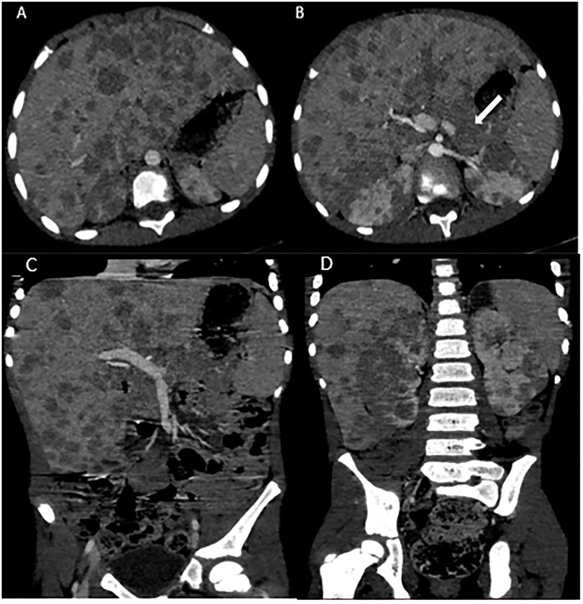
CT scan of the abdomen and pelvis injected in axial section (A and B) and coronal reconstruction (C and D) showing visceromegaly with multiple hepatic and renal nodules and pancreatic involvement.
At this stage, the diagnosis evoked was that of a malignant hemopathy or endocarditis complicated by septicemia.
A liver biopsy was performed under local anesthesia and the anatomopathological examination concluded to be Burkitt’s lymphoma (Figure 4).

Anatomopathological and immuhistochemical images of a liver biopsy in our patient. (A) HE Gx40, proliferation of round cells with ± crushed character by the biopsy procedure, round cells with hyperchromatic nucleated cytoplasm. (red star) bile duct. (B) Membrane labeling of tumor cells with CD10 antibodies. (C) Positive nuclear marking with anti-BCL 6 antibodies. (D) Diffuse membrane immunostaining of tumor cells with anti-CD20 antibodies.
The patient was admitted to the oncology department where he received chemotherapy with a good evolution of the gingival lesions. A scan after 1 month showed an almost complete regression of the lesions.
Discussion
Burkitt’s lymphoma (BL) is a high-grade non-Hodgkin’s lymphoma with significant tumor spread, particularly to the bone marrow and central nervous system, characterized by the proliferation of B-type lymphoid cells. 7
Historically, BL has been categorized into 3 subtypes, the endemic Burkitt’s lymphoma originating in Africa is the most common hemopathy in children and its incidence rate is about 50 times higher in Africa. Epstein-Barr virus (EBV) is implicated in this form. This type is characterized by a predominance of the maxillary location. 8
Our patient was originally from Morocco in North Africa.
Sporadic Burkitt’s lymphoma accounts for 30% of childhood lymphomas, occurring worldwide with a preferential abdominal location in 70% to 90% of cases. 9
The 3rd form occurs during HIV infection and is most common in people with human immunodeficiency virus/acquired immunodeficiency syndrome (HIV/AIDS). It can also occur in patients with inherited immune deficiencies or in those taking immunosuppressive drugs to prevent rejection after organ transplantation. The location is similar to that of the sporadic form.10,11
The recent definition of Burkitt lymphoma (BL) in WHO-HAEM5 (The 5th edition of the World Health Organization Classification of Haematolymphoid Tumors.) 5 remains mostly unchanged. It characterizes Burkitt’s lymphoma as a highly aggressive form of mature B-cell neoplasm consisting of medium-sized cells exhibiting a germinal center B-cell phenotype, including CD10 positivity, BCL6 positivity, weak or absent BCL2 expression, a high Ki67 index (>95%). Historically, BL has been categorized into 3 types. However, recent data suggests that Epstein-Barr-Virus. positive BL and Epstein-Barr-Virus. negative BL exhibit distinct molecular features, regardless of their epidemiological context or geographic location. Therefore, these molecular characteristics take precedence over the previous epidemiological subtyping. The distinction of the 2 subtypes, Epstein-Barr-Virus. positive Burkitt’s lymphoma versus Epstein-Barr-Virus.-negative Burkitt’s lymphoma, is recommended by WHO-HAEM5. 12
The clinical features of Burkitt’s lymphoma are variable and may manifest as endobuccal symptoms such as exophytic swelling of the gingival mucosa, associated with dental displacement and mobility, and altered general status. 9 Adenopathy is an inconsistent sign depending on the form of LB.
Radiologically, the oral involvement is manifested by tumor masses lysing the extensive bone, with displacement and mobility of the teeth. Abdominal involvement is manifested by organomegaly, with parenchymal nodules, thickening of the digestive tract, abdominal masses and periportal, and round ligament infiltration.
CT and ultrasound can be used to assess the extent of the lymphoma and for post-treatment monitoring. 13 Our patient had oral and maxillary involvement with endocranial extension and multivisceral involvement at the abdominal level including liver, kidney, and pancreas.
The positive diagnosis is confirmed by anatomopathological and immunohistochemical study which will show a monomorphic population of mature lymphocytes of intermediate size. The cells contain round nuclei with lacy chromatin and a basophilic cytoplasm with prominent vacuoles. numerous histiocytic cells giving a “starry sky” appearance consistent with Burkit’s lymphoma.
Chemotherapy is currently the mainstay of treatment due to the high chemosensitivity of this lymphoma. The major products used in the different therapeutic protocols are cyclophosphamide, methotrexate, cytarabine, vincristine, and doxorubicin. 9
The prognosis of this hemopathy depends on the initial extension of the disease and the speed of treatment.
The survival rate reaches 90% for all stages thanks to the new LMB protocols. 10
Our patient benefited from chemotherapy with a good evolution.
Conclusion
Burkitt’s lymphoma is a rare, very aggressive, fast-growing hematological malignancy that occurs frequently in children.
Its oral localization is rare, but it must be evoked in front of a gingival swelling with the ateration of the general state.
Footnotes
Acknowledgements
I would like to express my gratitude to my professors and all the colleagues who participated in the completion of this work.
Author’s Contributions
Declaration of Conflicting Interests
The author(s) declared no potential conflicts of interest with respect to the research, authorship, and/or publication of this article.
Funding
The author(s) received no financial support for the research, authorship, and/or publication of this article.
Ethics Approval
Our institution does not require ethical approval for reporting individual cases or case series.
Informed Consent
Written informed consent was obtained from a legally authorized representative for anonymized patient information to be published in this article.
Guarantor of Submission
The corresponding author is the guarantor of submission