
Abstract
This study aimed to explore differences in frontal lobe brain activity associated with two types of communication: task-oriented and life-worldly, the latter of which largely overlaps with everyday conversation. Using near-infrared spectroscopy, we explored differences by comparing oxygenated hemoglobin concentrations associated with periods of rest and conversation in two experimental groups comprising older and younger adults. Artifacts were removed from the signals using discrete wavelet transforms. Paired t-tests were used to compare the resulting data for the two types. The results showed that oxygenated hemoglobin levels during life-worldly communication were significantly higher than at baseline or during task-oriented communication, particularly for the older adult group. In addition, during life-worldly communication, relatively high levels of brain activity were found in the upper part of the Broca area and in the premotor cortex. These results, which suggest that life-worldly communication generates more activity in the frontal lobe, could potentially contribute to improving how caregivers communicate with older patients/residents in hospitals and nursing homes.
Introduction
Communication is a dynamic, systematic process in which meaning is created and shared as humans interact through using symbols (Wood, 2004). Many models exist for the functions of communication: we use communication to fulfill our physical and psychological needs, to obtain and share information, to develop and maintain relationships, to make decisions, and to persuade others (Steinberg, 2006). Based on a review of literature regarding the functions of communication, Vlăduțescu (2015) broadly classified them into two categories: those with a relationship (networking) function and those with a communication (sharing) function.
In day-to-day life, people have many kinds of conversational exchanges with their family, friends, acquaintances, coworkers, etc. Much of this everyday conversation overlaps with what is sometimes referred to as “small talk,” for example, about the weather, the seasons, one’s likes and dislikes, minor events, as well as gossip. Small talk has been defined as free conversation with no clear purpose that humans use for social interaction to create bonds and build friendships (Malinowski, 1999 [1926]) and is considered important for relationship-building (Coupland, 2000).
In the domain of older adult nursing care, communication plays a major role in developing and maintaining good caregiver-patient relationships (Balzer-Riley, 2012; Littlejohn & Foss, 2008). Nevertheless, despite the importance of psychosocial communication in nursing, it can be a low priority in actual nursing situations and the scarcity of social interaction in nursing homes has been considered an issue for some time (Armstrong-Esther & Browne, 1986; Grau et al., 1995; Nolan et al., 1995). It has been noted that older adults in long-term care facilities may suffer from psychosocial problems, such as loneliness, feeling isolated from their community, loss of identity, and unhappiness (Drageset et al., 2011; Mechakra-Tahiri et al., 2009). Not only are social isolation and loneliness major risk factors underlying depression in older adults (Jongenelis et al., 2004; McCusker et al., 2005), but they can negatively impact the immune system, potentially worsening one’s quality of life (Leonard, 2010). Furthermore, studies have indicated that patients want nurses to share more information with them and for their relationships with nurses to be less superficial, warmer, and more human (Bridges & Fuller, 2015; McCabe, 2004; Nakrem et al., 2011; Shattell, 2004).
Thus, nursing home communication deficiencies should be addressed in terms of both quantity and quality. Nevertheless, no previous studies seem to have explored deficiencies in communication between nurses and older patients by measuring the actual time spent communicating. Additionally, traditional research in nurse-patient communication has lacked a method for analyzing the quality of communication as a mutual interaction (Caris-Verhallen et al., 1999; Fleischer et al., 2009).
To address the deficiencies, in 2004, we conducted a study to identify the characteristics of caregiver-patient communication in long-term care facilities by analyzing its quantitative (duration and frequency) and qualitative (underlying interaction) aspects (Fukaya et al., 2004). Based on a survey of all verbal communication (conversations) between caregivers and residents during 1 day (from 9:00 a.m. to 5:00 p.m.), we found two different types of verbal communication occurring. Task-oriented communication occurred between the nurse and patient or resident and was related to their various tasks. Life-worldly communication gave meaning to these residents’ world as they went about their daily lives, including observations related to the season/weather, past life experiences, meals, hobbies, family, friends/acquaintances, social and other mundane matters, greetings, and expressions of affection. We found that these long-term care residents’ total utterance duration was around just 4 minutes a day because around 80% of all utterances were task-oriented.
Using conversation analysis (Schgloff, 2007), a qualitative method for analyzing verbal communications, we explored the mechanisms underlying interactions in the two communication types. Results showed that task-oriented communication was unilateral because what the nurse needed to do with (or say to) the patient was controlled by the objective of the care they had been tasked with. Therefore, residents’ utterance durations were remarkably short. However, when using life-worldly communication, caregivers showed interest and concern in what the residents said and allowed them to speak freely. Encouraging them to speak extended their communication duration. Therefore, we found that using life-worldly communication was effective in increasing the duration and frequency of resident speaking time (Fukaya et al., 2016). However, we found that nurses believed that this type of communication does not directly contribute to resolving health- or nursing care-related problems and perceived it not as communication necessary for a healthcare professional but as idle talk (Fukaya et al., 2009).
The context for this perception stems from ideas that deeply permeate clinical settings regarding what the professional objectives of communication should be. Verbal communication in institutions such as hospitals, courts, and schools, referred to as institutional talk, is characteristically oriented to specific institutional norms (Drew & Heritage, 1992). The structure and functions of an organization’s institutional talk differ according to the organization’s aims. Institutional conversations differ from free-flowing everyday conversation in that some users (e.g., patients) tend not to have the opportunity to start a conversation (Drew & Heritage, 1992). According to de Haes and Bensing (2009), the functions of healthcare-related communication, a type of institutional talk, are (1) fostering relationships, (2) gathering information, (3) getting information, (4) making decisions, (5) enabling disease and treatment-related behavior, and (6) responding to emotions. Thus, institutional talk in medical and nursing care has been structured based on the premise that the relationship between a doctor or nurse and a patient is not an ordinary relationship but is, rather, a therapeutic relationship aimed at resolving a health problem (Moore & Kuipers, 1992; Moreira et al., 1997; Roter & Larson, 2002). As a result, life-worldly communication, which largely overlaps with emotional/social communication, is considered a useful but indirect/secondary means to accomplish communication objectives in institutions (Gilbert & Hayes, 2009; Kasch & Lisnek, 1984) and is not seen as important in medical or nursing care communication.
However, daily conversation and small talk are part of a process fundamental to developing human relations by which a person confirms the social significance of their existence, experiences various emotions, sustains a life that is spiritually rich and secure, and finds satisfaction with intimacy and mutual acknowledgement in human relationships (Coupland, 2003; Wood, 2004). Life-worldly communication should be considered necessary in the care of older adults because it improves their emotional well-being and quality of life and can potentially reduce feelings of loneliness and isolation (Fukaya, 2017). In addition, it has been suggested that feeling lonely may contribute to cognitive functioning decline (Cacioppo, 2014), suggesting that a lack of life-worldly communication could contribute to reduced brain activity.
The different forms of non-invasive measurements of physiological brain activity can be divided into techniques that measure electrical activity in the central nervous system and techniques that measure changes in metabolic functions that support that activity. Near-infrared spectroscopy (NIRS) is an example of the latter and measures hemoglobin dynamics in tissue, as the brain’s hemodynamic response to increased neural activity leads to increased concentration of oxygenated hemoglobin (oxyHb)in activated brain areas. NIRS involves emitter and detector optodes being attached to the scalp. The principle for the measurement is based on the modified Lambert-Beer law, which relates the negative logarithm of the ratio of transmitted to emitted light intensity to the product of the path length and changes in hemoglobin concentration (Hoshi, 2007; Tsunashima et al., 2012). Compared to functional magnetic resonance imaging (fMRI), NIRS is less restricted in its usage (Cutini et al., 2012). However, there is more potential for noise in the signal, such as artifacts due to the distances between the emitters and detectors and changes in those distances, as well as to bodily movements and physiological artifacts, such as pulse. Strategies for dealing with these artifacts include adaptation of time-frequency domain analysis based on wavelet transforms (Tsunashima et al., 2012) and guidelines for eliminating irregular outlier values due to bodily movements (Cui et al., 2010; Fekete et al., 2011).
Studies have been conducted on NIRS use in psychiatric disorder diagnosis and in measuring brain activity during intelligence tests (Schecklmann et al., 2008; Suda et al., 2010; Takei et al., 2014). However, most such studies measured brain activity under experimental conditions following protocols that were restrictive and out-of-the-ordinary. To our knowledge, none have looked at brain activity under more ordinary conditions resembling everyday conversation, and none have applied this approach to communication between caregivers and older adults. Thus, this study aimed to explore differences in brain activity associated with task-oriented and life-worldly communication by age group with NIRS.
Research Design
Participants
Convenience sampling was used to recruit Japanese adults aged 65 or older and those aged 19 to 24 living in satellite cities of the Japanese capital (elderly population: 2.29 million representing 23.9% of the total population). Candidates were ineligible if: (1) they suffered from hearing loss; (2) they suffered from global aphasia; (3) they were diagnosed level IIa or higher (mild or more severe) on the Ministry of Health, Labour and Welfare in Japan (2003) Daily Life Independence Criteria for Elderly People with Dementia; or (4) their health was unstable due to being in the acute phase of treatment for a medical condition or being at the end of life. In Japan, 61% of older adults stay socially active for health reasons or to give their lives purpose by, for example, playing sports, pursuing hobbies, or attending community events (Cabinet Office in Japan, 2017). We, therefore, recruited healthy older adults who did not meet the exclusion criteria through community associations, sports clubs, and a cleaning and maintenance company that employs the elderly. We obtained informed consent from 43 older adults (11 through community associations, 12 through sports clubs, 11 through hobby and volunteer groups, and nine who worked part-time at a cleaning and maintenance inspection company). We recruited the younger adults by posting notices on university bulletin boards, and 24 applicants consented to participate.
Experimental Equipment
We used the NIRO-200NX NIRS device from Hamamatsu Photonics. We used four light emitters and nine photo-detectors to record oxygenated and deoxygenated hemoglobin concentrations (in μmol/l) in 16 channels (Figure 1). For the path length, we used the preset value for an adult head (18 cm). For the sampling frequency, we used 10 Hz. The 13 optodes were placed symmetrically left to right with the front row coinciding with the T3-Fpz-T4 line in the International 10 to 20 system for electroencephalography (Verner et al., 2013). A video camera simultaneously recorded (with audio) the communication during each trial.

NIRS optode positioning: The numbers are the channel numbers. Red circles represent the positions of the light emitters. Blue squares represent the positions of the photo-detectors. The interoptode distance was 3 cm. The optodes were placed symmetrically left to right with the front row placed along T3-Fpz-T4 in the International 10 to 20 system.
Procedure
The procedure for brain activity study by communication type was as follows. The task-oriented communication format was modeled on the communication that occurs in a medical interview to obtain information for medical or nursing care. This type had a unilateral pattern in which the researcher took the lead with closed-ended questions to which the subject responded (e.g., What time did you go to bed last night? Did you sleep well? What time did you get up?). The life-worldly communication format was modeled on small talk going on nearby. The researcher began by selecting a life-worldly communication topic (e.g., work, housework, hobbies, family) that would be relevant to the subject. The researcher showed interest in the subject to encourage them to talk, enabling them to pick their own topic and expand on the conversation, thus setting a bilateral communication pattern (e.g., Researcher: What kind of work did you do? Subject: I did “gaishou” (called on customers to take orders) for a department store. Researcher: Really? I’ve never shopped that way! Is it a difficult task to do? Subject: It’s not that difficult, but. . . [The subject continues talking]).
During measurement, the researchers engaged in conversations with subjects from a distance of one meter, following the protocol below. Additionally, to prevent the conversation type order from affecting the results, we started half of the cases with Type I and half with Type II. A total of 29 minutes per subject was spent going through the following protocol. NIRS measurements were taken during all five steps.
(1) Rest (3 minutes)
(2) Type I or II communication (10 minutes)
(3) Rest (3 minutes)
(4) Type II or Type I communication (10 minutes)
(5) Rest (3 minutes)
Analysis
NIRS data analysis
To remove noise from physiological artifacts from the recorded NIRS signal and artifacts due to bodily movements, we adapted the discrete wavelet-based multi-resolution analysis used by Tsunashima et al. (2012). For the multi-resolution analysis, we selected Daubechies wavelet 7 as the mother wavelet and decomposed the NIRS signal into a signal with 10 different center frequencies. Of these, we selected the signal with the center frequency of 0.433 Hz as the component most likely related to the conversation. To remove artifacts due to improperly fixed optodes, we calculated the running correlations and excluded hemoglobin concentrations with RC > 0.2 (Cui et al., 2010). Additionally, we Z-scored those signals and excluded values when z > 1 (Fekete et al., 2011) and eliminated non-measurable signals as errors. The NIRS signals with positive oxygenated hemoglobin concentrations (oxyHb > 0) were analyzed. Additionally, at baseline, we used signals for rest times that approached those of both task-oriented and life-worldly communication. Figure 2 shows an example of the multi-resolution analysis application and the calculation of running correlations for the NIRS signals.

An example of NIRS signal noise removal. Using multi-resolution analysis, the NIRS signal component related to talking was identified (upper box). Red and blue lines show the concentrations of oxygenated and de-oxygenated hemoglobin, respectively. The middle box shows detail for the part of time with larger waveforms. RC = Running correlation. The lower box shows the time waveform for RCs calculated to eliminate artifacts for that part of time.
Statistical analysis
We compared brain activity by communication type as follows:
(1) Mean baseline oxyHb levels were compared by age group and gender using t-tests.
(2) Mean oxyHb levels during each communication type were compared with mean baseline oxyHb levels.
(3) Mean oxyHb levels during task-oriented and life-worldly communication were compared using the difference in oxyHb levels means during each communication type and the baseline oxyHb levels.
(4) Results for three were compared by age group.
(5) Areas of brain activity during life-worldly communication were compared.
In (2) to (5), the comparisons were performed using paired t-tests.
SPSS version 24 was used for the statistical analysis.
Ethical Approval Considerations
This study was conducted according to the principles of the Declaration of Helsinki and Japanese Nursing Association code of ethics for nurses. Respondents were given a formal request to participate, and those who provided their written informed consent became study participants. The Ethical Review Board for Research on Human Subjects of Kanto Gakuin University approved the protocol for the study (approval no. H2017-2-3).
Results
The analyzed sample comprised 40 independently living older adults aged of 65 to 83 (mean = 71.73 ± 4.97) and 24 university students aged 19 to 24 (mean = 21.5 ± 1.06), yielding a total of 64 subjects. Of the 43 older participants, three (two males, one female) were excluded because we were unable to measure their brain activity. Thirty-four (53%) participants were male and 30 (47%) were female.
As shown in Table 1, significantly higher baseline means for oxyHb (in μmol/l) were found in the young adults compared to the older adults in eight of the 16 channels (ch1, 2, 5, 6, 7, 12, 15, 16). No significant gender-related difference was found.
Comparison of Mean Baseline oxyHb Levels by Age Group.

Note. CI = confidence interval; LL = lower limit; UL = upper limit.***p < .001. **p < .01. *p < .05.
Tables 2 and 3 show comparisons of mean oxyHb levels during task-oriented and life-worldly communication with the baseline levels (at rest). The differences between oxyHb levels during task-oriented communication and the baseline ranged from 0.00 to 0.02 μmol/l. For all channels, the task-oriented communication oxyHb levels were significantly higher than at baseline [t (62) = −2.14 to −4.60, p = .035 to .000]. For life-worldly communication, the difference with the baseline ranged from 0.01 to 0.05 μmol/l, and for all channels, the oxyHb levels were significantly higher during communication than at baseline [t (62) = −2.84 to −6.54, p = .006 to .000].
Differences in Mean oxyHb Levels During Task-Oriented Communication and at the Baseline (n = 64).

Note. Baseline = mean oxyHb level at baseline; Task-oriented = mean oxyHb level during task-oriented communication; CI = confidence interval; LL = lower limit; UL = upper limit. ***p < .001. **p < .01. *p < .05.
Differences in Mean oxyHb Levels During Life-Worldly Communication and at the Baseline (n = 64).

Note. Baseline = mean oxyHb level at baseline; LWC = mean oxyHb level during life-worldly communication; CI = confidence interval; LL = lower limit; UL = upper limit. ***p < .001. **p < .01. *p < .05.
Due to individual differences in baseline means, to compare brain activity during task-oriented and life-worldly communication, the oxyHb levels used for each communication type were the oxyHb levels for each type minus the baseline oxyHb levels. As shown in Table 4, the difference in mean oxyHb levels during task-oriented and life-worldly communication ranged from .00~.04 μmol/l, with oxyHb levels being significantly higher during life-worldly than task-oriented communication [t (62) = −2.07 to −4.90, p = .042 to .000].
Differences in Mean oxyHb Levels During Task-Oriented and life-Worldly Communication (n = 64).

Note. Task-oriented = mean oxyHb level during task-oriented communication—mean oxyHb level at baseline; LWC = mean oxyHb level during life-worldly communication—mean oxyHb level at baseline; CI = confidence interval; LL = lower limit; UL = upper limit. ***p < .001. **p < .01. *p < .05.
We then investigated whether the above difference in oxyHb levels for the two communication types differed by age group. For the older adults, the differences in oxyHb levels ranged from 0.00 to 0.04 μmol/l, and the levels were significantly higher for life-worldly communication than task-oriented communication in 13 channels, the exceptions being channels 10, 12, and 16 [t (39) = −2.25 to −4.53, p = .03 to .000] (Table 5). However, for the younger adults, the differences ranged from 0.00 to 0.02 μmol/l, and significant differences were found in only six channels (2, 4, 5, 7, 13, and 16) [t (23) = −2.09 to −3.75, p = .048 to .001].
Comparison of Differences in Mean oxyHb Levels During Task-Oriented and Life-Worldly Communication by Age Group.
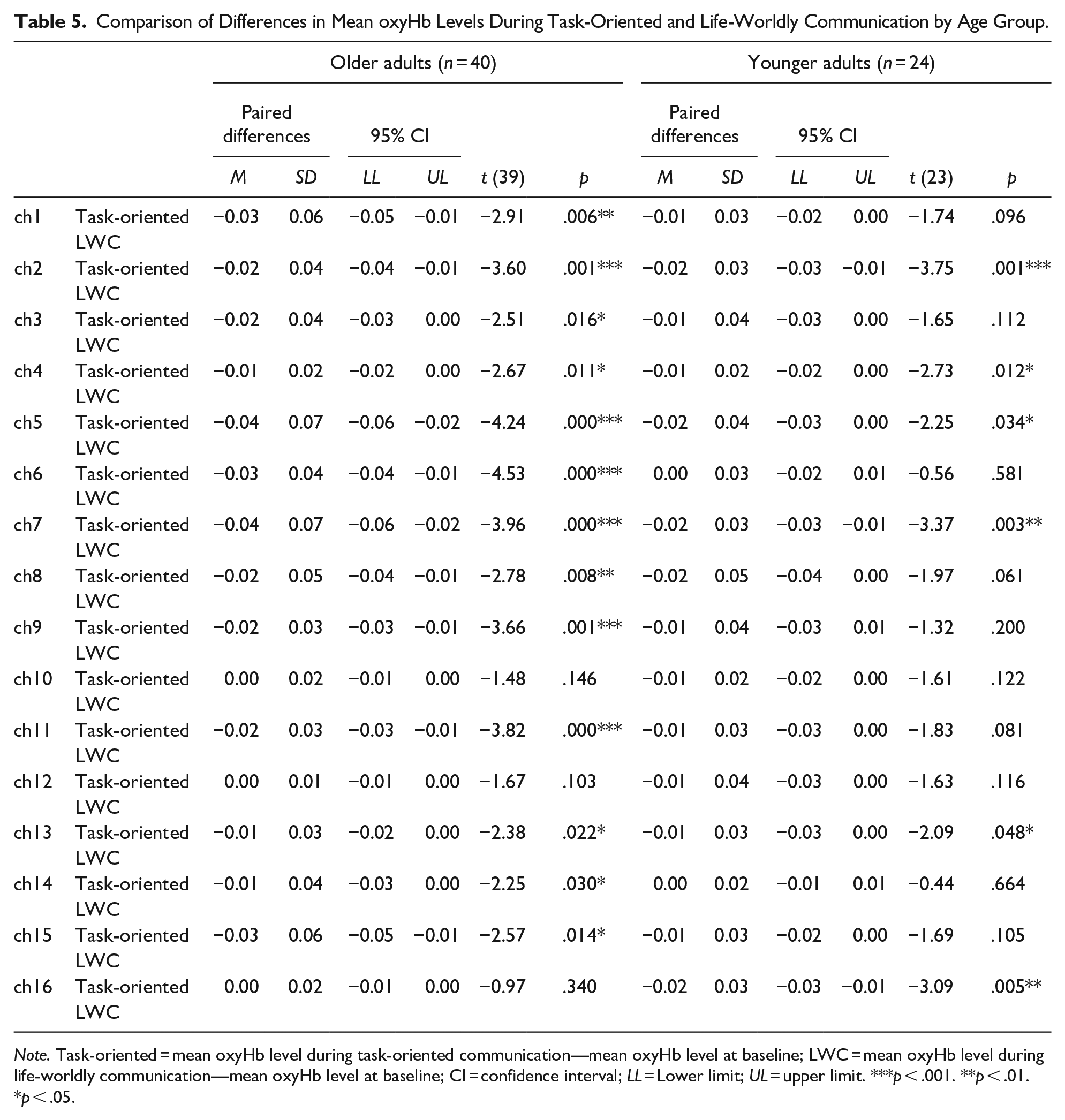
Note. Task-oriented = mean oxyHb level during task-oriented communication—mean oxyHb level at baseline; LWC = mean oxyHb level during life-worldly communication—mean oxyHb level at baseline; CI = confidence interval; LL = Lower limit; UL = upper limit. ***p < .001. **p < .01. *p < .05.
To observe the differences between channels during life-worldly communication, we examined differences in mean oxyHb between channels 10, 9, 2, and 1 on the left side, channels 16, 15, 8, and 7 on the right side, channels 10, 12, 14, and 16 in the frontal pole, and channels 1, 3, 5, and 7 in the premotor cortex. As shown in Table 6, the right side was slightly more active than the left. However, channel 2 on the left side, located slightly above the Broca area, showed significantly more activity than channel 8 on the right side [t (63) = 2.06, p = .04]. Vertical comparison prefrontal cortex activity showed that the premotor cortex was significantly more active than the frontal pole [t (63) = −2.50 to −3.93, p = .01 to .000].
Comparison of Differences in mean oxyHb Levels Between Channels During Life-Worldly Communication.
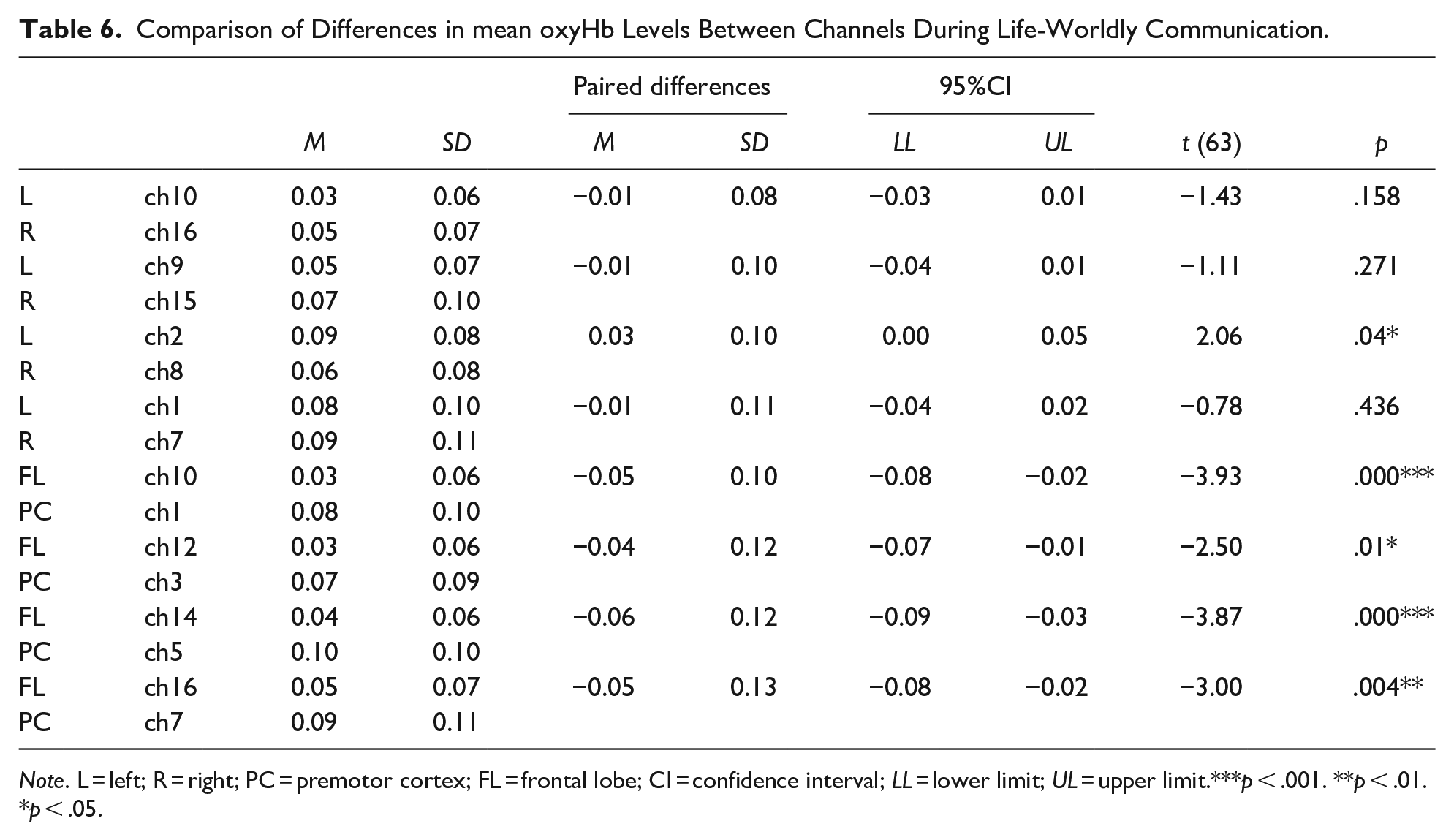
Note. L = left; R = right; PC = premotor cortex; FL = frontal lobe; CI = confidence interval; LL = lower limit; UL = upper limit.***p < .001. **p < .01. *p < .05.
Discussion
A comparison of mean baseline oxyHb levels by age found levels to be significantly lower for the older adults in eight channels, as supported by studies suggesting that cerebral blood flow declines with age in the gray matter (Mozley et al., 1997), the regions of the inferior frontal gyrus, both sides of the middle frontal gyrus, the left superior temporal gyrus, both sides of the cingulate gyrus (Takahashi et al., 2005), the parietal region, the left frontotemporal cortex, and the temporal cortex (Pagani et al., 2002). Although Takahashi et al. (2011) suggested that in the frontal region, blood flow in the scalp accounts for most NIRS signals, even our study found that from the parietal to the frontal region, there was a significant age-related decline in blood flow.
No significant gender-related difference in baseline oxyHb was found, supporting previous studies’ results showing no gender-related differences in cerebral blood flow in older adults (Liu et al., 2016; Schmidt et al., 2009).
Mean oxyHb for both communication types was significantly higher than baseline oxyHb for all of the channels, confirming activity in the prefrontal cortex (PFC). A commonly used test for frontal lobe function is the VFT. VFTs examine how many words a subject can speak or write that meet specified conditions in a prescribed amount of time. This kind of task explores a series of linguistic functions (e.g., recall from short-term memory), examining whether specific conditions are met and reproducing the words either verbally or in written form (Matsuo et al., 2004; Nishida et al., 2017). Studies using VFTs in samples of healthy individuals have shown activity in the frontotemporal cortex (Herrmann et al., 2017) and the PFC (Kubota et al., 2005). The PFC is also involved in executive processes related to working memory (Baddeley, 1992, 2000) and episodic memory recall (Lepage et al., 2000). Thus, the PFC activation seen in this study may be similar to that in VFTs, given that in both communication types the subjects had to recall things from memory, examine how those things would be uttered, and express them linguistically.
The comparison of the two communication types revealed that the mean oxyHb for life-worldly communication was higher than for task-oriented communication in all channels, indicating that it stimulated more PFC activity. In humans, the PFC is the area of the brain involved in working memory (Baddeley, 1992), a system that provides for the temporary storage and simultaneous manipulation of information. It enables various higher cognitive functions, such as understanding words and sentences, thinking, reasoning, and decision-making, which are essential in situations calling for the relating and execution of deliberate acts, displays of spontaneity, problem-solving, etc. (Funahashi, 2017). During task-oriented communication, subjects merely responded precisely to the question. However, during life-worldly communication, subjects had to decide what to talk about, consider ways to structure and organize it such that the listener (the researcher) would understand, make a decision, and carry it out, suggesting that in life-worldly communication there was more activity in the PFC working memory to retain and process information.
Another potential reason for the greater prefrontal activity increase during life-worldly communication is the effect of emotions. Studies on prefrontal activity and emotions have suggested that positive emotions activate cognitive functioning (e.g., in the PFC or insula) (Raschle et al., 2017), negative emotions reduce PFC activity (Aoki et al., 2013), anxiety reduces the hemodynamics of right PFC lateralization (Tseng et al., 2018), pleasant feelings amplify dorsolateral prefrontal cortex activity (Perlstein et al., 2002), and emotions and cognition interact to control thinking and behavior (Gray et al., 2002). In life-worldly communication, subjects choose topics that interest or concern them. The researcher demonstrating interest in that topic and encouraging them to elaborate expands the breadth of the conversation. This type of communication may have promoted activation of the subject’s PFC through the exchange of a variety of different emotions, thoughts, and ideas. Furthermore, life-worldly communication facilitates the subject’s construction of a world, which gives meaning to their existence. To confirm the social significance of one’s existence, as through this type of communication, feeling empathy for others is essential. Because the seat of empathy is located partly in the PFC (Meyer et al., 2013; Rameson et al., 2012), that region’s increased activity may have reflected its effect.
The comparison of the two communication types by age group showed that mean oxyHb levels for life-worldly communication were significantly higher for older adults, while significant differences were shown in only six channels for younger adults. A potential explanation for this could be that mean baseline oxyHb levels for the younger adults were significantly higher than for the older adults in nine of the channels, indicating that age-related differences in cerebral blood flow at baseline could have had an effect.
Regarding limitations, our results may contain sample-related biases due to the use of convenience sampling rather than random sampling for recruitment. In addition, the representativeness of the sample size was poor due to the fact that there were fewer younger participants (24) than older participants (40). Another limitation was that for the NIRS measurements, we used the NIRO-200NX. The optodes for this instrument are embedded in a cap. Although it can be adjusted to fit the subject’s head, depending on head shape, poor contact can generate signal noise. This resulted in considerable effort being needed for noise removal. Additionally, for subjects with little or no hair, measurements in many channels were not possible, which resulted in the exclusion of three participants. The development of better equipment to perform NIRS and better signal manipulation techniques would be helpful in the future.
Regarding the prefrontal cortex areas characteristically active during life-worldly communication, the results showed significant levels of activity in channel 2 on the left side and in the premotor cortex. The Broca area, which is involved in language processing, utterance calculation, and language comprehension, is usually located in the left prefrontal area (Brodmann areas 44 and 45) (Horwitz et al., 2003). Channel 2 was located slightly above the Broca area in Brodmann area 46. However, since the distance between the optodes in the NIRS device used in this study could not be adjusted to the shape of the subject’s head, there were slight variations between individuals in the areas measured. As a result, during continuous conversation, channel 2 activity may have been affected by Broca area activity.
The premotor cortex is also reportedly involved in complex actions such as voluntary choice, preparation, switching, and combining movements and active in speech perception and speaking (Callan et al., 2006; Iacoboni, 2008; Meister et al., 2007). Life-worldly communication is an intricate conversational activity that could involve premotor cortex activation. It develops as the speaker interacts with others, taking in information, such as the other person’s facial expressions and responses, and instantaneously deciding which complex conversational technique to use to continue the conversation.
Based on a literature review, Fratiglioni et al. (2004) concluded that, for older adults, an active and socially integrated lifestyle helps maintain cognitive functioning and prevent dementia. It has also been suggested that social relations and emotional support, especially from friends and family, contribute to the maintenance of cognitive functioning (Béland et al., 2005; Seeman et al., 2001). This study suggested that, not only does life-worldly communication play an important role in building social relations between people, but that, in older adults, the act of communicating itself can activate and potentially maintain cognitive functioning in older adults living in an eldercare facility. Moreover, given that older patients with depression may have low levels of oxyHb in the frontal region (Noda et al., 2012), encouraging life-worldly communication on a daily basis could potentially contribute to the prevention of depression.
Conclusion
This study was the first to look at brain activity related to life-worldly communication, a communication type largely ignored in medical and nursing care. Our results showed that life-worldly communication activates prefrontal cortex functioning in healthy community-dwelling older adults. If these results can be generalized to older adults who require nursing care, they could suggest that life-worldly communication would be an important part of care for older adults living in an eldercare facility. They may also contribute to rethinking and improving how caregivers in healthcare and public welfare communicate with older adults. Further, this study could be a steppingstone to develop future studies of the relationship between life-worldly communication and quality of life as well as dementia due to brain function loss through disuse.
Footnotes
Acknowledgements
We would like to express our deepest gratitude to everyone who supported the purpose of this study and participated in the survey.
Declaration of Conflicting Interests
The author(s) declared no potential conflicts of interest with respect to the research, authorship, and/or publication of this article.
Funding
The author(s) disclosed receipt of the following financial support for the research, authorship, and/or publication of this article: This work was funded by Japan Society for the Promotion of Science (KAKENHI Grant Number 17K12427).