
Abstract
Background:
Medial elbow joint instability is a critical concern among young baseball players and is associated with throwing-related injuries. Greater medial elbow joint space (MEJS) gapping has been proposed as a key indicator of valgus instability. However, age-specific differences remain unclear, particularly in elementary school-age pitchers.
Hypothesis/Purpose:
This study aimed to clarify age-related differences in MEJS gapping by comparing joint space changes between resting and gravity stress conditions among young baseball players. It was hypothesized that 12-year-old players would show greater changes in MEJS compared with younger players.
Study Design:
Cross-sectional study; Level of evidence, 3.
Methods:
This study included 190 young baseball players (mean age, 11 ± 0.6 years) from 22 teams. Ultrasonographic assessment of the MEJS was performed during a resting and a gravity-induced valgus stress test. The change in MEJS was defined as the distance between the medial humeral epicondyle and the sublime tubercle of the ulna measured under each condition. The Kruskal-Wallis test was used to compare changes in MEJS among 3 age groups (10, 11, and 12 years), and the Wilcoxon rank-sum test was used to assess differences between the throwing and nonthrowing sides. Multiple regression analysis was performed to identify factors associated with the change in MEJS on the throwing side.
Results:
Significant age-related differences in MEJS changes were observed (P = .03). Post hoc analysis revealed that 10-year-old players (median, 0.75 mm [interquartile range [IQR], 0.34–1.27]) exhibited significantly greater changes in MEJS on the throwing side compared with 12-year-old players (median, 0.47 mm [IQR, 0.25–0.70]; P = .03). The change in MEJS on the throwing side was significantly greater than that on the nonthrowing side across all age groups (P < .01). Change in MEJS on the nonthrowing side (β = 0.56; P < .01) and age (β = −0.14; P = .03) were associated with the change in MEJS on the throwing side (R2 = 0.38; P < .01).
Conclusion:
Our study demonstrated that 10-year-old players showed greater MEJS changes than 12-year-old players, highlighting the need for age-specific interpretation. These changes may reflect physiological joint laxity related to growth rather than pathological instability, with implications for ultrasonographic interpretation and injury risk.
Baseball-related throwing injuries, particularly elbow injuries, are common among school-aged athletes. 2 A survey 9 of young Japanese baseball players revealed that elbow pain was the most frequently reported injury (22%), exceeding pain in the shoulder (18%). Among elbow disorders, Little League elbow affects 20% to 40% of young pitchers.10-12 Moreover, an increasing number of cases require surgical intervention, underscoring the importance of early preventive measures— including pitch count regulation and throwing limitations.2,5
Previous studies indicate that >50% of teenage pitchers experience elbow pain during the season, 3 and 30.5% of baseball players aged 7 to 11 years report experiencing elbow pain. 14 Additionally, the proportion of children who experience elbow pain begins to increase at age 8, rises sharply at age 9, and reaches approximately 50% by age 12 years. 18 A systematic review identified several risk factors for elbow joint disorders—including age 9 to 11 years, pitcher or catcher position, training exceeding 16 hours per week, history of elbow pain, and pitching >100 innings per year. 1 Alarmingly, the mean age of onset of throwing-related elbow disorders has declined over the past decade from 11.6 to 10.7 years. 19
One proposed mechanism of throwing-related elbow injuries is repetitive valgus stress, which leads to progressive gapping of the medial elbow joint space (MEJS), increasing stress on the growth plate and predisposing young athletes to instability and injury. 6 Ultrasonographic evaluation enables real-time assessment of MEJS changes under dynamic stress, providing a noninvasive method for detecting early signs of instability and laxity. Notably, previous studies have reported that the MEJS gapping in young baseball players (throwing side: 0.7 mm, nonthrowing side: 0.5 mm) is greater than that in high school and collegiate athletes (throwing side: 0.5 mm, nonthrowing side: 0.2 mm), 13 suggesting that skeletal immaturity and increased valgus loading may contribute to joint instability.
Change in MEJS between resting and gravity-induced valgus stress test conditions is considered a key indicator of valgus instability and increased stress on the medial growth plate, which may lead to throwing-related elbow disorders. However, significant variability exists among elementary school-age athletes, particularly among those aged 10 to 12, owing to differences in physical development, skill level, and training intensity. Previous studies have identified height >150 cm and age ≥11 years as strong predictors of throwing-related injuries,5,24 highlighting the need for a granular, age-specific evaluation of MEJS changes in young baseball players. Therefore, this study aimed to clarify age-related differences in MEJS gapping by comparing joint-space changes between the resting and gravity-stress conditions in young baseball players. We hypothesized that 12-year-old players would show greater changes in MEJS compared with younger players.
Methods
Study Design
This cross-sectional study was conducted between February and August of both 2016 and 2024, involving young baseball players from 22 teams in Kanagawa Prefecture, Japan. This study utilized physical examination data collected as part of an annual, field-based medical screening program for young baseball athletes, routinely conducted by health care professionals from a local hospital and its affiliated institutions. The present analysis integrated data collected over 6 years (2016–2024), excluding 2020–2022, when measurements were suspended due to coronavirus disease 2019 (COVID-19) pandemic-related restrictions. This study was conducted in accordance with the Declaration of Helsinki and approved by the ethics committee of Kitasato University Kitasato Institute Hospital (approval number: 18044). Informed consent was obtained from the parents or legal guardians of all participants using the opt-out method.
Participants
This study included 181 young baseball players (male players: 175; female players: 6) aged between 10 and 12 years (mean age, 11 ± 0.6 years; height: 146.7 ± 7.4 cm; weight: 41.5 ± 8.6 kg). This study used medical screening data that excluded players <10 years. The eligibility criteria required that participants were actively playing baseball at the time of measurement and were members of the Japan Rubber Baseball Association. Players with a history of elbow injuries or current elbow discomfort were also included. All participants were affiliated with 22 teams in Kanagawa Prefecture, and their characteristics are summarized in Table 1. The participants completed an initial questionnaire regarding their dominant throwing side, years of baseball experience, and primary playing position (pitcher, catcher, or other). The exclusion criteria included missing data and players with osteochondritis dissecans.
Participant Characteristics a
Data are presented as means ± SD or n (%). BMI, body mass index.
Outcome Measures
Measurement of MEJS Gapping
All ultrasonographic measurements were performed by a single experienced ultrasonographer, a physical therapist (H.M.) with 9 years of experience as of 2024, using a standardized measurement protocol. Standardized protocols from previous studies on MEJS gapping were followed to enhance reproducibility.6,13,23 An assistant stabilized the elbow and forearm during imaging. The change in MEJS between resting and gravity-induced valgus stress conditions was measured using ultrasonography. Measurements obtained from 2016 to 2019 were performed using a SonoSite M-Turbo system (FUJIFILM SonoSite, Inc), whereas those obtained from 2023 to 2024 were performed using a diagnostic ultrasound system (KONICA MINOLTA, Inc).
Each participant was positioned supine on an examination table, with the shoulder abducted to 90°, the elbow flexed to 90°, and the forearm in a neutral position. Sonographic measurements were obtained under 2 conditions: resting condition (Figure 1A) and gravity stress (Figure 1B). The gravity stress method used in this study has previously been reported as an effective approach for assessing changes in MEJS (Figure 1C). 4 Measurements were performed sequentially, beginning with the throwing side elbow under resting conditions, followed by the same elbow under gravity stress. Subsequently, the nonthrowing side elbow was assessed under resting conditions, and finally, the nonthrowing side elbow was measured under gravity stress. An 11-MHz linear probe was placed over the medial epicondyle of the humerus and aligned along the long axis of the ulnar collateral ligament to visualize the medial epicondyle, trochlea, and sublime tubercle (Figure 1D). The ulnohumeral joint space was measured from the distal-medial corner of the trochlea of the humerus to the proximal edge of the medial tubercular portion of the ulnar coronoid process (Figure 1D). The fibrillar pattern of the ulnar collateral ligament was also evaluated. The dynamic motion of the medial elbow during valgus stress was recorded as a B-mode ultrasound video. If probe displacement was observed, the procedure was repeated. One successful video from each trial was transferred to a personal computer via an external hard drive. All ultrasonographic data were trimmed to the point at which the MEJS reached its maximum during the transition from rest to forearm self-weighted valgus stress.
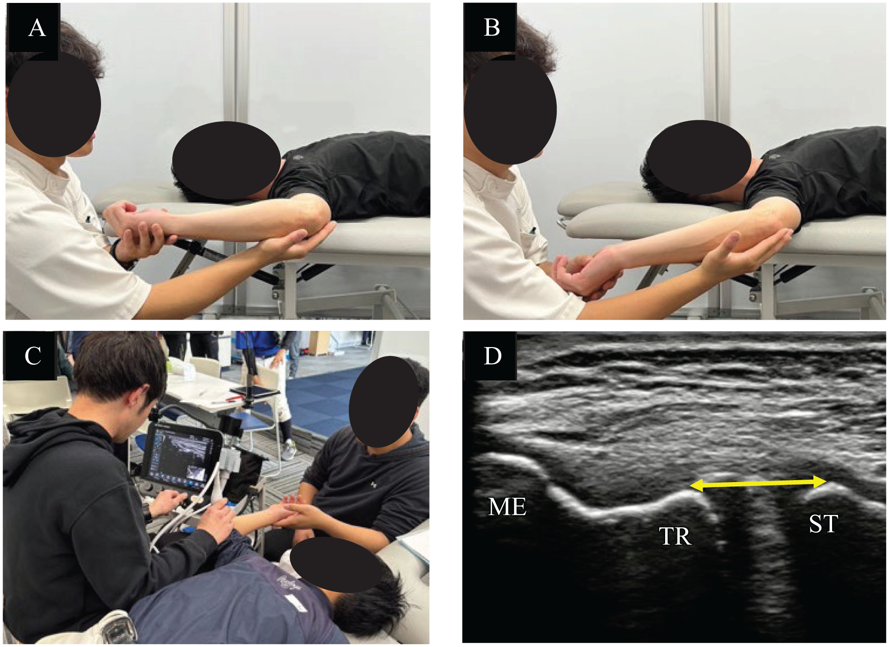
Measurement of the change in the MEJS. (A) MEJS in the resting condition. (B) MEJS under forearm self-weighted gravity-induced valgus stress conditions. (C) The MEJS gap measured from ultrasound images taken in the supine position. (D) Manual measurement of the MEJS between the trochlea and the sublime tubercle. ME, medial epicondyle; MEJS, medial elbow joint space; ST, sublime tubercle; TR, trochlea.
Measurements were performed using the ultrasonography distance measurement tool (minimum unit: 0.1 mm) and analyzed using ImageJ software (Version 1.48; National Institutes of Health). Change in MEJS was defined as the difference in the ulnohumeral joint space under gravity stress from the resting condition.
Statistical Analysis
All statistical analyses were performed using JMP Pro Version 17 (SAS Institute Inc). Data normality was assessed using the Shapiro-Wilk test. The Kruskal-Wallis test was used to compare MEJS change distributions across age groups for both the throwing and nonthrowing sides. When significant differences were detected, post-hoc pairwise comparisons were conducted using the Steel-Dwass test. Comparisons between the throwing and nonthrowing sides within each age group, as well as between pitchers/catchers and other players, were performed using the Wilcoxon rank-sum test. Multiple regression analysis was performed to identify factors associated with the change in MEJS on the throwing side. The dependent variable was the change in MEJS between the resting and gravity-stress conditions. Independent variables included MEJS change on the nonthrowing side, age, sex, baseball experience, and playing position. P < .05 was considered statistically significant.
Sample size calculations were conducted using G*Power (Version 3.1.9.6; Heinrich-Heine-Universität Düsseldorf; http://www.gpower.hhu.de/). The calculations were based on an alpha level of .05, a statistical power of 0.80, and an effect size of 0.25 (moderate). The results indicated that a minimum of 159 participants were required to detect statistically significant differences using the Kruskal-Wallis test.
Results
In total, 190 young baseball players were initially enrolled. We excluded 9 players because of missing data (n = 9) and osteochondritis dissecans (n = 0). Thus, 181 young baseball players were enrolled in this study. The characteristics of the participants are shown in Table 1. There were no significant differences in participant characteristics between the pre-COVID-19 period (2016–2019) and the post-COVID-19 period (2023–2024). Analysis of MEJS change on the throwing side across age groups revealed a significant difference between the 3 age groups (10-year-olds, 0.75 mm; 11-year-olds, 0.55 mm; and 12-year-olds, 0.47 mm) (P = .03; η2 = 0.033), indicating a small effect size, whereas no significant differences were observed for the nonthrowing side (Table 2). Post-hoc pairwise comparisons indicated that the MEJS changes on the throwing side in the 10-year-old group (0.75 mm) were significantly greater than those in the 12-year-old group (0.47 mm) (P = .03; r = 0.32) (Table 2; Figure 2), indicating a moderate effect size. Additionally, the MEJS change on the throwing side was significantly greater than that on the nonthrowing side across the age groups (P < .01; r = 0.33) (Figure 2), indicating a moderate effect size. There were no significant differences based on playing position for either the throwing (P = .69) or nonthrowing (P = .61) side (Figure 3).
Age-Based Difference of Change in MEJS (in mm) a

Data are presented as median [IQR]. Bold P values indicate statistical significance. IQR, interquartile range; MEJS, medial elbow joint space.
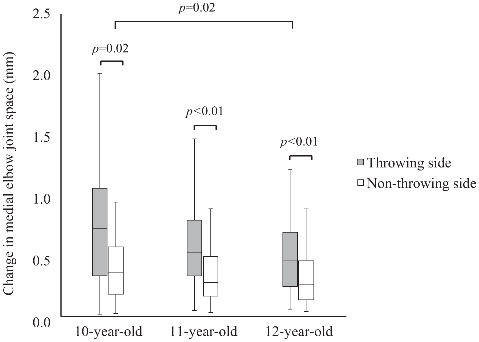
Age-based differences in MEJS changes between the throwing and nonthrowing side. MEJS, medial elbow joint space.

Differences in MEJS changes by playing position. MEJS, medial elbow joint space.
The results of the multiple regression analysis for the changes in MEJS on the throwing side are shown in Table 3. The changes in MEJS on the nonthrowing side (β = 0.56; P < .01) and age (β = −0.14; P = .03) were identified as factors associated with the changes in MEJS on the throwing side (R2 = 0.38; P < .01). There was no evidence of multicollinearity among the independent variables.
Factors Related to Changes in the MEJS on the Throwing Side by Multiple Regression Analysis, n = 181 a
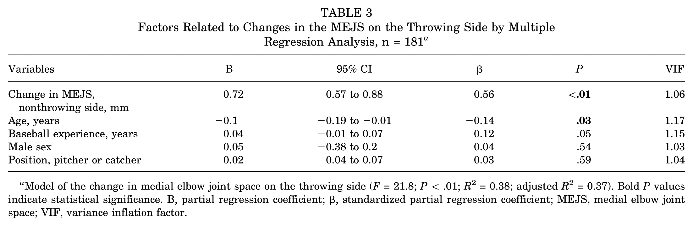
Model of the change in medial elbow joint space on the throwing side (F = 21.8; P < .01; R2 = 0.38; adjusted R2 = 0.37). Bold P values indicate statistical significance. B, partial regression coefficient; β, standardized partial regression coefficient; MEJS, medial elbow joint space; VIF, variance inflation factor.
Discussion
This study aimed to elucidate age-specific changes in MEJS between resting and gravity-stress conditions among school-aged baseball players. The major findings of our study demonstrate that MEJS changes on the throwing side in 10-year-old players (median, 0.75 mm) were significantly greater than those in 12-year-old players (median, 0.47 mm). These results suggest that younger players may experience greater MEJS changes, potentially due to developmental differences in joint laxity and mechanical stress.
Previous studies have reported MEJS changes in healthy school-aged baseball players ranging from 0.49 to 0.70 mm on the throwing side and 0.37 to 0.54 mm on the nonthrowing side.13,16,23 These findings are consistent with the results of the present study. Additionally, it has been reported that pitching velocity increases with age in school-aged players. 22 Based on this, we hypothesized that 12-year-old players would exhibit greater MEJS changes than 10-year-old players due to higher pitching velocity and frequency. However, our findings did not support this hypothesis. Instead, age and change in MEJS on the nonthrowing side were identified as significant factors associated with MEJS change on the throwing side, suggesting that MEJS changes are more pronounced in younger players. Age represents developmental changes, whereas the MEJS on the nonthrowing side reflects individual or joint laxity; therefore, these 2 variables may contribute to the MEJS on the throwing side through distinct mechanisms. In the league these players belong to, standardized balls are used across age groups. In youth baseball players, it has been demonstrated that throwing heavier balls generates greater elbow varus torque compared with lighter balls. 15 Therefore, 10-year-old players may experience relatively greater kinematic loads on the elbow joint during pitching compared with 11- and 12-year-old players, considering their developmental stage. This difference may have contributed to our results. Participant characteristics and measurement conditions were comparable throughout the study period, confirming the dataset's consistency. However, although the 0.30-mm difference between 10- and 12-year-old players was statistically significant, the effect size (r = 0.32) was moderate, and the magnitude of change appeared clinically minor compared with previous studies that reported differences of 0.50 mm between injured and noninjured groups. 23 In contrast, no significant age-related differences were observed in the MEJS changes on the nonthrowing side, likely due to the absence of repetitive pitching stress. Our findings provide important evidence that pitching stress can affect the MEJS even in 10-year-old players, emphasizing the need for age-specific monitoring and injury prevention strategies. Previous research has reported that pitchers, catchers, and players with greater baseball experience are at greater risk of elbow injuries.8,14,18,21,23 However, our findings suggest that younger age itself is associated with increased changes in MEJS, indicating that physiological factors may have a greater influence on changes in MEJS in school-aged baseball players than playing position or years of experience.
In recent years, the number of young baseball players in Japan has declined from 300,000 to 170,000 over the past decade. 7 With the decrease, it is possible that team sizes have become smaller, which may have led to longer playing time and greater exposure for each player. Consequently, the mean age of onset for elbow injuries has shifted earlier, from 11.6 years to 10.7 years. 9 Additionally, early sports specialization has been linked to increased sports-related injury risk, 20 with Japan reporting higher rates of early specialization than Western countries. 17 Given these trends, careful management of pitch counts and individualized injury prevention strategies is essential. Longitudinal research is needed to determine whether MEJS changes are directly associated with the risk of long-term elbow injury. These findings provide a foundation for developing targeted interventions based on physiological maturity and optimizing future injury prevention strategies.
Limitations
This study had several limitations. First, formal intra- and interrater reliability testing was not performed. However, all ultrasonographic measurements were conducted by a single experienced examiner (H.M.) using a standardized protocol. Given the operator-dependent nature of ultrasonography, future studies should further evaluate measurement reliability. Second, we did not assess physical and developmental factors that may influence changes in MEJS, such as muscle strength, bony morphological characteristics, growth and maturation status, height, weight, pitching mechanics, pitch count, and player characteristics, such as symptoms, medical history, and activity level. Future research should account for exposure-related and bony morphological factors by including them as covariates in the analysis. Third, although we did not specifically collect information regarding current or past elbow pain, it is possible that some participants with mild symptoms were included. However, none of the players had interrupted their participation in baseball activities due to elbow symptoms at the time of testing, suggesting that the influence of elbow pain on the results is likely minimal. Fourth, individual differences in growth and development were not taken into account. Given that joint laxity is present during the school-age period, future research should further investigate MEJS changes in relation to biological maturation to clarify the effects of pitching-related stress and joint stability. Finally, the clinical significance of the 0.30 mm difference in MEJS change between 10- and 12-year-old players remains unclear. Future longitudinal studies are warranted to investigate the relationship between MEJS and elbow pain or injury.
Conclusion
Our study demonstrated that 10-year-old players showed greater MEJS changes than 12-year-old players, highlighting the need for age-specific interpretation. These changes may reflect physiological joint laxity related to growth rather than pathological instability, with implications for ultrasonographic interpretation and injury risk.
Footnotes
Final revision submitted December 20, 2025; accepted January 4, 2026.
The authors declared that they have no conflicts of interest in the authorship and publication of this contribution. AOSSM checks author disclosures against the Open Payments Database (OPD). AOSSM has not conducted an independent investigation on the OPD and disclaims any liability or responsibility relating thereto.
Ethical approval for this study was obtained from the Ethics Committee of Kitasato University Kitasato Institute Hospital (approval number: 18044). Informed consent was obtained from participants using the opt-out method.