Abstract
Objectives:
The medial ulnar collateral ligament (UCL), the radiocapitellar joint, and the posteromedial olecranon are the constraints of the elbow that are often injured during the late cocking and early acceleration phases of throwing. Osteochondritis dissecans (OCD) of the capitellum usually occur in adolescents participating in baseball due to repetitive compression force and excessive valgus torque within the radiocapitellar joint. Currently, there is conflicting literature on how location and size of osteochondral defects effect elbow valgus angulation. The purpose of this study was to quantify elbow valgus angulation and radiocapitellar contact characteristics of different sized and located osteochondral defects throughout a full range of elbow flexion.
Methods:
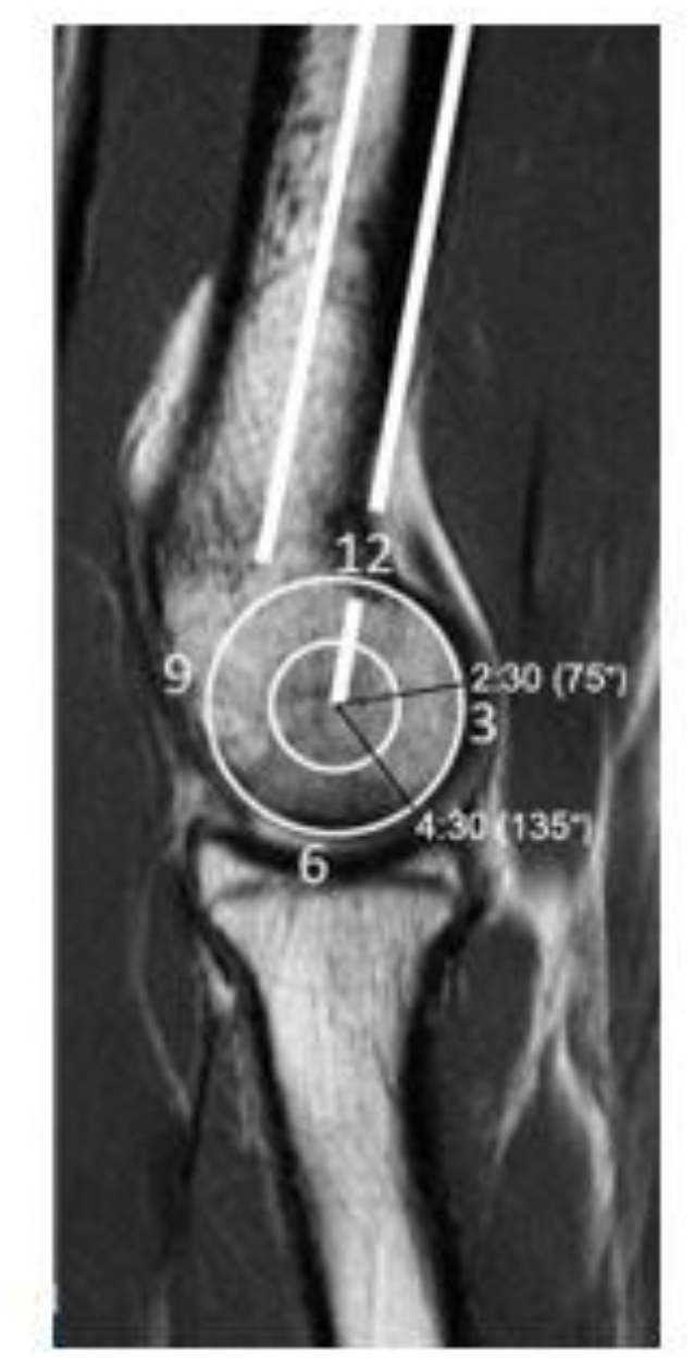
Eight cadaveric matched pair elbows were tested between 15° or max extension to 105° of elbow flexion in increments of 15°. Osteochondral defects were created following the clock-face model in which the most proximal part of the capitellum is 12 and the distal most part is 6 (Fig. 1). The matched pair elbows were divided randomly into a (1) 4:30 defect group and (2) 2:30 defect group. A capsulotomy was performed to access the capitellum surface in order to create a 4:30 or 2:30 lesion (135° and 75° anterior to the humerus). Each specimen was tested under (1) intact condition; (2) central defect; and (3) central lateral defect. Valgus angulation and contact characteristics were measured under no additional valgus loading, 2 Nm, and 3 Nm of valgus torque. For statistical analysis, a linear mixed effects model analysis of variance with a Tukey post hoc test was used and significance was set to P < 0.05.
Results:
Regardless of a 4:30 or 2:30 defect location, valgus angulation increased when applying more valgus torque. Elbow valgus angulation increased at all flexion angles under both 2 Nm and 3 Nm of valgus torque with the defect at the 4:30 position. For the 4:30 central defect, there was a significant increase in valgus angulation compared to intact at flexion angles of 15°, 45°, and 60° under 3 Nm of torque (P < 0.042). The central lateral defect significantly increased valgus angulation between 30°-75° of flexion (P < 0.022) under 3 Nm of torque. The 2:30 central (P = 0.012) and central lateral defects (P = 0.001) displayed significant increases in valgus angulation compared to intact at 105° of flexion. In addition, the central lateral defect showed significant increases in valgus angulation compared to the intact (P = 0.007) and central defect conditions (P = 0.014) at 45° of flexion. There was a decrease in contact area for both the 4:30 and 2:30 located defects with a larger decrease in contact area when the defect extends laterally (Fig. 2). The 4:30 located central lateral defect shows significant increase in contact and peak pressure compared to an intact capitellum under 3 Nm of torque (P < 0.035). There was no significant difference in contact or peak pressure between any of the conditions at the 2:30 defect.
Conclusions:
Osteochondral lesions show location and size dependent consequences in valgus angulation and radiocapitellar contact characteristics. In this study, the 4:30 lesion displayed that defect size has a role in valgus stability, particularly when a central defect is extended laterally, leading to significant differences in valgus angulation at later range of flexion angles. However, the specimens with a 2:30 defect did not show similar findings seen in the 4:30 defect group location. This may be due to the position of the lesion having a different flexion angle of engagement such that it becomes insignificant for valgus stability. The contact area between the capitellum and radial head would appropriately decrease at flexion angles that positioned the radial head near or over the defect. The peak pressure of the 4:30 central defect begins medial but as the elbow flexes from 60° to 75°, the peak pressure migrates anteriorly and as the elbow further flexes from 75° to 90° the peak pressure moves back to its original medial position. However, the migration of peak pressure was not observed when the defect was created in the 2:30 position. These findings demonstrate that peak pressure in elbow flexion is defect location dependent. This study provides new insight on the relationship between osteochondral defects and the UCL which is beneficial for orthopaedic surgeons to determine the treatment regimen for adolescent athletes.