
Abstract
Thank you for your thoughtful review and questions regarding our manuscript. Please see below for our responses to your questions.
Comparability of the PTS on FLL and SSK Radiographs We are curious whether the value of posterior tibial slope (PTS) is comparable on full-length lateral (FLL) and short standard knee (SSK) radiographs, given that both use different reference points for measurement.
Your first question is the main reason that this paper was written. We do not believe that measuring the PTS on FLL and SSK radiographs is interchangeable because of the significant difference in PTS values and the inconsistency between the 2 measurements. The unanswered question is whether the short leg or the long leg PTS is comparable, or when either value can be utilized. Some patients had the same SSK and FLL PTS, while others varied as much as 8°. This likely reflects a subtle osseous deformity that is not readily evaluated by using PTS (particularly SSK) as a measurement tool. We also do not know which measurement is a more reliable way to predict ACL injury, a question that we were unable to answer with this study. However, we have future studies in progress targeting that question. Our main intention was to bring to light that if the SSK PTS is solely relied upon, patients with elevated PTS using traditional deformity assessment principles (FLL) may be overlooked. However, many orthopaedic surgeons still measure the PTS on SSK radiographs, which leaves the door open to missing slopes that are actually elevated when measured on FLL radiographs.
2. Comparison of Anatomical and Mechanical Axes Is the anatomical axis for the proximal tibia (used in SSK for PTS measurement) similar to the mechanical axis of the tibia (used in FLL for PTS measurement)? If there is an inherent difference between these axes, wouldn't this inherently cause a difference in PTS measurements between SSK and FLL?
Yes, I think your question highlights the inherent differences in the various measurement techniques. They are all producing different measurements. The key point is to be consistent in the type of measurement method and to be specific in the reporting of those measurement techniques used when determining the PTS in the literature. I reference the article by Dean et al 1 to ensure that we are discussing the same measurement techniques. I agree with you that there are differences between the axes you referenced. My personal belief is that, regardless of whether you choose the mechanical or the anatomical axis, as long as you are using the long leg to make those measurements, there will be less variability than that from the short leg, and this is the key takeaway from this manuscript.
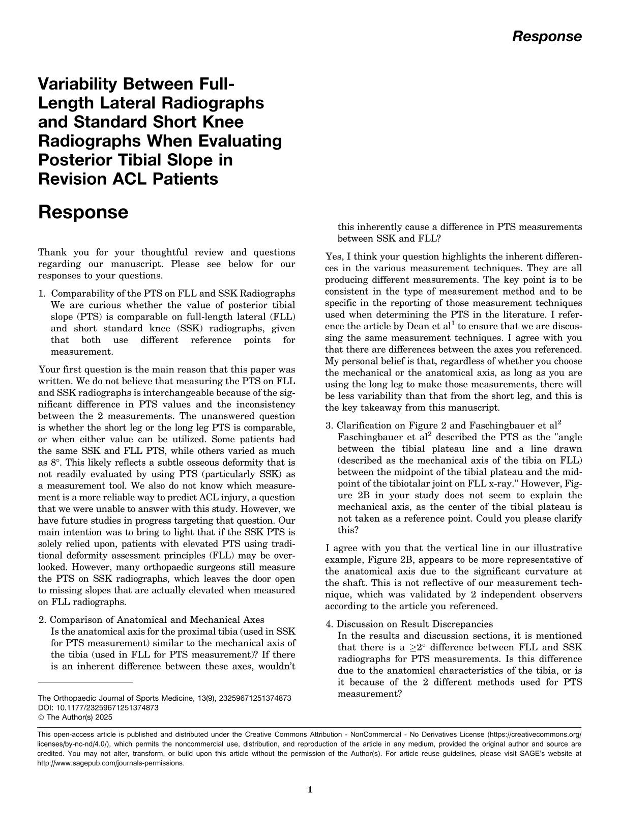
3. Clarification on Figure 2 and Faschingbauer et al
2
Faschingbauer et al
2
described the PTS as the "angle between the tibial plateau line and a line drawn (described as the mechanical axis of the tibia on FLL) between the midpoint of the tibial plateau and the midpoint of the tibiotalar joint on FLL x-ray.” However, Figure 2B in your study does not seem to explain the mechanical axis, as the center of the tibial plateau is not taken as a reference point. Could you please clarify this?
I agree with you that the vertical line in our illustrative example, Figure 2B, appears to be more representative of the anatomical axis due to the significant curvature at the shaft. This is not reflective of our measurement technique, which was validated by 2 independent observers according to the article you referenced.
4. Discussion on Result Discrepancies In the results and discussion sections, it is mentioned that there is a ≥2° difference between FLL and SSK radiographs for PTS measurements. Is this difference due to the anatomical characteristics of the tibia, or is it because of the 2 different methods used for PTS measurement?
We specifically reported that a difference of at least 2° existed between techniques in 66.7% of our cohort. However, there was a minimal difference between techniques in one-third of the cohort. We found that some measurements varied by as much as 7° or 8°, which should not be attributed solely to the measurement techniques, but rather to the inherent limitations of measuring on the partial length of a bone compared with the true deformity principles of using the entire bone for measuring.

Footnotes
The author declares no conflicts of interest in the authorship and publication of this contribution. AOSSM checks author disclosures against the Open Payments Database (OPD). AOSSM has not conducted an independent investigation on the OPD and disclaims any liability or responsibility relating thereto.