
Abstract
Background:
Whether the tibial slope or the concomitant meniscal tear is related to static knee position or anterior knee laxity remains controversial.
Purpose:
To investigate the association between medial and lateral posterior tibial slope, concomitant meniscal tear, static knee position using magnetic resonance imaging (MRI), and anterior knee laxity measured with the GNRB arthrometer.
Study Design:
Cross-sectional study; Level of evidence, 3.
Methods:
A total of 60 patients who underwent anterior cruciate ligament (ACL) reconstructions were retrospectively enrolled from May 2020 to October 2022. All patients underwent both MRI and GNRB arthrometry after the injury. The static knee position and medial and lateral tibial slopes were measured using MRI. The meniscal tear type was confirmed during arthroscopic surgery. Descriptive data were retrospectively reviewed from the medical records.
Results:
The side-to-side differences of anterior tibial translation at 134 N in the intact meniscus, isolated lateral meniscal tear, isolated medial meniscal tear, and both meniscal tear groups were 3.63 ± 1.4 mm, 4.61 ± 1.5 mm, 2.85 ± 1.5 mm, and 4.85 ± 1.6 mm, respectively (P = .003). The slopes of the force-displacement curve in the GNRB arthrometer were 6.55 ± 4.8 mm/N, 16.99 ± 5.6 mm/N, 9.69 ± 10.8 mm/N, and 10.89 ± 7.4 mm/N in the intact meniscus, lateral meniscal tear, medial meniscal tear, and both meniscal tear groups, respectively (P = .001). Subgroup analysis showed that patients with lateral meniscal tears tended to have greater anterior knee laxity based on the GNRB arthrometer tests. The medial and lateral tibial slopes were not correlated with static knee position or anterior knee laxity.
Conclusion:
Patients with ACL deficiency and concomitant lateral meniscal tears are more likely to exhibit greater anterior knee laxity, as measured using the GNRB. Clinicians should consider the concomitant lateral meniscal tear when planning surgery and arranging postoperative care. Tibial slopes were not found to be correlated with static knee position or anterior knee laxity.
The anterior cruciate ligament (ACL) plays an important role in both the anteroposterior and rotational stability of the knee. 9 Anteroposterior translations between the femur and tibia can be assessed by measuring static knee position and observing anteroposterior translation under force. Both the static knee position and anterior knee laxity are crucial in clinical practice. Static knee position can be measured using lateral radiographs or magnetic resonance imaging (MRI), 21 whereas anterior knee laxity can be measured using modern instruments such as the KT-1000 arthrometer, Telos, Rolimeter, and GNRB.3,15,18,22,25,28 The KT-1000 arthrometer and the Rolimeter are frequently used but are operator-dependent.4,13,22,25 The Telos device may be more precise than the KT-1000 arthrometer, 15 but the need for radiation exposure is a disadvantage.
The GNRB, which demonstrates performance comparable with that of the Telos, 3 features numerous advantages, such as automatic measurement recording, avoidance of radiation exposure, and precise transducer accuracy to the nearest 0.1 mm.16,29 Notably, the GNRB arthrometer can assess the anterior tibial translatory stiffness by utilizing the anterior laxity curve, which depicts the relationship between the anterior tibial displacement and the applied anterior translation force. 32 This stiffness indicates the strain in passive tissues resisting forward movement of the shin bone; it is a crucial biomechanical factor linked to ligamentous resistance, contributing to functional instability in the anterior knee. 32 Nevertheless, it is not yet clear whether the presence of a concomitant meniscal tear in an ACL-deficient knee affects anterior tibial translatory stiffness.
The posterior tibial slope was defined as the angle between the perpendicular of the tibial axis and the posterior inclination of the proximal surface of the tibial plateau. 6 It could be further classified as the medial posterior tibial slope when we measured the medial tibial plateau and as the lateral posterior tibial slope when measuring the lateral tibial plateau. 38 The existing literature presents inconsistent findings regarding the relationship between tibial slope and anterior tibial translation in the knee. Dejour et al 6 analyzed preoperative radiographs and revealed an association between a steeper tibial slope and increased anterior translation of the tibia in patients with ACL injuries. Concerning anterior tibial translation, some studies indicate a correlation between a steeper medial posterior tibial slope and increased anterior tibial translation measured using the KT-1000.30,31,35,37 Conversely, Li et al 19 reported that a greater lateral posterior tibial slope is also linked to an increased anterior tibial translation measured using the KT-1000. However, Zee et al 39 recently reported a weak or no correlation between anterior tibial translation and medial and lateral posterior tibial slopes in an in vivo kinematic motion analysis.
The aforementioned studies have certain limitations. First, previous studies commonly utilized the KT-1000 knee arthrometer to assess anterior translation,19,30,31,35,37 but concerns regarding low measurement reproducibility were evident. 5 Second, a separate evaluation of the effect of the medial and lateral tibial slopes on the static knee position was not addressed. 6 To address these limitations, the first goal of the present study was to investigate the association between concomitant meniscal tear, static knee position using MRI, and anterior knee laxity measured with the GNRB arthrometer. The second goal was to investigate the association with both medial and lateral posterior tibial slopes. We hypothesized that ACL injuries in patients with steeper tibial slopes and concurrent meniscal injuries would result in greater static anterior tibial translation on MRI and anterior knee laxity in the GNRB.
Methods
Patient Enrollment
This retrospective review was performed at a single tertiary medical center and approved by the institutional review board of the National Cheng Kung University Hospital (A-ER-109-121). All patients who had undergone ACL reconstruction (ACLR) surgery at our institution between May 2020 and October 2022 were retrospectively reviewed. The inclusion criteria were patients who had completed both an MRI and a GNRB arthrometer examination (Genourob). The exclusion criteria were low-resolution MRI, which was objectively defined by the image reviewer, incomplete data from the chart review, previous ipsilateral knee ligament injuries or surgeries, and previous contralateral knee injuries or surgeries. Relevant patient characteristics—including age, sex, and laterality—were retrospectively collected and analyzed.
Image Analysis
MRI was used to measure the patients’ static knee position, medial posterior tibial slope, and lateral posterior tibial slope. The MRI images were captured using the GE Discovery MR450 1.5T Scanner (GE Healthcare) in our institution. The patient laid on their back with the knee flexed at 10° to 15° and in a neutral rotation. Two operators (T-C.H. and C-K.H.) measured these signs twice, with a 2-week interval between evaluations. Intra- and interobserver reliability was determined by calculating intraclass correlation coefficients. Imaging measurements were obtained via a picture archiving and communication system (INFINITT PACS; INFINITT Healthcare Co).
The static knee position was measured under the midsagittal plane of the lateral femoral condyle, which was confirmed using the coronal plane. Two tangential parallel lines were drawn from the posterior aspect of the femoral condyle and the posterior aspect of the tibial condyle. The shortest distance between the 2 lines was defined as the anterior tibial translation.17,21
The medial and lateral posterior tibial slopes were measured using the 3-step method described by Hudek et al. 14 First, we determined the central coronal image by identifying the tibial attachment of the posterior cruciate ligament, intercondylar eminence, and anterior and posterior tibial cortices appearing in a concave shape. 2 Second, we drew a cranial circle that contacted the cranial tibial cortex and a caudal circle that contacted the anterior and posterior cortical borders of the tibia. The center of the caudal circle is located at the circumference of the cranial circle. Finally, the line between the centers of the cranial and caudal circles was considered the longitudinal axis of the tibia (Figure 1A). The angles between the line vertical to the longitudinal axis and the tangent line of the proximal medial tibial plateau and the proximal lateral tibial plateau are called the medial posterior tibial slope and the lateral posterior tibial slope, respectively (Figure 1, B-C).2,14

The methods for measuring medial and lateral tibial slopes in MRI. (A) The longitudinal axis was defined by the line between the cranial circle and the caudal circle. (B) The medial slope was measured by the angle between the tangent line of the medial plateau (blue line) and the vertical line to the longitudinal axis. (C) The lateral slope was measured by the angle between the tangent line of the lateral plateau (blue line) and the vertical line to the longitudinal axis. MRI, magnetic resonance imaging.
Knee Anterior Tibial Translation Measurement
Anterior tibial translation of the knee was evaluated using a GNRB arthrometer. Anterior tibial translation measurements were performed according to the recommendations of the GNRB manufacturer (Genourob). During measurement, the knee was placed in 20º of flexion at 0° rotation on a molded support to reproduce the typical Lachman test position.16,27-29 Both knees were evaluated to calculate the side-to-side difference (SSD) between the injured and healthy knees at each force level (89, 134, 150, and 200 N) for each subject with a 0.1 mm accuracy. The anterior tibial translation was measured simultaneously using the navigation system, and the data were recorded automatically using a computer. Three measurements were obtained for each knee for each applied force. A displacement-force curve was established, and the slope of the curve was calculated. Slope 1 (S1) was defined as the slope of the curve ranging between 0 and 100 N, and slope 2 (S2) was defined as the slope of the curve ranging between 100 N and the maximum force. The SSDs in S1 and S2 between injured and healthy knees were calculated for the analysis.
Meniscal Tear Type
The presence of the concomitant meniscal injury and the type of meniscal tear was confirmed during arthroscopy surgery.
Statistical Analysis
All statistical analyses were performed using the SPSS Statistics Version 20 software (IBM SPSS). Descriptive statistics—including means and standard deviations—were obtained. The chi-square test was used to compare between-group categorical data. The Spearman correlation coefficient was calculated to evaluate the relationship between nonparametric variables. Statistical significance was set as P≤ .05. The Mann-Whitney U test was used to compare between-group parameters as a post hoc analysis. As the Bonferroni correction was used in the post hoc analysis, P≤ .125 was considered statistically significant.
Results
Descriptive Data
A total of 97 patients underwent ACLR surgeries from May 2020 to October 2022. Eighteen patients were excluded due to a lack of GNRB examinations, 12 due to previous contralateral knee surgeries, 6 due to the absence of MRI scans in the imaging system, and 1 due to incomplete data from the chart review. A total of 60 patients (41 men, 19 women, mean age 26.63 ± 8.49 years) were included in the final analysis. A total of 17 patients (28.3%) had no concomitant meniscal tears, 14 (23.3%) had concomitant lateral meniscal tears, 16 (26.6%) had concomitant medial meniscal tears, and 13 (21.6%) had both medial and lateral meniscal tears (Table 1). In the lateral meniscal group, 4 patients (28.6%) had displaced bucket handle tears, 3 patients (21.4%) had posterior root tears, 3 patients (21.4%) had radial tears, 2 patients (14.3%) had flap tears, and 2 patients (14.3%) had peripheral longitudinal tears. In the medial meniscal tear group, 8 patients (50%) had displaced bucket handle tears, 5 patients (31.2%) had radial tears, and 3 patients (18.7%) had horizontal tears.
Descriptive Data of Different Meniscal Tear Locations a
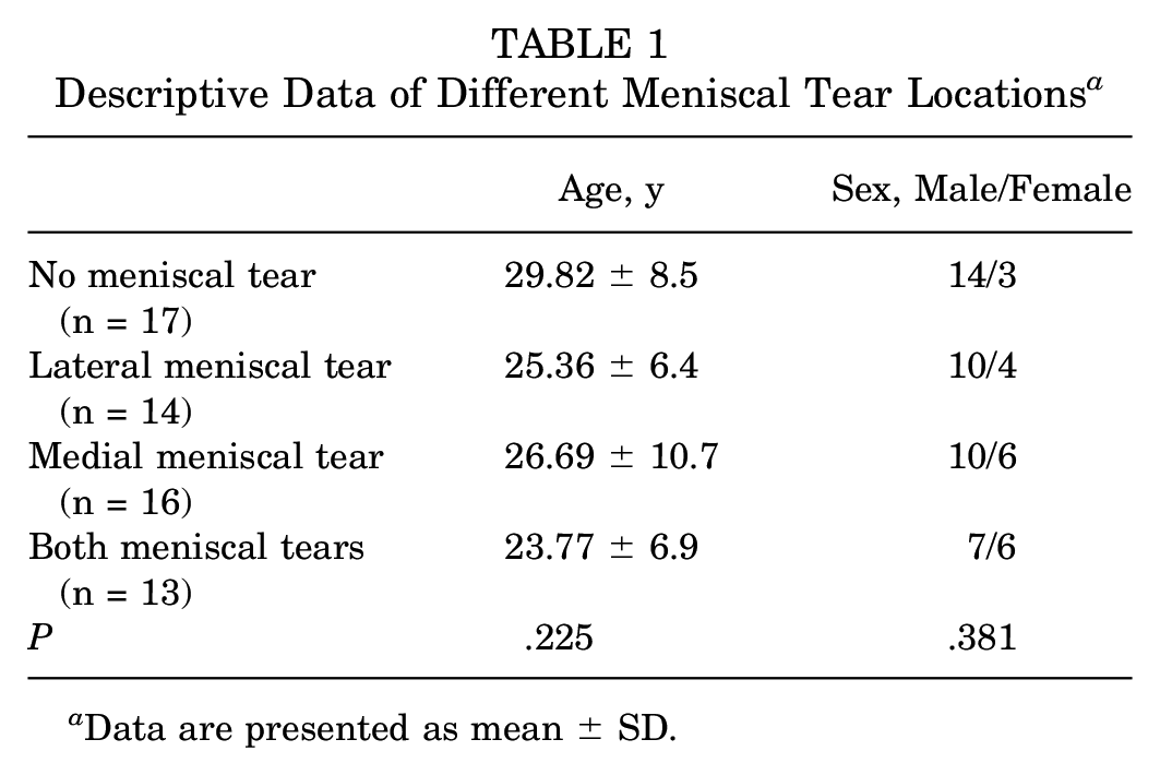
Data are presented as mean ± SD.
A total of 19 patients (31.7%) had MRIs at the faculty that had conducted the study, 26 patients (43.3%) had MRIs at nearby partner hospitals, 15 patients (25%) had MRIs at a medical center from different cities, and 1 patient (1.7%) had an MRI at a foreign country. The mean time of MRI to the operation was 3.3 ± 3.5 months. Also, 54 patients (90%) had surgery within 6 months after MRI examination.
Meniscal Tear Location on Static Knee Position and Anterior Knee Laxity
The SSD of anterior knee laxity, when applied with a load of 134 N, was 3.63 ± 1.4 mm, 4.61 ± 1.5 mm, 2.85 ± 1.5 mm, and 4.85 ± 1.6 mm in the no meniscal tear, only lateral meniscal tear, only medial meniscal tear, and both meniscal tear groups, respectively (P = .003). When loads of 150 N and 200 N were applied, significant differences were also noted among the different meniscal tear locations (P = .110 and P = .001, respectively). The detailed data are summarized in Table 2.
Anterior Knee Laxity and Static Knee Position of Different Meniscal Tear Locations a
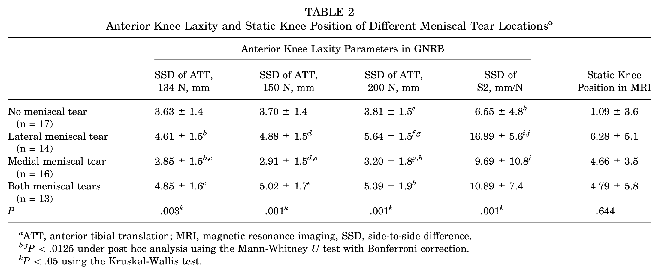
ATT, anterior tibial translation; MRI, magnetic resonance imaging, SSD, side-to-side difference.
P < .0125 under post hoc analysis using the Mann-Whitney U test with Bonferroni correction.
P < .05 using the Kruskal-Wallis test.
The SSD of S2 of 6.55 ± 4.8 mm/N, 16.99 ± 5.6 mm/N, 9.69 ± 10.8 mm/N, and 10.89 ± 7.4 mm/N in the patients without meniscal tears, with only lateral meniscal tears, with only medial meniscal tears, and both meniscal tears, respectively (P = .001). The differences in static knee position between the femur and tibia in MRI were 1.09 ± 3.6 mm, 6.28 ± 5.1 mm, 4.66 ± 3.5 mm, and 4.79 ± 5.8 mm in the no meniscal tear, only lateral meniscal tear, only medial meniscal tear, and both meniscal tear groups, respectively (P = .644).
A subgroup analysis was conducted to analyze the SSD of anterior knee laxity among the groups. When comparing the no meniscal tear group with the only lateral meniscal tear group, a significant difference was noted when the applied load was 200 N (P = .005), with the S2 slope (P < .001). When we compared the only lateral meniscal tear group with the only medial meniscal tear group, significant between-group differences were noted in the SSD of S2 (P = .005) and anterior translation with loads of 134 N (P = .004), 150 N (P = .002), and 200 N (P < .001). When we compared the only medial meniscal tear group with both meniscal tear groups, significant between-group differences were noted in the SSD of the anterior translation with loads of 134 N (P = .002), 150 N (P = .002), and 200 N (P = .004). No significant difference was found when comparing the no meniscal tear and only medial meniscal tear groups, as well as the no meniscal tear and both meniscal tear groups.
Medial/Lateral Tibial Slope on Static Knee Position on MRI and Anterior Knee Laxity
In our study, no significant correlation was found between the medial tibial slope and the anterior knee laxity parameters or static knee position on MRI. Also, no significant correlation was found between the lateral tibial slope and anterior knee laxity parameters or the static knee position on MRI. The detailed data are summarized in Table 3.
The Spearman Correlation of Medial/Lateral Tibial Slopes and Anterior Knee Laxity Parameters and Static Knee Position on MRI a

ATT, anterior tibial translation; MRI, magnetic resonance imaging; SSD, side-to-side difference.
Discussion
The most important finding of our study was that patients with lateral meniscal injury tended to have greater anterior knee laxity, as measured using the GNRB. Patients with greater medial or lateral tibial slopes measured using MRI did not have a greater anterior tibial translation or static knee positions on MRI. While previous cadaveric studies have shown that lateral meniscal injuries cause knee instability, there have been few in vivo studies.6,11,40 To the best of our knowledge, ours is the first study to investigate the relationship between meniscal tears and anterior knee laxity tested using the GNRB.
The meniscus is an important structure of the knee and provides stability in the anterior-posterior and rotation movements. Previous cadaveric studies have shown that both medial and lateral meniscal tears can cause significantly increased tibial translation.1,23,34 In contrast, the clinical studies still showed inconsistent results. In some studies, patients with ACL injuries and medial meniscal tears had significantly increased anterior knee laxity, but not in the lateral meniscal tear group.6,11 Zheng et al 40 found that patients with ACL injury with a concomitant posterior horn of the lateral meniscal tear had a greater anterior tibial translation, as tested using the KT-1000. In our study, we found that patients with ACL injury with concomitant lateral meniscal tears had a significantly greater anterior tibial translation, as tested using the GNRB. Although different clinical studies5,10,36 have shown inconsistent results, we believe that the GNRB arthrometer used in the present study is more reliable than the KT-1000.
Several factors—including injury, surgery, rehabilitation, and hormonal changes—can affect knee laxity.12,20,26,33 It is important to note that changes in anterior tibial translatory stiffness can assist in clinical decision-making regarding individualized rehabilitation programs and return to sports during ACL rehabilitation. 24 Some studies showed that hormonal changes during the menstrual cycle affect knee laxity and stiffness in healthy women.20,26,33 Our study further indicated that patients with a lateral meniscus had a significantly greater slope of the displacement-force curve, indicating the importance of the lateral meniscus in knee laxity. Clinicians should be cautious about anterior knee laxity in ACL-deficient patients with concomitant lateral meniscal tears when determining the personalized rehabilitation program and time to return to sports.
It has been established that a greater tibial slope is a risk factor for ACL tears, as well as a factor for ACL retear rate after reconstruction.6,19 However, a weakness of the abovementioned studies was that the tibial slope was measured on radiography, making it difficult to analyze the slopes of the medial and lateral tibial plateau separately. To overcome this disadvantage, we measured the tibial slope using MRI, in which both the medial and lateral slopes could be measured separately. Previous studies6-8 have reported that an increased tibial slope is associated with an increased static knee position on radiography and greater translation as measured by the KT-1000. On the contrary, the results of our study showed that medial and lateral slopes were not correlated with the static knee position on MRI or anterior knee laxity as measured by the GNRB. Because the values of the medial and lateral slopes were different, 2 using the methodology in the present study would be more reliable when evaluating the correlation between slopes and anterior knee laxity.
Limitations
First, this was a retrospective study. The study design was limited to previously collected data. Second, the sample size was relatively small and led to difficulties in further subgroup analyses, such as the type of meniscal tear and the sex differences. Future studies with larger sample sizes and data collected from different regions are warranted. Third, the consistency of MRI images could not be fully controlled. Patients were placed in the supine position with their knees in 0° to 15° of flexion and 15° of external rotation, as per the protocol of a standard knee MRI. However, some patients underwent MRIs from outside facilities, and we could not guarantee that they had followed the protocol. The strength of our study is that we used easily accessible, objective, and preoperative data to evaluate the clinical outcomes of the patients.
Conclusion
Patients with ACL deficiency tend to exhibit greater anterior knee laxity, as measured using the GNRB, if there is a concomitant lateral meniscal tear. Clinicians should consider the concomitant lateral meniscal tear when planning surgery and arranging postoperative care. Tibial slopes did not correlate with static knee position or anterior knee laxity.
Footnotes
Acknowledgements
The authors thank Skeleton Materials and Bio-compatibility Core Lab, Research Center of Clinical Medicine, National Cheng Kung University Hospital Tainan, Taiwan, for assistance with this project.
Final revision submitted November 5, 2024; accepted November 11, 2024.
One or more of the authors has declared the following potential conflict of interest or source of funding: This publication fee was funded by the National Cheng Kung University Hospital, Tainan, Taiwan (NCKUH-11404005 and NCKUH-11408016). AOSSM checks author disclosures against the Open Payments Database (OPD). AOSSM has not conducted an independent investigation on the OPD and disclaims any liability or responsibility relating thereto.
Ethical approval for this study was obtained from the National Cheng Kung University Hospital (–/ A-ER-109-121).