Abstract
Objectives:
The number of anterior cruciate ligament (ACL) injuries are increasing in pediatric patients. These ACL injuries are commonly associated with meniscal tears or injuries. The lateral meniscus (LMEN) is more commonly torn at the time of injury, whereas the medial meniscus (MMEN) is more commonly injured over time in an ACL-deficient knee. Some clinicians favor non-surgical treatment of pediatric ACL injuries, though the risk of degeneration in the joint is increased. Studies have suggested that meniscal injuries may be isolated to the posterior region after ACL tears, suggesting that regional changes to the meniscus after ACL injury should be investigated. Our previous studies showed menisci volume increased after ACL transection (ACLT), however, regional volumes of the meniscus were not investigated. The objective of this study is to determine regional changes in magnetic resonance imaging (MRI)-based size and signal parameters, histology, and biochemistry of the menisci after an ACLT using a juvenile porcine model.
Methods:
All animal protocols were approved by the institutional animal care and use committee. Seven female juvenile (3 month) Yorkshire crossbreed pigs underwent a unilateral ACLT arthroscopically and a sham incision was made on the contralateral joint. After 12 weeks, both hind limbs were imaged using the 3.0-T Siemens MAGNETOM Skyra MRI system (T2 SWI sequence, voxel size 0.5 x 0.5 x 0.8 mm) (
Results:
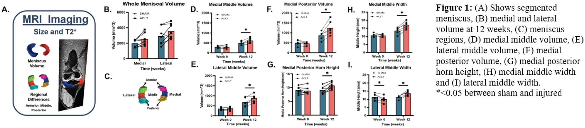
At 12 weeks post-ACLT, total volume of both medial and lateral menisci increased by 27% (P=0.090) and 22% (P = 0.111) relative to contralateral sham-operated controls (
Conclusions:
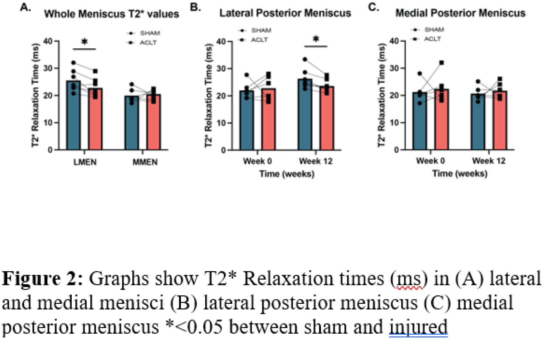
ACLT caused significant changes to the meniscus within 12 weeks of injury, specifically in the posterior and middle regions of the meniscus. Although meniscus remodeling has not been a primary metric for degeneration, studies have shown hypertrophy to be indicative of osteoarthritis. Our findings align with this. After an ACLT, the meniscus size increased. Furthermore, our study determined specific regions to better understand how the meniscus is impacted and where degeneration begins after an ACLT. Higher T2* values show greater disorganization in collagen fibers. Our results show a decrease in T2*, which could be a result of tissue remodeling in response to altered loading. Our results show greater differences when looking at size versus T2* values, which may be valuable since more complex imaging sequences would not be required. In future studies, T1rho mapping via MRI mapping can be used to associate changes in regional T1rho values with changes in GAGs due to degeneration. This could be an additional metric with histology to better quantify the compositional makeup of the meniscus after injury. Better understanding of where meniscal changes occur after ACL injury can lead to appropriate metrics for early detection of degeneration, which may aid in prevention of long-term changes, such as osteoarthritis.