
Abstract
Background:
Damaged cartilage can be treated using the creation of microfractures (MFxs) or the porcine-derived collagen-augmented chondrogenesis technique (C-ACT).
Purpose:
To provide the midterm results of a multicenter randomized controlled trial comparing MFx and C-ACT for knee cartilage defects.
Study Design:
Randomized controlled trial; Level of evidence, 1.
Methods:
The study cohort comprised 36 patients with medial femoral condyle cartilage defects who were followed up for 6 years with clinical and magnetic resonance imaging data (n = 14 treated with MFx alone, n = 22 treated with C-ACT). Clinical outcomes were assessed preoperatively and at 1, 2, and 6 years postoperatively using a visual analog scale (VAS) for pain, the Knee injury and Osteoarthritis Outcome Score (KOOS), and the International Knee Documentation Committee (IKDC) subjective score. Magnetic resonance imaging scans were performed preoperatively and at 1 and 6 years postoperatively, and the repaired cartilage tissue was evaluated using the magnetic resonance observation of cartilage repair tissue (MOCART) score. The repaired tissue/reference cartilage ratio was quantified using T2 mapping. Adverse events during follow-up were also evaluated.
Results:
In both groups, the VAS pain score improved and was maintained for 1, 2, and 6 years postoperatively compared with preoperatively (P < .05 for all). Although there were no significant differences between groups in the VAS pain, KOOS, or IKDC scores at any time point, the change in the IKDC—Activities of Daily Living subscore from preoperatively to 6 years postoperatively was better in the C-ACT group than the MFx group (P = .0423). At 6 years postoperatively, the MOCART assessment showed superior results regarding the surface of the repair tissue in the C-ACT group compared with the MFx group (P = .0288). There were no differences between the groups in the total MOCART score or other subscores.
Conclusion:
The study results suggest that C-ACT has similar effects to MFx in improving pain, joint function, and imaging findings and may be superior to MFx in improving daily life function and improving the quality of the surface of the cartilage tissue.
Registration:
ClinicalTrials.gov identifier: NCT02539030.
Cartilage is a biological tissue that is particularly difficult to recover to normal tissue after damage.33,48 Various techniques have been developed to supply cells for the recovery of damaged cartilage tissue,12,29 including bone marrow stimulation by multiple bone marrow drilling, microfracture (MFx), and transplantation of chondrocyte therapeutic agents. 35 Among the cartilage repair procedures, MFx is the most popular treatment for cartilage defects in the knee. 35 MFx creation is a relatively simple procedure with minimal medical expenses and is recognized as a standard treatment for cartilage defects. 35 However, MFx has limitations.12,35 The healing tissue induced by MFx is reported to be fibrous cartilage rather than hyaline cartilage, and the progress worsens over time.12,35 Studies have shown that the clinical outcomes of MFx deteriorate significantly after 2 years.12,35
To overcome these limitations, an improved MFx method, the collagen-augmented chondrogenesis technique (C-ACT), has been developed using atelocollagen (CartiFill; Sewon Cellontech Co Ltd).24,25,45 During the tissue healing process, it is important to supply an ideal collagen material.2,5,7 An ideal collagen material should have the triple helical structure of collagen existing in a living body, no immune action, and a high level of purity.2,5,7 Collagen present in living organisms forms a triple helix structure.2,5,7 Only the triple helix structure retains biocompatibility characteristics, including tissue structure support, cell activation, and platelet activation.2,5,7 To minimize the immune action of collagen, it is best to use atelocollagen from which telopeptides present at both ends of the triple helix structure and exhibiting antigenicity have been removed.4,34 In addition, using a collagen material with minimal impurities minimizes the inflammatory response.4,34
The C-ACT is very simple, safe, and as efficacious as MFx for the treatment of knee cartilage defects. 26 We previously reported stable outcomes and clinical development at 2 years after C-ACT performed in a multicenter randomized controlled trial (RCT). 24 The purpose of the present study was to report the midterm results of this multicenter RCT comparing MFx and C-ACT for knee cartilage defects. The hypothesis was that the quality of the repaired articular cartilage and clinical outcomes for C-ACT would be superior to those for MFx at 6 years postoperatively.
Methods
Initially, a multicenter RCT with 100 participants was conducted from 2013 to 2017. 24 The protocol for this previous study was approved by the institutional review board of each participating hospital, and all eligible patients were informed of the standardized information on clinical trials and provided written informed consent. This RCT was registered with ClinicalTrials.gov (ClinicalTrials.gov identifier: NCT02539030). The current study was conducted to evaluate the midterm safety and efficacy of MFx versus C-ACT for a medial femoral condyle cartilage defect of the knee at 6 years postoperatively. Inclusion criteria were age ≥15 years; presence of a knee cartilage defect (knee osteoarthritis or knee traumatic arthritis); and neutral alignment with the hip-knee-ankle angle of varus 5° or less, or correction of malalignment with osteotomy. Exclusion criteria were a personal or family history of autoimmune disease or anaphylactic reaction; sensitivity to transplants and/or porcine protein; current pregnancy or lactation; contraindication to the use of a fibrin sealant; or previous ligament surgery. Randomization was performed immediately before surgery using a computer-block randomization method to generate balanced control and intervention groups. High tibial osteotomy (HTO) was performed when correction was required due to varus deformity.9,21 HTO was performed in cases where the hip-knee-ankle angle was more than varus 5°.
Initially, 100 patients (52 patients in the intervention group who received C-ACT and 48 patients in the control group who received MFx) were followed up for 2 years 24 ; 22 patients in the C-ACT group and 14 patients in the MFx group were followed up for 6 years and included in the present study (Figure 1).
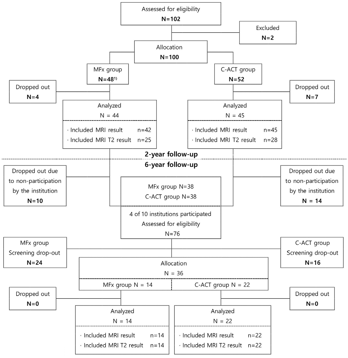
Study flow diagram. MFx, microfracture; C-ACT, collagen-augmented chondrogenesis technique.
Surgical Procedure and Rehabilitation
Arthroscopy and bone marrow stimulation MFx were performed in all patients. 26 To ensure stable cartilage margins, cartilage lesions and calcified cartilage layers were removed. MFx was performed using an MFx awl. For the C-ACT procedure, a 1-mL syringe was filled with a mixture of 0.9 mL of atelocollagen (CartiFill) and 0.1 mL of thrombin (50 IU). An additional syringe contained 1 mL of fibrinogen (Greenplast; Green Cross PD Co Ltd). The 2 syringes were assembled into a y-shaped mixing catheter connected to a 20-gauge needle. Under arthroscopic observation, the intra-articular fluid from the microfracture area was removed using suction and gauze, and after securing a dry field, the gel in a 2-way syringe was applied. The gel from a 2-way syringe was applied slowly to the MFx site during surgery. An initial atelocollagen layer was created, followed by an additional layer 1 to 2 minutes later. After 5 minutes, the knee was bent and extended to check the stability of the atelocollagen layer. 26 All cartilage lesions were confined to the medial femoral condyle.
Both groups followed the same postoperative rehabilitation protocol. The protocol started from the first postoperative day and comprised 30 minutes of knee exercises 4 times a day using a continuous manual exercise machine for 4 weeks. Only toe-touch weightbearing was allowed until 4 weeks postoperatively. Full weightbearing ambulation was allowed from 4 weeks postoperatively. After 4 weeks, walking ambulation was increased continuously and water exercise and gym cycling were recommended.
Outcomes Evaluation
Clinical outcomes were assessed using a 100-mm visual analog scale (VAS) for pain, the Knee injury and Osteoarthritis Outcome Score (KOOS), 38 and the International Knee Documentation Committee (IKDC) score, 39 preoperatively and at 1, 2, and 6 years postoperatively. The EuroQol 5 dimension scale (EQ-5D) was evaluated at 6 years postoperatively as an indicator of quality of life. 23
Magnetic resonance imaging (MRI) examinations were performed preoperatively and at 1 and 6 years postoperatively, and imaging outcomes were evaluated using the magnetic resonance observation of cartilage repair tissue (MOCART) score and T2 mapping.31,32 MOCART scores were assessed at 1 and 6 years postoperatively. The MOCART score for structural and morphological verification of regenerated articular cartilage was measured twice, 2 weeks apart, by 2 orthopaedic surgeons who were not involved in the study; each had more than 5 years of experience in orthopaedic radiography. The mean of the observers’ measurements was used. The reliability of the measurements was evaluated as intra-observer and inter-observer reliability using the intraclass correlation coefficient (ICC), and the intra- and inter-observer ICC values were >0.8. 32 T2 mapping is a noninvasive and proven method of judging cartilage quality and was used to determine the water content of cartilage and collagen structures.6,31 The repaired tissue/reference cartilage ratio was also used in the imaging evaluation. 8 The threshold for T2 by Youden index was 75.42%. Therefore, the cutoff value of the T2 ratio was defined as 75.42%. If the T2 ratio was >75.42%, the tissue was judged to have a biochemical and physiological structure that changed in a similar way to normal cartilage.
Adverse events that occurred during and after participation in this clinical trial were classified and investigated. Adverse events were confirmed through medical interviews (if necessary, adverse events were confirmed in the medical records of study participants). The primary efficacy evaluation variable was the pain VAS, and the secondary efficacy evaluation variables were the KOOS, IKDC score, EQ-5D score, MOCART score, and MRI cartilage-specific investigation.
Statistical Analysis
In this clinical trial, the full-analysis set and the per-protocol set were the same, so the full-analysis set values were substituted for the per-protocol set values. Background (eg, clinical history) and demographic characteristics were compared between treatment groups. Continuous variables were summarized with descriptive statistics consisting of sample size, means and standard deviations, and range. The VAS pain scores in the C-ACT and MFx groups at 6 years postoperatively were analyzed using the unpaired t test or Wilcoxon rank-sum test. The differences between the outcomes at 2 versus 6 years postoperatively were analyzed using the unpaired t test. The KOOS, IKDC score, EQ-5D score, MOCART score, and MRI cartilage-specific investigations were compared using the unpaired t test, Wilcoxon rank-sum test, chi-square test, or Fisher exact test. Repeated-measures analysis of variance and post hoc analyses were used to compare values over time. Power analysis for the surface of the repair tissue in MOCART scores showed results >70%, indicating sufficient statistical power to compare the quality of repaired articular cartilage between the 2 groups. The threshold for statistical significance was set at P < .05.
Results
There were no significant differences between the C-ACT and MFx groups regarding the size of the defect, International Cartilage Repair Society grade, number of patients who underwent concomitant HTO, and Kellgren-Lawrence grade (Table 1). In both groups, the VAS pain score improved compared with preoperatively, and this improvement was maintained for 1, 2, and 6 years postoperatively (P < .05 for all) (Table 2). The KOOS total score showed significant improvement in the MFx group at 2 and 6 years postoperatively compared with preoperatively (P < .05 for all) and showed significant improvement in the C-ACT group at 1, 2, and 6 years postoperatively compared with preoperatively (P < .05 for all). However, in the MFx group, the improvement in the IKDC total score from preoperatively was not maintained at 6 years postoperatively (P = .0616). There were no significant between-group differences in the VAS pain, KOOS, or IKDC scores at any time point (Table 2).
Patient Demographics and Defect Data a

Data are presented as mean ± SD (range) or n (%) unless otherwise indicated. BMI, body mass index; C-ACT, collagen-augmented chondrogenesis technique; HTO, high tibial osteotomy; ICRS, International Cartilage Repair Society; MFx, microfracture; OA, osteoarthritis.
Comparison of Clinical Outcomes a

Data are presented as mean ± SD (range). ADL, activities of daily living; C-ACT, collagen-augmented chondrogenesis technique; IKDC, International Knee Documentation Committee; KOOS, Knee injury and Osteoarthritis Outcome Score; MFx, microfracture; Post, postoperative; Pre, preoperative; QOL, Quality of Life; Sports/Rec, Sports/Recreation; VAS, visual analog scale.
The pre- to 6-year postoperative change in the pain VAS was 41.17 in the C-ACT group and 37.13 in the MFx group (P > .05) (Table 3). There was a significantly greater difference in the change in the IKDC-Activities of Daily Living (ADL) subscore from preoperatively to 6 years postoperatively in the C-ACT group compared with the MFx group (P = .0423) (Table 3). In the MFx group, there was a significant decrease in the IKDC total score from preoperatively to 6 years postoperatively compared with the change from preoperatively to 2 years postoperatively (P = .0443). In the C-ACT group, there were no differences in the changes in the IKDC total score from preoperatively to 1, 2, and 6 years postoperatively (all p > .05). The pre- to postoperative change in the EQ-5D score did not differ significantly between the MFx group (0.74) and the C-ACT group (0.73; P = .5659).
Changes in Clinical Outcomes a

Data are presented as mean ± SD (range). ADL, activities of daily living; C-ACT, collagen-augmented chondrogenesis technique; IKDC, International Knee Documentation Committee; KOOS, Knee injury and Osteoarthritis Outcome Score; MFx, microfracture; Post, postoperative; Pre, preoperative; QOL, Quality of Life; Sports/Rec, Sports/Recreation; VAS, visual analog scale. Bold indicates statistical significance.
The total MOCART score at 6 years postoperatively did not differ significantly between the MFx group (34.29 ± 14.92) and the C-ACT group (41.82 ± 17.83; P = .197). The subscore for part 3 (surface of the repair tissue) was significantly better in the C-ACT group (5.45 ± 3.05) than the MFx group (3.08 ± 2.53; P = .0288) (Table 4).
MOCART Scores at 6 Years Postoperatively in the Per-Protocol Set

C-ACT, collagen-augmented chondrogenesis technique; FSE, fast spin-echo; MFx, microfracture; TSE, turbo spin-echo.
A comparative analysis of the T2 values at 6 years postoperatively showed that the L2 (neocartilage) value did not differ significantly between the MFx group (32.38 ± 19.20 ms) and the C-ACT group (40.69 ± 11.15 ms; P = .1585). The L2/L1 ratio (ratio of repair tissue to reference cartilage) was also similar in the MFx group (0.77 ± 0.49) and the C-ACT group (0.96 ± 0.35; P = .1906). The L2/L1 ratio (ratio of repair tissue to standard cartilage) was 75.42% or more for 23 patients (63.89%), of which 7 (19.44%) were in the MFx group and 16 (44.44%) were in the C-ACT group. Although there was no significant difference between the 2 groups in the proportion of patients with an L2/L1 ratio of >75.42%, the C-ACT group showed a greater tendency than the MFx group to have improved repair with tissues such as hyaline cartilage rather than fibrocartilage (Figure 2).

Representative MRI scans (left) and T2 values (right) of (A, B) a 33-year-old man in the C-ACT group and (C, D) a 51-year-old woman in the MFx group. The T2 mapping results (B, D) showed that the defects in both patients were filled with repair tissue. Open arrowheads, reference cartilage; closed arrowheads, repaired tissue. C-ACT, collagen-augmented chondrogenesis technique; MFx, microfracture; MRI, magnetic resonance imaging.
However, the ratio of repair tissue/reference tissue (L2/L1) was 0.742 in the patient in the MFx group and 1.206 in the patient in the C-ACT group; this suggests that the defect was filled with fibrous tissue in the MFx group and filled with repair tissue such as hyaline cartilage in the C-ACT group. In the MFx group, patients underwent microfracture treatment alone; in the C-ACT group, patients received microfracture and collagen augmentation.
The adverse events after surgery comprised 1 case of effusion in 1 patient (2.78%) in the C-ACT group; the edema resolved within 7 days without additional treatment. No adverse events were reported in the MFx group.
Discussion
The most important finding of this study is that C-ACT was not superior to MFx for improving pain, joint function, and imaging findings. However, C-ACT showed some benefit in the IKDC-ADL subscore and the MOCART assessment of the surface of the repair tissue.
A known issue with MFx is whether the initial improvement in clinical outcomes will persist,1,47 as studies have shown that the clinical symptoms worsen over time.24,25 A systematic review found that MFx has good short-term efficacy for up to 2 years postoperatively, but that deterioration of clinical function occurs in 47% to 80% of patients after 2 years. 35 Various advanced MFx techniques have been introduced to overcome these limitations.24,29 However, it remains unclear whether the improvement in clinical outcomes continues after advanced MFx and other cartilage regeneration treatments.10,44 This gradual deterioration of the clinical outcome is a problem that has not yet been overcome, even by autologous matrix-induced chondrogenesis (AMIC).15,44 A 37-month follow-up study of AMIC in 27 patients with a mean defect size of 4.2 cm2 showed that both the Lysholm score and the Tegner activity scale were improved at 1 year postoperatively compared with preoperatively, and improved further in the second year postoperatively. 15 However, the Lysholm score decreased in the third year postoperatively compared with the value in the second year postoperatively, and showed a further decrease in the fourth year. 15 These findings are similar to the clinical course after MFx. 35 However, other studies have reported continued improvement of the clinical outcomes after AMIC, even after midterm follow-up of >5 years.16,46 To accurately determine the effect of the cartilage regeneration procedure, midterm follow-up results of >4 years are necessary.15,16,46 In consideration of these issues, the present RCT had a follow-up period of >6 years after C-ACT and MFx. Both the C-ACT and MFx groups showed continuous clinical improvements in the VAS, KOOS, and IKDC scores for up to 2 years postoperatively, and showed clinical improvement at 6 years postoperatively compared with preoperatively. However, the degree of improvement in the total IKDC score decreased at 6 years postoperatively compared with 2 years postoperatively in the MFx group. This indicates that the C-ACT technique may prolong the improvement of marrow stimulation.
The fact that the change in IKDC-ADL subscore from preoperative to 6 years postoperatively was significantly greater in the C-ACT group is considered to indicate better improvement in joint function in the C-ACT group than in the MFx group. The C-ACT group showed a significant improvement in IKDC total score compared with before surgery for up to 6 years after surgery, whereas the MFx did not. A systematic review and meta-analysis of studies evaluating AMIC and MFx with a follow-up of at least 2 years found no differences between the AMIC and MFx groups in the pre- to postoperative changes in the VAS, Lysholm score and Tegner activity scale. 22 However, the IKDC score was improved significantly in the AMIC group compared with the MFx group. 22 This is similar to our finding that the C-ACT group showed greater pre- to postoperative changes in the IKDC-ADL subscore than the MFx group. In addition, the improvement in the IKDC total score was maintained for 6 years postoperatively in the C-ACT group but not the MFx group. The reason for the different results is that the validated tools used to evaluate the improvement in clinical outcomes after cartilage repair do not perfectly reflect the clinical outcomes of C-ACT and MFx.14,19 However, because the IKDC score is a validated and excellent tool for evaluating the clinical outcomes of cartilage repair,18,19 the fact that the C-ACT group showed superior improvement and maintenance of the IKDC score compared with the MFx group is considered to be a clinically meaningful finding.
Although the MOCART scores did not differ significantly between the 2 groups, the C-ACT group had better defect filling and higher scores for the surface of the repaired tissue. Gille et al 15 reported that defect filling of >50% after AMIC was confirmed in 10 of 15 patients during a 37-month follow-up. Volz et al 46 conducted a 5-year RCT of 47 patients who underwent AMIC or MFx. At 2 years postoperatively, the AMIC group showed more complete defect filling than the MFx group. Furthermore, defect filling of more than two-thirds was observed in 60% of patients in the AMIC group and in 25% of patients in the MFx group. Volz et al 46 reported a greater degree of defect filling compared with our results, which may be because our study had a 6-year follow-up period. Previous studies have reported a total MOCART score of 58.3 points at a mean follow-up of 38 months after AMIC, 40 and 77.5 points at a mean follow-up of 49 months after AMIC. 16 Both of these previous studies reported slightly higher MOCART scores than our study results.
The treatment of chondral lesions often involves procedures such as realignment osteotomy in addition to the cartilage regeneration procedure.3,15,28 However, it remains unclear whether the clinical improvement in these cases is due to the cartilage regeneration procedure, the realignment procedure, or both.10,17,43 A systematic review suggested that performing both HTO and a cartilage regeneration procedure is effective in improving the clinical outcomes of the knee. 20 In contrast, another study demonstrated that no conclusion could be drawn as to whether a combination HTO and cartilage regeneration procedure had a clinical advantage over HTO alone. 13 Ferruzzi et al 11 conducted a study that followed up on patients for 11 years and found that patients who underwent HTO and autologous chondrocyte implantation and patients who underwent isolated HTO had superior clinical outcomes compared with patients who underwent HTO and MFx; there is no evidence that performing HTO and autologous chondrocyte implantation is superior to isolated HTO. 11 Kusano et al 28 analyzed 49 patients with knee cartilage lesions who underwent the AMIC technique and found no difference in clinical features between those with and without realignment osteotomy. Our study also included patients who underwent HTO in both groups. We believe that HTO may have a substantial impact on the improvement in clinical outcomes. However, as there was no significant difference between the 2 groups in the proportion of patients who underwent HTO in the present study, the performance of HTO is not a limitation in evaluating the clinical outcomes.
C-ACT is a cell-free type 1 collagen matrix that uses a mix of gel-type scaffold and fibrin glue. 41 In this study, a mini-arthrotomy was used, but this method can also be performed using an arthroscopic technique. 41 As a 1-stage procedure, it can be performed easily in combination with MFx. In the case of a solid scaffold, a process of suturing to the surrounding tissue is required, but since C-ACT is not a solid scaffold, this process does not need to be performed.24-26 The cost of C-ACT is much higher, at around US $1000, compared with MFx, but its long-term value is highlighted when considering clinical results and histological improvement. This suggests that the initial high cost of C-ACT may be offset by the potential for superior tissue recovery and longevity, reducing future healthcare costs. Therefore, despite the initial cost, C-ACT may offer significant value in terms of tissue quality and durability, making it a worthwhile investment in selected patients. However, an accurate answer to this appears to require more long-term follow-up results.
Limitations
Our study had several limitations. First, the sample size was small, and there was a decrease in the number of patients at 6 years postoperatively compared with the original patient population (followed up at 2 years postoperatively). The sample size was sufficient to reveal some differences between the C-ACT and MFx groups in radiological and clinical outcomes. However, the possibility of type 2 error exists for other measures, given the small sample size. A second limitation is the high proportion of women in the study cohort. This sex imbalance is particularly evident in Asian populations because the prevalence and incidence of osteoarthritis are higher in women than men, making it difficult to control the male-to-female ratio.27,30,33,36,37,42 Third, although the follow-up period was 6 years, it is necessary to confirm the long-term results after further follow-up. Finally, this study was a single-blinded RCT because a mini-arthrotomy was used to perform C-ACT. However, the operators and investigators were blinded during all final analyses. Despite these limitations, this study provides valuable information on the cartilage restoration effect after midterm follow-up, including the radiological and clinical outcomes of C-ACT.
Conclusion
Study findings indicated that C-ACT achieved similar effects to MFx regarding improvements in pain, joint function, and imaging and was superior to MFx in improving daily life function and improving the quality of the surface of the cartilage tissue. These results suggest that C-ACT is effective and durable in treating articular cartilage defects of the knee at a minimum follow-up of 6 years.
Footnotes
Acknowledgements
The authors thank Professor Dong Jae Kim (Division of Biostatics, Department of Medical Lifescience, College of Medicine, The Catholic University of Korea) for the statistical analysis.
Final revision submitted April 16, 2024; accepted April 30, 2024.
One or more of the authors has declared the following potential conflict of interest or source of funding: research support and materials for this study were received from Sewon Cellontech. AOSSM checks author disclosures against the Open Payments Database (OPD). AOSSM has not conducted an independent investigation on the OPD and disclaims any liability or responsibility relating thereto.
Ethical approval for this study was obtained from The Catholic University of Korea, Catholic Medical Center (reference No. XC13DSMI0025K), Wonkwang University College of Medicine Hospital (reference No. 1518), Samsung Medical Center (reference No. 2013-12-094-003), and Seoul Metropolitan Governance, Seoul National University Boramae Medical Center (reference No. 26-2013-36).