
Abstract
Background:
Diagnostic ultrasound can evaluate musculoskeletal structures for abnormalities that correlate with tendinopathy and fasciopathy. Previous literature suggests that sonographic screening of tendons and fascia can identify structures that are at risk for developing pain, although this has not been evaluated in collegiate athletes competing in a variety of sports.
Purpose:
To evaluate the use of ultrasound for screening the patellar tendon, Achilles tendon, and plantar fascia for time-loss injury in collegiate athletes during a full year.
Study Design:
Cohort study; Level of evidence, 2.
Methods:
A total of 242 National Collegiate Athletic Association (NCAA) Division I athletes from 3 institutions participated in this yearlong prospective observational study. Each athlete completed a brief demographic questionnaire, followed by an ultrasound examination of the bilateral patellar tendons, Achilles tendons, and plantar fascia performed at the beginning of the season. Ultrasound examinations assessed for tendon/fascia thickening, hypoechogenicity, and neovascularization. Athletes were monitored for any time-loss injury in these 3 structures throughout a full year.
Results:
Of the 242 athletes (n = 484 tendon/fascia) evaluated, the patellar tendon had the highest prevalence of sonographic abnormalities (37.4%), followed by the Achilles tendon (10.6%); plantar fascia abnormalities were rare (3.5%). The overall number of tendon/fascia structures that developed injury was low (<5%). The relative risk for a time-loss injury based on an initial abnormal ultrasound screening was 8.8, 17.2, and 13.2 for the patellar tendon, Achilles tendon, and plantar fascia, respectively (P < .01). Negative predictive values (99-99.6) far outweighed positive predictive values (10.3-14).
Conclusion:
NCAA Division I student-athletes with sonographic abnormalities on initial screening were more likely to develop a time-loss injury in the affected tendon/fascia during the competitive season. Further research on this topic should focus on identifying the specific sonographic abnormalities and their relationship with future injury.
Achilles tendinopathy, patellar tendinopathy, and plantar fasciopathy are common conditions in both athletes and the general population. †† These conditions are often chronic and are associated with overuse, although they can present acutely.15,26,49 Tendons and fascia are susceptible to developing pain, especially in the setting of high-impact activities—such as running, jumping, cutting, and pivoting.4,17,26,29,39,44 These conditions can clinically manifest as pain and decreased function, and can significantly affect an athlete's sports participation and performance.15,24-26,33,44,49 A study published in 2015 found that 23% of the reported collegiate overuse injuries were due to tendinopathy. For the >45% of the injuries caused by overuse, it took athletes >7 days to return to sports. 42 Patellar tendinopathy was reported in almost half of all patellofemoral injuries in 25 National Collegiate Athletic Association (NCAA) Division I sports. 45 During the 5-year study period, patellar tendinopathy led to the second-most cumulative days away from sports after patellar subluxation. 45 Furthermore, foot and ankle injuries represented 27% of musculoskeletal injuries reported in 37 NCAA Division I sports over 2 years; 8% and 4% of the injuries that led to time away from sports were due to the Achilles and plantar heel, respectively. 24
Collegiate athletes, specifically, train at a very high level and have long training sessions as well as competitive seasons. In this setting, there is concern for athletes developing pain associated with overuse. In clinical practice, ultrasonography has been utilized regularly to assess the etiology of musculoskeletal pain but typically after symptoms start. It allows for rapid, dynamic assessment of tendons, fascia, ligaments, muscles, nerves, vasculature, and bone.14,16,20,33,40 Tendon or fascial thickening, focal areas of hypoechogenicity, and abnormal vascularity are structural changes associated with tendinopathy or fasciopathy, and they can all be readily visualized with ultrasound.4,11,17,25,28,33,38,40 Evidence from systematic reviews has demonstrated that these abnormal findings on ultrasound can lead to an increased risk of developing pain. To our knowledge, no multicenter study has been conducted on sonographic tendon evaluations of collegiate athletes involving different sports. Moreover, there are no studies that involve sonographic evaluation of the plantar fascia and its development of future injury.
This study aimed to evaluate the use of ultrasound as a screening test for future injury in the Achilles tendon, patellar tendon, and plantar fascia of NCAA Division I athletes throughout a full year. It was hypothesized that ultrasound screening would be useful in predicting time-loss injury in these structures.
Methods
This was a prospective observational study that evaluated NCAA Division I student-athletes across 3 different institutions over a single year. The study protocol received institutional review board approval from all 3 participating institutions. NCAA Division I student-athletes from any sport were recruited consecutively at preparticipation physicals around the beginning of the 2021 academic year. The exclusion criteria were age <18 years and previous rupture/surgery of the affected tendon/fascia (the potential participant was not excluded, but the affected tendon/fascia was excluded from the analysis). An a priori power analysis based on an expected relative risk (RR) of 7 estimated a required sample size of 141 student-athletes. 33
Study Questionnaire
All consented participants completed a brief questionnaire immediately upon enrollment in the study. This included age, sex, height, weight, participating sports, years of competition in their sport, current pain in the structures under examination, previous pain in those structures, and previous surgery on those structures.
Ultrasound Video Acquisition
Evaluation of the Achilles tendon, patellar tendon, and plantar fascia was performed with a 12 to 18 MHz linear transducer (Logiq E9 R7, GE Healthcare; Sonosite PX, FujiFilm Sonosite; Sonosite X-porte, FujiFilm Sonosite; or Sonimage HS-1, Konica Minolta) and was performed by or directly supervised by an experienced sports medicine physician with at least 5 years of posttraining experience (D.M.C., M.F.). Videos—approximately 10 seconds in length for each plane of each structure, totaling 16 separate videos per participant—were recorded without personally identifiable information using the following protocol. The patellar tendons were scanned along their entire course first with the knees bent to 90° in a sagittal and axial plane, followed by another sagittal view with color Doppler with the knees extended and quadricep musculature relaxed. Next, the Achilles tendons were scanned, with the student-athlete in a prone position and knees extended. The ankle was passively flexed to neutral by the sonographer, and the Achilles tendon was scanned from the myotendinous junction to the insertion in both the sagittal and axial planes, followed by another sagittal view using power Doppler with the ankle relaxed without any passive tension. Finally, the plantar fasciae were scanned only in a sagittal plane, first with the ankle passively flexed to neutral, then using power Doppler with the ankle relaxed. All longitudinal scans were performed by sweeping the transducer lateral-to-medial and proximal-to-distal to obtain a complete view of all portions of the structure. Power Doppler was always performed with minimal transducer pressure and gain adjusted to maximize the likelihood of neovessel identification. 43
Ultrasound Video Analysis
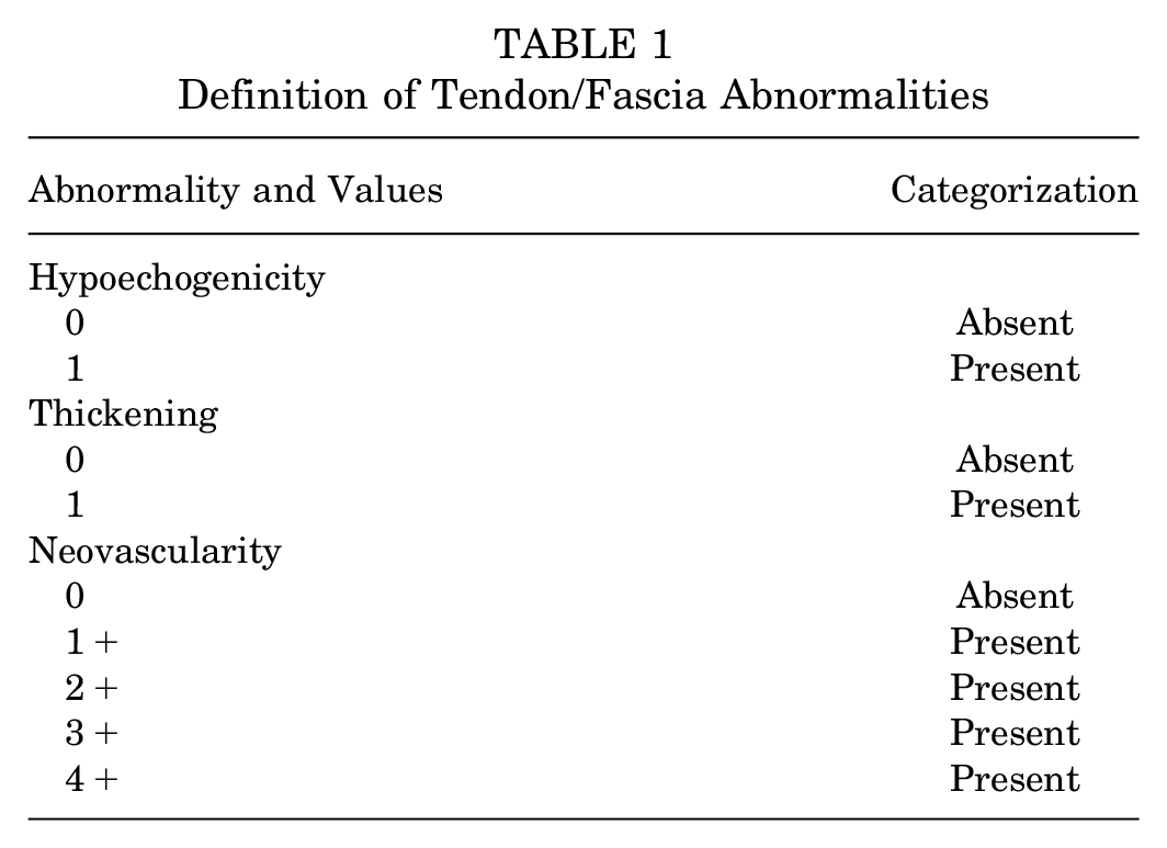
Video assessment was performed on a separate date by the primary author (D.M.C.), who has extensive experience reviewing the affected structures. The reviewer (D.M.C.) was blinded to all personal information related to the videos. The reviewer individually analyzed each video for the presence of focal hypoechogenicity (present/absent), morphologic thickening (present/absent), and neovascularity (0/1+/2+/3+/4+; analyzed as absent/present) (Table 1). 43 Structures were deemed abnormal if ≥1 of the 3 abnormalities existed.7,8,16,23,33,43
Definition of Tendon/Fascia Abnormalities
Patellar tendon thickness was measured from the deep layer of the patellar tendon to the superficial aspect, as it attaches to the patella. 37 Structural abnormalities were confirmed with multiple sonographic views for the Achilles and patellar tendon, while the plantar fascia abnormalities were confirmed solely in the sagittal plane, long axis to the fascia.
Interrater reliability was performed on a random selection of 20% of videos by a second experienced sonographer (S.F.E.), who was also blinded to participant information as well as the first reviewer's findings. Kappa values were calculated from categorical structure abnormality data (tendon/fascia: normal vs abnormal). Interrater kappa values are generally deemed <0.20 as slight, 0.21 to 0.40 as fair, 0.41 to 0.60 as moderate, 0.61 to 0.80 as substantial, and 0.81 to 1 as a near-perfect agreement between raters. 35 The kappa values were 0.556, 0.856, and 1 for the Achilles tendon, patellar tendon, and plantar fascia, respectively.
Injury Surveillance
All athletes were prospectively monitored over the entire academic year for the development of pain in the Achilles tendon, patellar tendon, and plantar fascia that caused time loss. The athletic training staff was notified of the study and the importance of reporting time-loss injuries to these structures; they were also queried individually throughout the year, quarterly, and at the end of the study time frame (ie, end of the academic year) for any additional injuries that had not been reported. The main outcome variable of interest was the development of pain in the affected structure that required missing practice or competition for any amount of time (= injury). Clinical diagnoses of load-related pain and tenderness in the target tendon/fascia were made by athletic trainers or sports medicine physicians.31,32 All student-athletes with unclear diagnoses underwent consultation with or were evaluated by a board-certified sports medicine physician. At the end of the study time frame, all student-athletes with diagnosed pain in 1 of the 3 structures were verified with the athletic training staff and/or sports medicine physicians. If an athlete sustained a rupture of 1 of the tendons, the decision was made a priori to exclude the athlete from the analysis, as the study was designed to evaluate overuse-type symptoms.
Also included were deidentified data collected as part of the Pacific-12 (Pac-12) collegiate athletic conference's Health Analytics Program (HAP), which were derived from clinical documentation in a Health Insurance Portability and Accountability Act (HIPAA)–compliant electronic medical record by sports medicine clinicians in the Pac-12. Data were deidentified using the HIPAA Safe Harbor method for deidentification (45 CFR 164.514). Resulting project data included deidentified records from only student-athletes that provided authorization for secondary research as part of the HAP.
Data Analysis
The primary outcome variable was injury (development of pain with time loss) in the Achilles tendon, patellar tendon, and plantar fascia, while the main predictor variable was the presence or absence of a sonographic abnormality at the preseason evaluation. Descriptive statistics were calculated for the demographic characteristics of athletes. Specifically, the mean with standard deviation or the median with interquartile range (IQR) were used for continuous variables, while categorical variables were summarized with frequencies and percentages. Ultrasound findings and current pain in the patellar tendon, Achilles tendon, and plantar fascia were also described using frequency and percentage, separately for the left and right tendons as well as for both tendons. A contingency table analysis was performed to examine the association between ultrasound findings and injury in these structures. Participants were only counted once if an injury was reported on ≥2 separate incidents in the same structure. In particular, an RR and its 95% CI, along with a P value and an absolute risk (AR), were computed from a generalized linear model for binomial distribution and log link, while using the cluster-robust variance estimator to calculate standard errors, to account for the potentially correlated data from the left and right tendons of each athlete.10,22,34,41,47,48 Also, each model was adjusted for previous injuries and the presence of pain at the time of ultrasound screening (= covariates) in a multivariate approach. Further, the Cox proportional hazards (PH) model with the cluster-robust standard errors was fit to the data to examine the association of injury to ultrasound findings, separately for the patellar and Achilles tendon.9,41,48 Meanwhile, the Cox PH model was not used for the data on the plantar fascia, as the number of cases in this structure was extremely low 46 (n = 4). A hazard ratio (HR) and its 95% CI were also calculated from the Cox PH model, adjusting for previous injury and pain at the time of ultrasound screening as covariates.
The above analyses were also repeated on asymptomatic tendon subgroups—including only the tendons from athletes without self-reported pain at the time of ultrasound screening. To examine ultrasound findings as a screening test for future injuries in these tendon structures, diagnostic summary measures—including sensitivity, specificity, positive predictive value (PPV), and negative predictive value (NPV)—were calculated for each tendon structure. Time loss was quantified as time to return to practice after the onset of symptoms and was compared by injury in each tendon structure using a Kruskal-Wallis test. P <.05 was used as the threshold for statistical significance, and all the analyses were conducted by a blinded statistician (M.T.) using Stata/MP 18.0 (StataCorp).
Results
Of the 243 athletes scanned by ultrasound in total during the 2021 academic year, 1 athlete sustained a tendon rupture during the study and was therefore excluded from the analysis. No participants were lost to follow-up. Consequently, data from 242 athletes with 484 tendons (ie, left and right tendons) were analyzed in this study. Participant characteristics are shown in Table 2. More than half of the athletes were women (57.8%); 51.7% of athletes engaged in explosive sports; and athletes from 14 different sports were included in the data.
Characteristics of the Included Athletes (N = 242) a
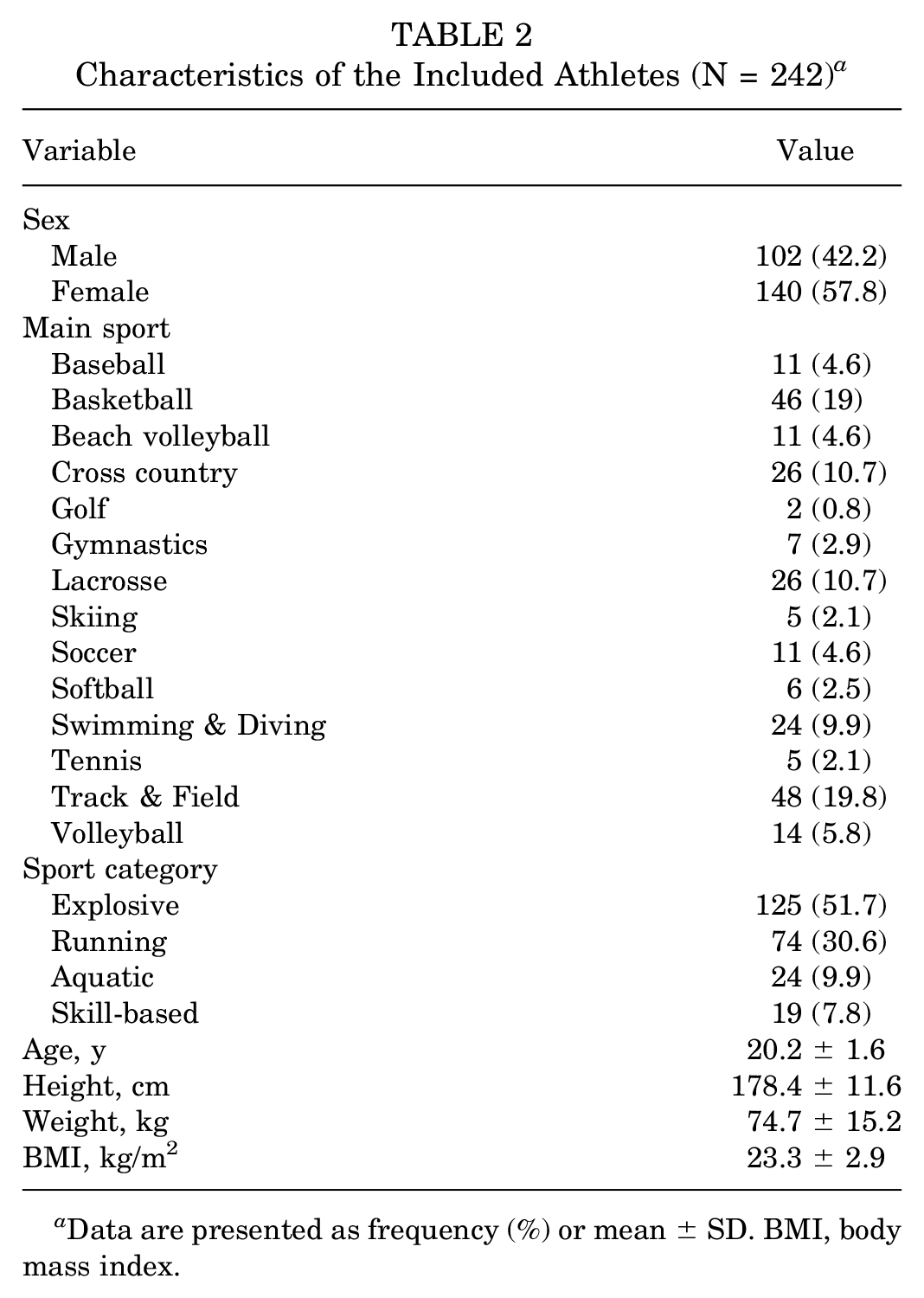
Data are presented as frequency (%) or mean ± SD. BMI, body mass index.
The ultrasound findings for the patellar tendon, Achilles tendon, and plantar fascia are reported in Table 3. Figure 1 demonstrates these corresponding findings. The patellar tendon had the highest prevalence of ultrasound abnormalities (range, 34.8%-40%) of the 3 structures. Ultrasound abnormalities in the plantar fascia were rare (range, 3.3%-3.7% of all participants), while the Achilles tendon showed abnormalities in around 10% of the participants (range, 9.9%-11.2%). A total of 24, 13, and 4 athletes reported injury (development of pain with time loss) for the patellar tendon, Achilles tendon, and plantar fascia, respectively, during the study. Among them, 1 athlete developed pain in the same patellar tendon for a second time, and 2 athletes developed pain in the same Achilles tendon for a second time; whereas none of the athletes sustained a repeat pain development in the plantar fascia. Hence, the analysis included a total of 23, 11, and 4 injuries in the patellar tendons, Achilles tendons, and plantar fasciae, respectively. Overall, the number of injuries in these structures was fairly low (≤5%) (Table 4). Athletes sustained injuries in the patellar tendon most frequently (4.6%-5%), followed by that in the Achilles tendon (2.1%-2.5%) and the plantar fascia (0.4%-1.2%).
Ultrasound Findings for Each Tendon Structure (N = 242 Athletes with 484 Tendons) a

Data are presented as frequency (%).
7 missing data points.
9 missing data points.
16 missing data points.
1 missing data point.
1 missing data point.

Examples of (A) patellar tendons, (B) Achilles tendons, and (C) plantar fasciae, taken as screenshots from videos. The top rows show normal findings: (a) normal long-axis image; (b) normal short-axis image; and (c) normal power Doppler image. The bottom rows show abnormal findings: (d/e) hypoechogenicity and thickening of the tendon (A) at the patellar pole, (B) just proximal to its insertion, or (C) hypoechogenicity and thickening of the plantar fascia; and (f ) neovascularity at that location.
Injury for Each Tendon Structure (N = 242 Athletes, 484 Tendons) a

Data are presented as frequency (%). Injury was defined as the development of pain with time loss.
Injury by ultrasound abnormality, separately for each structure, is summarized in Table 5. There was a significant association between ultrasound abnormality and injury for all tendon structures (P < .01). Specifically, athletes with ultrasound abnormalities in the patellar tendon, Achilles tendon, and plantar fascia were about 9 times (RR, 8.8 [95% CI, 1.9-42.1]; P = .006; AR, 8.4%), 17 times (RR, 17.2 [95% CI, 3-97.2]; P = .001; AR, 11.6%), and 13 times (RR, 13.2 [95% CI, 4.1-42]; P < .001; AR, 5.4%) more likely to develop pain than those without ultrasound abnormalities, respectively. According to the Cox PH models, the hazards of developing pain in the patellar and Achilles tendons for athletes with ultrasound abnormalities were about 9 times (HR, 9 [95% CI, 1.9-43.5]; P = .007) and 19 times (HR, 18.5 [95% CI, 3.8-102]; P = .001) higher than those without ultrasound abnormalities (Figure 2).
Contingency Table on Ultrasound Findings and Injuries (N = 242 Athletes/484 Tendons) a

Data are presented as frequency (%). Injury was defined as the development of pain with time loss. RR, relative risk.
Calculated from the generalized linear model for binomial distribution and log link with cluster-robust standard errors, adjusting for previous injuries and pain at the time of ultrasound screening. Bold values indicate statistical significance.

Cox proportional hazards model with cluster-robust standard errors on injury (pain with time loss) according to ultrasound findings in (A) patellar tendons and (B) Achilles tendons, adjusting for previous injury and pain at the time of ultrasound screening.
The results of the contingency table analysis and Cox PH models above held true even when the subgroup of only asymptomatic tendons was analyzed. Ultrasound abnormality was significantly associated with about 10 times (RR, 9.5 [95% CI, 2-45.2]; P = .005; AR, 8.9%) and 23 times (RR, 23.1 [95% CI, 2.5-209.4]; P = .005; AR, 10.7%) higher risk of pain development in the patellar and Achilles tendons, respectively (Table 6). Similarly, the patellar and Achilles tendons with ultrasound abnormalities were found to have over 9 times (HR, 9.5 [95% CI, 1.9-46.2]; P = .005) and 25 times (HR, 24.6 [95% CI, 2.7-226.3]; P = .005) greater hazards of pain development compared with those without ultrasound abnormalities (Figure 3).
Contingency Table on Ultrasound Findings and Injuries for Asymptomatic Tendons a

Data are presented as frequency (%). Injury was defined as the development of pain with time loss. NA, not applicable; RR, relative risk. Bold values indicate statistical significance.
Calculated from the generalized linear model for binomial distribution and log link with cluster-robust standard errors, adjusting for previous injuries.
Not calculable because of zero frequency in 1 cell.

Cox proportional hazards model with cluster-robust standard errors on injury (pain with time loss) according to ultrasound findings in asymptomatic (A) patellar tendons and (B) Achilles tendons.
Overall, ultrasound screening showed extremely high NPVs (≥90%) for all 3 tendon structures (Table 7). In other words, athletes with normal ultrasound findings were quite unlikely to develop pain in those structures. Ultrasound screening for the patellar tendon showed high sensitivity (85.7%), whereas that for the Achilles tendon and plantar fascia showed high specificity (>90%). Meanwhile, PPVs were fairly low for all 3 tendon structures (≤14%), indicating that most athletes with ultrasound abnormalities in these structures did not end up developing pain. Looking within types of abnormality, the highest PPVs were seen with neovascularity measurements, but they remained on the lower end.
Diagnostic Measures of Ultrasound Screening a
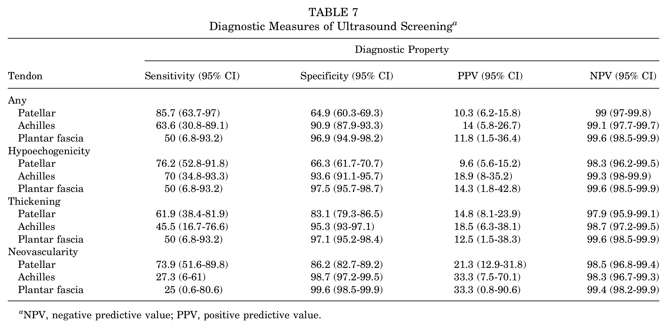
NPV, negative predictive value; PPV, positive predictive value.
The median time to return to practice after developing pain in the patellar tendon (n = 22), Achilles tendon (n = 10), and plantar fascia (n = 4) was 1 day (IQR, 1-4 days [range, 1-241 days]), 1 day (IQR, 1-14 days [range, 0-28 days]), and 8 days (IQR, 1.5-34 days [range, 1-54 days]), respectively. There were no significant between-group differences in time to return to practice (P = .432) among the 3 tendon structures, likely because of the large variability in the data.
Discussion
Previous literature on sonographic screening for tendon abnormalities in athletes has been focused primarily on sports involving running, jumping, cutting, or pivoting. 33 This multi-institutional study involved athletes from various collegiate sports and demonstrates that collegiate student-athletes with preseason ultrasound abnormalities in the patellar tendon, Achilles tendon, or plantar fascia are more likely to develop pain in those structures compared to those without preseason abnormalities.
Several prior studies have prospectively evaluated the likelihood of developing tendinopathic symptoms based on an ultrasound evaluation.11-13,19,33 Evidence from systematic reviews demonstrates that sonographic abnormalities significantly increase the risk of future Achilles and patellar tendon pain, though some discrepancies in the results have been reported between the studies. The present study is one of the largest of its kind to track athletes for a year and identified one of the highest RRs. The comparatively higher RR found in our study may relate to the population of interest: The present study included only NCAA Division I student-athletes, whereas prior studies examined both younger and older populations.6-8,11,12,14,16-18 Furthermore, most prior studies examined asymptomatic tendons, while the present study examined all tendons, but did include a subanalysis of asymptomatic tendons. The collegiate age may be the tipping point where abnormalities become symptomatic.
Though large RRs were noted for the development of pain in all 3 structures, its utility as a screening test should be viewed as having both strengths and limitations. All structures showed low PPVs (<15%) but had very high NPVs (≥99%). This could be related to 2 main causes. First, the overall percentage of athletes who developed pain remained low throughout the 3 structures. The patellar tendon, Achilles tendon, and plantar fascia developed pain in 4.8%, 2.3%, and 0.8% of athletes, respectively. Prior epidemiologic data have shown that Achilles and plantar heel injuries in collegiate athletes are relatively low compared to all foot and ankle injuries. 24 Second, the sonographic abnormality categories were broad—namely, thickening, hypoechogenicity, or neovascularity. Specific morphologic characteristics, such as location within the tendon, degree of abnormality, or shape, were not discretely analyzed; future studies may better elucidate which abnormalities best correlate to future pain.
In general, the patellar tendon had the highest percentage of sonographic abnormalities on the initial screening in this study. This is consistent with data from prior literature reporting the incidence of patellar tendinopathy being higher in elite adolescent athletes compared to the Achilles tendon. 4 Additionally, a recent study on ultrasound abnormalities of the patellar tendon in adolescent male basketball players showed that athletes developed ultrasound abnormalities during the peak height velocity (PHV) and post-PHV periods; none of the athletes demonstrated ultrasound abnormalities during the pre-PHV period. 5 This is relevant to the findings in the present study, as collegiate student-athletes typically hit their peak height velocity before starting college. 1 The ultrasound findings may indicate that the athletes have been at risk previously, but now the tendon fibers have matured, demonstrating the ultrasound abnormalities once they are in college. 5
Following the patellar tendon, the Achilles tendon was the structure with the second-highest percentage of ultrasound abnormalities in this study. Prior literature of elite athletes demonstrates an increased risk of developing pain in the Achilles tendon when ultrasound abnormalities were present on initial screening.15,16 On the other hand, although it was a smaller study, Jhingan et al did not find a significant difference between athletes with or without ultrasound abnormalities and future development of pain; the lack of association may be due to the smaller sample size. 26 With its larger sample size, the current study could add to the literature with more robust findings of ultrasound abnormalities in the Achilles tendon.
Overall, the plantar fascia had the smallest percentage of ultrasound abnormalities on initial screening. Of the 3 structures evaluated, the plantar fascia had a lower incidence of developing pain.2,20 To our knowledge, this is the first study examining the relationship between ultrasound abnormalities of the plantar fascia and its relation to future symptoms. Due to the relatively few cases of pain developing in this structure in the present cohort, it was not possible to determine with confidence if asymptomatic sonographic abnormalities related to future pain development. However, if symptomatic plantar fasciae were included, there was a significant association; this is in line with the patellar and Achilles tendon findings.
Pain in the patellar tendon, Achilles tendon, or plantar fascia required variable amounts of time away from sport before returning to practice. The median time away ranged between 1 and 8 days among all 3 structures, though the time away from practice had a larger range for some athletes. For example, the patellar tendon had the largest range of time away: from 1 to 241 days. In a previous study, collegiate athletes with patellar tendinopathy mostly spent 24 hours or less time away from competition. 45 The data from our study suggest that time away from sport is often low as well. This is significant to our study population as multiple weeks to months away from sport can impact a competitive season. The median number of days away from practice could be influenced by the timing of the injury and whether the injury occurred in season or the off season.
As previously mentioned, the main analysis for the present study included symptomatic and asymptomatic structures. Most previous data examine asymptomatic structures, as a true screening test is designed to identify a condition before symptoms. 33 However, there is still important information to be gained from including symptomatic tendons. A diagnosis is not always clear. Multiple similar conditions can masquerade as the tendinopathy/fasciopathy in this study, such as plantaris tendinopathy, retrocalcaneal bursitis, patellofemoral pain syndrome, fat pad impingement, tarsal tunnel syndrome, entrapment of the first branch of the lateral plantar nerve, and radiculopathy. The present study utilized self-reported patellar tendon, Achilles tendon, and plantar fascia pain; no diagnosis was made by a medical practitioner at the beginning of the study. Thus, this screening test still is effective without a medical diagnosis. When the analysis was run for only asymptomatic tendon structures, there was still a significantly higher risk of developing pain in the patellar tendon and Achilles tendon. The RR values for the asymptomatic tendon structures are smaller compared to the total group, and this can be attributed to the decrease in the overall number of structures included in the analysis, coupled with the higher risk of sonographic abnormality within athletes with self-reported symptoms in the structure. The RR for the plantar fascia could not be calculated, as there were only 2 asymptomatic structures that eventually developed pain, and they did not have any ultrasound abnormalities on initial screening.
Identification of at-risk structures is useful, and the next logical step is to incorporate interventions for injury prevention. Strengthening programs have been proposed to help decrease ultrasound abnormalities and pain. Fredberg et al 15 evaluated elite Danish soccer players who had asymptomatic patellar and Achilles tendons. Teams were randomized into an eccentric strengthening and stretching program, and the number of athletes with ultrasound abnormalities was decreased for the patellar tendon. 15 Contrary to their hypothesis, the risk of injury to the patellar tendon increased during the study period, while the Achilles tendon risk did not improve. 15 Further research is needed to evaluate the efficacy of a rehabilitation program to decrease the risk of developing pain in at-risk athletes.
Limitations and Strengths
There are limitations associated with the current study. First, our study included student-athletes from 14 different sports. There are more than 20 NCAA sports that compete for championships each year. 36 Despite a large number of study participants, we are not able to represent all sports with this data. Future research could be designed as a larger-scale, multi-institutional study with participants from all NCAA sports. Second, ultrasound abnormalities were recorded by either their presence or absence, without specification for the particular type morphology of ultrasound abnormality. While simplifying the protocol, this additional information could be useful in a future study to evaluate whether specific abnormal findings, such as abnormal vascularity, hypoechogenicity, or tendon/fascial thickening, are more likely to relate to future pain. For example, a study of Swedish high school volleyball players demonstrated that neovascularity in the region of other patellar tendon sonographic abnormalities was more indicative of symptomatic patellar tendinopathy. 17 Pain in the patellar tendon, Achilles tendon, or plantar fascia was described as either “yes” or “no.” More specifics on the type of injuries sustained could help understand any patterns in the type and severity of injuries that these athletes encounter.
In addition, operator experience plays a large role in the reliability of sonographic characterization. We speculate that the majority of discrepancies between experienced reviewers come from subtle differences between small hypoechogenic areas, which may be less likely to be related to future injury than larger, more obvious areas. As more potential abnormalities exist (eg, the patellar tendon, compared to the plantar fascia), reliability is lower; therefore, reliability may be lower in populations with a higher prevalence. Future studies are ongoing to identify specific causes of diminished reliability.
Despite the aforementioned limitations, the current study has multiple strengths that should be highlighted. This study included a large number of student-athletes from 3 different NCAA Division I institutions. Additionally, previous studies have been focused on individual groups of athletes, such as basketball, soccer, volleyball, or endurance athletes, while our study included athletes from a wide range of collegiate sports.8,11,14,17,18,20,27,30 This allows for a more robust generalization of the data among collegiate athletes. Close monitoring of the student-athletes was performed with the help of athletic training staff, coaches, and team physicians, with follow-up lasting an entire year. Last, the data collection was excellent for the 242 athletes with few missing data. Specifically, between the 3 structures that were evaluated (1452 possible data points), only 17 data points were missing, which accounted for <1% of the overall data.
Conclusion
Collegiate athletes with ultrasound abnormalities to the patellar tendon, Achilles tendon, and plantar fascia on the preseason screening tests were found to be more likely to develop pain in the respective structures during the competitive season. Of the 3 structures studied, the patellar tendon developed pain the most often, followed by the Achilles tendon. The NPV was >99% for all 3 structures demonstrating that athletes without ultrasound abnormalities on prescreening were extremely unlikely to develop pain, whereas PPVs were low (<15%). Preseason musculoskeletal ultrasound screening could be incorporated in collegiate athletes to evaluate for the risk of developing pain during the season. Further research on this topic should be focused on specifying high-risk sonographic abnormalities and identifying injury prevention strategies for those who demonstrate ultrasound abnormalities in the patellar tendon, Achilles tendon, and plantar fascia.
Footnotes
Acknowledgements
This publication contains materials created, compiled, or produced by the Pac-12 Health Analytics Program (HAP). The authors thank the tireless efforts of the athletic training staff at each institution for their insights, assistance, and coordination for this large study. In particular, they thank Lindsay Adams, Jessie Keiser, Trevor Jameson, Savannah Horning, Matt Delisi, and Tom Iriye. The authors also thank Cam Fausett, Alex Asay, Luke Johnson, Josh Larson, and Gabrielle Garruppo for their assistance with the study.
Final revision submitted April 11, 2024; accepted April 23, 2024.
One or more of the authors has declared the following potential conflict of interest or source of funding: This study was conducted with support from the Pac-12 Conference's Student-Athlete Health and Well-Being Initiative. Research reported in this publication was also supported by the National Center for Advancing Translational Sciences of the National Institutes of Health (award number UL1TR002538). AOSSM checks author disclosures against the Open Payments Database (OPD). AOSSM has not conducted an independent investigation on the OPD and disclaims any liability or responsibility relating thereto.
Ethical approval for this study was obtained from the University of Utah (reference No. 00130805).