
Abstract
Background:
Studies have shown that Kaplan fibers (KFs) play a role in controlling anterolateral rotatory knee laxity in anterior cruciate ligament (ACL) injury. However, recent clinical studies have suggested that KF injuries are not associated with a higher-grade pivot shift in knees with acute ACL injuries.
Purpose:
To compare the effect of KF and anterolateral ligament injury on anterolateral rotatory laxity as measured by the pivot-shift test in both adult and adolescent patients with acute ACL injury.
Study Design:
Cross-sectional study; Level of evidence, 3.
Methods:
This study consisted of patients who experienced primary acute ACL tears between January 2019 and December 2021. The magnetic resonance imaging examinations and pivot-shift tests for these individuals were conducted within 14 days after injury. Variables associated with concomitant injury were included in a binary logistic regression model to evaluate risk factors of a high-grade pivot shift and a KF injury.
Results:
The study included 540 patients with acute ACL tears (64 adolescent patients and 476 adults). The main risk factor for a high-grade pivot shift after acute ACL injury in adults was partial or total disruption of anterolateral ligament continuity (odds ratio [OR], 2.271; 95% CI, 1.499-3.442). In adolescent patients, the main risk factor was KF injury (OR, 19.98; 95% CI, 2.367-168.669), including edema and disruption of continuity.
Conclusion:
The main risk factor for a high-grade pivot shift after acute ACL injury differs between adolescent patients and adults: the KF injury sign in adolescent patients indicates a higher-grade pivot shift.
Keywords
According to statements from the International Anterolateral Complex (ALC) Consensus Group Meeting, 5 the ALC consists of the superficial and deep aspects of the iliotibial band with Kaplan fiber (KF) attachments on the distal femoral metaphysis, along with the anterolateral ligament (ALL), a capsular structure within the anterolateral capsule. The results of a biomechanical study using cadaveric knees support the contribution of KFs to controlling anterolateral rotatory laxity in anterior cruciate ligament (ACL)–deficient knees, which also indicates that KFs may function as a secondary restraint. 10 Additionally, another cadaveric study found that KFs played a greater role in controlling tibial internal rotation at higher flexion angles than did the ALL. 4 Similar to ALL, 8 KFs and KF injuries can be identified on magnetic resonance imaging (MRI), 1 and the rate of KF injury concomitant with ACL tears has been reported to be 17.6% to 60%. 20 In the field of ALL research, some studies to date have reported supporting the effects of ALL injury on rotatory knee laxity,14,16 while others have not.7,10,15,17 This phenomenon is also found in KFs, as the role of KFs in controlling anterolateral rotatory knee laxity remains uncertain. Although it has been shown that KFs can control anterolateral rotatory laxity in ACL-deficient knees, recent clinical studies have shown that KF injuries are not associated with a higher-grade pivot shift in knees with acute ACL injuries.2,20 Additionally, there have been no relevant reports on adolescent patients in the field of KF research. The available study involving adolescent patients is focused on the ALL. 12 For example, a study showed that lateral extra-articular tenodesis with single-bundle hamstring tendon autografting for ACL reconstruction (ACLR) in young patients significantly reduced graft rupture and persistent rotatory laxity at 2 years after surgery. 6 However, in this randomized controlled trial, the mean age of the patients was 18.9 years (range, 14-25 years); no imaging risk factors were specifically studied in adolescent patients with ACL injuries. Thus, despite KFs being part of the ALC, perhaps the uncertain relationship between KFs and rotatory laxity in ACL-deficient knees causes difficulties in the selection of the surgical approach in clinical practice. Meanwhile, the sample size of a recent imaging study 2 on KFs has remained a constraint in achieving a deeper understanding of KFs, and there have been no studies on KFs in adolescent patients. The purposes of the present study were to use MRI to detect concomitant KF and ALL injuries in knees with ACL injury in both adult and adolescent patients and then to compare the effect of KF injury and multiple risk factors (concomitant injury to the ALL, medial collateral ligament [MCL], or anterior, central, or posterior medial or lateral meniscus) on anterolateral rotatory laxity as measured by the pivot-shift test in a clinical setting. It was hypothesized that knees with acute ACL injury and concomitant KF injury would show a higher grade of manual pivot shift than knees with ACL injury without concomitant KF injury in both adult and adolescent patients.
Methods
Patients
This retrospective cohort analysis of collected data included patients with unilateral acute primary ACL tears who underwent primary ACLR at our institution. The diagnosis of ACL tears was made based on clinical findings and MRI and was confirmed arthroscopically. The inclusion criteria were as follows: unilateral acute primary ACL tear, considered when the patient reported knee injury occurring <2 weeks before the examination, and bone bruising on the femoral condyles and tibial plateau identified on MRI. The exclusion criteria were as follows: concomitant ligament (posterior cruciate ligament and/or posterolateral complex) procedures or realignment procedures, contralateral knee injury, previous injury or surgery affecting the ipsilateral knee, insufficient data from the electronic medical record system, lack of KF visualization on MRI, and contralateral positive pivot shift or preexisting rotatory laxity before injury. A previous study showed that a sample size of 84 knees was required to detect a difference during the pivot shift using a Mann-Whitney U test (effect size d = 0.80) at a power of 0.80 and significance level of .05. 20 Originally, 1944 patients were identified from medical records between January 2019 and December 2021. After reviewing the inclusion and exclusion criteria, 540 patients were included in the present study (Figure 1). The study was approved by our institution's research ethics committee and registered in the Protocol Registration and Results System (registry No. NCT06002308).

Flowchart of the exclusion process for the present study. KFs, Kaplan fibers; MR, magnetic resonance; MRI, magnetic resonance imaging.
Assessment of Concomitant Meniscal, Collateral Ligament, ALL, and KF Injury
Meniscal injuries were diagnosed via arthroscopy during surgery. These injuries included damage to the anterior, central, or posterior parts of the medial or lateral meniscus. All injuries to the posterior part of the meniscus were combined, despite differences between vertical longitudinal tears and root tears. Collateral ligament injuries were diagnosed based on MRI and clinical examinations. ALL and KF injuries were assessed by MRI using previously reported methods.18-20 Proton density–weighted imaging with and without fat suppression was performed on the sagittal, coronal, and axial planes. A single examiner (J.C.) assessed KF injury according to the report by Batty et al. 1 KF and ALL injuries were graded as follows: grade 1, mild periligamentous edema with identifiable, continuous low-signal-intensity fibers; grade 2, partial disruption or irregular contour with ligamentous edema; and grade 3, complete disruption. Segond fractures were considered grade 3 ALL injuries. 19
Pivot-Shift Test and Lachman Test
Pivot-shift and Lachman tests were performed with the patient under general anesthesia just before ACLR. Standardized pivot-shift tests were performed by 3 experienced surgeons (including S.C.). The results of pivot-shift testing are graded (grades 1-3), and grades 2 and 3 are considered as a high-grade pivot-shift test result. Operators were blinded to the MRI and arthroscopy results at the time of the pivot-shift evaluation.
Statistical Analysis
All analyses were conducted using SPSS Version 29.0 (IBM Corp). Categorical data were evaluated with the Pearson chi-square test. Statistical significance was set at P < .05. The mean ± SD are reported as basic descriptive statistics. Variables associated with concomitant injury were included in a binary logistic regression model to evaluate predictors of a high-grade pivot shift and a KF injury.
Results
The characteristics of the patients in the adolescent group and adult group are summarized in Table 1. In total, 64 adolescent patients with a mean age of 16.1 ± 7.8 years (range, 11-17 years) and 476 adult patients with a mean age of 30.9 ± 1.4 years (range, 18-55 years) were included. Significant differences were found between adolescent patients and adults in sex (P = .004), grade 1 pivot shift (42.9% vs 57.0%; P = .039), grade 3 pivot shift (30.2% vs 12.0%; P < .001), high-grade pivot shift (56.3% vs 42.2%; P = .034), grade 2 KF injury (39.5% vs 26.3%; P = .039), ALL injury (grades 1-3) (84.4% vs 94.1%; P = .004), and ALL disruption (grades 2 and 3) (39.1% vs 52.9%; P = .037). No significant differences were found between adolescent patients and adults in the period from injury to MRI (7.1 ± 2.8 vs 7.4 ± 2.9 days; P = .778), positive pivot shift (98.4% vs 98.1%; P = .855), grade 2 pivot shift (27.0% vs 31.0%; P = .523), KF injury (grades 1-3) (59.4% vs 51.9%; P = .260), grade 1 KF injury (52.6% vs 65.2%; P = .682), grade 3 KF injury (7.9% vs 8.5%; P = .920), KF disruption (grades 2 and 3) (28.1% vs 18.1%; P = .055), grade 1 ALL injury (53.7% vs 43.8%; P = .529), grade 2 ALL injury (31.5% vs 38.8%; P = .116), grade 3 ALL injury (14.8% vs 17.4%; P = .425), MCL injury (45.3% vs 45.0%; P = .957), anterior part of the lateral meniscus (LMA; 4.7% vs 0.0%; P = .520), anterior part of the medial meniscus (MMA; 0.0% vs 0.4%; P = .603), central part of the lateral meniscus (LMC; 28.1% vs 28.2%; P = .997), central part of the medial meniscus (MMC; 23.4% vs 23.5%; P = .987), posterior part of the lateral meniscus (LMP; 45.3% vs 35.5%; P = .126), or posterior part of the medial meniscus (MMP; 46.9% vs 36.3%; P = .102).
Baseline Characteristics of Patients a

Data are expressed as mean ± SD (range) or n (%) unless otherwise noted. Bold P values indicate statistical significance (P < .05). ALL, anterolateral ligament; KF, Kaplan fiber; LMA, anterior part of the lateral meniscus; LMC, central part of the lateral meniscus; LMP, posterior part of the lateral meniscus; MCL, medial collateral ligament; MMA, anterior part of the medial meniscus; MMC, central part of the medial meniscus; MMP, posterior part of the medial meniscus; MRI, magnetic resonance imaging. Blank cell indicates not applicable.
Chi-square test.
The concomitant injuries among patients with a high-grade pivot shift are summarized in Table 2. Significant differences were found between the 2 groups in the rate of ALL injury (86.1% vs 96.5%; P = .009), KF injury (88.9% vs 51.2%; P < .001), KF disruption (50.0% vs 18.4%; P < .001), and LMA injury (8.3% vs 2.0%; P = .038). No significant differences were found between the 2 groups in the rate of male sex (55.6% vs 68.7%; P = .125), ALL disruption (50.0% vs 64.2%; P = .106), MCL injury (44.4% vs 42.8%; P = .853), MMA injury (0.0% vs 0.5%; P = .671), LMC injury (27.8% vs 31.3%; P = .670), MMC injury (27.8% vs 24.9%; P = .712), LMP injury (52.8% vs 42.3%; P = .243), and MMP injury (52.8% vs 39.8%; P = .146).
Concomitant Injuries in Relation to the Presence of High-Grade Pivot Shift (n = 237) a

Data are expressed as n (%) unless otherwise noted. Bold P values indicate statistical significance (P < .05). ALL, anterolateral ligament; KF, Kaplan fiber; LMA, anterior part of the lateral meniscus; LMC, central part of the lateral meniscus; LMP, posterior part of the lateral meniscus; MCL, medial collateral ligament; MMA, anterior part of the medial meniscus; MMC, central part of the medial meniscus; MMP, posterior part of the medial meniscus.
Chi-square test.
Binary logistic regression analysis was used to identify risk factors associated with the presence of a high-grade pivot shift (Table 3). Of the 12 initially included predictive variables, the risk of a high-grade pivot shift was significantly increased in 4 variables: female sex (odds ratio [OR], 2.261 in adults and 2.025 in total), ALL disruption (OR, 2.271 in adults and 2.115 in total), KF injury (OR, 19.98 in adolescent patients), and LMP injury (OR, 1.699 in adults and 1.818 in total).
Risk Factors for High-Grade Pivot Shift a
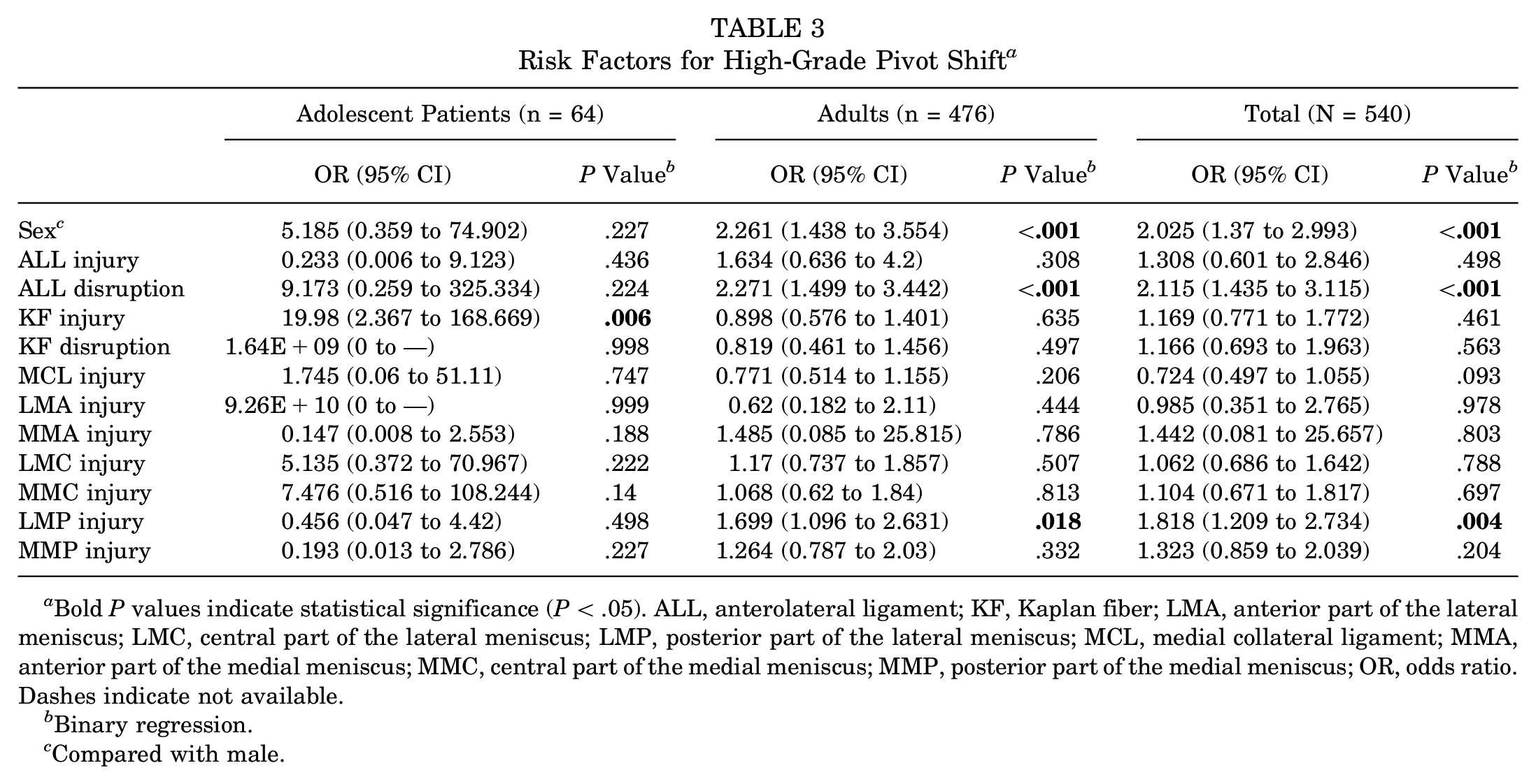
Bold P values indicate statistical significance (P < .05). ALL, anterolateral ligament; KF, Kaplan fiber; LMA, anterior part of the lateral meniscus; LMC, central part of the lateral meniscus; LMP, posterior part of the lateral meniscus; MCL, medial collateral ligament; MMA, anterior part of the medial meniscus; MMC, central part of the medial meniscus; MMP, posterior part of the medial meniscus; OR, odds ratio. Dashes indicate not available.
Binary regression.
Compared with male.
Binary logistic regression analysis was used to identify risk factors associated with KF injury (Table 4). Of the 10 initially included predictive variables, 5 significantly increased the risk of KF injury: ALL injury (OR, 5 in adults and 2.906 in total), ALL disruption (OR, 4.876 in adolescent patients, 1.808 in adults, and 1.903 in total), MCL injury (OR, 2.64 in adults and 2.342 in total), LMA injury (OR, 4.351 in adults), and LMP injury (OR, 1.632 in adults and 1.63 in total).
Risk Factors for KF Injury a

Bold P values indicate statistical significance (P < .05). ALL, anterolateral ligament; KF, Kaplan fiber; LMA, anterior part of the lateral meniscus; LMC, central part of the lateral meniscus; LMP, posterior part of the lateral meniscus; MCL, medial collateral ligament; MMA, anterior part of the medial meniscus; MMC, central part of the medial meniscus; MMP, posterior part of the medial meniscus; OR, odds ratio.
Binary regression.
Compared with male.
Discussion
Recently, investigators reported that concomitant KF injury did not have a significant effect on anterolateral rotatory laxity measured by the pivot-shift test in knees with acute ACL injuries.2,20 However, the present study found that the cause of a high-grade pivot shift after acute ACL injury differs between adolescent and adult patients. In adults, partial or total disruption of ALL continuity and LMP injury was associated with a high-grade pivot shift. This result is consistent with what was seen in the total population (including adolescent and adult patients). This finding is partly in line with those of recent clinical studies showing that KF injury is not associated with a high-grade pivot shift.2,20 However, in adolescent patients with acute ACL injury, neither ALL continuity nor LMP injury was a risk factor since the correlation between KF injury and a high-grade pivot shift was no longer statistically significant. Instead, the risk factor was a KF injury (including edema as well as disruption of continuity), where adolescent patients with a KF injury were nearly 20 times more likely to have a high-grade pivot shift than those without a KF injury.
The reason why the correlation between KF injury and a high-grade pivot shift was not statistically significant in adults may be because KF damage is the result of multiple factors in adults. It is possible that in adults, KF injuries are dependent on not only more severe and continuity-affecting ALL injuries (ALL disruption) but also common ALL injuries (eg, edema), resulting in KF injuries in adults being more irregular. Conversely, KF injuries were less irregular in adolescents and only associated with ALL injuries affecting continuity. Consequently, KF injury was associated with a high-grade pivot shift only in adolescents. In addition, a clue can be found in another important result of this paper: ALL injury, ALL disruption, MCL injury, LMA injury, and LMP injury significantly increased the risk of KF injury in both adults and the total population with acute ACL injury. However, in adolescent patients with acute ACL injury, only ALL disruption significantly increased the risk of KF injury. It is suggested that KF injury in adolescent patients is associated with higher-grade ALL injury (disruption of ALL continuity) rather than with grade 1 ALL injury; that is, lower-grade ALL injury in adolescent patients cannot significantly increase the risk of KF injury. This may be because KF injury is dependent on more severe and continuity-affecting ALL injuries (ALL disruption) and possibly due to the better extension capacity of the soft tissues in adolescent patients, necessitating more severe ALL injuries to cause damage to the KFs in adolescent patients. Therefore, when KF injuries in adolescent patients indicate severe damage to the stable structures of the knee, they result in a high-grade pivot shift.
One strength of our study is that the interval from injury to the MRI and pivot-shift examinations was <14 days, ensuring that the ALL injury had not yet started healing and that the examinations were performed in the postinjury state. In addition to the lack of adolescent patients in previous studies on rotatory laxity and KF assessment after ACL injury, these studies did not have a sufficiently short interval between patient injury and MRI and pivot-shift examinations. In a study by Musahl et al, 13 the mean time interval between the MRI examination and quantitative pivot-shift test was 43 ± 36 days. While the patients were in the acute phase of ACL injury at the time of MRI in that study, the pivot-shift test was performed up to 2 months from the time of injury. In a study by Miyaji et al, 11 the interval from ACL injury to MRI examination was >1 month. Furthermore, the authors still consider the association between KF injury and a high-grade pivot shift among adults with acute ACL injuries to be of great interest because the number of adolescent patients included in this paper is too small compared with the number of adults, accounting for only 15% of the latter.
Notably, even if KF injury is correlated with instability in adolescent patients with ACL injury, this does not indicate that adolescent patients with ACL injury are more in need of ALC reconstruction during ACLR, although there is not always a complete correlation between biomechanical instability and patient-reported outcomes after ACLR. 6 It can only be said that the contribution of KF injury to anterolateral rotatory knee laxity may not be limited to adolescent patients with ACL injury, which is in contrast to not only previous studies showing a significant contribution of KFs to controlling anterolateral rotatory knee laxity4,10 but also a recent study reporting “no contribution.” 20 This implies that the contribution of KF injury to anterolateral rotatory knee laxity remains uncertain, necessitating further research to gain insight. Even so, the KF injury sign in adolescent patients may still act as an alarming signal to surgeons in the surgical planning. Because the pivot-shift test is usually performed under general anesthesia, the preoperative finding of KF injury on the MRI scan obtained in an adolescent patient with ACL injury can guide the preoperative surgical planning (eg, with lateral extra-articular tenodesis surgery or not) as well as a more definitive preoperative consultation with the patient. In our country, a more definitive preoperative surgical plan would be more acceptable to patients.
Limitations and Strengths
There are several limitations to the present study. First, the total number of adolescent patients was relatively small, at just 64. However, we believe that this scenario reflects the current reality in daily clinical practice, since the number of adolescent patients with ACL injuries at our institution is significantly smaller than the number of adult patients with ACL injuries. Although studying young patients <25 years old instead of adolescent patients <18 years old could greatly increase the number of patients, according to a study by Getgood et al, 6 this approach was eventually abandoned for a more accurate study of rotational stability in adolescent patients. Further research is needed to obtain a larger number of adolescent patients for analysis. Second, we did not quantitatively evaluate the pivot shift using an electromagnetic measurement system. Furthermore, the manual pivot-shift test is subjective and widely variable since the pivot shift is multifactorial and can be affected by ALC lesions, meniscal tears, posterior tibial slope, and other osseous parameters. 3 Third, parameters of bone morphology (ie, posterior tibial slope), which might have affected the pivot-shift results, were not assessed or compared in the present study. Fourth, the pivot-shift tests were performed by 3 surgeons. However, these surgeons used a standardized technique, which diminished the variability between examiners. 9 Fifth, the retrospective methodological design of the present study involves an inherent bias. Last, we combined all injuries to the posterior part of the meniscus except for root tear.
The strengths of the present study are as follows: First, the correlation between anterolateral rotatory knee laxity and MRI findings in adolescent patients with ACL injury was assessed. Second, the MRI and clinical evaluations were performed <14 days after ACL injury in all 540 cases within the postinjury period. Third, edema and disruption of continuity were analyzed separately in KF and ALL injuries. Last, the MRI scans in the present study were all obtained with identical protocols at our institution.
Conclusion
The main risk factor for a high-grade pivot shift after acute ACL injury differs between adolescent patients and adults. In adults, the main risk factor is partial or total disruption of ALL continuity. In adolescent patients, the main risk factor is KF injury, including edema and disruption of continuity. The KF injury sign in adolescent patients would indicate a higher-grade pivot shift and would act as an alarming signal to surgeons in the treatment of these patients.
Footnotes
Final revision submitted May 24, 2024; accepted June 14, 2024.
One or more of the authors has declared the following potential conflict of interest or source of funding: This study was supported by the Scientific Research Program of Sichuan Medical and Health Care Promotion Institute (KY2022SJ0396) and Research Program of Sichuan Medical Association (Q22085). AOSSM checks author disclosures against the Open Payments Database (OPD). AOSSM has not conducted an independent investigation on the OPD and disclaims any liability or responsibility relating thereto.
Ethical approval for this study was obtained from Sichuan Provincial Orthopedic Hospital (KY2023-018-01).