
Abstract
Background:
Improvements in motor control of the scapular muscles are important for the prevention and rehabilitation of shoulder and elbow injuries in overhead athletes.
Purpose:
To clarify scapular muscle activity during multijoint compound movement exercises using fine-wire and surface electrodes.
Study Design:
Descriptive laboratory study.
Methods:
Sixteen healthy men performed 5 types of exercises (cat and dog, trunk rotation, A-exercise, T-exercise, and Y-exercise). Muscle activity was measured as percentage of maximum voluntary isometric contraction (%MVIC) by using fine-wire electrodes in the rhomboid major (Rhom) and using surface electrodes in the upper (UT), middle (MT), and lower (LT) trapezius and serratus anterior (SA) muscles. The Rhom/UT, MT/UT, LT/UT, and SA/UT muscle activity ratios were calculated. One-way analysis of variance was used to compare the %MVIC and muscle activity ratios between exercises.
Results:
There was no significant difference in Rhom activity between the exercises (34.6-54.2%MVIC; P = .25). LT activity was significantly greater in the trunk rotation (58.0 ± 24.6%MVIC) and Y-exercise (63.2 ± 40.1%MVIC) than in the cat and dog scapular retraction (19.6 ± 9.3%MVIC) and A-exercise (28.2 ± 14.2%MVIC) (P < .05). SA activity was significantly greater in the cat and dog scapular protraction (26.7 ± 11.0%MVIC) and Y-exercise (25.6 ± 19.3%MVIC) than in the other exercises (P < .05). The SA/UT activity ratio in the cat and dog scapular protraction exercise (9.64 ± 8.48) was significantly higher than in the other exercises (P < .05).
Conclusion:
All the exercises were effective for activating the Rhom. The trunk rotation and Y-exercise were effective for activating the LT, and the cat and dog scapular protraction exercise was effective for activating the SA while suppressing the UT.
Clinical Relevance:
These results enable exercise selection based on muscle activity characteristics (moderate [20%-50%MVIC] and high [>50%MVIC] levels contribute to muscle activation) to prevent and rehabilitate shoulder and elbow injuries.
Keywords
Shoulder injuries are common in overhead sports such as baseball, softball, tennis, and swimming. 25 In baseball, the incidence rates of shoulder and elbow injuries are high. 41 For example, baseball throwing is a high-load motion because it requires a large amount of torque on the shoulder and elbow joints and high muscle activities of the rotator cuff and scapular muscles. 14,16 In addition, overhead sports movements, including the throwing motion, require coordinated movements among the glenohumeral joint, scapulothoracic joint, and trunk. 15,16,31,46 Poor trunk and scapular motion such as decreased thoracic extension and scapular external rotation can increase the load on the shoulder and elbow joints. 21,45 Therefore, the mobility of each joint and proper motor control must be ensured for coordinated movements to prevent and rehabilitate shoulder and elbow injuries.
The common factors that cause shoulder injury in overhead athletes include strength deficits and motor control impairments, which are primarily found in the glenohumeral and/or scapulothoracic joints, 25,46 range-of-motion deficits in shoulder horizontal adduction and rotation, 6,44 increased thoracic kyphotic angle in the standing posture, 38,39 and scapular dyskinesis. 18 Scapular dyskinesis has been defined as the alteration of scapular position and motion. 22 Hickey et al 18 reported that athletes with scapular dyskinesis have a 43% greater risk of developing shoulder pain in the future. In scapular dyskinesis, the inferior angle and medial border prominence of the scapula are the most common, with high upper trapezius (UT) activity and low lower trapezius (LT) and serratus anterior (SA) muscle activities. 20 The medial border prominence of the scapula indicates increased internal scapular rotation, which can cause shoulder and elbow injuries in overhead sports. 21,30 Therefore, for shoulder and elbow injury prevention and rehabilitation in overhead athletes, improvement of scapular muscle impairment and obtaining proper motor control of the scapula are important.
Previous studies have noted the effectiveness of scapular muscle exercises for improving and preventing shoulder injury symptoms. 12,27,40 Sakata et al 39,40 studied a program to improve physical function that included scapular muscle exercises in youth baseball players and reported that the incidence rates of shoulder and elbow injuries were lower in the intervention group than in the nonintervention group. Lynch et al 27 applied an intervention in swimmers with scapular muscle exercises and observed improvements in rounded posture and shoulder muscle strength, which have been associated with shoulder injury, suggesting that the scapular muscle exercises were effective for improving shoulder injury.
There are several muscle activity studies on scapular muscle exercises. 37,42,46,47 Most of these studies reported the activity of superficial muscles such as the LT and SA, with a few reports on the rhomboid major (Rhom), which is located below the superficial muscles. Cools et al showed that overhead athletes with shoulder impingement had lower SA strength and LT activity, suggesting the importance of improving the LT and SA functions. 9,11 In addition to the LT and SA, the importance of the Rhom, which attaches to the medial border of the scapula and contributes to scapular motor control, has been suggested in overhead sports movements. 14,17,48 Most previous studies only considered exercises in a single plane that involve a single movement, such as shoulder forward flexion in side-lying and side-lying external rotation. 46 Few studies have shown Rhom activity during exercises with multijoint compound movement.
In the early phase of rehabilitation, the use of single-plane exercises below 90° of shoulder elevation in overhead athletes is recommended for suppressing UT overactivity that inhibits the proper activity of the LT and SA. 46 On the other hand, in the late rehabilitation phase and in the prevention phase, dynamic and multijoint compound movement exercises are required to prepare the athletes for return to overhead sports. 46 The scapula moves to an upward rotation, posterior tilt, and retraction during the arm-cocking phase of the throwing motion. 24 Shoulder maximum external rotation during throwing is the combined movement of glenohumeral external rotation, scapular posterior tilt, and thoracic spine extension. 31 Thus, scapular external rotation, posterior tilt, retraction, and thoracic spine extension are particularly important during the throwing motion. Information for exercise selection based on scapular muscle activity characteristics is important for athletes, athletic trainers, physical therapists, and coaches to improve and prevent shoulder and elbow injuries.
The purpose of this study was to clarify muscle activity during scapular muscle exercises with multijoint compound movement by using fine-wire and surface electrodes. Our hypothesis was that differences in joint position and joint movement during exercise influence differences in the muscle activity values of the Rhom, LT, and SA.
Methods
Participants
Sixteen healthy men (age, 21 ± 1 years; height, 171.6 ± 5.8 cm; weight, 67.4 ± 9.9 kg) participated in this study. Inclusion criteria were participants with no low back, shoulder, or neck pain in the previous 3 months or during the implementation of the exercises and no previous shoulder or spinal surgery. All the participants signed a written consent form before participation. This study was conducted in accordance with the principles of the Declaration of Helsinki and approved by the ethics review committee of our institution.
Electromyography Measurements
The muscle activity of the Rhom, UT, middle trapezius (MT), LT, and SA was measured using a wireless electromyography (EMG) telemeter system (BioLog DL-5000; S&ME Co). All the muscles were measured on the participant’s dominant-hand side. Rhom activity was measured using bipolar intramuscular fine-wire electrodes (Unique Medical Co, Ltd), the activity of the UT, MT, LT, and SA was measured using surface electrodes (Blue Sensor N-00-S; METS Co) with a sampling rate of 1000 Hz. The bipolar intramuscular fine-wire electrodes were covered with Teflon coating, except for their tips. The electrode was placed into a 23-gauge Cattelan needle (0.60 mm × 60 mm), and the tips were bent back to 3-5mm hooks. The Cattelan needle with a fine-wire electrode was sterilized by heating at 121°C for 20 minutes using a small fully automatic high-pressure steam sterilizer (Taneda Medical Instruments Co, Ltd).
The participants were placed In the prone position with the back of the dominant hand placed on their low back region (Figure 1A). After identification of the Rhom using ultrasonography (LOGIQe; GE), the insertion point was shaved and disinfected with alcohol. An experienced orthopaedist (K.K.) inserted the fine-wire intramuscular electrodes in the Rhom under ultrasonography guidance (Figure 1B). The insertion point was at the midpoint of the base of the spine of the scapula and the inferior angle of the scapula (Table 1). 1,2,36 After insertion in the Rhom, only the Cattelan needle was removed, and the fine-wire electrode was placed in the Rhom. To confirm accurate insertion, the Rhom electromyographic amplitude was checked with active scapular elevation and adduction. We attached surface electrodes parallel to the muscle fibers after shaving and rubbing the skin with alcohol to reduce skin impedance (Table 1). 23,43 The surface electrodes were attached with a 2-cm interelectrode distance.

Electrode Placement and Maximum Voluntary Isometric Contraction (MVIC) Trials for Electromyographic Normalization
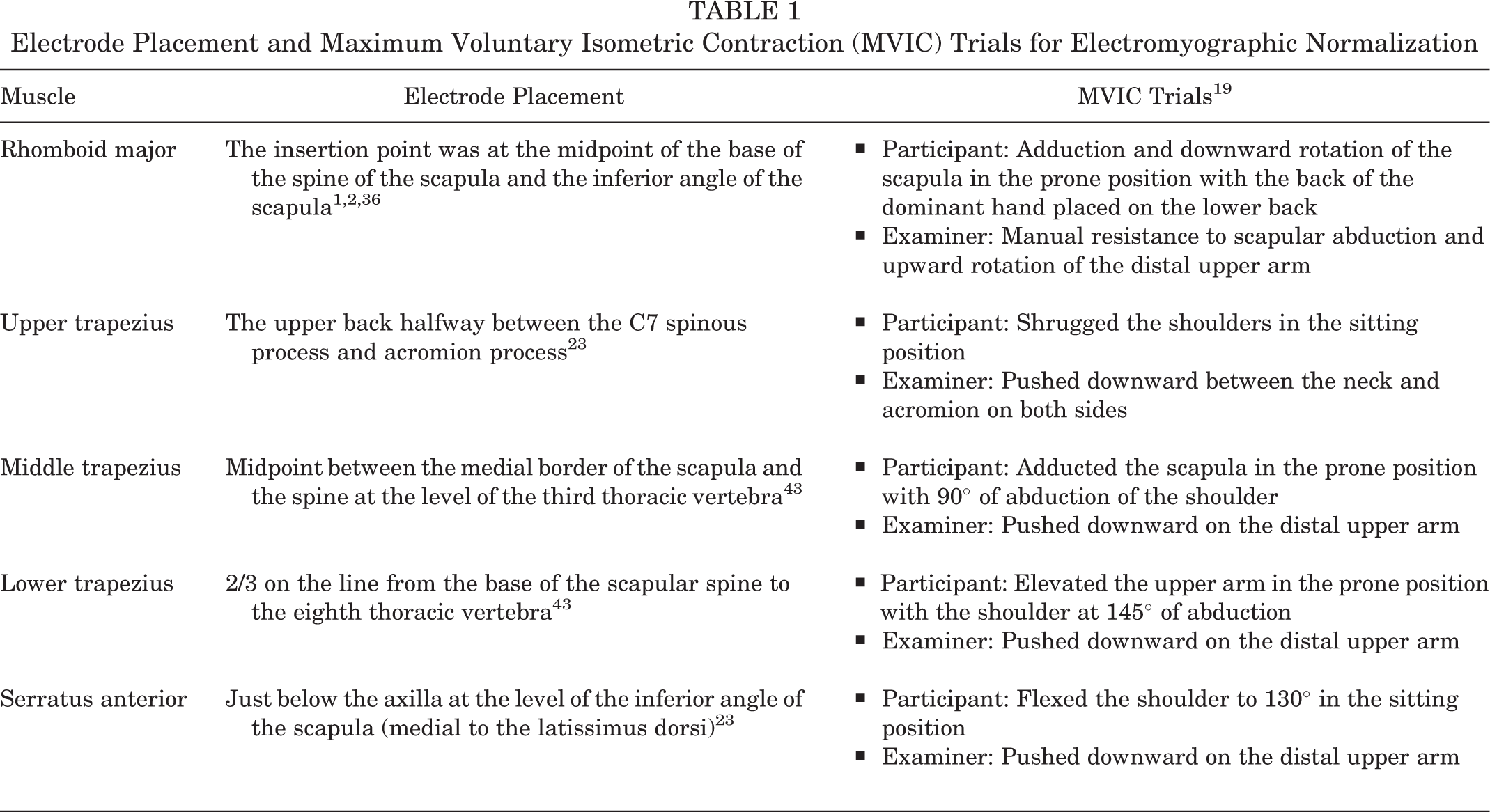
The maximum voluntary isometric contraction (MVIC) was measured as an index of normalization to compare the muscle activity of each exercise. The participants performed against the manual resistance applied by the same examiner (G.A.) in all the MVIC trials (Table 1). 19 The MVIC trials for each muscle were performed for 5 seconds in random order. A resting period of >1 minute was allocated between the MVIC trials.
Exercise Trials
The scapular muscle exercises are described in detail in Figure 2. These were multijoint compound movement exercises that combined the joint movement of the scapular and thoracic spine that are required for the throwing motion, aimed at activating the scapular muscles, such as the Rhom, LT, and SA. These exercises have been shown to contribute to a decrease in the incidence rates of shoulder and elbow injuries in intervention studies. 39,40 They are frequently performed as a warm-up, periscapular muscle strengthening exercise, and athletic rehabilitation exercise without using any tools in overhead sports, such as baseball. The participants performed 5 scapular muscle exercises in a random order. Prior to the muscle activity measurement in each exercise, detailed instructions regarding exercises and verbal feedback were given to each participant to perform exercises in proper motion. We also checked the proper exercise motion during muscle activity measurement. Each exercise was performed 5 times, and the motions were recorded using a high-speed camera (Exilim EX-100; Casio Computer Co, Ltd) at 120 Hz in the sagittal plane. The participants had >1 minute of rest time between the exercise trials. The 3-second motions in each exercise set from start to finish were controlled with a metronome.

Descriptions of the scapular muscle exercises.
Data Processing
The muscle activity data of the exercises were analyzed using biological information analysis software (BIMUTAS-Video; Kissei Comtec Co). The bandpass was filtered at 20 to 450 Hz to remove motion artifacts. 2 The filtered muscle activity data were used as the root mean square (RMS) value. The analysis was performed for each 3-second exercise motion from start to finish. The muscle activity during each exercise was represented as percentage of the MVIC (%MVIC). These values were normalized by the highest RMS value of the MVIC, calculated using the RMS during 1 second of the MVIC trials. The representative value for each exercise was the mean value of the measurements in the middle 3 of the 5 repetitions of each exercise.
The muscle activity levels were classified into 3 groups according to %MVIC as follows: low, <20%; moderate, 20% to 50%; or high, >50%. 5,29 In addition, the following muscle activity ratios were calculated: Rhom/UT, MT/UT, LT/UT, and SA/UT. The higher the ratio values, the more active each muscle was with suppressed UT activity.
Statistical Analysis
All data were reported as mean ± SD. Statistical analyses were performed using the SPSS Version 26.0 software (IBM Japan). One-way analysis of variance (ANOVA) on a factor (exercise trials) was used to compare the %MVIC and muscle activity ratios between the exercises. In the cat and dog exercise, the statistical analysis was performed by dividing motions into scapular retraction and scapular protraction. Therefore, in the cat and dog exercise, the motions with scapular protraction and retraction were used in the SA and Rhom, UT, MT, and LT muscle activity comparisons. When a significant main effect was detected, the Tukey test was performed as the post hoc test. The effect size (η2) for ANOVA was calculated to represent the magnitude of difference between muscle activity and muscle activity ratio for each exercise; η2 was defined as small, moderate, and large if the value ranged from 0.01 to <0.06, from 0.06 to <0.14, and up to >0.14, respectively. 8 The significance level was set at .05.
Results
All the muscle activities and muscle activity ratios of the exercises are presented in Table 2. The Rhom activity was the highest during the A-exercise (54.2 ± 23.7%MVIC). No significant main effect on Rhom activity was observed in the exercise trials (F (4,75) = 2.49; P = .25; η2 = 0.07). The UT activity was the lowest during cat and dog scapular retraction (13.1 ± 9.3%MVIC). The main effect on UT activity was significant in the exercise trials (F (4,75) = 8.15; P < .001; η2 =0.43) but significantly lower during cat and dog scapular retraction than during the A-exercise (35.2 ± 21.8%MVIC; P = .02), T-exercise (35.5 ± 16.9%MVIC; P = .02), and Y-exercise (53.9 ± 31.2%MVIC; P < .001). The MT activity was the highest during the T-exercise (46.7 ± 28.7%MVIC). We found a significant main effect on the MT activity in the exercise trials (F (4,75) = 5.23; P < .001; η2 = 0.27), which was significantly greater during T-exercise than during the cat and dog scapular retraction (16.6 ± 11.0%MVIC; P < .001). The LT activity was the highest during Y-exercise (63.2 ± 40.1%MVIC). A significant main effect on LT activity was observed (F (4,75) = 9.20; P < .001; η2 = 0.49), with the Y-exercise and trunk rotation exercise (58.0 ± 24.6%MVIC) showing significantly greater activity than cat and dog scapular retraction (19.6 ± 9.3%MVIC) and A-exercise (28.2 ± 14.2%MVIC) (P < .001). The SA activity was greatest during cat and dog scapular protraction (26.7 ± 11.0%MVIC). A significant main effect on SA activity (F (4,75) = 14.11; P < .001; η2 = 0.75) was observed, with cat and dog scapular protraction showing significantly greater than the trunk rotation exercise (9.1 ± 8.0%MVIC), and A-exercise (5.4 ± 5.7%MVIC), and T-exercise (6.2 ± 6.8%MVIC) (P < .001).
Comparison of Muscle Activities and Muscle Activity Ratios Between the Exercises a

a Values are presented as mean ± SD. LT, lower trapezius; MT, middle trapezius; %MVIC, percentage of maximum voluntary isometric contraction; Rhom, rhomboid major; SA, serratus anterior; UT, upper trapezius.
b Significantly higher than the cat and dog exercise (P < .05).
c Significantly higher than the trunk rotation exercise (P < .05).
d Significantly higher than the A-exercise (P < .05).
e Muscle activity during cat and dog scapular protraction.
f Significantly higher than the T-exercise (P < .05).
g Significantly higher than the Y-exercise (P < .05).
The Rhom/UT ratio was highest during cat and dog scapular retraction (5.85 ± 7.98). A significant main effect on the Rhom/UT ratio (F (4,75) = 3.91; P < .001; η2 =0.17) was observed in the exercise trials, but the Rhom/UT ratio during cat and dog scapular retraction was significantly greater than during the T-exercise (1.49 ± 1.10; P = .01) and the Y-exercise (1.00 ± 0.80; P < .001). The MT/UT and LT/UT ratios were the highest during cat and dog scapular retraction (2.32 ± 3.53 and 6.69 ± 17.48, respectively). We found no significant main effect on the MT/UT (F (4,75) = 1.37; P = .25; η2 = 0.06) or LT/UT ratios (F (4,75) = 1.27; P = .28; η2 = 0.06). The SA/UT ratio was highest in cat and dog scapular protraction (9.64 ± 8.48). A significant main effect on the SA/UT ratio (F (4,75) = 18.89; P < .001; η2 = 0.50) was observed, but the SA/UT ratio was significantly higher during cat and dog scapular protraction than during the trunk rotation (0.37 ± 0.25), A-exercise (0.26 ± 0.55), T-exercise (0.26 ± 0.37), and Y-exercise (0.58 ± 0.64) (P < .001).
Discussion
The purpose of this study was to clarify the muscle activity during scapular muscle exercises with multijoint compound movement by using fine-wire and surface electrodes. These were multijoint compound movement exercises that combined the joint movement of the scapula and thoracic spine that are required for the throwing motion, aimed at activating the scapular muscles, such as the Rhom, LT, and SA. The main findings of this study were as follows: First, Rhom activity was similar and not significantly different between the exercises (34.6-54.2%MVIC; P = .25). Second, LT activity was greater during the trunk rotation exercise and Y-exercise (58.0 ± 24.6%MVIC and 63.2 ± 40.1%MVIC, respectively; P < .05). Finally, SA activity was greater during cat and dog scapular protraction and Y-exercise (26.7 ± 11.0%MVIC and 25.6 ± 19.3%MVIC, respectively; P < .05), and the SA/UT activity ratio was higher during cat and dog scapular protraction (9.68 ± 8.48; P < .05) than during the other exercises. These exercises were performed without using any tools or equipment, and similar levels of muscle activity were observed in the scapular retraction muscles, such as the Rhom and LT, compared with muscle activity during the throwing motion.
In our study, Rhom activity was not significantly different between the exercises. Moseley et al 33 and Berckmans et al 4 reported that the main function of the Rhom was scapular retraction. The scapular external rotation movement in scapular retraction increases with trunk rotation. 47 Rhom muscle activity increases with increased scapular external rotation movement. 4 Exercises in the limb position parallel to the direction of the muscle fibers results in higher muscle activity. 32,37 In this study, the trunk rotation exercise, which required trunk rotation motion, and the A-exercise, in which the direction of movement was close to the direction of the oblique Rhom muscle fibers, were expected to show higher muscle activity, but no significant difference was found between the exercises. The reason is that all the study participants were young healthy men, and their motor control function was normal, maintaining the scapula in the proper position during the exercises; therefore, the Rhom was a tonic activity (sustaining activity of constant amount). Increased thoracic kyphotic posture is associated with shoulder and elbow injuries. 39,40 The Rhom is important in maintaining good posture in which the tragus and acromion are closer to the vertical line passing through the greater trochanter. 2 Moreover, the importance of the Rhom in overhead sports movement has been reported. 14,17,48 The activation of the Rhom may help prevent shoulder and elbow injuries by improving posture and proper motor control of the scapula during overhead sports movement in overhead athletes.
Previous studies have investigated Rhom activity in exercises using dumbbells, rubber tubing, and early phase rehabilitation activities. 3,4,7,33,34 These studies reported that Rhom activity was high in exercises involving scapular retraction and external rotation of the glenohumeral joint. The Rhom activity required during the baseball throwing motion was shown to be moderate in the early cocking phase (35% maximal manual muscle test [MMT]) and late cocking phase (41% MMT), 14 which was similar to the muscle activity levels during the exercises in this study. All the exercises performed in this study showed moderate (20%-50%) or high (>50%) Rhom activity, which suggests that the activity was sufficient to activate the Rhom. The Rhom/UT ratio was higher during cat and dog scapular retraction than during the T- and Y-exercises in this study. To obtain appropriate scapular motor control in overhead sports movement, activation of the Rhom in the suppressed position of the UT and then gradually activating the Rhom in the Y limb, which is close to the overhead sports limb, in cooperation with the MT, LT, and SA are important.
With regard to the LT activity, during the trunk rotation exercise and Y-exercise, the scapular retraction movements in high shoulder abduction and high scapular upward position may have resulted in high (>50%) LT activity. Yamauchi et al 47 reported the same results as in this study that accompanying trunk rotation during scapular retraction movement increased LT activity. This suggests that the accompanying trunk rotation in addition to performing scapular retraction exercises at a high shoulder abduction position may be effective in increasing LT activity. In a study examining the ratio of UT to LT activity in exercise, Cools et al 10 found that LT activity was high (>50%) and UT activity was low (<20%) or moderate (20%-50%) in the exercises performed in the prone and side-lying positions. De Mey et al 13 investigated scapular retraction exercises in various closed kinetic chain positions and reported that UT and most LT activities were low (<20%) for all exercises. Cools et al 10 and De Mey et al 13 recommended these exercises be performed in the early stage of rehabilitation to activate the LT while suppressing UT activity. However, it has been shown that during the throwing motion, the muscle activity of the UT and LT is moderate to high in the early cocking (UT, 64% MMT; LT, 39% MMT), late cocking (UT, 37% MMT; LT, 38% MMT), and acceleration phases (UT, 69% MMT; LT, 76% MMT), and LT activity is not necessarily higher than UT activity. 14 It is recommended that multijoint compound exercises be performed in the late rehabilitation and prevention phases, 46 and it may be necessary to activate the LT while allowing a certain amount of UT activity.
Regarding SA activity, cat and dog scapular protraction was performed with scapular protraction movement, and Y-exercise was performed in the scapular upward position, which may have resulted in moderate SA activity. Oyama et al 35 showed that an arm raise in 120° of shoulder abduction with external rotation in the prone position resulted in moderate SA activity, similar to the result of the Y-exercise (25.6 ± 19.3%MVIC). Maenhout et al 28 reported that SA activity was moderate at 31.6%MVIC during scapular protraction exercise on all fours, similar to the result of the cat and dog scapular protraction (26.7 ± 11.0%MVIC). Cat and dog scapular protraction not only activates the SA, but when used in combination with cat and dog scapular retraction, it alternates extension and flexion of the thoracic spine and mobilizes a large movement in the sagittal plane of the thoracic spine. Sakata et al 39,40 reported improvement in thoracic spine alignment with interventions including these exercises. Therefore, cat and dog scapular protraction and retraction may be more effective when performed in combination. Although the SA activity during both the Y-exercise and the cat and dog scapular protraction was moderate and sufficient to activate the SA, cat and dog scapular protraction was more effective in terms of the SA activity ratio to UT. Maenhout et al and Ludewig et al 26 similarly reported the effectiveness of scapular protraction exercises in improving the balance of muscle activity between the SA and UT. However, the SA activity during the throwing motion was moderate to high (early cocking, 44%MMT; late cocking, 69%MMT; acceleration, 60%MMT), and the SA activity was not necessarily higher than the UT activity. 14 Therefore, in order to aim for further activation and strengthening of the SA in the late rehabilitation and prevention phases, it may be necessary to perform exercises with high SA activity while allowing a certain amount of UT activity.
The exercises in this study were performed without using any equipment. Compliance with the exercise program has been reported to affect the intervention effect in reducing the incidence rate of shoulder and elbow injuries. 40 If the number of intervention exercises is greater and the exercise program time is longer, compliance can be reduced. 40 Similarly, if the exercise cannot be performed without the use of equipment and tools, compliance may be reduced. Myers et al 34 mentioned that one of the considerations for performing exercise is whether the exercise can be easily performed in the field setting. Therefore, the exercise in this study may be useful in increasing compliance with exercise performance.
Limitations
This study has several limitations. The participants performed the exercise trials with fine-wire electrodes placed on the Rhom and may have had some pain during the exercise trials, which may have affected the EMG data. Moreover, we did not measure the participants’ shoulder joint or trunk range of motion, and differences in range of motion may have affected the EMG data. The sample size was relatively small owing to the highly invasive nature of the fine-wire electrode experiment, and we did not perform a power analysis. All the participants were young healthy men. The results of this study may be different in healthy female patients, overhead athletes, and patients with scapular dyskinesis. Finally, we did not measure scapular or trunk kinematics and did not quantify actual scapular or trunk movements. Although scapular and trunk kinematics influence the scapular muscle activity, the relative position of the arms, scapula, and trunk that exhibit the highest muscle activity cannot be determined in this study.
Conclusion
All the exercises were effective for activating the Rhom, which suggests that the Rhom showed a tonic activity to optimize the scapular position during the exercises. The trunk rotation and Y-exercise were effective for activating the LT, and cat and dog scapular protraction was effective for activating the SA while suppressing the UT. These results will enable exercise selection based on the scapular muscle activity characteristics for athletes, athletic trainers, physical therapists, and coaches to improve and prevent shoulder and elbow injuries.
Footnotes
Final revision submitted July 31, 2022; accepted August 7, 2022.
One or more of the authors has declared the following potential conflict of interest or source of funding: This study was supported by a Grant-in-Aid for Scientific Research from the Japan Society for the Promotion of Science (grant No. 17K01767). AOSSM checks author disclosures against the Open Payments Database (OPD). AOSSM has not conducted an independent investigation on the OPD and disclaims any liability or responsibility relating thereto.
Ethical approval for this study was obtained from Waseda University (ref No. 2016-019).