
Abstract
Objectives:
Bone morphogenetic proteins (BMPs) belong to the transforming growth factor superfamily that were first discovered by Marshall Urist. There are 14 BMPs identified to date, each with distinct and versatile functional roles. Two pioneering studies compared the effects of 14 different BMPs on bone regeneration using an ectopic bone formation model and found that BMPs 2, 6, 7, and 9 efficiently induce bone formation using an adeno-viral BMP-transduced C2C12 cell line. This study also found that BMP3 can exert an inhibitory effect on bone formation induced by BMP2, 6, and 7, but not BMP9 [1,2]. Our research team previously showed that human muscle derived stem cells (hMDSCs) can promote bone regeneration in critical size calvarial bone defect when transduced with lenti-viral BMP2 [3-5]. However, the gene transduction of stem cells may limit its clinical translation due to safety concerns. Coacervate is a polymer designed to achieve local and sustained release of growth factors including for tissue engineering applications for bone and cartilage repair [6-8]. The aim of this study is to use the coacervate sustained release platform to identify the most potent BMPs for enhancement of bone regeneration in a critical sized calvarial bone defect.
Methods:
Results:
MicroCT results showed almost no bone formation in the PBS coacervate group (
Conclusions:
The current study reveals that the use of coacervate to deliver BMPs induced bone formation to different extents in a critical sized calvarial defect. BMP2 and BMP7 were the most potent BMPs in promoting bone formation. The new bone regenerated by the BMP2 group is equivalent to new bone regenerated using lenti-BMP2 transduced human muscle derived stem cells. More importantly, the newly regenerated bones are the same as natural host trabecular bone without any residual material.

MicroCT analysis of in vivo bone regeneration of different BMPs using coacervate sustain release system without using celts. A. MicroCT 3D overview of bone regeneration in the critical sized calvarial bone defect. BMP2 sustain-released with coacervate regenerated most amount of the new bone. BMP7 also regeneiated significant amount or new bone. BMP4.6 and 9 regenerated relative less new bone In the defect area. B Quantification of the new bone regenerated In the defect area. All BMPs regenerated significantly higher new bone volume than PBS control group at days 14,28 and 42. BMP2 regenerated significantly more new bone than BMP4.6.7.9 al all three time points after surgery. BMP7 also regenerated significantly higher new bone volume than BMP4,6,9. ANOVA, P<0.0001 for all three time points. "P<0.05, "P<0.01, ***P<0.001. **"P<0,0<301 for Tukey’s multiple comparisons.

Histology of the new regenerated bone in different BMPs groups sustained released by coacervate A. Herovicl’s staining showed new bone at the edge and middie of the defect. Areas between two red arrows showed the defect area at 20X magnification, Collagen 1 stained pink red while collagen 3 stained bright blue fiber, BMP2 group showed near complete healing at the edge of the defect. BMP7 group also showed more closure of the defect with new bone. Other groups showed less new bone formation. At the middie of the defect area, BMP2,4 and 7 showed new bone formation, but limited new bone formation in the other groups. At 100X magnification, new bone structure are more clear with intense collagen 1 positive matrix in pink red and collagen 3 showed in biue. B. H&E staining showed the general morphology of newly regenerated bone with typical normal trabecular bone structure with bone matrix and complete bone marrow, NB indicates new bone, Biue arrows point to the hematopoietic stem cells in the bone marrow and yellow arrows point to the red blood cells. Megakaryocytesare showed in the enlarged insets. Scale bars= 200pm for 100X and 1000pm for 20X magnification.
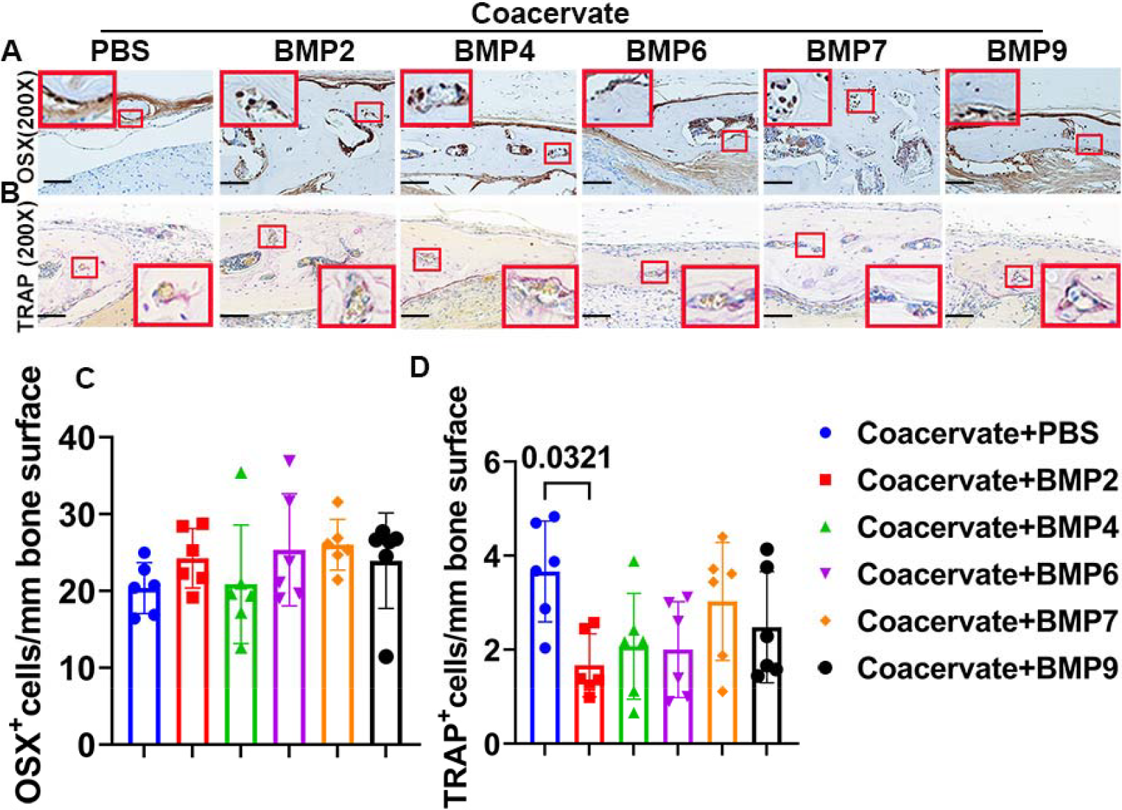
Histological analysis of the bone cells in regenerated new bone. A and C. immunohistochemistry of OSX and quantification. OSX is expressed in the osteoprogenitor cells on the bone surface as intense brown nuclear staining. Few osteocytes also expressed OSX. Some periosteum and fibrotic tissues in the defect area showed background staining. PBS control group staining actually are the host bone due to very limited new bone formation. All other groups showed new bone area. Insets are enlarged red boxed area to show OSX positive cells on bone surface. Quantification showed no Statistical difference between BMPs group and PBS group. B and D. TRAP staining for osteoclasts and quantifications. TRAP positive cells are showed in violet red multiple nuclei and single nuclear cells on the bone surface. Insets are enlarged red boxed area on each image to demonstrate positive osteoclasts. New bone in all BMPs groups showed presence of osteoclasts which indicate bone remodeling. PBS group are actually the left over host bone as very limited new bone formation are available to analyze. Quantification showed BMP2 group showed significantly lower number of osteoclasts ceils. ANOVA , P=0.034. Scale bars = 100ym.