Abstract
Objectives:
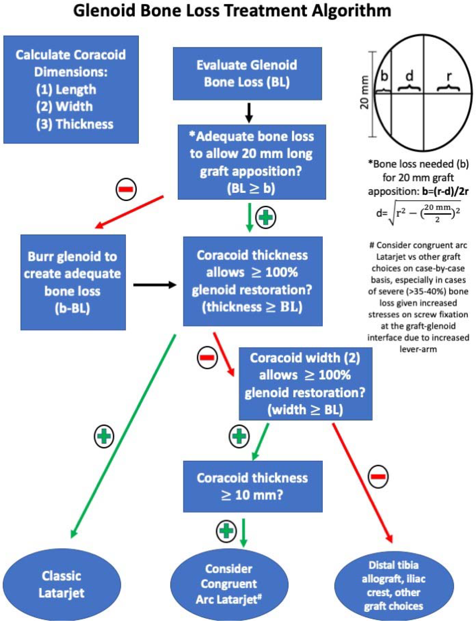
The concept of the “inverted pear” glenoid was introduced by Lo and Burkhart as an indicator of significant attritional glenoid bone loss that predisposes patients to recurrent instability and higher failure of arthroscopic Bankart repair. Coracoid transfer (Latarjet) is often performed in this setting, and evaluation of coracoid dimensions may improve patient-specific preoperative planning. The purpose of this study was to utilize computed tomography (CT) to determine a reproducible method for coracoid measurement and develop a preoperative planning algorithm for glenoid pear restoration using the traditional Latarjet technique or congruent arc modification (CAM). We hypothesized that classic Latarjet technique would sufficiently restore at least 100% of the glenoid diameter, up to 30% of anterior bone loss. We also hypothesized that most female patients would have a coracoid thickness <10 mm, which may preclude the use of the CAM due to a risk of graft fracture with screw insertion.
Methods:
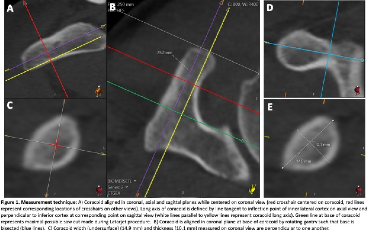
Multiplanar reconstructed (MPR) shoulder CT scans were reviewed for patients aged 18-45. This age group was selected to capture an adult population that typically undergoes anterior shoulder stabilization surgery. Patients were excluded if CT scan demonstrated glenoid or humeral osteophyte formation, glenoid dysplasia, coracoid fracture, or tumor involving the glenoid or coracoid. All CT scans were obtained with the arm at the patient’s side, palm facing up, and the shoulder in external rotation. CT scans were performed by acquiring thin-slice data from the volumetric CT in the axial plane with a Discovery CT750 HD scanner (GE Healthcare) and the following scan parameters: 0.625-mm slice thickness, 0.313-mm slice spacing, reconstruction matrix of 512 × 512 pixels, reconstruction field of view of 40 to 45 cm, and pitch factor of 0.516. All measurements were performed using a PACS system (Sectra Workstation, Sectra AB, Linkoping, Sweden) by a board-certified musculoskeletal radiologist and two orthopaedic surgeons. The coracoid base was aligned in the coronal plane, and the long axis of the coracoid was aligned in the axial and sagittal planes (
Results:
A total of 120 CT scans were reviewed, and three were excluded (two with osteophytes and one with severe glenoid dysplasia). Measurements were performed and dimensions recorded on 117 CT scans (69.2% males). Coracoid dimensions varied considerably among patients (length: 17.5-31.8mm, width: 9.1-20.5mm, thickness: 6.1-15.7mm,
Conclusions:
We describe a reliable method of measuring coracoid dimensions for preoperative planning of glenoid pear restoration. The traditional Latarjet technique reliably restores the glenoid AP diameter with bone loss of up to 30%. The majority of females have coracoid thickness <10mm which may increase the risk of graft fracture when using CAM. The decision to utilize the classic Latarjet technique or CAM considers each individual’s glenoid and coracoid dimensions with a goal of achieving at least 100% restoration of native glenoid diameter (