
Abstract
Background:
Equipment used to guide surgical incisions has been shown to be a source of bacterial contamination during surgery.
Purpose/Hypothesis:
To compare the culture-positive rates of sterile marking pens used before and after skin preparation for shoulder surgery. It was hypothesized that there will be no difference in culture-positive rates from marking pens used after skin preparation compared with before skin preparation.
Study Design:
Controlled laboratory study.
Methods:
Overall, 43 consecutive patients undergoing elective shoulder surgery were enrolled prospectively into this study. Each patient provided 2 samples: study pens (from marking the surgical site incision after skin preparation) and positive control pens (from marking the surgical site incision before skin preparation). In addition, there were 43 negative control pens evaluated (straight from the packaging without any patient contact). Cultures were evaluated at 4 and 21 days, and all positive cultures were further evaluated for speciation, if able. Standard descriptive summaries and Fisher exact tests were used to compare the study samples.
Results:
The average age of the 43 patients was 54 years (range, 18-76 years). There were 29 (67%) female patients, and 30 (70%) procedures were on the right shoulder. Of the 43 procedures performed, 29 (67.4%) were arthroscopic, 12 (27.9%) were open, and 2 (4.7%) were closed. Of the 43 study pens, 1 culture was positive for Propionibacterium acnes (2.3%). Of the 43 positive control pens, 2 cultures were positive for bacterial growth (4.7%): P. acnes and Gram-positive bacilli (no speciation could be obtained). Of the 43 negative control pens, none of the cultures were positive for bacterial growth (0%). There was no statistical difference in the culture-positive rate between the study pens and the positive or negative control pens (P ≥ .999).
Conclusion:
Study results indicated that sterile surgical marking pens used to plan incisions and to outline anatomic landmarks did not have a higher culture-positive rate compared with pens used on unprepared skin or pens straight from the packaging.
Clinical Relevance:
As a precaution, sterile surgical marking pens should be discarded after use on the skin surface and not placed on the sterile field.
Deep infection after shoulder surgery is rare, especially after arthroscopic procedures. 3 –5, 15,18 Infection after anatomic shoulder arthroplasty is reported to be up to 4.0% and is listed as the 7th most common complication. 4,8 Infection after reverse shoulder arthroplasty is reported to be up to 10% and is listed as the 3rd most common complication. 4–5 Infection after arthroscopic and open nonarthroplasty shoulder procedures is reported to be up to 0.6% and 1.7%, respectively. 3,15 However, even though deep infections are rare after shoulder procedures, this complication can be devastating for patients, and is quite costly as shoulder arthroplasty infections have been reported to lead to costs of up to $47,000. 8
Sources of bacterial contamination during surgery include gloves, gowns, knife blades, surgical site marking pens used preoperatively, tagging suture used during surgery, and marking pens used to guide surgical incisions as well as intraoperatively. 2,6–7,13–14,16–17 Specifically, marking pens used for arthroscopic anterior cruciate ligament reconstructions were found to have a 15% culture-positive rate despite sterile packaging and handling. 13 Shoulder skin flora, Staphylococcus aureus, Staphylococcus epidermidis, and Propionibacterium acnes (Cutibacterium acnes), make up the most frequent bacteria isolated in shoulder surgical site infections. 1,3,8,15,18 Therefore, once in contact with the patient’s skin, the marking pen is a potential source of bacterial transmission and infection. 13 With open and arthroscopic shoulder surgery, marking pens are used to guide surgical incisions and to identify anatomic landmarks preoperatively. (Figure 1)

A left shoulder draped and prepared for anatomic shoulder arthroplasty. Coracoid, clavicle, acromioclavicular joint, and acromion anatomical landmarks are marked with a surgical pen for guidance of a deltopectoral incision in the beach chair position.
Intraoperatively, marking pens are then used to mark grafts for measurements as with superior capsular reconstruction, awls for suture anchor placement as with rotator cuff repair, and humeral implants for version and humeral head offset as with shoulder arthroplasty (Figure 2).

An awl marked with a surgical pen to assist with suture anchor placement intraoperatively. Surgical marking pens are also used intraoperatively for marking grafts (as during superior capsular reconstruction or tendon transfers) as well as marking humeral-sided implants for rotation and version (as during shoulder arthroplasty).
To date, no study has evaluated marking pens as a potential bacterial contamination source during shoulder surgery. The purpose of this study was to evaluate sterile skin marking pens as a potential bacterial contamination source during shoulder surgery. The null hypothesis was that there will be no difference in culture-positive rates between sterile marking pens used to guide surgical incisions and identify anatomic landmarks after skin preparation (study pens) and sterile marking pens prior to skin preparation (control pens).
Methods
A total of 43 consecutive patients undergoing elective shoulder surgery were enrolled prospectively into this study. Patients undergoing shoulder surgery who were older than 18 years were included. Excluded were patients considered vulnerable (pregnancy), admitted to the hospital for surgery through the emergency room, undergoing surgery for an infection, had been administered antibiotics in the previous 2 weeks, or simply declined to be in the study. As normal shoulder skin flora is independent of the type of surgical procedure that the patient is undergoing, all closed, open, and arthroscopic shoulder surgeries were included. Each patient provided 2 samples, totaling 86 sterile skin marking pen cultures: study pens (from marking the surgical incision or anatomic landmarks from the operative shoulder after skin preparation) (Figure 3), and positive control pens (from marking the operative shoulder in the operating room prior to skin preparation). In addition, we included 43 negative control pens (directly from sterile packaging without marking the patient). This study was considered exempt from institutional review board approval.

A right shoulder draped and prepared for arthroscopic rotator cuff repair. The acromion, acromioclavicular joint, clavicle, coracoid, and proposed portal sites are marked with a sterile surgical pen once the operative shoulder is placed in the left lateral decubitus position.
All patients adopted a preoperative washing protocol that the senior author (M.S.) uses for all his surgeries starting a week prior to surgery. This protocol includes using Dial soap and Hibiclens Surgical Scrub (4% chlorhexidine gluconate, Molnlycke Health Care) once daily. All patients received prophylactic antibiotics 30 minutes before incision. Cefazolin was used unless the patient was allergic, in which case clindamycin was administered. According to the senior author’s preference, 3 ChloraPrep solutions (2% chlorhexidine gluconate, 70% isopropyl alcohol; CareFusion ChloraPrep) are used to prepare the patient’s skin prior to marking the incision or anatomic landmarks. Then 2 ChloraPrep solutions are used prior to draping the surgical upper extremity, and 1 after draping is completed and draping gloves have been exchanged.
All sterile skin marking pens (Devon Surgical Skin Markers, Cardinal Health) cultured for this study had the pen tip with ink chamber removed from the pen barrel in the operating room with a sterile surgical instrument (hemostat). The pen tip with ink chamber was placed into a sterile specimen cup with 3 drops of sterile normal saline. All samples were labeled appropriately with a subject number and letter code (A, positive control/unprepared skin; B, study pen/prepared skin; and C, negative control/directly from packaging). Short-term (4 days) and long-term (21 days) cultures were analyzed, and all cultures were held for 21 days for P. acnes (C. acnes). Positive culture specimens were further analyzed for bacterial speciation, if possible. Again, negative control pens were removed from sterile packaging directly, positive control pens were used to mark the operative shoulder prior to surgical skin preparation with ChloraPrep, and study pens were used to mark the intended surgical incision or anatomic landmarks after the use of 3 ChloraPreps on the operative shoulder. None of the marking pens sampled were used intraoperatively, which is the current practice of the senior author.
Standard descriptive measures were used (average, range, percent) for patients’ age, sex, laterality and type of case (open or arthroscopic procedure performed) enrolled into this study. Fisher’s exact test, as used with proportions with a low number of positive events per case, was used for the comparison between the study samples. A P value of .05 was considered statistically significant.
To have sufficient power (80%), it was necessary to include 40 patients based on a power analysis of marking pen culture-positive rate data and the sample size used in a similar study by Ridley et al, 13 which detected a 15% culture-positive rate at the knee.
Results
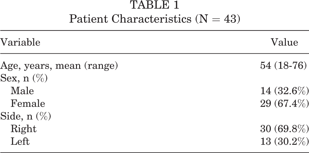
From February to August 2020, a total of 43 consecutive patients consented and enrolled prospectively to participate in our study. The average age of the patients was 54 years (range, 18-76 years), with the majority of the patients being female (67%) and the majority of the procedures being performed on the right shoulder (70%) (Table 1).
Patient Characteristics (N = 43)
The type of surgical procedures included in this analysis were manipulation under anesthesia (4.7%), revision shoulder arthroplasty (2.3%), open shoulder nonarthroplasty (subpectoral biceps tenodesis; 4.7%), shoulder arthroplasty (20.9%), and arthroscopic shoulder procedures (67.4%) (Table 2).
Types of Surgical Procedures Performed a

a aTSA, anatomic total shoulder arthroplasty, HA, hemiarthroplasty; rTSA, reverse total shoulder arthroplasty, SLAP, superior labral anterior to posterior.
Of the 43 cultures taken from prepped skin (study pens), 1 was positive for P. acnes (2.3%) (Table 3). With the positive control samples (pens that touched unprepared skin), 2 were positive for Gram-positive bacilli (no speciation could be obtained for this sample) and P. acnes (4.7%) (Table 3). With the negative control samples (pens that were cultured directly from the packaging), no samples were positive for any growth (0%) (Table 3). None of the samples showed any growth at 4 days, whereas study and positive control pens required the full 21 days to demonstrate growth. The culture-positive rate for study pens (2.3%) was not significantly different when compared with the culture-positive rate for positive controls (4.7%) or negative controls (0%) (P ≥ .99 for both).
Of the 43 Patients Prospectively Enrolled, Each Provided 3 Sets of Aerobic and Anaerobic Cultures

a All study pens were sampled after preparation of the surgical site, positive control pens were sampled before preparation of the surgical site, and negative control pens were sampled straight from the packaging without patient contact.
Discussion
This study failed to reject the null hypothesis as there was no statistical difference between study pens (prepped skin) to positive control pens (unprepped skin) and negative control pens (straight from packaging). From the 43 patients, 1 study pen sample was reported positive for bacterial growth (2.3%). Two positive control samples reported to be positive for bacterial growth (4.7%), while no negative control samples reported to be positive for bacterial growth (0%). The 3 positive samples included P. acnes (2) and Gram-positive bacilli (1) that could not be evaluated further for speciation. All bacterial growth was found with an extended culture protocol of 21 days, whereas no bacterial growth was found at 4 days for any sample. From a positive culture rate of 2.3% for the study pens (1 of 43 cases), a post hoc analysis found that over 800 cases would have been needed to achieve 80% power. Nonetheless, this study demonstrates cutaneous microorganisms of the shoulder can be cultured from a surgical marking pen and, thus, the surgical marking pen utilized to outline incisions and anatomic landmarks should be discarded and not retained on the sterile field for intraoperative use. This surgical marking pen may now serve as a potential source for bacterial contamination with shoulder surgery, both open and arthroscopic, despite appropriate cleansing protocols. Furthermore, as none of the negative control marking pens (from packaging) were positive for any growth, the bacteria cultured most likely represents contamination from the patient’s skin and not from the pen itself.
One prior study has evaluated surgical marking pens as a potential contamination source for orthopaedic-related procedures. 13 Ridley et al 13 cultured surgical marking pens from arthroscopic assisted anterior cruciate ligament reconstructions. Despite a presurgical wash and surgical prepping protocols, Ridley et al 13 found a 15% (3/20) culture-positive rate for study pens in their study. Furthermore, these authors found Staphylococcus growth with all 3 positive samples, while only 1 sample was positive for Staphylococcus and bacillus growth. 13 Compared with the current study, our results not only have a lower culture-positive rate for study pens but also a different bacterial growth pattern (P. acnes and Gram-positive bacilli). This can be explained by the different natural flora that exists surrounding the knee and shoulder.
In addition, there were different presurgical washing and surgical preparation protocols utilized in these studies. Other studies have evaluated cultures from deep capsular tissue as well as the freshly cut dermal edge during open shoulder surgery; however, these studies do not evaluate the marking pen as a source of bacterial contamination. 9 –12
The first limitation to this study is the small sample size. Despite a power analysis based on the study Ridley et al 13 , the culture-positive rate in this study was not as high as anticipated (15% vs 2.3%) and, therefore, a larger sample (>800 vs 40) would have been required to obtain an appropriate power. However, despite the limited sample size, the study results indicate that a sterile marking pen used for shoulder surgery can be a potential fomite and, as with Ridley et al 13 , the findings of our study were not limited to the knee. Furthermore, we acknowledge that the vast majority of P. acnes bacteria reside in the dermis rather than the epidermis, where our cultures were taken. Many studies have accounted for this by obtaining cultures within a freshly incised dermal edge. 10–11 However, we elected not to obtain cultures within the dermis, as our investigation was to determine if surgical marking pens could be a potential source of bacterial contamination. As surgical marking pens are used for outlining incisions and anatomical landmarks on the skin surface, sampling the epidermis was most appropriate.
Conclusion
Sterile surgical marking pens (study pens from prepped skin) used to plan incisions and to outline anatomic landmarks were not found to have a higher culture-positive rate compared with positive (unprepped skin) and negative (from packaging) control pens as this study failed to reject the null hypothesis. However, this finding should be confirmed with a larger sample size, which might provide stronger evidence for the frequency of sterile surgical marking pens that are cultured with bacterial growth. Although there can be many different sources of bacterial contamination, such as surgical instruments, as these sterile surgical marking pens are fairly inexpensive ($0.76 at our institution), as a precaution they should be discarded after their use on the skin surface and not placed in the sterile field. Additional sterile surgical marking pens should be used for intraoperative purposes, such as marking grafts as with superior capsular reconstructions or tendon transfers, awls for the placement of anchors for rotator cuff repairs, or humeral-sided implants for version and rotation with shoulder arthroplasty.
Footnotes
Final revision submitted June 7, 2021; accepted August 18, 2021.
One or more of the authors has declared the following potential conflict of interest or source of funding: K.M. has received hospitality payments from Zimmer Biomet. M.S. has received education payments from Arthrex and Pinnacle and hospitality payments from Arthrex, Medwest, and Zimmer Biomet. AOSSM checks author disclosures against the Open Payments Database (OPD). AOSSM has not conducted an independent investigation on the OPD and disclaims any liability or responsibility relating thereto.
Ethical approval for this study was waived by McLaren Health Care.