
Abstract
Background:
Quantifying native cartilage thickness in pediatric and adolescent knees can help match donor and recipient sites for articular cartilage restoration procedures such as osteochondral autograft transplantation (OATS) and osteochondral allograft transplantation (OCA).
Hypothesis/Purpose:
The purpose of the current study was to quantify articular cartilage thickness in pediatric and adolescent knees using magnetic resonance imaging (MRI). We hypothesized that cartilage thickness is inversely correlated with skeletal maturity and age.
Methods:
One hundred and twenty MRI scans were evaluated in a cohort of patients 9 to 18 years old without osteochondral lesions, chondral wear or pathology, intraarticular fractures, history of knee surgery, or inflammatory arthropathy. Measurements of articular cartilage thickness at the medial femoral condyle, lateral femoral condyle, lateral trochlea, and patella were made on axial, coronal, and sagittal MRI scans (
Results:
On the femur, cartilage was thickest at the lateral trochlea with mean articular thickness of 4.2 ± 1.4 mm in males and 3.6 ± 1.3 mm in females (p=0.015) (
Conclusion:
There is a strong inverse association between increasing age and cartilage thickness of the femoral condyles and lateral trochlea. In particular, pediatric knees demonstrate relatively thick cartilage at the lateral trochlea that decreases with age. This information will help surgeons understand recipient site anatomy and identify appropriate donor site tissue for articular cartilage restoration procedures such as OATS and OCA in children and adolescents.
Tables:
Mean articular cartilage thickness of the femur and patella

| Combined (n=120) | Male |
Female |
p value | |
|---|---|---|---|---|
| Medial femoral condyle | 3.2 ± 1.0 | 3.5 ± 1.0 | 2.9 ± 0.8 | 0.001 |
| Lateral femoral condyle | 2.8 ± 0.7 | 2.9 ± 0.7 | 2.7 ± 0.7 | 0.056 |
| Lateral trochlea | 3.9 ± 1.4 | 4.2 ± 1.4 | 3.6 ± 1.3 | 0.015 |
| Median ridge of patella | 4.2 ± 0.9 | 4.4 ± 0.9 | 4.1 ± 0.9 | 0.080 |
| Medial facet of patella | 4.7 ± 1.0 | 4.8 ± 1.0 | 4.6 ± 0.9 | 0.320 |
| Lateral facet of patella | 4.6 ± 0.8 | 4.7 ± 0.8 | 4.5 ± 0.8 | 0.185 |
Linear regression of articular cartilage thickness of the femur and patella by age

| R2 | B | p value | |
|---|---|---|---|
| Medial femoral condyle | 0.63 | 6.1 | <0.001 |
| Lateral femoral condyle | 0.64 | 4.9 | <0.001 |
| Lateral trochlea | 0.68 | 8.2 | <0.001 |
| Median ridge of patella | 0.02 | 4.8 | 0.090 |
| Medial facet of patella | 0.04 | 5.5 | 0.032 |
| Lateral facet of patella | 0.001 | 4.5 | 0.741 |
Figures:

Measurements of patellofemoral articular cartilage thickness
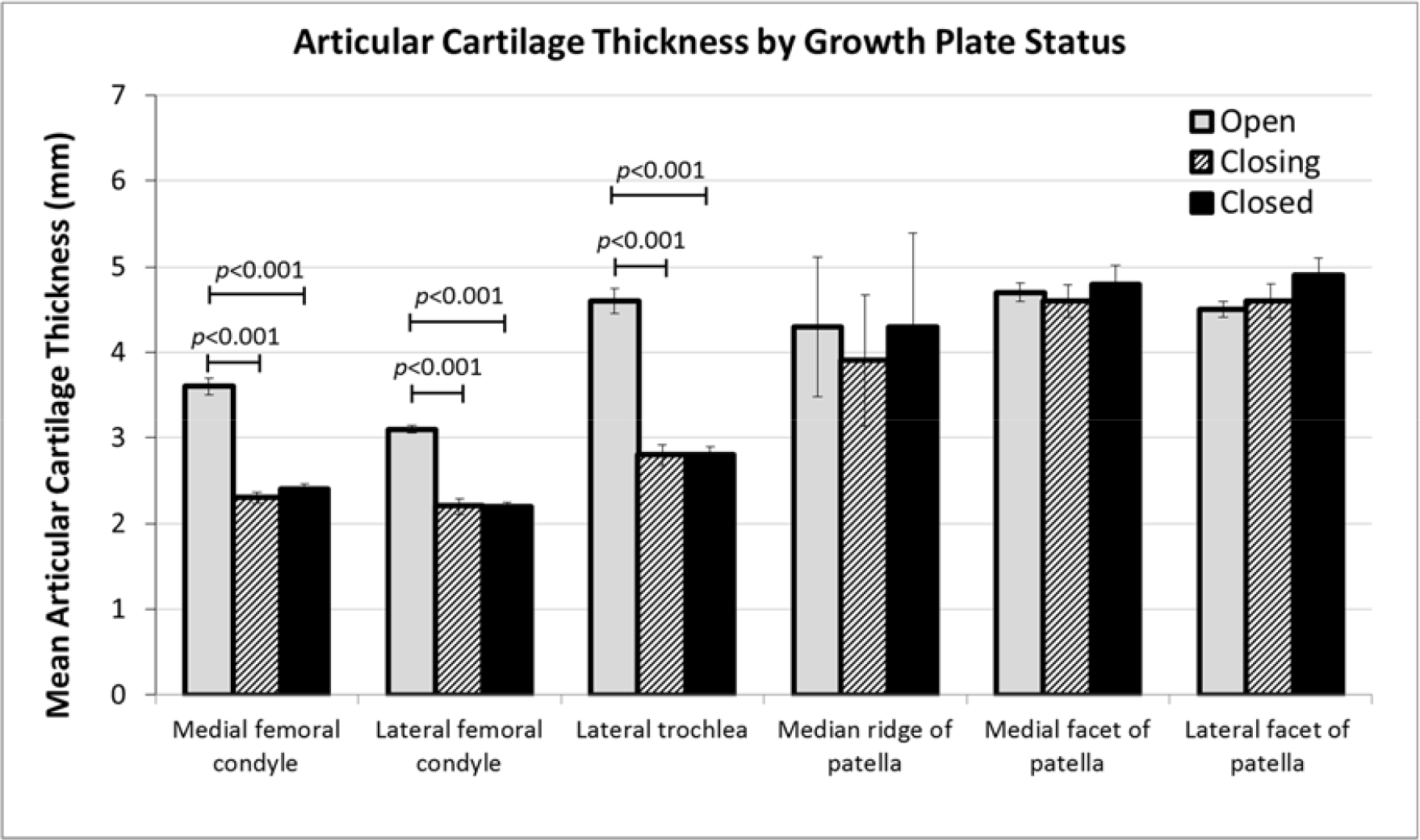
Mean articular cartilage thickness of the femur and patella by growth plate status

Mean articular cartilage thickness of the femur by age

Mean articular cartilage thickness of the patella by age