
Abstract
Background:
Cartilage lesions of the patellofemoral joint constitute a frequent abnormality. Patellofemoral conditions are challenging to treat because of complex biomechanics and morphology.
Purpose:
To develop a consensus statement on the functional anatomy, indications, donor graft considerations, surgical treatment, and rehabilitation for the management of large chondral and osteochondral defects in the patellofemoral joint using a modified Delphi technique.
Study Design:
Consensus statement.
Methods:
A working group of 4 persons generated a list of statements related to the functional anatomy, indications, donor graft considerations, surgical treatment, and rehabilitation for the management of large chondral and osteochondral defects in the patellofemoral joint to form the basis of an initial survey for rating by a group of experts. The Metrics of Osteochondral Allografts (MOCA) expert group (composed of 28 high-volume cartilage experts) was surveyed on 3 occasions to establish a consensus on the statements. In addition to assessing agreement for each included statement, experts were invited to propose additional statements for inclusion or to suggest modifications of existing statements with each round. Predefined criteria were used to refine statement lists after each survey round. Statements reaching a consensus in round 3 were included within the final consensus document.
Results:
A total of 28 experts (100% response rate) completed 3 rounds of surveys. After 3 rounds, 36 statements achieved a consensus, with over 75% agreement and less than 20% disagreement. A consensus was reached in 100.00% of the statements relating to functional anatomy of the patellofemoral joint, 88.24% relating to surgical indications, 100.00% relating to surgical technical aspects, and 100.00% relating to rehabilitation, with an overall consensus of 95.5%.
Conclusion:
This study established a strong expert consensus document relating to the functional anatomy, surgical indications, donor graft considerations for osteochondral allografts, surgical technical aspects, and rehabilitation concepts for the management of large chondral and osteochondral defects in the patellofemoral joint. Further research is required to clinically validate the established consensus statements and better understand the precise indications for surgery as well as which techniques and graft processing/preparation methods should be used based on patient- and lesion-specific factors.
Cartilage lesions of the patellofemoral joint are particularly common; however, surgery is not indicated in many cases, especially when they are asymptomatic. 11,20,26,58 Chondral and osteochondral injuries of the patellofemoral joint present several challenges for their treatment, as the morphology of the patella and trochlea can have wide variability between patients. In addition to its unique anatomy, the patellofemoral joint also experiences very high loads with daily activities, including 1.3 times the body weight (BW) during level ambulation, 3.3 times BW during stair ambulation, 5.6 times BW during running, and up to 7.8 times BW during a deep knee bend or squat. 28 Other factors that add additional complexity include excessive patellar malalignment, maltracking, patella alta or baja, and trochlear dysplasia, among others. 3,4,12,26,32,59
When indicated, multiple surgical options exist for large symptomatic defects. Matrix-induced autologous chondrocyte implantation (MACI) and osteochondral allograft transplantation (OCA) remain the most commonly utilized methods of treatment. 12,45 These treatments are used routinely in other compartments of the knee; however, the morphology, alignment, and biomechanics of the patellofemoral joint make some of these techniques particularly challenging. For example, the complex topography and variable anatomy of the patella and trochlea make OCA technically more demanding than autologous chondrocyte implantation (ACI) because of the need to match graft morphology to patient anatomy. As a result of this complexity, inferior outcomes have been reported with the treatment of symptomatic large patellofemoral lesions when compared with tibiofemoral cartilage treatments. 12,24 In addition, clinical studies, especially clinical trials, very often focus on the tibiofemoral joint, and the literature on cartilage restoration in the patellofemoral joint is much more limited. 24,40,80 For those reasons, significant debate remains regarding the indications and specifics of each technique for the surgical treatment of patellofemoral chondral defects.
Despite the growing body of literature on this topic, a standardized algorithm is lacking, leading to persistent controversy in the surgical treatment of large symptomatic patellofemoral cartilage injuries. In such instances, an expert consensus can be synthesized using a modified Delphi method. This allows for the development of a group-based consensus. The Delphi method provides several advantages over other group-based processes, including the preservation of participant anonymity that can reduce the effects of dominant participants. 39 Additionally, Delphi consensus statements conducted at a distance have been demonstrated to be as reliable as face-to-face panels, 93 with further advantages of greater participant flexibility. 42 For the abovementioned reasons, the purpose of this study was to develop a Delphi consensus statement on the functional anatomy, indications, donor graft considerations, treatment, and rehabilitation for the management of large chondral and osteochondral defects in the patellofemoral joint.
Methods
Study Design
A working group of 4 individuals (J.C., B.B.H., A.B.Y., J.F.) was responsible for facilitating the development of a consensus using modified Delphi techniques as previously described. 14 A comprehensive list of statements was generated under 5 categories: functional anatomy, surgical indications, donor graft considerations for osteochondral allografts, surgical technical aspects, and rehabilitation concepts for the management of large chondral and osteochondral defects in the patellofemoral joint. The Metrics of Osteochondral Allografts (MOCA) expert group was surveyed on 3 occasions to establish a consensus on the inclusion/exclusion of each statement.
Identification of Statements for Inclusion in the First-Round Survey
Potential statements for inclusion in the first-round survey were prepared by the working group on the basis of recently published studies, including systematic reviews and meta-analyses of patellofemoral joint cartilage treatment. 15,35,69,80 Online surveys were generated to allow respondents to vote whether statements should be included in an expert consensus document relating to the management of large chondral and osteochondral defects in the patellofemoral joint. There were 5 possible responses on a Likert 53 scale including “strongly agree,” “agree,” “neither agree nor disagree,” “disagree,” or “strongly disagree.” A free-text comment section was included to allow for suggested modifications or additional statements. The survey was piloted by 3 experts for face validity, understanding, and acceptability, resulting in minor modifications.
Establishing Consensus Using Delphi Methods
Delphi methods were used to establish a group consensus on whether statements should be included in an expert consensus document relating to the management of large chondral and osteochondral defects in the patellofemoral joint. 82 A total of 28 experts were included based on their comprehensive and authoritative knowledge of the topic, having a known clinical practice that utilizes cartilage restorative procedures for the patellofemoral joint (minimum of 30 cartilage cases per year), and having frequently published and/or lectured on the topic (>10 publications on patellofemoral chondral injuries). Experts were part of a previously established group (2016) of osteochondral allograft experts (MOCA group).
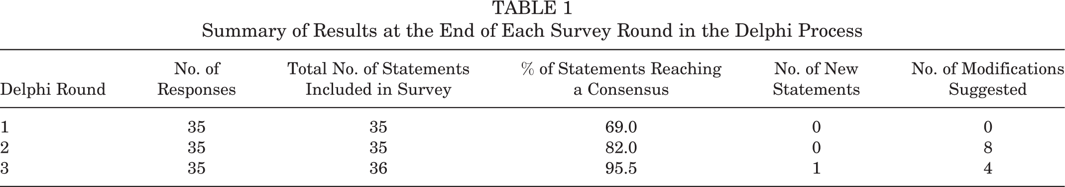
Experts participated in 3 rounds of surveys between February and April 2019 (Table 1). First-round surveys were analyzed, and participants were sent an anonymized summary of the results together with a second survey. In round 1, statements were categorized as “essential” and retained for round 2 if over 70% of respondents agreed and fewer than 20% disagreed. Statements not meeting these criteria were discarded or modified according to rater suggestions. The second-round survey also included any new statements suggested by experts in round 1. In round 2, participants were asked to rescore the statements and provide free-text comments. In round 2, responses were analyzed, retaining statements if over 70% of respondents agreed on their inclusion and fewer than 20% disagreed. Statements retained after round 2 were considered in round 3. Questionnaires were reanalyzed and the cycle repeated in round 3. For a consensus, defined a priori, statements were included in the final consensus document if over 75% of respondents agreed and fewer than 20% disagreed in the third-round Delphi survey (Table 2 and Figure 1).
Summary of Results at the End of Each Survey Round in the Delphi Process
Levels of Agreement and Disagreement in the Statements Included in the Third-Round Survey a

a ACI, autologous chondrocyte implantation; OCA, osteochondral allograft transplantation.
b Statements not reaching a consensus.
c Any implantation of cultured chondrocytes in a membrane; this includes all generations and membrane compositions such as Carticel, matrix-induced autologous chondrocyte implantation, ChondroCelect, Hyalograft C, and NeoCart, among others.

Stacked leaning bar chart representing the breakdown in agreement levels in the third-round Delphi survey. Bars to the left of the y-axis indicate disagreement, with bars to the right indicating agreement.
Discussion
The most important finding of this study was that a consensus among high-volume experts on the management of patellofemoral chondral injuries was reached on the majority of the statements (95.5%). Surgical indications were the most controversial topic with the highest disagreement reported. Specifically, the Delphi method failed to reach a consensus for indications for OCA versus ACI. This is most likely because of the lack of clinical studies comparing the 2 techniques, which results in surgeons’ decisions being based largely on personal experience. However, overall strong agreement was reached for all categories including functional anatomy of the patellofemoral joint, surgical indications, donor graft considerations for osteochondral allografts, surgical technical aspects, and rehabilitation concepts.
Functional Anatomy
The experts felt that the patella and trochlea were both high-loading (weightbearing) components of the knee. To this point, Flynn and Soutas-Little 28 reported forces of 1.3 times BW during level ambulation, 3.3 times BW during stair ambulation, 5.6 times BW during running, and up to 7.8 times BW during a deep knee bend or squat. As a significant weightbearing surface, these symptomatic lesions often require an intervention. However, given the unique mechanics and morphology, surgical techniques have greater technical demand when applied to the patellofemoral joint compared with the femoral condyles.
Indications
As loading areas, surgical treatment of symptomatic chondral defects in the patellofemoral joint should be considered when nonoperative measures fail. While the majority of experts agreed on most indication statements, 2 statements did not achieve agreement (indications of ACI vs OCA for the treatment of certain chondral defects). Several factors (cause, age, location, concomitant abnormalities, range of motion, and patient expectations) were considered important when deciding the timing and indications for surgery, even though the literature is conflicting on how those variables affect outcomes. 15 Evidence suggests that age, cause, and location likely do not affect outcomes, while female sex, lesions in the patella, large lesions, and bipolar chondral defects potentially lead to less optimal results. ¶ Other factors such as concurrent malalignment, maltracking, and patellar instability should be carefully evaluated during the workup of patients with chondral lesions, as they were deemed of utmost importance by the expert panel (they should be corrected before or at the time of the cartilage repair procedure). The importance of correcting coexisting abnormalities and anatomic abnormalities was initially highlighted by Peterson et al. 76 These authors reported on 224 patients who underwent ACI, recognizing that patients with patellar lesions had less than optimal outcomes when compared with patients with lesions in the femoral condyles. 11,74 However, when selective tibial tuberosity osteotomy was performed, the improvement in patients with patellofemoral lesions was similar to that in patients with lesions in the femoral condyles. 76 Several authors later reported comparable results, acknowledging the importance of correcting maltracking before or concurrently with the cartilage procedures. 35,46,81,85,89
Both ACI and OCA were considered valid treatments for large chondral defects in the patella and trochlea except in cases of end-stage osteoarthritis and/or restricted range of motion (<100°-110°). While end-stage arthritis is a contraindication to cartilage repair, subtotal loss or bipolar lesions without significant joint space narrowing can be treated with ACI or OCA, especially in young patients. # The addition of unloading osteotomy can reduce joint surface pressures up to 30% and is recommended when treating those patients with bipolar lesions. 7,35,78 Even though the sandwich technique has been described in the treatment of bone lesions concomitantly with ACI, there was no agreement on its utilization, and OCA was preferred for cases that include intralesional osteophytes, revision including prior failed microfracture, prior fracture malunion, significant subchondral stress reactions also known as “bone edema,” significant subchondral cyst formation, and bone loss due to fractures or osteochondritis dissecans fragment excision. 21,31,36,38,60,62,75,79
In the patellofemoral joint, morphology matching makes OCA technically more demanding than ACI. This is because of the complex topography, with highly variable shapes of the patella and trochlea. Furthermore, patients with patellofemoral cartilage lesions often have different morphology when compared with controls and potential donors. 56 This complicated morphology matching is more pronounced with the involvement of the central trochlear groove and median patellar ridge. Still, there was no agreement that ACI is preferred in those situations. With that being said, while OCA graft matching can be challenging in the patellofemoral joint, a consensus did not favor ACI in this setting.
Graft and Surgical Technique Considerations
A consensus was reached in multiple surgical technical aspects and on donor graft considerations for osteochondral allografts. When performing OCA, grafts from the same location are preferred, and matching can be performed by radiography with a sizing marker or magnetic resonance imaging. 8,19,23,47 Location matching has been reported to improve the congruence between the graft and recipient surfaces. 86
Experts agreed that chondrocyte viability is crucial to graft survival. To this point, the preservation technique (fresh grafts have greater chondrocyte viability than cryopreserved grafts), 19,47 timing (implantation in <28 days), 2,6,95 and technique of implantation (avoiding impaction) 9,71 can significantly affect chondrocyte viability. In this regard, higher impact loads may be encountered when the graft is thicker than 10 mm and when there is a >2-mm difference between the graft and recipient hole, and thus, thinner plugs are now recommended that match the depth of the socket. 71,91 Laboratory studies demonstrated that high-impact loads adversely affect cell viability, with less than 50% to 70% of the cells remaining viable in that setting. 71,91 The load of impact has a larger influence on chondrocyte death than the number of impacts. Thus, multiple low-load taps are preferred over single high-load taps if impaction cannot be avoided. 94 Therefore, the goal is to obtain a 6- to 10-mm graft that is flush with the cartilage surface and in contact with the bottom of the recipient hole, regardless of subchondral bone matching. ** This method decreases the subsidence of the graft and results in better restoration of the contact pressure in that compartment. 49
Another subject of debate in the 3 rounds of the Delphi consensus was the treatment of uncontained lesions in the patellofemoral joint. Experts stated that patellofemoral chondral injuries can be treated with ACI and OCA with appropriate modifications to ensure stability. These can be performed with transosseous sutures or anchors in ACI; however, MACI might not need any additional fixation if the uncontained portion is small. 25,34,35 In certain situations, OCA might need additional fixation when the graft is unstable. 10,13,37 Headless metal screws or absorbable internal fixation materials can be used for fixation, with the acknowledgment that metal screws will need to be removed once the OCA site has healed. 13,37 Other surgical technical aspects that had agreement were that dowels are preferred compared with shell grafts when possible 15 and that pulse lavage should be used because it may decrease the concentration of bone marrow elements. 44
Rehabilitation
Last, a sequential, staged rehabilitation program (range of motion, muscular endurance, strength, and power) was felt to be essential for a successful outcome among experts. 65 In early phases, a comprehensive patellar and tibiofemoral mobilization protocol is safe and should be implemented to avoid arthrofibrosis. 13,37,63 Notably, progressive weightbearing as tolerated with a knee brace locked in full extension does not excessively load the patellofemoral joint and is therefore considered safe if no associated osteotomy is performed. 35,55,77
This expert consensus statement fulfills established criteria for the reporting of Delphi studies 22 using a validated number of experts. 1 The 100% response rate across all 3 survey rounds highlights the commitment of these experts to establish a consensus on the management of patellofemoral chondral injuries. The Delphi technique has additional strengths as well as preserving participants’ anonymity and therefore shields participants from more influential expert opinions. Furthermore, the potential influence of any single participant was reduced by including more experts than most published Delphi studies. Last, the level of agreement required for final inclusion was higher than most health care Delphi studies 22 to ensure that only statements supported by over 95% of experts were included. Nevertheless, this study is not without limitations. As with any other consensus statement, although the statements were created from a review of the literature, the modifications and suggestions presented are not directly derived from data but from expert opinions. Additional research, including clinical outcomes data, is required to validate this consensus statement.
Conclusion
This study established a strong expert consensus document relating to the functional anatomy, surgical indications, donor graft considerations for osteochondral allografts, surgical technical aspects, and rehabilitation concepts for the management of large chondral and osteochondral defects in the patellofemoral joint. Further research is required to clinically validate the established consensus statements and better understand the precise indications for surgery as well as which techniques and graft processing/preparation methods should be used based on patient- and lesion-specific factors.
Footnotes
Notes
Final revision submitted November 24, 2019; accepted December 3, 2019.
One or more of the authors has declared the following potential conflict of interest or source of funding: J.C. has received research support and consulting fees from Arthrex, Conmed, and Smith & Nephew. B.B.H. has received research support from Arthrex and educational support from Arthrex and Elite Orthopaedics. A.B.Y. has received research support from Arthrex, Orthogenesis, Medwest, and Vericel; educational support from Arthrex, Medwest, and Smith & Nephew; and consulting fees from Aastrom Biosciences, JRF Ortho, Olympus, PatientIQ, Smith & Nephew, and Sparta Biomedical. J.F. has received consulting fees from Osiris Therapeutics, Zimmer Biomet, ISTO Technologies, Ceterix Orthopaedics, Biomet, Genzyme, MedShape, and Sanofi-Aventis; has received speaking fees from Arthrex and Zimmer Biomet; has received royalties from Arthrex, DePuy, Organogenesis, Springer, and Thieme Medical Publishers; and has stock/stock options in MedShape and Ortho Regenerative Technologies. W.D.B. has received consulting fees from Arthrex, DePuy, Insight Medical, JRF Ortho, OrthAlign, and Smith & Nephew; has received royalties from DePuy, Smith & Nephew, and Zimmer Biomet; and has stock/stock options in Insight Medical, Moximed, and OrthAlign. B.J.C. has received research support from Aesculap/B. Braun, Arthrex, and Regentis Biomaterials; has received educational support from Medwest; has received consulting fees from Acumed, Anika Therapeutics, Arthrex, Bioventus, Flexion Therapeutics, Geistlich Pharma, Genzyme, Pacira Pharmaceuticals, Regentis Biomaterials, Smith & Nephew, Vericel, and Zimmer Biomet; has received speaking fees from Arthrex, Carticept Medical, LifeNet Health, and Pacira Pharmaceuticals; has received hospitality payments from GE Healthcare; has received royalties from Arthrex, DJO, Elsevier, and Operative Techniques in Sports Medicine; and has stock/stock options in BandGrip, Ossio, and Regentis Biomaterials. D.C.C. has received research support from Histogenics, JRF Ortho, Moximed, and Zimmer Biologics and consulting fees from Arthrosurface, DePuy, Histogenics, JRF Ortho, and Moximed. J.E.F. has received research support from Arthrex and Smith & Nephew; educational support from Linvatec, Peerless Surgical, and Zimmer Biomet; and consulting fees and speaking fees from Smith & Nephew. A.G. has received research support from Aesculap/B. Braun, Arthrex, DePuy, Eupraxia Pharmaceuticals, Musculoskeletal Transplant Foundation, Ossur, and Smith & Nephew; consulting fees from Collagen Solutions, Olympus, Ossur, and Smith & Nephew; speaking fees from Conmed Linvatec, Ossur, and Smith & Nephew; and royalties from Smith & Nephew. A.H.G. has received research support from JRF Ortho and Vericel; consulting fees from Aesculap, Geistlich Pharma, Genzyme, JRF Ortho, Moximed, Smith & Nephew, and Vericel; speaking fees from Aastrom Biosciences, LifeNet Health, and Vericel; and royalties from Organogenesis. S.G. has received educational support from Arthrex and Goode Surgical and honoraria from Vericel. A.E.G. has received consulting fees, speaking fees, and royalties from Zimmer Biomet and has stock/stock options in Intellijoint Surgical. D.G.J. has received research support from Genzyme and Sanofi-Aventis; has received educational support from Arthrex; has received consulting fees from Aastrom Biosciences, Acumed, Amniox Medical, DePuy/Medical Device Business Services, Flexion, Genzyme, Vericel, and Zimmer Biomet; has received speaking fees from Arthrex, Conmed Linvatec, Flexion, Genzyme, Mitek, Musculoskeletal Transplant Foundation, TissueTech, Vericel, and Zimmer Biomet; and is a board or committee member of the Musculoskeletal Transplant Foundation. A.J.K. has received research support from Aesculap/B. Braun, Arthrex, Ceterix Orthopaedics, and Histogenics; has received consulting fees from Arthrex, DePuy, JRF Ortho, and Vericel; has received speaking fees from Arthrex; and is a board or committee member of the Musculoskeletal Transplant Foundation. C.L. has received honoraria from Arthrosurface; consulting fees from Aastrom Biosciences, Genzyme, JRF Ortho, Samumed, Sanofi-Aventis, Vericel, and Zimmer Biomet; and speaking fees from Vericel. B.R.M. has received consulting fees from Arthrex, BioMarin Pharmaceutical, DePuy, and Exactech; speaking fees from Arthrex; and royalties from Arthrex. R.M. has received research support from Arthrex and DJO, has received educational support from Arthrex, has received speaking fees from Arthrex, has received royalties from Zimmer Biomet, and has stock/stock options in AlignMed. T.S.M. has received research support from JRF Ortho, educational support from Arthrex, consulting fees from JRF Ortho and MicroAire Surgical Instruments, and speaking fees from JRF Ortho. J.D.P. has received research support from Ossur, Sanofi-Aventis, and Vericel; consulting fees from JRF Ortho; and speaking fees from DePuy. M.T.P. has received consulting fees from Arthrex and JRF Ortho (AlloSource), speaking fees from Arthrex, and royalties from Arthrex and SLACK. S.A.R. has received consulting fees from Flexion, has received speaking fees from Smith & Nephew, has received honoraria from Fidia Pharma, has received royalties from Zimmer Biomet, and has stock/stock options in Ortho Regenerative Technologies. O.S. has received research support from DePuy and consulting fees from Zimmer Biomet. S.L.S. has received consulting fees and research support from Arthrex Inc; is a paid consultant for Ceterix Orthopaedics, CONMED Linvatec, Flexion Therapeutics, GLG Consulting, JRF Ortho, Moximed, Olympus, Vericel, Linvatec, and RTI Surgical; has received hospitality payments from Linvatec and Smith & Nephew; and has received a grant (indirect) from DJO. E.D.S. has received research support from CartiHeal, Fidia Pharma, and NuTech; consulting fees from Arthrex, Fidia Pharma, Flexion, JRF Ortho, Organogenesis, Smith & Nephew, and Vericel; speaking fees from Arthrex, Organogenesis, and Vericel; and royalties from Jaypee Brothers Medical Publishers. C.J.W. has received consulting fees from Arthrosurface and Smith & Nephew and speaking fees from Arthrex, Arthrosurface, and Smith & Nephew. R.J.W. has received research support from Histogenics; has received consulting fees from Arthrex, JRF Ortho, and Lipogems; has received speaking fees from Arthrex; has received royalties from Arthrex; and has stock/stock options in CyMedica Orthopedics, Gramercy Extremity Orthopedics, Pristine Surgical, and RecoverX. AOSSM checks author disclosures against the Open Payments Database (OPD). AOSSM has not conducted an independent investigation on the OPD and disclaims any liability or responsibility relating thereto.