
Abstract
Background:
Passive glenohumeral range of motion may be characteristically limited to specific shoulder pathologies. While pain associated with loss of range of passive external glenohumeral rotation is recognized as a salient feature in adhesive capsulitis, restriction of glenohumeral range of motion in calcific tendinitis of the supraspinatus tendon has never been studied.
Hypothesis:
On the basis of clinical observation, we hypothesized that calcific tendinitis of the supraspinatus tendon is associated with loss of passive glenohumeral abduction without loss of external rotation.
Study Design:
Cohort study; Level of evidence, 3.
Methods:
Ranges of passive glenohumeral rotation and abduction, which are measured with a standardized protocol in our institution, were retrospectively reviewed and compared for patients diagnosed with either adhesive capsulitis or calcific tendinitis of the supraspinatus tendon. A total of 57 patients met the inclusion criteria for the calcific tendinitis, and 77 met the inclusion criteria for the adhesive capsulitis group.
Results:
When compared with the contralateral, unaffected shoulder, glenohumeral abduction in the calcific tendinitis group was restricted by a median of 10° (interquartile range [IQR], –20° to –5°) as opposed to glenohumeral external rotation, which was not restricted at all (median, 0°; IQR, 0° to 0°). The adhesive capsulitis group showed a median restriction of glenohumeral abduction of 40° (IQR, –50° to –30°) and a median restriction of passive glenohumeral external rotation of 40° (IQR, –60° to –30°).
Conclusion:
Calcific tendinitis of the supraspinatus does not typically cause loss of external rotation but is frequently associated with mild isolated restriction of abduction. This finding can be used to clinically differentiate adhesive capsulitis from calcific tendinitis.
Keywords
Certain pathologic conditions of the shoulder have distinctive clinical features. Adhesive capsulitis (frozen shoulder) is one of these conditions, wherein underlying anatomic changes in the capsule cause global loss of glenohumeral motion that, in the absence of glenohumeral arthritis or deformity, is considered to be specific for this pathology. Adhesive capsulitis affects men and women aged between 40 and 60 years and can be associated with diabetes mellitus or thyroid dysfunctions. 11,14,21,25,28,29
The pattern of potential loss of motion in calcific tendinitis has been described as being global, although also associated with impingement features. 3,4,12,17,26 This is caused secondarily by the underlying pathology, which is metaplastic change and calcium phosphate deposition in the supraspinatus tendon and, more rarely, other tendons of the rotator cuff. Calcific tendinitis is a common cause for shoulder pain and predominantly affects women between the ages of 40 and 60 years. 1 –3,5,7,15,18,24 We observed that calcific tendinitis is often associated with free passive glenohumeral external rotation but with limitations of passive abduction in the scapular plane. We hypothesized that in the absence of glenohumeral osteoarthritis, isolated limited abduction can distinguish calcific tendinitis from adhesive capsulitis, where diffuse restrictive changes cause global range loss, most noticeable in passive external rotation and abduction. 13,20,28
Methods
Institutional ethics committee approval (No. 2016-02156) was obtained for this study. A retrospective review of patients seen between April 2014 and September 2016 in our outpatient clinic, which specializes in shoulders, identified all patients with the confirmed diagnosis of adhesive capsulitis or calcific tendinitis.
Adhesive capsulitis was diagnosed if a patient had severe shoulder pain at night and severe restriction of movements but normal findings on anteroposterior, lateral, and axillary conventional radiographs, which specifically excluded osteoarthritis or calcific tendinitis. When magnetic resonance arthrography was available (47 patients, 60.3%), 3 factors were taken as confirming evidence of adhesive capsulitis: thickening of the rotator interval tissue in the anterosuperior joint, 6,16 thickening of the capsule, and a small capsular volume.
Calcific tendinitis was diagnosed if a patient had shoulder pain and when calcific deposits were identified on anteroposterior, scapular lateral, or axillary lateral radiographs, each taken in neutral, internal, or external rotation. Patients with radiographic signs of osteoarthritis or skeletal deformity were excluded, as well as patients suspected during clinical examination of having a rotator cuff tear.
The study inclusion criteria were unilateral symptoms and a physical examination with complete documentation for passive ranges of motion and strength. Patients were excluded if they had a symptomatic contralateral shoulder (control). Prior operative treatment was also an exclusion criterion.
Ranges of motion were measured with the patient sitting. External rotation was measured by the examiner facing the patient. The forearms were grasped immediately beneath the elbow, which was flexed to 90°. The upper arms were stabilized by pressing the elbow against the anterolateral border of the body so that the upper arm was vertical and could not be abducted from the trunk. Passive external rotation was then measured after the application of an external rotation to the lower arm. The amplitude of the external rotation was measured with a goniometer 23 for both shoulders.
Passive glenohumeral abduction was measured with the examiner standing behind the sitting patient. For the right shoulder, the scapula was stabilized by pressing it to the thorax with the left hand. The arm to be examined was bent to 90° at the elbow and held in neutral rotation. With his right hand, the examiner then abducted the arm of the patient in the scapular plane without releasing the pressure of his left hand on the scapula. The passive glenohumeral abduction, which could be obtained without any change in the position in the scapula, was the measured abduction.
Data were nonnormally distributed (except for external rotation in calcific tendinitis) according to Shapiro-Wilk tests; thus, the median (interquartile range [IQR]) is given. The Wilcoxon rank-sum test was used to compare range of motion between the calcific tendinitis and adhesive capsulitis groups. The Wilcoxon signed-rank test was used to compare range of motion within patient groups, and the paired t test was used for external rotation in the calcific tendinitis group, owing to normal distribution of data. The Wilcoxon rank-sum test was used to compare unpaired continuous data. The significance level was set at 5%. Stata (v IC/13.1; StataCorp LLC) was used for analysis.
Results
According to the defined criteria, 57 patients were included into the calcific tendinitis group and 77 into the adhesive capsulitis group. The median age was 49 years (IQR, 45-59 years) for the calcific tendinitis group, with 27 men and 30 (53%) women. The median age was 53 years (IQR, 47-59) for the adhesive capsulitis group, with 30 men and 47 women (61%) (Table 1). Median glenohumeral abduction was significantly restricted in both groups (P < .001). Calcific tendinitis was associated with a median reduction of passive glenohumeral abduction of 10° (IQR, –20° to –5°) and adhesive capsulitis, a median reduction of 40° (IQR, –50° to –30°; P < .001) (Table 2 and Figure 1). Median external rotation was unaffected in the calcific tendinitis group, with a median difference from the contralateral, asymptomatic side of 0° (IQR, 0° to 0°). Conversely, external rotation was reduced by a median of 40° (IQR, –60° to –30°) in the adhesive capsulitis group (P < .001) (Table 2 and Figure 2; the different values in the text and table are due to the calculation of medians [based on nonnormal distribution] and the value of 40° was reached by calculating the median of actual differences [from 1 value; ie, symptomatic minus asymptomatic] instead of reporting the difference between both medians [from 2 values]).
Patient Demographics a

a IQR, interquartile range.
Abduction and External Rotation in Calcific Tendinitis and Adhesive Capsulitis (N = 134) a

a Data are reported as median (interquartile range). ROM, range of motion.
b Wilcoxon signed-rank test for calcific tendinitis (nonnormally distributed data) and paired t test for adhesive capsulitis (normally distributed data).
c There were missing data for external rotation in 8 patients in the calcific tendinitis group.

Abduction in calcific tendinitis and adhesive capsulitis. Values are presented as medians, interquartile ranges, 95% CIs (whiskers), and outliers.

External rotation in calcific tendinitis and adhesive capsulitis. Values are presented as medians, interquartile ranges, 95% CIs (whiskers), and outliers.
Discussion
Our study confirms that adhesive capsulitis is associated with a substantial reduction of passive glenohumeral external rotation and abduction. Calcific tendinitis is also associated with loss of passive glenohumeral abduction, albeit less, but not with external rotation. The loss of external rotation with adhesive capsulitis occurs early on in the disease and can precede other clinical symptoms. 14,20,25,27,29 However, adhesive capsulitis may develop subsequent to calcific tendinitis and is a common complication in up to 18% of patients after surgical debridement of calcium deposits. 3,10 For the diagnosis of both conditions, conventional radiographs are usually obtained. If these radiographs show calcific tendinitis and if there is a restriction of passive glenohumeral external rotation, adhesive capsulitis and calcific tendinitis must be diagnosed. Those patients were excluded in our study. Conversely, restriction of glenohumeral abduction without restriction of external rotation essentially rules out adhesive capsulitis.
Our study does not explain the cause of abduction loss in calcific tendinitis. Features of impingement were postulated as causation for calcific tendinitis and were noted in prior studies. 8,17,18 An abduction contracture was described in a case report by Takahashi and Ogawa. 22 The loss of adduction resolved with resolution of the disease, and mechanical blocking was postulated as the cause for restricted range.
A block to abduction in calcific tendinitis could be due to direct impingement from the calcifications, the edematous tendon, or inflammation within the capsule or bursa. 9,19 The pathologic changes in calcific tendinitis are most commonly found in the supraspinatus tendon, 24 and isolated abduction loss was seen in our series. In the less common variants involving the anterior or posterior rotator cuff, rotational loss could theoretically be seen but was not brought out in our study. 1 –3,5,15,18
There are limitations to our study, including the number of patients excluded because of contralateral shoulder pathology. Also, our study only recruited 2 patient populations. Our study remains clinically relevant, as the range of motion for patients with calcific tendinitis has never been quantified in the literature. While the magnitude of abduction loss was moderate, the comparison with the contralateral shoulder was significant, with all shoulders showing equal or less range in abduction. The correlation for abduction loss and global motion loss for conditions other than calcific tendinitis further support our conclusion that a loss of range in multiple planes lends to the diagnosis of another pathology than calcific tendinitis.
This study is retrospective, which is a weakness, although it does help to limit bias because data were not collected specifically for this study. Despite these limitations, it appears that clinical isolated loss of glenohumeral abduction should raise a high level of suspicion for calcific tendinitis. If ipsilateral external rotation loss is present in the examination, the diagnosis is unlikely to be isolated calcific tendinitis, whether calcifications appear on radiographs or not.
Finally, our study does not show if the pathophysiologic mechanism that causes a restriction of the range of motion is due to a block of calcification, the edematous tendon, or inflammation within the capsule or bursa.
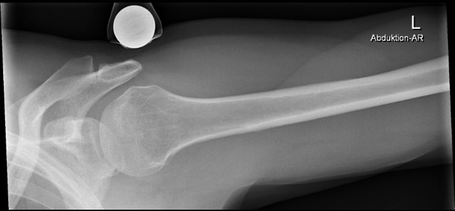
In Figures 3 to 5, we show radiographs of a patient with calcific tendinitis and the corresponding loss of abduction where the calcium deposit appears to be impinging against the glenoid.

Radiograph from a 45-year-old man with calcific tendinitis (yellow arrow) in the supraspinatus tendon.

The same 45-year-old man from Figure 3 with calcific tendinitis (yellow arrow) in the supraspinatus tendon. Radiograph in glenohumeral abduction shows a mechanical block.
The same 45-year-old man from Figure 4. Radiograph of the opposite side.
Conclusion
Adhesive capsulitis is associated with restriction of passive glenohumeral external rotation and abduction. In contrast, calcific tendinitis is not associated with loss of external rotation but is associated with isolated mild restriction of abduction.
Footnotes
The authors declared that they have no conflicts of interest in the authorship and publication of this contribution.
Ethical approval for this study was obtained from the Kantonale Ethikkommission Zürich (study No. BASEC Nr 2016-02156).