
Abstract
Background:
The incidence of superior labral surgery has increased in the past decade in the United States, and a contributing factor could be an increased rate of superior labral tears diagnosed with magnetic resonance imaging (MRI). Prior MRI studies of the asymptomatic shoulder have focused on rotator cuff pathology or pathology in a narrow and specific group of athletes. Labral abnormalities have not previously been thoroughly evaluated in asymptomatic middle-aged individuals.
Purpose:
To evaluate the prevalence of superior labral tears diagnosed by MRI in the asymptomatic shoulders of middle-aged people (age range, 45-60 years).
Study Design:
Cross-sectional study; Level of evidence, 3.
Methods:
A total of 53 asymptomatic adults (age range, 45-60 years) with no history of surgery or injury to either shoulder were included in the study. Physical examinations of all shoulders were performed. Noncontrast MRI (1.5 T) was performed in 1 randomly determined shoulder of each subject. Two fellowship-trained musculoskeletal radiologists who were blinded to the purpose of the study and ages of the subjects evaluated each MRI.
Results:
Radiologists interpreted the MRIs as consistent with superior labral tears in 55% and 72% of the cohort. Comparison of the radiological evaluations of the superior labra were moderate (κ = 0.410, P = .033). There were no differences in readings for superior labral tear regarding age (P = .87), sex (P = .41), whether the dominant shoulder underwent MRI (P = .99), whether the subject worked a physical job (P = .08), or whether the subject participated in overhead sports for a period of 1 year (P = .62).
Conclusion:
Superior labral tears are diagnosed with high frequency using MRI in 45- to 60-year-old individuals with asymptomatic shoulders. These shoulder MRI findings in middle-aged populations emphasize the need for supporting clinical judgment when making treatment decisions for this patient population.
Clinical Relevance:
To avoid overtreatment, physicians should realize that superior labral tears diagnosed by MRI in individuals between the ages of 45 and 60 years may be normal age-related findings.
Keywords
The diagnosis of superior labral tears can be challenging. Described mechanisms for the development of superior labral tears include overhead sports such as throwing and acute traumatic shoulder injuries. Physical examination tests for superior labral tears have demonstrated variable accuracies. 9,19 Regarding noncontrast magnetic resonance imaging (MRI), variable accuracy and sensitivity have been demonstrated for the diagnosis of superior labral tears. 6,7 The addition of arthrography to MRI has been reported to provide high accuracy levels for the diagnosis of superior labral tears. 2,4,14,17,34,35 However, other studies have demonstrated that noncontrast MRI and MR arthrography are neither highly sensitive nor specific for the diagnosis of superior labral anterior posterior (SLAP) lesions. 1,3,7,15,26,28
Initially thought to be an uncommon entity, the incidence of SLAP lesion surgery has increased in the United States in the past decade. 23,34,36,38 Such studies have demonstrated that a significant contingent of patients undergoing SLAP lesion surgeries were middle-aged. Since no standard and definitively accepted definition of “middle-aged” exists, this term may include people from ages 40 to 65 years. Onyekwelu et al 23 reported that the mean age for SLAP repairs in 2010 was 38.5 years in males and 44.1 years in females. Twenty-six percent of SLAP repairs were in patients older than 50 years. Weber et al 36 reported a mean age for SLAP repair in females to be 40.9 years and in males to be 36.4 years between 2003 and 2008 in American Board of Orthopaedic Surgery part II examination candidates. In a national insurance database study between 2004 and 2009, Zhang et al 38 reported that 64.6% of the SLAP lesions repaired were in patients 40 years and older. Between 2005 and 2009, Vogel et al 34 reported mean ages between 47 and 48 years annually in their analysis of a California database. Although studies report high rates of satisfactory results with SLAP lesion repair, 10,12 recent series have elucidated complications and less satisfactory results. 22,24,30,32,36 Because of this, it is important to diagnose clinically relevant superior labral tears as accurately as possible.
It is unclear why the incidence of superior labral surgeries has increased over the past decade. It is possible that increasing scientific knowledge has allowed surgeons to identify such pathology and correct it surgically when necessary in patients. It is also possible that difficulty diagnosing superior labral tears arthroscopically may influence superior labral surgery rates. Gobezie et al 13 and Wolf et al 37 elucidated this difficulty by demonstrating substantial intra- and interobserver variability for shoulder arthroscopists asked to provide diagnoses and treatment recommendations for arthroscopic videos. Furthermore, it is also possible that MRIs are being interpreted as consistent with superior labral tears more frequently. Possible reasons for this include that radiologists may be better trained to evaluate for such pathology and there may be a greater awareness of superior labral tears.
Cadaveric studies have demonstrated that detachment of the labrum from the glenoid occurs more frequently with aging. Histologically, increasingly extensive degenerative changes were noted in labra with increasing age. 25,27 These findings have not been evaluated radiologically in a middle-aged population.
The primary purpose of this study was to evaluate the frequency of superior labral tears diagnosed by MRI in the asymptomatic shoulders of people between the ages of 45 and 60 years. A secondary purpose of this study was to evaluate for other shoulder abnormalities in asymptomatic shoulders on MRI.
Methods
A total of 53 subjects without shoulder pain (age range, 45-60 years) were recruited throughout 2013 and included in the study. Subjects included patients being treated for lower extremity problems that did not require crutches, family members of patients, and some office staff. Subjects with a history of pain, instability, or injury to either shoulder were excluded. Institutional review board approval was obtained for this study. After informed consent was obtained, subjects were asked to fill out pain visual analog scales (VAS) for their shoulders. Subjects with a pain score greater than 0 were excluded.
Demographic information was documented, including patient age, sex, and dominant arm. Also, subjects were asked whether they had participated in overhead sports for a period of 1 year during the past 10 years. Subjects were also queried on whether they exert 20 to 50 pounds of force occasionally or 10 to 25 pounds of force frequently at their job at the time of the study. This coincides with the physical demands definition of “medium work” from the dictionary of occupational titles of the Department of Labor. 33 Any subject who performed a job with medium work or more at the time of study enrollment was considered to have a physical job.
A physical examination was performed on both shoulders by 1 of the authors (R.S., B.G.B., or B.L.R.). Standard examinations for range of motion, rotator cuff strength, and pain on palpation were performed. Tests performed for instability included apprehension sign, jerk test, anterior and posterior load and shift, and sulcus sign. Specific SLAP lesion tests performed included the active compression test and the crank test. Exclusion criteria included any physical examination abnormalities, refusal to participate in the study, and inability to obtain an MRI. The subjects meeting the above criteria were randomized to have 1 shoulder undergo MRI. Randomization was performed via a computer-generated randomized allocation program.
Noncontrast shoulder MRIs were performed on a GE Signa LX 1.5-T scanner (GE Healthcare) (RF coil, GE shoulder array coil; field of view, 16 cm; matrix size, 320 × 224; slice thicknesses, 3 mm with 1-mm gaps). Each shoulder MRI included T2 coronal oblique, sagittal, and axial sequences. Coronal oblique and axial proton density sequences were performed. In addition, a coronal STIR image was performed. No fat suppression was utilized.
Two musculoskeletal fellowship-trained radiologists (K.W.M., J.Y.W.) with 2 and 4 years of experience after training, respectively, evaluated the MRIs. Each radiologist was blinded to the purposes and nature of the study. They were blinded to all subject information, including age and sex. They were asked to specifically comment on the integrity of the rotator cuff, anterior labrum, superior labrum, posterior labrum, long head biceps tendon, and glenohumeral articular cartilage. They were encouraged to add any other thoughts they may have regarding the MRIs. One main indicator of a superior labral tear diagnosed by MRI was the following: If hyperintense signal intensity was present between the superior labrum and the intra-articular biceps tendon on coronal oblique images at the level of or posterior to the osseous anterosuperior pole of the glenoid, then the radiologists considered this a superior labral tear. Both radiologists used this finding as their main criterion for diagnosis of superior labral tears. They both relied more heavily on the coronal proton density images than on other sequences. A discrepancy between the radiologists regarding labral pathology interpretation involved the posterior labrum. One of the radiologists considered a tear of the posterior portion of the superior labrum to be a posterior labral tear. Thus, this radiologist counted posterior superior labral tears as superior and posterior labral tears.
The data were evaluated using descriptive statistics. Percentage agreement was calculated between the 2 raters. Correlation was also performed using Spearman correlation. The interrater agreement was calculated using the Cohen kappa coefficient. Significance was set at <.05. Evaluation was performed for associations between superior labral tears and sex, shoulder dominance, job physicality, and participation in overhead sports.
Results
Twenty-six men and 27 women between the ages of 45 and 60 years participated in the study. Only 11% of subjects had physical jobs, and only 9% participated in overhead sports (Table 1).
Patient Demographics (N = 53) a

a Data presented as n (%) unless indicated otherwise indicated.
b Two of the 4 (50%) subjects who participated in overhead sports had their dominant arm imaged.
Thirty-eight (72%) MRIs were interpreted by radiologist 1 as consistent with superior labral tears; 29 (55%) MRIs were interpreted by radiologist 2 as consistent with superior labral tears. The radiologists demonstrated moderate interrater reliability regarding their evaluations of superior labra (κ = 0.410, P = .001) (Table 2). Evaluation of the posterior labrum was the only area that the radiologists demonstrated poor interrater reliability (κ = 0.066, P = .631). The radiologists’ diagnoses regarding the anterior labrum, posterior labrum, rotator cuff, and long head biceps tendon are noted in Table 2.
Level of Agreement on MRI Evaluation for Different Anatomic Structures a

a MRI, magnetic resonance imaging; nc, cannot be calculated because there are no cases that are abnormal.
The radiologists’ MRI interpretations revealed no differences in their diagnoses of superior labral tears with regard to subject age, sex, whether the dominant arm was imaged, job physicality, or overhead sports participation (Table 3).
Evaluation Comparison of Data for Superior Labral Tears a

a Data are presented as n (%) unless otherwise indicated.
Other than the 5 anatomic areas in the shoulder that the radiologists were asked to evaluate, they commented on the following abnormalities. Radiologist 1 noted that 2 shoulder MRIs revealed rotator cuff calcific tendinopathy; radiologist 2 reported rotator cuff calcific tendinopathy in 1 of these 2 MRIs. Radiologist 1 reported a paralabral cyst in 1 of the MRIs with a reported superior labral tear; radiologist 2 noted the paralabral cyst in this same MRI as well as 3 additional MRIs.
Figures 1 and 2 are sample MRIs from this study depicting abnormal signal seen in the superior labrum consistent with a radiologist interpretation of a superior labral tear.
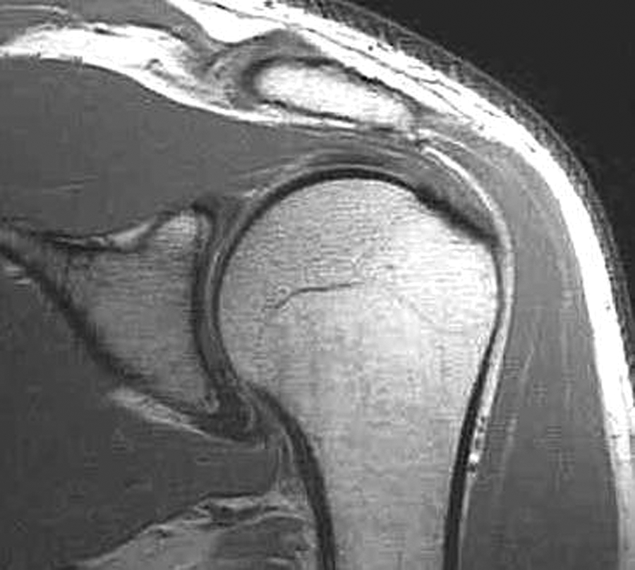
Coronal proton density magnetic resonance image depicting a left superior labral tear in a 54-year-old right hand–dominant male patient.

Coronal proton density magnetic resonance image depicting a right superior labral tear in a 53-year-old right hand–dominant female patient.
Discussion
Our study is the first to evaluate superior labra with noncontrast shoulder MRIs in 45- to 60-year-old asymptomatic people. Our study revealed that the noncontrast shoulder MRIs of 53 asymptomatic shoulders were interpreted as having superior labral tears by 2 fellowship-trained musculoskeletal radiologists at rates of 72% and 55%, respectively. One of the radiologists diagnosed a posterior labral tear in 55% of MRI examinations. Otherwise, the 2 radiologists did not find a high rate of anterior labral tears, partial-thickness rotator cuff tears, or full-thickness rotator cuff tears.
Prior published studies have reported on MRI of asymptomatic shoulders with a variety of findings. Chandnani et al 5 evaluated the shoulders of 20 asymptomatic volunteers and 20 symptomatic patients between the ages of 25 and 55 years. They reported “no abnormal morphology” regarding the glenoid labrum in any of the MRIs in the asymptomatic volunteers. However, they noted “abnormal internal signal within the labrum” in 10 of 20 shoulders in asymptomatic volunteers.
Iannotti et al 16 evaluated the sensitivity and specificity of shoulder MRI in 91 patients and 15 asymptomatic volunteers. They noted that 13 asymptomatic volunteers had normal MRIs and the other 2 revealed findings consistent with mild rotator cuff tendonitis. The ages of the asymptomatic volunteers were not noted. MRI studies in asymptomatic shoulders by Sher et al 31 and Miniaci et al 20 evaluated partial-thickness and full-thickness rotator cuff tears. Findings regarding the labrum were not reported in either study.
Several studies evaluated the labrum in more focused groups of asymptomatic subjects. Connor et al 8 evaluated the dominant and nondominant shoulders of 20 asymptomatic elite baseball and tennis players between the ages of 18 and 38 years with noncontrast 1.5-T MRI. They found a 7.5% prevalence of partial tears of the anteroinferior or superior glenoid labrum.
Miniaci et al 21 evaluated noncontrast MRIs of both shoulders of 14 asymptomatic professional baseball pitchers aged 18 to 22 years. Twenty-two of 28 shoulders imaged demonstrated signal abnormalities in the labrum. Of these 22 shoulders with abnormal labral signal, 5 (45%) of 11 throwing shoulders and 5 (45%) of 11 nonthrowing shoulders were diagnosed as having labral tears. Two (18%) labral tears were considered SLAP lesions.
Reuter et al 29 studied noncontrast shoulder MRIs in 16 symptomatic triathletes and 7 asymptomatic triathletes with mean ages of 35 and 39 years, respectively. No labral tears were identified in the MRIs of either group of triathletes. Fredericson et al 11 evaluated noncontrast MRIs of the asymptomatic dominant shoulders of 12 elite volleyball players and the right shoulders of 6 elite gymnasts. Mean age was 19 years. Fifty-eight percent of the volleyball players and 83% of the gymnasts were reported to have prominent changes in the labrum. Lesniak et al 18 evaluated 20 noncontrast MRIs and 1 MR arthrogram of the throwing shoulders of 21 asymptomatic professional baseball pitchers aged 20 to 39 years. Forty-seven percent were reported to have superior labral tears and 62% were reported to have either anterior or posterior labral tears.
Our study results regarding superior labral tears contrast with the findings in the study by Chandnani et al. 5 Their results regarding labral tears are quite different from the findings in our study. Although 50% of asymptomatic shoulders in their study revealed abnormal labral signal, none were considered to have labral tears. However, the age distribution of their asymptomatic cohort is unclear. The number of volunteers for each given age between 25 and 55 years was not reported. It is possible that the age distribution in the study by Chandnani et al may have involved volunteers on the younger end of their age range. Furthermore, their study did not include volunteers between the ages of 55 and 60 years. We believe that the possible age differences between their study and our study may explain the much higher prevalence of superior labral tears noted in our study. Another explanation for the differences in superior labral pathology may involve the evolution of technology. Although their study utilized an MRI scanner with a 1.5-T magnet, it is likely that the software has improved since their study was published in 1992. This could mean that the images with the 1.5-T strength magnet may be appreciably better than the images in the Chandnani study. Higher quality images may account for the greater amount of pathology visualized in the superior labra in our study.
Consistent with our study, several studies of asymptomatic athletes revealed large numbers of labral abnormalities. 11,18,21 In contrast with our results, other studies of asymptomatic athletes revealed few or no labral abnormalities. 8,29 Our study contrasts from these prior studies in that our study evaluated the asymptomatic shoulders of middle-aged people who are more representative of the general population than the narrow groups of athletes in the prior studies. Our unique study population compared with these previous studies’ populations likely explains the differences seen regarding labral abnormalities.
Our study has numerous strengths. Our study included a large number of asymptomatic shoulders in a specific age range of subjects who underwent physical examinations. The subjects were required to have had no prior or current symptoms in either shoulder as well as normal physical examinations. Furthermore, the subjects were questioned to determine their participation in overhead sports and their occupational demands. Also, a given subject’s shoulder selected for MRI was determined randomly to avoid any bias toward inclusion or exclusion of the dominant arm. Next, the MRIs were performed on the same 1.5-T scanner. The radiologists interpreting the MRIs were fellowship-trained musculoskeletal radiologists. These radiologists were blinded to the purposes of the study and were provided no clinical information about the subjects. This includes the sexes and ages of subjects.
Given that the MRIs were all performed on 1.5-T scanners, the data in this study may not generalize to MRI scanners with different strength magnets; 3.0-T scanners have emerged and there are still numerous scanners with magnet strengths below 1.5 T. It is very likely that the image quality from 3.0-T scanners exceeds that of lesser magnet strengths. Thus, it is possible that 3.0-T magnets might reveal a greater prevalence of signal consistent with superior labral tears. Furthermore, it is possible that the likely lesser quality images from scanners with less than 1.0-T magnets might reveal a lower prevalence of superior labral tear findings. To clarify these possibilities, our study population would need to be evaluated with MRI scanners with magnet strengths other than 1.5 T.
Our study also has a variety of weaknesses. First, the designation of the shoulders as asymptomatic relies on the integrity of the subjects’ histories. It is realized that although physical examinations were performed on both shoulders of all subjects, such examinations are imperfect in detecting all shoulder pathologies. It is possible that some subjects actually had prior shoulder problems that were not detected at the time of study enrollment. Second, our attempt to evaluate activity level of the subjects was limited to questioning regarding their involvement in overhead sports and physical demands of their jobs. Although this provides some characterization of the study population’s activity level, these assessments were not detailed or validated batteries. It is possible that subjects who were not performing physical demand jobs at the time of study enrollment may have engaged in physical jobs in the past. Also, we elected to question subjects regarding their participation in overhead sports only during the 10 years prior. Thus, subjects who participated in such sports at younger ages were not detected. This may have had an effect on the rate of superior labral abnormalities. Furthermore, the number of subjects who participated in overhead sports or whose occupations were considered physical was small. This minimizes the ability to make statistically significant comparisons among these groups. Next, all MRIs provided to the radiologists were shoulder MRIs of the asymptomatic subjects. This has the potential to introduce bias since the radiologists were not seeing a variety of pathologic changes. There were no additional MRIs provided with surgically documented pathology. However, the radiologists probably assumed that the MRIs they were reading were from symptomatic shoulders.
Another weakness is related to the radiologists’ evaluations of the posterior labra. Although the radiologists demonstrated moderate to good agreement overall in their MRI interpretations, their posterior labral evaluations demonstrated poor agreement. The explanation for this discrepancy revolves around the classification of posterior labral tears by radiologist 1. This radiologist considers posterosuperior labral tears to be posterior labral tears. Because of that, he interpreted many MRI scans as consistent with posterior labral tears that the other radiologist only considered to be superior labral tears. In addition to the discrepancy in posterior labral tear evaluations, radiologist 1 documented more pathology throughout the shoulder than radiologist 2. This could be because there exists a lack of consensus in definitions of pathologic conditions on shoulder MRIs. Furthermore, quality between radiologists and how aggressive they may interpret MRIs can differ.
Last, we were unable to confirm the radiologists’ MRI interpretations of superior labral tears. None of these subjects underwent shoulder surgery. Also, none of these subjects underwent follow-up history and physical examinations. Although it is possible that some subjects may have become or may become symptomatic at some point due to superior labral tears, it is likely that these superior labral MRI findings represent a normal aging process. Prior cadaveric studies provide support that these MRI findings may represent a degenerative phenomenon that is not always symptomatic. 25,27
Conclusion
There is a high prevalence of superior labral tears diagnosed by MRI in the asymptomatic shoulders of middle-aged people. These findings suggest that superior labral tears noted by MRI may not be the cause of symptoms in this patient group with shoulder pain. Hence, orthopaedic surgeons should be extremely vigilant to not base management decisions on MRI findings of superior labral tears in middle-aged people in the absence of supporting clinical findings. Better communication between orthopaedic surgeons and radiologists should be encouraged. Given the findings of this study, it may be prudent to not order shoulder MRIs for patients between the ages of 45 and 60 years who have normal shoulder examinations.
Footnotes
Acknowledgment
Dr Linda Papa is acknowledged for her help with the statistics.
One or more of the authors has declared the following potential conflict of interest or source of funding: The Orlando Orthopaedic Foundation provided gift cards to study subjects for their participation in the study. R.S. has received payment for lectures from Arthrex.