
Abstract
Composite resins represent the current state of the art in the field of restorative materials, for they present a standard aesthetic potential with satisfactory durability and are less cost-effective than a comparable ceramic restoration. The most recent addition to this is a nanohybrid direct composite that demonstrates exceptional handling properties and superior aesthetics.
These series of case reports illustrate the three different scenarios where nanohybrid composite was used as a direct restorative material owing to its high smoothness and favorable mechanical properties; the first case being the fracture of an upper lateral incisor, second of Class IV caries in upper central incisors, and lastly the discolored restoration and Class III caries in upper central incisors. The nanohybrids used provided an acceptable color match in all the three cases with a conservative technique and were relatively easy to maintain.
Introduction
Restorative dentistry is a continuously changing disciple and has progressed throughout its existence, dating back to silicate cements which were then replaced by acrylic resins and now eventually by composites. 1 Composite restorative materials denote one of the many accomplishments of contemporary biomaterials research since they substitute biological tissue in both appearance and function. 2 Over the past few years, the application of composite resin has tremendously increased for direct restorations in anterior and posterior teeth, chiefly due to the aesthetic demands of patients. Since composite resins require little to no preparation, minimally invasive procedures are possible, thus preserving tooth structure and providing natural-looking results. 3
Composite has indisputably attained a distinctive position amongst the different filling materials employed in restorative dentistry. Their significant aesthetic potentials provide diverse therapeutic indications, which continue to develop as a result of the great versatility of the presentations offered. Composite is indicated for direct anterior, posterior restorations, indirect restorations, aesthetic enhancement procedures, etc. However, the technique-sensitivity is one chief consideration which ought to be mentioned; hence-forth, the basic necessity to control certain aspects—proper case selection, adequate isolation, type of composite to be used, suitable technique, and sufficient curing—are crucial factors for expecting successful clinical results. 4
Nanohybrid is a hybrid resin composite with nanofiller in a prepolymerized filler form, which was recently launched, that involve a combination of high initial polishing and supreme polish and gloss retention. 5 The adhesion in nanohybrid composite occurs through chemomechanical interlocking by the diffusion of resins around demineralized enamel and partially demineralized dentin.
The aim of this article is to describe the case reports of direct composite restoration with emphasis on the nanohybrid composite material being used in all the three different aesthetic problems, namely fracture, caries, and old and discolored restorations.
Case I
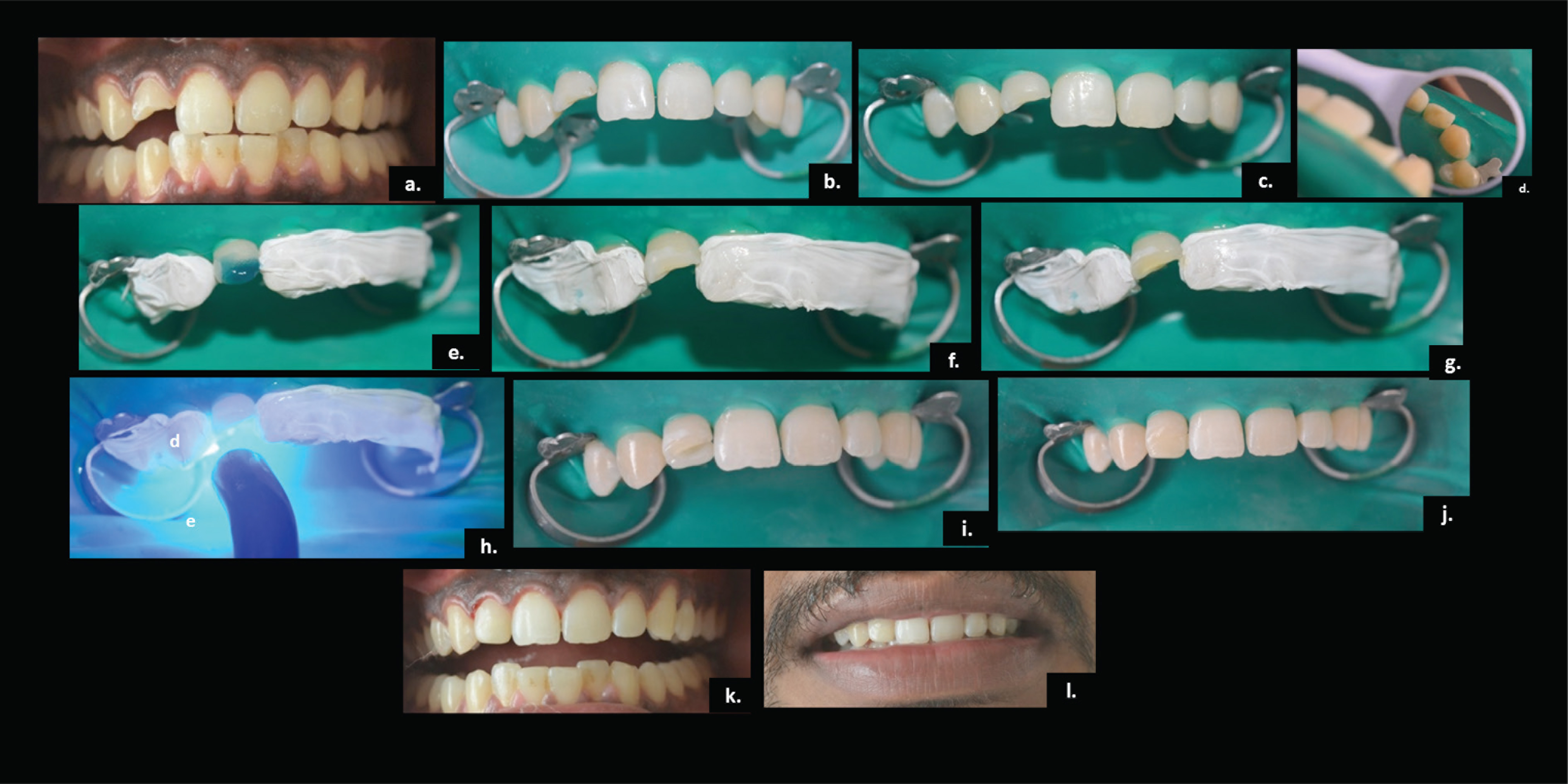
A 38-year-old male patient reported to the Department of Conservative Dentistry and Endodontics, with a chief complaint of fracture in the upper right front tooth. Dental history revealed that he met with an accident two days back, resulting in an injury. Extraoral examination revealed no significant findings. The intra-oral examination revealed a class-II Ellis fracture of the right maxillary lateral incisor. There was no other pathology related to the injury. Intraoral periapical radiograph clearly illustrated enamel and dentin fracture without the involvement of pulp in the tooth, and the sensibility test was positive for the same.
The treatment options were described to the patient but he wished for a direct composite restoration because of the immediate result and more affordable cost. A visual assessment was performed, and shade matching was done with A2 shade of Nanohybrid Universal Composite (GC-Solare Sculpt, Tokyo, Japan) being selected for the case. It was noted that only the right maxillary central incisor appeared to have a brighter shade than all other teeth (Figure 1(a)); hence shade matching was done with respect to the rest of the dentition, especially with the adjacent teeth, that is, maxillary right canine rather than with maxillary right central incisor. After achieving proper isolation, teeth were thoroughly cleaned and scrubbed (Figure 1(b)). A 2 mm wide bevel was created with a diamond point (Brassler, GA, USA) and finished with discs involving the enamel and dentin on the facial surface of the fractured tooth (Figure 1(c)). The palatal portion of the fracture line was smoothened to remove any overhanging enamel (Figure 1(d)). The adjacent teeth were covered with a Teflon tape and then the surface was etched with 37% phosphoric acid (Ivoclar N-Etch, Schaan, Liechtenstein) for 15 s (Figure 1(e, f)) after which the tooth was rinsed and dried, followed by the application of two coats of the bonding agent (Prime and Bond NT, Dentsply Sirona) onto the prepared surface, air-thinned and light-cured for 20 s (Figure 1(g, h)). The palatal shelf was created with A2 enamel (GC-Solare Sculpt, Tokyo, Japan) using a Mylar strip and index finger held palatally for support (Figure 1(i)). The shelf extended to the incisal edge and finished just short of the interproximal areas, after which the dentin A2 shade was placed in an accurate and precise position and sculpted with a composite instrument followed by curing each increment for 40 s. Lastly, A2 enamel (GC-Solare Sculpt, Tokyo, Japan) was placed onto the labial surface and sculpted accordingly (Figure 1(j)). Finally, after the removal of the rubber dam, the excess was removed. Then sequential grit abrasive disks (Sof-Lex Pop-on, 3M, Minnesota, USA) were used, followed by rubber points and, finally, composite polishing paste (Prisma-Gloss DENTSPLY Sirona) and the disk to achieve the final gloss (Figure 1(k)). The final appearance of the restored smile can be seen in Figure 1(l).
Case II
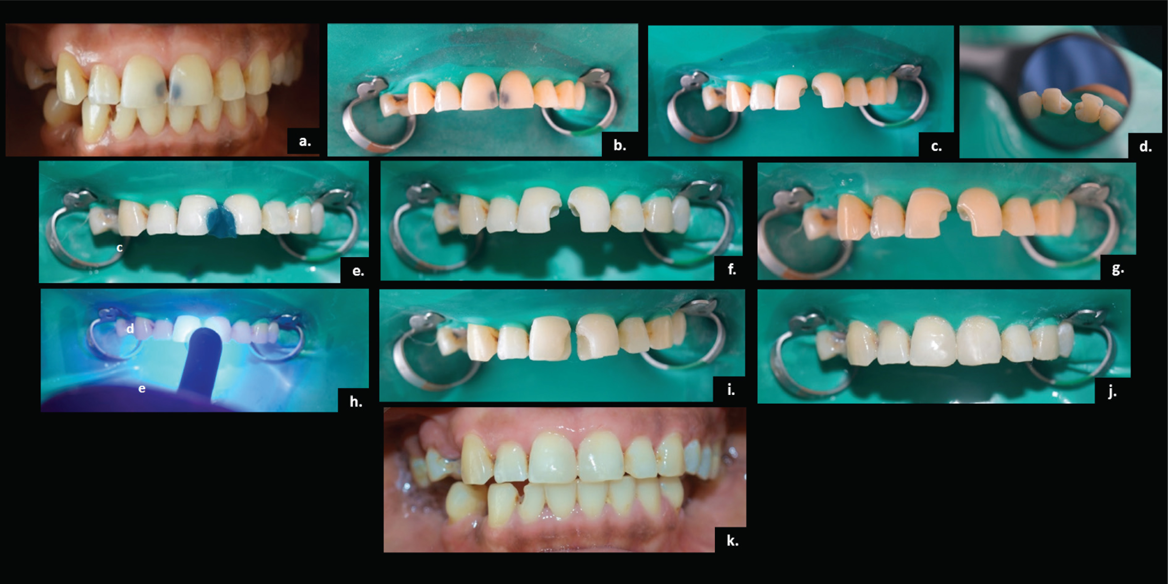
A 40-year-old female patient reported to the Department of Conservative Dentistry and Endodontics, with a complaint of decayed front teeth. Class-IV caries of right and left maxillary central incisors was diagnosed radiographically and the sensibility test showed that the tooth was vital. Due to the financial constraint, the direct composite restoration was chosen by her. Shade selection was done as mentioned before and A2 shade of Nano Hybrid Universal Composite (GC-Solare Sculpt, Tokyo, Japan) was selected.
The procedure was the same as described earlier (Figure 2(a, b)); a 2 mm bevel was placed on the facial aspects of both the teeth using a diamond point (Brassler, GA, USA) for a seamless transition (Figure 2(c)). Etching and bonding agent application was performed as described (Figure 2 (e–h)). A thin layer of A2 enamel resin (GC-Solare Sculpt, Tokyo, Japan) was used to create a palatal shelf using the Mylar strip, over which the dentin resin was placed in increments of not more than 2 mm, followed by final placement of the enamel resin (Figure 2(i, j)). This was followed by finishing and polishing as mentioned before (Figure 2(k)).
Case III
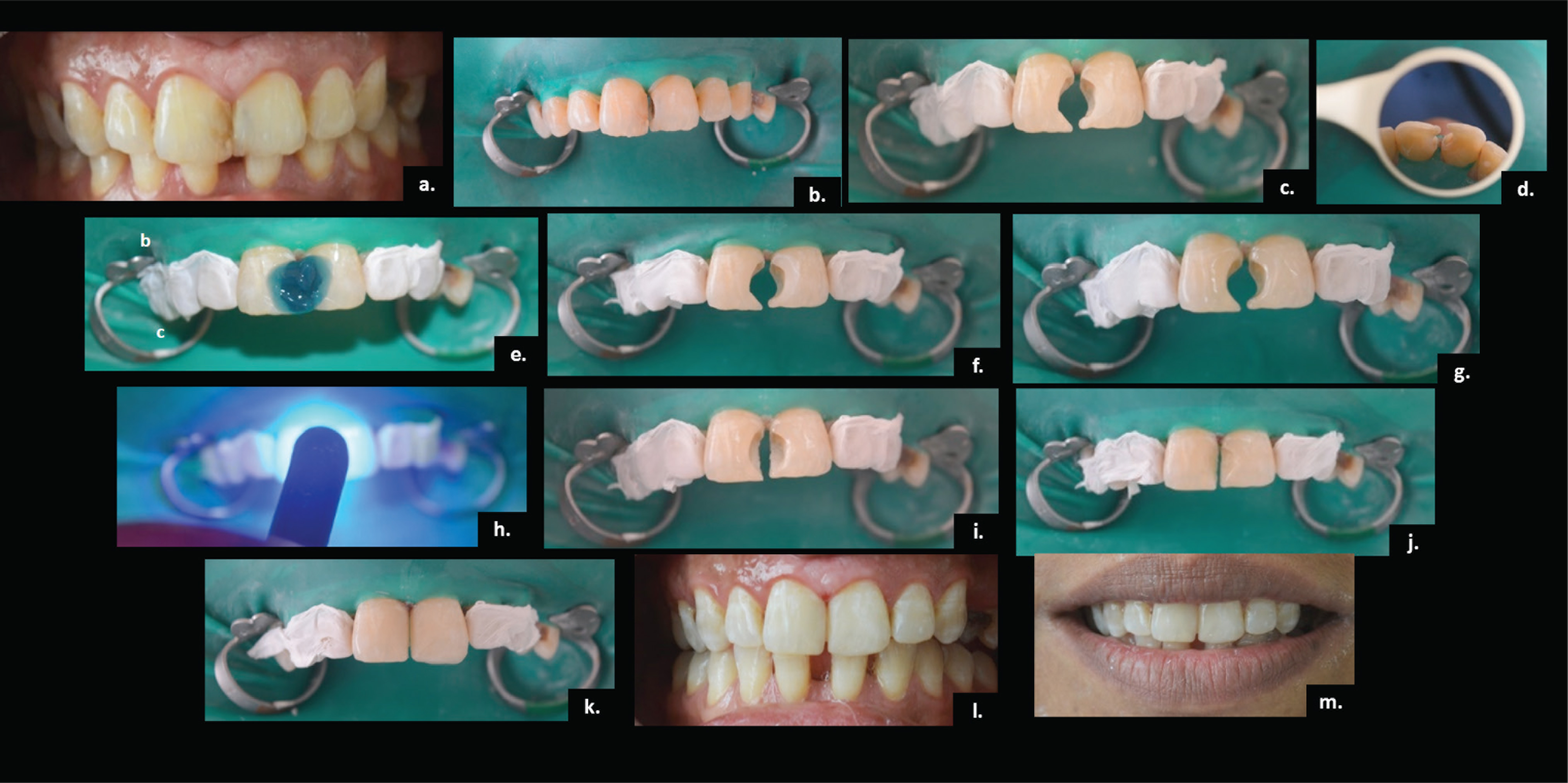
A 29-year-old female patient presented to the Department of Conservative Dentistry and Endodontics, with a discolored, previously placed restoration in her right maxillary incisor. On an intraoral examination, the existing restoration on the right maxillary central incisor was discolored with a poor color match and was also deficient. As for the left maxillary central incisor, Class III caries was diagnosed radiographically and appeared to be vital using the cold test. Taking into consideration the age of the patient, time, and financial aspect, direct composite restoration was chosen for the aesthetic rehabilitation.
In this case, A3 enamel shade (GC-Solare Sculpt, Tokyo, Japan) was selected and prior to the operative field isolation, the interproximal restorative material was removed with a carbide finishing bur (Brassler, GA, USA), which was then followed by removal of caries from the maxillary left central and the old restoration from the right using carbide finishing bur (Brassler, GA, USA) (Figure 3(a, b)). A fine diamond bur (Brassler, GA, USA) was used for surface preparation and 2 mm beveled margins were given with the same (Figure 3(c)). The adjacent teeth were protected with Teflon tape. Etching and bonding agent application was done as mentioned earlier (Figure 3(e–h)). The palatal shelf was created using Mylar strip with the A3 enamel shade (GC-Solare Sculpt Tokyo, Japan) (Figure 3(i)). Application of dentin and enamel resin composite was done incrementally and then followed by polymerization as mentioned earlier (Figure 3(j, k)). Finally, after the removal of rubber dam, gross finishing to adjust the incisal edge was done followed by final polishing as mentioned earlier (Figure 3(l)). The final appearance of the restored smile can be seen in Figure 3(m).
Discussion
Over the last two decades, the popularity of resin composites for aesthetic restorative procedures has enhanced owing to advances in adhesive systems and composite resin materials. The introduction of nanometer-sized particles has been one of the latest developments in the field which is thought to offer superior aesthetics and polishability required for anterior restorations in addition to excellent wear resistance and strength needed in the posterior teeth
Nonetheless, while nanofilled composites use nanosized particles in the resin matrix, nanohybrids take the approach of conjoining nanomeric and conventional fillers. 6 The presence of clusters formed by small particles seems to reduce the surface roughness of the materials and possibly increase the retention of smoothness after abrasion. 7 The inherent filler properties of nanocomposites are maximized while retaining excellent clinical handling properties and simultaneously reducing the amount of organic resin matrix, thus producing composites with significantly improved mechanical properties as well. These exhibit high translucency comparable to microfilled composites and physical properties similar to hybrid composite. Hence, these materials exhibit the potential to be used in all the diverse cases and conditions.
Durability of a restorative material in the oral environment is directly associated with its resistance to degradation. As far as nanocomposites are concerned, Kumar and Sangi in 2014 reported significantly higher water sorption and solubility for one nanocomposite in contrast with the other and a hybrid after 13-weeks water storage. Furthermore, lower strength values were reported for the nanocomposite that showed the highest sorption and solubility values. 8 However, Lopes et al. and Shin et al. reported no effect on sorption values of the tested composites when varying polymerization mode.9,10 The clinical performance of nanocomposites has been evaluated in a number of studies in terms of retention, color match, marginal discoloration, surface roughness, marginal adaptation, postoperative sensitivity, gross fracture, tooth integrity, and according to a review done in 2018, clinical performance of nanohybrids was shown to be comparable to microhybrids. 11
There are different techniques for placement of composite resin restorations which include direct and indirect techniques. The selection between direct and indirect techniques is a challenging decision-making process. In all the aforementioned cases, the direct restorative technique was used taking into consideration the patient concerns, the financial constraints and the type of case. However, indirect resin composites (IRCs) cover nowadays a wide range of indications, including inlays, onlays, and overlays. Nevertheless, there are some cases which come intermediately between direct and indirect where there is no clear demarcation for the technique to be used. Hence, in such cases it is completely a clinician’s call to take into account the amalgamation of all the factors and then decide in the best interest of the patient.
The advantages of IRCs include providing better proximal contour, improved wear resistance, reduced polymerization shrinkage, improved fracture resistance, and biocompatibility. 12 But these also come with some limitations such as requirement of an additional appointment, high cost, technician-related problems during shade harmonization, long chair time for simple fractures repair, and a technique-sensitive cementation procedure. The cost and the time factor are two of the major reasons why patients are inclined more toward the direct technique. However, the other merits include minimal tooth preparation, reversibility of treatment, and no need for an additional adhesive cementing system. Moreover, intraoral polishing is easy and has high potential for repair, immediately as well as intraorally. According to various studies done by Fennis et al., Cetin et al., and several others in the past, no significant difference was found between the performance of direct and indirect restoration.13,14
The placement of a cavosurface margin bevel on the enamel margins of anterior composite preparations traditionally has been recommended for retention and aesthetics. In all the three cases, 2 mm beveled margins were given to increase the surface area for the retention of the material as well as to obtain a better transition between composite resin and dental substrate inducing higher aesthetic pattern.
Successful restorations rely largely on the effective control of moisture and saliva from the tooth being restored as contamination remains an important cause of bond failure. Enamel surface contamination can occur at two critical times of the bonding procedure: After the tooth surface has been etched and after the primer has been applied. Contamination on an acid-etched enamel surface reduces the surface energy decreasing the wet ability and renders the surface less favorable for bonding resulting in a poor adhesive performance. 15 Contamination before priming would inevitably cause the formation of a smear layer. This layer, consisting mainly of proteins, covers the etched surface within seconds. Most of the porosities become plugged, and the penetration of the resin is impaired, which results in resin tags of insufficient number and lengths. The rubber dam technique has been one of the most widely used isolation methods in dental restorative treatments which were used for all the cases. However, it is noted from most of the past studies that the proper isolation is much more important than the method used for isolation. Thus, prior to application of bonding agents, the cavity should be rinsed thoroughly, dried, and kept isolated. This facilitates the bonding of the material to the tooth and improves the longevity of the restoration.
However, there were certain limitations in the cases presented. In the first case, only the right maxillary central incisor appeared to have a brighter shade than all other teeth; hence, shade matching for the right lateral incisor was done with respect to the rest of the dentition, especially with the adjacent teeth, that is, maxillary right canine rather than with maxillary right central incisor, hence the resulting mismatch between the central incisor and the lateral incisor. Additionally, in the second case, the oral prophylaxis and restoration for the mandibular right incisor could not be completed due to the time constraint for the patient.
Conclusion
Direct placement of resin composite to rehabilitate the anterior teeth has been shown to have good to short-term survival. Also, the nanohybrids used provided an acceptable color match with a conservative technique and were relatively easy to maintain. Although some disadvantages are noted, especially fragility and discolorations, when applied judiciously, nanohybrids can serve as material of choice for patients with aesthetic problems of anterior teeth and with maintainable hygiene practice.
Footnotes
Clinical Significance
The assessment of the clinical benefits of nanohybrid composite materials (i.e., improved surface luster and prolonged gloss retention) would be particularly useful in the front teeth region because it is the most esthetically relevant.
Acknowledgements
The authors would like to thank the patients for their cooperation and patience. We also wish to thank Solare Sculpt for assistance in providing the materials.
Declaration of Conflicting Interests
The authors declared no potential conflicts of interest with respect to the research, authorship, and/or publication of this article.
Funding
The authors received no financial support for the research, authorship, and/or publication of this article.
Patient Declaration of Consent
All participants were explained about the case study and written informed consents were taken prior to enroll them in the case study.