
Abstract
Purpose
Many factors in the etiology of anterior cruciate ligament (ACL) tears, predisposing factors related to knee morphology have also been reported. This study aimed to determine whether the Insall–Salvati (IS) index, which measures patella height, is a predisposing risk factor for ACL tears.
Methods
The IS index, patellar length (PL), and patellar tendon length (PTL) values of patients (study group) that underwent arthroscopic reconstruction for ACL tears obtained by preoperative magnetic resonance imaging (MRI) were compared with the index values in the preoperative MRIs of patients that underwent knee arthroscopy for reasons besides ACL tears. In addition, the anterior tibial translation (ATT) of both groups was also measured and compared on MRI images. The MRI findings of the subjects included in both study groups were arthroscopically confirmed.
Results
The mean ages of the study group (n = 120) and control group (n = 90) were 29.1 ± 8.2 years and 31.8 ± 9.8 years, respectively. There was a statistically significant difference between the study and control groups in terms of the PL and PTL values (p = 0.016 and p = 0.001, respectively). The IS index was statistically significantly higher in the study group with ACL tears (p = 0.009). The ATT was 8.61 ± 4.68 mm in the study group and 3.80 ± 1.92 mm in the control group. The ATT results of both groups were evaluated, and it was found that the study group was significantly higher than the control group (p = 0.001)
Conclusions
As a result of our current study, we observed higher IS index values in patients with ACL tears than in patients without ACL tears. It should be kept in mind that patella alta, which is associated with a high IS index as one of the factors of knee morphology associated with ACL tears, may play a role in the etiology of ACL tears.
Introduction
Anterior cruciate ligament (ACL) tears are one of the most common knee injuries. 1 They are particularly observed more frequently in sport activities that involve jumping, rotation, and direction changes, such as football and basketball.2,3 There are many studies in the literature that have evaluated in whom ACL tears occur more frequently and have investigated which factors are associated with ACL tears. These studies have focused on several factors, such as reduced width of the intercondylar notch, 4 Q angle, 5 volume of the ACL, 6 height of the lateral meniscus, 7 width of the femoral diaphysis, 8 muscle tone, 9 and sex differences. 10
Although some studies in the literature have reported that patella position (for both patella alta and baja) in the knee joint is also a risk factor for ACL tears,9,11no consensus could be suggested on the IS index in these studies. Singerman et al. 12 reported that patella baja further reduced contact stress compared to patella alta. They showed that, in some situations, such as torsion, valgus or varus, as well as flexion and extension of the knee joint, translation is increased to prevent further stress on the patella and femur, which causes strain and tear in the ACL. When the patellar tendon lengthens (high IS index), it places a load on the quadriceps tendon and reduces its strength, leading to ACL injury as a result of increased anterior tibial translation (ATT). 13
The Insall–Salvati (IS) index is an easily applied method in clinical practice. The IS index can also be measured by MRI and computed tomography (CT), as well as radiographic measurement. MRI, as one of these radiological imaging methods, has been reported to be more reliable than other techniques, since it can display patellar length (PL), patellar tendon length (PTL), and patellotrochlear cartilage more clearly.14,15 In addition, the IS index was found to be the most reliable method for measuring patella height. 16
In the present study, we aimed to evaluate whether the patellar height is a predictive risk factor in ACL tears by measuring the IS index in the preoperative knee MRIs of patients in the study group that underwent arthroscopic-guided ACL reconstruction and patients in the control group without ACL tears. To the best of our knowledge, this is the first study in the literature to examine the relationship between ACL tears and the IS index by evaluating MRIs only in men.
Methods
A total of 120 patients in the study group who underwent arthroscopic-guided ACL reconstruction using a hamstring graft and a total of 90 patients in the control group without ACL tears who were operated on for pathologies besides ACL tears between January 2014 and January 2020 were comparatively analyzed. Preoperative knee MRIs were examined, and PL, PTL, and IS index were comparatively measured by a radiologist experienced in reading images of the musculoskeletal system in both study groups. The ATT of the study and control groups was also measured on MRI.
The IS index reference interval was accepted as 0.8–1.2, according to the literature. Index values below 0.8 and over 1.2 were evaluated as patella baja and patella alta, respectively. 17 The ATT sign was positive when the ATT distance was more than 5 mm. 18
Patient selection
Study group
Patients who underwent arthroscopic interventions due to full-thickness ACL tears and whose skeletal maturation was completed were included in the study per the eligibility criteria. In addition, patients whose IS index measurements were made on these images with preoperative MRIs were included in the study.
Exclusion criteria were patients who had any additional bone, ligament or cartilage injury; moderate or severe knee arthrosis; and a history of previous surgery. In addition, patients with fractures, bipartite patella, patellar dislocation, multiple ligament injuries, previous ACL surgery, patellar tendon elongation, and preoperative MRI images were excluded from the study.
There were two female patients who underwent ACL reconstruction and were excluded from our study. One of them had multiple ligament injuries, and the other was excluded from the study due to the homogeneity of our study. As a consequence, a total of 120 male patients met the inclusion criteria of the study.
Control group
Our hospital records included the data of the patients operated on for non-ACL tears, for reasons such as meniscopathy, synovial hypertrophy, chondral injury, and plica. The control group was comprised of 90 patients after the exclusion of female patients, patients with osteoarthritis, and patients without available preoperative MRIs from the study. In addition, patients with a history of previous patellar fracture were excluded from the study.
MRI protocol
The images were acquired with a 1.5 T MRI device (MagnetomAmira, Siemens Medical Systems, Forcheim, Germany) using lower extremity coils. The standard knee MRI protocol was implemented in all the patients, and IV contrast agent was not administered.
The evaluation of the images
All the acquired images were read by a radiologist experienced in reading images of the musculoskeletal system. The measurements were performed in the midsagittal plane in the sagittal T1-weighted images, since the maximum lengths of the patella and patellar tendon can be acquired in the midsagittal plane and insertions of the patellar tendon into the tibia and patella can be distinguished most optimally in the non–fat-suppressed T1-weighted sequences. The length measurements were obtained using standard measurement calipers in the PACS software of our hospital.
Insall–Salvati index and anterior tibial translation measurement
The PTL was measured from the deep posterior surface of the lower tip of the patella to the tibial tubercle in the MRI. The PL was defined as the greatest diagonal length of the patella. The IS index is the ratio of PTL/PL (Figure 1). Insall–Salvati index is calculated by the PTL/PL ratio.
Anterior tibial translationATT distance measurement was performed on a sagittal T1-weighted image in the midsagittal plane of the lateral femoral condyle. The distance between two parallel lines drawn from the posterior cortex of the lateral condyle of the femur and the posterior cortex of the tibia lateral plateau was defined as ATT. The lines were drawn in the cephalocaudal direction, parallel to the long axis of the image to the lateral plateau plane of the tibia (Figure 2).
19
ATT is measured distance from the posterior border of the medial tibial condyle to the femoral condyle.
Results
Demographic data of the patients.
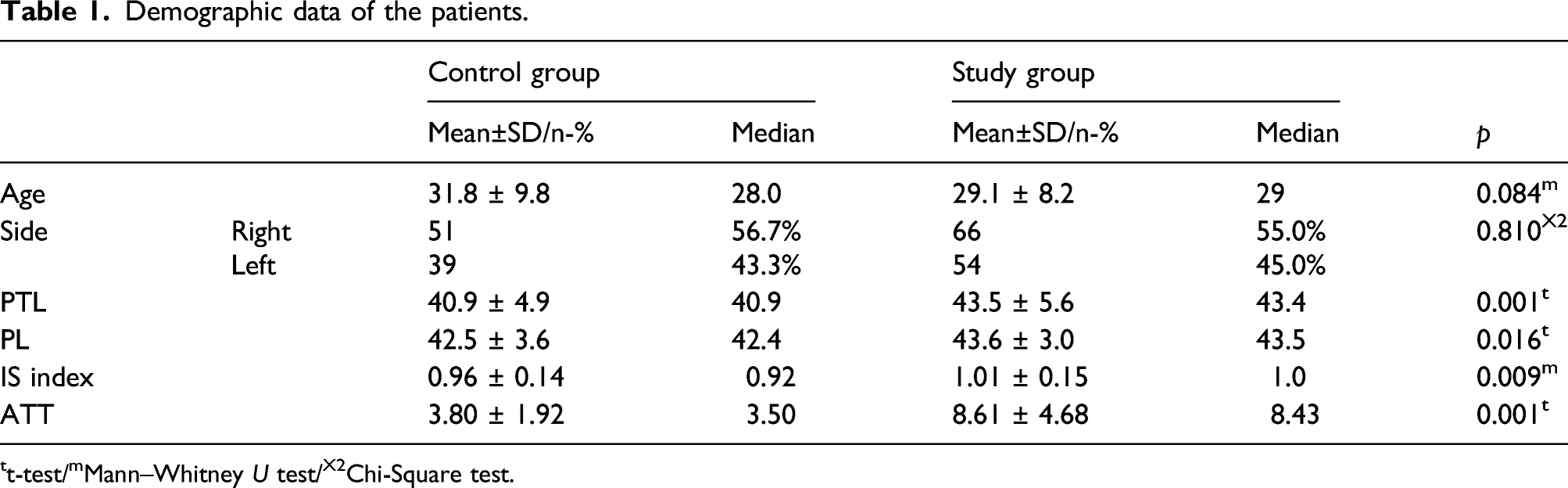
tt-test/mMann–Whitney U test/X2Chi-Square test.
According to Table 1; PL, PTL, and IS index values of the study group were statistically significantly higher than those in the control group (p = 0.016, p = 0.001, and p = 0.009, respectively). These comparisons are shown in Figures 3, 4, and 5. The comparison between the group with ACL tear and control group in terms of patellar length. The comparison between the group with ACL tear and control group in terms of patellar tendon length. The comparison between the group with ACL tear and control group in terms of IS index.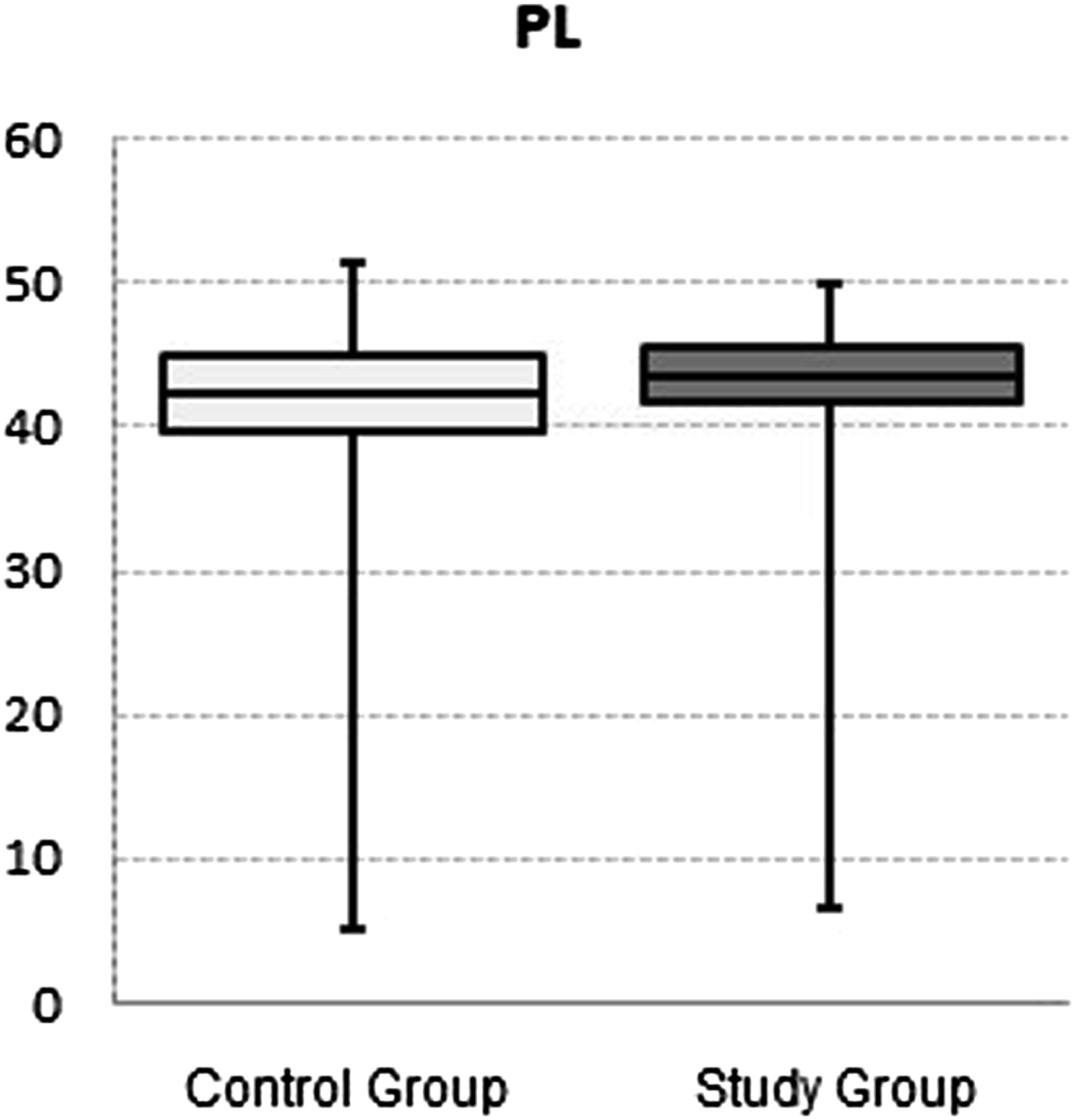


When the ATT results of both groups were evaluated, it was found that the ATT of the study group was significantly higher than that in the control group (p = 0.001) (Table 1).
Discussion
In our study, the IS index values were found to be 1.01 ± 0.15 and 0.96 ± 0.14 in the study group (with ACL tears) and control group (without ACL tears), respectively. Akgün et al. 20 detected a mean IS index of 1.11 ± 0.08 vs 1.08 ± 0.06 in similar study and control groups, respectively, and observed that the mean IS index value of the study group was statistically significantly higher than that in the control group. The PTL value was also found to be higher in the study group (with ACL tears), similarly to our study. In that study, sagittal knee MRIs were retrospectively evaluated, and a comparison was performed between the groups with and without ACL tears. A study that compared the findings of ACL tears obtained by MRI with arthroscopic findings determined that the sensitivity and specificity values of MRI for ACL tears were 77.8% and 93–96%, respectively. 21 The difference in our study compared with the study of Akgün et al. 20 was that all the cases were included in our study after arthroscopic confirmation.
Many predisposing factors have been suggested for ACL tears. The well-documented morphological risk factors for the knee include narrowing of the intercondylar notch, sagittal changes in femoral condyles, increased tibial slope, decreased size of tibial eminence, weak tibiofemoral compliance, and decreased volume of the ACL and IS index.11,22 It has also been reported that increased ATT is associated with ACL tears. We found that ATT was higher in the study group in relation to the IS index. 23
In our study, the ATT was 8.61 ± 4.68 mm in the study group and 3.80 ± 1.92 mm in the control group. Numkarunarunrote et al. 23 found that the length of ATT was 1.5 ± 3.6 mm in individuals with an intact ACL, 5.0 ± 2.8 mm in patients with partial ACL tears and 7.6 ± 3.0 mm in patients with complete ACL tears. In another study, it was reported that quadriceps/hamstring imbalance and the tendency to lengthen the hamstring muscles may cause ACL tears as a result of increased ATT. 24 In individuals with high patella, it causes a load on the quadriceps and a decrease in its strength. In addition, ACL tears may occur secondary to decreased tibial support and increased ATT in relation to the PTL.
In the literature, there are studies that have been performed with X-rays and MRIs based on the measurement technique of the IS index. 25 Verhuslt 16 emphasized that the IS index was better in conventional radiography in a study he conducted with MRI, conventional radiography, and CT. Miller 26 stated that MRI was more valuable in measuring the IS index, independent of knee flexion. Additionally, Shabshin et al. 27 emphasized that knee MRI gave less erroneous images and was partly more efficient in measuring PTL. The major advantages of MRI include measurement of patellar height (particularly by sagittal MRI), displaying patellofemoral joint cartilage, better visualization of additional soft tissue injuries, and viewing patellotrochlear cartilage overlap, which is clinically more frequently associated with patella alta. 28
Female and male gender were usually evaluated together in the studies that addressed the fact whether the IS index may be a predisposing factor for ACL tears in the literature.9,11,20 It has been noted that the incidence of ACL tears increased due to a partial increase in the laxity of the ACL, resulting from the changes in the estrogen levels caused by the menstrual cycle in women. 29 It has been reported that the mean values of the IS index were higher in females than in males, 9 and the morphology of the intercondylar notch, valgus alignment, shorter flexion range of motion, and increased quadriceps activity also increase the risk for ACL tears.30,31 All the groups (study and control) included only male patients in our study; thereby, the aspect of gender difference was completely eliminated. Our study may be considered as a different study with this same characteristic.
Degnan et al.11found statistically significantly higher IS index values in the group with ACL tears compared with those in the group without ACL tears in their study carried out in a pediatric patient population (p < 0.001). In the study by Degnan et al., the IS index values were statistically higher in the group with ACL tears, similarly to our study. Degnan et al. 11 determined statistically significantly higher PTL values in the group with ACL tears (p < 0.001), whereas the evaluated PL values were statistically insignificant (p = 0.523). Additionally, the diagnostic MRI findings were arthroscopically confirmed in all patients. This study was different from our study, since it was carried out in a pediatric population with a smaller number of patients. It has been noted in the literature review that traction apophysitis (Sinding–Larsen–Johannson disease) located on the inferior pole of the patella in adolescent children may lead to confusion in MRI.32,33
Lin et al. 9 conducted an evaluation based on X-rays in 2005 and reported that the IS index was lower in the group with ACL tears. Thus, patella infera is a predisposing risk factor. The patella and patellar tendon can be evaluated more optimally by MRI. More accurate measurements can be obtained, since the patellar tendon is visualized directly.11 In our study, the measurements of all the patients were performed based on MRIs, and it was concluded that patella alta may be a risk factor, in contrast to the study of Lin.
Limitations
The number of patients in the study group may be considered to be relatively small. However, Lin et al. 9 and Akgün et al. 20 carried out their studies on 115 patients and 110 patients with ACL tears, respectively. Another study by Degnan et al. 11 was conducted on 34 patients. Therefore, the number of our cases may be considered higher than the other studies mentioned that measured the IS index. On the other hand, arthroscopic confirmation of ACL tears in the patients, as well as MRI findings, was the strong feature of our study. The lack of an evaluation related with the time interval between the occurrence (acute or chronic) and reconstruction of the ACL may be considered another limitation of our study. Some of the patients included in our study were athletes, and some of them were young individuals who did not do active sports. If all individuals included in the study were professional athletes, perhaps more homogeneity could have been achieved. A novel study can be conducted to determine how this time interval affects the IS index to provide a contribution to the literature. Another limitation of our study may be the absence of measurement of the body mass index (BMI). Some studies have reported that an elevated BMI is a risk factor for ACL tears.2,34 There was no obesity in any of the patients in our study.
In our study, there was a higher IS index value in patients with ACL tears compared to those without ACL tears. It should be kept in mind that patella alta may play a role in the etiology of ACL tears as one of the factors of knee morphology associated with ACL tears. The determination of such factors, performing hamstring strengthening exercises and testing adductor and abductor balance may reduce the probability of ACL tears with respect to seasonal competition, particularly in the subjects with sports activities. The regular muscle strengthening exercises in the subjects detected to have high IS index values allows these subjects to keep performing sports activities, as well as prevents the negative psychosocial impacts on these subjects due to ACL tears.
Footnotes
Declaration of conflicting interests
The author(s) declared no potential conflicts of interest with respect to the research, authorship, and/or publication of this article.
Funding
The author(s) received no financial support for the research, authorship, and/or publication of this article.
Ethical approval
Our study was approved by the Non-invasive Research Local Ethics Committee (approval dated 10/07/2020 and No: 2020/04–31)