
Abstract
A ring-shaped meniscus is a very rare anatomical variant among all meniscal abnormalities. Additionally, an accessory meniscus is extremely rare, and only a few cases have been reported. We herein report a case involving the combination of these two features in a single lateral meniscus. These abnormalities were found during arthroscopic surgery for removal of an osteochondral fragment that had detached from the patellar bone and plication of the medial patellofemoral ligament in a patient with acute patellar dislocation. To our knowledge, each variant is extremely rare and the combination of the two variants has not been reported.
Introduction
Various types of meniscal anomalies have been reported. Anomalies of the lateral meniscus are more frequently found than those of the medial meniscus. Among them, a discoid meniscus is the most common anomaly; other anomalies are rarely reported. Other anomalies of the lateral meniscus include a double-layered meniscus, accessory meniscus, and ring-shaped meniscus. Although a few cases of these anomalies have been reported separately, 1 –4 combinations of these aberrations are extremely rare. To our knowledge, no reports have described the combination of a ring-shaped aberration and accessory meniscus in a single joint. We herein describe a patient with a ring-shaped lateral meniscus combined with an accessory meniscus.
Case report
A 16-year-old boy presented with sudden-onset knee pain and joint swelling after the patella was briefly hit during recreational Korean wrestling. There was no direct trauma and no previous knee joint symptoms such as catching, effusion, locking, or giving way. The patient’s medical history was unremarkable, but unrecognized joint laxity was found on physical examination. Additionally, the range of motion of the knee joint was restricted to 0°–60°, with the patella positioned laterally, and ecchymosis and tenderness were present on the medial side of the patella. Radiological examination revealed lateral subluxation of the patella with a displaced fragment (Figure 1). Magnetic resonance imaging (MRI) showed a discoid lateral meniscus with a small cystic alteration under the anterior root of the lateral meniscus along with a portion of meniscal tissue at the medial portion of the lateral compartment with the regular border adjacent to the tibial spine (Figure 2). These findings suggested a combined alteration of the lateral meniscus, bucket handle tear, and anterior root tear of the lateral meniscus.

(a) Plain radiograph showing patellar subluxation and a displaced fragment (white arrow) on the medial side of the patella. (b) A loose body (osteochondral fragment, black arrow) is seen on a coronal magnetic resonance image.

(a) Focal scalloping of the lateral tibial plateau adjacent to the medial inferior portion of the cyst (white arrow). (b) Width of the midbody of the lateral meniscus (double-headed arrow) indicates a discoid meniscus. (c) Redundant meniscal tissue (arrowhead) on the medial aspect of the lateral meniscus mimics a bucket handle tear.
We decided to perform an arthroscopic procedure. First, we performed osteochondral fragment removal and imbrication of the medial patellar retinaculum as an arthroscopic all-inside technique as described by Halbrecht 5 (Figure 3). However, under arthroscopic examination of the lateral compartment, we found a normally inserted and stable ring-shaped lateral meniscus without injury. Its insertion to the anterior and posterior horn was firmly attached. The inner portion of the meniscus was not mobilized when pulled with a probe. We considered the patient to have an aberration of the lateral meniscus. Additionally, we found an accessory structure under the lateral meniscus; this structure was exposed by pulling the anterior horn of the lateral meniscus anteriorly. It appeared as a cartilage structure that arose from the anterolateral portion of the lateral tibial plateau and inserted at the inner portion of the ring-shaped lateral meniscus. Its shape resembled a comma. The texture of the accessory meniscus felt normal under examination with a probe. No tears or injuries of the accessory meniscus were found (Figure 4). We considered this to be an incidental finding. Therefore, no arthroscopic procedure was performed, and the ring-shaped meniscus and accessory meniscus were preserved intact.

Arthroscopic findings. (a) Osteochondral fracture of the patella (white arrow). (b) Removal of osteochondral fragment using a forceps. (c) Sutures are placed along the medial retinaculum for arthroscopic all-inside medial imbrication.
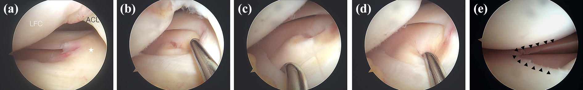
Arthroscopic findings of the lateral compartment of the right knee. (a) Ring-shaped lateral meniscus with an inter-horn bridge (white star). (b) Stabilized inner portion of the ring-shaped meniscus. (c) Exposed comma-shaped accessory meniscus. (d) The accessory meniscus arises from the lateral tibial plateau with no injury. (e) Lateral, anterior, and posterior portions of the ring-shaped lateral meniscus with a smooth margin (arrows), and no evidence of tear. LFC: lateral femoral condyle; ACL: anterior cruciate ligament.
Immobilization of the knee in full extension was applied for 3 weeks postoperatively. The range of motion of the knee joint was then gradually increased. The patient returned to activities of daily living 3 months postoperatively and regained his previous sports activity level 6 months postoperatively. Two years after surgery, he had no complaints regarding his knee and had experienced no recurrence of patellar dislocation.
The patient and his family provided informed consent to submit the data from his case for publication.
Discussion
The lateral meniscus is more morphologically variable than the medial meniscus, and the most common lateral meniscal aberration is the discoid meniscus. Its incidence is very low, except in Asian populations, in which it has an incidence of about 16.6%. In contrast, it occurs in less than 5% of the Caucasian population. 6,7 Other anomalies, such as a ring-shaped meniscus, double-layered meniscus, and accessory meniscus, are relatively rare. In a large cadaver study, Ryu et al. 2 reported that the prevalence of a ring-shaped meniscus was 0.9% and that of a double-layered meniscus was 0.5%.
A ring-shaped meniscus previously reported is almost on the lateral side and usually asymptomatic. It has a firm structure and is connected to the surrounding soft tissue. This may contribute to the lack of clinical symptoms. MRI has been reported to be a valuable diagnostic tool to detect the lateral meniscal variants, but it is important to bear in mind the possibility of a ring-shaped meniscus in the differential diagnosis of a bucket handle tear or a central tear of a discoid meniscus. In the sagittal plane of MRI, “central bow tie sign” is useful in detecting the normal meniscal tissue onto the inner portion of the lateral compartment of the knee. 8 In the coronal plane, the inner portion of the lateral meniscus is in the medial portion of the lateral compartment and not in the intercondylar notch, and the presence of the “mirror sign” in which the inner portion of the ring-shaped lateral meniscus is the mirror image or reflection of the normal appearing body of the meniscus. 8
A double-layered meniscus is extremely uncommon and one of the forms of accessory meniscus. In all cases reported in the literature, it was overlying the normal lateral meniscus. Its clinical features remain unclear, however mechanical obstruction of the hypermobile components of the accessory meniscus may have often been associated with symptoms including pain and mechanical symptoms such as clicking, locking, and giving way. 4,9 A review of the literature showed that the symptoms were significantly improved by resection of the hypermobile upper accessory meniscus. However, all reported double-layered lateral menisci have no specific radiological finding, thus careful arthroscopic examination is needed to reveal this uncommon abnormality and achieve a clinical improvement.
An accessory meniscus arising from the tibial plateau is extremely rare, with only two cases having been published in the literature. 1,10 One of them has clinical symptoms, pain and mild swelling, but the other has no clinical symptom associated with a lateral accessory meniscus. They have no abnormality on plain radiograph and MRI. Such aberrations are considered to result from a congenital rather than developmental process, although the clinical presentations, pathology, and epidemiology of these variations are still unclear. 9,11
In the arthroscopic findings, a ring-shaped meniscus is apt to be misinterpreted as a bucket handle tear of a normal C-shaped lateral meniscus or a central tear of a discoid meniscus. However, a ring-shaped meniscus has some differences from a bucket handle tear or a central tear. The displaced medial portion of the bucket handle tear is usually movable and reducible into the lateral portion of the meniscus, and detachment from the torn edge is observed. A central tear has an irregular or degenerative inner margin that is not tapered and sharp. In the present case, the ring-shaped meniscus was characterized by the presence of a bridge between the two horns of the meniscus, and the inner portion of the ring-shaped meniscus was not mobilized when we tried to move it laterally within the intercondylar notch using a probe. It also had the same appearance as that of a normal meniscus, with a sharp and tapering inner margin.
Additionally, the combined accessory meniscus in our case arose from the anterolateral portion of the lateral tibial plateau and inserted jointly with the inner portion of the ring-shaped meniscus; however, no anomaly such as a tear or degenerative change was found. The patient’s presenting symptoms before the surgery were caused by patellar dislocation, and the aberrations of the lateral meniscus were found incidentally during arthroscopy. We believe that these aberrations were probably a congenital anomaly. We also found no clear evidence of a relationship between the symptoms and the morphological anomalies. Thus, we decided to leave these malformations untouched. Their true role in producing clinically significant knee symptoms requires further research and long-term follow-up.
Conclusion
To the best of our knowledge, the present case is the first description of a combined ring-shaped meniscus with an accessory meniscus of the lateral side with no other anomalies. A diagnosis of rare aberrations, as ring-shaped or accessory menisci, should be carefully made after thorough probing to differentiate them from other anomalies. Usually, ring-shaped and accessory menisci are asymptomatic and found incidentally. Preoperative history taking and assessment of symptoms of the knee joint are essential to determine whether to treat the aberration.
Footnotes
Declaration of conflicting interests
The author(s) declared no potential conflicts of interest with respect to the research, authorship, and/or publication of this article.
Funding
The author(s) received no financial support for the research, authorship, and/or publication of this article.