
Abstract
Purpose:
We aimed to determine the factors that influence the symptoms of naviculo-cuneiform (NC) coalition using radiography and computed tomography (CT).
Methods:
We retrospectively reviewed the radiographic and CT findings of 37 NC coalition cases. The existence of a large pit (depth >3 mm), irregular articular surface, joint space narrowing, dorsal bony spur, subchondral sclerosis, multiple subchondral bony cysts, and intra-articular loose body were evaluated on radiographs or CT. The size of the largest subchondral bony cyst was also measured using CT. All cases were divided into two subgroups according to the symptoms. Fisher’s exact test was used to distinguish the factors influencing the symptoms.
Results:
Twenty-three and fourteen feet were enrolled into the symptomatic and asymptomatic groups, respectively. The rates of the large pit on either radiograph (47.83 vs. 21.43%) or CT (65.22 vs. 28.57%) were significantly different between both groups (p = 0.001). The mean size of the largest subchondral bony cyst on CT was also significantly greater in the symptomatic group (4.25 vs. 1.53 mm, p = 0.005).
Conclusion:
A large deep pit and huge subchondral bony cyst on the radiograph or CT can be related to symptoms for the patient with NC coalition. A CT is highly recommended for a more accurate evaluation in patients with NC coalition.
Keywords
Introduction
Naviculo-medial cuneiform (NC) coalition is an extremely rare condition and is therefore quite likely to go unrecognized. 1 –3 A coalition is an abnormal fusion of fibrous, cartilaginous, or osseous tissue between two bones, which can be either congenital or acquired. In Western countries, the talo-calcaneal and calcaneo-navicular coalitions are more common in the tarsal bones of the hindfoot and midfoot, accounting for more than 90% of all tarsal coalitions. Therefore, several case reports exist on the treatment associated with NC coalition. 4 –7
Some reports that reveal radiographic and computed tomographic (CT) features of NC coalition are about cases from Eastern countries. 8 –10 Choi et al. 9 reported that the characteristic radiological, CT, and MRI features of an NC coalition include an irregular articular surface with possible secondary degenerative changes in the plantar margin of the joint. In addition, Lee et al. 10 evaluated 19 feet with symptomatic NC coalition and stated that CT is the most reliable test for evaluating NC coalition and for demonstrating its characteristic morphology.
The purpose of the present study was to evaluate and compare the radiographic and CT findings between symptomatic and asymptomatic patients with NC coalition to determine the factors influencing its symptoms. To the best of our knowledge, there are no previous reports comparing the CT findings between symptomatic and asymptomatic feet with NC coalition. We hypothesized that CT would provide accurate and valuable information regarding the factors associated with symptomatic NC coalition.
Materials and methods
Patient selection
A total of 23 feet of 16 consecutive patients, aged above 15 years, who visited our outpatient clinic for symptomatic NC coalition between March 2008 and March 2018 were included in this study. Patients complained of various degrees of symptoms, from dull and vague to sharp and aching pain during weight bearing, with or without focal tenderness on the plantar side of the NC joint. The operations for NC coalition were performed for 9 feet in the symptomatic group after at least 6 months of conservative treatment that included painkillers, physical therapy, and orthoses. Simple excision surgeries were performed for 3 feet and NC arthrodeses were performed for 6 feet (Figure 1).

Radiograph of a 17-year-old male patient with aching pain under the NC joint reveals an NC coalition (a). An arthrodesis of the NC joint was performed (b) when 6 months of conservative treatment had failed. A postoperative radiograph after 3 years (c) shows a solid fusion. Currently, the patient is in a pain-free state. NC: naviculo-medial cuneiform.
During the same period, we enrolled 14 feet of 13 patients whose NC coalitions were found incidentally without any presenting symptoms. All feet were related to acute fractures of either the metatarsals or phalanges. Patients were initially asked about pain of the NC joint and finally included in the study, unless they complained of any symptoms after they were allowed to bear full weight with complete bony union at the fracture site. All patients in both groups underwent the same weight-bearing radiographic investigations and nonweight-bearing CT evaluation.
No patient in either group had a history of systemic inflammatory conditions such as rheumatoid arthritis or gout.
Assessments
First, weight-bearing anteroposterior (AP) and lateral foot radiographs were assessed for the existence of a large pit with a depth > 3 mm, irregular articular surface, joint space narrowing, dorsal bony spur, subchondral sclerosis, multiple subchondral bony cysts, and intra-articular loose bodies (Figure 2). In addition, the talo-first metatarsal angle (Meary’s angle) was measured using a lateral radiograph. Using CT, the size of the largest subchondral bony cyst was measured in addition to the aforementioned radiographic findings (Figure 3).

Various features of NC coalition on radiograph. Irregular articular surface with small subchondral bony cysts (a), joint space narrowing with bony spur (b), large pits with depth of >3 mm partially involving NC joint (c), irregular articular surface with joint space narrowing and a single huge subchondral cyst (d), large pits with depth of >3 mm involving the entire NC joint (e), and large pits with multiple subchondral cysts (f). NC: naviculo-medial cuneiform.

Various features of NC coalition on computed tomography. Irregular articular surface with small subchondral bony cysts (a), large pits with depth of >3 mm (b), large pits with multiple subchondral cysts (c), and irregular articular surface with joint space narrowing and a single huge subchondral cyst (d). NC: naviculo-medial cuneiform.
The location of each NC coalition was described according to the findings of Byun et al. 11 The NC joint was divided into four parts, marked 1, 2, 3, and 4 from medial to lateral, and A, B, C, and D from the plantar to the dorsal aspect (Figure 4).
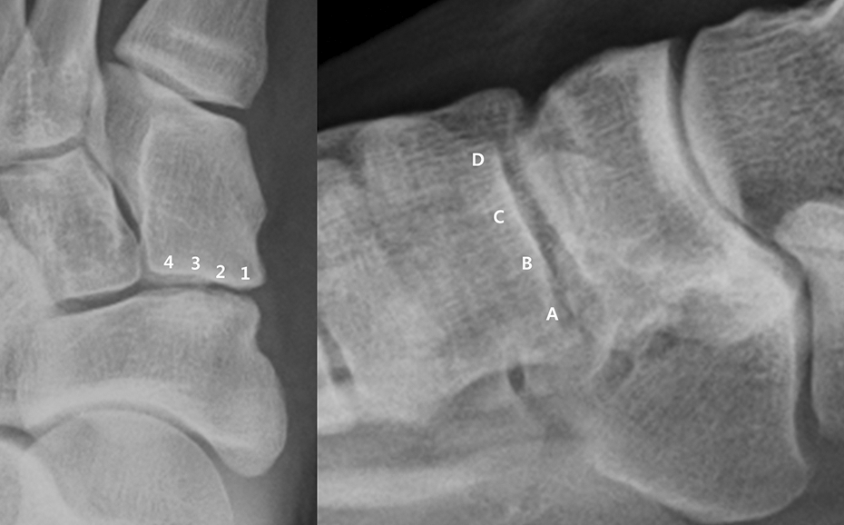
The location of each NC coalition has been described according to the findings of Byun et al. 11 The NC joint has been divided into four parts, marked 1, 2, 3, and 4 from medial to lateral, and A, B, C, and D from the plantar to the dorsal aspect. NC: naviculo-medial cuneiform.
Ethical considerations
Informed consents were obtained in writing from all the patients before enrolling them in the study. This study was approved by our institutional ethical review committee and conformed to the guidelines of the Declaration of Helsinki.
Statistical analyses
A descriptive analysis was performed for all the variables, including calculation of the mean and standard deviation or frequency. Data normality was tested using a Kolmogorov–Smirnov test. The Mann–Whitney test was used to compare continuous variables between the symptomatic and asymptomatic groups (age, Meary’s angle, and mean size of the largest subchondral cyst on CT). Fisher’s exact test was used to analyze differences in the ratio of each radiographic and CT finding.
To validate both radiographic and CT findings, one orthopedic surgeon and one radiologist graded 10 randomly selected feet twice, with a 2-month interval between the assessments. Random selection was performed by a blinded practitioner who was not involved in this study. The interobserver reliability and the intraobserver reproducibility are shown in Table 1 using intra-class correlation coefficients (ICCs).
The statistics for interobserver reliability and intraobserver reproducibility for the 10 randomly selected cases.

ICC: intra-class correlation coefficients; CI: confidential interval; CT: computed tomography.
Statistical analyses were performed using SPSS software, version 19 (IBM Corp., Armonk, New York, USA). Statistical significance was determined as a p-value less than 0.05 for all analyses.
Results
The overall parameters of radiographs and CT images are presented in Table 2. Data and comparison of the two subgroups divided according to presence of symptoms are demonstrated in Table 3. The mean age was significantly younger in the symptomatic group (p = 0.001). The values for Meary’s angle were significantly greater in the asymptomatic group (p = 0.042), although both values were within the normal range. The ratios of the large pit on both radiograph and CT were significantly greater in the symptomatic group (p = 0.001, for each), whereas the other radiographic and CT features were not significantly different. The mean size of the largest subchondral bony cyst on CT was 4.25 ± 2.17 mm in the symptomatic group, which is significantly greater than that in the asymptomatic group (p = 0.005).
Parameters of overall 37 cases in 25 patients.

CT, computed tomography
Parameters of two subgroups divided by symptom.

n: number of cases; CT: computed tomography.
The coalition sites of each group on radiograph and CT are summarized in Table 4.
The coalition location of each group.

(): number of cases; CT: computed tomography.
Discussion
Our data suggested that the symptoms related to NC coalition usually presented in the ages between adolescence and young adulthood. Likewise, almost every operation for NC coalition was performed for this age group. Conversely, NC coalition was mainly found incidentally in middle- and old-aged individuals.
Kumai et al. 12 divided NC coalitions into three categories according to the CT morphology: irregular, cystic, and combined pattern. We believed that this categorization was insufficient to explain the factors that could be related to the symptoms and the operative treatment. Our data suggested that a large pit with a depth of >3 mm along the joint space or a huge subchondral bony cyst could be related to the symptoms in patients with NC coalition. Choi et al. 9 reported that subchondral cysts were observed on plain radiography and CT imaging in 86.4% and 76.9% of symptomatic and asymptomatic individuals with NC coalition, respectively, without a statistical comparison. However, we determined that the number of CT scans (n = 14) in their study was too small to reach an exact conclusion.
While we were performing this research, we were curious about the contents of the large pits that were the cause of the NC coalition symptoms. According to the findings of Byun et al., 11 histological examination of biopsied samples showed degenerated fibrocartilaginous tissue or degenerated cartilaginous tissue with calcification. Kumai et al. 12 also reported the same histopathological findings, wherein fibrocartilaginous or cartilaginous material was found. In both these studies, it was hypothesized that the initial irregular coalitions gradually developed into cystic, and finally, combined patterns. In our study, some patients presented with large pits and huge subchondral bony cysts at a young age; therefore, we could assume that other factors also had an influence on the development of the combined pattern.
The pain associated with NC coalition is known to be caused by weakness of the fused region relative to the weight-bearing force. 12 –15 According to the findings of Kumai et al., 13 an incomplete coalition produces microfractures and remodeling at the boundaries between bone and the coalition, which further leads to degenerative changes. This mechanical abnormality appears to induce pain via free nerve endings in the periosteum and in the articular capsule surrounding the coalition. On this basis, we concluded that a large pit could be related to the symptoms caused by mechanical instability of the coalition site. Symptoms may not appear in the early stages because the NC coalition mostly occurs in joints with a small range of motion.
Subchondral bony cysts can also be caused by stress-induced bone resorption in osteoarthritic conditions, 16 which indicates that the degree of arthritic change in the affected joint has been progressing. Therefore, we concluded that a huge subchondral bony cyst was a result of a progressing arthritis of the NC joint related to coalition.
Regarding the location of the coalition, NC coalitions usually involve the medial plantar aspect of the NC joint initially and then spread dorsally and laterally as the disease progresses. These finding matched the symptoms seen in our patients in that the pain and focal tenderness began in the plantar and medial aspect. Choi et al. 9 reported that possible secondary degenerative changes in NC coalition could be observed at the plantar margin of the joint. Moreover, Byun et al. 11 stated that NC coalitions were located primarily in the medial plantar area, and none were located in the dorsolateral aspect of the NC joint. In cases of advanced coalition with arthritis, the dorsal portion of the NC joint was obliterated with large dorsal bony spurs.
Interestingly, the ratios of other radiographic and CT findings were not different between the symptomatic and asymptomatic groups, including irregular articular surface, joint space narrowing, dorsal bony spur, subchondral sclerosis, multiple subchondral cysts, or intra-articular loose bodies. These findings were understood to be a normal degenerative process of NC coalition that was unlikely to influence the symptoms, unless a large pit or huge subchondral cyst developed. However, since the differences were frequently noted between radiographic and CT findings (Figure 5), CT was highly recommended for a more accurate evaluation in patients with NC coalition.

A 27-year-old female patient with dull pain under the NC joint on weight bearing. Standing anteroposterior radiograph of the foot (a) shows joint space irregularity, whereas computed tomography (b) shows clear features of NC coalition with subchondral cysts. NC: naviculo-medial cuneiform.
Kumai et al. 12 reported that the incidence of isolated NC coalition was much greater than previously reported. We fully agree with their findings as we encountered similar patients with NC coalitions who visited the hospital for other reasons. We believe that the incidence of this condition would be higher if practitioners examine patients with a greater level of attention.
The present study has certain limitations, such as the small sample size and retrospective design. To conduct the study with adequate power, more than 60 patients in each group would be required. Another limitation was that we did not perform a histopathological examination. Nevertheless, this study is meaningful because, to the best of our knowledge, it is the first study that compares radiographic and CT findings between symptomatic and asymptomatic NC coalition to identify the factors that influence the symptoms.
In conclusion, a large pit with a depth of >3 mm and a huge subchondral bony cyst on radiograph or CT can be associated with the symptoms of patients with NC coalition. A CT is highly recommended for a more accurate evaluation in patients with NC coalition.
Footnotes
Declaration of conflicting interests
The author(s) declared no potential conflicts of interest with respect to the research, authorship, and/or publication of this article.
Funding
The author(s) received no financial support for the research, authorship, and/or publication of this article.