
Abstract
Aim:
We aimed to evaluate clinical and functional outcomes of indirect fracture reduction performed by coracoclavicular fixation with minimal invasive double button lift-up system in Neer type IIa unstable fractures of distal clavicle.
Material and methods:
22 patients with Neer type 2 distal clavicle fracture were enrolled in that prospective study. All patients underwent indirect reduction and osteosynthesis performed by coracoclavicular fixation with minimal invasive double button lift-up system. Postoperative follow-up was carried out clinically and radiologically with plain X-rays and utilization of Constant and American Shoulder and Elbow Surgeons Standardized Shoulder Assessment (ASES) shoulder scores. Mean follow-up time was 15 months. A standard sling was applied for 2 weeks, postoperatively. Rehabilitation program was started on postoperative day 1.
Results:
Mean age was 39 (range: 21–60), 18 of the patients were male. Right dominant extremity was affected in 14 patients. Mean duration of the surgical intervention was 40 min (range: 30–55 min). Mean union time was found to be 14 weeks (range: 7–21 weeks). Mean postoperative ASES and Constant scores were 79.9 (66.9–88.3) and 82.2 (71–100), respectively. The duration of return to normal daily activities were found to be 4.5 months. Any loss of reduction, AC joint arthrosis, and clavicular shortening were not detected in X-rays.
Conclusion:
This study has demonstrated that indirect osteosynthesis performed by coracoclavicular fixation with double button lift-up system in the treatment of unstable Neer type IIa fractures of the distal clavicle had successful clinical, radiological, and functional outcomes.
Keywords
Introduction
Fractures of the clavicle are quite commonly seen injuries, constituting about 5–10% of all fractures, among which further 15% of them are distal fractures. 1 Underlying mechanism of these fractures often involves a direct trauma onto the shoulder, usually during sport activities such as cycling, skiing, and other contact sports. 2 Nevertheless, it may also occur secondary to simple falls in elderly. 3
Neer classified these fractures as three types, described according to the localization of fractured fragments with respect to the anatomical insertion point of the coracoclavicular ligament. 4 Type I fractures are stable where an intact coracoclavicular ligament is present. The fracture is located lateral to the ligament with a minimal displacement. Type II fractures, located medial to the ligament, are divided into two by Craig and Rockwood: In type IIa fractures, both ligaments (conoid and trapezoid) are inserted into the lateral fragment whereas in type IIb fractures, conoid ligament is separated from lateral fragment. Type III fractures, also located lateral to the fragment, are intra-articular fractures extending up to the acromioclavicular (AC) joint. 5
Type II distal clavicle fractures are associated with high nonunion rates. 6 Failure to prevent downward pull of the distal fragment by shoulder joint and superior migration of the medial fragment under the influence of trapezius muscle collectively leads to instability, decreasing effective union rates. 6 –8 Yet incidence of nonunion is high in both operative and nonoperatively treated patients. 9,10
In the management of minimally displaced or nondisplaced fractures, up to 98% of success rates are reported to be achieved by nonoperative treatment. 11 Displaced unstable fractures, however, had up to 33% of nonunion rates when nonoperative therapy was preferred. 8 Furthermore, prolonged immobilization may result in a permanent shoulder dysfunction. 7
Many surgical technique has been described for the fixation of unstable distal fractures of the clavicle, including fixation by Kirschner wire, coracoclavicular screw fixation, low-profile plate or hook plate applications, or coracoclavicular fixation by various materials (anchor, TightRope). 12
This study aims to evaluate clinical and functional outcomes of indirect fracture reduction performed by coracoclavicular fixation with minimal invasive double button lift-up system in Neer type IIa unstable fractures of distal clavicle.
Materials and methods
This study was designated in a prospective fashion, enrolling 22 patients in whom surgery was indicated upon a diagnosis of Neer type IIa distal clavicle fracture between January 2012 and December 2014. All patients underwent indirect reduction and osteosynthesis performed by coracoclavicular fixation with minimal invasive double button lift-up system (TightRope™ system, Arthrex, Naples, Florida, USA) under fluoroscopy guidance. Postoperative follow-up was carried out clinically and radiologically with plain X-rays including anteroposterior, axillary, and lordotic views and utilization of Constant and American Shoulder and Elbow Surgeons Standardized Shoulder Assessment (ASES) scores. Mean duration of follow-up was 15 months (range: 12–18 months).
Surgical technique
All patients were operated by the same surgeon under general anesthesia. Prophylaxis was administered as 1 g cefazolin IV infusion 1 h prior to the procedure. Patients were prepared in beach chair position provided that the scapula was positioned outside the table. The extremity was covered in sterilized manner and draped. After stabilization of patients and induction of anesthesia, AC joint and clavicle were visualized by fluoroscopy. All anatomical landmarks were marked by a board marker. A straight line perpendicular to the long axis of the clavicle was drawn over the coracoid process. A 2-cm incision was performed longitudinally along the Langer lines extending from the part of this line between anterior and posterior border of the clavicle (Figure 1). After skin and subcutaneous tissue were passed through, anteriorly deltoid and posteriorly trapezius muscles on the clavicle were removed in full thickness by cautery and superior surface of the clavicle was exposed by the rugine. The procedure was carried out medially to the fracture line to preserve fracture hematoma. A 2.9 mm guidewire was introduced over the superior aspect of the clavicle ensuring that it passed through the middle of coracoid process, as confirmed by the fluoroscope (Figure 2). Bone tunnels were opened by 4.5-mm cannulated drill over that guidewire ensuring that it was aimed into the middle of the base of the coracoid at 30° of shoulder abduction to avoid any potential injury to neurovascular structures including the axillary nerve, musculocutaneous nerve, lateral cord of the brachial plexus, and the axillary artery 13 (Figure 3). The tunnel was used to deliver double button lift-up system by a 3.5-mm carrier guidewire. After ensuring that the button was emerged inferior to the coracoid as confirmed by the fluoroscope, it was rolled over and locked under the coracoid process (Figure 4). After temporary reduction with the help of manual pressure on the medial clavicle, lift-up system was tightened by pushing the extremity and shoulder superiorly, followed by tightening of the rope passing through the second button over the clavicle, ensuring a reduction without separation of fracture line. The reduction was confirmed by the fluoroscope (Figure 5). Satisfactory reduction was verified by continuity of the inferior border of the lateral clavicle before the system was locked by the ropes passing through the button (Figure 6). Carrying sutures were cut and removed. Skin and subcutaneous tissue were sutured after ensuring that there was no bleeding. Shoulder X-rays were taken before patients were discharged from theater.

A 2-cm incision between anterior and posterior border of the clavicle over the axis of coracoid process.
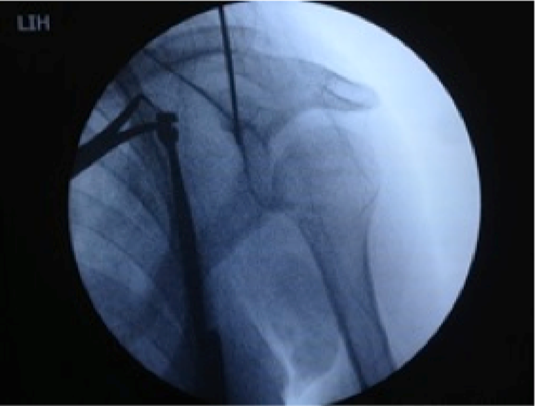
Entering guidewire over the superior surface of the clavicle ensuring that it passed through the middle of coracoid process under fluoroscopy.
Opening bone tunnels by drill over guidewire.

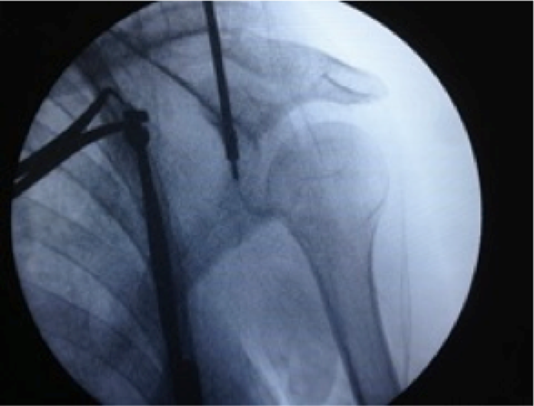
The button was rolled over and locked under the coracoid process.

After tightening the lift-up system, the reduction was confirmed with the fluoroscopy.

Verification of satisfactory reduction before the system was locked by the ropes.
Rehabilitation and follow-up
Standard sling was applied for 3 weeks, postoperatively. Pendular shoulder exercises and elbow exercises were initiated on postoperative day 1. Patients were called to come for outpatient visit 2 weeks after the surgery, where sutures were removed and passive range of motion exercises were started. In addition, control X-rays were taken and clinical appointments were arranged for every 2 weeks till week 6, when active range of motion and muscle strengthening exercises were started, and the outpatient visits were scheduled once a month. Constant and ASES shoulder scores were obtained and recorded for every patient at postoperative 4 and 12 months. With radiographic evidence of healing (bony bridging across fracture site) and gaining full range of motion, muscle strength of 90% of the contralateral extremity was aimed for contact sports. Patients were not allowed to deal with heavy workloads including lifting, carrying, pushing, and pulling on the operated side for 6 months.
IRB/Ethical Committee Approval: All patients gave the informed consent to this study. This study conforms to the Declaration of Helsinki and was authorized by the ethical committee of the authors’ institution.
Results
Of 22 patients, 19 enrolled in the study were male. The mean age was 39 (range: 21–60). Mean duration for the surgical intervention was 40 min (range: 30–55 min. Mean union time was found to be 14 weeks (range: 7–21 weeks). Right dominant extremity was affected in 14 patients, and remaining 8 patients had fracture in their left extremity. The reasons for fracture were detected to be traffic accident in 14 cases, falling from height in four cases, falling from a horse in two cases, and secondary to sport injury in the others. Mean duration from the index event till the surgery was 5 days (range: 3–8 days). Early wound complication or loss of reduction was not seen in any cases. No AC joint arthrosis and clavicular shortening were detected in X-rays during follow-up (Figures 7 and 8).

Preoperative AP view of the patient with distal clavicle fracture.

Postoperative AP X-ray of the same patient at first year follow-up.
Mean postoperative ASES and Constant scores were 79.9 (66.9–88.3) and 82.2 (71–100), respectively. Return to normal daily activities was found to be at an average of 4.5 months. Eighteen patients experienced a complete functional recovery with normal range of motion and pain relief. Two patients had prolonged physical therapy due to limited range of motion at shoulder joint, who achieved normal range of motion at the end of year 1. Two patients were not able to return to work because of weakness and persistent shoulder pain with no difficulty in performing daily activities.
Discussion
The distal clavicle fractures are frequently encountered in the clinical setting. Since the proximal fragment is highly unstable and has no ligamentous attachment, with the tendency for displacement of the fragments due to muscle forces and weight of the arm, type 2 fractures have higher rate of nonunion as compared to the other types. Various surgical procedures have been described for the fixation of unstable distal fractures of the clavicle, including fixation by Kirschner wire, coracoclavicular screw, low-profile plate or hook plate, or coracoclavicular fixation by various materials (anchor, TightRope).
Coracoclavicular stabilization, hook plate, intramedullary fixation, interfragmentary fixation, and K-wire plus tension band wiring were compared in a retrospective meta-analysis study consisting of 425 patients, where complication and nonunion were shown to be 1.6–22% with these open surgical techniques. 14,15
Although hook plates are shown to be effective in prevention of translation of the clavicle superiorly, they may lead to osteolysis and fracture in acromion in its inferior aspect, and even rotator cuff tear, which may necessitate a second surgical intervention. 16 –18 Our technique does not require such reoperation to remove the implant.
Quite a few studies reported successful union rates after coracoclavicular fixation performed with cannulated screw introduced from proximal clavicular fragment. Nonetheless, it needs a second surgical procedure for the screw to be removed prior loosening. 6,14,19 –22
Usually, outcomes of osteosynthesis are also relatively satisfactory when performed by transacromial Knowles pin with coracoclavicular ligament repair or reconstruction. 23 –25 However, asymptomatic radiographic lucency around the pin and 1–5 mm lateral migration of the pin were demonstrated during removal of pin. In addition, 12% of patients were found to have coracoclavicular heterotopic ossification. 26
Either arthroscopy assisted or in open fashion, numerous techniques have been described in coracoclavicular repair and reconstruction. Fixation techniques involve sutures, anchors, TightRope systems, and button devices, yet with limited number of studies in the literature. 27 –30 Weber and Harris published a case series of 11 patients where they used Dacron graft in coracoclavicular fixation, achieving successful outcomes. 31 Li et al. reported 100% of union rates after fixation performed with a cerclage wire inserted from a drill tunnel opened between the clavicle and the coracoid. 32
Although the cerclage of the clavicle around coracoid is one of the therapeutic options in long oblique fractures, it may not provide an effective fixation against distraction forces in comminuted fractures. 26
In a biomechanical study on fresh frozen cadaver, Rieser et al. found the fixation using combination of locked plate with TightRope to be biomechanically superior to other techniques. 26 Another study with five patients reported successful results after button-assisted coracoclavicular fixation performed with locked plate. 33 Drawbacks of this technique include the need for large incision for locked plating and excessive stripping of the bone from surrounding soft tissue. The technique is especially recommended in comminuted fractures where stabilization could not be ensured with single plate. The disadvantages related with the addition of TightRope are risks that may develop secondary to dissection around the coracoid and prolonged duration of the surgery. 26 The surgical technique we suggested allows for concurrent correction of coracoclavicular congruence with reduction of the fracture, whereby the healing of torn coracoclavicular ligaments is also intended.
Intramedullary fixation has some advantages over plate fixation such as small incision, less soft tissue stripping, less irritation, and easy performance for hardware removal. Despite these, intramedullary fixation was reported to have relatively high nonunion rates and AC arthrosis due to pin penetration. 34
Outcomes of fixation performed with TightRope technique by Hague et al. were highly successful; however, a direct reduction was applied after opening fracture line. 35 In our study, fracture line was not opened, performing an indirect reduction under the guidance of the fluoroscope as well as preserving fracture hematoma.
In 2013, Loriaut et al. published results of osteosynthesis performed with arthroscopic double button technique in distal clavicle fractures in a case series of 21 patients. The authors reported satisfactory clinical and functional outcomes in 81% of patients. A 3-cm incision was performed apart from arthroscopic portals. 36 We did not need arthroscopy in our study. Proper use of the fluoroscope was regarded as sufficient for the technique and confirmation of the reduction. This both significantly shortens the duration of the operation and offers minimizing the size of incision.
The limitations of our technique include difficulty in controlling the tension of ropes and buttons over the fracture site and it may not be appropriate for comminuted fractures. Although satisfactory reduction was described as the continuity of the inferior border of the lateral clavicle under fluoroscopy, further biomechanical studies may be necessary to assess the optimum tension for maintenance of the reduction until fracture healing.
Conclusion
This study has demonstrated that indirect osteosynthesis performed by minimal invasive coracoclavicular fixation with double button lift-up system in the treatment of unstable Neer type IIa fractures of the distal clavicle had successful clinical, radiological, and functional outcomes. While a 2-cm incision provided a satisfactory result in terms of cosmetic view and wound healing, closed reduction without opening fracture line allowed for achievement of union with the preservation of fracture hematoma. In addition, no second operation was required for the removal of the implant.
Footnotes
Declaration of conflicting interests
The author(s) declared no potential conflicts of interest with respect to the research, authorship, and/or publication of this article.
Funding
The author(s) received no financial support for the research, authorship, and/or publication of this article.